Embed Size (px)
Citation preview

Synthesis of Se-Adenosyl-L-Selenohomocysteine Selenoxide and Potential Gram-
Negative Antibacterial Analogs
By Dillon Cleary
B.S. in Chemistry, Northeastern University
A thesis submitted to
The Faculty of
the College of Science of
Northeastern University
in partial fulfillment of the requirements
for the degree of Master of Science
April 9, 2015
Thesis directed by
James Aggen
Associate Professor of Medicinal Chemistry

ii
Dedication
First, I want to thank my family and, specifically, my parents, Cornelius and Emily, and
my siblings, David, Molly, and Julianne. It is from them that I draw my motivation and without
their support my education would not have been the same.
Julie Miquel has been another major source of support and has always been there to help
me overcome my toughest challenges.
My high school chemistry teacher, Dr. Melanie Furlong-Gates, deserves the credit for
introducing me to chemistry and being a major influence in my decision to become an organic
chemist. I also thank her for continuing to be a positive role model in my life.
My fellow colleagues in Sunnyland, Professor Rick Duclos, Kalli Catcott, Wanlu Qu, and
Shanshan Liu who worked with me on my project and were always willing to lend a helping
hand.
Professor Sunny Zhou deserves thanks for taking me into his lab as an undergraduate and
allowing me to the opportunity to improve my organic chemistry knowledge and skills. I
appreciate his honesty and for continuing to look after my best interests.
My fellow colleagues in Professor Aggen’s Laboratory, Andrew Spaulding, Dr. Khuloud
Takrouri, Dr. Harold Cooper, Dr. Poornachandran Mahalingam, Westley Tear, Matthew
Dowgiallo, and Nick Elias who have all provided me with valuable experiences and camaraderie
and I thank them for that.
I also owe a large amount of gratitude to Professor James Aggen for a long list of
reasons. Starting with allowing me the opportunity to work in his laboratory, he has also been
inspirational in helping me find the direction I want to move forward with in my career. He is
always giving time to answer questions, give advice, and review project work. Working for him
has been a pleasure and I can’t express my appreciation enough.

iii
Acknowledgements
Aggen Laboratory
Professor James Aggen
Dr. Khuloud Takrouri
Dr. Harold Cooper
Dr. Poornachandran (Poorna) Mahalingam
Andrew Spaulding
Westley Tear
Matthew Dowgiallo
Nick Elias
Microbiology Laboratory
Dr. Elizabeth Hirsch
Dr. Paola Zucchi
Biochemistry Laboratory
Professor Penny Buening
Bilyana Koleva
Sunnyland
Professor Zhaohui Sunny Zhou
Professor Richard Duclos Jr.
Kalli Catcott
Wanlu Qu
Shanshan Liu
William Devine
Dr. Jason Guo
Professor George O’Doherty
Department of Chemistry and Chemical Biology, Northeastern University

iv
Abstract
This paper includes work performed on two separate projects. The first project is the
synthesis and characterization of a new analog of S-adenosylmethionine (AdoMet or SAM)
called Se-adenosyl-L-selenohomocysteine selenoxide (SeAHO). This work outlines the total
synthesis starting from adenosine and the characterization data collected along the way. The
work culminated with the first successful synthesis of the desired selenoxide analog that can now
be studied further for its potential biological activity. The second project involves medicinal
chemistry efforts in synthesizing analogs of the oxazolidinone scaffold with desirable
characteristics for potential treatment of Gram-negative bacterial infections. The oxazolidinone
scaffold has already been shown to be successful in treating Gram-positive bacterial infections
and the goal of our research is to be able to convert it to one that also has activity against Gram-
negative bacteria. With the goal of developing a novel approach to making compounds with this
activity, we look at specific modifications made to certain areas of the scaffold and how these
changes impacted the activity against different strains of bacteria through our biological assay.

v
Table of Contents
Dedication ii
Acknowledgements iii
Abstract of Thesis iv
Table of Contents v
List of Figures /Tables vi
Chapter 1: Synthesis and Characterization of SeAHO 1
Chapter 2: Introduction to Gram-negative bacteria 21
Chapter 3: Oxazolidinone analog synthesis 25
References 51
Appendices 55

vi
List of Figures
Figure 1.1: SeAHO Reaction Scheme 3
Figure 1.2: TLC of SeAHO step 1 4
Figure 1.3: TLC of SeAHO step 2 5
Figure 1.4: HPLC SeAHO vs. SAHO 7
Figure 1.5: Possible Cyclization Structures 8
Figure 1.6: 1H NMR SeAH 10
Figure 1.7: 1H NMR and COSY SeAHO 11
Figure 1.8: LC-MS SeAH 12
Figure 1.9: LC-MS SeAHO 13
Figure 1.10: Total Ion Chromatograph SeAHO 14
Figure 1.11: Extracted Ion Chromatograph SeAHO 15
Figure 1.12: MS Mass Fragments SeAHO 15
Figure 2.1: Growing Resistance Graph 22
Figure 2.2: Gram-Negative Bacteria Outer Membrane 23
Figure 2.3: Oxazolidinone Leading Compounds 24
Figure 3.1: Oxazolidinone Analog General Synthetic Scheme 25
List of Tables
Table 3.1: Oxazolidinone Analog Structures and Data 32
Table 3.2: Gradient for 2to30in30 HPLC method 50
Table 3.3: Gradient for 2to50in30 HPLC method 50
Table 3.4: Gradient for 2to100in30 HPLC method 50
Table 3.5: Gradient for 2to100in8PosM LC-MS method 50

1
Chapter 1: Synthesis and Characterization of Se-adenosyl-L-selenohomocysteine
selenoxide
This work has been previously published in the Journal of Sulfur Chemistry. Synthesis and
Characterization of Se-adenosyl-L-selenohomocysteine selenoxide. 2015; 36: 135-144.1
http://dx.doi.org/10.180/17415993.2014.979173
Authors
Richard Duclos Jr. – Synthesis/characterization work and lead author
Dillon Cleary – Synthesis/characterization work
Kalli Catcott – Characterization and biological studies
Zhaohui Sunny Zhou – Corresponding author
2
Introduction
S-adenosylmethionine (SAM or AdoMet) is a well-known, naturally occurring
methylating agent that is involved in many chemical processes in the body. S-
adenosylmethionine dependent methyltransferases are enzymes that are necessary to catalyze the
donation of the methyl group to other compounds, such as proteins or small molecules, and, in
the process, form S-adenosylhomocysteine (SAH or AdoHcy).2-5
There has been much research
in this field on the synthesis of analogs of S-adenosylmethionine at its reactive sulfur center.
Some such analogs include keto-AdoMet, 6 propargyl-AdoMet,
7 and Adovin.
8 Developing new
analogs is of interest for many reasons which include tagging proteins, studying the functions of
methyltransferases, and forming new compounds for the activation or inhibition of certain
enzymes.
Another analog that has been synthesized and studied for its role as an inhibitor is S-
adenosylhomocysteine sulfoxide (SAHO). This compound was first synthesized in 19709
and has
been cited as showing inhibition for enzymes such as mRNA methyltransferase,10
protein
methyltransferase II,11
and catechol-O-methyltransferase.12,13
However, the sulfoxide analog has
not shown to be as effective an inhibitor as S-adenosylhomocysteine. While this may be the case,
that does not stop analogs of similar structure from being of interest to researchers in this area.
Along with these modifications of the methyl group, another series of analogs that has
begun to be explored are those replacing the sulfur with closely related selenium. Selenium plays
an important role as a part of many metabolites found in living organisms.14-16
Though the
amount of selenium present is much less than that of sulfur, these analogs are still of interest
because of how little is known about them.

3
A recent paper published by Willnow et al. outlines a new synthesis for the selenium
analog of S-adenosylhomocysteine as they discuss the modification of proteins through copper-
catalyzed azide-alkyne cylcloaddition (CuAAAC) click chemistry.7 As part of this work, they
synthesized a new selenium AdoMet analog with a propargyl group and showed that the
selenium center led to better stability and activity. Compared to S-adenosylmethionine and S-
adenosylhomocysteine, not much is known about the biochemical activity and redox chemistry
of Se-adenosylselenohomocysteine. Also, the selenoxide analog had not been previously
synthesized before our group reported it in a recent publication.1 In the next chapter, I describe
the synthesis and characterization of Se-adenosyl-L-selenohomocysteine selenoxide.
Synthesis
Figure 1.1: Synthetic route for SeAHO.
Step 1
The first step of this synthetic route, preparation of 5’-chloro-5’-deoxyadenosine (ClDA),
was run on a 5.0 gram scale using a procedure adapted from the literature.17
This was a two part

4
procedure that involved first making a sulfinyl intermediate and then the desired ClDA product.
The first part resulted in 5.435 grams (88% yield) of the sulfinyl intermediate. The next step
involved removing the protecting group from the intermediate and a recrystallization that yielded
4.68 grams (100% yield) of the desired product.
Melting Point, TLC, and NMR analysis were all performed to determine structure and
purity. The product began decomposing between 185-190 °C and this matched up with the
literature values. TLC in 3:1 chloroform: methanol, under UV (Figure 1.2), showed one spot for
the product and no starting material present.
A proton NMR (Figure 1.6) was run, using a 700 MHz instrument, with the product in
deuterated DMSO and the resulting spectrum showed the desired peaks. Some additional solvent
peaks showed that there was some residual water and methanol present in the sample. Overall
compound characterization proved that we made our desired product with overall 90% yield.
Figure 1.2: TLC under UV of starting material (adenosine), product (ClDA), and a co-spot.
Step 2
The next step of this route was to make the selenium analog of S-adenosylhomocysteine
based on a recently published synthesis.6 In order to carry out this step, however, we first had to

5
synthesize selenohomocysteine from selenomethionine.18
This reaction was run on a 700 mg
scale and the unpurified product was used for the reaction with 926 mg of CIDA.
The reaction gave crude Se-adenosylselenohomocysteine (SeAH) that was dried under
vacuum overnight to give a yellowish white solid. The solid was then purified by two hot
filtration/crystallizations in methanol and this resulted in 330 mg (24% yield) of crystals. During
the second recrystallization, some of the crystals, about 115 mg, would not dissolve and these
were set aside to be analyzed separately.
The melting point of the SeAH crystals was found to be about 204 °C and this is a new
value not previously reported in the literature. This step of the synthesis was monitored by a TLC
method adapted from the literature. Originally a solvent system of 12:5:3 isopropanol: water:
acetic acid was used but was changed to a ratio of 12:1:3 of the same solvent system because it
proved to give better separation. TLC of the crystals that would not dissolve showed them to be
relatively pure when compared to the crude reaction product (Figure 1.3).
Figure 1.3: TLC plates stained with potassium permanganate (left) and ninhydrin (right). Each
has six spots starting from the left: CIDA, selenomethionine, Step 2 before crystallization,
crystals that didn’t dissolve during recrystallization, crystals after recrystallization, and methanol
mother liquor.
6
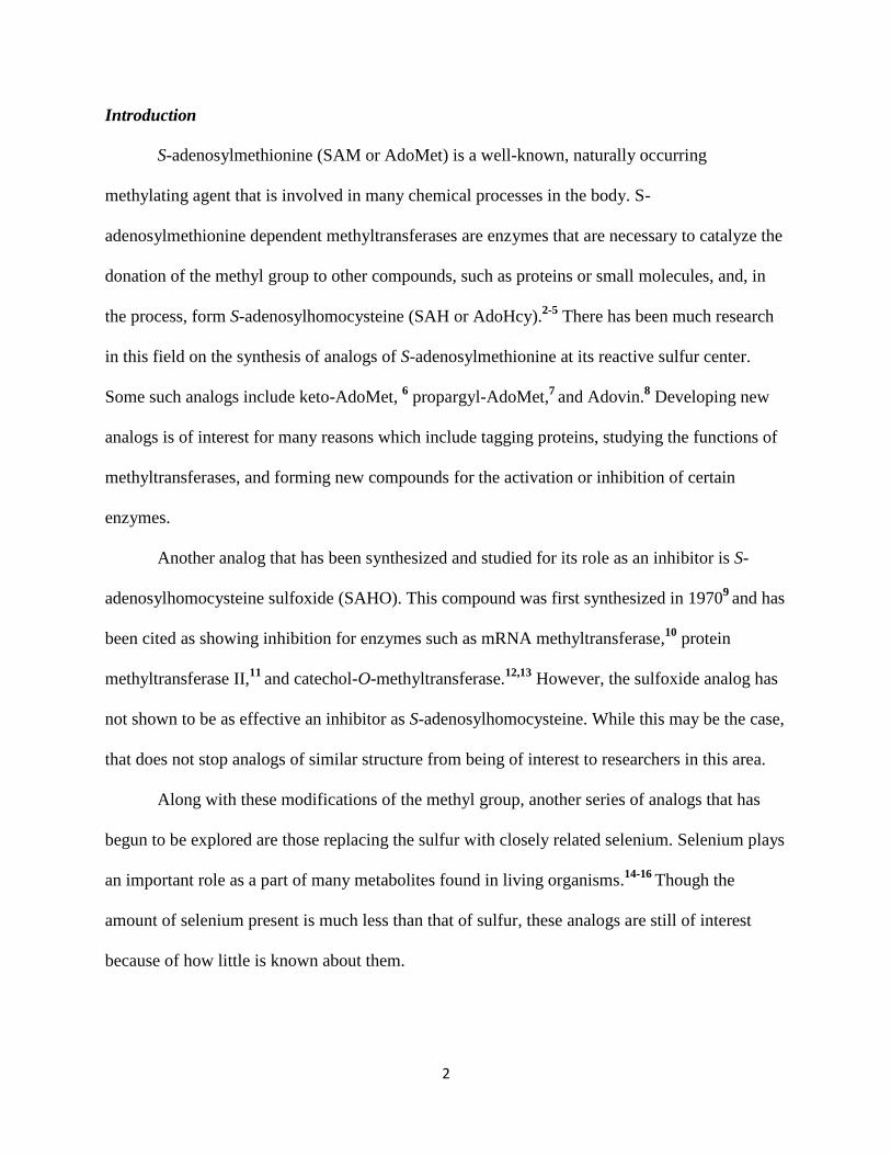
Step 3
TLC analysis showed that SeAH was a more polar molecule consistent with the desired
product and so a small amount (5 mg) was used in a reaction in order to attempt to make the
selenoxide analog. The procedure used for this reaction was one that was adapted from the
literature for the synthesis of S-adenosylhomocysteine sulfoxide.12
An excess of hydrogen
peroxide (30%) was used to oxidize selenium and, while the Borchardt paper allowed the
reaction with SAH to go overnight, by monitoring with TLC we were able to see the new product
spot appear almost instantaneously. While this reaction was going, we also made the sulfoxide
analog, SAHO, from SAH in order to compare to the selenoxide analog.
Upon seeing that the selenoxide product, SeAHO, had formed, we tried to precipitate it
out of solution with methanol as done in the Borchardt paper for the sulfoxide analog. The
sulfoxide successfully precipitated as reported, however, the selenoxide did not and so we had to
obtain a solid through lyophilization. The solution was frozen using dry ice and the solvent
removed under high vacuum over one hour to give a quantitative yield.
Discussion
Se-adenosyl-L-selenohomocysteine selenoxide (SeAHO) was run on high performance
liquid chromatography (HPLC) and compared to samples of SAH, SeAH, and SAHO. These
traces appear in Figure 1.4 and we see the product elute around 5 minutes. While we also see
some later eluting peaks that would seem to be from degradations products, this analysis shows a
clear difference between the starting material and the oxidized product.

7
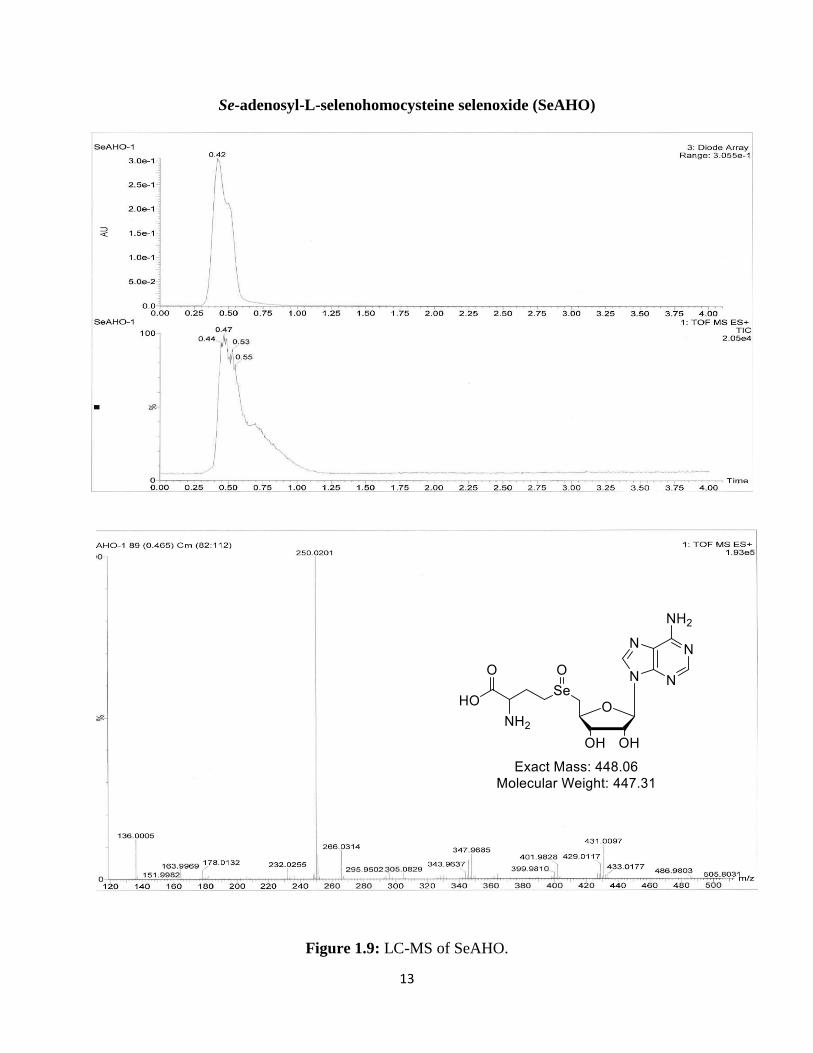
Mass Spectrometry data was also collected for these compounds using a liquid
chromatography mass spectrometer (LC-MS), but SeAHO was the only product not to give the
desired molecular ion, showing an (M-18)+ peak, however.
NMRs of each compound were also taken in acetic acid-d4 and these led to some
interesting results. When comparing the NMR for the SeAHO to the other spectra, the movement
of the peaks may suggest that a cyclization with the amine and/or carboxylic acid occurred.19
The
structures in Figure 1.5 suggest possible cyclization products that may have formed based on the
observed molecular ion.
Figure 1.4: HPLC 1 is of S-adenosylhomocysteine (SAH) and the peak appears at 11.967 min,
HPLC 2 shows Se-adenosylselenohomocysteine (SeAH) and this peak elutes at 12.817 min,
HPLC 3 is of S-adenosylhomocysteine sulfoxide (SAHO) and the peak appears at 5.054, and
HPLC 4 is of Se-adenosylselenohomocysteine selenoxide (SeAHO) with the peak eluting at
5.114. Column: Alltech Econosil C18, 4.6mm x 250mm.

8
Figure 1.5: Structures of possible cyclization products formed during reaction
In order to see if we could observe the desired molecular ion (449) the sample was run on
MALDI MS and LC-MS/MS instruments. The data observed did reveal the desired molecular
ion along with that observed previously. From this data we see our desired mass of 449 and
fragmentation to 431 and 467, which are the M-18 and M+18 peaks respectively. A mass
difference of 18 signifies a gain or loss of water. The mass of 467 clearly indicates the hydrated
form of the compound or the dihydroxyselenide ion. The 431 ion peak is a little more difficult to
explain but, as previously stated, the literature does show precedent for the formation of cyclic
analogs. The structures shown in Figure 1.5 are possible cyclization products that may have
occurred and their masses are about 431.
Conclusion
From the NMR and mass spectral data, we are able to say confidently that we
successfully synthesized Se-adenosyl-L-selenohomocysteine selenoxide for the first time. This
compound is of interest as a possible inhibitor of certain S-adenosylmethionine dependent
methyltransferases, other enzymes, and proteins due to its close similarity to the sulfoxide analog
of SAM. Further studies of this compound have shown its stability in aqueous environments and

9
its ability to be reduced by glutathione and cysteine.1 This work offers further opportunities for
selenium analogs of SAM to be studied in order to gain a better understanding of selenium’s
biological functionality compared to sulfur.
10
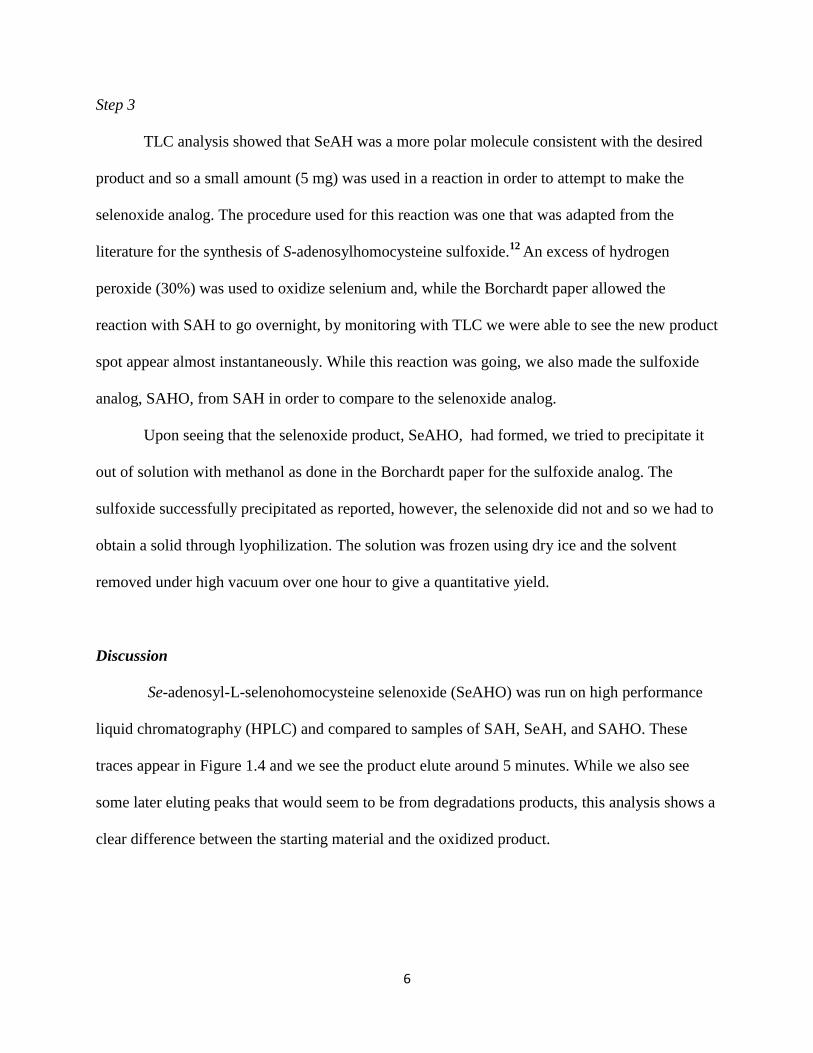
Data
Se-adenosylselenohomocysteine (SeAH) in HOAc-d4
Figure 1.6: Proton NMR of SeAH in HOAC-d4.
20130906_SAH_RD.004.001.1r.esp
8 7 6 5 4 3 2 1 0
Chemical Shift (ppm)
0
0.25
0.50
0.75
Norm
alized Inte
nsity
1.041.072.092.161.041.031.031.020.951.001.00
acetic acid
beta
gamma
5'
alpha
4'
3'2'
M03(d)M01(s)
M02(s)
M04(t)M06(q)
M05(t)
M11(m)
M10(d)M07(br. s.)
M09(m)
M08(m)
8.5
0(1
2)
8.4
4(7
)
6.1
76.1
6(2
3)
4.8
6(2
)
4.4
9(4
,3)
4.3
8(5
)4.1
5(2
1)
3.4
2
3.0
7(1
7)
2.7
9(1
9)
2.7
8
2.3
5(2
0)
2.2
5(2
0)
2.0
4
N13
12
N11
10
9
14
N6
7
N8
NH223
4 3
2O15
OH15
OH16
17
Se1819
20
21
NH222
CO2H24

11
Se-adenosyl-L-selenohomocysteine selenoxide (SeAHO) in HOAc-d4
Figure 1.7: a) Proton NMR of SeAHO in HOAC-d4. b) COSY of SeAHO.
20130906_SAH_RD.006.001.1r.esp
8 7 6 5 4 3 2 1 0
Chemical Shift (ppm)
0
0.05
0.10
0.15
0.20
0.25
0.30
0.35
Norm
alized Inte
nsity
1.001.130.883.372.031.281.291.251.331.09
2'
alpha
5'
gamma beta
beta
acetic acid
M02(s)
M01(s)
M03(d)
M05(t)
M04(dd)
M08(m)
M06(m)
M07(m)
M09(br. s.)
M10(m)
8.4
8(1
2)
8.4
5(7
)
6.2
1(2
0)
4.9
54.9
4(2
)4.7
24.7
2(5
,4,3
)4.6
2(2
3)
3.9
0(1
7)
3.6
5(2
1)
2.7
8(2
2)
2.6
5(2
2)
N13
12
N11
10
9
14
N6
7
N8
NH220
4 3
2O15
OH15
OH16
17
Se1821
22
23
NH224
CO2H25
O19

12
Se-adenosylselenohomocysteine (SeAH)
Figure 1.8: LC-MS of SeAH
13
Se-adenosyl-L-selenohomocysteine selenoxide (SeAHO)
Figure 1.9: LC-MS of SeAHO.

14
SeAHO Sample (Full scan):
Figure 1.10: Total ion chromatography (TIC) of SeAHO.

15
Figure 1.11: Extracted ion chromatography (XIC) of 431.0, 449.0 and 467.0 m/z in SeAHO
sample.
Figure 1.12: MS at 2.16, 2.25 and 4.74 min in SeAHO sample.
16
Experimental
General Data
TLC was carried out on plastic-backed silica gel 60, PE SIL G Whatman Plates, UV254. HPLC
used an Agilent/Hewlett Packard Series 1100. NMR spectra were recorded on a Bruker 700 MHz
spectrometer. Mass Spectrometry Instrumentation used included: (1) LC-MS-ToF Waters LCT
Premier ToF MS; (2) LCQ-MS: Finnegan LCQ ESI ion trap MS; (3) MALDI-MS: AB SCIEX
5800 TOF/TOF
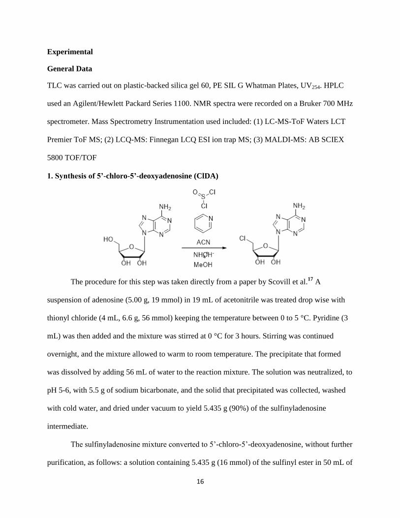
1. Synthesis of 5’-chloro-5’-deoxyadenosine (ClDA)
The procedure for this step was taken directly from a paper by Scovill et al.17
A
suspension of adenosine (5.00 g, 19 mmol) in 19 mL of acetonitrile was treated drop wise with
thionyl chloride (4 mL, 6.6 g, 56 mmol) keeping the temperature between 0 to 5 °C. Pyridine (3
mL) was then added and the mixture was stirred at 0 °C for 3 hours. Stirring was continued
overnight, and the mixture allowed to warm to room temperature. The precipitate that formed
was dissolved by adding 56 mL of water to the reaction mixture. The solution was neutralized, to
pH 5-6, with 5.5 g of sodium bicarbonate, and the solid that precipitated was collected, washed
with cold water, and dried under vacuum to yield 5.435 g (90%) of the sulfinyladenosine
intermediate.
The sulfinyladenosine mixture converted to 5’-chloro-5’-deoxyadenosine, without further
purification, as follows: a solution containing 5.435 g (16 mmol) of the sulfinyl ester in 50 mL of
17
methanol was treated with 5 mL of concentrated ammonium hydroxide. Crystals formed upon
standing for an hour at room temperature. These were collected, washed with cold
methanol/ammonium hydroxide solution, and dried under vacuum to yield 4.68 g (99%) of
colorless needles of 5’-chloro-5’-deoxyadenosine. 1H NMR in Appendix A1.
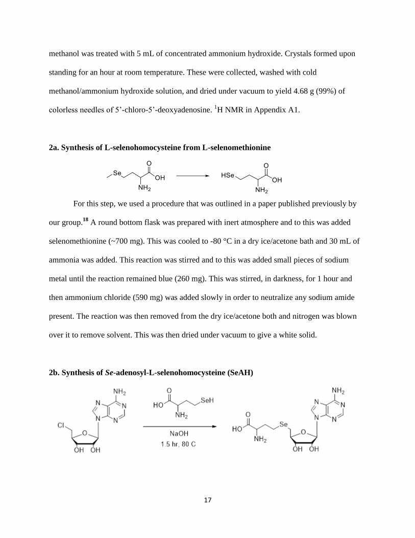
2a. Synthesis of L-selenohomocysteine from L-selenomethionine
For this step, we used a procedure that was outlined in a paper published previously by
our group.18
A round bottom flask was prepared with inert atmosphere and to this was added
selenomethionine (~700 mg). This was cooled to -80 °C in a dry ice/acetone bath and 30 mL of
ammonia was added. This reaction was stirred and to this was added small pieces of sodium
metal until the reaction remained blue (260 mg). This was stirred, in darkness, for 1 hour and
then ammonium chloride (590 mg) was added slowly in order to neutralize any sodium amide
present. The reaction was then removed from the dry ice/acetone both and nitrogen was blown
over it to remove solvent. This was then dried under vacuum to give a white solid.
2b. Synthesis of Se-adenosyl-L-selenohomocysteine (SeAH)

18
The procedure for this step was adapted from a paper by Willnow et al.7 A fresh batch of
L-selenohomocysteine (700 mg) was dissolved in water (4 ml) and to this was added 5’-chloro-
5’-deoxyadenosine (926 mg). To this mixture was then added 2.5 mL of a 10% sodium
hydroxide solution and more water (5 mL). This reaction was let stir for 1.5 hours at 80 °C. The
solution remained cloudy for 15 minutes before becoming homogeneous. Acetic acid (~1.25 mL)
was then added drop wise until the solution pH was between 5 and 6. The reaction was put under
vacuum and let dry overnight to give a yellowish white solid. This solid was dissolved in an
ethanol/toluene solution and again dried under vacuum to remove residual solvent.
The reaction was monitored by TLC using a 12:5:3 (from literature) and 12:3:3
isopropanol:water:acetic acid solvent system. The product was purified through recrystallization
with hot filtration in methanol. As much of the product as possible was dissolved in boiling
methanol. The solid that didn’t dissolve was filtered off and the remaining solution was boiled
down until crystals started forming. This sat overnight before the mother liquor was filtered off
and the crystals were washed with cold methanol.
A second recrystallization was run in the same way. The mother liquor was also saved as
partially purified SeAH. TLC of the final crystals showed a single UV spot lower than the ClDA
starting material spot. Additional plates were run and stained with potassium permanganate and
ninhydrin. The adenine group was visible by UV254 TLC plates with a hand-held short wave
UV lamp, potassium permanganate staining showed thioether, selenoether, and ribosyl groups,
and the ninhydrin stain showed the amino group. The pure crystals of SeAH acetate salt were
isolated in a final yield of 330 mg (24%). MP 204-205 °C. TLC: 12:1:3 isopropanol:water:acetic
acid: ClDA Rf 0.80, selenomethionine Rf 0.73, and SeAH Rf 0.44. TLC 12:5:3
isopropanol:water:acetic acid: SeAH Rf 0.72. DAD UV spectrum λmax 258 nm. 1H NMR (700
19
MHz, CD3CO2D) δ 2.22-2.30 (m, 1H, βb), 2.32-2.39 (m, 1H, βa), 2.74-2.84 (m, 2H, γ), 3.03 (dd,
J=13.2, 6.2 Hz, 1H, 5b’), 3.05 (dd, J=13.2, 5.7 Hz, 1H, 5a’), 4.16 (m, 1H, α), 4.38 (ddd, J=4.3,
5.7, 6.2 Hz, 1H, 4’) 4.49 (dd, J=4.3, 5.4 Hz, 1H, 3’), 4.86 (dd, J=4.4, 5.4 Hz, 1H, 2’), 6.16 (d,
J=4.4Hz, 1H, 1’), 8.44 (s, 1H), 8.50 (s, 1H, Ar) (Figure 1.6)
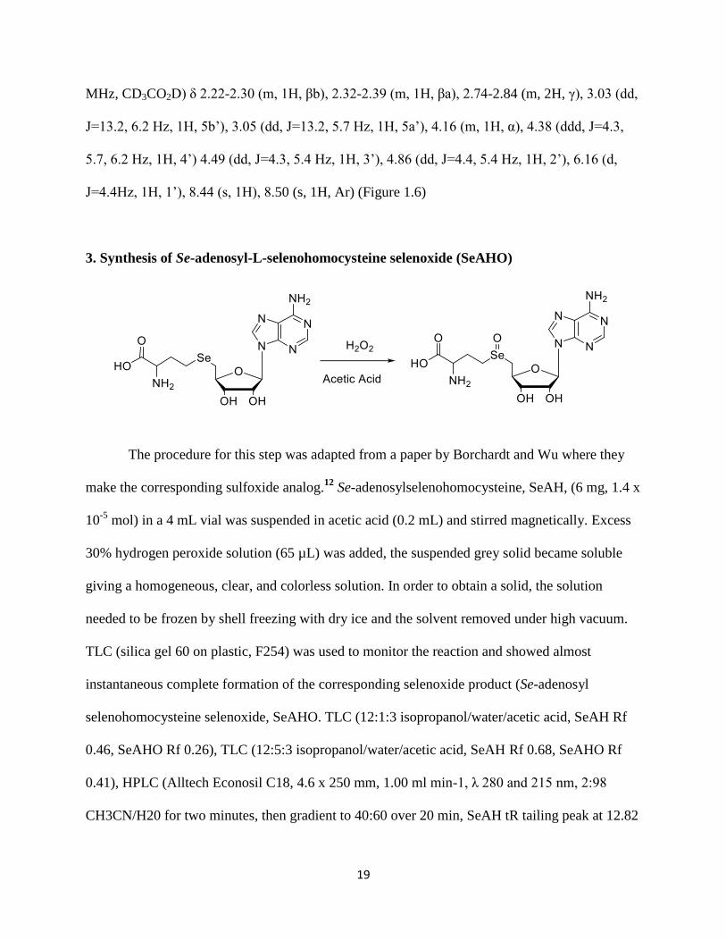
3. Synthesis of Se-adenosyl-L-selenohomocysteine selenoxide (SeAHO)
The procedure for this step was adapted from a paper by Borchardt and Wu where they
make the corresponding sulfoxide analog.12
Se-adenosylselenohomocysteine, SeAH, (6 mg, 1.4 x
10-5
mol) in a 4 mL vial was suspended in acetic acid (0.2 mL) and stirred magnetically. Excess
30% hydrogen peroxide solution (65 µL) was added, the suspended grey solid became soluble
giving a homogeneous, clear, and colorless solution. In order to obtain a solid, the solution
needed to be frozen by shell freezing with dry ice and the solvent removed under high vacuum.
TLC (silica gel 60 on plastic, F254) was used to monitor the reaction and showed almost
instantaneous complete formation of the corresponding selenoxide product (Se-adenosyl
selenohomocysteine selenoxide, SeAHO. TLC (12:1:3 isopropanol/water/acetic acid, SeAH Rf
0.46, SeAHO Rf 0.26), TLC (12:5:3 isopropanol/water/acetic acid, SeAH Rf 0.68, SeAHO Rf
0.41), HPLC (Alltech Econosil C18, 4.6 x 250 mm, 1.00 ml min-1, λ 280 and 215 nm, 2:98
CH3CN/H20 for two minutes, then gradient to 40:60 over 20 min, SeAH tR tailing peak at 12.82

20
min, SeAHO tR 9.3 min as a broader peak with shoulders at 8.9 min and 10.2 min for the more
polar compound), HPLC-ESI MS 431, 402, 348, 250, DAD UV spectrum λmax 260 nm, NMR:
1H NMR (700 MHz, CD3CO2D) δ2.60-2.70 (m, 1 H, βa), 2.75-2.85 (m, 1 H, βb), 3.65 (br d, 1 H,
J = 10.7 Hz, 5a’), 3.84-3.95 (m, 3 H, 5b’ and both γ), 4.67-4.58 (m, 2 H, α, 4’), 4.72 (dd, J = 5.7,
4.7 Hz, 1 H, 3’), 4.94 (dd, J = 4.7, 3.7 Hz, 1 H, 2’), 6.20 (d, J = 3.7 Hz, 1 H, 1’) 8.45 (s, 1 H, Ar)
8.48 (s, 1 H, Ar).(Figure 1.7)

21
Chapter 2: Introduction to Multidrug Resistant Gram-Negative Bacteria
Finding compounds that act as potent antibacterial agents has long been an arduous task
of scientists and, in particular, medicinal chemists. In the middle of the 20th
century, this area of
research reached its peak success as many of the antibiotics still used today, such as penicillin
and vancomycin, were discovered. The “golden age” of antibiotics produced many antibacterial
agents that led to a revolution in the field of medicine. Unfortunately, in the half a century or so
since that time period, the number of antibiotics developed has severely plummeted and the
current pipeline is not all too encouraging.20-21
This is mainly to do with a lack of urgency and
financial incentive for major pharmaceutical companies; many of whom have already left behind
this area of drug development. Both of these factors need to change (and seem to be heading in
that direction) in the immediate future as the development of bacterial resistance is an area of
growing concern.22
In the years since the golden age of antibiotics, bacteria have evolved to fight off all of
the antibiotics we have at our disposal. The consistent use, and over prescription, of the same
antibiotics have led certain strains of bacteria to mutate and develop mechanisms of resistance
against all antibacterial agents we have on the market. From 1999 to 2010, the resistance levels
of Acinetobacter baumanii, a Gram-negative bacterium, against carbapenems (a common class
of antibiotic used as the last line of defense against multidrug-resistant bacteria) rose from 5% to
40% (as seen in Figure 2.1) and the assumption is that it has only increased further in the five
years since then. A 2013 report by the Centers for Disease Control and Prevention stated that two
million Americans are infected with resistant bacteria each year and 23,000 of them die as a
direct result.23
This is not to say that all bacteria are resistant to the antibiotics we currently have,
but the numbers are growing and the problem must be addressed.

22
Figure 2.1: Graph of growing bacterial resistance to carbapenem antibiotics from 1999-2010.
(Data used to generate graph was obtained from the Center for Disease Dynamics, Economics &
Policy website)24
The biggest threat posed by these strains comes from multidrug resistant Gram-negative
bacteria (GNB) as these are responsible for the majority of hospital acquired infections.25
Unlike
Gram-positive bacteria (GPB), GNB have an additional layer of protection to ward against
antibiotics. This extra layer is a lipopolysaccharide containing outer membrane that does not
allow compounds with antibacterial activity (or anything) to penetrate inside to the bacteria.
These bacteria do need certain nutrients in order to survive, however, and so this membrane does
have small entrance points along it called porins. These porins represent an area to exploit when
developing Gram-negative antibiotics.26
That being said, these porins limit the size and properties of the compounds that can pass
through and are not the final obstacle potential antibiotics need to overcome. Within the
membrane of these bacteria are efflux pumps that effectively pump out any undesired
compounds that happen to find their way through the porins. A potential Gram-negative
antibiotic needs to have properties that allow it to pass through the porins and get to the bacteria

23
without being transported back outside the membrane by the efflux pumps (Figure 2.2). The lack
of development of potential Gram-negative antibiotics to this point may be due to an incomplete
understanding of these bacteria. A rational approach to making these antibiotics does not
currently exist and the goal of our group is to change that.
The approach is two-fold as we want to: 1) develop compounds that show they are able to
penetrate the outer membrane and remain inside and 2) develop a biochemical assay that can not
only show this but also be an effective predictor of how the compound will work once inside
whole cells. The assay being used currently is a minimum inhibitory concentration (MIC) assay
and involves comparing the activity of compounds against gram positive bacteria vs. strains of
wild type GNB (Escherichia coli) and modified mutants. The mutants are efflux pump knockout
strains and membrane permeabilized variations that allow us to see how modifications made to
our target scaffold affect its activity against each individual barrier.
Figure 2.2: Diagram of the outer membrane of gram-negative bacteria.
Our idea to target compounds with only desired physicochemical properties so that they
can penetrate and remain in the cell means we should be able to take a scaffold already used

24
against GPB and apply it toward this problem. The oxazolidinone class is a good starting point
because of the amount of literature already published on its SAR activity and toxicity. This class
derives its activity from its ability to disrupt protein synthesis by blocking aminoacyl tRNA
substrate binding at the A-site of the peptidyl transferase center through competitive inhibition at
the 50S ribosomal subunit.26
Linezolid and tedezolid phosphate are the two drugs of this class
that have market approval as GPB antibiotics (shown in Figure 2.3). Radezolid is a compound
that has shown activity against weaker strains of GNB (H. influenzae) and proves that this class
has potential as an antibacterial against GNB.
Looking at the structure, we can divide the oxazolidinone scaffold into an A, B, and C
ring. The A ring contains the center of the scaffold in the oxazolidinone ring and an R group
where modifications are typically made. The C ring is the other area of this scaffold where
modifications well tolerated. Minimal modifications can be made to the B ring due to tolerability
issues but there are some that can be explored. Based on previous work done, the ideal candidate
will have a low molecular weight (< 400), high polarity (Log P ~0), and multi-charge character
at physiological pH. Here I include the compounds that I have synthesized and the biological
assay MIC results associated with them.
Figure 2.3: Structures and properties of oxazolidinones linezolid, radezolid, and tedezolid.

25
Chapter 3: Synthesis and Analysis of Potential Gram-Negative Antibacterials
Figure 3.1: General scheme for oxazolidinone compound synthesis.
General Syntheis
The general procedure for the synthesis of these oxazolidinone structures requires 3-4
main steps (Figure 3.1). Depending on the modifications in functionalities that we are looking at
this can change but for the compounds that I made this outlines the common synthetic scheme.
We begin by purchasing an aniline with the desired components and protecting it with benzyl
chloroformate. This is done by adding benzyl chloroformate dropwise to a solution of the desired
aniline starting material and potassium carbonate in tetrahydrofuran (THF) at 0° C. After being
stirred for two hours the reaction is quenched with water and extracted with ethyl acetate. The
reaction yields the desired CBz protected compound (1) at about 95%and is ready for direct use
in step two.
From the protected aniline can be formed the oxazolidinone ring. Compound 1 is
reddissolved in THF and cooled to -78 °C in a dry ice acetone bath. To this is slowly added n-

26
butyl lithium or lithium bis(trimethylsilyl)amide (LHMDS) and the reaction is stirred for an hour
at the same temperature. After this point, (R)-(-)-glycidyl butryate is added dropwise and let
warm to room temperature. It is important to use this form of glycidyl butryate in order to
generate the desired stereochemistry of the tail coming off the A ring. Upon completion of this
step (as determined by LC-MS) the reaction is quenched with water and stirred for an hour
before being worked up by the same conditions used in step 1. The reaction does not always go
all the way to completion as it has been found to stall with some intermediate still remaining.
Also, depending on the compound, a recrystallization in 20% ethyl acetate/hexanes may be
necessary in order to get a solid after concentrating the organic layer down.
The next step is determined by the commercial availability of desired C-rings as boronic
acids or bromides. The boronic acid form can be coupled directly to compound 2 by a Suzuki
(also known as a Suzuki-Miyaura) coupling. However, sometimes these are not readily available
for purchase and the bromide must be bought instead. In this case, the boronic ester/acid (3) must
be synthesized first. For this step, compound 2 is dissolved in dry DMSO and mixed with ,
bis(pinacolato)diboron, potassium acetate, and 1,1’-[bis-
(diphenylphosphino)ferrocene]dichloropalladium(II) (Pd(dppf)Cl2). This is heated to 80 °C
under nitrogen and let stir overnight before being cooled to room temperature upon completion.
The product is then recrystallized by adding methanol and water to the mixture and letting it sit
at 4 °C.
As it was just mentioned, the final step of our general synthesis of these oxazolidinone
compounds is a suzuki coupling to add the desired C-ring. The literature is overflowing with
hundreds of possible conditions with which to run a suzuki cross-coupling reaction and often
times chemists must try multiple procedures before finding the most efficient for their

27
compounds. Our laboratory has found a procedure that is efficient in a majority of our suzuki
couplings with these oxazolidinone intermediates. Exceptions exist to every rule, however, and
sometimes the conditions need to be tweaked or a new procedure attempted depending on the
functionalities contained on our C-rings.
The typical conditions for our suzuki couplings involve the use of Pd(dppf)Cl2 (0.05-0.1
equiv) as our catalyst and potassium carbonate (4 equiv) as our base. These reagents along with
the boronic ester/acid (1.2 equiv) are added to the arylbromide starting material (1 equiv)
dissolved in 9:1 dioxane:water. These reactions are stirred between 85-90 °C and typically are
done after 1-2 hours (occassionally being run longer or even overnight). LC-MS is used to
determine when the reactions are done and, upon completion, they are quenched with water and
worked up in ethyl acetate. The crude final product is then purified by reverse-phase HPLC (RP-
HPLC) using 0.1% TFA in water and 0.1% TFA in acetonirile as the solvents. Fractions
containing the purified material are collected, combined, and frozen in a dry ice/acetone bath
before being lyophilized (freeze-dried). Once dry, the material is tested by analytical HPLC to
determine purity and then 2 mg/mL samples in 12% DMSO/water are made for submission to be
tested.
Reasoning behind making chosen analogs
The structures of the compounds that I synthesized along with their antibacterial activity
generated from our assay are listed in Table 3.1. The focus of the modifications I made to the
oxazolidinone scaffold were at the B and C rings. Changes at the C ring may be the easiest to
make of the three areas mentioned earlier because of the wealth of commercially available
arylbromides and boronic acids for coupling. In some instances (such as DP-305 and 314),
however, additional synthetic steps were required in order to get the desired functional groups in
28
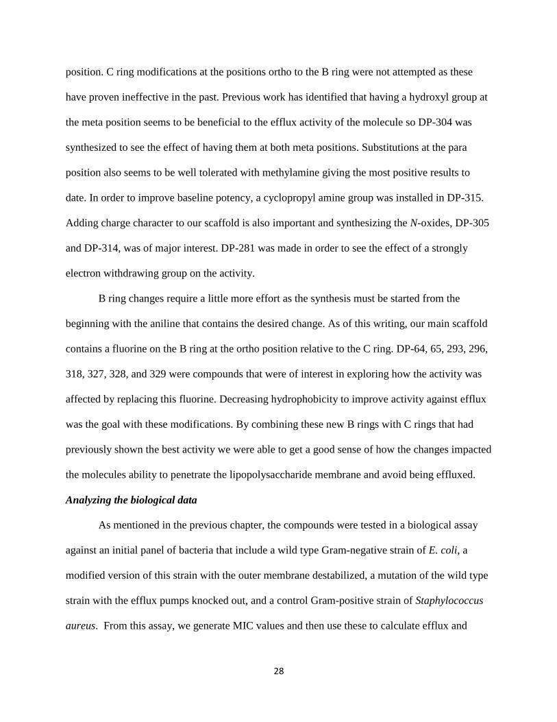
position. C ring modifications at the positions ortho to the B ring were not attempted as these
have proven ineffective in the past. Previous work has identified that having a hydroxyl group at
the meta position seems to be beneficial to the efflux activity of the molecule so DP-304 was
synthesized to see the effect of having them at both meta positions. Substitutions at the para
position also seems to be well tolerated with methylamine giving the most positive results to
date. In order to improve baseline potency, a cyclopropyl amine group was installed in DP-315.
Adding charge character to our scaffold is also important and synthesizing the N-oxides, DP-305
and DP-314, was of major interest. DP-281 was made in order to see the effect of a strongly
electron withdrawing group on the activity.
B ring changes require a little more effort as the synthesis must be started from the
beginning with the aniline that contains the desired change. As of this writing, our main scaffold
contains a fluorine on the B ring at the ortho position relative to the C ring. DP-64, 65, 293, 296,
318, 327, 328, and 329 were compounds that were of interest in exploring how the activity was
affected by replacing this fluorine. Decreasing hydrophobicity to improve activity against efflux
was the goal with these modifications. By combining these new B rings with C rings that had
previously shown the best activity we were able to get a good sense of how the changes impacted
the molecules ability to penetrate the lipopolysaccharide membrane and avoid being effluxed.
Analyzing the biological data
As mentioned in the previous chapter, the compounds were tested in a biological assay
against an initial panel of bacteria that include a wild type Gram-negative strain of E. coli, a
modified version of this strain with the outer membrane destabilized, a mutation of the wild type
strain with the efflux pumps knocked out, and a control Gram-positive strain of Staphylococcus
aureus. From this assay, we generate MIC values and then use these to calculate efflux and
29
permeability ratios that allow us to see how well each compound is performing against these
individual barriers. This data is shown in Table 3.1 and we can calculate each ratio through a
simple formula that involves dividing the MIC value generated against the bacterial strain by the
MIC value for the Gram-positive bacterial strain. Understanding this we can then analyze the
modifications made to our scaffold as I do below.
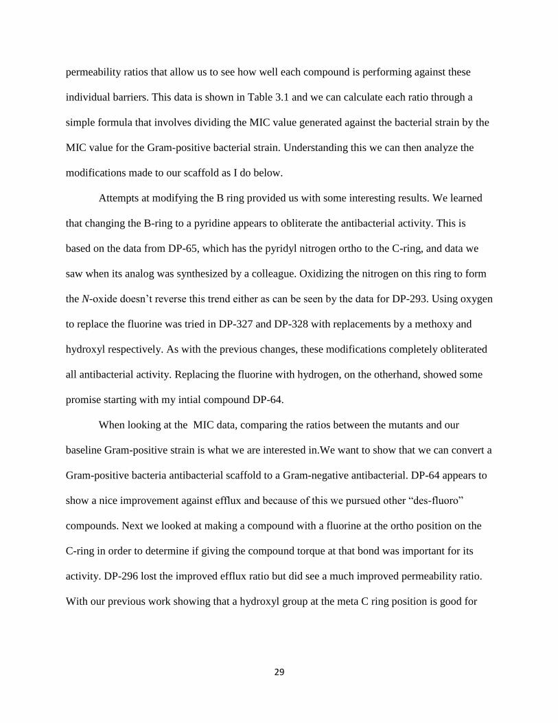
Attempts at modifying the B ring provided us with some interesting results. We learned
that changing the B-ring to a pyridine appears to obliterate the antibacterial activity. This is
based on the data from DP-65, which has the pyridyl nitrogen ortho to the C-ring, and data we
saw when its analog was synthesized by a colleague. Oxidizing the nitrogen on this ring to form
the N-oxide doesn’t reverse this trend either as can be seen by the data for DP-293. Using oxygen
to replace the fluorine was tried in DP-327 and DP-328 with replacements by a methoxy and
hydroxyl respectively. As with the previous changes, these modifications completely obliterated
all antibacterial activity. Replacing the fluorine with hydrogen, on the otherhand, showed some
promise starting with my intial compound DP-64.
When looking at the MIC data, comparing the ratios between the mutants and our
baseline Gram-positive strain is what we are interested in.We want to show that we can convert a
Gram-positive bacteria antibacterial scaffold to a Gram-negative antibacterial. DP-64 appears to
show a nice improvement against efflux and because of this we pursued other “des-fluoro”
compounds. Next we looked at making a compound with a fluorine at the ortho position on the
C-ring in order to determine if giving the compound torque at that bond was important for its
activity. DP-296 lost the improved efflux ratio but did see a much improved permeability ratio.
With our previous work showing that a hydroxyl group at the meta C ring position is good for
30
efflux, and DP-64 seemingly backing that up, and the data from DP-296 we decided to make DP-
318 in hopes that we would see additive effects.
While we didn’t see the improvements we hoped for in terms of efflux and permeability
we did see DP-296 and DP-318 give us a nice return to the baseline activity against our S. aureus
strain as compared to DP-64. This was important because it may suggest that having a fluorine
(or possibly a similar sized functional group) at the ortho position on the C ring can work as a
substitution for the fluorine on the B ring. This may be due to the torque effect on the rotatable
bond caused by having a group adjacent to it or to the possibility of a binding pocket located in
the target that a substitutent at either position can fit into.
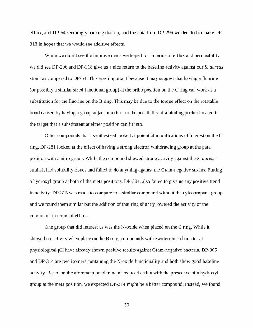
Other compounds that I synthesized looked at potential modifications of interest on the C
ring. DP-281 looked at the effect of having a strong electron withdrawing group at the para
position with a nitro group. While the compound showed strong activity against the S. aureus
strain it had solubility issues and failed to do anything against the Gram-negative strains. Putting
a hydroxyl group at both of the meta positions, DP-304, also failed to give us any positive trend
in activity. DP-315 was made to compare to a similar compound without the cylcopropane group
and we found them similar but the addition of that ring slightly lowered the activity of the
compound in terms of efflux.
One group that did interest us was the N-oxide when placed on the C ring. While it
showed no activity when place on the B ring, compounds with zwitterionic character at
physiological pH have already shown positive results against Gram-negative bacteria. DP-305
and DP-314 are two isomers containing the N-oxide functionality and both show good baseline
activity. Based on the aforemetnioned trend of reduced efflux with the prescence of a hydroxyl
group at the meta position, we expected DP-314 might be a better compound. Instead, we found

31
both had the same activity against the permeabilized membrane strain and that DP-305 appeared
to be better at penetrating the lipopolysaccharide membrane.
Conclusions
While none of the compounds I synthesized have led to the desired activity numbers our
group is looking for, they have revealed some trends that can be used in future work. From the
modifications made to the B-ring of our oxazolidinone scaffold, we can conclude that changes in
this area are not highly tolerated. Substitutions of the fluorine, on this ring, with anything other
than hydrogen appears to make the compound totally inactive against all bacteria. Replacing the
fluorine with hydrogen gave us some interesting data and the compounds I synthesized showed
potential in improving permeability or efflux. However, combining modifications that gave these
improvements individually did not provide us with the desired additive effects we expected to
see.
In regards to the C-ring modifications I made, only the introduction of an N-oxide
appeared to have interesting potential. Particularly at the para position, the N-oxide showed
decent permeability and good baseline potency and this may be attributed to its charge character.
Further work with the n-oxide has not been attempted at this point in time.
I have reviewed the compounds that I synthesized personally and the data that was
produced from the biological assays that these targets were tested in. None of these compounds
reached the desired levels of potency (4 µg/mL) against the Gram-negative bacterial strains we
tested against. However, our group has had success with other analogs that have not been
included in this paper. We hope to include this work in future publications.
32
ID Structure E Coli
Mg1665
Mg1665
PMB-n
Efflux
Ratio
Mg1665
ΔacrAB
Perm.
Ratio
S.aureus
MSSA
LZD
128 32 16 8 2 4
DP-64
>256 64 4 128 8 16
DP-65
>256 >256 >4 >256 >4 >64
DP-281*
>64 >64 >128 >64 >128 0.5
DP-293
>256 >256 >4 >256 >4 64
DP-296*
>64 >64 >16 8 2 4
DP-304
>256 >256 >32 64 8 8
DP-305
128 64 64 8 8 1
DP-314
256 64 64 32 32 1
DP-315
>256 64 32 16 8 2
DP-318
>256 32 32 16 16 1
DP-327
>256 >256 >4 >256 >4 >64
DP-328
>256 >256 >4 >256 >4 >64
DP-329
>256 64 16 32 8 4
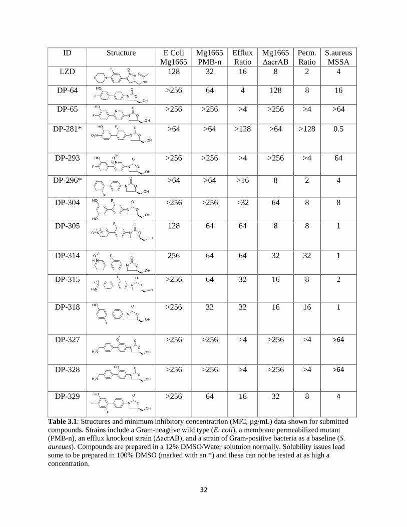
Table 3.1: Structures and minimum inhibitory concentratrion (MIC, µg/mL) data shown for submitted
compounds. Strains include a Gram-neagtive wild type (E. coli), a membrane permeabilized mutant
(PMB-n), an efflux knockout strain (ΔacrAB), and a strain of Gram-positive bacteria as a baseline (S.
aureues). Compounds are prepared in a 12% DMSO/Water solutuion normally. Solubility issues lead
some to be prepared in 100% DMSO (marked with an *) and these can not be tested at as high a
concentration.

33
Experimental
Step 1: Benzyl chloroformate protection of 4-bromo-3-fluoroaniline (1)28
A solution of 3-fluoroaniline (18.7 g, 168.3 mmol) in tetrahydrofuran (THF, 150 mL)
was treated with sodium bicarbonate (NaHCO3, 46.45 g, 336.6 mmol, 2.0 equiv) and cooled to
~0 °C in an ice bath before a solution of benzyl chloroformate (CBzCl, 31.58 g, 185.1 mmol,
26.1 mL, 1.1 equiv) in THF (50 mL) was dropwise added into the reaction mixture under N2.
The resulting reaction mixture was allowed to warm to room temperature and then stirred for 2
hours. When the reaction was judged to be complete by LC-MS analysis, the reaction mixture
was partitioned between water (100 mL) and ethyl acetate (EtOAc, 100 mL). The two layers
were separated, and the aqueous layer was extracted with EtOAc (2×100 mL). The combined
organic extracts were washed with water (2×100 mL) and saturated aqueous sodium chloride
(1x100 mL), dried over magnesium sulfate (MgSO4), and concentrated in vacuo. The residue
was further dried in vacuo to afford the desired 1 (39.2 g, 95% yield) as pale-yellow oil.
Observed mass M+H = 326.0 amu (Appendix B1). This product was directly used in subsequent
reactions without further purification.
1a. Protection of 4-bromoaniline

34
Compound 1a was synthesized using the step 1 conditions. Beginning with 2 grams of 4-
bromoaniline, we were able to produce the CBzCl protected aniline in 90% yield (3.198 g).
Observed mass M-H = 305.8 amu (Appendix B2).
1b. Protection of 6-bromopyridin-3-amine
Compound 1b was synthesized using the step 1 conditions. Beginning with 2 grams of 6-
bromopyridin-3-amine, we were able to produce the CBzCl protected aniline in 100% yield (3.55
g). Observed mass M+H = 308.0 amu (Appendix B3).
1c. Protection of 4-bromo-3-methoxyaniline
Compound 1c was synthesized using the step 1 conditions. Beginning with 2 grams of 4-
bromo-3-methoxyaniline, we were able to produce the CBzCl protected aniline in 100% yield
(3.33 g). Observed mass M+H = 335.9 amu (Appendix B4).

35
Step 2: Oxazolidinone ring formation (2)28
A solution of 1 (39.2 g, 160.0 mmol) in anhydrous THF (300 mL) was cooled to -78° C
in a dry-ice/acetone bath before a solution of LHMDS (1 M solution in THF, 176 mmol, 1.1
equiv) was added dropwise under N2. The resulting reaction mixture was subsequently stirred at
-78 °C for 1 hour before a solution of (R)-(-)-glycidyl butyrate (25.37 g, 24.6 mL, 176 mmol, 1.1
equiv) in anhydrous THF (100 mL) was added dropwise into the reaction mixture at -78 °C
under N2. The resulting reaction mixture was stirred at -78 °C. for 30 min before being gradually
warmed to room temperature for 12 h under N2. When LC-MS showed the reaction was
complete, the reaction mixture was quenched with H2O (200 mL), and the resulting mixture was
stirred at room temperature for 1 hour before ethyl acetate (200 mL) was added. The two layers
were separated, and the aqueous layer was extracted with ethyl acetate (2×100 mL). The
combined organic extracts were washed with water (2×100 mL) and saturated aqueous sodium
chloride (1x100 mL), dried over MgSO4, and concentrated in vacuo. White crystals precipitated
from the concentrated solution when most of the solvent was evaporated. The residue was then
treated with 20% EtOAc/hexane (100 mL) and the resulting slurry was stirred at room
temperature for 30 min. The solids were collected by filtration and washed with 20% ethyl
acetate/hexane (2×50 mL) to afford the desired compound 2 (24.4 g, 72.3% yield) as white
crystals. Observed mass M+H = 290.0 amu (Appendix B5). This product was directly used in
subsequent reactions without further purification. 1H NMR (399 MHz, DMSO-d6) ppm 3.52 -

36
3.60 (m, 1 H) 3.63 - 3.73 (m, 1 H) 3.77 - 3.88 (m, 1 H) 4.08 (t, J=9.16 Hz, 1 H) 4.67 - 4.77 (m, 1
H) 5.24 (t, J=5.50 Hz, 1 H) 7.36 (d, J=8.79 Hz, 1 H) 7.62 - 7.75 (m, 2 H)
2a. Des-Fluoro B-Ring
Compound 2a was synthesized using the step 2 conditions. Beginning with 3.198 grams
of compound 1a, we were able to form the oxazolidinone 2a in76% yield (2.15 g). Observed
mass M+H = 271.8 amu (Appendix B7). 1H NMR (400 MHz, DMSO-d6) ppm 3.50 - 3.60 (m,
1 H) 3.61 - 3.69 (m, 1 H) 3.79 (m, 1 H) 4.05 (t, J=9.16 Hz, 1 H) 4.63 - 4.72 (m, 1 H) 5.19 (t,
J=5.50 Hz, 1 H) 7.47 - 7.58 (m, 4 H)
2b. Pyridyl B-Ring
Compound 2b was synthesized using the step 2 conditions. Beginning with 3.55 grams of
compound 1b, we were able to form the oxazolidinone 2b in 50% yield (1.572 g). Observed
mass M+H = 273.0 amu (Appendix B9). 1H NMR (400 MHz, DMSO-d6) ppm 3.50 - 3.62 (m,
1 H) 3.66 - 3.77 (m, 1 H) 3.81 - 3.93 (m, 1 H) 4.11 (t, J=8.79 Hz, 1 H) 4.76 (d, J=3.66 Hz, 1 H)
5.24 (t, J=5.50 Hz, 1 H) 7.67 (d, J=8.79 Hz, 1 H) 7.94 - 8.16 (m, 1 H) 8.45 - 8.65 (m, 1 H)
37
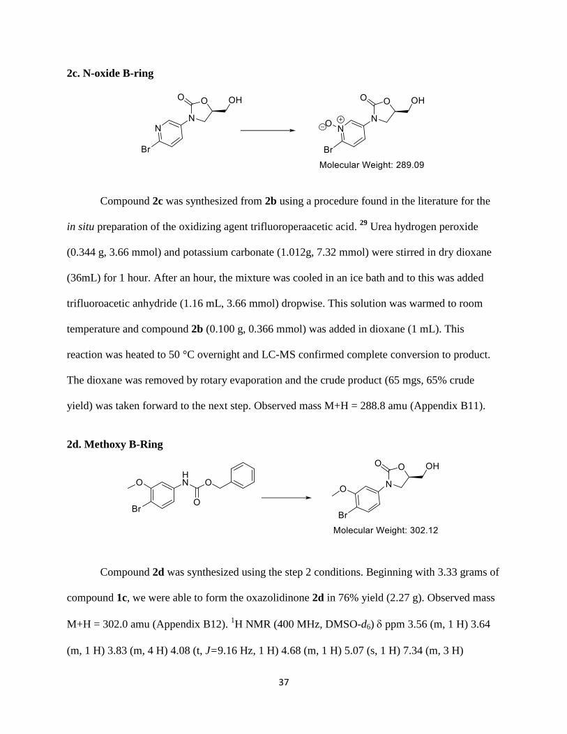
2c. N-oxide B-ring
Compound 2c was synthesized from 2b using a procedure found in the literature for the
in situ preparation of the oxidizing agent trifluoroperaacetic acid. 29
Urea hydrogen peroxide
(0.344 g, 3.66 mmol) and potassium carbonate (1.012g, 7.32 mmol) were stirred in dry dioxane
(36mL) for 1 hour. After an hour, the mixture was cooled in an ice bath and to this was added
trifluoroacetic anhydride (1.16 mL, 3.66 mmol) dropwise. This solution was warmed to room
temperature and compound 2b (0.100 g, 0.366 mmol) was added in dioxane (1 mL). This
reaction was heated to 50 °C overnight and LC-MS confirmed complete conversion to product.
The dioxane was removed by rotary evaporation and the crude product (65 mgs, 65% crude
yield) was taken forward to the next step. Observed mass M+H = 288.8 amu (Appendix B11).
2d. Methoxy B-Ring
Compound 2d was synthesized using the step 2 conditions. Beginning with 3.33 grams of
compound 1c, we were able to form the oxazolidinone 2d in 76% yield (2.27 g). Observed mass
M+H = 302.0 amu (Appendix B12). 1H NMR (400 MHz, DMSO-d6) ppm 3.56 (m, 1 H) 3.64
(m, 1 H) 3.83 (m, 4 H) 4.08 (t, J=9.16 Hz, 1 H) 4.68 (m, 1 H) 5.07 (s, 1 H) 7.34 (m, 3 H)

38
2e. Hydroxy B-Ring
Compound 2e was synthesized from compound 2d using a general procedure for
deprotection of methyl ethers. Compound 2d (300 mgs, 0.993 mmol) was dissolved in DCM (~5
mL) and cooled to -78 °C in a dry ice/acetone bath. The reaction was kept under N2 and boron
tribromide (0.160 mL, 1.688 mmol) in DCM was then added slowly. The reaction went
overnight forming a brown precipitate that would prove to be the desired product. The solvent
was removed by filtration and the product was collected to give 257 mgs (90% yield). Observed
mass M+H = 287.8 amu (Appendix B14). This was taken on to the next step without further
purification. 1H NMR (400 MHz, DMSO-d6) ppm 3.55 (dd, J=12.46, 2.93 Hz, 1 H) 3.67 (dd,
J=12.46, 2.20 Hz, 1 H) 3.76 (t, J=7.33 Hz, 1 H) 4.03 (t, J=8.79 Hz, 1 H) 4.60 - 4.80 (m, 1 H)
6.82 (d, J=8.79 Hz, 1 H) 7.30 - 7.49 (m, 2 H) 10.39 (br s, 1 H)
Step 3: Making the boronic ester/acid (3 and 3’)30
To a solution of 2 (3.37 g, 10 mmol) in 20 mL of dry DMSO, bis(pinacolato)diboron (20
mmol, 2 equiv), potassium acetate (50 mmol, 5 equiv), and Pd(dppf)Cl2 (0.5 mmol, 0.05 equiv)

39
were added. The mixture was heated at 80 oC under nitrogen overnight. When HPLC/MS
showed the reaction was complete, it was cooled to room temperature. Then 20 mL of methanol
were added and the mixture was filtered through celite. To this filtrate was added 20 mL of water
and this was allowed to stand overnight at 4 oC. The precipitate formed was isolated by filtration
with a Buchner funnel and washed with water. The filtrate was further concentrated to get a
second crop of precipitate. The solids were combined and recrystallized in methanol/water. The
formed precipitate was filtered and washed with water and dried under vacuum to give a mixture
of products 3 and 3’ as a brown powder. Observed masses M+H = 255.9 (3.166 min) amu and
338.0 (4.644 min) amu (Appendix B16). This was used in subsequent reactions without further
purification. 1H NMR (400 MHz, DMSO-d6) ppm 1.28 (br s, 12 H) 3.57 (d, J=9.53 Hz, 1 H)
3.66 (d, J=10.99 Hz, 1 H) 3.84 (m, 1 H) 4.09 (t, J=9.16 Hz, 1 H) 4.73 (br s, 1 H) 7.36 (d, J=6.60
Hz, 1 H) 7.47 (d, J=6.60 Hz, 1 H) 7.63 (t, J=7.69 Hz, 1 H)
3a: Des-fluoro boronic ester/acid
Compound 3a was synthesized using the conditions outlined in step 3. Crystallization in
water/methanol proved unsuccessful, however, and so the product was extracted in ethyl acetate.
Ethyl acetate was removed by rotary evaporation and then the product was place under vacuum
to complete drying. Beginning with 905 mgs of compound 2a, we yielded 514 mgs (48%) of
flakey, brown material which was used in subsequent reactions without further purification.

40
Observed mass M+H = 320.0 amu (Appendix B18). 1H NMR (400 MHz, DMSO-d6) ppm 1.27
(s, 12 H) 3.49 - 3.60 (m, 1 H) 3.61 - 3.71 (m, 1 H) 3.82 (dd, J=8.43, 623 Hz, 1 H) 4.02 - 4.12 (t,
J=9.16 Hz, 1 H) 4.63 - 4.73 (m, 1 H) 5.13 - 5.26 (m, 1 H) 7.50 - 7.62 (m, 2 H) 7.62 - 7.71 (m, 2
H)
Step 4: General Suzuki Coupling reaction conditions (4)
The desired bromide (100mgs, 1.0 equiv) was dissolved in 2-3 mL of 9:1 dioxane: water.
To this solution was added the corresponding boronic acid/ester (1.2 equiv), Pd(dppf)Cl2 (0.1
equiv), and potassium carbonate (4 equiv). This mixture was then heated to 85-90°C and let stir.
The reaction was monitored by LC-MS and, generally, was complete within 1-2 hours. Upon
completion, the reaction was quenched with water (5 mL) and the product extracted with ethyl
acetate (1x10 mL). The aqueous layer was back-extracted with ethyl acetate (1x5 mL) and the
organic layers were combined. These were washed with saturated sodium chloride (1x10 mL),
dried over magnesium sulfate, and concentrated to dryness by rotary evaporation. The crude
product was dissolved in 1 mL of DMF and purification was performed using reverse phase
HPLC (water and acetonitrile with 0.1% TFA as solvents for mobile phase). Those fractions
containing pure product were pooled and lyophilized to give the pure product as a white solid.

41
DP-64
DP-64 was synthesized by Suzuki coupling 4-fluoro-3-hydroxy boronic acid with
compound 2a using the reaction conditions outlined in step 4. Purification resulted in the desired
compound eluting at ~30%B. Analytical HPLC using method 2to100in30 (Table 3.4) showed the
compound was 96% pure at 254 nm with a retention time of 10.879 minutes. Observed mass
M+H = 304.1 amu. HPLC and LC-MS chromatograms are available in the Appendix (B20 and
B21). 1H NMR: not available due to lack of material after submission for testing.
DP-65
DP-65 was synthesized by Suzuki coupling 4-fluoro-3-hydroxy boronic acid with
compound 2b using the reaction conditions outlined in step 4. Purification resulted in the desired
compound eluting at ~17%B. Analytical HPLC using method 2to50in30 (Table 3.3) showed the
compound was 95% pure at 254 nm with a retention time of 12.126 minutes. Observed mass
M+H = 305.1 amu. HPLC and LC-MS chromatograms are available in the Appendix (B22 and

42
B23). 1H NMR (400 MHz, DMSO-d6) δppm 3.60 (dd, J=12.46, 4.40 Hz, 1 H) 3.69 (dd,
J=12.09, 3.30 Hz, 1 H) 3.93 (m, 1 H) 4.18 (m, 1 H) 4.65 - 4.98 (m, 3 H) 7.23 (m, 1 H) 7.45 (br s,
1 H) 7.69 (m, 1 H) 7.92 (d, J=8.79 Hz, 1 H) 8.12 (m, 1 H) 8.80 (br s, 1 H)
DP-281
DP-281 was synthesized by Suzuki coupling 5-bromo-2-nitrophenol with compound 3
using the reaction conditions outlined in step 4. Purification resulted in the desired compound
eluting at ~38%B. Analytical HPLC using method 2to50in30 (Table 3.2) showed the compound
was 95% pure at 254 nm with a retention time of 12.692 minutes. Observed mass M+H = 348.8
amu. HPLC and LC-MS chromatograms are available in the Appendix (B25 and B6). 1H NMR
(400 MHz, DMSO-d6) δ ppm 3.59 (m, 1H) 3.70 (m, 1 H) 3.88 (m, 1 H) 4.14 (t, J=8.79 Hz, 1 H)
4.76 (m, 1H) 5.25 (br s, 9 H) 7.17 (m, 1 H) 7.31 (br s, 1 H) 7.50 (d, J=8.06, 11 Hz, 1 H) 7.58 -
7.72 (m, 2 H) 8.00 (d, J=8.06, 11 Hz, 1 H)
DP-293
43
DP-293 was synthesized by Suzuki coupling 4-fluoro-3-hydroxy boronic acid with
compound 2c using the reaction conditions outlined in step 4. Purification resulted in the desired
compound eluting at ~18%B. Analytical HPLC using method 2to100in30 (Table 3.4) showed the
compound was 90% pure at 254 nm with a retention time of 6.646minutes. The impurity present
was known to be DP-65. Prior testing showed that compound to be inactive so we moved
forward with submission at this point. Observed mass M+H = 320.9 amu. HPLC and LC-MS
chromatograms are available in the Appendix (B28 and B29). 1H NMR (400 MHz, DMSO-d6) δ
ppm 3.59 (m, 1 H) 3.68 (m, 1 H) 3.88 (m, 1 H) 4.12 (m, 1 H) 4.78 (br s, 1 H) 7.19 (br s, 1 H)
7.24 (s, 1 H) 7.55 - 7.64 (m, 3 H) 8.66 (s, 1 H) 10.09 (br s, 1 H)
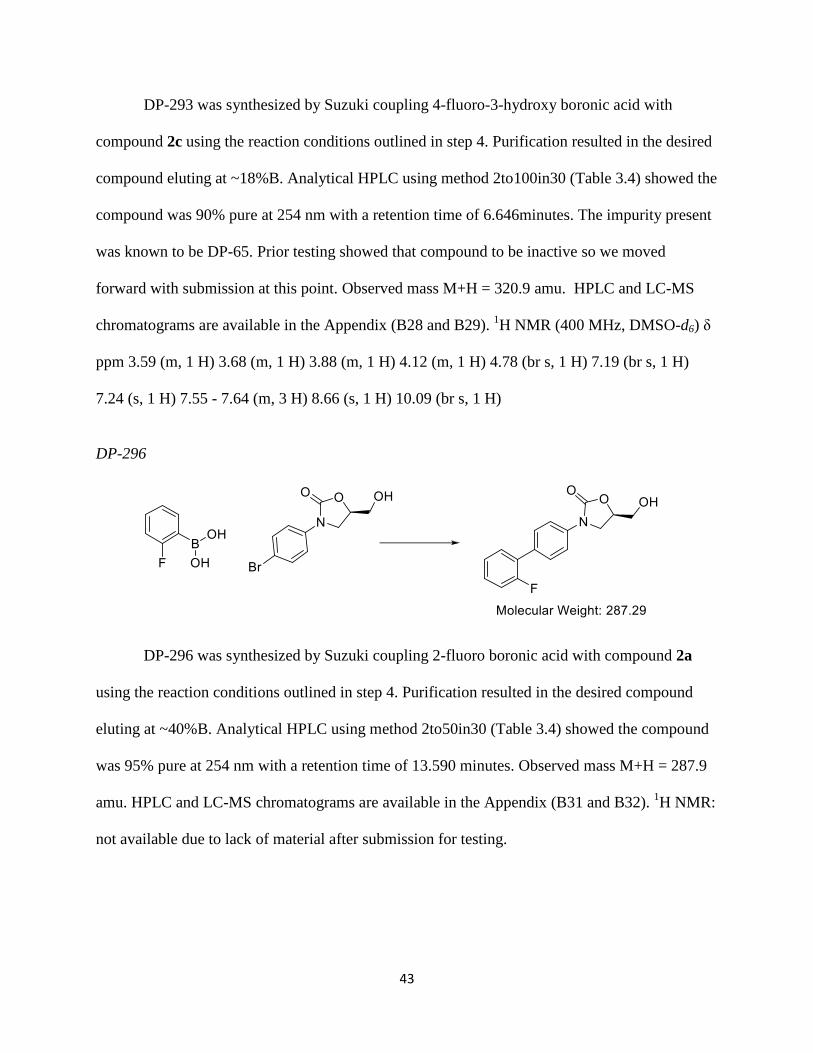
DP-296
DP-296 was synthesized by Suzuki coupling 2-fluoro boronic acid with compound 2a
using the reaction conditions outlined in step 4. Purification resulted in the desired compound
eluting at ~40%B. Analytical HPLC using method 2to50in30 (Table 3.4) showed the compound
was 95% pure at 254 nm with a retention time of 13.590 minutes. Observed mass M+H = 287.9
amu. HPLC and LC-MS chromatograms are available in the Appendix (B31 and B32). 1H NMR:
not available due to lack of material after submission for testing.

44
DP-304
DP-304 was synthesized by Suzuki coupling 5-bromoresorcinol with compound 2 using
the reaction conditions outlined in step 4. Purification resulted in the desired compound eluting
at ~17%B. Analytical HPLC using method 2to50in30 (Table 3.4) showed the compound was
95% pure at 254 nm with a retention time of 12.545 minutes. Observed mass M+H = 319.9 amu.
HPLC and LC-MS chromatograms are available in the Appendix (B33 and B34). 1
H NMR: not
available due to lack of material after submission for testing.
DP-305
DP-305 was synthesized by first Suzuki coupling 4-bromopyridine with compound 3
using the reaction conditions outlined in step 4. This was followed by oxidation by
trifluoroperaacetic acid that was generated in situ.29
Urea hydrogen peroxide (0.483 g, 5.14
mmol) and potassium carbonate (1.421g, 10.28 mmol) were stirred in dry dioxane (40mL) for 1
45
hour. After an hour, the mixture was cooled in an ice bath and to this was added trifluoroacetic
anhydride (1.4 mL, 5.14 mmol) dropwise. This solution was warmed to room temperature and
then our previously coupled intermediate was added in dioxane (1 mL). This reaction was heated
to 50°C overnight and LC-MS confirmed complete conversion to product. The dioxane was
removed by rotary evaporation and the crude product was then dissolved in water and purified.
Purification resulted in the desired compound eluting at ~11%B. Analytical HPLC using method
2to30in30 (Table 3.2) showed the compound was 95% pure at 254 nm with a retention time of
8.187 minutes. Observed mass M+H = 304.9 amu. HPLC and LC-MS chromatograms are
available in the Appendix (B35 and B36). 1
H NMR (400 MHz, DMSO-d6) ppm 3.52 - 3.63 (dd,
J=12.09, 4.03 Hz, 1 H) 3.69 (dd, J=12.46, 4.03 Hz, 1 H) 3.91 (m, 1 H) 4.14 (t, J=9.16 Hz, 1 H)
4.75 (m, 2 H) 7.51 (dd, J=8.06, 2.20 Hz, 1 H) 7.68 (m, 2 H) 7.72 (m, 2 H) 8.33 (d, J=6.60 Hz, 2
H)
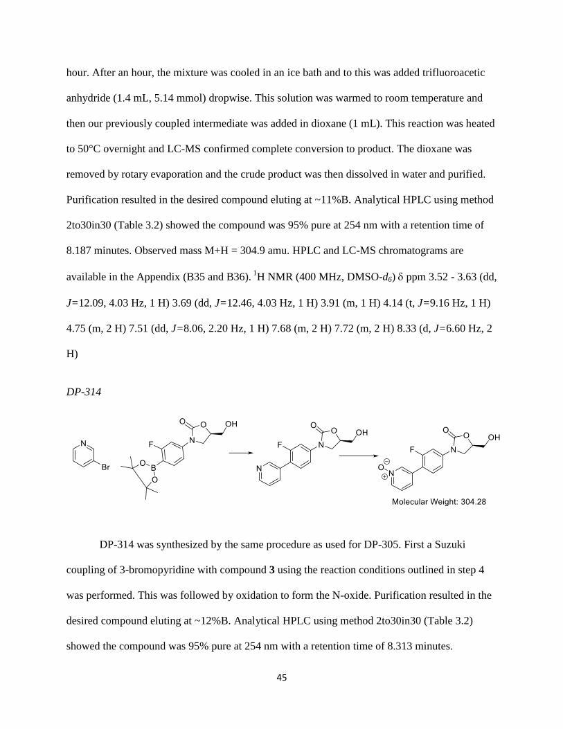
DP-314
DP-314 was synthesized by the same procedure as used for DP-305. First a Suzuki
coupling of 3-bromopyridine with compound 3 using the reaction conditions outlined in step 4
was performed. This was followed by oxidation to form the N-oxide. Purification resulted in the
desired compound eluting at ~12%B. Analytical HPLC using method 2to30in30 (Table 3.2)
showed the compound was 95% pure at 254 nm with a retention time of 8.313 minutes.

46
Observed mass M+H = 304.9 amu. HPLC and LC-MS chromatograms are available in the
Appendix (B38 and B39). 1
H NMR (400 MHz, DMSO-d6) ppm 3.59 (d, J=3.66 Hz, 1 H) 3.68
(d, J=3.66 Hz, 2 H) 3.89 (m, 1 H) 4.14 (t, J=9.16 Hz, 1 H) 4.74 (m, 1 H) 7.54 (m, 3 H) 7.69 (m,
2 H) 8.26 (d, 5.13 Hz, 1 H) 8.44 (s, 1 H)
DP-315
DP-315 was synthesized by Suzuki coupling 4-bromo styrene oxide with compound 3
using the reaction conditions outlined in step 4. Purification resulted in the desired compound
eluting at ~23%B. Analytical HPLC using method 2to30in30 (Table 3.2) showed the compound
was 95% pure at 254 nm with a retention time of 11.601 minutes. Observed mass M+H = 343.2
amu. HPLC and LC-MS chromatograms are available in the Appendix (B41 and B42). 1
H NMR
(400 MHz, DMSO-d6) ppm 1.27 (m, 2 H) 1.36 (m, 2 H) 3.59 (m, 1 H) 3.69 (m, 1 H) 3.89 (m, 1
H) 4.13 (t, J=9.16 Hz, 1 H) 4.76 (m, 1 H) 5.26 (br s, 1 H) 7.43 - 7.54 (m, 3 H) 7.54 - 7.69 (m, 4
H) 8.69 (br s, 2 H)
DP-318
47
DP-318 was synthesized by Suzuki coupling 4-fluoro-3-bromo phenol with compound 3a
using the reaction conditions outlined in step 4. Purification resulted in the desired compound
eluting at ~33%B. Analytical HPLC using method 2to30in30 (Table 3.2) showed the compound
was 95% pure at 254 nm with a retention time of 11.601 minutes. Observed mass M+H = 303.9
amu. HPLC and LC-MS chromatograms are available in the Appendix (B44 and B45). 1
H NMR
(400 MHz, DMSO-d6) ppm 3.59 (dd, J=12.46, 3.66 Hz, 1 H) 3.68 (dd, J=12.46, 2.93 Hz, 1 H)
3.87 (m, 1 H) 4.13 (t, J=8.79 Hz, 1 H) 4.72 (m, 1 H) 5.22 (br s, 1 H) 6.75 (m, 1 H) 6.84 (m, 1 H)
7.09 (m, 1 H) 7.54 (d, J=7.33 Hz, 2 H) 7.66 (d, J=8.79 Hz, 2 H) 9.50 (s, 1 H)
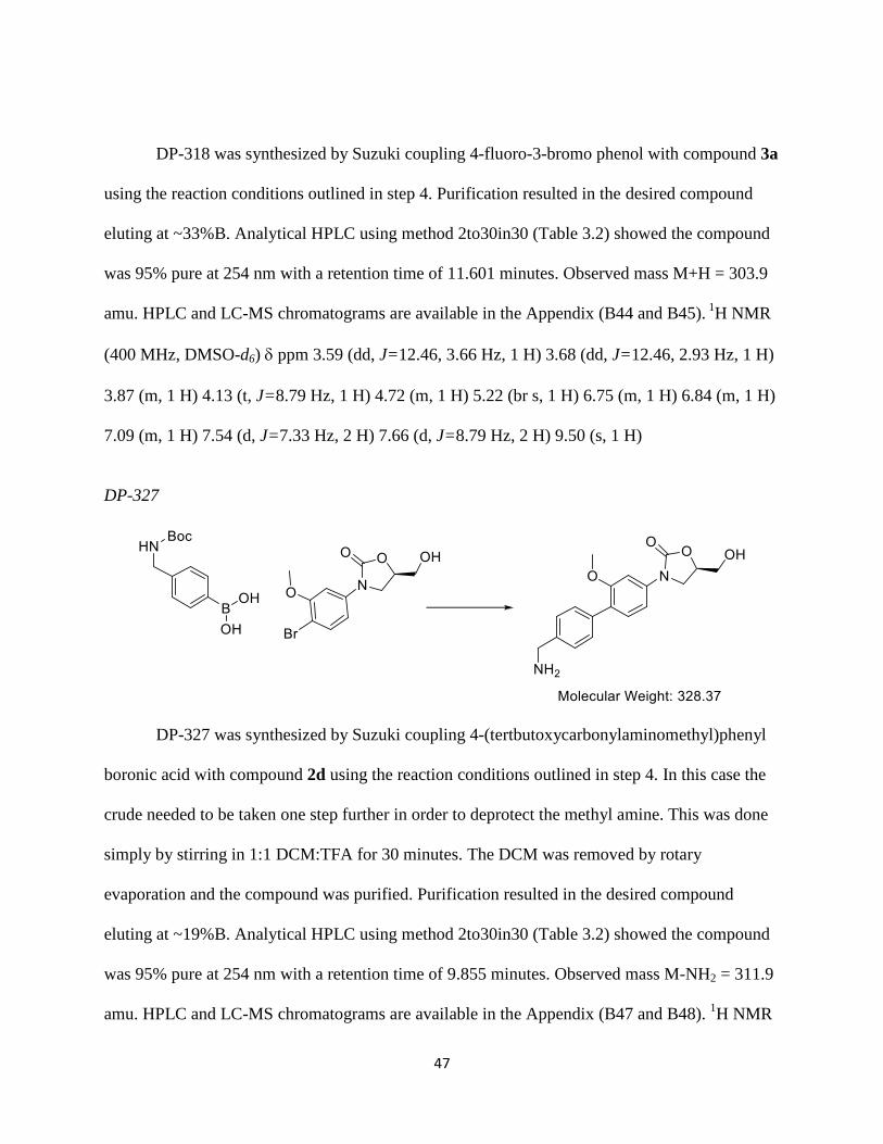
DP-327
DP-327 was synthesized by Suzuki coupling 4-(tertbutoxycarbonylaminomethyl)phenyl
boronic acid with compound 2d using the reaction conditions outlined in step 4. In this case the
crude needed to be taken one step further in order to deprotect the methyl amine. This was done
simply by stirring in 1:1 DCM:TFA for 30 minutes. The DCM was removed by rotary
evaporation and the compound was purified. Purification resulted in the desired compound
eluting at ~19%B. Analytical HPLC using method 2to30in30 (Table 3.2) showed the compound
was 95% pure at 254 nm with a retention time of 9.855 minutes. Observed mass M-NH2 = 311.9
amu. HPLC and LC-MS chromatograms are available in the Appendix (B47 and B48). 1H NMR

48
(400 MHz, DMSO-d6) ppm 3.58 (m, 1 H) 3.70 (m, 1 H) 3.76 (s, 3 H) 3.88 (m, 1 H) 4.05 (br s,
2 H) 4.12 (m, 1 H) 4.73 (m, 1 H) 5.26 (br s, 1 H) 7.12 (m, 2 H) 7.29 (m, 2 H) 7.35 - 7.57 (m, 3
H) 8.26 (br s, 2 H)
DP-328
DP-328 was synthesized similarly to DP-327. First a Suzuki coupling was performed
with 4-(tertbutoxycarbonylaminomethyl)phenyl boronic acid and compound 2e using the
reaction conditions outlined in step 4. This was followed by a boc-deprotection to give us our
desired product. Purification resulted in the desired compound eluting at ~19%B. Analytical
HPLC using method 2to30in30 (Table 3.2) showed the compound was 95% pure at 254 nm with
a retention time of 8.685 minutes. Observed mass M-NH2 = 298.1 amu and M+H = 315.1 amu.
HPLC and LC-MS chromatograms are available in the Appendix (B50 and B51). 1
H NMR (400
MHz, DMSO-d6) ppm 3.52 (m, 2 H) 3.66 (m, 1H) 3.79 (m, 1 H) 4.02 (m, 2 H) 4.69 (m, 1 H)
5.28 (br s, 1 H) 6.99 (d, J=8.06 Hz, 1 H) 7.27 (d, J=8.79 Hz, 1 H) 7.37(m, 1 H) 7.48 (m, 2 H)
7.58 (m, 2 H) 8.45 (br s, 2 H) 9.93 (s, 1 H)

49
DP-329
DP-329 was synthesized by Suzuki coupling 5-bromo-2,4-difluoro phenol with
compound 3a using the reaction conditions outlined in step 4. Purification resulted in the desired
compound eluting at ~35%B. Analytical HPLC using method 2to30in30 (Table 3.2) showed the
compound was 95% pure at 254 nm with a retention time of 17.575 minutes. Observed mass
M+H = 321.9 amu. HPLC and LC-MS chromatograms are available in the Appendix (53 and
B54). 1H NMR (400 MHz, DMSO-d6) ppm 3.51 - 3.62 (m, 1 H) 3.66 - 3.74 (m, 1 H) 3.83 -
3.92 (m, 1 H) 4.13 (t, J=9.16 Hz, 1 H) 4.66 - 4.77 (m, 1 H) 5.24 (t, J=5.50 Hz, 1 H) 7.03 (t,
J=8.79 Hz, 1H) 7.24 - 7.34 (m, 1 H) 7.51 (d, J=8.79 Hz, 2 H) 7.67 (d, J=8.06 Hz, 2 H) 9.97 (br
s, 1 H)
Characterization
The raw data generated for the characterization of the oxazolidinone analogs detailed in
this paper are included in the Appendix. HPLC chromatograms from our HP 1100 analytical
HPLC system are included and used to determine the purity (goal of 95%) of the final
compounds. These are thirty minute methods that use one of the following gradients dependent
on retention time of the compound:

50
Time (min) % Mobile Phase B
0 0
25 30
25.01 100
27 100
27.01 2
30 2
Table 3.2: Gradient of HPLC runs using 2to30in30 method. Mobile phase A: 0.1% TFA in water and B: 0.1% TFA in acetonitrile.
Time (min) % Mobile Phase B
0 0
25 50
25.01 100
27 100
27.01 2
30 2
Table 3.3: Gradient of HPLC runs using 2to50in30 method. Mobile phase A: 0.1% TFA in water and B: 0.1% TFA in acetonitrile.
Time (min) % Mobile Phase B
0 0
25 100
27.01 2
30 2
Table 3.4: Gradient of HPLC runs using 2to100in30 method. Mobile phase A: 0.1% TFA in water and B: 0.1% TFA in acetonitrile.
LC-MS data is also included from our HP 1100 HPLC-MS system that we use to monitor
our reactions by providing us with the desired molecular weight of our target compounds. These
are eight minute methods run in the positive mode that use the following gradient:
Time (min) % Mobile Phase B
0 2
4 100
6 100
6.1 2
8 2
Table 3.5: Gradient of LC-MS runs of 2to100in8min-PosM and 2to100in8min-NegM methods. Mobile phase A: 0.1% FA in water and B: 0.1%FA in acetonitrile.

51
LC-MS and HPLC data were used as our main sources of characterization. When
material was available, proton NMR, using a 400 MHz Varian NMR spectrometer, were run to
confirm the structures. For all structures, d6-DMSO with 0.03% TMS was used.

52
References
1) Duclos RI, Cleary DC, Catcott KC, Zhou ZS. Synthesis and Characterization of Se-
adenosyl-L-selenohomocysteine selenoxide. J. Sulfur Chem. 2015; 36: 135-144
2) Loenen WAM. S-adenosylmethionine: jack of all trades and master of everything?
Biochem Soc Trans. 2006; 34:330-3.
3) Chiang PK, Gordon RK, Tal J, Zeng GC, Doctor BP, Pardhasaradhi K, McCann PP. S-
Adenosylmethionine and methylation. FASEB J. 1996; 10:471-80.
4) Struck A-W, Thompson ML, Wong LS, Micklefield J. S-Adenosyl-methionine-
dependent methyltransferases: highly versatile enzymes in biocatalysis, biosynthesis and other
biotechnological applications. ChemBioChem. 2012; 13:2642-55.
5) Stolowitz ML, Minch MJ. S-Adenosyl-L-methionone and S-Adenosyl-L-homocysteine,
an NMR study. J Am Chem Soc. 1981; 103:6015-9.
6) Lee BW, Sun HG, Zang T, Kim BJ, Alfaro JF, Zhou ZS. Enzyme-catalyzed transfer of a
ketone group from an S-adenosylmethionine analogue: a tool for the functional analysis of
methyltransferases. J Am Chem Soc. 2010; 132:3642-3
7) Willnow S, Martin M, Luscher B, Weinhold E. A selenium-based click AdoMet
analogue for versatile substrate labeling with wild-type protein methyltransferases.
ChemBioChem. 2012; 13:1167-73.
8) Kenton N. Design and Synthesis of Methionine Analogues for the Enzymatic Synthesis
of S-Adenosyl-Methionine Analogues: Tools for the Analysis of Methyltransferase Substrate
Specificity. Chemistry Master’s Thesis. 2012.
9) Duerre JA, Salisbury L, Miller CH. Preparation and characterization of sulfoxides of S-
adenosyl-L-homocysteine and S-ribosyl-L-homocysteine. Anal Biochem. 1970; 35:505-15.
10) Pugh CS, Borchardt RT. Effects of S-adenosylhomocysteine analogues on vaccinia viral
messenger ribonucleic acid synthesis and methylation. Biochemistry. 1982; 21:1535-41.
11) Gillet L, Looze Y, Deconinck M, Leonis J. Binding capacities of various analogues of S-
adenosyl-L-homocysteine to protein methyltransferase II from human erythrocytes. Experientia.
1979; 35:1007-9.
12) Borchardt RT, Wu YS. Potential inhibitors of S-adenosylmethionine-dependent
methyltransferases. 1. Modification of the amino acid portion of S-adenosylhomocysteine. J Med
Chem. 1974; 17:862-8.
53
13) Coward JK, D'Urso-Scott M, Sweet WD. Inhibition of catechol-O-methyltransferase by
S-adenosylhomocysteine and S-adenosylhomocysteine sulfoxide, a potential transition-state
analog. Biochem Pharmacol. 1972; 21:1200-3.
14) Mudd SH, Cantoni GL. Selenomethionine in enzymatic transmethylations. Nature. 1957;
180:1052.
15) Rayman MP. Selenium and human health. Lancet. 2012; 379:1256-68.
15) Hatfield DL, Tsuji PA, Carlson BA, Gladyshev VN. Selenium and selenocysteine: roles
in cancer, health, and development. Trends Biochem Sci. 2014; 39:112-20.
17) Scovill JP, Thigpen II DL, Lemley PV. A convenient method for the synthesis and raney
nickel desulfurization of 5'-deoxy-5'-methylthioadenosine. Phosphorus, Sulfur, and Silicon.
1993; 85:149-52.
18) Zhou ZS, Smith AE, Matthews RG. L-Selenohomocysteine: one-step synthesis from L-
selenomethionine and kinetic analysis as substrate for methionine synthases. Bioorg Med Chem
Lett. 2000; 10: 2471-5.
19) Ritchey JA, Davis BM, Pleban PA, Bayse CA. Experimental and theoretical evidence for
cyclic selenurane formation during selenomethionine oxidation. Org Biomol Chem. 2005; 3:
4337-42.
20) Boucher HW. Challenges in anti-infective development in the era of bad bugs, no drugs:
a regulatory perspective using the example of bloodstream infection as an indication. Clinical
infectious diseases: an official publication of the Infectious Diseases Society of America 2010;
50 Suppl 1: S4-9.
21) Spellberg B, Shlaes D. Prioritized current unmet needs for antibacterial therapies. Clin
Pharmacol Ther 2014; 96: 151-3
22) Spellberg B, Guidos R, Gilbert D et al. The epidemic of antibiotic-resistant infections: a
call to action for the medical community from the Infectious Diseases Society of America.
Clinical infectious diseases: an official publication of the Infectious Diseases Society of America
2008; 46: 155-64.
23) Hampton T. Report reveals scope of US antibiotic resistance threat. JAMA 2013; 310:
1661-3.
24) The Center for Disease Dynamics, Economics & Policy. www.cddep.org/
25) Peleg AY, Hooper DC. Hospital-acquired infections due to gram-negative bacteria. The
New England journal of medicine 2010; 362: 1804-13.

54
26) Nakae T, Ishii JN, Tokunaga H et al. The solute selectivity of porin pores of Escherichia
coli and Salmonella typhimurium. Tokai J Exp Clin Med 1982; 7:Suppl: 141-8.
27) Shaw KJ, Barbachyn MR. The oxazolidinones: past, present, and future. Ann N Y Acad
Sci 2011; 1241: 48-70.
28) Wu Y, Shili C, Chen Y, Hanselmann R, Lou R, Zhou, J. Process for the Synthesis of
Biaryl Oxazolidinones. U.S. Pat. Appl. Publ., 20100022772, 28 Jan 2010.
29) Rong D, Phillips VA, Rubio RS, Castro MA, Wheelhouse RT. A safe, convenient and
efficient method for the preparation of heterocyclic N-oxides using urea hydrogen peroxide. Tet.
Lett. 2008; 49: 6933-6935
30) Reck F, Zhou F, Eyermann CJ, Kern G, Carcanague D, Ioaniddis G, Illingworth R, Poon
G, Gravestock MB. Novel Substituted (Pyridin-3-yl)phenyloxazolidinones: Antibacterial Agents
with Reduced Activity against Monoamine Oxidase A and Increased Solubility. JMC 2007; 50
(20): 4868-4881

55
Appendix A
Additional Characterization Data of SeAHO Intermediates and Comparators
A1: Proton NMR of ClDA in DMSO-d6.

56
A2: Proton NMR of SAH in DMSO-d6.

57
A3: Proton NMR of SAHO in DMSO-d6
A4: COSY of SAHO
5.5 5.0 4.5 4.0 3.5 3.0 2.5
F2 Chemical Shift (ppm)
3
4
5
F1 C
hem
ical S
hift (ppm
)

58
A5: LC-MS data for SAH
A6: LC-MS of SAHO

59
60
Appendix B
LC-MS Data for Intermediates
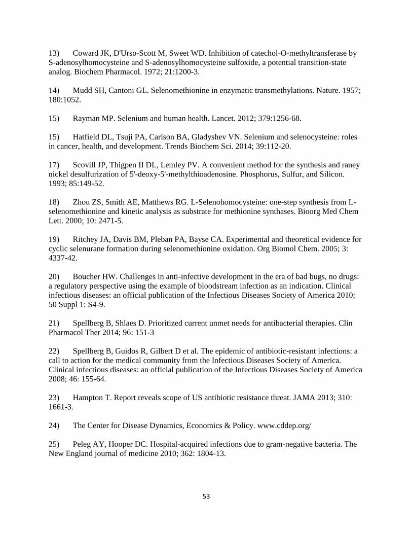
B1: Compound 1 HPLC-MS chromatogram and mass fragment using method 2to100in8min-PosM

61
B2: Compound 1a HPLC-MS chromatogram and mass fragment using method 2to100in8min-NegM

62
B3: Compound 1b HPLC-MS chromatogram and mass fragment using method 2to100in8min-PosM

63
B4: Compound 1c HPLC-MS chromatogram and mass fragment using method 2to100in8min-PosM

64
B5: Compound 2 HPLC-MS chromatogram and mass fragment using method 2to100in8min-PosM

65
B6: Compound 2 1H NMR
DC-fl-oxaz.esp
8 7 6 5 4 3 2 1 0
Chemical Shift (ppm)
0
0.05
0.10
0.15
0.20
0.25
0.30
0.35
0.40
0.45
0.50
0.55
0.60
Norm
alized Inte
nsity
1.000.970.960.960.870.831.061.57
TMS
DMSOWater
7.7
37.7
1(9
,12)
7.6
97.6
97.6
6
7.3
7(1
3)
7.3
5
5.2
55.2
4(7
)5.2
2
4.7
34.7
2(2
)4.7
1
4.1
14.0
8(3
)3.8
53.8
3(3
)3.6
6(6
)3.5
7(6
)3.5
63.5
4
0.0
0
11
12 13
8
910
N4
5
O14
O1
23
6OH7
Br16
F15

66
B7: Compound 2a HPLC-MS chromatogram and mass fragment using method 2to100in8min-PosM

67
B8: Compound 2a 1H NMR

68
B9: Compound 2b HPLC-MS chromatogram and mass fragment using method 2to100in8min-PosM

69
B10: Compound 2b 1H NMR
DP-65 precursor (DC-18).esp
8 7 6 5 4 3 2 1 0
Chemical Shift (ppm)
0.1
0.2
0.3
0.4
0.5
0.6
0.7
0.8
0.9
Norm
alized Inte
nsity
1.000.961.031.060.890.900.840.920.80
Water
DMSO
TMS
8.5
68.5
5(9
)
8.0
78.0
68.0
5(1
3)
8.0
4
7.6
87.6
5(1
2)
5.2
55.2
4(7
)5.2
2
4.7
74.7
6(2
)
4.1
34.1
1(3
)4.0
93.8
83.8
6(3
)3.6
8(6
)3.5
9(6
)3.5
83.5
63.5
53.3
3
2.5
0
0.0
0
11
12 13
8
9N10
N4
5
O14
O1
23
6OH7
Br15

70
B11: Compound 2c HPLC-MS chromatogram and mass fragment using method 2to100in8min-PosM
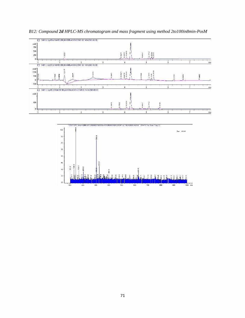
71
B12: Compound 2d HPLC-MS chromatogram and mass fragment using method 2to100in8min-PosM

72
B13: Compound 2d 1H NMR
The NMR was taken of the crude product. Additional peaks present are due to the presence of a reaction
intermediate (structure below) that did not go to completion. The peak can be seen in the LC-MS above at
retention time 4.07 minutes.

73
B14: Compound 2e HPLC-MS chromatogram and mass fragment using method 2to100in8min-PosM

74
B15: Compound 2e 1H NMR
DC-43-hydroxy.esp
10 9 8 7 6 5 4 3 2 1 0
Chemical Shift (ppm)
0.1
0.2
0.3
0.4
0.5
0.6
0.7
0.8
Norm
alized Inte
nsity
1.000.850.971.071.141.091.550.76
TMS
DMSO
10.3
9(1
5) 7
.46
7.4
4(9
,12)
6.8
3(1
3)
6.8
1
4.6
8(2
)4.6
7
4.0
54.0
3(3
)3.7
83.7
6(3
)
3.6
5(6
)3.5
73.5
6(6
)
3.5
4
3.5
3
0.0
0
11
12 13
8
910
N4
5
O14
O1
23
6OH7
Br16
OH15

75
B16: Compounds 3/3’ HPLC-MS chromatogram and mass fragment using method 2to100in8min-PosM
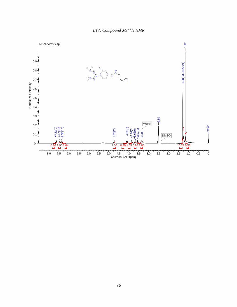
76
B17: Compound 3/3’ 1H NMR
NE-9-borest.esp
8.0 7.5 7.0 6.5 6.0 5.5 5.0 4.5 4.0 3.5 3.0 2.5 2.0 1.5 1.0 0.5 0
Chemical Shift (ppm)
0
0.1
0.2
0.3
0.4
0.5
0.6
0.7
0.8
0.9
Norm
alized Inte
nsity
6.5310.231.060.951.050.991.001.041.060.96
Water
DMSO7.6
3(9
)7.4
7(1
2)
7.3
6(1
3)
4.7
3(2
)
4.0
9(3
)
3.8
4(3
)3.6
6(6
)3.5
7(6
)
3.3
4
2.5
0
1.2
8(2
3,2
4,2
2,2
1)
1.1
7
0.0
0
11
12 13
8
910
N4
5
O14
O1
23
6OH7
B16
O17
O20
19
18
2324
21
22
F15

77
B18: Compounds 3a HPLC-MS chromatogram and mass fragment using method 2to100in8min-PosM
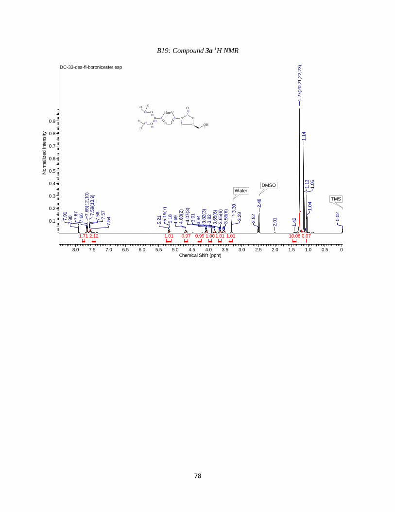
78
B19: Compound 3a 1H NMR
DC-33-des-fl-boronicester.esp
8.0 7.5 7.0 6.5 6.0 5.5 5.0 4.5 4.0 3.5 3.0 2.5 2.0 1.5 1.0 0.5 0
Chemical Shift (ppm)
0.1
0.2
0.3
0.4
0.5
0.6
0.7
0.8
0.9
Norm
alized Inte
nsity
0.0710.081.011.011.000.990.971.012.121.71
WaterDMSO
TMS
7.9
17.9
0 7.6
77.6
6 7.6
5(1
2,1
0)
7.5
9(1
3,9
)7.5
87.5
77.5
4
5.2
15.1
9(7
)5.1
84.6
94.6
8(2
)
4.0
7(3
)3.9
13.8
43.8
2(3
)3.8
2
3.6
5(6
)3.6
5(6
)3.5
6(6
)
3.3
03.2
9
2.5
2
2.4
8
2.0
1
1.4
21.2
7(2
0,2
1,2
2,2
3)
1.1
41.1
31.0
51.0
4
-0.0
2
11
10 9
8
1312
N4
5
O14
O1
23
6OH7
B15
O16
O19
18
17
2223
20
21
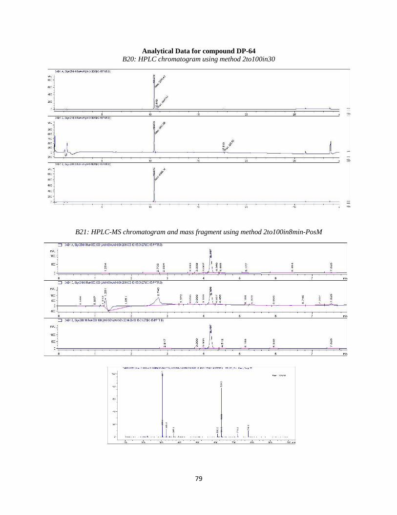
79
Analytical Data for compound DP-64 B20: HPLC chromatogram using method 2to100in30
B21: HPLC-MS chromatogram and mass fragment using method 2to100in8min-PosM

80
Analytical Data for compound DP-65
B22: HPLC chromatogram using method 2to50in30
B23: HPLC-MS chromatogram and mass fragment using method 2to100in8

81
B24: 1H NMR
DC-31.esp
9 8 7 6 5 4 3 2 1 0
Chemical Shift (ppm)
0.05
0.10
0.15
0.20
0.25
0.30
0.35
0.40
0.45
0.50
0.55
0.60
Norm
alized Inte
nsity
1.131.091.061.313.131.031.081.021.071.010.99
DMSO
TMS
8.8
0(9
)
8.1
2(1
3)
7.9
2(2
0)
7.6
9(1
2)
7.4
5(1
6)
7.2
3(1
9)
4.9
8(7
,21)
4.7
7(2
)4.7
6
4.1
8(3
)
3.9
3(3
) 3.6
9(6
)3.6
0(6
)
15 11
12 13
8
9N10
N4
5
O14
O1
23
6OH7
1617
18
19 20
F22
OH21
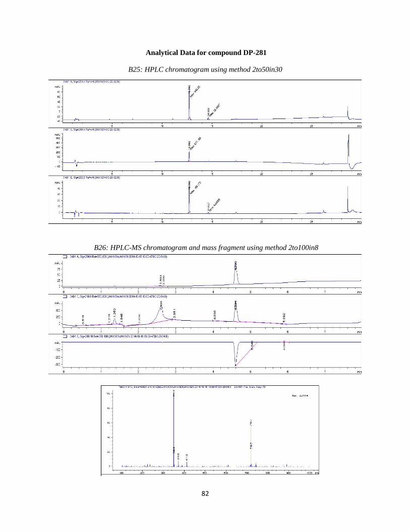
82
Analytical Data for compound DP-281
B25: HPLC chromatogram using method 2to50in30
B26: HPLC-MS chromatogram and mass fragment using method 2to100in8

83
B27: 1H NMR
DP-281.esp
8 7 6 5 4 3 2 1 0
Chemical Shift (ppm)
0
0.01
0.02
0.03
0.04
0.05
0.06
0.07
0.08
0.09
0.10
Norm
alized Inte
nsity
-0.130.270.630.821.001.041.161.241.212.611.20
TMSDMSO
water
DMSOWater8.0
1(2
0)
7.9
97.6
87.6
57.6
57.6
3(9
,12)
7.4
9
7.3
1(1
7)
7.1
87.1
6(2
1)
5.2
5(7
)
4.7
64.7
5(2
)
4.1
74.1
43.9
1(3
)3.8
93.8
93.7
2
3.6
8(6
)3.5
93.5
6(6
)
16 11
12 13
8
910
N4
5
O14
O1
23
6OH7
1718
19
20 21
F15
NO223
OH22
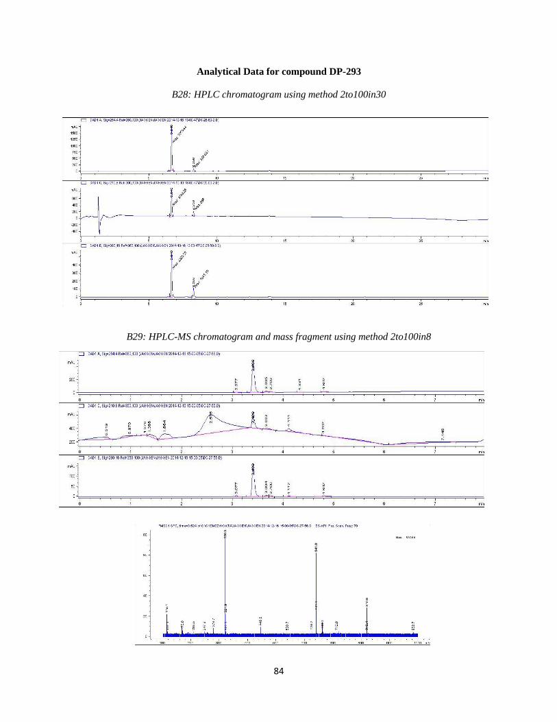
84
Analytical Data for compound DP-293
B28: HPLC chromatogram using method 2to100in30
B29: HPLC-MS chromatogram and mass fragment using method 2to100in8

85
B30: 1H NMR

86
Analytical Data for compound DP-296
B31: HPLC chromatogram using method 2to50in30
B32: HPLC-MS chromatogram and mass fragment using method 2to100in8

87
Analytical Data for compound DP-304
B33: HPLC chromatogram using method 2to50in30
B34: HPLC-MS chromatogram and mass fragment using method 2to100in8

88
Analytical Data for compound DP-305
B35: HPLC chromatogram using method 2to30in30
B36: HPLC-MS chromatogram and mass fragment using method 2to100in8
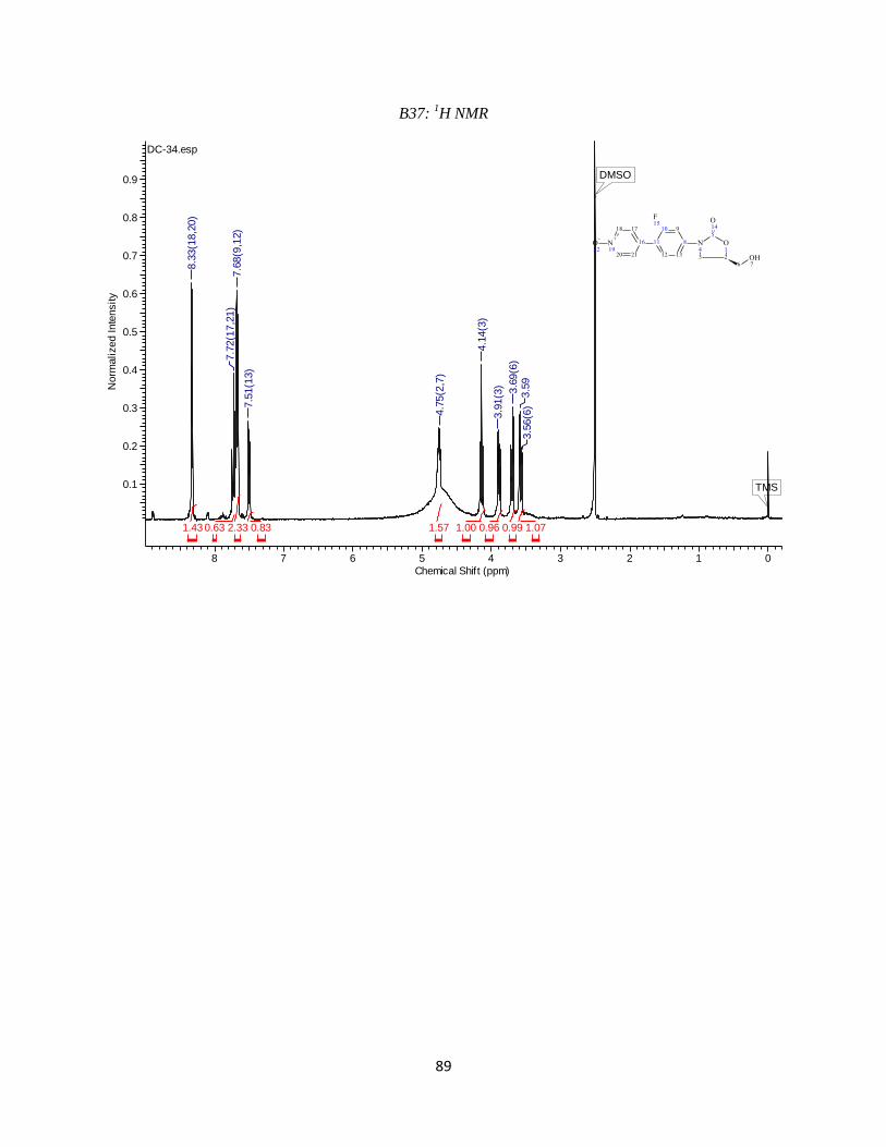
89
B37: 1H NMR
DC-34.esp
8 7 6 5 4 3 2 1 0
Chemical Shift (ppm)
0.1
0.2
0.3
0.4
0.5
0.6
0.7
0.8
0.9
Norm
alized Inte
nsity
1.070.990.961.001.570.832.330.631.43
TMS
DMSO
8.3
3(1
8,2
0)
7.7
2(1
7,2
1)
7.6
8(9
,12)
7.5
1(1
3)
4.7
5(2
,7)
4.1
4(3
)
3.9
1(3
) 3.6
9(6
)3.5
93.5
6(6
)
16 11
12 13
8
910
N4
5
O14
O1
23
6OH7
1718
N+
1920 21
F15
O-
22
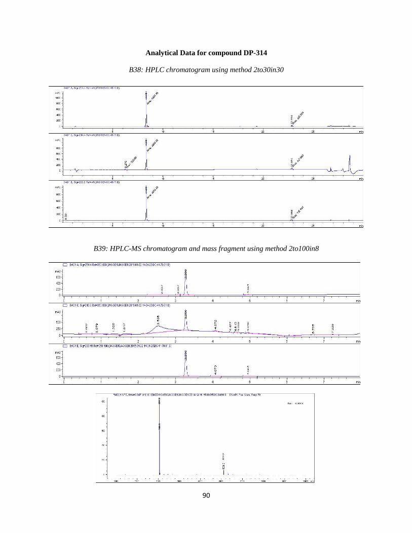
90
Analytical Data for compound DP-314
B38: HPLC chromatogram using method 2to30in30
B39: HPLC-MS chromatogram and mass fragment using method 2to100in8

91
B40: 1H NMR
DC-41.esp
8 7 6 5 4 3 2 1 0
Chemical Shift (ppm)
0
0.05
0.10
0.15
0.20
0.25
0.30
Norm
alized Inte
nsity
2.561.976.362.021.070.923.072.400.971.04
TMS
DMSO
8.4
4(1
7)
8.2
6(1
9)
7.6
9(9
,21)
7.5
4(2
0,1
3,1
2)
4.7
4(7
)
4.1
4(2
)
3.8
9(3
)
3.6
8(6
)3.5
9(6
)
0.0
0
16 11
12 13
8
910
N4
5
O14
O1
23
6OH7
17N+
1819
20 21
O-
22F15

92
Analytical Data for compound DP-315
B41: HPLC chromatogram using method 2to30in30
B42: HPLC-MS chromatogram and mass fragment using method 2to100in8

93
B43: 1H NMR
DC-37.esp
9 8 7 6 5 4 3 2 1 0
Chemical Shift (ppm)
0.05
0.10
0.15
0.20
Norm
alized Inte
nsity
2.381.991.001.040.910.991.010.712.653.422.31
TMS
DMSOWater
8.6
9(2
5)
7.6
67.6
2(1
7,2
1,1
8,2
0)
7.5
27.5
0(1
2)
7.4
7(9
)7.4
5(1
3)
6.5
6
5.2
6(7
)
4.7
6(2
)
4.1
3(3
)
3.8
9(3
)
3.6
9(6
)3.5
9(6
)
1.3
6(2
3,2
4)
1.2
7(2
3,2
4)
0.0
0
16 11
12 13
8
910
N4
5
O14
O1
23
6OH7
1718
19
20 21
F15
22
NH225
23
24
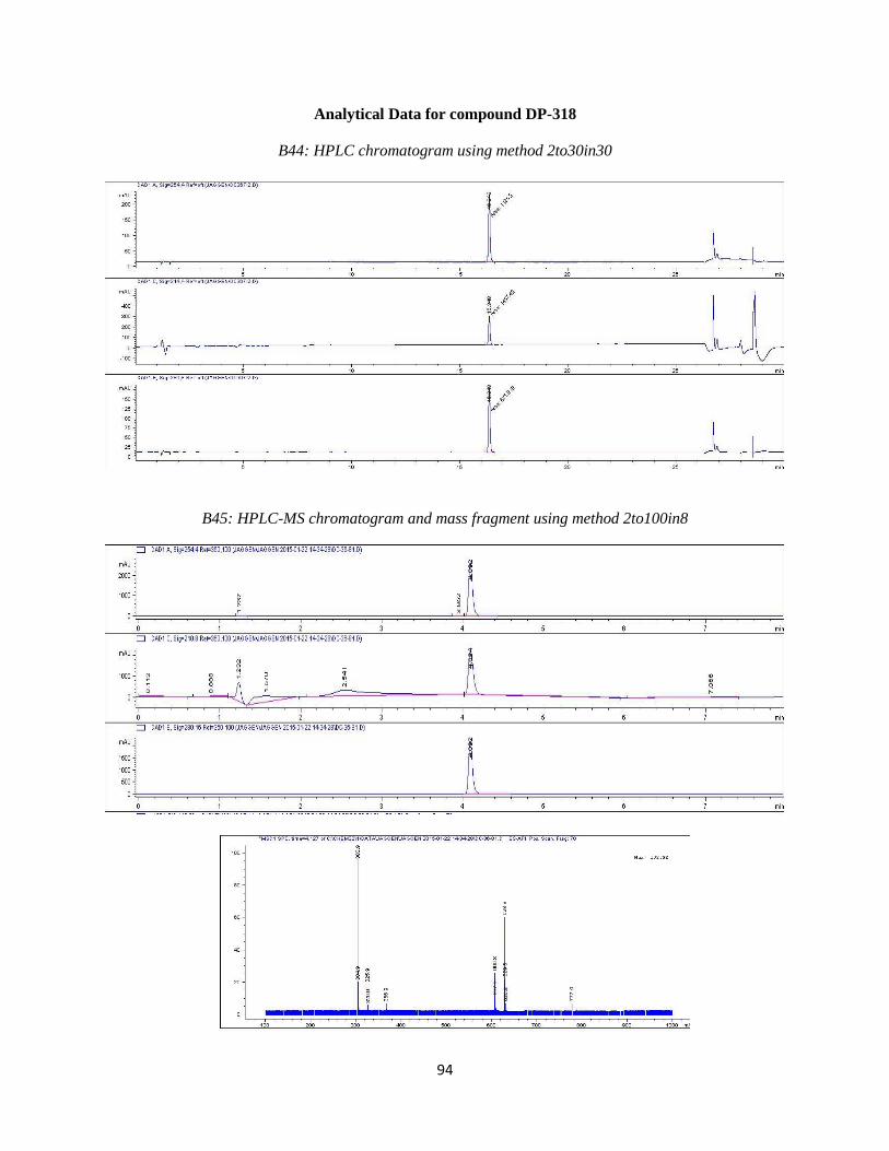
94
Analytical Data for compound DP-318
B44: HPLC chromatogram using method 2to30in30
B45: HPLC-MS chromatogram and mass fragment using method 2to100in8

95
B46: 1H NMR
DC-36.esp
9 8 7 6 5 4 3 2 1 0
Chemical Shift (ppm)
0
0.1
0.2
0.3
0.4
0.5
0.6
0.7
0.8
0.9
Norm
alized Inte
nsity
1.000.991.010.951.000.680.780.930.871.652.190.95
Water
DMSO
TMS
9.5
0(2
2)
7.6
6(1
3,9
)7.5
4(1
0,1
2)
7.0
9(2
0)
6.8
4(1
7)
6.7
5(1
8)
5.2
2(7
)
4.7
2(2
)
4.1
3(3
)
3.8
7(3
)
3.6
8(6
)3.5
9(6
)
0.0
0
15 11
10 9
8
1312
N4
5
O14
O1
23
6OH7
2019
18
17 16
OH22
F21

96
Analytical Data for compound DP-327
B47: HPLC chromatogram using method 2to30in30
B48: HPLC-MS chromatogram and mass fragment using method 2to100in8
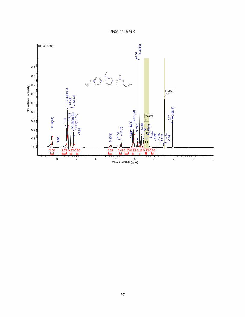
97
B49: 1H NMR
DP-327.esp
8 7 6 5 4 3 2 1 0
Chemical Shift (ppm)
0
0.1
0.2
0.3
0.4
0.5
0.6
0.7
0.8
0.9
Norm
alized Inte
nsity
0.900.822.260.822.300.680.380.500.633.782.00
DMSO
Water
8.2
6(2
4)
7.9
3
7.5
07.4
9(1
3,9
)7.4
87.4
7(1
2)
7.4
27.2
8(1
9,2
1)
7.1
7(1
8,2
2)
7.1
5
5.2
6(2
)
4.7
34.7
1(7
)
4.1
54.1
2(3
) 4.0
5(2
3)
3.8
9(3
)3.7
6 3.7
5(1
6)
3.6
7(6
)3.5
83.5
8(6
)3.5
52.8
72.8
72.7
12.7
12.5
32.0
7 2.0
6(7
)
17 11
12 13
8
910
N4
5
O14
O1
23
6
OH7
1819
20
21 22
O15
23
NH224
16
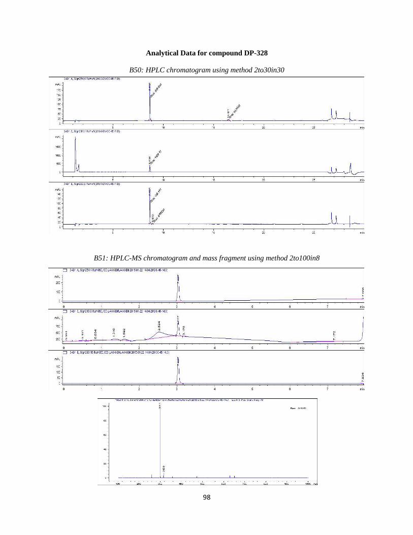
98
Analytical Data for compound DP-328
B50: HPLC chromatogram using method 2to30in30
B51: HPLC-MS chromatogram and mass fragment using method 2to100in8
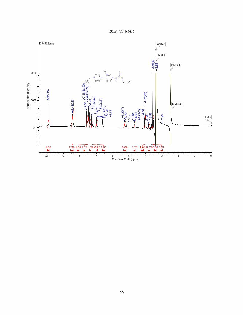
99
B52: 1H NMR
DP-328.esp
10 9 8 7 6 5 4 3 2 1 0
Chemical Shift (ppm)
0
0.05
0.10
Norm
alized Inte
nsity
1.510.040.351.880.730.821.000.751.091.721.592.361.02
Water
DMSO
TMS
DMSO
Water
9.9
3(1
5)
8.4
5(2
3)
7.5
87.5
6(1
8,2
0)
7.4
9(1
7,2
1)
7.4
77.4
0(1
3)
7.2
8 7.2
6(1
2)
7.0
0(9
)6.9
86.6
3
5.2
9(7
)5.2
75.1
44.6
94.6
94.6
7(2
)
4.0
64.0
2(2
2)
3.8
3 3.8
13.5
6(6
)
3.3
3
2.9
9
16 11
12 13
8
910
N4
5
O14
O1
23
6OH7
1718
19
20 21
OH15
22
NH223

100
Analytical Data for compound DP-329
B53: HPLC chromatogram using method 2to30in30
B54: HPLC-MS chromatogram and mass fragment using method 2to100in8

101
B55: 1H NMR
DP-329.esp
10 9 8 7 6 5 4 3 2 1 0
Chemical Shift (ppm)
0
0.05
0.10
0.15
0.20
Norm
alized Inte
nsity
0.860.810.880.920.990.921.000.971.932.191.01
TMS
DMSOWater
9.9
7(2
3)
7.6
87.6
6(9
,13)
7.5
2(1
2,1
0)
7.4
97.2
9(2
0)
7.0
67.0
3(1
7)
7.0
1
5.2
55.2
45.2
3(7
)4.7
4 4.7
34.7
2(2
)
4.1
54.1
3(3
)4.1
1 3.8
8(3
)3.6
7 3.6
03.5
53.3
23.3
0
0.0
7
15 11
10 9
8
1312
N4
5
O14
O1
23
6OH7
2019
18
17 16
F22
OH23
F21