Embed Size (px)
Citation preview

Neurochemistry Voi. I, pp. 29-42. Pergamon Press Ltd. 1980. Printed in Great Britain.
SYNTHESIS AND TURNOVER OF LIPID AND PROTEIN COMPONENTS OF FROG RETINAL ROD OUTER SEGMENTS
Robert E. Anderson, Paula A. Kelleher, Maureen B. Maude and Tom M. Maida
Cullen Eye Institute Baylor College of Medicine Houston, Texas 77030 U.S.A.
ABSTRACT
The incorporation and turnover of several lipid precursors in frog retina lipid and protein were studied using biochemical and autoradiographic techniques. Lipid and protein are synthesized on the photoreceptor microsomes from where some migrate to the base of the rod outer segments (ROS) and are incorporated into basal discs. Most of the labeled protein is confined to a discrete group of discs which is dis- placed apically as newly synthesized unlabeled discs are added. The specific radio- activity of ROS protein is constant throughout this period, which in the present study was determined to be 39 days. Conversely, lipid, once incorporated into the basal discs, diffuses throughout the entire ROS as evidenced by an exponential decline in specific radioactivity. The half-life of ROS lipid is shorter than the renewal rate for protein, suggesting that lipid turns over faster than protein in the ROS.
KEY WORDS
Phosphatidyl serine; phosphatidyl ethanolamine; rod outer segments; lipid synthesis; lipid turnover; membranes; renewal.
INTRODUCTION
Over a decade ago Young (1967) demonstrated that the proteins of rod outer segments (ROS) are constantly being renewed. Following injection of radioactive smino acids, a band of silver grains representative of newly synthesized proteins was observed at the base of the ROS. With time, these labeled proteins were displaced apically until they reached the tips of the ROS where they were shed and phagocytized by the pigment epithelium (reviewed by Young, 1973 and 1976; and O'Brien, 1978). In an elegant study involving biochemistry and electron microscopic autoradiography, Hall, Bok and Bacharach (1969) followed the fate of injected 3H-leucine over a 9-week period. They showed by autoradiography that the label was first incorpo- rated into proteins of the ribosomes of the inner segment, from which it moved to the Golgi body and finally percolated through the mitochondrial-dense ellipsoid region to the base of the ROS. Biochemical analysis of the radioactive products showed that most of the label was in rhodopsin. By carefully monitoring the tempo- ral relationship between autoradiographic pattern and the specific radioactivity of
29

30 R.E. Anderson cC ~.
rhodopsin in isolated ROS, they demonstrated that a molecule of rhodopsin, once incorporated into a disc, remained with that disc until it was shed.
Other studies on the biosynthesis of ROS proteins have come from the laboratory of Papermaster and co-workers (1975). Following intravitreal injection of 3H-leucine, the specific activity of frog opsin peaked in two non-ROS membrane fractions of retina prior to reaching maximum activity in ROS. Since opsin was never found in a soluble form, delivery of newly synthesized opsin to the growing ROS was indi- cated to occur via a membrane-bound intermediate.
Renewal of lipids in rod outer segments is not so well understood. The autoradio- graphic studies of Bibb and Young (1974a) following the fate of injected tritiated palmitic, stearic, or arachidonic acids in frogs showed some early banding in the basal discs which quickly diffused throughout the entire ROS. In another auto- radiographic study (Bibb and Young, 1974b), glycerol was rapidly incorporated into lipid in the rod inner segment which subsequently migrated to the ROS. As observed in the fatty acid study, the lipid was diffusely spread throughout the ROS. Some sustained banding observed in the outer segments in the glycerol study was shown to be due to protein. They concluded that ROS lipids are renewed by both molecular and membrane replacement, and that ROS lipids are renewed more rapidly than pro- tein. Biochemical studies by Hall, Basinger and Bok (1973) on the fate of injected 33po4 and 3H-choline in frog ROS phospholipids showed that, with the exception of phosphatidyl inositol in which a rapid incorporation and turnover was observed, there was a slow incorporation of both labels into phospholipids. Maximum specific activity of 33P-phospholipids was not achieved until 30 days after injection and remained constant through day 63, and only a slight decline in tritiated phospha- tidyl choline (PC) specific activity was noted by day 73. There was no indication from these biochemical studies that ROS phospholipids were turning over within the time frame of protein renewal, in contrast to Bibb and Young's (1974b) glycerol autoradiography data.
In an attempt to gain a greater insight into the renewal of lipid in ROS, we have carried out a series of biochemical and autoradiographic experiments in which the biosynthesis and turnover of retinal lipids were studied by following the fate of several radioactive lipid precursors. In this paper we discuss the incorporation of 32p04, 33PO4, 3-3H-serine, and 2-3H-glycerol into protein and phosphatidyl serine of the frog retina.
METHODOLOGY
The details of the methodology will be presented in subsequent publications; only a brief outline will be given here.
Frogs acclimated to a body temperature of 24.5 ° in a metabolic chamber under a light cycle of 14L:IOD were injected in the dorsal lymph sac with either 33P0~, 32P0~, 2-3H-glycerol or 3-3H-serine. Eyes were removed at various times and pro- cessed for biochemistry or autoradiography. For biochemistry, retinas were gently homogenized in sucrose (1.17 gm/cc) containing i0 mM Tris and 2 mM MgClz (pH 7.35) and overlaid with 1.15, 1.13 and 1.11 gm/cc sucrose in Tris and MgCI2. After centrifugation at 82,000 x g x 60 min., ROS were collected at the 1.11/1.13 g/cc interface. The remaining volume of sucrose was diluted i:I with buffer without sucrose and centrifuged at 18,800 x g x 15 min. The resulting supernatant was centrifuged at 96,000 x g x 90 min. to obtain a microsomal pellet.
Lipids from the ROS and microsomal fractions were extracted with chloroform: methanol (2:1, v/v) and separated into classes by preparative thin-layer chroma- tography. Individual lipid classes visualized after exposure to iodine vapors were assayed for lipid phosphorus and an aliquot counted for the determination of
Synthesis and Turnover of Lipid and Protein Components 3|
specific radioactivity. Phospholipid classes identified after spraying with water were methylated and the esters analyzed by gas chromatography.
In some instances, intact retinas were homogenized in chloroform:methanol (2:1) and again after the addition of 0.2 vols of 0.9% NaCI. Aliquots of the upper water: methanol phase were taken for determination of water soluble phosphorus and radio- activity. The specific radioactivity of water soluble precursors per micromole of phosphorus was determined. Protein was determined by the standard Lowry procedure.
For autoradiography of retinal lipids, the procedure of Gould and Dawson (1976) was followed with some modifications which will be described in detail in a subsequent publication. Several experiments showed that all of the radioactive lipid was extracted from the tissue designated "lipid out", while less than 8% was removed during the "lipid in" procedures.
BIOSYNTHESIS OF RETINAL PROTEIN FROM SERINE AND GLYCEROL
Serine and glycerol were incorporated into both protein and lipid in the frog retina. Figures i and 2 are the specific activities of microsomal and ROS protein following injection of 3H-serine and 3H-glycerol, respectively. In the microsomes, maximum specific radioactivity of protein was reached on the first day and decreased exponentially with time. This was different from the labeling pattern of ROS pro- tein where maximum labeling occurred between days one and three and remained fairly constant until some time between days 24 and 40.
5 0
4 0
' 0
z 30 W p- o cc
o_
~ zo
Q. O
I0
3 H - S E R I N E
I o M,C.OSOMES J
/
+ I 0
I 1 I I I I I0 2 0 SO 4 0 5 0 6 0
DAYS
Fig. i. Specific radioactivity of microsomal and ROS pro- tein following injection of 3-SH-serine.

32
6
,'o x
¢o ,., 5
o
o n- o4
.. ~D '0
× 3
0
_z2 F- 0
o, I
IE
121 0
1
+ i
O
R. B. Anderson. L ~i,.
I
H-GLYCEROL
I" os; 0 MICROSOMES
0 ~ ~ 0 . . . . . . . . . . . . . . . . . O-
l ; i I
I0 20 30 4 0 DAYS
Fig. 2. Specific radioactivity of microsomal and ROS pro- tein following injection of 2-3H-glycerol.
These data are consistent with previous demonstrations that ROS proteins are syn- thesized in the inner segments of rod visual cells and migrate to the base of the ROS where they are incorporated into newly forming discs (Hall and co-workers, 1969). Maintenance of constant specific radioactivity in this dynamic membrane system where protein is daily being added at the base of the ROS and shed at the apical tips indicates that the radioactive proteins, once incorporated into ROS discs, do not diffuse throughout the ROS. If they did, an exponential decline in specific radioactivity similar to that observed for microsomal protein would also be evident for ROS protein. Such is not the case.
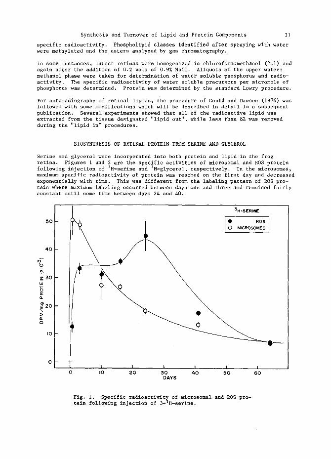
Autoradiograms from retinas of animals sacrificed one or 17 days after the injec- tion of 3H-serine are shown in Figs. 3A-D. Several features are evident in these figures: (i) A band of radioactivity indicative of the assembly of newly synthe- sized membrane components is present at the base of the rods but not the cones at day one. These bands persist in the delipidated tissue suggesting that most of the banded material is protein. The band has been displaced nearly half the length of the ROS by day 17, consistent with a 39-day turnover for integral disc proteins at 24.5°C. (2) At day one, the paraboloid regions of the accessory members of the double cones are highly labeled relative to the other cells (see arrows). This labeling pattern was observed by Bibb and Young (1974b) following tritiated glycerol injection, as well as by Basinger (personal communication) with 32po4, 3H-inositol, and 3H-glycerol. Since the paraboloid contains large amounts of glycogen, Bibb and Young (1974b) suggested that the label was in glucose derived by gluconeogenesis.
Synthesis and Turnover of Lipid and Protein Components 33
Fig. 3. Autoradiograms of frog retinas following the in- jection of 3-3H-serine. (A) Day I, lipid in; (B) Day i, lipid out; (C) Day 17, lipid in; (D) Day 17, lipid out.


Synthesis and Turnover of Lipid and Protein Components 35
However, label must also be in other components since lipid precursors not readily converted to glucose are also concentrated in this area. Determination of grain densities over the paraboloid region relative to the protein bands of adjacent ROS at day one for "lipid in" and "lipid out" tissue revealed that the densities in the paraboloid region were the same for both treatments, suggesting that the label was in protein. This labeling pattern was not observed following leucine injection, and its significance is not known. (3) Radioactive proteins accumulated in the synaptic region of the frog retina on the first day and were still seen, although somewhat diminished in intensity, at day 17. (4) Although lipid accounted for about half of the tissue radioactivity at both day one and day 17, unique labeling patterns were not seen for lipid. Rather, lipid seems to be diffusely incorporated into all of the cellular membranes, including the ROS. This point will be dis- cussed in following sections.
BIOSYNTHESIS OF RETINAL PHOSPHOLIPIDS
Tritiated serine is incorporated into phospholipids of the frog retina. A pre- cursor-product relationship is indicated by the higher specific activity of PS in the microsomes than in the ROS at the early time points, followed by a reversal in this pattern at day 2 (Fig. 4).
6
~4
0 3 =, ~2
0
' j ! ' ' ' 3 ; 3 . - s ; R , . E ' _ ' o '- " P s - ' - R o s ' - ' ~ A PS" MICROSOMES
I' ~ ~ • PE-MICROSOMES
iI ~/'~~/f / ~ 1 ~ ~ ~ _ _ _ . . . . . . . . . Me c
Jl 1 ". , . I 1 ~ ~
- - . . . . . . . . . . . . - - - - - . . - z - -I till . . . . . --
- +
I l I I I I I I I I I I I
0 4. 8 12 16 20 24 TIME (DAYS)
Fig. 4. Biosynthesis of phosphatldyl serine and phospha- tidyl ethanolamine in mierosomes and ROS following the injection of 3-3H-serine.

36 R.E. Anderson e$ a~.
This is consistent with the synthesis of phospholipid on retinal microsomes and the subsequent incorporation of this lipid into ROS disc membranes. A similar labeling pattern for both the microsomal and ROS PS was observed following the injection of tritiated glycerol (data not shown). Whether the serine is incorporated during de novo synthesis of phospholipids or by base exchange reactions remains to be determined. However, preliminary data from our laboratory have shown an active serine base exchange system in bovine retinal microsomes.
DECARBOXYLATION OF PHOSPHATIDYL SERINE TO PHOSPHATIDYL ETHANOLAMINE
One of the pathways for the formation of phosphatidyl ethanolamine in some tissues is the enzymatic decarboxylation of PS. The present study demonstrated an active decarboxylase in the frog retina. Although the site of decarboxylation cannot be unequivocally established from these in vivo studies, it is likely to be both the microsomes and the ROS. Evidence for this is as follows: (I) The specific activities of ROS or microsomal phosphatidyl serine and phosphatidyl ethanolamine are nearly the same 24 days after injection of labeled serine (Fig. 4), but these 24-day values are different for the two subcellular fractions indicating that decarboxylation occurred in each fraction. (2) Although a precursor-product relationship exists in both fractions for the decarboxylation reaction, and for the synthesis of phosphatidyl serine, one does not exist for transfer of microsomal phosphatidyl ethanolamine to ROS. In unpublished studies, following the injection of labeled ethanolamine, the specific activity of microsomal phosphatidyl ethanola- mine was several times that of ROS phosphatidyl ethanolamine at early time points. Thus, if a precursor-product relationship existed between microsomal and ROS phosphatidyl ethanolamine formed by the decarboxylation of microsomal phosphatidyl serine, it should have been easily identified.
Although not as pronounced as the data obtained following injection of labeled serine, the conversion of phosphatidyl serine to phosphatidyl ethanolamine in ROS was also demonstrated following injection of other lipid precursors. As shown in Fig. 5 and 6, the specific activity of phosphatidyl serine in the ROS following the injection of 32PO 4 or 2-3H-glycerol was clearly higher than that of phosphatidyl ethanolamine at the early time points, and only after several weeks did the specific activities of these two lipids become the same.
The fatty acid composition of phosphatidyl ethanolamine and phosphatidyl serine given in Table i is also consistent with an interconversion of these two lipid classes. The phosphatidyl serine contains slightly less long-chain polyunsaturated fatty acids than the phosphatidyl ethanolamine while the phosphatidyl ethanolamine has a slightly higher percentage of palmitic acid. However, a remarkable similar- ity in fatty acid composition does exist.

Synthesis and Turnover of Lipid and Protein Components 37
I0
9
8
~" 4
0
I I v I
" • PS o PE
I I I I I
0 5 I0 15 20 DAYS
I
25
Fig. 5. Specific activities of phosphatidyl serine and phosphatidyl ethanolamine in ROS following the injection of 32p04.

38 R.E. Anderson et a~.
--- 7 I
0 x 6
0') D 5 n- O -r 4 Q_ or) 0 -r 3 n
IJJ --J 2 0 ~E
I ~E (3_
0
2 - 3 H - G L Y C E R O L - ROS
• PS o PE
i I I ; , I i
0 5 I0 15 2O 25 DAYS
Fig. 6. Specific activities of phosphatidyl serine and phosphatidyl ethanolamine in ROS following the injection of 2-3H-glycerol.
TABLE 1 Composition of the Major Fatty Acids of Phospha- tidyl Ethanolamine and Phosphatidyl Serine from Frog ROS.
Acid PS PE
16:0 4 6
18:0 19 13
18:1 3 6
20:4~6 2 7
22:4~6 7 7
22:5~3 + ~6 8 5
22:6~6 46 51
(Anderson and Risk, 1974)

Synthesis and Turnover of Lipid and Protein Components 39
TURNOVER OF PHOSPHATIDYL SERINE IN ROD OUTER SEGMENTS
The incorporation and turnover of 2-3H-glycerol,33PO4, and 3-3H-serine in phospha- tidyl serine in rod outer segments are shown in Fig. 7.
A
x
n- lz.
rw IJJ IJJ _J (/')
" X
LIJ 0
¢.9 ~ 0
' f , ' ' '. . . . . . ' ' '
rn r ' , = l \ ~ o ° , ' , °ocM,N," ", j
I I I I I I I, I I I I I I I
0 2 0 4 0 6 0 8 0 I 0 0 120 T I M E ( D A Y S )
Fig. 7. Turnover of phosphatidyl serine in ROS following the injection of 2-3H-glycerol, 33P04, or 3-3H-serine.
As previously observed by Hall and co-workers (1973), the incorporation of labeled phosphorus into ROS phosphatidyl serine was quite slow, requiring 20 days to achieve maximum specific activity, and the loss of label due to turnover was equally slow. It was impossible to calculate turnover times for the membrane phospholipids from the labeled phosphorus data. On the other hand, both glyserol and serine rapidly labeled the ROS phospholipids with glycerol achieving maximum specific activity three days and serine six days after injection of the isotopes. The reason for the distinctive labeling pattern of phosphate is evident from the data in Fig. 8 where the percent of maximum specific activity of water soluble radioactivity from frog retinas (DPM per micromole phosphorus) is plotted against time. Although the chemical identities of the labeled water soluble products were not established, it is clear that glycerol and serine are rapidly catabolized in the retina and thus are true "pulses" of radioactive lipid precursors whereas labeled phosphate remains available for lipid synthesis for weeks. Thus, the rapid labeling observed autoradiographically by Bibb and Young (1974b) following tritiated glycerol injection was the result of a pulse of radioactive precursor being presented to the tissue, whereas the slow labeling following phosphorus administration resulted from the presence of a long-lived retinal pool of radio-

40
active phosphate.
R. E. Anderson eg a~.
I 0 0 - ,
>- 80 F-
o
o 60 o 0c
~E
~E 4 0 x <
o 2O
I i
• 32 po 4
'! - +
I
0
@k• \ ~ 0 . . . . . O_ @ ~ Q . _ . . . . . 0 . . . . . . . . . 0
! l I
5 I0 15 T I M E (DAYS)
i
RETINA
3-3H-SERINE O--
@--
I I
20 25
Fig. 8. Time course of labeling of water soluble precursors of retinal lipids.
Returning to the data in Fig. 7, the specific activity of phosphatidyl serine from both glycerol and serine declined from their maximum value in an exponential manner. Linear regression analysis of the log of specific activity versus time for each precursor gave identical 23-day half-lives for phosphatidyl serine in the ROS. A similar analysis for phosphatidyl ethanolamine derived from phosphatidyl serine gave a half-life of 38.5 days. The different half-lives of phosphatidyl serine and phosphatidyl ethanolamine are probably due to the fact that phosphatidyl serine is lost through decarboxylation and disc shedding while phosphatidyl ethanolamine is lost through disc shedding, but is replaced by decarboxylation.
DISCUSSION
Protein and lipid are synthesized on the photoreceptor microsomes and migrate to the base of the ROS where they are incorporated into the basal infoldings of the

Synthesis and Turnover of Lipid and Protein Components 4!
plasma membrane, which eventually pinch off to become free-floating discs. From this time on, the protein is "trapped" and remains with the same disc throughout the 5½ week period of apical displacement. Lipid, on the other hand, is not con- fined to any particular disc. The exponential decline in specific radioactivity of ROS phospholipids is consistent with a diffuse rather than a discrete labeling of ROS by lipid and supports the earlier reports of Bibb and Young (1974a and 1974b).
Since the labeled lipid diffuses throughout the ROS, one-half of the label should be lost in one complete turnover of outer segment protein if disc shedding is the only means of losing lipid, and calculations of t½ for lipid should give a value comparable to the ROS turnover rate. We calculated a value of 39 days for turn- over of ROS discs, which is longer than the half-life determined for phosphatidyl- serine (23 days), phosphatidyl choline (24 days, unpublished) and phosphatidyl inositol (< 5 days, unpublished). (The value of 38.5 days calculated for phospha- tidyl ethanolamine is longer than the actual value, since phosphatidyl serine is slowly converted to phosphatidyl ethanolamine, and thus is not derived from a "pulse" of radioactive precursor.) It appears then that ROS lipid is turning over at a rate faster than protein, a conclusion also reached by Bibb and Young (1974b).
Lipid and protein are probably delivered and inserted into the growing membrane as a package, since newly synthesized frog opsin is not found in a soluble form (Papermaster and co-workers, 1975). How the lipid diffuses throughout the ROS is not known, but it may be mediated by phospholipid exchange proteins, which we recently described in bovine retina (Dudley and Anderson, 1978). Whether or not the diffusion involves only interdisc transfer of lipid originally delivered to the growing membrane as a lipoprotein complex, as opposed to lipid exchange with other rod cellular organelles, remains to be determined. However, based on half- life determinations of ROS phospholipids discussed above, the latter possibility seems reasonable.
An unexpected observation in these in vivo studies was the decarboxylation of ROS phosphatidyl serine, which suggests an active lipid metabolism in ROS. In studies to be reported elsewhere, phosphatidyl ethanolamine was methylated to phosphatidyl choline, apparently in the ROS, while the entire molecule of phosphatidyl inositol completely turned over in less than seven days. Enzymes of lipid metabolism have not previously been reported in ROS (Swartz and Mitchell, 1973, 1974; Mizuno, 1976). However, these investigations utilized vigorous homogenization and exten- sive washing procedures during the isolation of ROS which may have led to losses of some loosely bound proteins. We are currently searching for the subcellular localization of the enzymes of lipid metabolism responsible for the various inter- conversions demonstrated in these in vivo experiments.
ACKNOWLEDGEMENTS
This research was supported in part by a grant from the Retina Research Foundation (Houston), grants EY 00871 and EY 07001 from the National Eye Institute, and departmental grants from Research to Prevent Blindness, Inc. and The Brown Foundation (Houston).
REFERENCES
Anderson, R. E. and M. Risk (1974). Lipids of ocular tissues. IX. The phospho- lipids of frog photoreceptor membranes. Vision Res., 14, 129-131.
Bibb, C. and R. W. Young (1974a). Renewal of fatty acids in the membranes of visual cell outer segments. J. Cell Biol., 61, 327-343.
N . C . I . ~/I-4 D
42 R. E . A n d e r s o n :~:~i.
Bibb, C. and R. W. Young (1974b). Renewal of glycerol in the visual cells and pigment epithelium of the frog retina. J. Cell Biol., 62, 378-389.
Dudley, P. A. and R. E. Anderson (1978). Phospholipid transfer protein from bovine retina with high activity towards retinal rod disc membranes. FEBS Letters, 95, 57-60.
Gould, R. M. and R. M. C. Dawson (1976). Incorporation of newly formed lecithin into peripheral nerve myelin. J. Cell Biol., 68, 480-496.
Hall, M. O., S. F. Basinger and D. Bok (1973). Studies on the assembly of rod outer segment disc membranes. In H. Langer (Ed.), Biochemistry and Physiology of Visual Pisments, Springer-Verlag, New York, pp. 319-326.
Hall, M. O., D. Bok and A. D. E. Bacharach (1969). Biosynthesis and assembly of the rod outer segment membrane system. Formation and fate of visual pigment in the frog retina. J. Molec. Biol., 45, 397-406.
Mizuno, A. (1976). Incorporation of serine and etL1anolamine into the phospho- lipids of rabbit retina. J. Biochem., 80, 45-57.
O'Brien, P. J. (1978). Rhodopsin: A light-sensitive membrane glycoprotein. In P. Cuatrecasas and M. F. Greaves (Eds.) Receptors and Recognition, Series A, Vol. 6, Chapman and Hall, London, pp. 109-150.
Papermaster, D. S., C. A. Converse and J. Siu (1975). Membrane biosynthesis in the frog retina: Opsin transport in the photoreceptor ceil. Biochem., 14, 1343-1352.
Swartz, J. G. and J. E. Mitchell (1973). Phospholipase activity of retina and pigment epithelium. Biochem., 12, 5273-5278.
Swartz, J. G. and J. E. Mitchell (1974). Acyl transfer reactions of retina. Biochem., 13, 5053-5059.
Young, R. W. (1967). The renewal of photoreceptor cell outer segments. J. Cell Biol., 33, 61-72.
Young, R. W. (1973). Renewal systems in rods and cones. Ann. Ophthalmol., 5, 843-854.
Young, R. W. (1976). Visual cells and the concept of renewal, l_nvest. Ophthalmol 15, 700-725.