Embed Size (px)
Citation preview

International Journal of Environment, Agriculture and Biotechnology (IJEAB) Vol-2, Issue-3, May-Jun- 2017
http://dx.doi.org/10.22161/ijeab/2.3.10 ISSN: 2456-1878
www.ijeab.com Page | 1084
Synthesis and Evaluation of the Cytotoxic and
Anti-Proliferative Properties of Dox-ZnO
Quantum Dots Loaded Chitosan Nanoparticles
against MCF-7 and SKBR-3 Human Breast
Cancer Cells Srikanth Jagadeesan1 *, Roshini A2 *, Kim Kyung Hwan2, Yang-Hoi Doh1, Yoon-Kyu Lim3,
Kyung Hyun Choi1*
1 Department of Advanced Convergence Technology and Science, Jeju National University.63243, Korea. 2 Department of Mechatronics Engineering, Jeju National University, 63243, Korea.
3 Department College of Veterinary Medicine, Jeju National University, 63243, Korea.
Abstract— Doxorubicin (Dox) is a potent chemotherapeutic
agent used in the treatment of cancer. In the present study,
pH responsive chitosan polymer coated Dox nanoparticle
(Composite) was developed to investigate targeted drug
delivery against breast cancer. The anticancer drug DOX-
ZnO QDs was loaded to the chitosan nanoparticles. The
synthesized free and drug loaded nanoparticle were
analyzed using Fourier transmission electron microscopy
(FTIR) and UV-Visible spectroscopy(UV-Vis). The particle
size was measured using Transmission Electron Microscopy
(TEM). Further, the composite was evaluated for its
anticancer effects. Drug release analysis showed
significantly larger amount of drug released in acidic pH of
5.0 compared to pH 7.4. The composite was significantly
more cytotoxic to the breast cancer cells MCF-7 and SKBR-
3. The composite was however, less toxic to HEK-293
human embryonic kidney cells confirming minimum side
effects on normal cells andcytotoxic to tumor cells. DAPI
staining showed nuclear degradation in composite treated
breast cancer cells. The cellular uptake of the composite
was analyzed by confocal microscopy. The composite
induced a G0/G1 phase arrest in breast cancer cells and the
number of colonies formed by the composite treated breast
cancer cells formed less number of colonies compared to
free NP. Our results showed that our composite could
serve as a promising therapeutic approach to improve
clinical outcomes against various malignancies.
Keywords— Dox-ZnO QDs, iron-oxide, chitosan,
Nanoparticle, Apoptosis, Breast cancer.
I. INTRODUCTION
Chemotherapy is the most preferred treatment for various
types of cancers; however, the low therapeutic efficacy and
high toxicity are major drawbacks [1]. Drug resistance
exhibited by the tumor cells is also major challenge for
effective treatment of cancer [2]. Nowadays, there are many
anticancer drugs applied for cancer treatment such as
Doxorubicin, a well-known anticancer drug for treatment of
several types of cancers including breast
cancer.Doxorubicin, a potent anticancer drug interacts with
DNA by intercalation and inhibition of macromolecular
biosynthesis. This inhibits the progression of topoisomerase
II, an enzyme which relaxes supercoils in DNA for
transcription [3].Doxorubicin is reported to induce
apoptosis of the cancer cells. It controls the cancer cell
proliferation [4] .Dox is reported to induce cell cycle arrest
at G0/G1 phase [5,6]Despite such significant anticancer
activity, the major drawbacks are the side effects, cellular
uptake and drug resistance of cancer cells[7,8]
Nanotechnology based approach is growing extensively in
recent years in every research areas such chemistry, physics,
medicine, mechanical, optical, chemical, and electronics
respectively[9–11]. In clinical approach, extensive research
is carried out on nano biotechnology based applications
predominantly diagnostic and therapeutic applications
[12,13]. In cancer research, nanocarriers offers huge
opportunity for anticancer drug delivery. Nanocarriers are
developed through various organic and inorganic materials
especially those that are bio degradable and nontoxic to

International Journal of Environment, Agriculture and Biotechnology (IJEAB) Vol-2, Issue-3, May-Jun- 2017
http://dx.doi.org/10.22161/ijeab/2.3.10 ISSN: 2456-1878
www.ijeab.com Page | 1085
normal cells [14,15]. Iron oxide, an organic nanoparticle
offers multiple applications in various fields including
biomedical applications [16]. For clinical aspect, iron oxide
nanoparticle is applied for different approaches such as
imaging, targeted drug delivery and for driving the drug to
target tissues [17]. This is due to high magnetic property
and particle size distribution that favors iron oxide
nanoparticle for targeted cancer drug delivery and
hyperthermia application [18]. Also, iron oxide
nanoparticles are biocompatible and its 1000 times smaller
than human cells so it can easily penetrate into the cells,
enzymes, receptor and proteins [19]. Current approaches
focus on applying iron oxide nanoparticle for magnetically
driven drug delivery particularly for cancer treatment [20].
However, the low stability and aggregating nature of
magnetic iron oxide nanoparticles owes to the practical
difficulties to use this as a carrier. To surmount this, surface
modification by biocompatible materials are widely
accepted method[21]. Chitosan is a natural, bio-degradable,
non-toxic polymer derived from deaceylation of chitin.
Chitosan has several reports for its clinical applications
such as burns, wounds, nanoparticle based treatments and
tissue regeneration [22].
ZnO quantum dots also finds significance for its
applications in many areas for its attractive florescence
properties [23]. ZnO quantum dots have superior
florescence properties than the other semiconductors
quantum dots. Basically semiconductor ZnO nanoparticle is
soluble in water phase so in targeted drug delivery cancer
treatment it is applicable for imaging and diagnostic
properties [24]. In conventional drug delivery system have
many issues such as the drug leakage, instability of the
carrier etc. [25]. Hence, the modern day cancer therapy uses
stimuli responsive drug delivery system such as pH, thermal,
magnetic, optical, redox, etc. Among these, pH responsive
delivery system is highly suitable for targeted drug delivery.
Due to the acidic pH exhibited by cancer cells, change in
pH benefits drug to be released at these sites [26].
In this study, pH responsive polymer encapsulated
Doxorubicin-ZnO QDs was synthesized. The composite
characterization was performed. The composite was used
for analyzing its cytotoxicity in MCF-7 and SKBR-3 breast
cancer cells and also HEK-3 human embryonic kidney cells.
The effects of the composite on cancer cell proliferation
was also evaluated.
II. MATERIALS
Chemicals and reagents
Chitosan, Doxorubicin hydrochloride and ZnAc2.2H2O
were all purchased from Sigma-Aldrich (St. Louis, MO,
USA). Iron (2) chloride tetrahydride, iron (3) chloride
hexahydrate and ammonium hydroxide were purchased
from Merck & Co., Inc. (West Point, PA, USA). DAPI
(4’,6-Diamidino-2-Phenylindole, Dihydrochloride),
Fluoromount™ Aqueous Mounting Medium, Crystal violet
solution, Propidium iodide, DMSO, MTT (Thiazolyl Blue
Tetrazolium Blue)were all purchased from Sigma-Aldrich
(St. Louis, MO, USA).
Cell lines
HEK-293 human embryonic kidney cell line, human breast
cancer cell lines MCF-7 and SKBR-3 were purchased from
Korea Cell Line bank (South Korea). All the cells were
grown in RPMI-1640 media supplemented with 10 % Fetal
bovine serum (FBS) and 1% Penicillin/streptomycin. Cells
were maintained at 37° C with 5% CO2 in a humidified
incubator. The cells were washed with 1x Dulbecco's PBS
and trypsinized with Trypsin-EDTA solution (1x). All the
reagents used for cell culture were purchased from
WELGENE (South Korea).
III. METHODS
Preparation of chitosan encapsulated iron oxide
nanoparticle:
Chitosan encapsulated Iron oxide nanoparticles were
synthesized through co-precipitation method as per previous
reports [19,27]. Accordingly, Iron (II) chloride tetra hydride
Fe(II) 1.99 g and iron (III) chloride hexahydrate Fe (III)
5.41 g precursors were dissolved in 100ml of water. This
mix was transferred to five necked glass balloons provided
with heating mantle. The solutions were mechanically
stirred continuously. Additionally, nitrogen gas was
continuously supplied to the five necked balloon to prevent
oxidation. To the solution, 30ml Tripolyphosphate was
added and NH4OH was added dropwise in order to achieve
uniform and small sized nanoparticles. Finally, the chitosan
coated colloidal solution was washed with deionized water.
Preparation of ZnO Quantum dot
ZnO quantum dots was prepared from ZnAc2.2H2O.
10mmol of ZnAc2.2H2O was mixed to anhydrous ethanol
and the solution was refluxed at 70°C for about 4h. On the
other hand, 3mmol LiOH.2H2O precursor solution was
dissolved in 10ml anhydrous ethanol. This precursor

International Journal of Environment, Agriculture and Biotechnology (IJEAB) Vol-2, Issue-3, May-Jun- 2017
http://dx.doi.org/10.22161/ijeab/2.3.10 ISSN: 2456-1878
www.ijeab.com Page | 1086
solution was actively added into the ZnZc2.2H2O precursor
solution to achieve the ZnO quantum dots. To this, 20ml N-
hexane was added to ZnO quantum dots solution and
centrifuged for 10-15min. The precipitated particle was
dissolved using anhydrous ethanol and clearing top solution
was separated and kept for drying to obtain ZnO quantum
dots powder. This ZnO quantum dots was dispered in N-
methylpyrrolidone and sonicated to get uniform
suspension[28].
Preparation of DOX-ZnO QDs loaded on chitosan
NMP dispersed ZnO QDs was prepared by adding 20µl of
3-Aminoprpyltriethoxysilane and the mixture were stitred at
100°C for 20 min. The amine modified ZnO QDs was
collected and washed with NMP. 1 mg of folic acid was
dissolved in dimethyl sulfoxide (DMSO), to this 1-(3-
dimethylaminopropyl)-3-ethylcarbodiimide hydrochloride
was added and stirred for 30 min, after that the amine
functionalized ZnO QDs were added and stirred. DOX was
mixed into the amine functionalized ZnO QDS and the
reaction was continued for some time by continues stirring.
Finally, ZnO-DOX mixture was transferred in to the
chitosan coated colloid solution and continuously stirred for
12 hours(Fig.1).
Characterization of the composite
The morphology, physical and chemical characteristics of
the prepared composite was analyzed through different
characterization methods such as Transmission electron
microscopy(TEM), Fourier transmission infrared
microscopy(FTIR), Ultraviolet -visible spectroscopy (UV-
Vis) and Photoluminescence spectroscopy (PL).
Time based drug -release study:
The Dox drug release from the carrier nanoparticle was
investigated in time dependent manner in different buffers
at pH 7.4 and pH 5.0 for 12 hours at 37° C. Accordingly, 1
ml of drug loaded carrier solution was transferred to the
dialysis membrane tube (3.5 KDa). The released drug in the
different pH buffers was analyzed at different time intervals
by using the UV-visible spectrometer at 480 nm.
In vitro cytotoxicity assay
Cytotoxicity was evaluated on HEK-293 human embryonic
cells, MCF-7 and SKBR-3 breast cancer cells. Cells were
seeded on a 96-well plate in RPMI-1640 medium with 10%
FBS and 1% P/S at cell density of 1x10⁴ cells per well and
incubated overnight. Cells were treated with Doxorubicin-
loaded chitosan nanoparticles containing Dox
concentrations at 1µM, 2µM, 3µM were mixed with RPMI-
1640 media. After 48h incubation, cells morphology was
taken images using Phase contrast microscope. Following
this, 20 µl of MTT reagent was added to each well. After 4
h of incubation in 37° C, the medium was removed and 100
µl of DMSO was added to each well. The OD was
measured at 570nm using Microplate reader.
DAPI staining
MCF-7 and SKBR-3 cells were seeded on a coverslip in a
35mm dish and allowed to adhere for 24h. Cells were then
treated with empty and doxorubicin-loaded chitosan
nanoparticles for 48h at 37° C. The cells were fixed with
100% methanol for 20min. Cells were washed three times
with 1x DPBS and stained with DAPI for 20sec. The cells
were then washed with 1x DPBS and mounted. Nuclear
morphology was taken images using confocal laser
scanning microscope.
Cellular uptake
Cellular uptake of doxorubicin loaded chitosan
nanoparticles was assessed. Briefly, MCF-7 and SKBR-3
cells were seeded on a coverslip in a 35mm dish and
allowed to adhere for 24h. Cells were treated with Empty
and doxorubicin-loaded chitosan nanoparticles for 48h at 37°
C. The cells were fixed with 100% methanol for 20min.
Following this, cells were washed three times with 1xDPBS
and stained with DAPI for 20sec. The cells were further
washed thrice with 1xDPBS and mounted. The cellular
uptake of Doxorubicin was observed using confocal laser
scanning microscope.
Colony formation assay
Breast cancer cells MCF-7 and SKBR-3 were harvested
from log phase of growth and plated at densities of 500 cells
in a 60mm dish. Next, 24h after plating, the medium was
replaced with fresh medium with empty or doxorubicin-
loaded iron oxide nanoparticle. After two weeks, the cells
were washed in 1x DPBS and fixed with 100% methanol
for 15min. The fixed cells were further washed twice with
1x DPBS and stained with 0.5% crystal violet for 15min at
room temperature. The cells were washed until the stain
was removed completely. The plated showing the number
of colonies in the empty nanoparticle and doxorubicin-
loaded iron oxide nanoparticle treated cells were
photographed with a digital camera.

International Journal of Environment, Agriculture and Biotechnology (IJEAB) Vol-2, Issue-3, May-Jun- 2017
http://dx.doi.org/10.22161/ijeab/2.3.10 ISSN: 2456-1878
www.ijeab.com Page | 1087
Cell cycle analysis
MCF-7 and SKBR-3 cells were seeded into 6-well plates
and treated with empty and doxorubicin-loaded iron oxide
nanoparticles. The cells were incubated for 48h at 37° C.
Cells were trypsinized, centrifuged at 3000rpm for 3min.
Cells were washed twice with ice cold 1x DPBS and fixed
with 70% cold ethanol for 1h. Following this, cells were
washed twice with ice cold 1x DPBS and stained with 10µl
Propidium iodide (1mg/mL) for 30 min at 37° C in dark.
The amount of cells in each cell cycle phase was analyzed
using flow cytometry BD LSRFortessa Instrument.
Statistical analysis
All the experiments were performed in replicates of three.
Results are presented as means ± standard deviations (SDs).
Statistical significance was evaluated using the Student’s t-
test and P values of < 0.05 were considered significant.
IV. RESULTS AND DISCUSSION
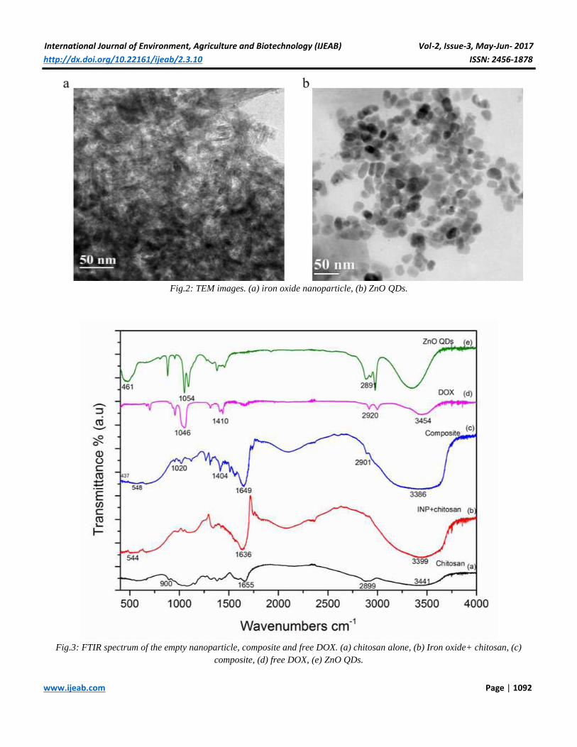
Morphology analysis
The size and shape of iron oxide nanoparticle was analyzed
using TEM. Results showed the total size of the particle
approximately 30 nm and the shape of the particle
expressed irregularly arranged iron oxide nanoparticle (Fig.
2a). Approximately 90% of the iron oxide nanoparticle
revealed needle like nano rods similar to the previous
reports [29]. The morphology and size of the ZnO QDs
appeared sphere like quantum dots structure and size of the
QDs is approximately 8-10nm (Fig. 2b).
Analysis of chemical composition of composite using
FTIR
FTIR analysis was done for chitosan, iron oxide + chitosan,
free Dox and Dox loaded with Iron oxide and Zinc oxide
nanoparticles. Simple Iron oxide and ZnO showed band at
560 cm-1 and 463 cm-1 indicating the Fe-O and Zn-O
stretching. Bands at 3369 cm-1 and 1630 cm-1and the
absence of band at 560 cm-1 in INP-Chitosan composite
confirms the encapsulation of iron oxide by chitosan. The
observed FTIR band for free DOX assigned as follow, 3454
& 2920 cm-1and band at1410 cm-1 is due to the presence of
C-C, and at 1046 cm-1is due to C-O. In Dox loaded
composite nanoparticles, FTIR spectrum peak is shifted to
3386 cm-1due to overlapof N-H and O-H peaks and showed
broader peaks at 2901 cm-1 (C-H) ,1408 cm-1 (C-C) and
1020 cm-1 (C-O). These peaks confirmed successful loading
of Dox in the composite. Iron showed absorption peak at
560 cm-1and ZnO quantum dots at 461 cm-1in FTIR
spectrum (Fig. 3).
UV-Visible spectrometry analysis of the composite
The UV-Visible analysis of the free Dox and Dox loaded
composite nanoparticle was analyzed by the UV-Visible
spectrometer. Free Dox exhibited the two major peaks at
234nm and 487 nm. However, the composite showed peak
shift at 256 nm and 570nm. Also the composite peak
exhibited a slight hump peak at 358nm exhibiting the
presence of ZnO (Fig. 4). These results confirmed that Dox
was successfully loaded in the composite. These results
further confirmed the FTIR results.
Photoluminescence
Photoluminescence spectrum of the ZnO quantum dot and
composite nanoparticle showed multiple emission peaks.
ZnO quantum emission attribute at 376nm 408 and 558nm.
The peak 376nm denoted ZnO QDs. The peak at 558nm
denoted the oxygen vacancy level. Composite nanoparticle
also showed three major peaks at 366 ,422 and 580nm
respectively. The peak at 366nm exhibit ZnO QDs peak
shift from 376nm to 366nm in the composite. Chitosan peak
was seen at 422nm and finally DOX emission peak was
seen at 580nm (Fig. 5).
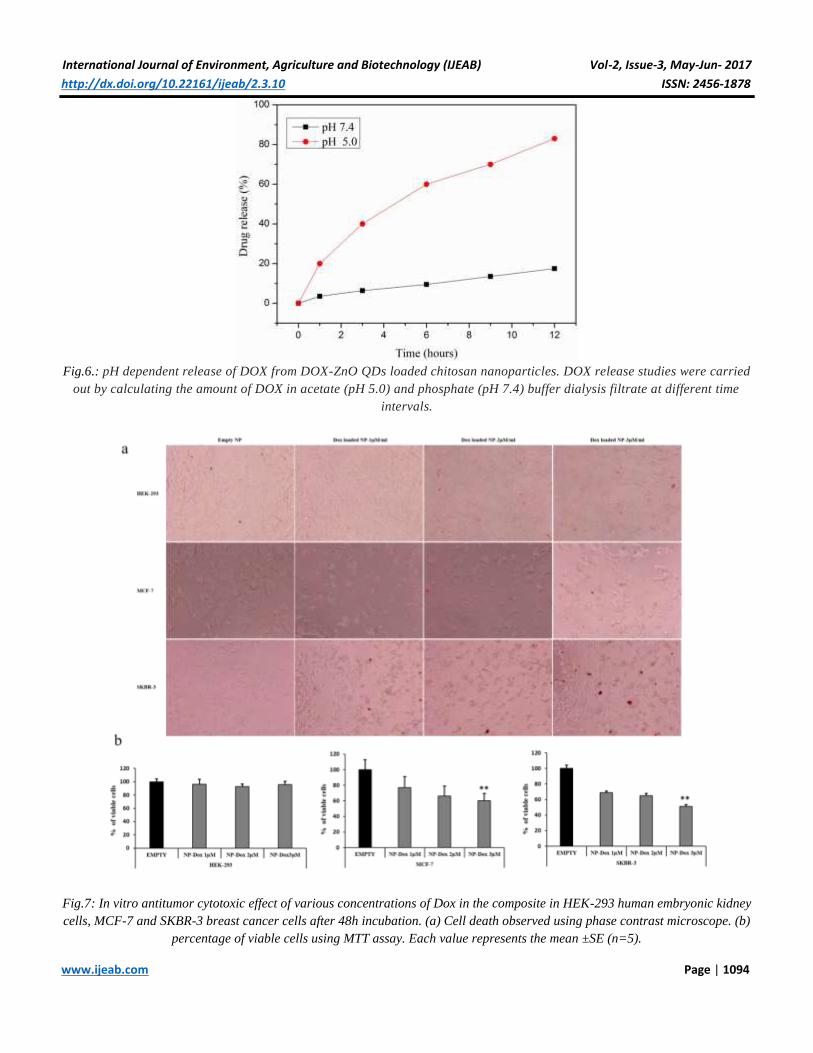
Drug release from carrier nanoparticle
The DOX drug released from the carrier nanoparticle in
different pH levels at 37°C in different pH buffered solution
at pH 5.0 and pH 7.4 is shown in (Fig. 6). Maintaining the
temperature at 37°C mimics the normal physiological
temperature. Poor drug release was observed at pH 7.4 at 12
hours but at pH 5.0 the drug released was nearly 80%. The
release study suggested the carrier was completely acidic
pH responsive thus releasing the drug at acidic pH.
The cytotoxicity of Doxorubicin- loaded iron oxide
nanoparticles
MTT assay was performed to determine the cytotoxicity of
Doxorubicin-loaded iron oxide nanoparticles. Dox
concentrations ranging from 1µM to 3µM was treated to
HEK-239 embryonic kidney cells and MCF-7 and SKBR-3
breast cancer cells. The percentage of viable cells after 48h
of treatment was calculated (Fig. 7 a, b). Doxorubicin when
introduced as nanoparticle composites to the cells, is shown
to confer less toxicity to the normal cells. There are
previous reports that Doxorubicin-loaded colloidal
nanoassembles confer less cytotoxicity compared to direct

International Journal of Environment, Agriculture and Biotechnology (IJEAB) Vol-2, Issue-3, May-Jun- 2017
http://dx.doi.org/10.22161/ijeab/2.3.10 ISSN: 2456-1878
www.ijeab.com Page | 1088
treatment with Doxorubicin in NIH3T3 mouse fibroblast
cells [30]. Corresponding to this, our results from MTT
assay showed Doxorubicin-loaded iron oxide nanoparticles
induced cytotoxicity to the MCF-7 and SKBR-3 breast
cancer cells at specific concentrations. However, HEK-293
human embryonic kidney cells were not affected at the
same concentrations. Thus the composite can be effective in
inducing cytotoxicity to the cancer cells while leaving the
normal cells unharmed The results confirmed a significant
cytotoxicity of the composite containing Dox concentration
at 3µM in MCF-7 and SKBR-3 breast cancer cells. On the
other hand, this concentration did not show any cytotoxicity
to HEK-293 cells.
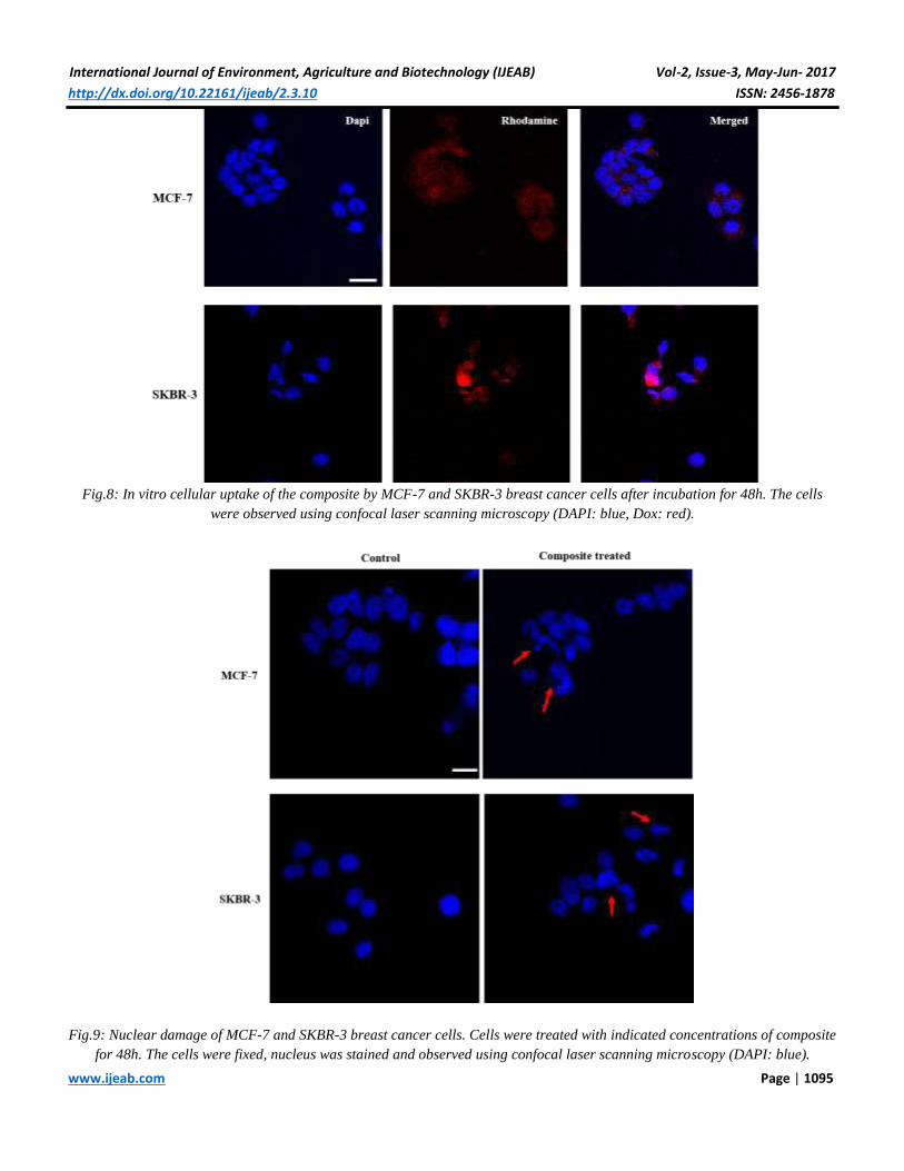
Cellular uptake analysis using CSLM
The cellular uptake of Dox was assessed by CSLM. The
nucleus is stained with DAPI and cellular uptake of Dox
was seen as red fluorescence inside cells. The intensity of
the red fluorescence was also seen to be increased in the
cells whose nuclear morphology is changed indicating
apoptosis in these cells induced by Dox (Fig. 8). The most
important criteria for developing different drug-loaded
nanoparticle composites is increased cellular uptake by the
cancer cells [31] The mechanism through which chitosan
encapsulated nanoparticles enter the cancer cells are
discussed extensively. The most common being endocytosis.
[32] The particles are endocytosed into human cancer cells,
drug is then released and increases cytotoxicity than free
drug. Images from the CSLM of the composite treated
MCF-7 and SKBR-3 breast cancer cells showed immense
cellular uptake of the composite 48h after treatment.
Doxorubicin-loaded iron oxide nanoparticles induce
nuclear breakage
Condensed and fragmented nuclei are characteristics of
apoptosis. MCF-7 and SKBR-3 breast cancer cells treated
with Doxorubicin-ZnO QDs loaded chitosan nanoparticles
for 48h were subjected to DAPI staining. The nuclear
morphology of the empty nanoparticle treated and
Doxorubicin NP treated cells was evaluated using Confocal
microscope. The morphology of the cells treated with empty
nanoparticle remained intact and regular. On the other hand,
nuclear condensation and abnormal nucleus are seen in
Doxorubicin-loaded chitosannanoparticles indicating cell
apoptosis (Fig. 9). Similar to these results, changes in
nuclear morphology of cancer cells upon Dox treatment is
previously reported. MCF-7 and SKBR-3 breast cancer
cells treated with the composite showed similar nuclear
degradation as observed in confocal images upon DAPI
staining. There are several reports on different molecular
mechanisms underlying this [33].
Doxorubicin-loaded chitosan nanoparticles control the
cell proliferation of breast cancer cells
One important feature of Doxorubicin is the regulation of
cancer cell proliferation. To confirm the regulation of breast
cancer cell proliferation by the composite, colony formation
assay was performed. MCF-7 and SKBR-3 breast cancer
cells were treated with empty and composite for 48 h and
the number of colonies formed was evaluated after two
weeks. MCF-7 and SKBR-3 cells treated with empty NP
formed many colonies. On the other hand, MCF-7 and
SKBR-3 cells treated with composite formed significantly
less number of colonies than empty NP treated cells. These
results indicate that composite controls the breast cancer
cell proliferation (Fig. 10 a). Dox treatment is reported to
control the cell proliferation of many types of cancer cells
such as colon, lung, liver, breast, etc.[34][4]. Here, we have
shown that our composite could appreciably reduce the cell
proliferation.
Doxorubicin-loaded chitosannanoparticles induce G1
phase arrest in breast cancer cells
Besides apoptosis, our composite also inducted cell cycle
arrest in breast cancer cells. MCF-7 and SKBR-3 breast
cancer cells were treated with empty NP and composite.
48h after treatment, cells were subjected to Propidium
iodide (PI) staining and flow cytometry analysis was used to
determine the cell cycle distribution. MCF-7 and SKBR-3
breast cancer cells treated with composite showed a
significant reduction of cells in G0/G1 phase compared to
the empty NP treated cells (Fig.10 b).
V. CONCLUSION
In summary, DOX-ZnO QDs loaded with the chitosan
nanoparticles were successfully developed and evaluated
for their anticancer effects. The composite was nanosized at
around 30nm and with uniform needle like nano rods
morphology. The drug was loaded to the shell effectively.
Drug release analysis showed significantly larger amount of
drug released in acidic pH of 5.0 compared to pH 7.4. The
composite induced significant cytotoxicity only to MCF-7
and SKBR-3 but very less toxic to HEK-293 cells at
particular concentrations emphasizing cytotoxic effect
against breast cancer cells specifically. The cellular uptake
of the composite in the breast cancer cells were quite

International Journal of Environment, Agriculture and Biotechnology (IJEAB) Vol-2, Issue-3, May-Jun- 2017
http://dx.doi.org/10.22161/ijeab/2.3.10 ISSN: 2456-1878
www.ijeab.com Page | 1089
significant. The composite also induced nuclear damage
upon treatment. The proliferation of the breast cancer cells
drastically decreased compared to the control. Further, the
composite induced cell cycle arrest at the G1 phase in these
cells. Based on our results, we believe that our composite
can effectively kill the breast cancer cells in vitro and needs
for more in vivo studies in future to translate this approach
into the clinic (Fig.11). Iron oxide in the composite
facilitates driving the composite to cancer cells specifically
using magnetic field and ZnO quantum dots can act as a dye
for imaging purpose in in vivo models. Additionally, there
are several reports on the anticancer properties of ZnO
therefore this Dox-ZnO QDs formulation will have higher
anticancer effect compared to other Dox formulations.
Hence, as a next step, we intend to continue the effective
targeted therapy of the composite against breast cancer in
xenograft models.
CONFLICTS OF INTEREST
The authors report no conflicts of interest about this work.
ACKNOWLEDGMENT
This study was supported by Jeju National University in
2017.
REFERENCES
[1] Hofmann M, Guschel M, Bernd A, Bereiter-Hahn J,
Kaufmann R, Tandi C, et al. Lowering of Tumor
Interstitial Fluid Pressure Reduces Tumor Cell
Proliferation in a Xenograft Tumor Model. Neoplasia.
2006;8:89–95.
[2] Szakacs G, Paterson JK, Ludwig JA, Booth-Genthe C,
Gottesman MM. Targeting multidrug resistance in
cancer. Nat Rev Drug Discov. 2006;5:219–34.
[3] Nitiss JL. Targeting DNA topoisomerase II in cancer
chemotherapy. Nat. Rev. Cancer. 2009;9:338–50.
[4] Silden E, Hjelle SM, Wergeland L, Sulen A, Andresen
V, Bourdon J-C, et al. Expression of TP53 Isoforms
p53β or p53γ Enhances Chemosensitivity in TP53(null)
Cell Lines. Gartel AL, editor. PLoS One. San
Francisco, USA: Public Library of Science;
2013;8:e56276.
[5] Mosieniak G, Sliwinska MA, Alster O, Strzeszewska
A, Sunderland P, Piechota M, et al. Polyploidy
Formation in Doxorubicin-Treated Cancer Cells Can
Favor Escape from Senescence(). Neoplasia.
Neoplasia Press; 2015;17:882–93.
[6] Mohammad N, Vikram Singh S, Malvi P, Chaube B,
Athavale D, Vanuopadath M, et al. Strategy to
enhance efficacy of doxorubicin in solid tumor cells
by methyl-β-cyclodextrin: Involvement of p53 and Fas
receptor ligand complex. Sci. Rep. The Author(s);
2015;5:11853.
[7] Ha JS, Byun J, Ahn D-R. Overcoming doxorubicin
resistance of cancer cells by Cas9-mediated gene
disruption. Sci. Rep. The Author(s); 2016;6:22847.
[8] Smith L, Watson MB, O'Kane SL, Drew PJ,
Lind MJ, Cawkwell L. The analysis of doxorubicin
resistance in human breast cancer cells using antibody
microarrays. Mol. Cancer Ther. 2006;5:2115 LP-2120.
[9] Logothetidis S. Nanotechnology: Principles and
Applications. In: Logothetidis S, editor.
Nanostructured Mater. Their Appl. Berlin, Heidelberg:
Springer Berlin Heidelberg; 2012. p. 1–22.
[10] Stanković A, Dimitrijević S, Uskoković D. Influence
of size scale and morphology on antibacterial
properties of ZnO powders hydrothemally synthesized
using different surface stabilizing agents. Colloids
Surfaces B Biointerfaces. 2013;102:21–8.
[11] Farokhzad OC, Langer R. Nanomedicine: Developing
smarter therapeutic and diagnostic modalities. Adv.
Drug Deliv. Rev. 2006;58:1456–9.
[12] Mohanty C, Arya G, Verma RS, Sahoo SK.
Nanobiotechnology: Application of Nanotechnology
in Therapeutics and Diagnosis. Int. J. Green
Nanotechnol. Biomed. Taylor & Francis; 2009;1:B24–
38.
[13] Alharbi KK, Al-sheikh YA. Role and implications of
nanodiagnostics in the changing trends of clinical
diagnosis. Saudi J. Biol. Sci. 2014;21:109–17.
[14] Falagan-Lotsch P, Grzincic EM, Murphy CJ. New
Advances in Nanotechnology-Based Diagnosis and
Therapeutics for Breast Cancer: An Assessment of
Active-Targeting Inorganic Nanoplatforms. Bioconjug.
Chem. American Chemical Society; 2017;28:135–52.
[15] Pérez-Herrero E, Fernández-Medarde A. Advanced
targeted therapies in cancer: Drug nanocarriers, the
future of chemotherapy. Eur. J. Pharm. Biopharm.
2015;93:52–79.
[16] Costo R, Bello V, Robic C, Port M, Marco JF, Puerto
Morales M, et al. Ultrasmall Iron Oxide Nanoparticles
for Biomedical Applications: Improving the Colloidal
and Magnetic Properties. Langmuir. American
Chemical Society; 2012;28:178–85.
[17] Lin J, Li Y, Li Y, Wu H, Yu F, Zhou S, et al.
Drug/Dye-Loaded, Multifunctional PEG–Chitosan–

International Journal of Environment, Agriculture and Biotechnology (IJEAB) Vol-2, Issue-3, May-Jun- 2017
http://dx.doi.org/10.22161/ijeab/2.3.10 ISSN: 2456-1878
www.ijeab.com Page | 1090
Iron Oxide Nanocomposites for Methotraxate
Synergistically Self-Targeted Cancer Therapy and
Dual Model Imaging. ACS Appl. Mater. Interfaces.
American Chemical Society; 2015;7:11908–20.
[18] Patra S, Roy E, Karfa P, Kumar S, Madhuri R, Sharma
PK. Dual-Responsive Polymer Coated
Superparamagnetic Nanoparticle for Targeted Drug
Delivery and Hyperthermia Treatment. ACS Appl.
Mater. Interfaces. American Chemical Society;
2015;7:9235–46.
[19] Javid A, Ahmadian S, Saboury AA, Kalantar SM,
Rezaei-Zarchi S. Chitosan-Coated Superparamagnetic
Iron Oxide Nanoparticles for Doxorubicin Delivery:
Synthesis and Anticancer Effect Against Human
Ovarian Cancer Cells. Chem. Biol. Drug Des.
2013;82:296–306.
[20] Kuo C-Y, Liu T-Y, Chan T-Y, Tsai S-C, Hardiansyah
A, Huang L-Y, et al. Magnetically triggered
nanovehicles for controlled drug release as a colorectal
cancer therapy. Colloids Surfaces B Biointerfaces.
2016;140:567–73.
[21] Lachowicz D, Szpak A, Malek-Zietek KE, Kepczynski
M, Muller RN, Laurent S, et al. Biocompatible and
fluorescent superparamagnetic iron oxide
nanoparticles with superior magnetic properties coated
with charged polysaccharide derivatives. Colloids
Surfaces B Biointerfaces. 2017;150:402–7.
[22] Azuma K, Izumi R, Osaki T, Ifuku S, Morimoto M,
Saimoto H, et al. Chitin, Chitosan, and Its Derivatives
for Wound Healing: Old and New Materials.
Jayakumar R, editor. J. Funct. Biomater. MDPI;
2015;6:104–42.
[23] Muhammad F, Guo M, Guo Y, Qi W, Qu F, Sun F, et
al. Acid degradable ZnO quantum dots as a platform
for targeted delivery of an anticancer drug. J. Mater.
Chem. The Royal Society of Chemistry;
2011;21:13406–12.
[24] Hosseinzadeh G, Maghari A, Saboury AA, Moosavi-
Movahedi AA. Unfolding of insulin at the surface of
ZnO quantum dots. Int. J. Biol. Macromol.
2016;86:169–76.
[25] Qin G, Li Z, Xia R, Li F, O’Neill BE, Goodwin JT, et
al. Partially polymerized liposomes: stable against
leakeage yet capable of instantaneous release for
remote controlled drug delivery. Nanotechnology.
2011;22:155605.
[26] Karimi M, Eslami M, Sahandi-Zangabad P, Mirab F,
Farajisafiloo N, Shafaei Z, et al. pH-Sensitive
stimulus-responsive nanocarriers for targeted delivery
of therapeutic agents. Wiley Interdiscip. Rev.
Nanomedicine Nanobiotechnology. John Wiley &
Sons, Inc.; 2016;8:696–716.
[27] Unsoy G, Khodadust R, Yalcin S, Mutlu P, Gunduz U.
Synthesis of Doxorubicin loaded magnetic chitosan
nanoparticles for pH responsive targeted drug delivery.
Eur. J. Pharm. Sci. 2014;62:243–50.
[28] Zubair M, Mustafa M, Ali A, Doh YH, Choi KH.
Improvement of solution based conjugate polymer
organic light emitting diode by ZnO--graphene
quantum dots. J. Mater. Sci. Mater. Electron.
2015;26:3344–51.
[29] K. Rastogi S, F. Jabal JM, Zhang H, M. Gibson C.
Antibody@Silica Coated Iron Oxide Nanoparticles:
Synthesis, Capture of E.coli and Sers Titration of
Biomolecules with Antibacterial Silver Colloid. J.
Nanomed. Nanotechnol. 2011;2:2–8.
[30] Tomankova K, Polakova K, Pizova K, Binder S,
Havrdova M, Kolarova M, et al. In vitro cytotoxicity
analysis of doxorubicin-loaded/superparamagnetic
iron oxide colloidal nanoassemblies on MCF7 and
NIH3T3 cell lines. Int. J. Nanomedicine. Dove
Medical Press; 2015;10:949–61.
[31] Dhar S, Reddy EM, Prabhune A, Pokharkar V, Shiras
A, Prasad BL V. Cytotoxicity of sophorolipid-gellan
gum-gold nanoparticle conjugates and their
doxorubicin loaded derivatives towards human glioma
and human glioma stem cell lines. Nanoscale. The
Royal Society of Chemistry; 2011;3:575–80.
[32] Xiang S, Zhang X. Cellular Uptake Mechanism of
Non-Viral Gene Delivery and Means for Improving
Transfection Efficiency. Gene Ther. - Tools Potential
Appl. Rijeka: InTech; 2013. p. Ch. 0.
[33] Demidenko ZN, Vivo C, Halicka HD, Li CJ, Bhalla K,
Broude E V, et al. Pharmacological induction of
Hsp70 protects apoptosis-prone cells from doxorubicin:
comparison with caspase-inhibitor- and cycle-arrest-
mediated cytoprotection. Cell Death Differ.
2005;13:1434–41.
[34] Jin X, Zou B, Luo L, Zhong C, Zhang P, Cheng H, et
al. Codelivery of thioridazine and doxorubicin using
nanoparticles for effective breast cancer therapy. Int. J.
Nanomedicine. Dove Medical Press; 2016;11:4545–52.

International Journal of Environment, Agriculture and Biotechnology (IJEAB) Vol-2, Issue-3, May-Jun- 2017
http://dx.doi.org/10.22161/ijeab/2.3.10 ISSN: 2456-1878
www.ijeab.com Page | 1091
Fig.1: Schematic diagram showing preparation of Dox-ZnO QDs and subsequent loading of Dox-ZnO QDs to Chitosan
nanoparticles.
International Journal of Environment, Agriculture and Biotechnology (IJEAB) Vol-2, Issue-3, May-Jun- 2017
http://dx.doi.org/10.22161/ijeab/2.3.10 ISSN: 2456-1878
www.ijeab.com Page | 1092
Fig.2: TEM images. (a) iron oxide nanoparticle, (b) ZnO QDs.
Fig.3: FTIR spectrum of the empty nanoparticle, composite and free DOX. (a) chitosan alone, (b) Iron oxide+ chitosan, (c)
composite, (d) free DOX, (e) ZnO QDs.

International Journal of Environment, Agriculture and Biotechnology (IJEAB) Vol-2, Issue-3, May-Jun- 2017
http://dx.doi.org/10.22161/ijeab/2.3.10 ISSN: 2456-1878
www.ijeab.com Page | 1093
Fig.4: Absorption spectra of the DOX and DOX loaded nanoparticle. (a) Iron oxide, (b) Chitosan, (c) ZnO QDs, (d) free DOX, (e)
Composite.
Fig.5: Fluorescence spectra. (a) ZnO QDs, (b) DOX loaded composite nanoparticle.
International Journal of Environment, Agriculture and Biotechnology (IJEAB) Vol-2, Issue-3, May-Jun- 2017
http://dx.doi.org/10.22161/ijeab/2.3.10 ISSN: 2456-1878
www.ijeab.com Page | 1094
Fig.6.: pH dependent release of DOX from DOX-ZnO QDs loaded chitosan nanoparticles. DOX release studies were carried
out by calculating the amount of DOX in acetate (pH 5.0) and phosphate (pH 7.4) buffer dialysis filtrate at different time
intervals.
Fig.7: In vitro antitumor cytotoxic effect of various concentrations of Dox in the composite in HEK-293 human embryonic kidney
cells, MCF-7 and SKBR-3 breast cancer cells after 48h incubation. (a) Cell death observed using phase contrast microscope. (b)
percentage of viable cells using MTT assay. Each value represents the mean ±SE (n=5).
International Journal of Environment, Agriculture and Biotechnology (IJEAB) Vol-2, Issue-3, May-Jun- 2017
http://dx.doi.org/10.22161/ijeab/2.3.10 ISSN: 2456-1878
www.ijeab.com Page | 1095
Fig.8: In vitro cellular uptake of the composite by MCF-7 and SKBR-3 breast cancer cells after incubation for 48h. The cells
were observed using confocal laser scanning microscopy (DAPI: blue, Dox: red).
Fig.9: Nuclear damage of MCF-7 and SKBR-3 breast cancer cells. Cells were treated with indicated concentrations of composite
for 48h. The cells were fixed, nucleus was stained and observed using confocal laser scanning microscopy (DAPI: blue).

International Journal of Environment, Agriculture and Biotechnology (IJEAB) Vol-2, Issue-3, May-Jun- 2017
http://dx.doi.org/10.22161/ijeab/2.3.10 ISSN: 2456-1878
www.ijeab.com Page | 1096
Fig.10: (a) Composite inhibited cell proliferation of breast cancer cells. (a) MCF-7 and SKBR-3 cells were treated with
indicated concentrations of composite for 48h. Clonogenic assay was performed to validate colony formation ability of treated
cells. (b) Cell cycle analysis was performed after treating MCF-7 and SKBR-3 cells with indicated concentrations of composite
for 48h using flow cytometry.

International Journal of Environment, Agriculture and Biotechnology (IJEAB) Vol-2, Issue-3, May-Jun- 2017
http://dx.doi.org/10.22161/ijeab/2.3.10 ISSN: 2456-1878
www.ijeab.com Page | 1097
Fig.11: Schematic illustration of mechanism for the interaction of ZnO QDs-breast cancer cells and their cytological death.