Embed Size (px)
Citation preview

Synthesis and Evaluation of Indole Cyanoacrylic
Acids as Anticancer Agents
A THESIS
SUBMITTED TO THE FACULTY OF
UNIVERSITY OF MINNESOTA
BY
KEBREAB SAMUEL
IN PARTIAL FULFILLMENT OF THE REQUIREMENTS
FOR THE DEGREE OF
MASTER OF SCIENCE
Dr. Joseph L. Johnson
DECEMBER 2016

© Kebreab Samuel 2016

i
ACKNOWLEDGEMENTS
I would like to thank my advisors Dr. Joseph Johnson and Dr. Venkatram Mereddy for
their valuable effort, time, and energy through my graduate school career. I am grateful to
my advisors for their support, encouragement and patience. I would also like to thank my
committee member Dr. Paul Siders for devoting his valuable time and support.
I am grateful to my colleagues Sravan Jonnalagadda, Lucas Solano, Shirisha Gurrapu,
Conor Ronayne, Grady Nelson and Min Kyung Sohn. Especially, I am greatly indebted to
Shirisha for all her valuable input in writing this paper.
It is a pleasure to thank the department of Chemistry and Biochemistry of University of
Minnesota Duluth for giving me the opportunity of a lifetime to further my education. I
would equally like to express my gratitude to all faculty members of the chemistry
department.
Finally, I would like to thank my dearest wife Lul, my parents Rediet and Hadas, and all
my family members including Rebecca, Moses and Martha for making my dream come
true through their continuous love and support in every step of my life.

ii
ABSTRACT
Cancer is the second leading cause of death in the United States. Breast cancer is
common among women, and triple negative breast cancer accounts 10-20% of all breast
cancer patients. Cancer has seemingly unlimited proliferative capacity and can spread
easily to other parts of the body, making it difficult to treat. Cancer cells meet their high-
energy requirement by changing their metabolic pathway in a process called “Warburg
effect” via increased glycolysis independent of the presence or absence of oxygen. The
increased glycolysis, which fuels hypoxic cancer cells for their high-energy demand,
produces lactate. Transport of lactate and pyruvate through the plasma membrane is
mediated by a group of integral membrane transport proteins, which are called
monocarboxylate transporters (MCTs). Inhibition of MCT results in the starvation of
tumor cells and, eventually, leads to their death. Therefore targeting MCTs could be an
effective way to reduce aggressive spread of cancer in the body. In this thesis, a library of
indole based small molecules have been synthesized and characterized. Furthermore their
MCT1 inhibition has been evaluated in RBE4 cell line. It was found that most of the
indole cyanoacrylic acids exhibited good MCT1 inhibition while compounds 1, 11-13
didn’t show any activity. Based on their good MCT1 inhibition, compounds 9 and 10
were selected as the lead candidate compounds. Future studies will include the evaluation
of the lead candidates for their systemic toxicity, and in vivo anticancer efficacy in tumor
xenograft models.

iii
TABLE OF CONTENTS
I. ACKNOWLEDGMENTS i
II. ABSTRACT ii
III. TABLE OF CONTENTS iii
IV. LIST OF SCHEMES iv
V. LIST OF FIGURES v
VI. LIST OF TABLES vi
VII. LIST OF ABBREVATIONS vii
VIII. CHAPTER 1: INTRODUCTION 1
IX. CHAPTER 2: RESULTS AND DISCUSSION 17
X. CHAPTER 3: EXPERIMENTAL PROCEDURES AND SPECTRAL
CHARACTERIZATION 31
XI. CONCLUSION 60
XII. REFERENCES 61
XIII. APPENDIX 68

iv
LIST OF SCHEMES
Scheme Title of the scheme Page number
Scheme 1 Synthesis of (E)-2-cyano-3-(1H-indol-3-yl)acrylic acid 18
Scheme 2 Synthesis of (E)-2-cyano-3-(1-(4-substituted benzyl)
-1H-indol-3-yl) acrylic acids 19
Scheme 3 Synthesis of (E)-2-cyano-3-(1-(4-substituted phenyl)
-1H-indol -3-yl) acrylic acids 21
Scheme 4 Synthesis of indolyl amide using benzoyl chloride 25
Scheme 5 Synthesis of indolyl amides using benzoic acids 26
Scheme 6 Synthesis of indolyl amido aldehydes 27
Scheme 7 Synthesis of indole amido cyanoacrylic acids 28
v
LIST OF FIGURES
Figure Description of the figure Page number
Figure 1 Hallmarks of cancer: Acquired capabilities 2
Figure 2 Glucose metabolism in normal and cancer cells 6
Figure 3 Fluorodeoxyglucose (18
F) 6
Figure 4 Stromal-epithelial metabolic coupling and reverse Warburg effect 10
Figure 5 α-Cyano-4-hydroxycinnamate 12
Figure 6 Examples of CHC derived compounds with MCT1 IC50 values 14
Figure 7 Important indole containing molecules 15
Figure 8 Clinically used indole molecules 16

vi
LIST OF TABLES
Table 1: IC50 (nM) values of indole derivatives for MCT1 inhibition 22
Table 2: IC50 values of indole derivatives in MDA-MB-231 and 29
4T1 cell lines using MTT assay
vii
LIST OF ABBREVIATIONS
ATP adenosine triphosphate
bFGF Fibroblast growth factor-basic
CHC α-cyano-4-hydroxycinnamate
CH2Cl2 (DCM) methylene chloride (dichloromethane)
CH3CN acetonitrile
DMAP 4-N,N-dimethylaminopyridine
DMF N,N-dimethylformamide
DMSO dimethyl sulfoxide
ECM extracellular matrix
EGFR epidermal growth factor receptor
EPO erythropoietin
Et3N triethylamine
EtOAc ethylacetate
FDG fluorodeoxyglucose
GLUT glucose transporters

viii
HBTU 2-(1H-benzotriazol-1-yl)-1,1,3,3-tetramethyluronium
hexafluorophosphate
HCOONH4 ammonium formate
HER2 human epidermal growth factor receptor 2
HIF-1 hypoxia-inducible factor 1
K2CO3 potassium carbonate
MCTs monocarboxylate transporters
OxPhos oxidative phosphorylation
Pd/C Palladium on charcoal
PET positron emission tomography
POCl3 phosphoryl chloride
TAT1 T-type amino-corrosive transporter-1
THF tetrahydofuran
TNBC triple negative breast cancer
VEGF vascular endothelial growth factor

1
CHAPTER 1: INTRODUCTION
For a body to function normally, cells have to grow, divide and die. These
processes happen in a controlled fashion. Sometimes, cells undergo uncontrolled growth
and interfere with normal functioning of the body by reaching a critical mass or by
spreading to another location via metastasis. This abnormal growth eventually leads to
the death of the individual. This condition is called “cancer”.
Hallmarks of cancer: A focus on tumor metabolism
Many cancers have some common underlying features, but they also possess
unique pathologies and differences in their metabolic processes. An increased
dependency on glycolysis for ATP production is considered to be one of the important
hallmarks of tumor cells.1-2
Understanding and targeting these hallmarks of cancer is
broadly accepted as a promising strategy for therapeutic intervention to prevent, treat and
eliminate cancer and provides a set of very attractive targets to guide the design of new
drugs.1-2
The following are some of the crucial hallmarks of cancer that can be targeted to
develop new treatments for cancer (Figure 1).

2
Figure 1: Hallmarks of cancer: Acquired capabilities (Modified from ref 1)
Acquired growth signal independence or self-sufficiency: Normal cells need growth
signals to undergo proliferation from a quiescent state, and they do not undergo
proliferation without these stimulatory signals. However, tumor cells do not depend on
external growth stimulation. Three common strategies through which cancer cells
become independent of outside growth signals are: a) changing of extracellular growth
signals; b) transcellular transduction of those signals; and c) intracellular circuits that
translate those signals into action. A cancer cell is capable of making its own growth
factors to facilitate its proliferation and becomes independent of other cells. The cell
surface receptors that transduce growth stimulatory signals into the cell interior become
unregulated during the progression of the tumor. Vascular endothelial growth factor
The Six Hallmarks of Cancer
Self-sufficiency in growth signals
Insensitivity to anti-growth
signals
Tissue invasion & metastasis
Limitless replicative potential
Sustained angiogenesis
Evading apoptosis

3
(VEGF), epidermal growth factor receptor (EGFR), and human epidermal growth factor
receptor 2 (HER2) are some of the examples of induced growth signal transduction.1-2
Insensitivity to anti-growth signals: Anti-growth signals control the proliferation of
normal cells by forcing them to enter in to quiescent state and also reduces their growth
potential by inducing them into a post mitotic state. However, cancer cells develop
strategies to bypass the antigrowth signals and divide uncontrollably.1-2
Evading apoptosis: Apoptosis is a programmed cell death which is a critical process for
the survival of multicellular organisms. Cancer cells bypass it to grow and divide
uncontrollably by avoiding cellular death pathways. p53 is a cancer suppressor gene and
a major part of the DNA damage sensor that can instigate the apoptotic cascade. When
p53 is mutated or missing, cancer cells evade apoptosis. Cancer cells are also known to
weaken the activity of p53 by inhibiting it and silencing its activators.1-2
Limitless replicative potential: Normal cells are able to keep track of the number of their
cell divisions. Usually after 40-60 divisions, cell growth slows down and eventually
stops. This state, known as senescence, is irreversible. Cancer cells generally deviate
from a normal cellular growth program and breach the limit of replication and divide
endlessly.1-2
Sustained angiogenesis: Angiogenesis is a coordinated growth of new blood vessels from
pre-existing blood vessels. Tumors are limited in their growth beyond a certain size due

4
to the lack of oxygen and access to other essential nutrients. Tumors induce blood vessel
formation by secreting growth factors such as fibroblast growth factor-basic (bFGF) and
VEGF that initiate capillary growth into the tumor, which in turn allows for tumor
expansion by supplying nutrients.1-2
Tissue invasion and metastasis: Tumors are surrounded by extracellular matrix (ECM).
After a certain stage, tumors cannot grow and hence it is possible for a few tumor cells to
evade the contact with the ECM, escape, and be transported to new areas of the body
such as brain, lungs, liver, bones, etc. These new regions have the nutrients to support the
growth of the metastasized tumor cells to form secondary tumors.1-2
5
Warburg effect in cancer cells
Generally, in normal cells, oxidative phosphorylation (OxPhos) of pyruvate to CO2
and H2O takes place in the mitochondria to generate energy in the form of ATP.
Similarly, cancer cells must also produce energy that can allow them to proliferate.
Tumor cells often alter their metabolic pathways to bypass the checkpoints in cell
division. Some of the changes in metabolic pathways serve to shift the generation of ATP
from OxPhos to glycolysis.3-9
Glucose metabolism in cancer cells is essentially
distinguished by two principal biochemical phenomena: an increased glucose uptake and
increased levels of glycolysis. Glycolysis encompasses the conversion of glucose to
pyruvate and lactate. Under aerobic conditions, glycolysis typically favors the production
of pyruvate, which then feeds into the OxPhos pathway where ATP production is coupled
with the reduction of O2 to H2O. Conversely, glycolysis under anaerobic cellular
conditions typically favors the formation of lactate as the terminal step of glycolysis. In
cancer cells, glycolysis is the preferred source of ATP production even in the presence of
oxygen, a process termed as “Warburg effect” (Figure 2).3-9
Cancer cells require
significant amount of glucose for their rapid proliferation. Adaptation of glycolysis is a
survival mechanism for all advanced stage tumors and starving the cancer cell of
necessary energetic requirement for rapid and uncontrolled proliferation can inhibit
glycolysis. Thus, inhibition of glycolysis can be a useful target for the development of
anticancer agents for cancer treatment.10-11

6
Figure 2: Glucose metabolism in normal and cancer cells
The elevated glucose requirement of cancer cells compared to normal cells is
applied in 18
F fluorodeoxyglucose (FDG, Figure 3) based positron emission tomography
(PET). FDG-PET is a screening method used in distinguishing malignant cancer cells
from normal cells by quantitatively analyzing glucose metabolism. As stated before,
malignant cancers have high rate of glucose metabolism.12-13
Figure 3: Fluorodeoxyglucose (18
F)

7
Tumor hypoxia:
Tumor cells require excessive nutrients to proliferate continuously and, hence,
they develop new blood vessels to meet energy requirements. As the tumor continues to
grow, some regions of tumor are deprived of oxygen and this condition is called
“hypoxia”. Hypoxia is a common characteristic of numerous solid tumors. The
adjustment of cancer cells to hypoxic conditions is mediated by hypoxia-inducible
factor 1 (HIF-1), which is a major transcription factor involved in upregulating a series of
genes including VEGF, angiogenesis, erythropoietin (EPO), glucose transporters
(GLUT), and several glycolytic enzymes involved in glucose metabolism. Hypoxia-
induced gene expression in cancer cells has been correlated with malignant
transformation. However, studies suggest that lactate and pyruvate have the ability to
direct hypoxia-inducible gene expression without the need for hypoxic conditions by
expressing HIF-1α. Tumor hypoxia also correlates with treatment failure, relapse and,
patient mortality as these cells are generally resistant to standard chemo- and radiation
therapies.14-16
Monocarboxylate transporters (MCTs):
In hypoxic cells, metabolism of glucose to lactate generates only 2 ATPs per
molecule of glucose whereas oxidative phosphorylation generates up to 36 ATPs.
Therefore, in order to overcome this inefficient energy production, tumor cells have to
consume large amounts of glucose at a rapid pace to fuel the hypoxic cells and produce
lactate.14-16
Transport of lactate and pyruvate through the plasma membrane is catalyzed
by a group of integral membrane transport proteins, which are called the

8
monocarboxylate transporters (MCTs). 17-20
Not only do the cancer cells consume a large
amount of glucose, but they also overexpress/upregulate the related enzymes including
MCT’s. The MCT family is made up of 14 members, namely MCTs 1–9, MCTs 11–14
and T-type amino-corrosive transporter-1. MCTs 1-4 are involved in transporting proton-
linked monocarboxylates such as lactate, pyruvate, acetoacetate and β-hydroxybutyrate
through the plasma membrane. MCT8, known as SLC16A2, is a thyroid hormone
transporter and MCT-10 is an aromatic amino acid transporter. The function of the other
MCTs is not clearly understood.17-20
MCT1 consists of 494 amino acids with an atomic weight of ~53 KDa and 12
transmembrane domains. Substrate conditions and pH can significantly influence the
ability of MCT1 to facilitate the movement of lactate in and out of the cell. The
transporter can trade one monocarboxylate molecule for another without a net exchange
of protons.17-20
However, in other tissues like the red blood cells, white skeletal muscle,
and tumor cells, it transports lactic acid out of the cell where the metabolism is highly
glycolytic. This is crucial for cancer cells which primarily depend on glycolysis due to
mitochondrial dysfunction and lack of ATP production via OxPhos as a result of hypoxic
conditions.14-16
MCT4 has a molecular weight of ~43 KDa and also spans the membrane. Tissues
in the human body with high expression of MCT4 are white skeletal muscle fibers
astrocytes, white blood cells, chrondrocytes and tumor cells, which are known for their
high glycolytic rates.21-22
Both MCT1 and MCT4 have great similarities in terms of tissue
distribution, regulation and substrate/inhibitor specificity. MCT4 has a low affinity for a

9
variety monocarboxylates when compared to MCT1. The Km of MCT4 for most
monocarboxylates such as pyruvate is more than 100 mM, except for L-lactate, which is
about 30 mM. These lower affinities of MCT4 for different substrates compared to
MCT1 was also observed for many inhibitors.21-22
MCT4 expression is increased in
cancer cells as a result of delayed hypoxia through a transcriptional regulatory component
involving HIF-1α.23
High levels of MCTs are commonly expressed in a range of human
tumors, including breast, head and neck, prostrate, brain tumors.24-32

10
Stromal-epithelial metabolic coupling, and reverse Warburg effect:
Recent studies by Lisanti et al. on metabolic processes in epithelial cells show
that the tumor microenvironment consists of stromal fibroblasts and epithelial cancer
cells. These cancer cells use oxidative stress as a means to get nutrients from surrounding
fibroblasts. Epithelial cancer cells initiate oxidative stress in neighboring stromal
fibroblasts by releasing hydrogen peroxide resulting in mitochondrial dysfunction and
increased dependence upon glycolysis and the associated increased production of lactate
and pyruvate. These metabolic products are transported by MCTs to feed and proliferate
adjacent epithelial cancer cells, which can generate ATP via oxidative phosphorylation.
Thus, they form parasite-host relationship in which the aggressive cancer cells are
parasites (Figure 4).33-36
Figure 4: Stromal-epithelial metabolic coupling and reverse Warburg effect
Triple negative breast cancer
Triple negative breast cancer (TNBC) accounts for 10-20% of all breast cancers.
The term triple negative is an operational term which refers to all heterogeneous breast
cancers and commonly affects young women of African-American, Hispanic origins

11
and/or those with a BRCA1 gene mutation.37,38
Generally, breast cancer drugs are
targeted towards hormone receptor markers such as estrogen receptor (ER), progesterone
receptor (PR), and human epidermal growth factor receptor 2 (HER2) for treatment.
Tamoxifen and raloxifen are the drugs that target hormone receptors, whereas, herceptin
is used to target HER2. The absence of well-defined biomarkers makes the treatment of
TNBC more difficult. The standard treatment for TNBC includes surgery, radiation and
chemotherapy with anthracyclins and taxane-based medications. The fact that these drugs
are not cancer cell selective in addition to their serious side effects makes them inefficient
in the treatment of cancer. Most patients initially respond to the chemotherapy but a
majority relapse, and the cancer becomes drug resistant. Subsequently, novel therapeutics
that are specifically harmful to tumor cells and ideally that work on drug resistant cells
are highly critical.
Cancer treatment, which has long depended on the prevention of rapid
proliferation of tumor cells for effective treatment, was found to be ineffective due to the
undesirable side effects caused by the lack of specificity in the approach. Cancer cells
shift to a dependence upon glycolytic metabolism as an adaptation mechanism to hypoxia
in order to fulfill the high bioenergetic and bisosynthetic demand of proliferating cells,
which also creates a lactate gradient. MCT1 and MCT4 are largely expressed in cancer
cells for the transport of metabolites, and are especially upregulated in response to
hypoxia. By inhibiting MCT1 or MCT4, the transport of lactate is inhibited in hypoxic
regions, and the aerobic cancer cells cannot proliferate due to the lack of lactate. In
addition, the pH in hypoxic cells decreases resulting in acidosis and cell death. The
aerobic cells can easily be treated with chemotherapy and radiation. Therefore, targeting

12
both MCT4, which is a direct transcriptional target of HIF-1α, and MCT1 could hinder
the transport of lactate and effectively target the tumor cells.39, 40
Recent studies on MCT inhibitors:
In recent years, numerous molecules have been synthesized and evaluated as
MCT inhibitors. The following is a brief insight into these inhibitors.
α-Cyano-4-hydroxycinnamate (CHC, Figure 5) is a known MCT1 inhibitor. CHC
specifically and significantly inhibits pyruvate transport in liver mitochondria and
erythrocytes. The fact that it specifically inhibits pyruvate but not acetate or butyrate
transport suggests that MCT1 is specifically involved in the transport of lactate across
plasma membranes and across the inner membrane of mitochondria. Pyruvate transport
inhibition studies by CHC were conducted using mitochondrial incubation media at very
low concentrations (<200 µM) and the rate of pyruvate oxidation by mitochondria from
blowfly flight muscle and rat heart was determined.41
Figure 5: α-Cyano-4-hydroxycinnamate
U-87 is a well-established model for glioblastoma multiforme and show elevated
MCT1 expression. CHC was evaluated for its anticancer efficacy in glioblastoma tumor
xenograft model in immunodeficient rats.42
Rats were stereotactically implanted with
glioma cells and CHC was administered via osmotic pump for the treatment. MRI images

13
were obtained for the treated and untreated rats. The tumor burden was significantly
decreased in rats that received treatment with CHC on day-120 whereas in the untreated
group, tumor size increased by day-20. This clearly suggests that CHC inhibits MCT1 in
glioblastoma tumor model.
In another study, a Lewis lung carcinoma tumor xenograft model, which also
overexpresses MCT1, was treated with CHC (MCT1 inhibitor) and the tumor growth was
slowed over an 8 day period compared to the vehicle group.16
AZD3965, an MCT1 inhibitor, was also implicated in the radiosensitzation of
small cell lung carcinoma (SCLC) model.43
Immunocompromised CD-1 mice were
inoculated with H526 cells and mice were treated with 100 mg/kg of AZD3965
compound for three days, followed by the treatment with 2Gy radiation. The mice were
monitored for an additional period of time until the tumor volumes reached 1000 mm3.
Treated groups showed a took an additional 4 days to reach 1000 mm3
(12 days)
compared to the group treated with radiation only, which took 8 days.
Based on the above studies, MCT inhibition leads to a decrease in tumor growth
and can radiosensitize the tumor cells and improves the survival of rodent tumor
xenograft models. Hence, MCT is an extremely important glycolytic target to treat
advanced stage tumors. In connection with the above statement, many CHC derivatives
have been synthesized in recent years with good in vitro MCT1 inhibition and in vivo
efficacy.44

14
In cyanocinnamate compounds, the substitution of the OH group on the benzene
ring by the N,N-dialkyl groups lead to an exponential increase in activity (Figure 6).
These compounds showed better IC50 values while maintaining great MCT1 inhibitory
potency compared to CHC. Above all, the easiness of their synthesis, water solubility and
non-toxicity to other cells makes them very desirable and opens the way for further
research and synthesis of other cyanocinnamate derived compounds.44
Figure 6: Examples of CHC derived compounds with MCT1 IC50 values
Therefore this project was designed on the hypothesis that synthesis and
biological evaluation of compounds which have both the indole moiety and
cyanocinnamic acid unit could achieve better anti proliferative and MCT1 inhibitory
potency. In this regard, we have synthesized novel indole based cyanocinnamate
derivatives and evaluated them for their MCT1 inhibition.
Indole is a pharmacologically privileged chemical structure and many indole-

15
based pharmaceuticals are in the clinical usage for the treatment of various diseases.
Hence, we envisioned to synthesize several indole based compounds as potential MCT
and tumor metabolism inhibitors.
The indole moiety is a critical pharmacological unit that is found in essential
amino acid tryptophan, neurotransmitter tryptamine, serotonin, melatonin, etc. (Figure
7).45-47
Figure 7: Important indole containing molecules
Indole is also an important pharmacophore in clinically used drugs such as 5-THS3
antagonist alosetron for the treatment of irritable bowel syndrome in women,
indomethacin used for rheumatoid arthritis, sumatriptan for migraine management,
ondansetron which is used as an antiemetic to control nausea and vomiting in patients
who undergo chemotherapy for cancer treatment (Figure 8).48

16
Figure 8: Clinically used indole molecules

17
CHAPTER 2: RESULTS AND DISCUSSION
As mentioned above, indole based compounds are pharmacologically and
pharmaceutically privileged nitrogen-containing heterobicyclic compounds. These
compounds are responsible for a variety of biological activities and sometimes mimic
endogenous substrates by binding to proteins. In fact, indole is the core of the amino acid
tryptophan and other important derivatives such as melatonin and serotonin. Hence,
indole has a vital role in the central nervous system.
Hypothesis and purpose of the work
Owing to the great success of indole based pharmaceuticals in clinic, we
hypothesized that indolyl cyanoacrylates would be excellent compounds for the inhibition
of MCT function. We envisioned that if these novel compounds exhibited potent MCT1
or MCT4 inhibition or cytotoxicity, they could be developed as potential tumor
glycolysis inhibitors. The main purpose of this project is to synthesize and explore
indolyl cyanoacrylates as anticancer agents with potential mechanisms in the disruption
of tumor glycolysis and other metabolic pathways.

18
Synthesis of (E)-2-cyano-3-(1H-indol-3-yl)acrylic acid
(E)-2-cyano-3-(1H-indol-3-yl)acrylic acid 3 was synthesized using indole as a
starting material. 1H-indole 1 was formylated using Vilsmeier Haack condition to obtain
1H-indole-3-carbaldehyde 2. This product 2 was further reacted with cyanoacetic acid via
Knoevenagel condensation to get indole cyanoacrylic acid 3 in 69% yield (Scheme 1).
Scheme 1: Synthesis of (E)-2-cyano-3-(1H-indol-3-yl)acrylic acid
Synthesis of (E)-2-cyano-3-(1-(4-substituted benzyl)-1H-indol-3-yl)acrylic acids
1-(4-substituted benzyl)-1H-indole cyanoacrylic acids 4-8 were synthesized
starting from 1H-indole 1 and substituted benzyl bromides. We have utilized benzyl
bromide, 4-ethylbenzyl bromide, 4-methoxybenzyl bromide, 4-chlorobenzyl bromide and
4-bromobenzyl bromide. First, benzyl bromides were prepared by reducing substituted
benzaldehyde with sodium borohydride and subsequent bromination of alcohol with
PBr3. Then, 1 was alkylated using these corresponding bromides followed by Vilsmeier
Haack formylation and Knoevenagel condensation to obtain (E)-2-cyano-3-(1-(4-
substituted benzyl)-1H-indol-3-yl)acrylic acids in 50-70% yields (Scheme 2).

19
Scheme 2: Synthesis of (E)-2-cyano-3-(1-(4-substitutedbenzyl)-1H-indol-3-yl)acrylic
acids

20
Synthesis of (E)-2-cyano-3-(1-(4-substituted phenyl)-1H-indol-3-yl)acrylic acids
(E)-2-cyano-3-(1-(4-substituted phenyl)-1H-indol-3-yl)acrylic acids 9-13 were
synthesized starting from 1H-indole 1 and 1-iodo-4-substituted benzenes. We utilized
simple iodobenzene, 1-iodo-4-methoxybenzene, 2-bromopyridine, 3-bromopyridine and
2-iodobenzoic acid. Here, 1 was reacted with 1-iodo-4-substituted benzene in presence of
K2CO3, CuI, L-proline to obtain 1-(4-substituted phenyl)-1H-indole, which was
converted to aldehyde and condensed to cyanoacrylic acids 9-13 (50-78% yield) using
Vilsmeier Haack formylation and Knoevenagel condensation, respectively (Scheme 3).
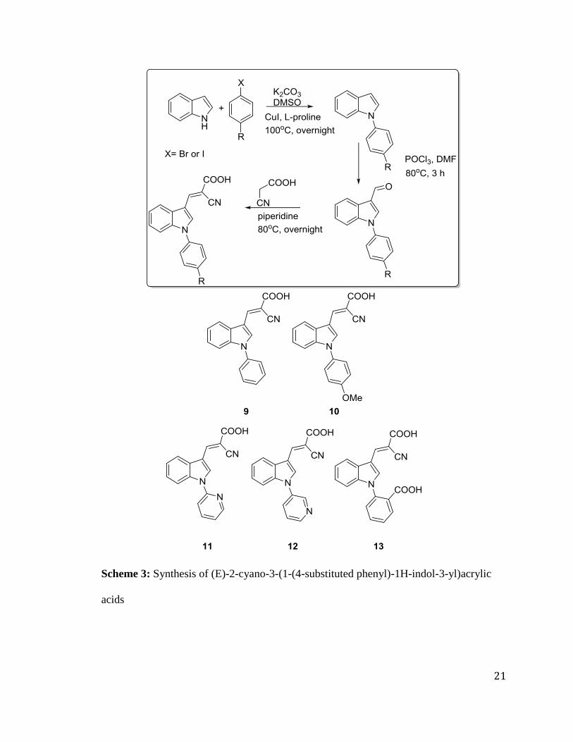
21
Scheme 3: Synthesis of (E)-2-cyano-3-(1-(4-substituted phenyl)-1H-indol-3-yl)acrylic
acids

22
Biological Evaluation
To evaluate the biological activity of indole cyanoacrylic acids, we carried out
MCT1 inhibition assay on rat brain endothelial 4 (RBE4) cell line using sodium salt of
[14
C]-L-lactic acid. RBE4 is a non-cancerous cell line with high MCT1 expression.44
Approximately 2x10⁵
cells/well were plated in 24-well plate and incubated in 5%
CO2 atmosphere for 16 -24 hours. The cells were washed with HEPES buffer. The buffer
was removed and test compounds in [14
C]-L-lactate solution were added for influx of
lactate for 15 minutes. The test compounds were removed and stop buffer was added and
kept on ice. The cells were then lysed with 0.1 M triton-X Solution. Uptake values of the
lysed cells were obtained in dpm (disintegrations per minute) using Scintillation counter.
Table 1: IC50* (nM) values of indole derivatives for MCT1 inhibition
Compound No. Compound MCT1 IC50
3
>1000
4
65.9±10.9

23
5
36.3±14.1
6
107.0±10.5
9
12.8±2.6
10
21.9±2.9
11
>1000

24
12
>1000
13
>1000
*IC50 is expressed in avg±SEM nM, average of minimum 3 experimental values

25
Based on the interesting biological results obtained for indole cyanoacrylic acids,
we also prepared indole benzamido cyanoacrylic acids 21 and 22 with a phenyl spacer
between amide and indole groups.
Synthesis of N-(4-(1H-indol-1-yl)phenyl)benzamide:
Indolyl amide 15 was synthesized using indole as a starting material. The first
step involved nucleophilic ipso substitution of 1H-indole 1 by 4-fluoro nitrobenzene
under basic conditions to obtain 1-(4-nitrophenyl)-1H-indole 16. The nitro group was
further reduced using ammonium formate and palladium-carbon to obtain the
corresponding amine 17. Benzoylation of the indole amine using benzoyl chloride
resulted in the formation of indolyl amide 18 (Scheme 4).
Scheme 4: Synthesis of indolyl amide using benzoyl chloride

26
4-Methoxy indolyl amide derivative 18 was synthesized starting from 4-methoxy
benzoic acid utilizing HBTU coupling in basic conditions with catalytic DMAP (Scheme
5).
Scheme 5: Synthesis of indolyl amides using benzoic acids
27
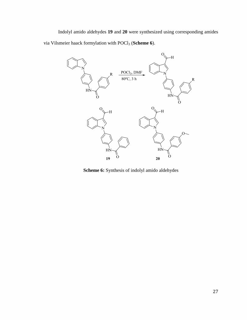
Indolyl amido aldehydes 19 and 20 were synthesized using corresponding amides
via Vilsmeier haack formylation with POCl3 (Scheme 6).
Scheme 6: Synthesis of indolyl amido aldehydes

28
Indolyl amido cyanoacrylic acids 21 and 22 were obtained via Knoevenagel
condensation with corresponding indole amido aldehydes 19 and 20 using cyanoacetic
acid in presence of piperidine (Scheme 7).
Scheme 7: Synthesis of indole amido cyanoacrylic acids
Cytotoxicity evaluation of indolyl amide derivatives:
To evaluate the biological activity of indolyl amide derivatives, we carried
out cytotoxicity studies based on 3-[4,5-dimethylthiazol-2-yl]-2,5- diphenyltetrazolium
bromide (MTT) assay. This assay measures cell viability based on the conversion of
MTT to insoluble purple formazan via cleavage of the tetrazolium ring of MTT by the
action of mitochondrial reductase in living cells.

29
MDA-MB-231 and 4T1 cell lines were chosen for preliminary cytotoxicity
studies. We have chosen these two cell lines because we are targeting MCT1 and MCT4
for the inhibition of glycolysis. MDA-MB-231 is a triple negative breast cancer cell line
that predominantly expresses MCT4, and 4T1 is a murine stage IV metastatic breast
cancer cell line that predominantly expresses MCT1. The test results indicate that all the
compounds 15-22 are non-cytotoxic in MDA-MB-231 and 4T1 cell lines even at 25 µM
concentration (Table 2). MCT1 IC50 values of compounds 21 and 22 will be evaluated
in the future.
Table 2: IC50* values of indole derivatives in MDA-MB-231 and 4T1 cell lines using
MTT assay
Compound
No.
Compound MDA-MB-231 4T1
17
>25 >25
18
>25 >25

30
19
>25 >25
20
>25 >25
21
>25 >25
22
>25 >25
*IC50 is expressed in avg±SEM µM, average of 3 experimental values

31
CHAPTER 3: EXPERIMENTAL PROCEDURES AND SPECTRAL
CHARACTERIZATION
Synthesis of indole cyanoacrylic acids
To a mixture of 1H-indole 1 (10 mmol) in DMF (60 mmol) was added POCl3 (12
mmol) at 0 oC and the reaction mixture was heated at 80
oC for 3 hours. Upon completion
(TLC), the reaction mixture was poured into a saturated solution of Na2CO3 at 90 oC and
stirred for 1 hour. The solid obtained was filtered and washed with ether and hexane
mixture to obtain the 1H-indole-3-carbaldehyde 2. To a solution of aldehyde 2 (1 mmol)
and cyanoacetic acid (1.5 mmol) in acetonitrile (10 mL), piperidine (1.3 mmol) was
added and the reaction mixture was refluxed at 80 oC for 8 hours. After conformation that
the reaction was complete, the mixture was poured in 20 mL of ice-cold 3N HCl and
stirred for 20 minutes. The resulting solid was collected by filtration and washed with
hexanes. The solid was suspended in chloroform (10 mL) and stirred overnight to remove
the impurities. Finally pure product 3 was obtained upon filtration in 69% yield.

32
Compound 3: (E)-2-cyano-3-(1H-indol-3-yl)acrylic acid
1H NMR (500 MHz, DMSO-d6): δ 12.55 (s, 1H), 8.58-8.53 (m, 2H), 7.98-7.93 (m, 1H),
7.60 (d, J = 8.5 Hz, 1H), 7.30 (m, 2H).
13C NMR (125 MHz, DMSO-d6): δ 165.13, 146.62, 136.67, 132.53, 127.39, 124.01,
122.51, 118.94, 113.34, 110.30, 94.16.

33
Representative procedure for synthesis of (E)-2-cyano-3-(1-(4-substituted benzyl)-
1H-indol-3-yl)acrylic acids:
To synthesize substituted benzyl bromides, substituted benzaldehyde (10 mmol)
was dissolved in THF (20 mL) and cooled to 0 oC and NaBH4 (5 mmol) was added very
slowly and the reaction was completed in 30 mins. To the substituted benzyl alcohol (10
mmol) in diethylether (20 mL) was added PBr3 (3 mmol) dropwise very slowly at 0 o
C.
The reaction mixture was quenched with saturated NaHCO3 and extracted with ether-
water. The organic layers were collected, dried with anhydrous MgSO4 and evaporated
under vacuum to obtain substituted benzyl bromide.
To a mixture of 1H-indole 1 (10 mmol) in DMSO (10 mL) was added benzyl
bromide (20 mmol) and K2CO3 (20 mmol) and stirred at 100 o
C overnight. Upon the
completion of the reaction, the reaction mixture was poured into water and stirred to
obtain solid, which was then filtered and washed with hexanes to yield 1-(4-
benzylphenyl)-1H-indole. To a mixture of this benzylated indole and DMF (60 mmol),
POCl3 (12 mmol) was added dropwise at 0 o
C and the reaction mixture was heated at 80
oC for 3 hours. Upon completion (TLC), the reaction mixture was poured into a saturated
solution of Na2CO3 at 90 o
C and stirred for 1 hour. The solid obtained was filtered and
washed with ether and hexane mixture to obtain the 1-(4-benzylphenyl)-1H-indole-3-
carbaldehyde. To this aldehyde (1 mmol) and cyanoacetic acid (1.5 mmol) in acetonitrile
(10 mL), piperidine (1.3 mmol) was added and the reaction mixture was refluxed at 80 oC
for 8 hours. After conformation that the reaction was complete, the mixture was poured in
20 mL of ice-cold 3N HCl and stirred for 20 minutes. The resulting solid was collected
by filtration and washed with hexanes. The solid was suspended in chloroform (10 mL)
34
and stirred overnight to remove the impurities. Finally pure product 4 was obtained upon
filtration in 57% yield.

35
Compound 4: (E)-3-(1-benzyl-1H-indol-3-yl)-2-cyanoacrylic acid
1H NMR (500 MHz, DMSO-d6): δ 8.61 (s, 1H), 8.46 (s, 1H), 7.87 (d, J = 7.0 Hz, 1H),
7.55 (d, J = 7.0 Hz, 1H), 7.29-7.25 (m, 7H), 5.54 (s, 2H).
13C NMR (125 MHz, DMSO-d6): δ 165.33, 146.08, 137.02, 136.79, 135.01, 129.50,
128.64, 128.31, 127.97, 124.51, 123.21, 119.41, 118.96, 112.35, 110.06, 94.95, 50.79.

36
Compound 5: (E)-2-cyano-3-(1-(4-ethylbenzyl)-1H-indol-3-yl)acrylic acid
1H NMR (500 MHz, DMSO-d6): δ 8.68 (s, 1H), 8.52 (s, 1H), 7.97 (d, J = 7.5 Hz, 1H),
7.64 (d, J = 7.5 Hz, 1H), 7.33-7.17 (m, 6H), 5.60 (s, 2H), 2.57-2.51 (m, 2H), 1.12 (t, J =
6.0 Hz, 3H).
13C NMR (125 MHz, DMSO-d6): δ 165.33, 146.04, 144.25, 136.93, 135.10, 134.55,
128.86, 128.46, 128.12, 124.47, 123.11, 119.55, 118.98, 112.55, 110.11, 95.12, 50.64,
28.54, 16.25.

37
Compound 6: (E)-2-cyano-3-(1-(4-methoxybenzyl)-1H-indol-3-yl)acrylic acid
1H NMR (500 MHz, DMSO-d6): δ 8.67 (s, 1H), 8.51 (s, 1H), 7.97 (d, J = 7.5 Hz, 1H),
7.68 (d, J = 8.0 Hz, 1H), 7.34-7.27 (m, 4H), 6.92 (d, J = 8.5 Hz, 2H), 5.57 (s, 2H), 3.72
(s, 3H).
13C NMR (125 MHz, DMSO-d6): δ 165.28, 159.64, 146.07, 136.84, 134.95, 129.69,
129.06, 128.45, 124.38, 123.11, 119.50, 118.93, 114.88, 112.55, 110.01, 94.92, 55.80,
50.32.

38
Compound 7: (E)-3-(1-(4-chlorobenzyl)-1H-indol-3-yl)-2-cyanoacrylic acid
1H NMR (500 MHz, DMSO-d6): δ 8.70 (s, 1H), 8.52 (s, 1H), 7.97 (d, J = 8.0 Hz, 1H),
7.61 (d, J = 8.0 Hz, 1H), 7.42 (d, J = 8.0 Hz, 1H), 7.32-7.27 (m, 4H), 5.67 (s, 2H).
13C NMR (125 MHz, DMSO-d6): δ 165.23, 146.01, 136.80, 136.32, 135.07, 133.22,
129.91, 129.50, 128.41, 124.51, 123.19, 119.58, 118.85, 112.42, 110.20, 95.38, 50.03.
39
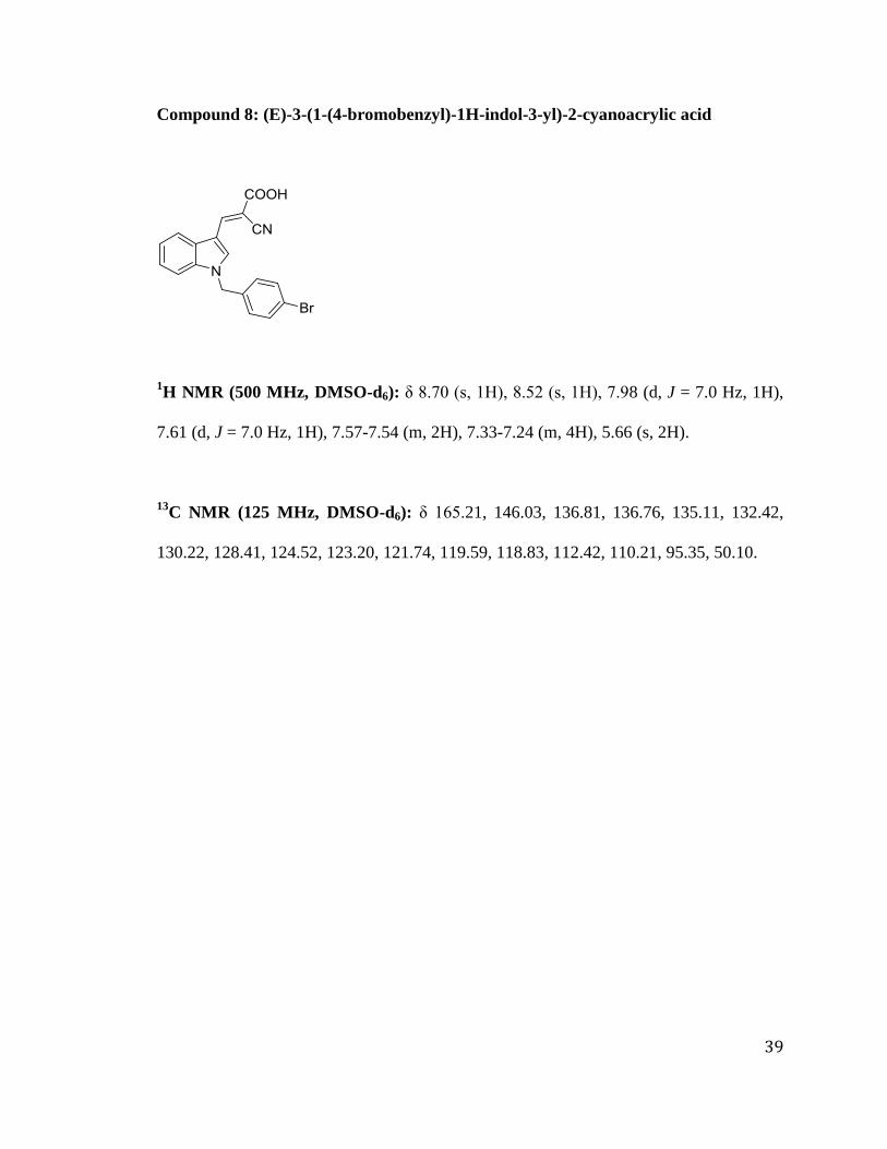
Compound 8: (E)-3-(1-(4-bromobenzyl)-1H-indol-3-yl)-2-cyanoacrylic acid
1H NMR (500 MHz, DMSO-d6): δ 8.70 (s, 1H), 8.52 (s, 1H), 7.98 (d, J = 7.0 Hz, 1H),
7.61 (d, J = 7.0 Hz, 1H), 7.57-7.54 (m, 2H), 7.33-7.24 (m, 4H), 5.66 (s, 2H).
13C NMR (125 MHz, DMSO-d6): δ 165.21, 146.03, 136.81, 136.76, 135.11, 132.42,
130.22, 128.41, 124.52, 123.20, 121.74, 119.59, 118.83, 112.42, 110.21, 95.35, 50.10.

40
Representative example for the synthesis of (E)-2-cyano-3-(1-(4-substituted phenyl)-
1H-indol-3-yl)acrylic acids:
To a solution of 1H-indole 1 (10 mmol) in DMSO (25 mL), were added
iodobenzene (15 mmol), K2CO3 (20 mmol), CuI (2 mmol), L-proline (1 mmol) and
refluxed at 100 o
C overnight. The reaction mixture was extracted in ethyl acetate-water
mixture (3x100 mL) and the organic layers were collected and dried in anhydrous MgSO4
and evaporated under vacuum. The resulting liquid was purified using column
chromatography in ethyl acetate-hexanes mixture (1:9 ratio) to obtain 1-phenyl-1H-
indole. To a solution of phenyl indole (10 mmol) and DMF (60 mmol), POCl3 (12 mmol)
was added dropwise at 0 o
C and the reaction mixture was heated at 80 o
C for 3 hours.
Upon completion (TLC), the reaction mixture was poured into a saturated solution of
Na2CO3 at 90 o
C and stirred for 1 hour. The solid obtained was filtered and washed with
ether and hexane mixture to obtain the 1-(4-benzylphenyl)-1H-indole-3-carbaldehyde. To
this aldehyde (1 mmol) and cyanoacetic acid (1.5 mmol) in acetonitrile (10 mL),
piperidine (1.3 mmol) was added and the reaction mixture was refluxed at 80 oC for 8
hours. After conformation that the reaction was complete, the mixture was poured in
20 mL of ice-cold 3N HCl and stirred for 20 minutes. The resulting solid was collected
by filtration and washed with hexanes. The solid was suspended in chloroform (10 mL)
and stirred overnight to remove the impurities. Finally pure product 9 was obtained upon
filtration in 65% yield.

41
Compound 9: (E)-2-cyano-3-(1-phenyl-1H-indol-3-yl)acrylic acid
1H NMR (500 MHz, DMSO-d6): δ 8.63 (s, 1H), 8.60 (s, 1H), 8.10-8.09 (m, 1H), 7.71-
7.66 (m, 4H), 7.59-7.56 (m, 2H), 7.39-7.38 (m, 2H).
13C NMR (125 MHz, DMSO-d6): δ 164.66, 138.72, 138.48, 135.89, 130.63, 129.83,
128.54, 128.31, 124.85, 124.38, 122.41, 121.42, 119.27, 112.01, 111.54, 108.90.

42
Compound 10: (E)-2-cyano-3-(1-(4-methoxyphenyl)-1H-indol-3-yl)acrylic acid
1H NMR (500 MHz, DMSO-d6): δ 8.60 (d, J = 8.0 Hz, 2H), 8.31 (s, 1H), 8.05-8.03 (m,
1H), 7.56 (d, J = 8.5 Hz, 2H), 7.47-7.45 (m, 1H), 7.36-7.34 (m, 2H), 7.18 (d, J = 8.5 Hz,
1H), 3.86 (s, 3H).
13C NMR (125 MHz, DMSO-d6): δ 165.05, 159.86, 145.85, 137.04, 134.12, 130.88,
128.27, 126.82, 125.10, 123.51, 119.80, 118.92, 115.88, 112.18, 111.28, 96.60, 56.26.

43
Compound 11: (E)-2-cyano-3-(1-(pyridin-2-yl)-1H-indol-3-yl)acrylic acid
1H NMR (500 MHz, DMSO-d6): δ 8.95 (s, 1H), 8.62 (d, J = 4 Hz, 1H), 8.52 (s, 1H),
8.14 (d, J = 8 Hz, 1H), 8.00 (t, J = 7.5 Hz, 1H), 7.90 (s, 1H), 7.86 (d, J = 7 Hz, 1H), 7.68
(d, J = 8 Hz, 1H), 7.40-7.32 (m, 3H).
13C NMR (125 MHz, DMSO-d6): δ 164.80, 151.05, 149.71, 145.08, 139.87, 135.48,
131.18, 129.28, 125.25, 123.74, 122.96, 118.93, 118.23, 116.44, 114.17, 112.50, 98.50.

44
Compound 12: (E)-2-cyano-3-(1-(pyridin-3-yl)-1H-indol-3-yl)acrylic acid
1H NMR (500 MHz, DMSO-d6): δ 9.13 (s, 1H), 8.87 (d, J = 5.0 Hz, 1H), 8.72 (s, 1H),
8.60 (s, 1H), 8.47 (d, J = 8.5 Hz, 1H), 8.13-8.11 (m, 1H), 7.93-7.91 (dd, J = 8.0, 5.0 Hz,
1H), 7.63-7.61 (m, 1H), 7.46-7.41 (m, 1H).
13C NMR (125 MHz, DMSO-d6): δ 164.74, 147.37, 145.78, 144.29, 136.73, 136.46,
135.81, 134.08, 128.35, 126.65, 125.61, 123.99, 120.17, 118.42, 112.41, 112.09, 98.24.
45
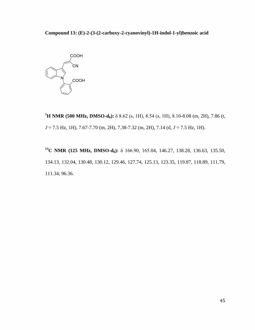
Compound 13: (E)-2-(3-(2-carboxy-2-cyanovinyl)-1H-indol-1-yl)benzoic acid
1H NMR (500 MHz, DMSO-d6): δ 8.62 (s, 1H), 8.54 (s, 1H), 8.10-8.08 (m, 2H), 7.86 (t,
J = 7.5 Hz, 1H), 7.67-7.70 (m, 2H), 7.38-7.32 (m, 2H), 7.14 (d, J = 7.5 Hz, 1H).
13C NMR (125 MHz, DMSO-d6): δ 166.90, 165.04, 146.27, 138.28, 136.63, 135.50,
134.13, 132.04, 130.48, 130.12, 129.46, 127.74, 125.13, 123.35, 119.87, 118.89, 111.79,
111.34, 96.36.

46
Synthesis of 1-(4-nitrophenyl)-1H-indole:
A mixture of (1H)indole 1 (1.0 eq, 42.7 mmol) and 4-fluoro nitrobenzene 14 (1.1
eq, 46.9 mmol) were first stirred at room temperature in 30 mL DMSO, followed by the
addition of K2CO3 (3 eq, 128 mmol). The reaction mixture was stirred for 12 hours at
90 ºC. After it was confirmed that the reaction was complete using a TLC (20%
EtOAc/hexane), water was added to the reaction mixture in order to dissolve the excess
potassium carbonate, and the resultant precipitate was collected by filtration. The yellow
solid was washed with hexane to afford the product 1-(4-nitrophenyl)-1H-indole 15 in
80% yield.
47
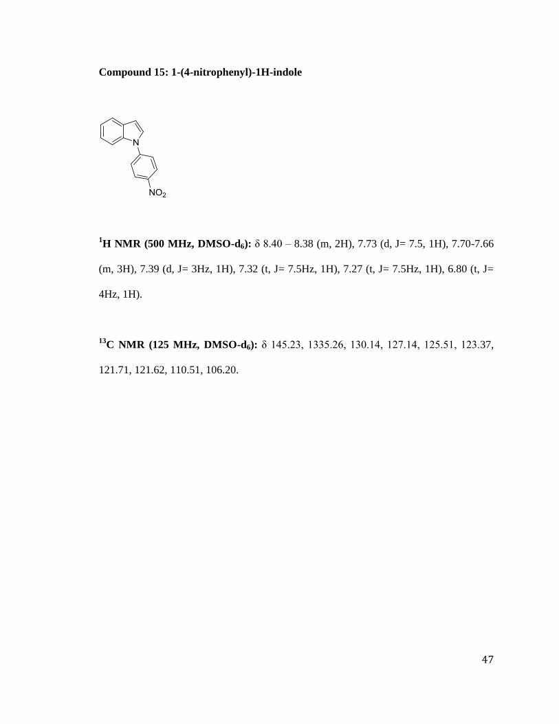
Compound 15: 1-(4-nitrophenyl)-1H-indole
1H NMR (500 MHz, DMSO-d6): δ 8.40 – 8.38 (m, 2H), 7.73 (d, J= 7.5, 1H), 7.70-7.66
(m, 3H), 7.39 (d, J= 3Hz, 1H), 7.32 (t, J= 7.5Hz, 1H), 7.27 (t, J= 7.5Hz, 1H), 6.80 (t, J=
4Hz, 1H).
13C NMR (125 MHz, DMSO-d6): δ 145.23, 1335.26, 130.14, 127.14, 125.51, 123.37,
121.71, 121.62, 110.51, 106.20.

48
Synthesis of 4-(1H-indol-1-yl) aniline:
The above-formed 1-(4-nitrophenyl)-1H-indole 15 was reduced to obtain 4-(1H-
indol-1-yl) aniline 16 by a catalytic hydrogenation using palladium charcoal catalyst and
ammonium formate and THF (100 mL) as a solvent. Compound 15 (1 eq, 17.9 mmol)
was dissolved in 50 mL THF. Then, Pd/C (0.3 eq, 75.2mmo) was added followed by
addition of ammonium formate (6 eq, 107.5 mmol) and refluxed for 6 hours. The reaction
mixture was cooled to room temperature. Finally, the reaction mixture was fast flushed
through silica gel to get rid of excess ammonium formate and Pd/C and concentrated
under vacuum to obtain 16 as a viscous, dark greenish compound in 83% yield.

49
Compound 16: 4-(1H-indol-1-yl)aniline
1H NMR (500 MHz, DMSO-d6): δ 8.18 (d, J= 6Hz, 1H), 7.93 (d, J= 7Hz, 1H), 7.66-
7.7.63 (m, 3H), 7.54 (d, J= 8Hz, 2H), 7.10 (s, 1H), 3.77 (s, 2H).
13C NMR (125 MHz, DMSO-d6): δ 145.98, 136.98, 130.87, 129.46, 129.09, 126.31,
122.64, 121.62, 120.60, 116.00, 111.21, 103.10.

50
Synthesis of N-(4-(1H-indol-1-yl)phenyl)benzamide:
To a mixture of 16 (1.00 eq, 5.7 mmol) and triethylamine (3 eq, 17.1 mmol)
dissolved in THF, benzoyl chloride (1.2 eq, 6.84 mmol) was added drop wise at 0 ˚C. The
reaction mixture was brought back to room temperature and further stirred for 2 hours by
which time the reaction was complete. The reaction mixture was extracted with ethyl
acetate (EtOAc) and water followed by extraction with NaHCO3 to quench any acid
formation to get white precipitate. Finally, the solid was filtered and evaporated under
vacuum, and was stirred overnight in ether and the resultant solid was filtered to obtain
pure product 17 in 70% yield.

51
Compound 17: N-(4-(1H-indol-1-yl)phenyl)benzamide
1H NMR (500 MHz, DMSO-d6): δ 8.12 (s, 1 H), 7.91 (d, J = 8.5 Hz, 2 H), 7.81 - 7.79
(m, 2 H), 7.71 (d, J = 7.5 Hz, 1 H), 7.59 - 7.54 (m, 2H), 7.52 - 7.48 (m, 2H), 7.32 (d, J = 3
Hz, 1H), 7.26 - 7.17 (m, 2H), 6.69 (d, J= 3.5, 1H).
13C NMR (125 MHz, DMSO-d6): δ 163.96, 149.76, 140.20, 136.82, 135.64, 129.33,
128.38, 127.83, 127.80, 124.91, 123.99, 122.53, 121.79, 121.27, 120.53, 110.33, 103.88.

52
Representative procedure for indole amides synthesized from corresponding acids.
Compound 16 (1eq, 4.85 mmol) was first dissolved in THF and cooled to 0 ˚C,
followed by addition of triethylamine (3 eq, 14.55 mmol). Then, 4-methoxy benzoic acid
(1.5 eq, 7.3 mmol) was added slowly to the reaction mixture at 0 ˚C. The reaction mixture
was brought to room temperature before the addition of HBTU (1.6 eq, 7.76 mmol) and a
catalytic amount of DMAP, and the reaction was complete after stirring for 2 hrs. The
reaction mixture was extracted with EtOAc and water, and then some NaHCO3 was
added to the EtOAc/product extract to remove any acid formed and was dried by
anhydrous MgSO4. Finally the product was concentrated by rotary vacuum evaporation
and was stirred overnight in CHCl3 to remove impurities. Filtration of the product from
CHCl3 afforded the pure product 18 in 65% yield.

53
Compound 18: N-(4-(1H-indol-1-yl)phenyl)-4-methoxybenzamide
1H NMR (500 MHz, DMSO-d6): δ 10.30 (s, 1H), 8.02-7.98 (m, 4H), 7.65 (d, J= 8Hz,
1H), 7.62 (s, 1H), 7.57-7.54 (m, 3H), 7.21-7.18 (m, 1H), 7.13-7.07 (m, 3H), 6.68 (d,
3.5Hz, 1H), 3.85 (s, 3H).
13C NMR (125 MHz, DMSO-d6): δ 165.46, 162.45, 138.23, 135.69, 134.81, 130.13,
129.37, 129.00, 127.31, 124.57, 122.66, 121.79, 121.35, 120.56, 114.11, 110.80, 103.63,
55.91.

54
Representative procedure for synthesis of indolyl amido-aldehydes:
POCl3 (2.5 eq, 0.65 mL) was slowly added at 0 ˚C to compound 17 (1 eq, 2.8
mmol) dissolved in N,N-dimethyl formamide (4 mL). The reaction mixture was refluxed
at 80 ˚C for 3 hours, at which time TLC analysis (10% EtOAc/Hexane) indicated the
completion of the reaction. The reaction mixture was poured in a saturated sodium
carbonate solution and stirred for 30 minutes. The product was collected by filtration and
was stirred in chloroform to remove the impurities and filtered again to obtain solid
product 19 in 70% yield.

55
Compound 19: N-(4-(3-formyl-1H-indol-1-yl)phenyl)benzamide
1H NMR (500 MHz, DMSO-d6): δ 10.57 (s, 1H), δ 10.03 (s, 1H), δ 8.57 (s, 1H), 8.21
(dd, J = 6.5 Hz, 1 H), 8.06 (d, J = 9 Hz, 2 H), 8.01 (d, J = 8.5Hz, 2 H), 7.67-7.65 (d, J =
10 Hz, 2 H), 7.61-7.60 (t, J = 5 Hz, 1 H), 7.67-7.54 (m, 6 H), 7.37-7.34 (m, 2 H).
13
C NMR (125 MHz, DMSO-d6): δ 185.72, 166.28, 141.02, 139.51, 137.38, 135.16,
133.32, 132.23, 128.90, 128.20, 125.53, 125.48, 124.89, 123.65, 121.80, 119.08, 111.83.

56
Compound 20: N-(4-(3-formyl-1H-indol-1-yl)phenyl)-4-methoxybenzamide
1H NMR (500 MHz, DMSO-d6): δ 10.11 (s, 1H), 8.38 (d, J= 8Hz, 1H), 8.08 (s, 1H),
7.92-7.87 (m, 6H), 7.52 (d, J= 8.5Hz, 2H), 7.47 (d, J= 7.5 Hz, 1H), 7.39-7.27 (m, 2H),
7.02-7.01 (d, J=9Hz, 2H), 3.89 (s, 3H).
13C NMR (125 MHz, DMSO-d6): δ 184.96, 165.38, 162.81, 138.24, 138.17, 137.63,
133.93, 129.04, 126.57, 125.58, 125.47, 124.64, 123.48, 122.23, 121.34, 119.61, 114.12,
111.03, 55.52.
57
Representative example for the synthesis of (E)-2-cyano-3-(1-(4-(4-sunstituted-
benzamido)phenyl)-1H-indol-3-yl) acrylic acids:
To the aldehyde 19 (1 mmol) and cyanoacetic acid (1.5 mmol) in acetonitrile (10
mL), piperidine (1.3 mmol) was added and the reaction mixture was refluxed at 80 oC for
8 hours. After conformation that the reaction was complete, the mixture was poured in
20 mL of ice-cold 3N HCl and stirred for 20 minutes. The resulting solid was collected
by filtration and washed with hexanes. The solid was suspended in chloroform (10 mL)
and stirred overnight to remove the impurities. Finally pure product 21 was obtained
upon filtration in 68% yield.

58
Compound 21: (E)-3-(1-(4-benzamidophenyl)-1H-indol-3-yl)-2-cyanoacrylic acid
1H NMR (500 MHz, DMSO-d6): δ
1H NMR (500 MHz, DMSO-d6): δ 10.54 (s, 1H),
8.57 (s, 1H), 8.55 (s, 1H), 8.05-7.97 (m, 5H), 7.66-7.54 (m, 6H), 7.37-7.34 (m, 2H).
13
C NMR (125 MHz, DMSO-d6): δ 166.36, 164.75, 145.64, 139.75, 136.56, 135.07,
133.67, 133.08, 132.28, 128.9, 128.19, 128.06, 125.48, 125.04, 123.43, 121.92, 119.65,
118.64, 112.07, 111.24, 96.62.

59
Compound 22: (E)-2-cyano-3-(1-(4-(4-methoxybenzamido)phenyl)-1H-indol-3-yl)
acrylic acid
1H NMR (500 MHz, DMSO-d6): δ 10.38 (s, 1H), 8.57 (d, J= 10Hz, 2H), 8.06-7.99 (m,
5H), 7.65-7.63 (m, 1H), 7.56-7.54 (dd, J= 6Hz, 2H), 7.37-7.34 (m, 2H), 7.08-7.05 (m,
2H), 3.83 (s, 3H).
13C NMR (125 MHz, DMSO-d6): δ 165.62, 164.75, 162.53, 145.68, 140.03, 136.59,
133.71, 132.82, 130.19, 128.08, 127.11, 125.38, 124.99, 123.38, 121.74, 119.65, 118.62,
114.11, 112.09, 111.23, 96.49, 55.90.

60
CONCLUSION
MCTs play an important role in glycolysis that has many therapeutic implications
in cancer treatment. In this regard, we have designed, synthesized and evaluated various
indole derivatives as potential therapeutics for cancer. Indole cyanoacrylic acids and
indolyl amide cyanoacrylic acids have been synthesized and characterized using NMR
spectroscopy. We have also evaluated MCT1 inhibition of these derivatives in a non-
cancerous RBE4 cell line. Most of the synthesized compounds were found to have potent
MCT1 inhibition in the range of 12-100 nM, whereas compounds 1, 11, 12 and 13 did not
show any activity. From these studies, compounds 9 and 10 were chosen as lead
candidate compounds due to their potent MCT1 inhibition. Future studies include
systemic toxicity study in healthy CD-1 mice, with further evaluation of anticancer
efficacy in a tumor xenograft models.

61
REFERENCES
1. Hanahan, D.; Weinberg, R. A. “Hallmarks of Cancer: The Next Generation.” Cell.
2011, 144, 646-674.
2. Cantor, J. R.; Sabatini, D. M. “Cancer Cell Metabolism: One Hallmark, Many
Faces”. Canc Discov. 2012, 2, 881-98.
3. Hsu, P. P.; Sabatini, D. M. “Cancer Cell Metabolism: Warburg and Beyond”.
Cell. 2008, 134, 703-707.
4. Ganapathy, V.; Thangarajue, M.; Parasad, P. D. “Nutrient transporters in cancer:
Relevance to Warburg hypothesis and beyond”. Pharm & Ther. 2009, 121, 29-
40.
5. Rob A. C.; Isaac S. H.; Tak, W. M. “Regulation of cancer cell metabolism”.
Nature Reviews Cancer. 2009, 11, 85-95.
6. Olivier, F. “Pyruvate into lactate and back: From the Warburg effect to symbiotic
energy fuel exchange in cancer cells”. Radiother and Onco. 2009, 92, 329-333.
7. Warburg O. “On the origin of cancer cells”. Science. 1956, 123, 309-14.
8. Vander, H. M. G.; Cantley, L C.; Thompson C. B. “Understanding the Warburg
effect: the metabolic requirements of cell proliferation”. Science. 2009, 324,
1029-1033.
9. Kim, H. H.; Kim, T.; Kim, E.; Park, J. K.; Park, S. J.; Joo, H.; Kim, J.H. “The
Mitochondrial Warburg Effect: A Cancer Enigma” Interdisciplinary Bio Central.
2009, 1, 1-7.
10. Ganapathy-Kanniappan, S.; Geschwind, J. “Tumor glycolysis as a target for
cancer therapy: progress and prospects”. Mol. Cancer. 2013, 12, 152.

62
11. Pelicano, H.; Martin, D.; Xu, R.; Huang, P. “Glycolysis inhibition for anticancer
treatment”. Oncogene. 2006, 25, 4633-4646.
12. Kelloff, G. J.; Hoffman, J. M.; Johnson, B.; Scher, H. I.; Siegel, B. A.; Cheng, E.
Y.; Cheson, B. D.; O'shaughnessy, J.; Guyton, K. Z.; Mankoff, D. A.; Shankar,
L.; Larson, S. “M.; Sigman, C. C.; Schilsky, R. L.; Sullivan, D. C. “Progress and
promise of FDG-PET imaging for cancer patient management and oncologic drug
development.” Clin. Cancer Res. 2005, 11, 2785–2808.
13. Som, P.; Atkins, H. L.; Bandoypadhyay, D.; Fowler, J. S.; MacGregor, R. R.;
Matsui, K.; Oster, Z. H.; Sacker, D.F.; Shiue, C. Y.; Turner, H.; Wan, C. N.;
Wolf, A. P.; Zabinski, S. V. “A fluorinated glucose analog, 2-fluoro-2-deoxy-D-
glucose (F-18): Nontoxic tracer for rapid tumor detection” Nucl. Med. 1980, 21,
670–675.
14. Lu, H.; Forbes, R. A.; Verma, A. “Hypoxia-inducible Factor 1 Activation by
Aerobic Glycolysis Implicates the Warburg Effect in Carcinogenesis”. Biol.
Chem. 2002, 277, 23111-5.
15. Ward, C.; Langdon, S. P.; Mullen, P.; Harris, A. L.; Harrison, D. J.; Supuran, C
T.; Kunkler, I. H. “New strategies for targeting the hypoxic tumour
microenvironment in breast cancer”. Cancer Treatment Reviews 2013, 39, 171–
179.
16. Sonveaux, P.; Vegran, F.; Schroeder, T.; Wegrin, M. C.; Verrax, J.; Rabbini, Z.
N.; De Saedler, C. J.; Kennedy, K.M.; Diepart, C.; Jordan, B.F.; Kelley, M.J.;
Gallez, B.; Wahl, M.L.; Feron, O.; Dewhirst, M. W. “Targeting lactate-fueled
63
respiration selectively kills hypoxic tumor cells in mice” Clin. Inv. 2008, 11,
3930-3942.
17. Halestrap, A. P.; Price, N. T. “The proton-linked monocarboxylate transporter
(MCT) family: structure, function and regulation”. Biochem. 1999, 343, 281–299.
18. Spanier, J. A.; Drewes, L. R. “Monocarboxylate Transporters in Drug
Transporters: Molecular Characterization and Role in Drug Disposition (eds G.
You and M. E. Morris), John Wiley & Sons, Inc., Hoboken, NJ, USA. 2007, 147-
170.
19. Pinheiro, C.; Longatto-Filho, A.; Azevedo-Silva, J.; Casal, M.; Schmitt, F. C.;
Baltazar, F. “Role of monocarboxylate transporters in human cancers: state of the
art”. Bioenerg. Biomembr. 2012, 44, 127-39.
20. Enerson, B. E.; Drewes L.R. “Molecular features, regulation, and function of
monocarboxylate transporters: implications for drug delivery”. Pharm. Sci. 2003,
92, 1531-44.
21. Morris, M. E.; FelmLee, M. A. “Overview of the proton-coupled MCT (SLC16A)
family of transporters: characterization, function and role in the transport of the
drug of abuse gamma-hydroxybutyric acid”. AAPS J. 2008, 10, 311-21.
22. Arend, B. “The expression of lactate transporters MCT1 and MCT4 in heart and
muscle”. Applied Physiology. 2001, 86, 6-11.
23. Ullah, M. S.; Davies A. J.; Halestrap, A. P. “The plasma membrane lactate
transporter MCT4, but not MCT1, is up-regulated by hypoxia through a HIF-
1alpha-dependent mechanism”. Biol. Chem. 2006, 281, 9030–9037.
64
24. Sonveaux, P.; Copetti, T.; Saedeleer, C. J. D.; Vegran, F.; Verrax, J.; Kennedy, K.
M.; Moon, E. J.; Dhup, S.; Danhier, P.; Frerart, F.; Gallez, B.; Ribeiro, A.;
Michiels, C.; Dewhirst, M. W.; Feron, O. “Targeting the Lactate Transporter
MCT1 in Endothelial Cells Inhibits Lactate-Induced HIF-1 Activation and Tumor
Angiogenesis” PLoS ONE. 2012, 7, e33418.
25. Doherty, J. R.; Yang. C.; Scott, K. E.; Cameron, M. D.; Fallahi, M.; Li, W.; Hall,
M. A.; Amelio, A. L.; Mishra, J. K.; Li, F.; Tortosa, M.; Genau, H. M.;
Rounbehler, R. J.; Lu, Y.; Dang, C. V.; Kumar, K. G.; Butler, A. A.; Bannister, T.
D.; Hooper, A. T.; Unsal-Kacmaz, K.; Roush, W. R.; Cleveland, J. L. “Blocking
Lactate Export by Inhibiting the Myc Target MCT1 Disables Glycolysis and
Glutathione Synthesis” Cancer Res. 2014, 4, 908-20.
26. Critchlow, S. E.; Tate, L. “Use of a MCT1 Inhibitor in the Treatment of Cancers
Expressing MCT1 over MCT4”. PCT Int. Appl. 2010, WO 2010089580 A1
20100812.
27. Draoui, N.; Schicke, O.; Fernandes, A.; Drozak, X.; Nahra, F.; Dumont, A.;
Douxfils, J.; Hermans, E.; Dogné, J. M.; Corbau, R.; Marchand, A.; Chaltin, P.;
Sonveaux, P.; Feron, O.; Riant, O. “Synthesis and pharmacological evaluation of
carboxycoumarins as a new antitumor treatment targeting lactate transport in
cancer cells” Bioorg. Med. Chem. 2013, 21, 7107–7117.
28. Pertega-Gomes, N.; Vizcaino, J. R.; Miranda-Goncalves, V.; Pinheiro, C.; Silva,
J.; Pereira, H.; Monteiro, P.; Henrique, R. M.; Reis, R. M.; Lopes, C.; Baltazar, F.
“Monocarboxylate transporter 4 (MCT4) and CD147 overexpression is associated
with poor prognosis in prostate cancer”. BMC Cancer. 2011, 11, 1-9.

65
29. Hao, J.; Chen, H.; Madigan, M. C.; Cozzi, P. J.; Beretov, J.; Xiao, W.; Delprado,
W. J.; Russell, P. J.; Li, Y. “Co-expression of CD147 (EMMPRIN), CD44v3-10,
MDR1 and monocarboxylate transporters is associated with prostate cancer drug
resistance and progression”. Br. J. Cancer. 2010, 103, 1008-1018.
30. Sanita, P.; Capulli, M.; Teti, A.; Galatioto, G. P.; Vicentini, C.; Chiarugi, P.;
Bologna, M.; Angelucci, A. “Tumor-stroma metabolic relationship based on
lactate shuttle can sustain prostate cancer progression”. BMC Cancer. 2014, 14, 1-
14.
31. Wang, H.; Yang, C.; Doherty, J. R.; Roush, W. R.; Cleveland, J. L.; Bannister, T.
D. “Synthesis and Structure Activity Relationships of Pteridine Dione and Trione
Monocarboxylate Transporter 1 Inhibitors”. Med. Chem. 2014, 57, 7317-7324.
32. Baek, G.; Tse, Y. F.; Hu, Z.; Cox, D.; Buboltz, N.; McCue, P.; Yeo, C. J.; White
M. A.; DeBerardinis, R. J.; Knudsen, E. S.; Witkiewicz, A. K. “MCT4 defines a
glycolytic subtype of pancreatic cancer with poor prognosis and unique metabolic
dependencies”. Cell Rep. 2014, 9, 2233-49.
33. Agnieszka, K. W.; Whitaker-Menezes, D.; Dasqupta, A.; Philp, N. J.; Lin, Z.;
Gandara, R.; Sneddon, S.; Martinez-Outschoorn, U. E.; Sotqia, F.; Lisanti, M. P.
“Reverse warburg Effect to identify high-risk breast cancer patients: stromal
MCT4 predicts poor clinical outcome in triple-negative breast cancers”. Cell
Cycle. 2012, 11, 1108-1117.
34. Pavlides, S.; Whitaker-Menezes, D.; Castello-Cros, R.; Flomenberg, N.;
Witkiewicz, A. K.; Frank, P.G.; Casimiro, M.C.; Wang, C.; Fortina, P.; Addya,
S.; Pestell, R. G.; Martinez-Outschoorn, U.E.; Sotgia, F.; Lisanti, M.P. “The

66
reverse Warburg effect: Aerobic glycolysis in cancer associated fibroblasts and
the tumor stroma”. Cell Cycle. 2009, 8, 3984-4001.
35. Martinez-Outschoorn, U. E.; Pavlides, S.; Howell, A.; Pestell, R.G.; Tanowitz,
H.B.; Sotgia, F.; Lisanti, M.P. “Stromal–epithelial metabolic coupling in cancer:
Integrating autophagy and metabolism in the tumor microenvironment”. Int. J.
Biochem. Cell Biol. 2011, 43, 1045–1051.
36. Witkiewicz, A. K.; Casimiro, M. C.; Dasgupta, A.; Mercier, I.; Wang, C.;
Bonuccelli, G. “Towards a new “stromal based” classification system for human
breast cancer prognosis and therapy”. Cell Cycle. 2009, 8, 1654-8.
37. Ontilo, A.A.; Engel, J.M.; Greenlee, R.T.; Mukesh, B.N. “Breast cancer subtypes
based on ER/PR and Her2 expression: Comparison of clinicopathologic features
and survival”. Clin Med & Res. 2009, 7, 4-13.
38. De Laurentiis, M.; Cianniello, D.; Caputo, R.; Stanzione, B.; Arpino, G.; Cineri,
S.; Lorusso, V.; De Placido, S. “Treatment of triple negative breast cancer
(TNBC): current options and future perspectives”. Canc Tre Rev. 2010, 36S3,
S80–S86.
39. Tennant, D.A.; Durant, R.V.; Gottlieb, E. “Targeting metabolic transformation for
cancer therapy”. Nature Reviews Cancer. 2010, 10, 267-277.
40. Doherty, J.R.; Cleveland, J.L. “Targeting lactate metabolism for cancer
therapeutics”. J. Clin. Investigation. 2013, 123, 3685-92.
41. Halestrap, A. P.; Denton, R.M. “Specific Inhibition of Pyruvate Transport in Rat
Liver Mitochondria and Human Erythrocytes by a-Cyano-4-hydroxycinnamate”
Biochem. J. 1974, 138, 313-316.

67
42. Colen, C. B.; Shen, Y.; Ghoddoussi, F.; Yu, P.; Francis, T. B.; Koch, B. J.;
Monterey, M.D.; Galloway, M.P.; Sloan, A.E.; Mathupala, S.P. “Metabolic
targeting of lactate efflux by malignant glioma inhibits invasiveness and induces
necrosis: an in vivo study”. Neoplasia. 2011, 13, 620-32.
43. Bola, B.M.; Chadwick A. L.; Michopoulos, F.; Blount, K.G.; Telfer, B.A.;
Williams, K.J.; Smith, P.D.; Critchlow, S.E.; Stratford, I. J. “Inhibition of
monocarboxylate transporter-1 (MCT1) by AZD3965 enhances radiosensitivity
by reducing lactate transport”. Mol. Cancer. Ther. 2014, 13, 2805-16.
44. Gurrapu, S.; Jonnalagadda, S. K.; Alam, M. A.; Nelson, G. L.; Sneve, M. G.;
Drewes, L. R.; Mereddy, V. R. “Monocarboxylate Transporter 1 Inhibitors as
Potential Anticancer Agents”. ACS Med. Chem. Lett. 2015, 6, 558−561.
45. Young, V. R. “Adult amino acid requirements: the case for a major revision in
current recommendations”. J Nutr. 1994, 124(8 Suppl), 1517S-1523S.
46. Jones, R. S. “Tryptamine: a neuromodulator or neurotransmitter in mammalian
brain?” Progress in neurobiology. 1982, 19, 117–139.
47. Young, S.N. “How to increase serotonin in the human brain without drugs”. Rev.
Psychiatr. Neurosci. 2007, 32, 394–99.
48. Kaushik, N. K.; Kaushik, N.; Attri, P.; Kumar, N.; Kim, C.H.; Verma, A. K.;
Choi, E. H. “Biomedical importance of indoles”. Molecules. 2013, 18, 6620-62.

68
Appendix

69

70

71

72

73

74

75

76

77

78

79

80

81

82

83

84

85

86

87

88

89

90

91

92

93

94

95

96

97

98

99

100

101
102

103
104

105

106