Embed Size (px)
Citation preview
Synthesis and Chemistry of Kinamycins
and Related Antibiotics
by
Nan Chen
A thesis
presented to the University of Waterloo
in fulfillment of the
thesis requirement for the degree of
Doctor of Philosophy
in
Chemistry
Waterloo, Ontario, Canada, 2010
© Nan Chen 2010

ii
AUTHOR'S DECLARATION
I hereby declare that I am the sole author of this thesis. This is a true copy of the thesis,
including any required final revisions, as accepted by my examiners.
I understand that my thesis may be made electronically available to the public.
Nan Chen

iii
Abstract
The kinamycin antitumor antibiotics, discovered in Japan in the early 1970s as secondary
metabolites of the soil bacterium Streptomyces murayamaensis, were at first believed to be
derivatives of the N-cyanobenzo[b]carbazole ring system. In 1994, studies in this laboratory revealed
that the initial structural assignment was incorrect. The true structures of the kinamycins are based on
a novel diazobenzo[b]fluorene ring system, a fused 6-6-5-6 carbon skeleton bearing an unusually
stable diazo moiety along with a para-quinone and other functionalities. Additionally, this group
revised the structure of isoprekinamycin (IPK), a metabolite from Streptomyces murayamaensis
previously considered to be a fully aromatized diazobenzo[b]fluorene. IPK was shown to be an
isomeric diazobenzo[a]fluorene possessing a fused 6-5-6-6 carbon skeleton and incorporating an
ortho-quinonediazide moiety. These observations stimulated much research elsewhere in regard to the
synthesis and biological activity of these structurally novel natural products. Among the notable
discoveries in other groups was the isolation and characterization of the lomaiviticins, metabolites of
the marine bacterium Micromonospora lomaivitiensis that are dimeric diazobenzo[b]fluorene
analogues, which are even more potent than the kinamycins as anticancer and antibacterial agents.
The present project was designed to develop new synthetic methods to improve access to the
diazobenzo[b]fluorenes, with a focus on (1R,2R,3R,4S)-11-diazo-1,2,3,4,9-pentahydroxy-2-methyl-
3,4-dihydro-1H-benzo[b]fluorene-5,10-(2H,11H)-dione, also called kinamycin F. The present project
was also designed to carry out experimental and theoretical studies to gain insights into the structures
and chemical properties of kinamycins, to better understand their biological properties and to identify
how such properties might be optimized through specific structural alterations.
A synthetic study was carried out on 2-methyl-1,4-naphthoquinone as a model for a possible
biomimetic generation of the highly oxygenated D-ring of the kinamycins as found in kinamycin F.

iv
Epoxidation of the model quinone, followed by stereoselective reduction of both keto-carbonyl
groups and ring opening of the epoxide with acetate as the nucleophile in a novel process involving
tetramethylammonium triacetoxyborohydride provided (1R*,2R*,3R*,4S*)-2-methyl-1,2,3,4-
tetrahydronaphthalene-1,2,3,4-tetraol in good yield. Comparison of the proton NMR characteristics of
the model tetrol with those of the D-ring of kinamycins led to the conclusion that kinamycin F, unlike
other kinamycins with some of their D-ring oxygen(s) bearing acyl groups, prefers a D-ring
conformation in which the hydroxyl group that is nearest the diazo group is in a pseudo-equatorial
orientation such that the C-O bond is approximately parallel with the diazo group. Ab initio molecular
orbital calculations at the RHF 6-31G level led to the conclusion, supported by experimental
measurements of diazo IR stretching frequencies, that the diazo group of kinamycin F has an
enhanced diazonium ion character in this favoured conformation. This observation is of potential
significance since the electrophilicity of the diazo group may play a role in the mode-of-action of the
kinamycins, and since there is evidence to suggest that the other kinamycins may undergo conversion
into kinamycin F in vivo before exerting their biological effect.
A strategy for applying the results of the model study to the total synthesis of kinamycin F is
disclosed. In addition, the construction of 6-hydroxy-8-methoxy-3-methyl-7,12-dioxo-7,12-
dihydrotetraphen-4-yl methanesulfonate from readily available starting materials is described and
suggestions as to how this compound might serve as a key intermediate in the biomimetic synthesis of
kinamycin F are provided. A critical analysis of this synthetic strategy to the kinamycins in contrast
with several other approaches that have been reported by other groups during the course of this thesis
research is presented. Additionally it is pointed out that this synthetic method could provide
(1S,2R,3R,4R)-5-diazo-1,2,3,4,8-pentahydroxy-3-methyl-1,2,3,4-tetrahydrotetraphene-6,7,12(5H)-
trione as a key intermediate, which might well represent a novel analogue of the kinamycins with
potentially intrinsic anticancer and antibacterial activity of its own, since this compound possesses a

v
6-6-6-6 carbon skeleton containing an ortho-quinonediazide that could serve as an unique hybrid
between the 6-6-5-6 diazobenzo[b]fluorene and the 6-5-6-6 diazobenzo[a]fluorene systems.
A semi-synthetic method for generating kinamycin F from other natural kinamycins by applying
a modified Zemplen deacylation condition is reported. Electrospray mass spectrometry was employed
to identify products from interaction of kinamycin F with glutathione on a very small scale.
Kinamycin F was found to form a covalent adduct with this thiol that is ubiquitous in mammalian
cells. A discussion of the potential biological significance of this process as well as possible
interactions with other biologically important thiols in specific potential target proteins is provided.
A systematic ab initio molecular orbital analysis at the RHF 6-31 G level of the influence of
substituents in the aromatic D-ring of isoprekinamycin was also carried out. The results have led to
the suggestion of specific structural alterations that might be employed to fine tune the
electrophilicity of the diazo group, which might affect the biological activity of such compounds.
Despite the very high potency of the lomaiviticins as anticancer and antibacterial agents, progress
towards badly needed practical drugs in these areas has been frustrated by a lack of access to adequate
quantities of these complex secondary metabolites either through in vitro fermentation or total
synthesis at the moment. In the hope that a prediction of the three dimensional properties of the
lomaiviticins might inspire the design and synthesis of simpler analogues with comparable biological
activities, a systematic ab initio molecular orbital study at the RHF 6-31G level was undertaken. In
the end, predictions of the most likely conformations of lomaiviticins A and B were achieved and are
provided as potential starting points for medicinal chemists to design simpler but equally potent and
much more accessible analogues.

vi
Acknowledgements
Like always and the way it should be, the first, the biggest and the most sincerely “Thank you”
should be devoted to my supervisor Dr. Gary I. Dmitrienko for numerous reasons, which I could only
briefly mention so that this would not turn into a lengthy acknowledgement. When I look back at my
fairly long stay at Waterloo, there are so many valuable memories and experience that always have
Gary somewhere in the picture. Gary was the instructor of my very first graduate course at Waterloo,
and he also served as my research committee member when I was a Master candidate (under the
supervision of Dr. Monica Barra). My observation and communication with Gary convinced me that
he would be the supervisor of my choice if I would like to continue my graduate (Ph.D.) studies at
Waterloo in synthetic organic chemistry, which I did. I could not thank Gary enough for everything
that he has done for me, from the great kinamycin project, the important financial support and his
irreplaceable guidance to handle the research challenges, to his patient tolerance of my mistakes and
failures over the years. I could only wish that I had worked harder and better to deserve his kindness
and the opportunity of being his graduate student. Gary shall also be thanked for his extreme help to
go through many versions of the very long drafts, which makes the thesis as readable as it is now.
Past and present members of the Dmitrienko group, Dr. Radoslaw Laufer, Mr. Justin Yiji Wu, Mr.
Matthew Brown, Mr. Matthew Buck, Dr. Muhong Shang, Mr. Darryl Evanoff, Dr. Jeff Kent, Dr.
Anthony Krismanich, Dr. Ahmad Ghavami, Dr. Wei Liu, Mr. Glenn Abbott, Mr. Jarrod Johnson, Mrs.
Valerie Goodfellow, Mrs. Miriam Heynen, Dr. O. Adidayo and Dr. Laura Marrone, shall be thanked
for their various help on the project and wonderful team work all the time. I am really lucky to meet
and work with so many great colleagues that I could ask for no more. Undergraduate students who
have participated in the kinamycin project, Herlina Lim, Raymond Chu and Janet Simons, shall also
be acknowledged for their individual contributions.
vii
All the committee members, Dr. Monica Barra, Dr. Mike Chong, Dr. Don Mackay and Dr. William
Tam (University of Guelph), shall be thanked for their great help with not only the research projects
(and thesis) along the way, but also their great guidance in many of the courses that I took with them.
Many other professors of the Chemistry Department, particularly Dr. Thammaiah Viswanatha (who
unfortunately passed away a while ago), Dr. Russell Rodrigo and Dr. Steve Forsey shall be thanked
for their frequent and helpful discussions on many little issues of research.
The supporting staff of the Chemistry Department, from everyone in the ChemStore to the
secretaries in the department office, has made my study at UW a very enjoyable process because of
their excellent services. Ms. Jan Venne and Dr. Mike Ditty shall be acknowledged for their
tremendous technical support on NMR experiments. I am also in debt to Dr. Nicholas Taylor (who
sadly passed away more than a year ago) and Dr. Jalil Assoud for their superior service with X-ray.
Dr. Richard Smith shall be greatly thanked for his work on not only the routine acquisition of mass
spectra but also his particular help with the ESI-MS instrument, which built a significant part of this
thesis. Collaboration on the kinamycin projects with Dr. Thorsten Dieckmann on NMR studies of
kinamycin-DNA interaction and Dr. Brian Hasinoff (University of Manitoba) on biological studies of
kinamycin F and isoprekinamycin shall also be greatly acknowledged.
Aside from scientific research, my personal experience at Waterloo has been indeed very
wonderful because of the precious friendship and time together that many friends shared with me, for
which I am so grateful. Last but not least, I would like to thank my parents for their greatest love and
support ever since I was born.

viii
Dedication
Dedicated to my dearest parents for their endless love and support.

ix
Table of Contents
AUTHOR'S DECLARATION ...............................................................................................................ii
Abstract .................................................................................................................................................iii
Acknowledgements ...............................................................................................................................vi
Dedication ...........................................................................................................................................viii
Table of Contents .................................................................................................................................. ix
List of Figures ......................................................................................................................................xii
List of Schemes .................................................................................................................................... xx
List of Tables...................................................................................................................................xxviii
List of Abbreviations.........................................................................................................................xxxi
Chapter 1 Introduction............................................................................................................................ 1
1.1 Natural Products, Secondary Metabolites, Antibiotics and Streptomyces.................................... 1
1.2 Overview of the Natural Kinamycin Antibiotics and Related Compounds.................................. 3
1.3 Isolation, Characterization and Biosynthesis of the Kinamycins ............................................... 13
1.3.1 Production and Isolation of Natural Kinamycins ................................................................ 14
1.3.2 Characterization and Structure Elucidation of the Kinamycins .......................................... 15
1.3.3 Biosynthesis of the Kinamycins .......................................................................................... 21
1.4 Biological Activities and Speculation on the Mode-of-Action of the Kinamycins.................... 34
1.4.1 Biological Activities of the Kinamycins and Lomaiviticins................................................ 34
1.4.2 Speculation of the Mode-of-Action of the Kinamycin Antibiotics ..................................... 36
1.5 Reported Syntheses of Kinamycins and Related Compounds.................................................... 58
1.5.1 Synthetic Efforts towards the N-cyanobenzo[b]carbazole System...................................... 58
1.5.2 Synthetic Efforts towards the Diazobenzo[b]- and Diazobenzo[a]fluorenes ...................... 59
Chapter 2 A Simple Biogenetically-Inspired Synthesis of a Ring-D Model of Kinamycin F: Insights
into the Conformation of Ring-D ......................................................................................................... 69
2.1 Synthetic vs Natural Biopathway towards the D-Ring of Kinamycins ...................................... 69
2.2 A Simple Biogenetically-Inspired Synthesis of a Ring-D Model of Kinamycin F .................... 74
2.3 Conformational Analysis of the D-Ring of Kinamycin F and Model Compounds .................... 92
2.4 Conclusion.................................................................................................................................. 99
2.5 Experimental Details ................................................................................................................ 100
2.5.1 General Information .......................................................................................................... 100

x
2.5.2 Detailed Experimental Procedures.................................................................................... 101
Chapter 3 Synthetic Attempts towards the Kinamycin Antibiotics and Analogues .......................... 113
3.1 Synthetic Work towards the Kinamycin Benzo[b]fluorene Skeleton ...................................... 113
3.1.1 New Retrosynthesis of the Benzo[b]fluorene System ...................................................... 117
3.1.2 Synthetic Work on the Kinamycin AB-Ring Precursor .................................................... 120
3.1.3 Synthetic Work on the Kinamycin D-Ring Precursors ..................................................... 127
3.1.4 Coupling of the Kinamycin AB- and D-Ring Precursors and the Following Steps.......... 136
3.2 Recent Literature Concerning The Total Syntheses of Kinamycins ........................................ 154
3.2.1 Literature Total Synthesis of the Diazobenzo[b]fluorene type of Kinamycins................. 157
3.2.2 Total Synthesis of the Diazobenzo[a]fluorene type of Kinamycins by the Dmitrienko
Group ......................................................................................................................................... 169
3.3 Conclusion ............................................................................................................................... 176
3.4 Experimental Details................................................................................................................ 177
3.4.1 General Information.......................................................................................................... 177
3.4.2 Detailed Experimental Procedures.................................................................................... 177
Chapter 4 Chemistry of Kinamycin F: Studies on the Mode-of-Action of the Diazobenzo[b]fluorene
Type of Antibiotics ............................................................................................................................ 190
4.1 Kinamycin F: the Unique Diazobenzo[b]fluorene Antibiotic.................................................. 191
4.2 Mechanistic Study of Kinamycin F by Means of ESI-MS ...................................................... 197
4.2.1 Reactions of Thiols with Kinamycin F ............................................................................. 197
4.2.2 Potential Significance of the Reaction of Kinamycin F with Thiols................................. 227
4.2.3 Recent Literature Work on the Mode-of-action of Kinamycins ....................................... 232
4.3 NMR Studies on the Interaction of Kinamycin F and Double-stranded DNA ........................ 253
4.4 Ab initio MO Calculations and Solution IR Studies of Kinamycin F...................................... 257
4.5 Investigation of Intramolecular H-bonds within Diazobenzo[b]fluorenes .............................. 269
4.6 Conclusion ............................................................................................................................... 280
4.7 Experimental Details................................................................................................................ 281
Chapter 5 Chemistry of the Diazobenzo[a]fluorene of Isoprekinamycin .......................................... 286
5.1 Structural Properties of Isoprekinamycin ................................................................................ 287
5.2 Computation Studies of the Influence of Structure on the Diazonium Ion Character of
Isoprekinamycin Analogues........................................................................................................... 290
5.2.1 MO Calculations of IPK-C4-aliphatic Analogues ............................................................ 301

xi
5.2.2 MO Calculations of IPK-C4-halogen Analogues .............................................................. 308
5.2.3 MO Calculations of IPK-C4-nitrogen Analogues ............................................................. 310
5.2.4 MO Calculations of IPK-C4-sulfur/oxygen Analogues .................................................... 321
5.2.5 MO Calculations of IPK-C4-carbonyl Analogues............................................................. 336
5.2.6 Design of Simple Diazobenzo[a]fluorene Analogues ....................................................... 354
5.3 Theoretical Analysis of Interaction of Proximal Electron Rich Heteroatoms with Diazonium
Groups ............................................................................................................................................ 365
5.4 Conclusion................................................................................................................................ 370
5.5 Experimental Details ................................................................................................................ 371
Chapter 6 Conformational Studies of Lomaiviticins.......................................................................... 372
6.1 MO Calculations for Lomaiviticin A and B ............................................................................. 372
6.1.1 Conformational Analysis of the Carbohydrates of Lomaiviticins..................................... 376
6.1.2 Conformational Preferences of the Monomeric Diazobenzo[b]fluorene Core of
Lomaiviticins.............................................................................................................................. 382
6.1.3 Conformational Preference of the Lomaiviticin Monocore Containing Sugars ................ 385
6.1.4 Conformational Preference of the Dimeric Core of Lomaiviticins ................................... 393
6.1.5 Conformational Preference of Lomaiviticin A and B ....................................................... 403
6.2 Vibrational Circular Dichroism Spectra of Kinamycins .......................................................... 424
6.3 Conclusion................................................................................................................................ 429
Appendix A. Summary of Antibiotic Activities of Natural Kinamycins in The Literature ............... 431
Appendix B. X-ray Crystallographic Structure of Epoxy Diester 2.30 .............................................. 441
Appendix C. X-ray Crystallographic Structure of Epoxy Diester 2.31 .............................................. 446
Appendix D. X-ray Crystallographic Structure of Isoprekinamycin (1.5) ......................................... 450
Appendix E. X-ray Crystallographic Structure of Isoprekinamycin Analogue 3.236........................ 453
Appendix F. Optimized and Possible Conformations of Lomaiviticin Model Compound 6.5 .......... 456
References ........................................................................................................................................ 465

xii
List of Figures
Figure 1-1. Structures of all known natural kinamycins (refer to Table 1-1 on next page for detailed
name, variation of substituents and source Streptomyces of the kinamycins) ………………......…… 3
Figure 1-2. Other naturally-occurring diazo compounds besides kinamycins ….……………...…… 10
Figure 1-3. Natural products (besides kinamycins) possessing benzo[b]- and benzo[a]fluorene
skeletons ……………………………………………………………………………………..……… 12
Figure 1-4. Possible arrangements of the triatomic moiety within kinamycins ………………..…… 16
Figure 1-5. Properties of the diazo groups within some diazo compounds ……………..…………... 49
Figure 4-1. +ESI-MS of kinF (1.1f) in 1:1 MeCN/H2O (from top to bottom, a–d): (a) MS of kinF +
LiOAc; (b) MS of kinF + 0.2% HCOOH; (c) MSMS of [kinF + Li]+; (d) MSMS of [kinF +
H]+ .…………..………………………………………………………………………………...…… 201
Figure 4-2. +ESI-MS of GSH (4.1) in 1:1 (v/v) MeCN/H2O (pH of the aqueous GSH solution was
adjusted to 7.60 with LiOH before adding MeCN): (top) GSH alone; (bottom) GSH + 0.2%
HCOOH ……………………………………………………………………………...………..…… 202
Figure 4-3. Time-resolved (as labeled on each MS spectrum) +ESI-MS spectra of the reaction
mixture of kinF (1.1f) and GSH (4.1) (sample KINFGSH0, Table 4-1) …………………..………. 205
Figure 4-4. +ESI-MS spectra of the reaction mixtures of kinF (1.1f) and GSH (4.1) incubated for 24
hr at different temperatures (as labeled on each spectrum, samples KINFGSH1–4, Table 4-
1) …………………………………….………………………………….………….………………. 207
Figure 4-5. Time-resolved (as labeled on each spectrum) +ESI-MS spectra of the reaction mixture of
kinF (1.1f) and GSH (4.1) (sample KINFGSH6, Table 4-1) ……………….………...……………. 208

xiii
Figure 4-6. +ESI-MS spectra of the cluster ion [kinF + GSH +H]+ with m/z ~ 678.18 (sample
KINFGSH12, Table 4-1). Top spectrum: collision energy = 10 eV; Middle spectrum: collision energy
= 2 eV; Bottom spectrum: 1:10 dilution of the sample with collision energy = 2 eV .…………….. 210
Figure 4-7. +ESI-MSMS of mass peak with m/z of ca. 678.18 that corresponds to the cluster ion
species of [kinF + GSH +H]+ under different mass collision energies (as labeled on each
spectrum) ……………………..……………………………………………………………………. 211
Figure 4-8. Mass fragmentation pattern of (a) [GSH + H]+ 4.20; (b) protonated etoposide-GSH adduct
4.21 (literature result) and kinF-GSH adduct 4.18 (expected) ……………………..……………… 213
Figure 4-9. +ESI-MS spectra of a new (non-cluster) type of ion species with m/z of ca. 678.18
(sample KINFGSH13, Table 4-1, acidified after 20 min of mixing) under different mass collision
energies (from top to bottom, a–d): (a) 10 eV; (b) 2 eV; (c) 4x zoom-in of spectrum (a) ; (d) 4x zoom-
in of spectrum (b) …………………………………………………………………………….……. 215
Figure 4-10. +ESI-MSMS of the new (non-cluster) ion species with m/z of ca. 678.18 under different
mass collision energies (as labeled on each spectrum) ……………………………………………. 216
Figure 4-11. Optimization of the +ESI-MSMS spectra (only 330 < m/z < 560 were shown) of the new
(non-cluster) ion species with m/z of ca. 678.18 under fixed collision energy (20 eV) but variable
LM/HM (as labeled on each spectrum) ……………………………………………………………. 217
Figure 4-12. +ESI-MSMS spectra of the mass peak with m/z of ca. 549.1 under different mass
collision energies and settings of LM/HM (as labeled on each spectrum, from top to bottom, a–
e) ………………………………………………………..……………………………………….…. 218
Figure 4-13. +ESI-MSMS spectra of the mass peak with m/z of ca. 339.1 under different mass
collision energies (as labeled on each spectrum) …………………………………..……………… 219
Figure 4-14. +ESI-MS spectra of an ion species with m/z of ca. 556.1 observed with kinF (1.1f) and
4.17 in 1:1 MeCN/H2O (sample KINFCYS2, 12 hr at 37 °C, Table 4-1). Top spectrum: reaction

xiv
sample with no formic acid; bottom spectrum: reaction sample acidified with 0.2%
HCOOH …………………………………………………………………………..……………...... 225
Figure 4-15. +ESI-MSMS spectra of mass peak with m/z of ca. 556.1 under different mass collision
energies (as labeled on each spectrum) ……………………………………………….…………… 226
Figure 4-16. Comparison of optimized conformations at RHF/6-31G level of prekinamycin (1.4),
kalafungin (4.37b) and isoprekinamycin (1.5, from X-ray, see Chapter5 for more details) ………. 232
Figure 4-17. Docking of kinF (1.1f) into a model DNA d(TGGCCA) ….……………………….... 254
Figure 4-18. Time-resolved NMR spectra of kinF (1.1f) and a self complementary dsDNA 5'-
d(CGTACG) in aqueous buffer (conditions as marked on each spectrum; for clarity, only the NMR
region 6–14 ppm that corresponds to the aromatic and DNA-imino protons is shown) ….……….. 256
Figure 4-19. Possible binding sites within dsDNA for kinF (1.1f) ………………….……….……. 257
Figure 4-20. Semi-quantitative conformational analysis of kinE (1.1e) and kinF (1.1f) …….….… 260
Figure 4-21. Calculated diazo IR stretching frequencies and N≡N bond lengths (RHF/6-31G//6-31G)
for the proposed simple kinamycin analogue 4.118 with different equatorial C4-substituents
……………………………………………………………………………………………………… 269
Figure 5-1. ORTEP plots of single crystals of natural IPK (1.5) and its synthetic analogue
3.236 …………….………………….……………………………………………………………… 288
Figure 5-2. Superimposition of the diazobenzo[a]fluorene of IPK (1.5) and the diazobenzo[b]fluorene
of PK (1.4) …………………………..……………………………………………………………... 290
Figure 5-3. (a) Repulsion of lone pair of electrons within IPK analogue 3.236 (conformation II) and
(b) π-π stacking of 3.236 (conformation I) in its single crystals …………….…………………….. 298

xv
Figure 5-4. Different preference of diazo bending within diazobenzo[a]- and
diazobenzo[b]fluorenes ………………….……………..………………………………………….. 299
Figure 5-5. Substituent effects on diazonium ion character within IPK-C4-alkyl analogues
5.1 ………………………………….………………….…………………………………………… 302
Figure 5-6. Substituent effects on diazonium ion character within IPK-C4-vinyl analogue
5.2a ………………………………………………………..……………………………………….. 305
Figure 5-7. Substituent effects on diazonium ion character within IPK-C4-acetylene analogue
5.2b ………………………………..………………………….……………………………………. 306
Figure 5-8. Substituent effects on diazonium ion character within IPK-C4-cyano analogue
5.2c …………………………………………..………………………………………….…………. 307
Figure 5-9. Optimized geometry of IPK-C4-NH2 analogue 5.4a …….....………………………… 313
Figure 5-10. SPE calculations of IPK-C4-NH2 analogue 5.4a ………………….………………… 314
Figure 5-11. Close distances between C4-halogen and C5-diazo group with IPK-C5-halogen
analogues 5.3a–5.3c suggesting lone pair-diazo interactions ……………………..………………. 316
Figure 5-12. Lone pair-diazo interactions within the two conformers of IPK-C4-nitroso analogue
5.4c ………...………………………………………………………………………………………. 318
Figure 5-13. Optimized (partial) geometry of the IPK-C4-nitro analogue 5.4d ……………..……. 320
Figure 5-14. Resonance forms of the nitro group …….…………………………………………… 321
Figure 5-15. Optimized geometries of IPK-C4-sulfur analogues 5.5a (C4-SH) and 5.5b (C4-SMe)
….……………………………..……………………………………………………………………. 323

xvi
Figure 5-16. Optimized geometries of IPK-C4-oxygen analogues 5.5c (C4-OH) and 5.5d (C4-
OMe) …………….…………………….…………………………………………………………… 325
Figure 5-17. Optimized geometries of IPK-C4-OH-C3-H 5.8a and IPK-C4-OMe-C3-H 5.8b …… 327
Figure 5-18. SPE calculations of IPK-C4-OH analogue 5.5c ……..………………………………. 328
Figure 5-19. Model compounds and resonance forms of IPK-C4-O- analogue 5.5e ………..…….. 330
Figure 5-20. Optimized geometries of the two conformers of IPK-C4-tosylate analogue 5.5h …... 334
Figure 5-21. Substitution effects within IPK-D-ring-quinone analogue 5.7 on diazonium ion
character ……………………………………………………..…………………………………….. 335
Figure 5-22. Optimized (partial) geometries of the two conformers of IPK-C4-CHO analogue 5.6a
……………………………………………..……………………………………………………….. 338
Figure 5-23. Optimized (partial) geometries of the two conformers of IPK-C4-carbamide analogue
5.6c suggesting no (carbamide-nitrogen) lone pair-diazo interaction …………………………….. 340
Figure 5-24. Optimized (partial) geometries of the two conformers of IPK-C4-carbamide analogue
5.6c suggesting interactions between carbamide-carbon and the diazo moiety …………………... 341
Figure 5-25. Optimized (partial) geometries of the four conformers of IPK-C4-carboxylic acid
analogue 5.6d ………………………………………………………..…………………………….. 347
Figure 5-26. Comparison of geometries and diazo IR frequency between similar conformers of IPK-
C4-carboxylic acid 5.6d and IPK-C4-ester 5.6e …..………………………………………….…… 351
Figure 5-27. (a) Resonance of (aromatic) carboxylate anion and (b) optimized (partial) geometry of
IPK-C4-carboxylate analogue 5.6f ………………………………………………………………… 354
Figure 5-28. Comparison between IPK (1.5) and a simpler analogue 5.13 ………..……………… 359

xvii
Figure 5-29. Comparison of optimized (partial) geometries of analogues 5.14 and 5.6f …..……... 361
Figure 5-30. Intramolecular electron-rich proximal group interacting with the diazonium moiety
within the 8-substituted-naphthalene-1-diazonium cations 5.18 and the corresponding Bürgi-
Dunitz/INA model of 5.19 ….……………………………………………………………………… 366
Figure 5-31. The INA (Nα–attraction) model vs. the 1,3-bridging attraction model for diazonium
ion ….………………………………………………..………………………….………………….. 367
Figure 5-32. Computed model of 5.20a in consistent with the 1,3-bridging attraction model ……. 369
Figure 5-33. Analysis of the kinamycin F (1.1f) conformer having pseudo-eq. C4-OH group by using
the 1,3-bridging attraction model of Glaser …......………………………………………………... 370
Figure 5-34. Computed MO orbital of IPK-C4-NH2 analogue 5.4a suggesting possible lone pair-diazo
interactions ……………………..…………………………………………………….…………… 371
Figure 6-1. Published literature drawing of lomaiviticin A (1.11a) from the original reference with no
alternation except the added stereochemical labels (R/S) for carbohydrates ………..……………. 377
Figure 6-2. Optimized geometries of the methyl glycoside of α-L-oleandrose 6.1 and the methyl
glycoside of β-D-N,N-dimethylpyrrolosamine 6.2 ………..……………………………………….. 381
Figure 6-3. Possible conformations of 6.4 based on its D-ring conformation and C4-sugar
orientation ……..…………………………………………………………………………………… 387
Figure 6-4. SPE of each conformer of 6.4 as a function of the dihedral angle of C4-O-C1’-
O6’ ……………….……………………………………………………………………………..….. 392
Figure 6-5. Schematic diagram of the nine possible starting geometries for 6.5 ……..…………… 394
Figure 6-6 (part I). Optimized conformer I–IX of lomaiviticin model compound 6.5 (Refer to
Appendix F (Figure F1–9) for more detailed conformation diagram displayed in multiple viewing

xviii
angles. Due to the structural complexity, the molecule of 6.5 is displayed in “Tube” format instead of
the previous “Ball and Stick” format. For further clarity, all hydrogens on the D-ring C4/C4’-OMe
methyl groups and C3/C3’-Et groups are omitted) …...………………………..………………….. 396
Figure 6-6 (part II). Optimized conformer I–IX of lomaiviticin model compound 6.5 (Refer to
Appendix F (Figure F1–9) for more detailed conformation diagram) ….…………………….…… 397
Figure 6-7a. Optimized conformer I of lomaiviticin A (1.11a). For further clarity, all hydrogens
within the molecule are omitted. The partial dimeric D-ring structures, however, show some critical
hydrogens including the C2/C2’ and C4/C4’-Hs, and this “display rule” also applies to the rest
……………………………………….……………………………………………………….…...... 406
Figure 6-7b. Optimized conformer II of lomaiviticin A (1.11a) ……..……………………………. 407
Figure 6-7c. Optimized conformer III of lomaiviticin A (1.11a) ……..…………………………… 408
Figure 6-7d. Optimized conformer IV of lomaiviticin A (1.11a) …..……………….………….…. 409
Figure 6-7e. Optimized conformer V of lomaiviticin A (1.11a) ………..…………………….…… 410
Figure 6-8. Structural features within conformer V of lomaiviticin A (1.11a) favouring the “normal-
normal” D-rings …………………………………………………………………………………… 413
Figure 6-9. Comparison of the optimized conformations of the double protonated and neutral
lomaiviticin A (1.11a) ………..……………………………………………………………………. 418
Figure 6-10. Optimized conformation of the lomaiviticin B (1.11b) having “flipped-flipped” D-rings
…..…………………………………………………………………………………………………. 420
Figure 6-11. Detailed geometric information of partial structure from the optimized conformation of
lomaiviticin B (1.11b) with “flipped-flipped” D-rings ..……………………………….…………. 422

xix
Figure 6-12a. Computed VCD spectra of kinamycin F (1.1f) with a “normal” D-ring at RHF/6-31G
level in different medium. (Note: 1. IR frequencies were corrected with a scaling factor of 0.8929; 2.
PCM model was used for solution calculations.) .……………………………………………….... 427
Figure 6-12b. Computed VCD spectra of kinamycin F (1.1f) with a flipped D-ring at RHF/6-31G
level in different medium. (Note: 1. IR frequencies were corrected with a scaling factor of 0.8929; 2.
PCM model was used for solution calculations; 3. Calculation in acetone failed.) ……….……… 428

xx
List of Schemes
Scheme 1-1. Conversion of natural kinamycins (boxed) to some semi-synthetic derivatives ….….… 8
Scheme 1-2. NMR difference between the “N-cyano carbon” of kinamycin D (1.1d) and other N-
cyano species and synthesis of the N-cyanobenzo[b]carbazoles by (a) Echavarren and (b)
Dmitrienko ………………………………………………………………………………..……….… 19
Scheme 1-3. Biosynthetic origins of carbon, oxygen and hydrogen atoms of kinamycins ……….… 22
Scheme 1-4. (a) Proposed mechanism for the oxidative elaboration of kinamycin D-ring; (b) stepwise
biosynthetic transformation sequence between kinamycins; and (c) experiments confirming
stealthin C (1.15c) as a biosynthetic intermediate for kinamycins …………………………..….….. 25
Scheme 1-5. Overall biosynthetic pathway for benzo[b]fluorene-type kinamycins ………….....….. 27
Scheme 1-6. (a) Proposed reversible interconversion between diazobenzo[b]fluorene and diazo-
benzo[a]fluorene skeletons and (b) isotope labeling results of benzo[a]fluorene-type kinamycin … 29
Scheme 1-7. Proposed biosynthetic conversions from kinobscurinone (1.16a) to stealthin C
(1.15c) ………………………………………………………………………..……………..………. 31
Scheme 1-8. Literature examples for formation of N-N bonds in nature via hydrazine
intermediates ……………………………………………………………………….…..…..……….. 32
Scheme 1-9. Proposed mechanism by the Dmitrienko group for diazo formation in kinamycins
through the nitric oxide (NO) trapping pathways ……………………………..……..……..……….. 33
Scheme 1-10. Proposed mode-of-action of (a) mitomycin C (1.62) and (b) kinamycin C (1.1c)
through two electron reductive activation ……………………………………………….………….. 37
Scheme 1-11. Two mechanisms for the formation of aryl radicals from aryldiazonium salts …….... 40
xxi
Scheme 1-12. Stock’s proposed mechanism for reactions of aryldiazonium ions with adenines …... 41
Scheme 1-13. Product distribution of reactions between aryldiazonium ions and guanines .….…… 42
Scheme 1-14. Proposed mechanism by Gannett for DNA damage caused by aryldiazonium ions
………………………………………………………………………………………………….……. 44
Scheme 1-15. Proposed mode-of-action involving the diazo moiety of the kinamycin model
compound 9-diazofluorene (1.109) ………………………………………………………...……….. 46
Scheme 1-16. Photolytic DNA cleavage by diazofluorene-type compounds ………………………. 46
Scheme 1-17. Attempted rearrangement of an isoprekinamycin model compound 1.123 to the
corresponding diazobenzo[b]fluorene structure 1.124 …………………………………..………….. 50
Scheme 1-18. Comparison of diazo reactivity and electrophilicity of isoprekinamycin (1.5) and model
compound 1.123 via azo-coupling reactions with β-naphthol (1.128) ……………………………… 52
Scheme 1-19. Intramolecular H-bonding model for isoprekinamycin (1.5) ………………...……… 55
Scheme 1-20. Possible mechanisms proposed by the Dmitrienko group for DNA cleavage by
kinamycins (a) via triazene intermediates; (b) through diazo reduction ……………………………. 57
Scheme 1-21. Retrosynthetic analysis of the generic benzo[b]fluorene structure 1.152 of
kinamycins …………………………………………………………………………………..………. 60
Scheme 1-22. Total synthesis of kinobscurinone (1.16a) and stealthin C (1.15c) …………..……… 62
Scheme 1-23. Total synthesis of dimethyl stealthin A (1.180) and dimethyl stealthin C
(1.181) .………………………………………………………………………………………...……. 63
Scheme 1-24. Synthesis of (a) prekinamycin analogues 1.186 and (b) kinafluorenone derivative
1.165 ………………………………………………………………………………………………… 64

xxii
Scheme 1-25. Synthesis of (diazo)benzo[b]fluorenes via base-induced double condensation ……... 65
Scheme 1-26. Synthesis of benzo[b]fluorenes via thermal biradical cyclization of polyenynes ….... 67
Scheme 2-1. Non-stereoselective construction of kinamycin D-ring by Kumamoto and Ishikawa
……………………………………………………………………………………………………….. 70
Scheme 2-2. (a) Biosynthetic pathway to the D-ring of the kinamycins in Streptomyces; and (b)
proposed biomimetic model synthesis of the kinamycin D-ring by the Dmitrienko group
……………………………………………………………………………………………………….. 73
Scheme 2-3. Synthesis of the model epoxy diol 2.28 from the model para-quinone 2.25 …………. 75
Scheme 2-4. Synthesis of various ester derivatives of epoxy diol 2.28 .............................................. 77
Scheme 2-5. (a) Proposed H-bonding model for the observed stereoselectivity on reduction of 2.27;
(b) literature examples of non-stereoselective reduction of cyclic epoxy ketones by NaBH4; (c)
literature example of reduction of Vitamin K1 epoxide 2.41 ……………………………………….. 79
Scheme 2-6. Proposed mechanism for the observed regio- and stereoselective ring opening of the
epoxydiol 2.28 in the presence of Me4NBH(OAc)3 and related literature examples ………….……. 83
Scheme 2-7. Literature example of acyl migration under mild basic conditions ………………….... 84
Scheme 2-8. Derivatization of the triol acetates 2.45 ……………………………….………………. 86
Scheme 2-9. ERO reaction of 2.28 by halogen nucleophile in the presence of Me4NBH(OAc)3 and the
potential synthetic applications of 2.61 as a model compound ……………………………………... 88
Scheme 2-10. Proposed mechanism for the improved asymmetric epoxidation of 2.25 …………… 91
Scheme 2-11. Preparation of kinamycin F (1.1f) from natural kinamycin D (1.1d) …………...…… 95
xxiii
Scheme 2-12. Calculated two energy minimum conformations of kinamycin F (1.1f) in which the
conformation A is preferred ……………………………………………………………………….... 99
Scheme 3-1. (a) Original retrosyntheses of the benzo[b]fluorene skeleton by the Dmitrienko lab; (b)
synthesis of kinamycin model compounds based on retrosyntheses in (a) …………………..……. 114
Scheme 3-2. Synthetic attempts towards the benzo[b]fluorene of prekinamycin (1.4) based on
retrosynthetic plans of Scheme 3-1a ………………………………………………………….….… 116
Scheme 3-3. New retrosynthetic analysis of the diazobenzo[b]fluorenes of kinamycins ……….… 118
Scheme 3-4. Literature examples of ring contraction through (a) Wolff rearrangement and (b) base-
induced rearrangement related to the benzo[b]fluorene skeleton of kinamycins ………………….. 120
Scheme 3-5. (a) Several reported syntheses of compounds suitable as AB-ring precursor 3.38; (b)
proposed mechanism of NBS bromination of 3.55 by Grunwell and (c) by Jung ……….…….….. 122
Scheme 3-6. Synthesis of the bromojuglone 3.7a (this work) …………………………………….. 123
Scheme 3-7. Simplified synthesis of the aryl bromide 3.58 from the bromojuglone 3.7a ……..…. 125
Scheme 3-8. Attempted one-pot reduction/methylation of bromojuglones under modified
conditions ……………………………………………………………………..……………...……. 126
Scheme 3-9. Synthesis of the AB-ring boronic acid precursor 3.59 ……………………....………. 127
Scheme 3-10. Retrosynthetic analysis of the D-ring precursor 3.39 …………………………...…. 127
Scheme 3-11. Synthesis of the aryl iodide 3.80 and its attempted Suzuki coupling with
3.59 …………………………………………………………………………………………...…… 129
Scheme 3-12. Proposal of the new D-ring precursor 3.83 and its retrosynthetic analysis ………... 130
Scheme 3-13. Attempted syntheses of the new D-ring precursor(s) such as 3.88 and 3.91 ………. 131

xxiv
Scheme 3-14. (a) Literature example of Pd-catalyzed ortho-iodination of substituted benzoic acid; (b)
attempted Pd(II)-catalyzed ortho- iodination of 3.86 and 3.95 …………………………………..... 133
Scheme 3-15. (a) Pd(II)-catalyzed ortho-iodination of the mesylbenzoic acid 3.97; (b) proposed
literature mechanism for Pd(II)-catalyzed ortho-iodination of substituted benzoic acids ……….... 135
Scheme 3-16. Synthesis of the benzyl cyanide 3.107 as the D-ring precursor ………………...….. 136
Scheme 3-17. Suzuki coupling of the AB-ring precursor 3.59 and the D-ring precursor 3.107 ….. 137
Scheme 3-18. Various attempted acidic/basic hydrolysis (Table 3-1) of the cyanide 3.109 ……… 137
Scheme 3-19. (a) Literature example of benzyl cyanide cyclization in conc. H2SO4 and (b) a possible
mechanism for the partial demethylation and cyano hydrolysis of 3.109 under acidic (HCl)
conditions ……………………………………………………………………………..…………… 140
Scheme 3-20. Plausible literature mechanism for the conversion of nitrile to ester by
TMSCl/alcohol …………………………………………………………………………………….. 141
Scheme 3-21. Products and mechanism of the solvolysis of 3.109 (refer to Table 3-2) …….…….. 143
Scheme 3-22. Mild basic hydrolysis (Table 3-3) of ester 3.120 ………..…………………….…… 145
Scheme 3-23. Attempted direct intramolecular cyclization/acylation of the ester 3.120 ………..… 147
Scheme 3-24. MSA-catalyzed hydrolysis and in situ cyclization (Table 3-4) of 3.120 …………… 149
Scheme 3-25. MSA-catalyzed direct cyclization of 3.120 (Table 3-5) …………………………..... 151
Scheme 3-26. Synthetic plan towards kinamycins from the intermediate 3.131 and some recent
progress (from 3.131 to 3.141b) made by Janet Simons according to the plan ………………...…. 153
Scheme 3-27. Summary of the current synthetic efforts towards the kinamycins ……………...…. 156
xxv
Scheme 3-28. (a) Total synthesis of prekinamycin (1.4) via base-induced double condensation by
Birman’s group and (b) synthesis of benzo[b]fluorenone via base-induced condensation and
intramolecular cross coupling by Estevez’s group ………………………………………………… 159
Scheme 3-29. Total synthesis of kinamycin C (1.1c) by the Porco group ……………………….... 162
Scheme 3-30. Total synthesis of (±)-methyl kinamycin C 3.176 by the Kumamoto/Ishikawa
group ………………………………………………………………………………….……………. 163
Scheme 3-31.Synthesis of kinamycin ABC-ring precursor analogues through Diels-Alder reactions by
the Kumamoto/Ishikawa group …………………………………………………………….……… 164
Scheme 3-32. Total synthesis of kinamycin C (1.1c) by the Nicolaou group ……………..………. 166
Scheme 3-33. Total synthesis of kinamycin F (1.1f) by the Herzon group ……………………….. 168
Scheme 3-34. Total synthesis of isoprekinamycin analogue 1.123 by Laufer …………………….. 172
Scheme 3-35. Total synthesis of isoprekinamycin analogue 3.236 ………………………….…...... 174
Scheme 3-36. Total synthesis of isoprekinamycin (1.5) …………………………………………... 175
Scheme 4-1. Thiol-activated mechanisms for DNA cleavage by enediynes ………………………. 196
Scheme 4-2. (a) Literature examples of nucleophilic addition of thiols to aromatic diazonium salts; (b)
proposed nucleophilic GSH-activation mechanism for DNA cleavage by kinF (1.1f) ……………. 198
Scheme 4-3. Proposed fragmentation mechanism (part I) for the protonated kinF-GSH adduct
4.18 ……………………………………………………………………………………………….... 220
Scheme 4-4. Proposed fragmentation mechanism (part II) for the protonated kinF-GSH adduct
4.18 ……………………………………………………………………………………………….... 221

xxvi
Scheme 4-5. Proposed mechanism for the inhibition of AKT by PNQ lactones in the
literature ……………………………………………………………………………………………. 229
Scheme 4-6. Proposed mode-of-action for possible inhibition of PKB/AKT by the kinamycins … 231
Scheme 4-7. Proposed mechanism for kinamycins based on one electron reduction pathway ….... 234
Scheme 4-8. Products and possible reductive radical mechanism for prekinamycin (1.4) and its
derivatives in the presence of Bu3SnH (one electron reductant) and AIBN (initiator) ……………. 236
Scheme 4-9. (a) Two possible mechanisms proposed by Ayra for the mode-of-action of kinamycins;
(b) photochemical reaction of diazo-oxochlorin (as kinamycin model compounds) involving a
carbine ……………………………………………………………………………………………... 239
Scheme 4-10. (a) Observed DNA cleavage by diazofluorenes in the presence of DTT (4.2); (b)
proposed two electron reductive DNA cleavage mechanisms for kinamycins by
Melander …………………………………………………………………………………...………. 242
Scheme 4-11. Proposed two electron reduction mechanism for prekinamycin (1.4) by Skibo ….... 243
Scheme 4-12. Experimental results by Skibo and co-worker in support of the proposed two electron
reduction mechanism for the kinamycins ……………………………………………….………… 244
Scheme 4-13. (a) Skibo’s argument on the pKa of carbonyl oxygen and phenolic OH of the quinone
methide; (b) known acid-base equilibrium of momofulvenones (1.14) favouring the enolate; and (c)
possible acid-base equilibrium of the quinone methide generated from lomaiviticin A
(1.11a) ………………………………………………………………………………...…………… 247
Scheme 4-14. Possible products and +ESI-MS ion species derived from kinF (1.1f) by following two
quinone reductive activation processes for kinamycins proposed in the literature ………………... 251
Scheme 4-15. Possible resonance delocalization of C5 negative charge within kinD (1.1d) …..…. 273
xxvii
Scheme 4-16. Resonance forms of prekinamycin (1.4) to delocalize the C5 negative charge ……. 279
Scheme 4-17. Experimental procedure to study reactions between kinF (1.1f) and thiols by +ESI-
MS …………………………………………………………………………………..…………….. 283
Scheme 6-1. Proposed (a) literature biosynthetic pathways of L-oleandrose and D-desosamine
component of oleandomycin (6.6) and (b) hypothetic pathways of biosynthesis for L-oleandrose and
D-N,N-dimethylpyrrolosamine of lomaiviticins by this work ……………………………….……. 380

xxviii
List of Tables
Table 1-1. Name, substituent variation and source Streptomyces for all known natural
kinamycins …………………….…………………………………………………………………...…. 4
Table 1-2. Calculated diazo frequencies and N-N bond lengths of various kinamycin structures ….. 54
Table 2-1. Summary of failed attempts on ERO reactions of 2.28 and 2.29 ……..……………...….. 81
Table 2-2. Literature JH-H of some natural kinamycins, derivatives and related model compounds
………………………………………………………………………………………………..……… 93
Table 2-3. Measured JH1–H2 of some natural kinamycins and model compounds in various
solvents ………………………………………………...……………………………………………. 97
Table 3-1. Detailed experimental conditions and results for the hydrolysis of 3.109 (Scheme 3-
18) ………………………………………………………………………………………..………… 138
Table 3-2. Detailed workup conditions and results for the solvolysis of 3.109 (Scheme 3-
21) ………………………………………………………………………………………..………… 144
Table 3-3. Detailed experimental conditions and results for the hydrolysis of 3.120 (Scheme 3-22)
…..…………………………………………………………………….….………………………… 145
Table 3-4. Detailed experimental conditions and results for the MSA-catalyzed hydrolysis and
cyclization of 3.120 (Scheme 3-22) ……..…………………………………………………………. 150
Table 3-5. Detailed experimental conditions and results for the MSA-catalyzed cyclization of 3.120
(Scheme 3-25) .…….…………..………………..………………………………………………..... 152
Table 4-1. Experimental conditions for the reactions between kinF (1.1f) and thiols …………..… 204
Table 4-2. A-Values for some substituted cyclohexenes and cyclohexanes ……………….…..….. 259
Table 4-3. Gas phase ab initio MO calculations of kinE (1.1e) and kinF (1.1f) ……………..……. 262

xxix
Table 4-4. Ab initio MO calculations of kinF (1.1f) and kinE (1.1e) in various solvents …............ 263
Table 4-5 (part I). Diazo IR frequency of kinF (1.1f) in organic solvents ………………..……….. 266
Table 4-5 (part II). Diazo IR frequency of kinF (1.1f) in organic solvents ………….…..………… 267
Table 4-6. Calculated diazo IR frequency and bond length of kinF (1.1f) in gas phase and solutions
...………………………………………………………………………………………………...….. 268
Table 4-7. Calculated diazo IR frequencies and bond lengths of some diazobenzo[b]fluorenes ….. 270
Table 4-8. Gas phase MO calculations of kinD (1.1d) with different conformations ……..………. 272
Table 4-9. Calculated and experimental diazo IR frequencies of some diazobenzo[b]fluorenes
…………………………………………………………………………………..………………….. 275
Table 4-10. Gas phase MO calculations of prekinamycin (1.4) with different conformations ….... 277
Table 4-11. HPLC gradient conditions for chromatographic separation of kinamycin
antibiotics ……………………………………………………..…………………………………… 282
Table 5-1. Ab initio MO calculations and experimental measurements of IPK (1.5) and its simple
synthetic analogue 3.236 …………………………………………………………..………………. 295
Table 5-2. Ab initio MO calculations of IPK-C4-alkyl analogues 5.1a–5.1d ……………..……… 301
Table 5-3. Ab initio MO calculations of IPK-C4-carbon (unsaturated) analogues 5.2a–5.2c ……. 303
Table 5-4. Ab initio MO calculations of IPK-C4-halogen analogues 5.3a–5.3c ………………..… 309
Table 5-5. Ab initio MO calculations of IPK-C4-nitrogen analogues 5.4a–5.4d ………………..... 311
Table 5-6. Ab initio MO calculations of IPK-C4-sulfur analogues 5.5a–b …………….…….…… 322
Table 5-7. Ab initio MO calculations of IPK-C4-oxygen analogues 5.5c–e ………………..…….. 324
Table 5-8. Ab initio MO calculations of IPK-C4-oxygen analogues 5.5f–h ………..…………….. 331
Table 5-9. Ab initio MO calculations of IPK-C4-carbonyl analogues 5.6a–c ……………..……… 337

xxx
Table 5-10. Ab initio MO calculations of IPK-C4-carbonyl analogues 5.6d–f …......……..……… 344
Table 5-11. Ab initio MO calculations of some simpler IPK-based diazobenzo[a]fluorene
analogues …………………………………………………………………………………..………. 357
Table 5-12. Ab initio MO calculations of some simpler 5.6f-based diazobenzo[a]fluorene
analogues ………………………………………………………………………………….…….…. 358
Table 5-13. Calculated diazo IR stretching frequency of C4- and C2- analogues of 1.123 ….….… 363
Table 6-1. Comparison of the two D-ring conformers of the monocore model 6.3 …………..…… 384
Table 6-2. Optimized geometries of the monocore of lomaiviticin A with two sugars 6.4 .............. 388
Table 6-3. Summary of some calculated physical properties of the nine possible conformers of 6.5
……………………………………………..………………………………………………….….… 398
Table 6-4. Summary of some calculated physical properties of the possible conformations of 1.11a
……………………………………………………………………………………………………… 411

xxxi
List of Abbreviations
heating
μW microwave
↑↓ reflux
[H] reducing agent
[O] oxidizing agent
A absorbance
Ac acetyl
AIBN 2,2-azobisisobutyronitrile
aq. aqueous
Ar aryl
ATCC American Type Culture Collection (Manassas, VA, USA; www.atcc.org)
atm atmosphere
ATP adenosine triphosphate
a.u. atomic unit (Hartree), 1 a.u. = 627.5095 kcal/mol
ax. axial
Bn benzyl
BIA biochemical induction assay
BINAP 2,2´-bis(diphenylphosphino)-1,1´-binaphthyl
Boc tert-butoxycarbonyl
br broad
BSA bovine serum albumin
t-Bu tert-butyl
calc. calculated

xxxii
CAN ceric ammonium nitrate
cat. catalytic amount
CCDC Cambridge Crystallographic Data Centre (Cambridge, UK)
CCRC Culture Collection and Research Center (Hsinchu, Taiwan, R.O.C)
CDK cyclin-dependent kinase
CHO Chinese hamster ovary (cell)
CI chemical ionization (mass spectrometry)
conc. concentrated
m-CPBA m-chloroperbenzoic acid
CSD Cambridge Structural Database
Cy cyclohexyl
CW continuous wave
d doublet
d distance
dba dibenzylideneacetone
DBU 1,8-diazabicyclo[5.4.0]undec-7-ene
DCE dichloroethane
dd doublet of doublets
D-DIPT D(-)-diisopropyl tartrate
DDQ 2,3-dichloro-5,6-dicyano-1,4-benzoquinone
decomp. decomposition
DIBAL diisobutylaluminum hydride
DIPEA diisopropylethylamine (Hünig’s base)
DMA N,N-dimethylacetamide

xxxiii
DMAP 4-N,N-dimethylaminopyridine
DMDO dimethyldioxirane
DME 1,2-dimethoxyethane
DMF N,N-dimethylformamide
DMSO dimethylsulfoxide
DNA deoxyribonucleic acid
DON 6-diazo-5-oxo-L-norleucine
dr diastereomeric ratio
dTDP deoxythymidine diphosphate
DTT dithiothreitol
ee enantiomeric excess
Eh redox potential
EI electron impact (mass spectrometry)
eq equivalent
eq. equatorial
ERO epoxide ring opening
+/-ESI (positive/negative) electrospray ionization
ESR electron spin resonance
EPR electron paramagnetic resonance
Et ethyl
expt. experimental
FT Fourier transform
FVP flash vacuum pyrolysis
g gram

xxxiv
GGT γ-glutamyl transpeptidase
GI50 growth inhibitory activity
GSH L-glutathione
GSSG glutathione disulfide (oxidized form of GSH)
hr hour
HOMO highest occupied molecular orbital
HMBC heteronuclear multiple bond correlation
HMDS 1,1,1,3,3,3-hexamethyldisilazane
HMPA hexamethyl phosphoramide
HMQC heteronuclear correlation through multiple quantum coherence
HPLC high-performance liquid chromatography
HRMS high resolution mass spectrum
HSAB hard and soft acids and bases
Hz Hertz
IC50 half maximal (50%) inhibitory concentration
Im imidazole
INA incipient nucleophilic attack
IR infra-red
ISP International Streptomyces Project
J-MOD J-modulated
KAT kinamycin acetyltransferase
L liter
LC liquid chromatography
LD50 half maximal (50%) lethal dose

xxxv
LDA lithium diisopropylamide
LRMS low resolution mass spectrum
m multiplet
Me methyl
mg milligram
MIC minimum inhibitory concentration
min minute
mL milliliter
mp melting point
MMA methyl methacrylate
mmol millimole
MO molecular orbital
MOM methoxymethyl
Ms methanesulfonyl, mesyl
MS mass spectrum
MW molecular weight
MSA methanesulfonic acid
NADP+ nicotinamide adenine dinucleotide phosphate
NADPH reduced form of nicotinamide adenine dinucleotide phosphate
NaHMDS sodium bis(trimethylsilyl)amide
NBO natural bond orbital
NBS N-bromosuccinimide
ng nanogram
NIS N-iodosuccinimide

xxxvi
nM nanomolar
NMR nuclear magnetic resonance
NMO N-methylmorpholine N-oxide
nOe nuclear Overhauser effect
ORTEP Oak Ridge (National Lab) Thermal Ellipsoid Program (Molecular Modeling)
PCC pyridinium chlorochromate
PCM Polarizable Continuum Model
PDB protein data bank, http://www.rcsb.org/pdb/
PDC pyridinium dichromate
Ph phenyl
PKB protein kinase B
PKS polyketide synthase
PNB para-nitrobenzyl
PNQ pyrano-naphthoquinone
PPA polyphosphoric acid
PTC phase transfer catalyst
PTSA p-toluenesulfonic acid
i-Pr iso-propyl
Py pyridine
R conventional R-factor (X-ray)
Rw Hamilton weighted R-factor (X-ray)
RHF restricted Hartree-Fock
RNA ribonucleic acid
rpm revolutions per minute

xxxvii
rt room temperature
s singlet
sept septet
STP standard temperature and pressure
t triplet
TBAF tetra-n-butylammonium fluoride
TBDMS tert-butyldimethylsilyl
TBS tert-butyldimethylsilyl
TEBAC triethylbenzyl ammonium chloride
Tf trifluoromethanesulfonyl
TFA trifluoroacetic acid
TFAA trifluoroacetic anhydride
THF tetrahydrofuran
TIPS triisopropylsilyl
TLC thin layer chromatography
TMS tetramethylsilane (NMR) or trimethylsilyl (protecting group in synthesis)
TMSE 2-(trimethylsilyl)ethyl
Ts p-toluenesulfonyl, tosyl
UDP uridine 5’-diphosphate
UV-vis ultraviolet-visible
VCD vibrational circular dichroism
vdW van der Waals
VOA vibrational optical activity
ZPVE zero point vibrational energy


1
Chapter 1
Introduction
1.1 Natural Products, Secondary Metabolites, Antibiotics and Streptomyces
By unrestricted definition in the widest sense, the term “natural products” refers to any organic
substances that are obtained from natural living species such as microorganisms, plants and animals.
In principle, natural products can be either the entire or partial organism (e.g., a bacterium/fungus or
its extraction, a whole plant or its leaf/root/flower/seed, and an entire animal or its organs), as well as
pure organic compounds isolated from such natural sources that are commonly known as
metabolites.1 However, modern chemistry has limited the term “natural products” to be used almost
exclusively for the last case, referring to a large number of structurally varied small organic
molecules produced by the living organisms, typically having a molecular weight (MW) less than
1500.1 Most organic molecules in the natural product category are known as secondary metabolites,
which are typically categorized based on their structural characteristics and/or biosynthetic origins.
Secondary metabolites mainly include alkaloids, terpenoids, steroids, carbohydrates, glycosides,
polyketides, phenolics, fatty acids, and some biopolymers such as non-life-essential DNA
(deoxyribonucleic acid), RNA (ribonucleic acid), peptides and proteins.
On the other hand, certain naturally-occurring carbohydrates, amino acids, proteins and nucleic
acids are common key intermediates and crucial products of metabolism for all living organisms,
which are essential for life and therefore are classified as primary metabolites. Secondary metabolites
are not directly involved in the normal growth or reproduction of the source organisms, unlike the
primary ones, and the absence of secondary metabolites would not lead to immediate death of the
organism. Such a situation, however, may still impair the organism’s long-term survivability and

2
reproductivity, since, as a result of biological evolution of the organisms to accommodate the
environment, secondary metabolites frequently act as either toxins against various biological threats
(e.g., predators/parasites, diseases and interspecies competition), or promoters to facilitate the
reproduction processes (e.g., colouring the organisms or producing distinct smells and flavours as
commonly found within plants that lure insects for pollination).
Interest in natural products (secondary metabolites) mainly comes from the fact that many such
compounds have broad and significant biological and pharmacological activities on living organisms,
making them useful as medicines. Among thousands of secondary metabolites, chemical substances
that are capable of inhibiting the growth of bacteria or other microorganisms are more commonly
known as the antibiotics (antibacterial agents). The antibiotics may possess inhibitory activities either
in vitro (i.e., in an artificial environment outside the living organism) or in vivo (i.e., within a living
organism), and an antibiotic with high inhibitory activity but low toxicity in vivo would be extremely
valuable in treating various bacterial infections. The most capable species to produce antibiotics are
the various microorganisms including bacteria (particularly the Actinomycetes), fungi, algae, and in
some cases sponges and soft corals, while plants are the second major source of antibiotics.2 So far
there are more than 16,500 natural antibiotics produced alone by various microorganisms that have
been reported in the literature, not including thousands of semi-synthetic derivatives and synthetic
analogues of these natural products, with about 90% of such antibiotics characterized and assigned
with a molecular structure.2
As the most important and largest genus within the Actinomycetes, the Streptomyces are Gram-
positive and aerobic bacteria but they seldom lead to human infections. The Streptomyces are
typically found in soil that gives them the characteristic earthy odor and they play an essential role in
soil ecology. More importantly, they can produce many clinically useful and scientifically important

3
antibiotics, such as streptomycin, tetracycline, mitomycin and vancomycin, as well as the kinamycins,
which are the subject of this thesis.3
1.2 Overview of the Natural Kinamycin Antibiotics and Related Compounds
The entire family of the natural kinamycin antibiotics consists of a total of about 20 compounds
(1.1a–p, 1.2a–b and 1.3–1.5, Figure 1-1 and Table 1-1) so far, which are only produced by certain
species of Streptomyces or Actinomycetes. The kinamycins were found to be strongly active against
Gram-positive bacteria but much less effective towards the Gram-negative ones, while some members
have also demonstrated moderate to significant antitumor activities and other interesting biological
activities as well (section 1.4.1). Interest in the kinamycins mainly arises from three aspects: (1) their
structural novelty from a synthetic point of view; (2) the intriguing but poorly understood mode-of-
action of their biological activities; and (3) the unique biosynthetic pathways.
O
OH
R1O
OR3
O
OR4
Me
OR2
N
O
OH
HOO
O
OR1
Me
N2
O
OH
HO
O
Me
N2
O
OH
HOO
O
O
Me
N2
HO
O OH
Me
N2O
N
O
OH
R1O
OR3
O
OR4
Me
OR2
CNN
1.1a-p
original/incorrect
structure
(1970-1994)
1.1a-p
current/correct
structure
(1994-now)
1.5 isoprekinamycin1.2a FL-120B (R = Ac)
1.2b FL-120B'(R = COi-Pr)
1.3 ketoanhydrokinamycin 1.4 prekinamycin
Figure 1-1. Structures of all known natural kinamycins (refer to Table 1-1 on next page for detailed
name, variation of substituents and source Streptomyces of the kinamycins)
4
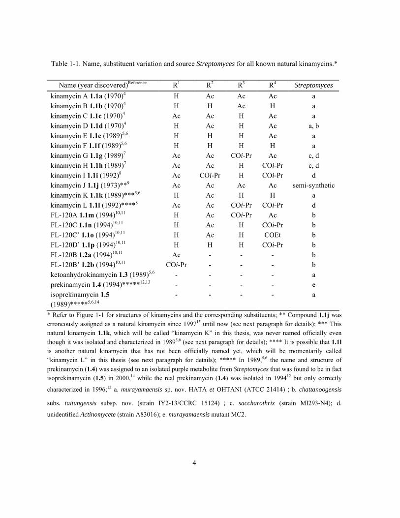
Table 1-1. Name, substituent variation and source Streptomyces for all known natural kinamycins.*
Name (year discovered)Reference R1 R2 R3 R4 Streptomyces
kinamycin A 1.1a (1970)4 H Ac Ac Ac a kinamycin B 1.1b (1970)4 H H Ac H a kinamycin C 1.1c (1970)4 Ac Ac H Ac a kinamycin D 1.1d (1970)4 H Ac H Ac a, b kinamycin E 1.1e (1989)5,6 H H H Ac a kinamycin F 1.1f (1989)5,6 H H H H a kinamycin G 1.1g (1989)7 Ac Ac COi-Pr Ac c, d kinamycin H 1.1h (1989)7 Ac Ac H COi-Pr c, d kinamycin I 1.1i (1992)8 Ac COi-Pr H COi-Pr d kinamycin J 1.1j (1973)**9 Ac Ac Ac Ac semi-synthetic kinamycin K 1.1k (1989)***5,6 H Ac H H a kinamycin L 1.1l (1992)****8 Ac Ac COi-Pr COi-Pr d FL-120A 1.1m (1994)10,11 H Ac COi-Pr Ac b FL-120C 1.1n (1994)10,11 H Ac H COi-Pr b FL-120C’ 1.1o (1994)10,11 H Ac H COEt b FL-120D’ 1.1p (1994)10,11 H H H COi-Pr b FL-120B 1.2a (1994)10,11 Ac - - - b FL-120B’ 1.2b (1994)10,11 COi-Pr - - - b ketoanhydrokinamycin 1.3 (1989)5,6 - - - - a prekinamycin 1.4 (1994)*****12,13 - - - - e isoprekinamycin 1.5 (1989)*****5,6,14
- - - - a
* Refer to Figure 1-1 for structures of kinamycins and the corresponding substituents; ** Compound 1.1j was erroneously assigned as a natural kinamycin since 199715 until now (see next paragraph for details); *** This natural kinamycin 1.1k, which will be called “kinamycin K” in this thesis, was never named officially even though it was isolated and characterized in 19895,6 (see next paragraph for details); **** It is possible that 1.1l is another natural kinamycin that has not been officially named yet, which will be momentarily called “kinamycin L” in this thesis (see next paragraph for details); ***** In 1989,5,6 the name and structure of prekinamycin (1.4) was assigned to an isolated purple metabolite from Streptomyces that was found to be in fact isoprekinamycin (1.5) in 2000,14 while the real prekinamycin (1.4) was isolated in 199412 but only correctly
characterized in 1996;13 a. murayamaensis sp. nov. HATA et OHTANI (ATCC 21414);b. chattanoogensis
subs. taitungensis subsp. nov. (strain IY2-13/CCRC 15124) ; c. saccharothrix (strain MI293-N4); d.
unidentified Actinomycete (strain A83016); e. murayamaensis mutant MC2.

5
It should be noted that there are several old but still on-going mistakes in the literature regarding
the names of natural kinamycins (footnotes of Table 1-1), which have never been pointed out and
corrected until the writing of this thesis. First, Gould officially named compound 1.1j as “kinamycin
J” in his very thorough and detailed review of kinamycins published in 1997,15 in which he referred to
the original work of Smitka and co-workers in 19928 as the literature source of this presumed
“natural” kinamycin. However, a more careful review of Smitka’s original paper8 indicates that no
kinamycin J (1.1j) was isolated or identified back then. In fact, the molecule having the shown
structure of kinamycin J (1.1j) was never found from nature (fermentation of bacteria), but it was first
generated upon acylation of natural kinamycin A (1.1a) by Ōmura in 1973 (Scheme 1-1),9 and more
recently synthesized by the Nicolaou group in 2007.16 Therefore, kinamycin J (1.1j) should be
considered as a (semi-)synthetic derivative of natural kinamycins. Second, Gould isolated and fully
characterized a unique natural kinamycin compound 1.1k in 1989,5,6 but interestingly, it was not even
mentioned in his own 1997 review of kinamycins.17 No one, including the discoverer, has ever
provided the natural 1.1k with an official kinamycin name, which will be called “kinamycin K” in
this thesis. The last problem was caused by the Smitka group who isolated three natural kinamycins
in 1992,8 among which two had been unambiguously identified as kinamycin G (1.1g) and kinamycin
I (1.1i). Smitka claimed8 the third kinamycin to be the previously discovered kinamycin H (1.1h),7
but the published chemical drawing for this “kinamycin H” was identical to the one shown in Table
1-1 for compound 1.1l, which matched neither the real structure of kinamycin H (1.1h) nor the
corresponding name of “3-O-isobutyrylkinamycin C” (common name for 1.1h) used in his own
paper.8 This inconsistency would suggest two possibilities. First, the mistake could be nothing but a
simple editorial error with the published chemical structure, although Smitka’s paper provided no
spectroscopic data of 1.1l to prove such. Recent (June 2008) personal communications with Dr.
Smitka regarding this issue unfortunately did not clarify the confusion either, due to the

6
inaccessibility of the original spectroscopic data at the moment. Therefore, a second possibility is that
1.1l might be another natural kinamycin that has not been officially recognized yet, but only if the
spectroscopic data of 1.1l are different from 1.1h and prove that the original chemical drawing of 1.1l
in Smitka’s paper is correct.
There are several distinct structural features of the molecules, among which the most notable one is
the existence of a very rare (for natural products) and unusually stable diazo functionality (>C=N+=N-
↔ >C--N+N). This C-diazo moiety was previously assigned as an N-cyano (>N-CN) group based
on limited spectroscopic data (including a low quality X-ray of 1.1s, Scheme 1-1) and misleading
chemical evidence at the time of the initial discovery,9,18,19 when the very first four members of the
kinamycin antibiotics, kinamycins A, B, C and D (1.1a-d), were isolated from the fermentation broth
of Streptomyces murayamaensis by Ōmura and Hata in 19704,20 and subsequently characterized in
1971.18 This error in the kinamycin structure was finally discovered 23 years later, owing to the
higher quality of X-ray evidence from the (+)--methylbutryrate of natural kinamycin D (1.1v in
Scheme 1-1) from the Gould lab21 and the simultaneous synthetic and spectroscopic work from the
Dmitrienko lab.22 The long-believed N-cyano structure for kinamycins was then revised to the current
C-diazo assignment (see section 1.3.2 for details). In addition, prekinamycin (1.4) and
isoprekinamycin (1.5) are indeed structural isomers with a difference in arrangement of their
carbocyclic skeletons, which were not distinguished until 2000 when another structural revision of
kinamycins (section 1.3.2) was suggested by the joint investigation of the Dmitrienko lab and the
Proteau lab.14
Structures of some simple semi-synthetic derivatives of natural kinamycins, which were made in
the process of their structure determination during the early stage of research in the area (section
1.3),5,9,18,21 are shown in Scheme 1-1. These semi-synthetic compounds were typically named as
derivatives of the corresponding mother kinamycin. It is interesting to note, in retrospect, that the
7
diazo group in these natural products survives the rather harsh (e.g., strong acidic) conditions engaged
in some of the derivatizations, since most diazo compounds are unstable under such conditions. Later,
some of these semi-synthetic derivatives were found to be natural kinamycins as well. For example,
deacetylkinamycin C (1.1f in Scheme 1-1), derived in 1973 from kinamycin C (1.1c) upon basic
hydrolysis,9 is identical to natural kinamycin F (1.1f in Figure 1-1) that was isolated as a bacterial
metabolite in 1989.5 In addition, a few more synthetic derivatives of kinamycins were prepared in
recent years when several total syntheses of the kinamycins were achieved (section 3.2).16,23-26

8
Scheme 1-1. Conversion of natural kinamycins (boxed) to some semi-synthetic derivatives.
Naturally-occurring diazo compounds are rare, and besides the kinamycins, there are only a
handful of other examples reported in the literature. As summarized in Figure 1-2, other known

9
natural diazo compounds include several modified α-amino acids (e.g., 6-diazo-5-oxo-L-norleucine
(DON, 1.6a),27,28 azaserine (1.6b),29-35 alazopeptin (1.6c)36 and its natural derivative OS-3256-B (no
given structure)37, duazomycin A (N-acetyl DON, 1.6d) and B (1.6e),38 N-(L-alanyl)azaserine (LL-
D05139β, 1.6f),39 O-[(3R)-2-diazo-3-hydroxybutyryl]-L-serine (thrazarine/FR900840, 1.6g)40-44,
cremeomycin (1.7),45,46 SQ30957 (1.8),47 lagunamycin (1.9),48,49 SF2415A1, A2, and A3 (1.10a–c)50,51
and lomaiviticin A (1.11a) and B (1.11b).52 All these naturally-occurring diazo compounds were
produced by certain strains of Streptomyces or Actinomycetes except 1.8, which was isolated from a
fungus, and in most cases they possess activity against various bacteria and tumor cells. The last two
natural diazo compounds mentioned above are particularly noteworthy. Lomaiviticin A (1.11a) and B
(1.11b) were isolated by He and collaborators from the fermentation broth of Micromonospora
lomaivitiensis (Actinomycete strain LL-37I366).52 In this case the bacterium was hosted by
Polysyncraton lithostrotum, a Fijian marine ascidian that is more commonly known as a sea squirt,
and these natural products were identified in an investigation that originally targeted the marine
invertebrates as a possible source of enediyne-type anticancer agents.52 Lomaiviticin A (1.11a) is a
dimeric glycoside analogue of the kinamycins while lomaiviticin B (1.11b) is the bis-hemiketal
formally derived from lomaiviticin A (1.11a). Both lomaiviticin A (1.11a) and B (1.11b) possess
extremely potent activities with an ultra low level of IC50 of about 0.01–98 ng/mL against a very
broad range of cancer cells and bacteria.52

10
O
XCOOHR
N
N
1.6a DON R = H; X = CH2; R' = H
1.6b azaserine R = H; X = O; R' = H
1.6c alazopeptin R = H; X = CH2; R' =
1.6d duazomycin A R= H; X = CH2; R' = Ac
1.6e duazomycin B R= H; X = CH2; R' =
1.6f LL-D05139 R = H; X = O; R' =
1.6g thrazarine R = ; X = O; R' = H
NHR'
O
N
CO2H
CH3ON
1.7 cremeomycin
O
NH2
CH3
H3C
OH
NH
O
N
O
O
O
1.9 lagunamycin
N
OH
OH
O
O
N O
O
O
O
O
R'
H
H
R
H
Et
EtO
R
H
R'
N
O
O
OH
OH
N
N
OH
OH
O
O
N O
O
O
R'
H
H
H
Et
EtO
H
R'
N
O
O
OH
OH
N
N
HO
OH
1.11a lomaiviticin A 1.11b lomaiviticin B
OH
O
N
N
O
O
Cl
OH
OH
O
N
N
O
O
O
OH
O
N
N
O
O
O
ClCl
1.10a SF2415A1 1.10b SF2415A2 1.10c SF2415A3
O
O
H
N2O
H2N
CH3
O
NH
O
H
N2O
HO2C
NH2
O
N
CH3O
N
1.8 SQ30957
OOH
NMe2
R' =
O OHOMe
R =
Figure 1-2. Other naturally-occurring diazo compounds besides kinamycins.
The second distinct structural feature of the kinamycins comes from their unusual skeletons
(Figure 1-3). All kinamycins possess a carbocyclic skeleton containing three 6-membered rings and

11
one 5-membered ring, among which a typical fluorene core 1.12 with a fused 6-5-6 ring arrangement
is common to all kinamycins. Depending on the relative orientation of the remaining 6-membered
ring, the kinamycin skeleton can be defined as either a benzo[a]fluorene core 1.13a or a
benzo[b]fluorene core 1.13b. The other possible isomeric benzo[c]fluorene core 1.13c has never been
found within the natural kinamycins. The four rings of the kinamycins are denoted as ring A, B, C
and D consecutively, with the A-ring bearing a single –OH group in all natural kinamycins. The
numbering system of the kinamycins, as suggested by Gould14,21 and shown in Figure 1-3, follows the
one employed for benzo[a]anthraquinone biosynthetic precursors53 and that of Chemical Abstracts.
All natural kinamycins except isoprekinamycin (1.5) possess the benzo[b]fluorene skeleton but with
various substituents and different degrees of oxidation of their D-rings. Most kinamycins (1.1a–p, 15
out of 20) contain a saturated D-ring bearing four acyloxy and/or hydroxy substituents and three other
kinamycins (1.2a, 1.2b and 1.3) have an epoxide within the saturated D-ring. In the case of
prekinamycin (1.4), a fully aromatized D-ring is present. On the other hand, isoprekinamycin (1.5), a
structural isomer of prekinamycin (1.4), is the only member of the kinamycin family that possesses
the benzo[a]fluorene skeleton. Both the benzo[b]- and benzo[a]fluorene cores are very rare structures
that occur within natural products, and only a few other examples are reported in the literature for
each case (Figure 1-3), which includes momofulvenone A (1.14a) and B (1.14b)54, stealthin A (1.15a),
B (1.15b)55 and C (1.15c)17, kinobscurinone (1.16a)56 and kinafluorenone (1.16b)57,58, seongomycin
(1.17),59 cysfluoretin (1.18),60 shikometabolin A (1.19a) and B (1.19b),61,62 fluostatin A (1.20a), B
(1.20b),63 C (1.14c), D (1.14d) and E (1.14e).64,65 Not only are all of these natural products closely
related to the kinamycins in structure, but also they (except 1.19a–b) are produced exclusively by
certain species of Streptomyces. Among them, 1.15c and 1.16a were found to be key intermediates in
the biosynthesis of kinamycins.

12
O
OH
R1O
OR3
O
OR4
Me
OR2
N2
1
5
7
113
10 2
4
68
9
A B CD11a
11b
5a
4a
6a
10a
HO
O OH
Me
N2O
1
5
7
113
102
46
8
9
A BC
D11a
11b
6a4a
6b
10a
1.13a benzo[a]fluorene
1.13b benzo[b]fluorene
1.13c benzo[c]fluorene
1.12 9H-fluorene
OH
M+O-
O
O
CH3
OH
RO OH
M = Na, K, Mg, Ca, H1.14a momofulvenone A
(R = H)1.14b momofulvenone B
(R = Ac)
OH
O O
O
CH3
HO
1.20a fluostatin A
OH
O HO
O
CH3
HO
OH
OH
O RO
O
CH3
HO
O
OH
O HO
O
CH3
HO
OH
Cl
OH
OH
O
HO
NH2
1.15a stealthin A(R = CH2OH)
1.15b stealthin B(R = CHO)
1.15c stealthin C(R = CH3 )
R
OR1
O
O
R2O
O
CH3
OH
OH
O
HO
S
CH3
NHAcHO2C
O
OMe OH
OH
OMe
SCO2H
AcHN
OH
OH
OH
O
O
O
HO
OH
HOH
1.18 cysfluoretin1.19a shikometabolin A
1.17 seongomycin
1.16a kinobscurinone(R1 = R2 = H)
1.16b kinafluorenone(R1 = CH3, R2 = H)
1.20c fluostatin C (R = H)1.20d fluostatin D (R = COiPr)
1.20b fluostatin B 1.20e fluostatin E
OH
OH
OH
OO
O
OH
HO
HOH
1.19b shikometabolin B
Figure 1-3. Natural products (besides kinamycins) possessing benzo[b]- and benzo[a]fluorene
skeletons.*
* 1.16a/b are shown in their oxidized quinone forms, but spectroscopic evidence suggested that they may actually co-exist in the
corresponding hydroquinone or semiquinone radical forms as well. (57) Cone, M. C.; Melville, C. R.; Gore, M. P.; Gould, S. J. J. Org. Chem. 1993, 58, 1058-1061. (66) Gould, S. J.; Melville, C. R. Tetrahedron Lett. 1997, 38, 1473.

13
The significant bioactivity of the kinamycins, along with their unique structures, has made these
antibiotics the focus of multi-disciplinary research. Since the initial discovery of kinamycins in 1970,
many laboratories worldwide, including this research (Dmitrienko) group, have devoted much effort
to various aspects of kinamycins over the past few decades, from isolation, structure characterization,
and mechanism-of-action studies, to total synthesis.
1.3 Isolation, Characterization and Biosynthesis of the Kinamycins
In 1970, Ōmura, Hata, Ohtani and co-workers obtained a strain of Streptomyces from a soil sample
collected in Murayama (Saitama-ken, Japan),4 a small town situated in the north of Tokyo
Metropolis’ central area that became the city of Musashimurayama in the same year. Therefore, the
bacteria strain was named as Streptomyces murayamaensis sp. nov. HATA et OHTANI, which was
found to be capable of producing some orange crystalline antibiotics that were later identified and
named as kinamycin A, B, C and D (1.1a–d).4 Not long after its discovery, Streptomyces
murayamaensis was patented and samples were deposited to the American Type Culture Collection
(ATCC, Manassas, VA, USA; www.atcc.org) by Kyowa Fermentation Industrial Co., Ltd. under the
registration number of ATCC 21414, which was then made available to the general scientific public.
Until early 1989, Streptomyces murayamaensis (ATCC 21414) was the only bacterium known to
produce the kinamycins. Later, the discovery of various kinamycins from Streptomyces saccharothrix
(strain MI293-N4, 1989),7 an unidentified actinomycete (strain A80316, 1992)8 and Streptomyces
chattanoogensis subsp. taitungensis (strain IY2-13/CCRC 15124, 1994)11 significantly enlarged the
source pool for the natural kinamycins. Although each Streptomyces mentioned above would produce
unique natural kinamycins individually, some kinamycins were isolated and identified from more
than one Streptomyces source. For example, kinamycin D (1.1d), initially obtained from Streptomyces

14
murayamaensis,4 was also isolated much later from Streptomyces chattanoogensis subsp.
Taitungensis.11
1.3.1 Production and Isolation of Natural Kinamycins
The initially discovered Streptomyces murayamaensis4 (ATCC 21414) is the most frequently used
bacterium to produce the valuable natural kinamycins through fermentation, which needs to be first
grown on appropriate agar media prior to further use.6 Fermentation of the Streptomyces
murayamaensis is a rather complicated and very sensitive biological process, and the outcome is
affected significantly by even small variations in experimental conditions (constituents of media and
the conditions of cultivation), which were examined extensively by the Hata/Ōmura team20,67 and the
Gould group.6 Organic extraction using benzene or toluene of the treated (acidified, concentrated,
centrifuged and sonicated) fermentation broth followed by repetitive chromatographic separation
would give the desired kinamycins usually in very low yields, along with isolation of other
structurally and biosynthetically related metabolites such as dehydrorabelomycin (1.21) and
murayaquinone (1.22).6
O
OOH OH
HO Me
1.21 dehydroabelomycin
OH O
OH
O
O
1.22 murayaquinone
Streptomyces capable of producing kinamycins have not been genetically engineered and
optimized for such purpose, even though the genes in Streptomyces murayamaensis that are
responsible for producing kinamycins were already identified, cloned and heterologously expressed
by the Gould group in 1998,58 and some similar genetic work was patented later on by the Diversa Co.
15
(CA, USA) in 2003.68 In both studies, however, the gene(s) responsible for the formation of the
kinamycin’s diazo group were neither identified nor expressed.
1.3.2 Characterization and Structure Elucidation of the Kinamycins
1.3.2.1 Structural Determination and Revision of Kinamycins Containing the
Benzo[b]fluorene-type Skeleton
After the isolation of kinamycin A, B, C and D (1.1a–d), Ōmura and co-workers carried out
extensive spectroscopic characterization of these compounds by means of 1H-NMR, mass
spectrometry (MS), infrared (IR), UV-vis spectrometry and chemical derivatization and degradation.
The bulk of the tetracyclic skeleton of kinamycins and major substituents and functional groups were
assigned by following the common spectroscopic rules.9,18 However, a linear triatomic moiety within
the kinamycins containing two nitrogen and one carbon atoms (X-Y-Z, Figure 1-4), which has a
corresponding strong IR absorption band at 2155 cm-1 (common to 1.1a–d), remained mysterious.
There are only three possible arrangements for this triatomic moiety, i.e. a C-diazo (>C=N+=N- ↔
>C--N+N), an N-cyano (>N-CN) or an N-isonitrile (>N-N+C-). The possibility of a C-diazo group
was never considered by Ōmura, likely because of the perceived instability of this functional group
and the very few examples of diazo-containing natural products known at the time (1.6a–d, early
1970s). Qualitative chemical tests were used to distinguish between the N-cyano and the N-isonitrile
groups due to the lack of further spectroscopic evidence. It was found that hydrolysis of kinamycin C
(1.1c)18 and deacetylkinamycin C (1.1f, kinamycin F)9 in refluxing 30% aqueous KOH liberated
ammonia but not formic acid,18 and reaction of deacetylkinamycin C (1.1f, kinamycin F) under acidic
conditions (refluxing 10% HCl-MeOH)9 led to the apparent detection of ammonia but no formic acid.
Since the cyano group would produce ammonia but not formic acid under either acidic or basic
hydrolysis, and the isonitrile group would lead to formic acid but not ammonia either way, such

16
chemical observation would “suggest and confirm” the presence of an N-cyano group rather than an
N-isonitrile group, when only considering these two options. Apparently this incorrect N-cyano
structure of kinamycin also gave a modest fit to the X-ray crystallographic data (R = 8.9%) in the case
of the p-bromobenzoate of kinamycin C (1.1s in Scheme 1-1), which established not only the absolute
stereochemistry of the D-ring but also confirmed the relative orientation between the A-ring and D-
ring.19 However, the instrumental limitation of NMR spectroscopy in the early 1970s (i.e., most low
field CW(continuous wave)-NMR instruments at the time were not capable of acquiring 13C spectra)
led to the lack of 13C-NMR spectra of the kinamycins, which may have prevented assigning the
incorrect N-cyano structure if such data had been available. Another possible contributing factor to
this wrong structural assignment is likely the lack of spectroscopic data of other N-cyano compounds
to compare with at the time when kinamycins were discovered. This proposed N-
cyanobenzo[b]carbazole structure of kinamycins could reasonably explain most of the observed
spectroscopic data and chemical behavior, therefore, it was believed to be correct for a long time until
later studies discovered more conflicting and inconsistent observations in regards to this N-cyano
structure.
X
O
OH Y
AcO
OH
O
OAc
CH3
OAc
Z
X Y Z
N C N
C N N
N N C
1.1c kinamycin C
N-cyano
C-diazo
N-isonitrile
Figure 1-4. Possible arrangements of the triatomic moiety within kinamycins.
In the mid 1980s when Gould initiated biosynthetic studies on the kinamycins, he was able to
observe and assign 13C NMR resonances for all of the carbons of kinamycin D (1.1d) except the
cyanamide carbon, which was expected to have a chemical shift of ca. 110–120 ppm.69,70 Fortunately,

17
when Gould fed the Streptomyces with 15N-labeled ammonium sulfate as the sole nitrogen source, a
doubly-labeled [15N2]kinamycin D (1.1d) was produced. For the first time, this [15N2]kinamycin D
enabled Gould to locate the “missing” NMR signal of the “cyanamide carbon” of kinamycins, which
appeared as a doublet of doublets (dd, J = 21.2 Hz and 5.4 Hz) at chemical shift of 78.5 ppm due to
the coupling between 13C and the two adjacent 15N atoms.71 Further reexamination of previously
recorded spectra of non-isotope-labeled natural kinamycin D (1.1d) revealed the presence of a very
weak singlet at 78.5 ppm that was barely noticeable due to the nearby very strong solvent peak of
CDCl3 at 77 ppm and originally thought to be caused by impurities.71 This newly discovered carbon
signal at 78.5 ppm was then assigned to the “N-cyano carbon” despite its significant deviation from
the expected chemical shift of 110–120 ppm. This dramatic difference of about 30 ppm was attributed
to possible electronic effects of the indolequinone (within the original/incorrect structure of
benzo[b]carbazole skeleton of kinamycins) on the cyanamide carbon.71 However, literature data
indicate that the 13C atoms attached to diazo groups in some diazo compounds have chemical shifts in
the 60–80 ppm region,72 which matches the observed 13C chemical shift of the “N-cyano carbon” of
kinamycin D (78.5 ppm) much more closely than the 13C chemical shifts of the N-cyano compounds.
Such findings would suggest a necessary reconsideration of the previously ignored possibility of
having a diazo functional group rather than the long-believed N-cyano moiety within the kinamycins.
In the meantime (early to mid-1990s), other research groups had been working towards the total
synthesis of kinamycins based on the incorrect benzo[b]carbazole skeleton, which led to the
preparation of N-cyanoindoles (e.g., compound 1.23 and 1.24 in Scheme 1-2, made by the Dmitrienko
lab) as well as other advanced intermediates containing both the N-cyano group and the
benzo[b]carbazole skeleton.73,74 The 13C NMR chemical shifts of ca. 105–112 ppm for such N-cyano
carbons match very well with the expected values, but poorly with the observed corresponding signal
for kinamycins. In addition, such synthetic N-cyano compounds all have a characteristic IR band in

18
the region of ca. 2240–2250 cm-1 for their N-cyano moiety, while the IR band of the “N-cyano” in
kinamycins appeared at ca. 2155 cm-1 (a difference of ca. 100 cm-1). Among the available examples
of N-cyano compounds, two literature reports are worthy of some additional comments. One work is
the first synthetic route (Scheme 1-2a) towards the benzo[b]carbazole of prekinamycin (1.4), which
was reported by Echavarren’s group briefly in 199374 followed by a more detailed disclosure in
1997.75 The other work was carried out by Mithani from the Dmitrienko lab, who reported another
total synthesis of N-cyanobenzo[b]carbazole-based prekinamycin derivative 1.30 in 1994 (Scheme 1-
2b).22 Detailed spectroscopic analysis and comparison of 1.30 and a variety of other model N-cyano
compounds with natural kinamycins clearly ruled out the possibility of kinamycin possessing the N-
cyano group, and instead, a diazobenzo[b]fluorene quinone skeleton was proposed by the Dmitrienko
lab as the final answer to the confusing structure of the kinamycins. Independent and simultaneous
results, from a very careful and rigorous X-ray crystallographic study on the (+)--methylbutyrate of
natural kinamycin D (1.1v in Scheme 1-1) performed by Gould’s group, also led to the same
conclusion.21 Fitting of X-ray data of 1.1v provided significantly better statistical R and Rw factors
when the C-diazo group (R = 5.18%, Rw = 5.63%) is used for the triatomic linear structure rather than
the other two possibilities, i.e. N-cyano group (R = 5.69%, Rw = 6.35%) and N-isonitrile group (R =
5.73%, Rw = 6.41%). The quality of Gould’s X-ray data is superior when compared with Ōmura’s X-
ray result of the kinamycin C derivative 1.1s that has a very large R of 8.9%, which made the
corresponding conclusion more convincing and trustworthy.

19
MeO Me
NHBoc
O
OMeOMe3Sn
Br
1.25 1.26
RO
Me
N
O
ORO CN
1.4 (R = H); 1.27 (R = Me)
(a)
OAc
Me
N
O
OOMeOMe
NH2
O
HO Me
1.301.28 1.29
CN
(b)
105.0 ppm
O
O
NCN
Me
Me
O
O
NCN
OAc
OAc
1.23
104.7 ppm 103.6 ppm
1.24
N
O
OH C
HO
OH
O
OAc
CH3
OAc
N
1.1d (original structure)78.5 ppm
Scheme 1-2. NMR difference between the “N-cyano carbon” of kinamycin D (1.1d) and other N-
cyano species and synthesis of the N-cyanobenzo[b]carbazoles by (a) Echavarren and (b) Dmitrienko.
This structure revision of kinamycins in 1994, from N-cyanobenzo[b]carbazoles to
diazobenzo[b]fluorenes, has had a profound influence on many aspects of the chemistry and
biochemistry of the kinamycin antibiotics. Not only did the synthetic strategies to kinamycins have to
be redirected towards the very rare benzo[b]fluorene skeleton as well as the unique and potentially
reactive diazo group, but also the previous knowledge and explanation of the biosynthesis and
mechanism (mode-of-action) related to kinamycin’s biological activities required reexamination.
20
1.3.2.2 Structural Determination and Revision of Isoprekinamycin - Kinamycin
with a Benzo[a]fluorene-type Skeleton
In 1989, Gould’s group isolated a purple solid metabolite, designated as compound “P1” in the
original papers, from the bacterial fermentation broth of Streptomyces murayamaensis.5,6 The
corresponding name and structure of prekinamycin (1.4 with the original N-cyanobenzo[b]carbazole
skeleton) was then assigned to compound “P1” based on the spectroscopic data obtained. Evidence
available at that time would suggest this so-called “prekinamycin” has the common N-
cyanobenzo[b]carbazole skeleton but with an aromatized D-ring, which might be converted
biosynthetically in a stepwise fashion into other kinamycins bearing the saturated D-ring with the
necessary substituents.5
After the structural revision of kinamycins in 1994, the structure of this “prekinamycin”
corresponding to compound “P1” was also updated accordingly (1.4 changed from the incorrect N-
cyanobenzo[b]carbazole to the correct diazobenzo[b]fluorene). This compound “P1” (isolated in 1989)
was also known as “Cpd A” within Gould’s group,13 and its structure was not challenged until 1996,
when Hauser and Zhou achieved the first total synthesis (section 1.5) of a product having the exact
structure as the proposed prekinamycin (1.4).76 The spectroscopic data of Hauser’s synthetic
prekinamycin, however, did not match the already reported literature values for the presumed natural
“prekinamycin”. Gould and co-workers then found that Hauser’s synthetic prekinamycin indeed
matched perfectly in all spectroscopic methods used (1H-NMR, UV-vis and HPLC) with another
metabolite, which had been previously observed from the extracts of Streptomyces murayamaensis
mutant MC2 and designated as “Cpd B” in Gould’s group but never characterized structurally.12 As a
result of this discovery, “Cpd A” lost the proposed name and structural assignment of prekinamycin
(1.4) to “Cpd B”. The true identity of “Cpd A” remained unclear until four years later, when joint
studies by the Dmitrienko group and the Proteau group finally characterized the mysterious “Cpd A”

21
(compound “P1”) as a structural isomer of prekinamycin (1.4, “Cpd B”). “Cpd A” was found to
possess the rare diazobenzo[a]fluorene skeleton rather than the relatively common
diazobenzo[b]fluorene skeleton among other kinamycins, therefore, “Cpd A” was named as
isoprekinamycin (1.5, Scheme 1-1) afterwards.14
1.3.3 Biosynthesis of the Kinamycins
Experiments to explore the biosynthetic pathway to the kinamycins were carried out from the mid
1970s to the late 1990s. Initially, the Ōmura group used IR to suggest the origin of the “cyanamide
carbon” of kinamycins. Later, Gould’s group used extensive isotope labeling of precursors and NMR
analysis to establish the biosynthetic pathways of kinamycins. Most studies on kinamycin
biosynthesis were accomplished before the structural revision of kinamycins in 1994, which resulted
in a reevaluation of the known biosynthetic mechanisms at the time, particularly in terms of
intermediate metabolites involved. It was found that the new diazobenzo[b]fluorene structure for
kinamycins was perfectly consistent with all the known results of biosynthetic studies, which were
previously explained using the old and incorrect structure of N-cyanobenzo[b]carbazole. The
corresponding results, reviewed thoroughly by Gould in 199715 and 1998,58 now provide quite deep
understanding in this regard.
1.3.3.1 Biosynthetic Origins of the Kinamycin Skeleton and D-ring Substituents
The biosynthetic origins of the kinamycins were determined by Gould and co-workers upon
feeding Streptomyces murayamaensis with various isotope-labeled sodium acetates and metabolite
intermediates (Scheme 1-3), and the derived isotope-containing kinamycin C (1.1c) and D (1.1d)
were subjected to NMR spectroscopic analysis to locate the distribution of isotopic atoms.5,53,70,71,77
Several fundamental conclusions were made based on these results (Scheme 1-3). The observed 13C

22
enrichment (Scheme 1-3a/b/c) indicated that the entire skeleton of kinamycin was derived from
acetate through a polyketide pathway and the origins of all carbons except the “cyanamide carbon”
were identified. In addition, oxygen at C-1, C-6 and C-7 was derived from the acetate, oxygen at C-3,
C-4 and C-11 came from the air (O2) and the C-2 oxygen originated from water (Scheme 1-3c/d). In
addition, the previously isolated dehydrorabelomycin (1.21) was confirmed to be a key biosynthetic
intermediate for kinamycins (Scheme 1-3e/f).
X
O
OH
RO
O
CH3
OH
O
O
CH3 CO2Na
CD3 CO2Na
X
O
OH
HO
O
CD2H
OH
OCOCD2H
OCOCD2H
D
DY Y
Z Z
= 13C labeled
1.1c kinamycin C (R = Ac)1.1d kinamycin D (R = H)
X Y Z
N C N
C N N
Streptomycesmurayamaensis
H3C CO2Na + 18O2
X
18O
OH
HO
O
CH3
18OH
OAc
18OAc
2
43
11
YZ
H3C C18O2Na
X
O
18OH
R18O
18O
CH3
OH
OCCH3
OCCH3
YZ
18O
18O
65
HO CH3O
OOH OH
1.21 dehydrorabelomycin
HOCH3
O
O
X
Y
OCOCH3
OH
OCOCH3
OH
H
CH3 CO2Na
1
5
32
4
Z
(original structure)
(revised structure)
1.1d kinamycin D
1.1d kinamycin D
1.1c kinamycin C (R =1.1d kinamycin D (R =H)
CCH3)
18O
1.1d kinamycin D
(a)(b)
(c)
(d)(e)
1
67
HO CH3O
OOH OH
1.21 (D-labeled)
D
D
DD
D
X
O
OH
HO
O
CH3
OH
OAc
OAc
D
DYZ
(f)
1.1d kinamycin D
D
2
CH3
O
O
CH3
Scheme 1-3. Biosynthetic origins of carbon, oxygen and hydrogen atoms of kinamycins.

23
To tackle the biosynthetic origin of the “cyanamide carbon” (diazo carbon) of kinamycins, initial
work was performed by Ōmura in 1976, who fed CH313COONa and 13CH3COONa to Streptomyces
murayamaensis, and the derived kinamycin D (1.1d) were analyzed by IR spectroscopy.78 An isotope-
induced shift from 2155 to 2139 cm-1 of the IR peak, corresponding to the stretching frequency of the
13C-N triple bond within the N-cyano group (now assigned to the diazo stretching), was only observed
when CH313COONa was used but not in the case of 13CH3COONa, suggesting the “cyanamide
carbon” (>N-CN, now known as the diazo carbon >C--N+N) was derived from C-1 (carbonyl
carbon) of the acetate.* Later, Gould and co-workers directly observed the “cyanamide carbon” within
13C-labeled kinamycin D (1.1d) that were also produced by feeding bacteria with either CH313COONa
or 13CH3COONa.71 However, the more convincing NMR evidence suggested the other alternative, i.e.
the “cyanamide carbon” of kinamycins should be derived from C-2 (methyl carbon) of the acetate
instead, since 13C enrichment of the “cyanamide carbon” was only observed by NMR with kinamycin
D (1.1d) derived from 13CH3COONa but not CH313COONa.71 Since the observed level of the isotope
enrichment for this “cyanamide carbon” was similar to other carbons of the kinamycin skeleton, it
was further proposed that the “cyanamide carbon” was derived from C-5 of dehydrorabelomycin
(1.22) (Scheme 1-3e).71
Results of the above labeling studies also led to a plausible proposal for the oxidative elaboration
of the D-ring of kinamycins as given by Scheme 1-4a, in which the lack of deuterium labeling at C-4
of the non-aromatic D-ring of kinamycin D (Scheme1-3b) would indicate first a hydroxylation
occurred at C-4 of the precursor with an aromatic D-ring, and produced the hydroquinone 1.31
followed by oxidation to the epoxide 1.32. The epoxide may then undergo stereospecific
tautomerization to epoxyquinol 1.33 or 1.34, and a stereospecific reduction of either 1.33 or 1.34
* Preliminary computational results from this work indicated that this presumed “isotope-induced” diazo IR shift as observed by Ōmura
must be caused by other reasons but not the claimed one.

24
would lead to the epoxy diol 1.35. Finally, trans attack of water on the epoxy group of 1.35 at the less
hindered C-2 position shall yield kinamycin with a fully oxygenated D-ring structure 1.36 having the
correct stereochemistry and with each atom derived from the correct source as found by the labeling
studies (Scheme 1-3).77 This D-ring evolution mechanism was further supported by the later (1989–
1994) isolation of minor metabolites such as ketoanhydrokinamycin (1.3b), prekinamycin (1.4),
kinamycin F (1.1f) and kinamycin E (1.1e) from Streptomyces murayamaensis and its mutant
(Scheme 1-4b).5,6,12 In addition, Gould’s group isolated an apparently large enzyme (MW > 669 kDa)
from Streptomyces murayamaensis in 1996, which was responsible for two consecutive acetylations
of kinamycin F (1.1f), first at C-4 leading to kinamycin E (1.1e) and then at C-2 leading to kinamycin
D (1.1d). Therefore, the enzyme was named as kinamycin acetyltransferase I (KAT I),79 but at least
two more KATs were required by the minimum pathway to account for the remaining three other
acetylation patterns on the kinamycin D-ring.15 Furthermore, the isolation of stealthin C (1.15c) in
199717 as a minor metabolite from Streptomyces murayamaensis mutant MC212 by Gould’s group,
has provided deeper insight into the biosynthesis of kinamycins. Feeding of deuterium-labeled
stealthin C (1.15c) to Streptomyces murayamaensis (not the mutant MC2 from which 1.15c was
produced) led to the corresponding deuterium-labeled kinamycin D (1.1d), which confirmed the role
of stealthin C (1.15c) as a critical intermediate in kinamycin biosynthesis (Scheme 1-4c).
Interestingly, feeding of synthetic compounds such as 1.37 that were essentially the same as 1.15c
except the difference in nitrogen substituents, led to no production of labeled kinamycin D (1.1d)
(Scheme 1-4c).17

25
HO
Me
HO
Me[18O]
18OH
HO
Me
18OH
O18
[H]HO
Me
18OH
O18HOMe
18OH
18OH
H2O*
*OH
HO
Me
18O
O18
X
O
OOH Y
Z
4
2
[18O]
O
Me
18OH
O18
(a)
(b)
OH O
OMe
HO
1.4 prekinamycin
OH O
OMe
HOO
O
O
OH
HO
OH
O
OH
Me
OH
O
OH
HO
OH
O
OAc
Me
OAc
N2
1.1d kinamycin D
1.3 ketoanhydrokinamycin 1.1f kinamycin F
AcSCoA
KAT I
N2 N2 N2
O
OH
HO
OH
O
OAc
Me
OH
N2
1.1e kinamycin E
AcSCoA
KAT I
1.31 1.32 1.33
1.341.351.36
24
X Y Z
N C N
C N N
1.4
HO
Me
HO
O NH2
D
D
Streptomycesmurayamaensis
HO
Me
O
O N2OH
D
D OH
OAc
OAc
HO
MeO
O NOHOH
D
DHO
MeOH
O NOOH
D
D
1.37a
X
1.37b 1.15c stealthin C
Na2S2O4
1.1d kinamycin D
Streptomycesmurayamaensis
(c)
Scheme 1-4. (a) Proposed mechanism for the oxidative elaboration of kinamycin D-ring; (b) stepwise
biosynthetic transformation sequence between kinamycins; and (c) experiments confirming
stealthin C (1.15c) as a biosynthetic intermediate for kinamycins.

26
Later on, the understanding of kinamycin biosynthesis on the level of molecular biology was
greatly promoted, after Gould’s group successfully cloned the genomic DNA (genes) of Streptomyces
murayamaensis and heterogeneously expressing the genes when the DNA was transferred into
Streptomyces lividinas ZX7.58 The genetic studies identified two clusters of polyketide synthase (PKS)
genes, among which only one was responsible for producing the kinamycins through a decaketide
intermediate 1.38a, while the other was likely responsible for producing murayaquinone (1.22), a
metabolite previously isolated from Streptomyces murayamaensis, through a different (but unknown)
decaketide 1.38b pathway (Scheme 1-5). Expression of the particular kinamycin gene cluster in
Streptomyces lividinas ZX7, which was not complete but sufficient to biogenetically synthesize the
benzo[b]fluorene skeleton of kinamycins, smoothly led to accumulation and identification of several
known metabolites/intermediates of kinamycin biosynthesis such as dehydrorabelomycin (1.21),
kinobscurinone (1.16a) and stealthin C (1.15c), as well as two shunt metabolites, namely
kinafluorenone (1.16b) and seongomycin (1.17) (Scheme 1-5).58 On the other hand, isolation of
momofulvenone A (1.14a) and B (1.14b) from Streptomyces diastatochromogenes (strain Tü 2895)
and the corresponding 13C labeling experiments in 1995 indicated that these two species, whose
counter ions vary depending on the salts used in the growing medium, were also involved in the same
biosynthetic sequence as the kinamycins (Scheme 1-5).54 All these biosynthetic studies have now led
to a hypothetical and overall biosynthetic mechanism for kinamycins with the benzo[b]fluorene
skeleton, as shown in Scheme 1-5.

27
= 13C label
1.15c stealthin C
1.4 prekinamycin
HOCH3
O
O N2
O
OH
O
1.1d kinamycin D1.3 ketoanhydrokinamycin
OH
HO CH3O
OOH OH OH
OHCH3HO
CHOCO2H
O O O
O O
CO2H
O
O
O
O
1.38a decaketide
H
CH3
O
CH3
O
1.21 dehydrorabelomycin
OH
O
OHCH3
HO
OHO
OOH
H
[O]
O
OCH3
HO
OH O
[O]
O
OHCH3
HO
OH NH2
O
OCH3
HO
OH N2
[O]
O
O
CH3
HO
OH N2
O
O[H]
56
1.16a kinobscurinone
CH3 CO2Na OH O
OH
O
O
1.22 murayaquinone
Str eptomyces 1.38b decaketide(different from 1.38a)
1.39
1.40
- CO2
1.16b kinafluorenone
O
OCH3
HO
CH3O O
OH
OH
O
HO
S
CH3
NHAcHO2C
1.17 seongomycin
OH
M+O-
O
O
CH3
OH
RO OH
M = Na, K, Mg, Ca, H1.14a momofulvenone A (R = H)1.14b momofulvenone B (R = Ac)
then[H]
SCoA
O
Scheme 1-5. Overall biosynthetic pathway for benzo[b]fluorene-type kinamycins.
The dehydrorabelomycin (1.21), derived from the polyketide precursor 1.38a upon aromatization
and decarboxylation, is the first key intermediate of the entire biosequence, which then undergoes an

28
oxidative ring opening at C-5 and C-6 followed by reduction of the quinone moiety leading to 1.39.
An electrophilic cyclization of 1.39 would afford structure 1.40 containing a benzo[b]fluorene
skeleton for the first time. The dihydroquinone obtained from 1.40 upon decarboxylation (not shown
in the scheme) would eventually lead to momofulvenones (1.14a–b) in Streptomyces
diastatochromogenes, while in the case of Streptomyces murayamaensis this dihydroquinone would
be further oxidized to kinobscurinone (1.16a), the second key intermediate of the kinamycin
biosynthesis, which could be derivatized to other (shunt) metabolites such as kinafluorenone (1.16b),
seongomycin (1.17) and most importantly stealthin C (1.15c), which now has an essential and critical
amino group at C-5. The latter then may undergo an oxidative incorporation of the second nitrogen
atom and produce prekinamycin (1.4), followed by stepwise oxidation, reduction and acetylation to
alter the kinamycin D-ring. However, the genes and corresponding products responsible for the final
step(s) in formation of the diazo group have not been identified.
Finally, co-production of both benzo[a]fluorene and benzo[b]fluorene types of kinamycins and
their comparable stability (ab initio calculation predicted that 1.4 is more stable than 1.5 by ca. 2.4
kcal/mol) had led Dmitrienko and Proteau to propose a hypothesis of reversible interconversion
between these two types of ring skeletons through a possible enzyme-catalyzed rearrangement
(Scheme 1-6a).14 Such a proposal was consistent with some unpublished isotopic labeling results
from Gould’s group of isoprekinamycin (1.5) (Scheme 1-6b), when Streptomyces murayamaensis was
fed with 13CH313COONa (Scheme 1-6b).80

29
O
OHO
Me
HO
N2
1.5 isoprekinamycin
O
OH O
Me
HO
1.4 prekinamycin
CH3 CO2Na
N2
Str eptomycesmur ayamaensis = 13C label
OH O
O
Me
N2
HO
O
O
N2
H
OHHO
O
N2
H
O
O
OOH
Me
HO
N2
Me
HO
Me
HO
OHOH
1.4 prekinamycin
1.41 1.42
1.5 isoprekinamycin
H2O
+ H2O - H2O
(a)
(b)
Scheme 1-6. (a) Proposed reversible interconversion between diazobenzo[b]fluorene and diazo-
benzo[a]fluorene skeletons and (b) isotope labeling results of benzo[a]fluorene-type kinamycin.
1.3.3.2 Biosynthetic Origins of the C-Diazo Moiety of Kinamycins
Although the bulk of the kinamycin biosynthetic mechanism as introduced in section 1.3.3.1 is
quite well understood by now, there are still some unclear steps and conversions within this particular
biosynthesis that deserve more detailed consideration and efforts, among which the most intriguing
ones are the biosynthetic steps leading to the formation of the rare diazo group at C-5 of the
kinamycins.
Early isotope labeling experiments carried out in Gould’s lab in the late 1980s, by using
(15NH4)2SO4 as the sole nitrogen source for Streptomyces murayamaensis, indicated that both nitrogen

30
atoms of the diazo group (believed to be the N-cyano group at that time) within kinamycins were
incorporated with the 15N isotope.71 This observation suggested a stepwise incorporation of the two
nitrogen atoms into the kinamycin structure, which is consistent with the later identification of the
key biosynthetic intermediate stealthin C (1.15c), whose C-5 amino group was eventually
transformed into the corresponding diazo group of kinamycins.
It is still not clear how the biosynthetic transformation from kinobscurinone (1.16a) to stealthin C
(1.15c) (Scheme 1-5) is performed by the bacteria, but the cloned DNA from Streptomyces
murayamaensis by Gould does contain the gene(s) responsible for introducing the first nitrogen of the
eventual diazo group of the kinamycins.58 Without identification of any possible amination substrates
and intermediates, Gould proposed a likely but unproven hypothesis that kinobscurinone (1.16a)
might exist in the corresponding quinone form 1.16a, semi-quinone form (not shown) and
hydroquinone form 1.46 at ambient temperature due to the rather small redox potential differences
between such structures. In the presence of appropriate and necessary enzymes such as pyridoxal
phosphate-dependent transaminase or glutamine-/ammonia-dependent transaminase among others,
1.16a and its various redox forms may be converted biosynthetically to stealthin C (1.15c) through
several possible pathways (Scheme 1-7).58

31
HO
MeOH
OH OOH
1.16a kinobscurinone
HO
MeOH
OH NHOH
HO
MeOH
OH NCH2ArOH
1.45
HO
MeOH
O NH2OH
HO
MeO
O OOH
HO
MeO
O NHOH
HO
MeO
O NCH2ArOH
Glutamine/NH3
Amidotransaminase
TautomeraseOxidase
HydrolaseTautomerase
[1,2]reductasetautomerase
or [1,6]reductase
1.43
1.15c stealthin C
1.46 1.47
TautomeraseHydrolase
NH
HO
Me
NH2
OPO32-
1.44
(PMP)
Transaminase
Glutamine/NH3
Amidotransaminase
NH
HO
Me
NH2
OPO32-
(PMP)
Transaminase
Ar =NH
HO
Me
OPO32-
Scheme 1-7. Proposed biosynthetic conversions from kinobscurinone (1.16a) to stealthin C (1.15c).
Unfortunately, there are no experimental results observed so far that are related to the biosynthetic
pathway leading to the introduction of the second nitrogen within the diazo moiety of kinamycins.
Moreover, current knowledge about the biosynthetic formation of N-N bonds in nature is very limited
despite the relatively wide existence of such functionalities.81 There are a few examples in the
literature indicating that the formation of natural N-N bonds could involve hydrazine intermediates,81
and enzymes capable of oxidizing the arylhydrazine to aryldiazonium salts are known as well.82,83
Therefore, in principle, the potential existence of a hydrazine intermediate in the kinamycin
biosynthesis is possible but yet to be confirmed. Two such recent examples are valanimycin (1.51)

32
and agaritine (1.52). The former is an azoxy natural product isolated from Streptomyces viridifaciens
MG456-hF10, and it is derived biosynthetically through oxidation of a hydrazine intermediate 1.50,
which is produced from the condensation of isobutylhydroxylamine (1.48) and serine (1.49) (Scheme
1-8a).84 The later example of agaritine (1.52) is present abundantly in the most popular edible
mushroom Agaricus bisporus in North America (more commonly known by the name of
white/button/pizza mushroom), and biosynthetic studies (Scheme 1-8b) indicated that 1.52 could be
initially hydrolyzed to the free hydrazine 1.53 by either mammalian or fungal enzymes such as γ–
glutamyl transpeptidase (GGT), which was then consequently oxidized to the corresponding
aryldiazonium salt 1.54.82,83,85
NHOH H2NCO2H
HO
NH
HN
HO2C
OHN N
HO2C
OH
O
1.48 isobutylhydroxylamine 1.49 serine 1.50 1.51 valanimycin
[O](a)
(b) NHNH
CH2OH
O
CO2HH2N
1.52 agaritine
GGT
NHNH2
CH2OH
1.53
cytochromes
P450
N
CH2OH
1.54
N
Scheme 1-8. Literature examples for formation of N-N bonds in nature via hydrazine intermediates.
On the other hand, the known behavior of soil bacteria to interconvert nitrate and ammonia
through a complex series of redox reactions86 with the radical byproduct of nitric oxide (NO) that can
have various deleterious effects such as DNA damage in vivo,87 has led to a reasonable speculation in
the Dmitrienko lab, that N-nitrosation might be involved in the formation of the diazo group
originating from the primary amine group of stealthin C (1.15c) (Scheme 1-9), since stealthins are
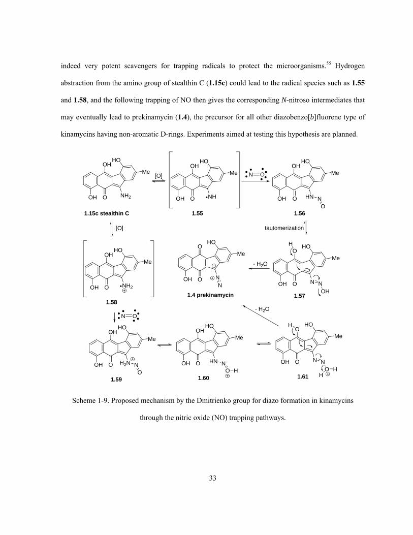
33
indeed very potent scavengers for trapping radicals to protect the microorganisms.55 Hydrogen
abstraction from the amino group of stealthin C (1.15c) could lead to the radical species such as 1.55
and 1.58, and the following trapping of NO then gives the corresponding N-nitroso intermediates that
may eventually lead to prekinamycin (1.4), the precursor for all other diazobenzo[b]fluorene type of
kinamycins having non-aromatic D-rings. Experiments aimed at testing this hypothesis are planned.
HO
MeOH
O NH2OH
1.15c stealthin C
N O
HO
MeOH
O NHOH
HO
MeOH
O HNOH
HO
MeO
O NOH NOH
HO
MeO
O NOHN
- H2O
1.55
tautomerization
1.56
1.571.4 prekinamycin
NO
HO
MeOH
O NH2OH
1.58
[O]
N O
HO
MeOH
O H2NOH
1.59
NO
H
HO
MeOH
O HNOH
1.60
NO H
HO
MeO
O NOH
1.61
NO H
H
H
- H2O
[O]
Scheme 1-9. Proposed mechanism by the Dmitrienko group for diazo formation in kinamycins
through the nitric oxide (NO) trapping pathways.

34
1.4 Biological Activities and Speculation on the Mode-of-Action of the
Kinamycins
1.4.1 Biological Activities of the Kinamycins and Lomaiviticins
The reported antimicrobial activities of natural and semi-synthetic kinamycins indicate that they are
strongly active against Gram-positive bacteria but (much) less effective towards the Gram-negative
ones.4,6-10,18,52 Lomaiviticins (1.11a/b), the dimeric analogues of kinamycins, were found to be even
more potent towards the Gram-positive bacteria. A detailed summary of the reported antimicrobial
activities of all kinamycins in the literature is provided in the appendix section (Appendix A). As
antibiotics, the intravenous acute toxicity (LD50) of natural kinamycins in mice was found to be
moderate in general. For kinamycin A–D (1.1a–d), LD50 is about 30-40 mg/kg each,4 and for FL-
120A (1.1m), FL-120B (1.2a) and FL-120C’ (1.1o), LD50 is 35.36 mg/kg, > 50 mg/kg, and 6.97
mg/kg, respectively.11 A lethal dose in mice of 0.5 mg per mouse was reported for kinamycin H (1.1h)
but a reduction of the dose by half showed no toxicity.7
When compared with some common antibiotics such as streptomycin, mitomycin C and
vancomycin, kinamycin D (1.1d) exhibits a moderate antimycoplasmal activity but with unknown
mode-of-action.88 In addition, kinamycin D (1.1d) possessed moderate antifungal activities at a
concentration of 10–100 μg/mL against certain plant-pathogenic and agriculture-related fungi,
comparable to other quinone-type antibiotics such as antimycin, nanaomycin A, oligomycin and
ikutamycin.89 This particular kinamycin also demonstrated significant herbicidal activity (30–80%
inhibition of plant growth) against radish. Such antifungal and herbicidal activity of kinamycin D
(1.1d) was considered to be effective through possible inhibition of the biosynthesis of cellulose.89
The other important biological activity of kinamycins, and lomaiviticins in particular, is their
antitumor activity. Early antitumor tests of kinamycin C (1.1c) and D (1.1d) towards EHRLICH

35
ascites carcinoma and sarcoma-180 indicated that, 1.1c showed some survival effect by
intraperitoneal injection at 0.1 and 1 mg/kg, yet 1.1d was found to have no tumor inhibition effect.20
Later, kinamycin G (1.1g) and H (1.1h) were found to possess cytotoxicity against L1210 leukemia
(IC50 1.1g: 0.72 μg/mL; 1.1h: 0.38 μg/mL) and IMC carcinoma cells (IC50 1.1g: 0.88 μg/mL; 1.1h:
0.72 μg/mL) in suspension culture, and cytotoxicity against LX-1 human lung carcinoma (IC50 1.1g:
0.542 μg/mL; 1.1h: 0.790 μg/mL) and SC-6 human stomach carcinoma (IC50 1.1g: 2.50 μg/mL; 1.1h:
0.760 μg/mL) in a clonogenic assay.7 The dimeric diazobenzo[b]fluorenes of lomaiviticin A (1.11a)
and B (1.11b) were found to possess very potent DNA-damaging activity (minimum induction
concentration ≤ 0.1 ng/spot) in biochemical induction assay (BIA) and extreme cytotoxicity (0.01–98
ng/mL, equivalent to 0.007–72 nM) against a broad panel of cancer cell lines.52 Moreover, the
cytotoxicity profile of 1.11a in a 24-cancer cell line panel was unique when compared with other
known DNA-damaging anticancer drugs such as adriamycin and mitomycin C, indicating a different
and still unknown mechanism of interaction with DNA. Studies also indicated that lomaiviticin A
(1.11a) is capable of cleaving double-stranded DNA under reducing conditions.52 The strong and
broad anticancer activity of lomaiviticins, along with their unknown mode-of-action, have stimulated
a great deal of research interests in the diazobenzo[b]fluorene systems. More recent work from the
Dmitrienko lab in collaboration with the Hasinoff lab indicates that isoprekinamycin (1.5)
significantly inhibits the growth of Chinese hamster ovary (CHO) cells (IC50 = 5.8 μM) and K562
human leukemia cells (IC50 = 6.4 μM), comparable to the clinically useful anticancer agent etoposide
(CHO: IC50 = 1.4 μM; K562: IC50 = 3.4 μM).25 In addition, during recent physical organic studies on
prekinamycin (1.4) and simpler analogues, such compounds have been screened against a 60-cell
human cancer panel and some of them demonstrated significant growth inhibitory activity (GI50)
against pancreas, breast, lung, colon and prostrate tumor cells at a concentration of ca. 0.41–7.3
μg/mL.90
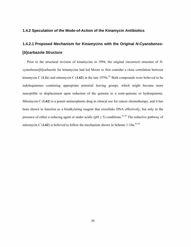
36
1.4.2 Speculation of the Mode-of-Action of the Kinamycin Antibiotics
1.4.2.1 Proposed Mechanism for Kinamycins with the Original N-Cyanobenzo-
[b]carbazole Structure
Prior to the structural revision of kinamycins in 1994, the original (incorrect) structure of N-
cyanobenzo[b]carbazole for kinamycins had led Moore to first consider a close correlation between
kinamycin C (1.1c) and mitomycin C (1.62) in the late 1970s.91 Both compounds were believed to be
indolequinones containing appropriate potential leaving groups, which might become more
susceptible to displacement upon reduction of the quinone to a semi-quinone or hydroquinone.
Mitomycin C (1.62) is a potent antineoplastic drug in clinical use for cancer chemotherapy, and it has
been shown to function as a bisalkylating reagent that crosslinks DNA effectively, but only in the
presence of either a reducing agent or under acidic (pH ≤ 5) conditions.91-93 The reductive pathway of
mitomycin C (1.62) is believed to follow the mechanism shown in Scheme 1-10a.91-93
37
N
O
O
H2N
Me NH
OCONH2
OMe 1. 2 [H]
N
OH
OH
H2N
Me NH
OCONH2
N
O
OH
H2N
Me NH
OCONH2
H
N
OH
OH
H2N
MeNH2
OCONH2
DNA
N
OH
OH
H2N
MeNH2
DNA
DNA
1.62 mitomycin C 1.63
1.66
1.68
2. + H+,- MeOH
H
N
O
OH
H2N
MeNH2
OCONH2
1.65
DNAN
OH
OH
H2N
MeNH2
DNA
DNA
N
O
O
H2N
MeNH2
DNA
DNA
1.69
InterstrandCross-link
- 2 [H]
1.67
1.64
(a)
N
CN
O
OOH
OAc
Me
OH
OAcAcO
N
CN
OH
OHOH
OAc
Me
OH
OAcAcO
N
CN
O
OHOH
Me
OH
OAcAcO
NCN
O
OOH
Me
OH
OAcDNA
NCN
O
OOH
Me
OH
OAc
DNA
DNA
2 [H] - HOAc
- HOAc
NCN
O
OHOH
Me
OH
OAc
DNA
DNA
H+1. H+
2. - 2 [H]
1.1c kinamycin C 1.70 1.71
1.721.731.74
(b)
Scheme 1-10. Proposed mode-of-action of (a) mitomycin C (1.62) and (b) kinamycin C (1.1c)
through two electron reductive activation.
Reduction of 1.62 to the corresponding hydroquinone 1.63 facilitates an acid-induced elimination
of the angular methoxy group, and leads to the subsequent formation of the indole 1.64. Ring-opening
of the aziridine produces the quinone methide 1.65, which is reactive towards nucleophilic attack by
species such as deoxyguanosine of DNA. Further elimination of carbamate from 1.66 then affords a
38
Michael acceptor 1.67 capable of being attacked by DNA again. Although both DNA-alkylation steps
(i.e., 1.65 to 1.66 and 1.67 to 1.68) may be reversible, oxidation of 1.68 to the quinone 1.69 prevents
the detachment of DNAs and therefore stabilizes the interstrand cross-link of DNAs. An analogous
mechanism for kinamycin C (1.1c) was then proposed by Moore (Scheme 1-10b).91,92 The in vivo
reduction of 1.1c may lead to the corresponding hydroquinone 1.70, which then undergoes two
consecutive eliminations of acetate groups off the D-ring and forms a double Michael acceptor 1.72
that can further interact with DNAs. In principle, Moore’s hypothesis requires no participation of the
“N-cyano/C-diazo” moiety of kinamycins, so its validity shall not be affected by the structural
revision of kinamycins. However, once the kinamycins were shown to be diazobenzo[b]fluorenes and
not N-cyanobenzo[b]carbazoles in 1994, their diazo moieties became the focus of speculation
concerning the possible mechanism(s) involved in the anticancer and antibacterial activity.
Shortly before the discovery of the diazo moiety within kinamycins, the isolation and
identification of kinafluorenone (1.16b) from a mutagenic strain (MC1) of Streptomyces
murayamaensis led to some surprising observations. Both the natural 1.16b and its synthetic triacetate
derivate 1.16c possess the benzo[b]fluorene skeleton but lack the C-diazo moiety (considered as the
N-cyano group at the time), whose structures were unambiguously established based on spectroscopic
evidence and single crystal X-ray studies of 1.16c.57 Kinafluorenone (1.16b) showed no detectable
antibiotic activity against Bacillus subtilis (ATCC 6633), which is very sensitive to the kinamycins
(MIC = 0.01–0.39 μg/mL).57 In addition, 1.16c also showed no detectable antibiotic activity, even at a
fairly high concentration of 64 μg/mL, against many bacteria that were known to be inhibited by
kinamycins at (much) lower concentrations.57 This observation would indicate that, the presence of a
diazo functionality within a benzo[b]fluorene skeleton, is very likely involved and essential in the
biological activity of the diazobenzo[b]fluorenes of kinamycins.

39
CH3O
OH
OH
HO
O
CH3
CH3O
AcO
OAc
AcO
O
CH3
1.16b 1.16c
1.4.2.2 Role of Diazo and Diazonium Moiety in Biological Activities
Cleavage of single and double stranded DNA upon exposure to simple (para-substituted)
phenyldiazonium salts has been observed but with no substitution effect.94,95 It is generally believed
that the aryl radicals, generated in situ from the corresponding diazonium salts, are the active species
that cause the DNA cleavage94,95 and are responsible for their genotoxicity.96 The exact pathway
leading to the aryl radicals from the aryldiazonium ions in aqueous solution is still not very clear;
however, both an inner-sphere and an outer-sphere electron transfer mechanisms are commonly
considered (Scheme 1-11).97-99 The inner-sphere path involves no apparent reductant but an electron
donor (Nu-) with high oxidation potentials (e.g., -OH), which adds to the electrophilic diazonium ion
(Ar-N2+) and forms a transient species 1.75 of Ar-N=N-Nu(OH). It then either directly dissociates in a
homolytic manner to produce the corresponding aryl radical Ar• along with the hydroxyl radical HO•,
or reacts further in its anionic form 1.89 with another molecule of the diazonium ion to form a dimer
1.90, still affording the same Ar• upon the loss of an arylazoxy radical 1.78 and N2. The outer-sphere
mechanism would apply if there exists an appropriate and readily oxidizible (by the diazonium ion)
reductant, which could directly transfer an electron to the diazonium ion resulting in the formation of
aryl diazenyl radical Ar-N=N• (1.79) that further decomposes to the Ar• upon the loss of N2.

40
Ar N2+ + -OH Ar N N OH Ar N N O- H+
Ar N2Ar N N O N N Ar
Ar
OH
Ar N N Oe-
1.75
Inner-sphere mechanism:
Outer-sphere mechanism:
Ar N2+ - N2
- N2Ar N2
1.76 1.77
1.78
1.79
- N2 -
-
Scheme 1-11. Two mechanisms for the formation of aryl radicals from aryldiazonium salts.
Early model studies by Stock and co-workers on reactions of aryldiazonium salts with the purine
units of DNA such as adenine, guanine and their derivatives revealed details on how the diazo
compounds may interact with DNA.100,101 Adenine (1.80a) and its derivates such as adenosine (1.80b)
and 5’-adenylic acid (1.80c) react readily with aryldiazonium salts under basic conditions, and the
corresponding purine-based C6-triazene 1.81a–c were obtained as the initial and isolated products
(Scheme 1-12).100 The triazenes (1.81a as an example) decomposed either quickly at room
temperature under acidic conditions (pH 3) by following the typical acid-catalyzed triazene
degradation mechanism, or very slowly (3 days) even at elevated temperature (60–100 °C) under
basic conditions (pH 10) and yielded C8-aryladenines 1.90 via a free radical mechanism. The free-
radical mechanism, however, was proposed to involve initially a heterolytic and acid-catalyzed
cleavage of the triazene 1.81a leading back to the adenine (1.80a) and the aryl diazonium ion 1.83.
The latter could exist in equilibrium with 1.82 and 1.84 that eventually lead to the aryl radicals 1.86
and 1.87 through the aforementioned inner-sphere mechanism. Further radical substitution and
hydrogen abstraction would give the final C8-arylated product 1.89 causing the irreversible DNA
damage.

41
N
NNH
N
NH2
H
XC6H4
N
NN
N
NHNN
N
NN
N
NH2
pH 10-11
0 oCRR
XC6H4
1.80a-c 1.81a-c(60-85%)
1.88
XC6H4N2+
N
NNH
N
NH2
1.89X = H, CH3,Br, SO3H
XC6H4
pH 10
60-100 oC
H2ON
NNH
N
NH2
OHNN
XC6H4
+
XC6H4 + XC6H4N2O
1.82 1.80a
1.871.86
ONN
XC6H4
1.84
NNXC6H4
1.83
-OH-OH
NNXC6H4 O N N C6H4X- N2
1.85
13
5
8
67
9
1.80a adenine(R = H)
1.80b adenosine(R = ribose)
1.80c adenylic acid(R = ribose 5'-phosphate)
O
OHOH
HHHH
HO
ribose
1'
5'
2''
Scheme 1-12. Stock’s proposed mechanism for reactions of aryldiazonium ions with adenines.
On the other hand, guanine (1.90a) and its derivatives such as guanosine (1.90b) and 5’-guanylic
acid (1.90c) demonstrated more complex results when reacting with aryldiazonium salts at pH 10–11
(Scheme 1-13). The corresponding DNA cleavage mechanism, however, was believed to still follow
the same radical pathway involving triazene intermediates (Scheme 1-12).101 Guanine (1.90a) led to
quick (15 min) formation of C8-arylazoguanines 1.91 at low temperature (0 °C), which could survive
both very acidic (pH 1) and harsh basic conditions (pH 10, 24 hr, 90–100 °C), yet it was easily
reduced to the corresponding amines of 1.92 and 1.93. Even at slightly higher temperature (25 °C),
guanosine (1.90b) still reacted much more slowly (24 hr) with the diazonium salts than guanine
(1.90a), but in this case the C8-arylguanosines 1.94 were obtained as the final product with
concurrent production of N2. The 5’-guanylic acid (1.90c) reacted even more slowly with the
diazonium salts than 1.90b, and under the same conditions (24 hr, 25 °C) it only afforded the C2-
triazenes 1.95 in low yield. Heating of the reaction, however, caused radical arylation at C8 to occur
to a certain extent to generate 1.96 instead. The observed difference in the reactivity among guanine
(1.90a) and its derivatives 1.90b/c, as well as the adenines 1.80a–c, were explained in terms of the

42
relative nucleophilicities of the C6 (of 1.80) and C2 (of 1.90) amino groups, and the relative tendency
to undergo an electrophilic reaction at the C8 positions of such purines, which would be significantly
retarded due to unfavorable polar (inductive) and steric effects if a sugar such as ribose and ribose-5’-
phosphate is present at the adjacent N9.101 A later study also showed that 4-carbamoyl-
benzenediazonium chloride can form a C2-triazene with the deoxyguanine bases of DNA (i.e., dGMP,
deoxyguanosine monophosphate), whose structure was confirmed by both NMR and mass
spectrometry.102 Spectroscopic evidence for the involvement of a DNA-triazene adduct, both in vitro
and in vivo, through the reaction of benzenediazonium ion (formed in situ from N-nitroso-N-
methylaniline) and DNA has also been reported.103
pH 10-11
0 oC, 15 min
XC6H4N2+
+ 1.92
1.90a
N
NHN
N
H
O
NH2N
NHN
N
H
O
NH2
NNX
1.91 (60-85%)X = H, CH3, Br, SO3H
Na2S2O4 N
NHN
N
H
O
NH2
H2N
XC6H4NH2 (X = Br)
1.93
1.90b
N
NHN
N
O
NH2
O
OHOH
HHHH
HOpH 10-11
25 oC, 24 hr
XC6H4N2+
1.94 (20-32%)X = H, CH3, Br, SO3H, NO2
N
NHN
N
O
NH2
O
OHOH
HHHH
HO
X + N2 + H+
N
NHN
N
O
NH2
O
OHOH
HHHH
-2O3PO pH 10-11
25 oC, 24 hr
XC6H4N2+
1.95 (10%)X = SO3H, NO2
N
NHN
N
O
NH
O
OHOH
HHHH
-2O3PO
+ H+
NN X
1.90c
pH 10-11,
XC6H4N2+
1.96 (10-20%)X = Br, SO3H
N
NHN
N
O
NH2
O
OHOH
HHHH
-2O3PO
X
1
3
5
8
7
9 2
Scheme 1-13. Product distribution of reactions between aryldiazonium ions and guanines.
43
Further in vitro and in vivo studies on the reactions of arenediazonium ions p-X-C6H5N2+ (1.97, X
= CH3, CH2OH, CH2OCH3, CO2H) with DNA bases, nucleosides, DNAs and cells by the Gannett
group agreed well with Stock’s proposal (Scheme 1-12) in general, but with some new discoveries
(Scheme 1-14).99,104-106 Both aryldiazonium ions 1.97 and the corresponding aryl radicals 1.98
generated upon either chemical or metabolic reduction of 1.97 are genotoxic and mutagenic. The
dominant bioactive species, however, is determined by the reduction potential of the diazonium ion,
i.e. a diazonium ion with a low reduction potential (difficult to reduce) is more important than the
corresponding radical and vice versa.104 Reaction of the aryldiazonium ions with adenine and/or
guanine leads to the formation of either isolated or detectable triazenes 1.98, which decompose at
both physiological and elevated temperatures (25–75 °C) in weak basic (pH 7.4) aqueous buffers.105
Triazene decomposition leads to not only the aryl radicals but also reactive hydroxyl radicals, as
characterized by their ESR (electron spin resonance) spectra.99,104-106 The possibility that these
concurrent oxygen radicals participate in the DNA damage was ruled out based on observations from
competitive inhibition experiments.106 The aryl radicals, most likely generated by concurrent multiple
routes, including but not limited to stepwise acid-catalyzed decomposition of triazenes (1.99 to 1.98
via 1.103), homolytic radical cleavage (1.99 to 1.98 via 1.100), and direct diazo reduction (1.97 to
1.98), then give the corresponding DNA adducts upon arylation at C8 of adenines (cf. 1.102) and
guanines (cf. 1.107), ultimately resulting in the DNA damage.

44
N2+XC6H4
+ e-
- N21.97 (X = CH3, CH2OH, CH2OCH3, CO2H)
DNA bases / nucleosides / DNAs
(in vitro and in vivo)
N
NN
N
NHNN
R
XC6H4
1.99R = H, ribose,
2'-deoxyribose, DNA
N
NHN
N
O
N
H
N N C6H4X
guaninesadenines
R
N
NN
N
NH
1.100
XC6H4 +
1.98
N
NN
N
NH
1.101
XC6H4
H
R
R
N
NN
N
NH2
1.102DNA damage
XC6H4
R
N
NN
N
NH2
1.103
R
XC6H4
1.98
- N2
N
NN
N
NH2
1.105R
H
XC6H4
- e- -H+
N
NHN
N
O
NH2R
+ H+, + e- - N2+ H+
N
NN
N
NH2NN
R
XC6H4
N
NN
N
NH2
NN
R
XC6H4
or
N
NN
N
NH2
R
NNXC6H4
+ e- - N2
1.97
1.1051.104
1.106
- e- -H+
H
XC6H4
N
NHN
N
O
NH2R
XC6H4
1.106
1.107DNA damage
XC6H4
1.98
DNA bases/nucleosides/DNAs (in vitro and in vivo)
Scheme 1-14. Proposed mechanism by Gannett for DNA damage caused by aryldiazonium ions.
In the meantime, Kikugawa and coworkers carried out a series of biological and mechanistic
studies on some mutagenic diazo compounds, most of which are carcinogenic and capable of cleaving
single-strand DNA in vitro under physiological conditions. Such examples include 3-diazo-N-

45
nitrosobamethan,107,108 diazo-N-nitrosoetilefrin,109 o- and p-diazoquinones,110-112 fluorene-2-diazonium
salt,113,114 5-diazouracil,115 4-(hydroxymethyl)benzenediazonium salt (1.54),116,117 and 6-diazo-5-oxo-
L-norleucine (DON, 1.6a).118 The observed biological activities were not affected by molecular
oxygen but inhibited by common hydrogen donors, nucleophiles and reductants such as alcohols,
phenols, azide and thiols,114,115,118 amino acids and proteins,119 even unsaturated fatty acids and
lipoprotein.120 Despite the structural diversity involved in these diazo compounds, their mode-of-
action was confirmed by ESR experiments to rely on the carbon-centered radicals generated in situ
from the corresponding diazo compounds but not the concurrently produced oxygen radicals,
consistent with Gannett’s observations. The carbon radicals would react with not only purines (free or
within DNA) still at C8 but also pyrimidine bases (free or within DNA). In addition, diazo-initiated
bond breakage of phosphodiester of DNA was also observed and attributed to the action of the carbon
radicals as well.
Soon after the structural revision of the kinamycins from the N-cyanobenzo[b]carbazole to the
diazobenzo[b]fluorenes, Jebaratnam’s group reported some initial evidence suggesting the possible
diazo involvement in the bioactivity of kinamycins when 9-diazofluorene (1.109) was used as a
simple model compound.121 It was found that 1.109 did not induce any DNA cleavage either alone
(no cleavage at pH 4–7 and cleavage observed at pH < 4 was caused by acid rather than 1.109) or in
the presence of Cu2Cl2 (i.e., Cu+). However, in the presence of an oxidant such as cupric acetate
Cu(OAc)2, 1.109 led to successful DNA cleavage while the Cu(II) salt itself did not cleave DNA
(Scheme 1-15). These results were rationalized as an one-electron oxidation of 1.109 with Cu2+
followed by loss of N2 to produce a corresponding carbon radical 1.110, which was suggested to be
the active species that initiates DNA cleavage, likely by hydrogen atom abstraction from DNA.121
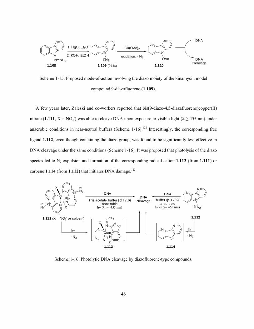
46
N2 DNACleavage
1.109 (91%)
OAc
1.1101.108
N
1. HgO, Et2O
2. KOH, EtOHNH2
Cu(OAc)2
oxidation, - N2
DNA
Scheme 1-15. Proposed mode-of-action involving the diazo moiety of the kinamycin model
compound 9-diazofluorene (1.109).
A few years later, Zaleski and co-workers reported that bis(9-diazo-4,5-diazafluorene)copper(II)
nitrate (1.111, X = NO3-) was able to cleave DNA upon exposure to visible light (λ ≥ 455 nm) under
anaerobic conditions in near-neutral buffers (Scheme 1-16).122 Interestingly, the corresponding free
ligand 1.112, even though containing the diazo group, was found to be significantly less effective in
DNA cleavage under the same conditions (Scheme 1-16). It was proposed that photolysis of the diazo
species led to N2 expulsion and formation of the corresponding radical cation 1.113 (from 1.111) or
carbene 1.114 (from 1.112) that initiates DNA damage.123
N
N N2N
NN2
Cu(II)
X
X
1.111 (X = NO3- or solvent)
DNADNA
cleavage
NN
N2
1.112
Tris acetate buf fer (pH 7.6)anaerobic
h ( 455 nm)
DNA
buffer (pH 7.6)anaerobic
h ( 455 nm)
NN
- N2N
NN
N
Cu(I)
X
X
h
1.1141.113
- N2
h
Scheme 1-16. Photolytic DNA cleavage by diazofluorene-type compounds.

47
The involvement of either carbon radical, radical cation or carbene as diazo-derived active
intermediates in the above two mechanisms, presented to have relevance to the possible mode-of-
action of the kinamycins, resembles the previous knowledge of how the simple diazo(nium) species
interact with DNA. However, both Jebaratnam’s and Zaleski’s hypotheses require the mandatory
presence of either an oxidant (Cu2+ in both cases) or the condition of photolysis to facilitate the
transformation of the kinamycin-like diazo compound to the active species. Such artificial conditions
are barely available (i.e., Cu2+ is less commonly available in biosystems as oxidant) or even
unnecessary (e.g., light) in the actual biological systems where the bioactivities of kinamycins were
observed. Therefore, both mechanisms (particularly the Zaleski model) are of questionable value.
These considerations led to a logical suggestion by the Dmitrienko group that the diazo moieties of
kinamycins are probably diazonium-like and electrophilic to a significant extent, and that similar
chemical behaviour analogous to that exhibited by aryl diazonium ion as described above may occur
with these natural diazobenzo[a]fluorenes and diazobenzo[b]fluorenes.80,124 Reactivities of diazonium
and diazo species with various organic and inorganic nucleophiles have been well studied and
documented ever since the discovery of the first diazo compound in 1858.97,125-128 Among the
numerous examples, reactions such as azo coupling, thiol-addition and amine-addition are of
particular importance, since such reactions might be involved in the bioactivity and modes-of-action
of the diazo compounds such as the kinamycins. Experimental and computational evidence from the
Dmitrienko lab, which are to be described in the following section, are in good agreement with such a
proposal.
1.4.2.3 Mechanistic Studies of Kinamycins in the Dmitrienko Lab
It is known in the literature that ortho- (1.115) and para-hydroxy aryldiazonium salts can be
readily deprotonated even under acidic conditions to produce the corresponding o- (1.117) and p-

48
quinonediazides (Figure 1-5).80 X-ray crystallographic data of some simple diazo compounds (Figure
1-5) clearly indicate a concomitant decrease of the bond order for the C-N linkage with the increasing
N-N triple bond character in the order of diazoketone 1.118 < substituted o-quinonediazide 1.119 <
substituted p-quinonediazide 1.120 < diazonium salt 1.121, suggesting that the chemical reactivity of
the diazo group within quinonediazides would typically fall in between those from diazoketones and
aryl diazonium salts. Quinodiazides can undergo azo coupling with phenols in both weakly acidic and
alkaline solution, but ortho-quinodiazides are less reactive than their para-isomers and even less so
than diazonium salts for such reactions, which may require the use of strong and activated
nucleophiles. The presence of electron withdrawing groups on the quinodiazide nucleus would
accelerate the corresponding azo coupling reactions, since the diazonium character is enhanced when
the negative charge on the diazo carbon is stabilized by the electron withdrawing factor(s). Moreover,
DNA cleavage by quinonediazides has also been reported.110-112 In this regard, kinamycins such as the
isoprekinamycin (1.5) could be recognized as a structure incorporating a 1,2-naphthoquinone-1-
diazide (1.122), which may demonstrate similar chemical properties and behaviour as mentioned
above. A likely correlation between the biological activities of kinamycins and their corresponding
diazo reactivities should be considered.

49
N2
O
1.122 1,2-naphtho-quinone-1-diazide
O
OOH
Me
HO
N2
1.5 isoprekinamycin
O
N2
1.117
O
N2
1.116
OH
N2- H+
1.115
O
N2
ClClO
O
O
N2
N2
O
1.119a 1.120a
dC-N (Å)
N2 Cl
1.121a
O
O
H N2
H
N2
1.118a
(1.385c)1.418
(1.099c)1.093
1.313
1.114
1.357
1.106
1.363(1.373b)
1.104(1.095b)
a. Bond lengths were measured directly from the corresponding .cif f ile of the compound depositedin the CCDC database; b. Two different geometries of 1.120 co-exist in the same crystal; c.1.121 co-crystallized with acetic acid.
dN-N (Å)
Figure 1-5. Properties of the diazo group within some diazo compounds.
In 2002, Laufer from the Dmitrienko lab synthesized a model compound 1.123 (Scheme 1-17),
possessing the same benzo[a]fluorene skeleton and the essential o-quinonediazide moiety as in the
natural isoprekinamycin (1.5) but lacking its A-ring and D-ring substituents.80,124 Attempts to
rearrange the benzo[a]fluorene frame of 1.123 to the isomeric benzo[b]fluorene of 1.124 failed under
both basic and acidic conditions (Scheme 1-17), but 1.123 survived the acidic conditions intact and it
was converted to a hydrodediazoniation product 1.127 exclusively under the basic conditions. This
led to the conclusion that the hypothetical interconversion equilibrium between the
diazobenzo[a]fluorene and the diazobenzo[b]fluorene structures (Scheme 1-6a), if it occurs in vivo,
would be an enzyme-catalyzed rather than a spontaneous chemical transformation. In addition, the
known base-induced hydrodediazoniation of aryldiazonium species129,130 would suggest a plausible
mechanism (Scheme 1-17) for the conversion of 1.123 to 1.127. A diazohydroxide intermediate 1.125
may be obtained upon nucleophilic attack of the diazo moiety of 1.123 by the HO-, which
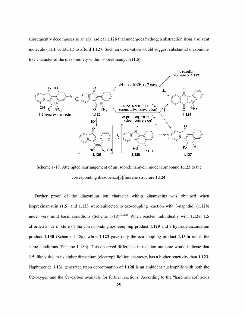
50
subsequently decomposes to an aryl radical 1.126 that undergoes hydrogen abstraction from a solvent
molecule (THF or EtOH) to afford 1.127. Such an observation would suggest substantial diazonium-
like character of the diazo moiety within isoprekinamycin (1.5).
Scheme 1-17. Attempted rearrangement of an isoprekinamycin model compound 1.123 to the
corresponding diazobenzo[b]fluorene structure 1.124.
Further proof of the diazonium ion character within kinamycins was obtained when
isoprekinamycin (1.5) and 1.123 were subjected to azo-coupling reaction with β-naphthol (1.128)
under very mild basic conditions (Scheme 1-18).80,124 When reacted individually with 1.128, 1.5
afforded a 1:2 mixture of the corresponding azo-coupling product 1.129 and a hydrodediazoniation
product 1.130 (Scheme 1-18a), while 1.123 gave only the azo-coupling product 1.134a under the
same conditions (Scheme 1-18b). This observed difference in reaction outcome would indicate that
1.5, likely due to its higher diazonium (electrophilic) ion character, has a higher reactivity than 1.123.
Naphthoxide 1.131 generated upon deprotonation of 1.128 is an ambident nucleophile with both the
C2-oxygen and the C1-carbon available for further reactions. According to the “hard and soft acids

51
and bases” (HSAB) theory,131 the C2-oxygen is a hard nucleophile (Lewis base) while the C1-carbon
is a soft one, since the former bears more negative charge than the latter. The terminal nitrogen of the
diazo moiety in 1.5, on the other hand, bears significant positive charge, which makes it a hard
electrophile (Lewis acid), as indicated by its LUMO coefficients obtained from MO calculation. So
when 1.5 reacts with 1.131, the competition between a hard-hard interaction (path A, Scheme 1-18a)
and a soft-hard interaction (path B, Scheme 1-18a) is expected. Since hard acid prefers to react with
hard base and vise versa, preference of path A is predicted by the HSAB theory and the
corresponding product 1.132 then may undergo a hydrodediazoniation to afford 1.130 as the final
major product, likely via the mechanism outlined in Scheme 1-17. On the contrary, theoretical
calculation by means of NBO (natural bond orbital) population analysis indicated that the terminal
nitrogen of the diazo group in 1.123 is significantly less positive and therefore softer than that in
1.5.80 This prediction is consistent with the result that the reaction of 1.123 and 1.128 only led to
1.134a as the only/major product (Scheme 1-18b), which should be derived from the preferred soft-
soft interaction (path B) in this case. When a 1:1 mixture of 1.5 and 1.123 was subjected to the azo
coupling conditions, TLC (thin layer chromatography) and 1H-NMR evidence indicated a
significantly preferential consumption of 1.5 over 1.123. This observation suggests a strong
correlation between the diazonium ion characters of kinamycins and their reactivities. Another
parallel competition experiment was carried out by simultaneously treating 1.5 and 1.123 with 1.128
under exactly identical and strictly controlled conditions but in separate flasks, and both reactions
were monitored until the complete consumption of the diazo compounds (Scheme 1-18c).
Isoprekinamycin (1.5) was found to be much more reactive than its simpler analogue 1.123, as
demonstrated by the shorter reaction time and lower reaction temperature needed to complete the azo-
coupling. Moreover, unpublished results from Laufer also indicated that under the same conditions,
isoprekinamycin (1.5) reacted with guanosine triacetate and kinamycin D (1.1d) reacted with β-

52
naphthol (1.128).80 Complete consumption of the kinamycins due to reactions with the nucleophiles
were observed in both cases.
Scheme 1-18. Comparison of diazo reactivity and electrophilicity of isoprekinamycin (1.5) and model
compound 1.123 via azo-coupling reactions with β-naphthol (1.128).

53
To account for the observed difference in chemical behaviour of isoprekinamycin (1.5) and the
model compound 1.123 from a structural point of view, Laufer also carried out extensive ab initio
MO (molecular orbital) calculations on kinamycins and related structures,80,124 with a focus on the
diazo IR stretching frequency and the N-N bond length. Such properties were found to be a suitable
indication for the corresponding diazonium ion character. Comparison of the computational results
with available experimental values showed satisfying consistency, and a clear tendency of increasing
diazo IR frequency and diazonium ion character with concurrent decreasing N-N bond length (Table
1-2), in the order of 9-diazofluorene (1.109) < 2,1-napthoquinodiazide (1.122) < isoprekinamycin
model 1.123 < isoprekinamycin (1.5) < kinamycin B (1.1b) < lomaiviticin A model 1.138 < PhN2+Cl-
(1.121), matches roughly the increasing biological activities in essentially the same order, i.e.
isoprekinamycin (1.5) < kinamycin B (1.1b) < lomaiviticin A (1.11a). It is clear, however, that many
factors other than diazonium ion character may also influence the biological activity of kinamycins
and their analogues.

54
Table 1-2. Calculated diazo frequencies and N-N bond lengths of various kinamycin structures.a
Compound Calculated
NN (cm-1)
Experimental
NN (cm-1)
Calculated
dN≡N (Å)
Experimental
dN≡N (Å)
N2 1.109
1906
2000 (KBr)
1.133
1.126
ON2 1.122
2056
1.111
O
OOH
HO
Me
N2 1.4
2058
2119 (film)
1.111
O
O N2
Me
O
O
Me
Me 1.135
2079
2113 (CH2Cl2
solution)
1.109
O
O N2 1.123
2087
2105 (film)
1.108
O
O N2
Me
HO
1.136
2101
1.107
O
O N2OH
Me
1.137
2125
1.105
O
O N2
Me
HO
OH 1.5
2139
2162 (film)
1.103
1.104
O
OOH N2
Me
HOOAc
OH
OH
1.1b
2188
2155 (KBr)
1.099
1.1s: 1.080b 1.115b
O
OH N2
O
OH
O
OH
OH
1.138
2212
1.097
PhN2+Cl- 1.121 2212 2260 1.100 1.099
a. All MO calculations were performed at the RHF/6-31G//6-31G level. Although the calculated absolute IR frequencies for diazo groups may differ significantly from the experimental values, the computed difference in IR frequency (Δ) as a result of structural change matches the observed experimental Δ fairly well. For example, experimental Δ (1.5 - 1.123) = 57 cm-1, and the corresponding computed Δ (1.5 - 1.123) = 52 cm-1; b. Two different diazo bond lengths co-exist in the crystal of 1.1s (refer to Scheme 1-2 for structure of 1.1s).

55
The diazonium ion character of isoprekinamycin (1.5) is significantly enhanced by not only the
electron-withdrawing keto group on its B-ring, as evident by comparison of the diazo IR frequency of
1.123 and 1.5 with that of 1.122, but also the intramolecular hydrogen bonding (H-bonding)
interactions, as evident by comparison of diazo IR of 1.123, 1.136, 1.137 and 1.5.80,124 The presence
of strong intramolecular H-bond(s) within natural kinamycins, both the diazobenzo[a]- and
diazobenzo[b]fluorene type, is well known since all A-ring and D-ring phenolic protons of
kinamycins have characteristic down-field NMR chemical shifts of ca. 11–13 ppm.5,9,76 As a
comparison, typical phenolic protons without inter-/intramolecular H-bonding interactions exhibit
chemical shifts of only ca. 4–7.5 ppm.132 Therefore, the diazonium ion character of 1.5 is greatly
enhanced by the presence of double intramolecular H-bonding interactions (Scheme 1-19), which
delocalize the negative charge from the diazo carbon in the quinonediazide structure and stabilize the
negative charge on the oxygen atom as in the phenolic diazonium form.80,124 This theoretical model
provided a reasonable rationale for the observed difference in reactivities between isoprekinamycin
(1.5) and the analogue 1.123. The observed much lower diazo IR frequencies of isoprekinamycin
diacetate 1.5a (2119 cm-1)5 and isoprekinamycin dimethyl ether 1.135 (2113 cm-1) than that of the
mother compound 1.5 (2162 cm-1),5 both of which lack the intramolecular H-bonding interactions, are
entirely consistent with the discussion above.
Scheme 1-19. Intramolecular H-bonding model for isoprekinamycin (1.5).

56
The significant structural influence on the diazonium ion character of the diazo moiety within
kinamycins, as indicated by the above structure-activity studies, led the Dmitrienko group to propose,
at the time, that kinamycins (and lomaiviticins) may react with DNA in a manner similar to that of the
aryl diazonium salts as discussed in section 1.4.2.2.80 The purine units of DNA (adenine-based 1.139
and guanine-based 1.140) may react with the diazo group of kinamycins to form the corresponding
triazenes (1.141 and 1.142) initially (Scheme 1-20a), followed by dediazotization with loss of N2
(1.141 as an example) to yield the kinamycin-derived radical 1.143 and purine-derived radical 1.144.
Such radicals can react further through complex sequences, such as possible depurination via 1.145
and C8-arylation to form 1.150, ultimately leading to DNA damage. It was also realized that the
structural features such as in the case of isoprekinamycin (1.5) that enhance diazonium ion character
and, hence, the electrophilicity of the diazo group, also enhance the electron affinity. This might
favour a reductive pathway to radical 1.151 via the electron transfer in vivo (Scheme 1-20b).80
The speculations concerning the mechanism of action of the kinamycins outlined above represent
the state of thinking in the field when the present research project was initiated. Since then other
speculations based on model chemical studies as well as biochemical and whole cell experiments
have provided additional thoughts on the subject. These more recent results are presented and
discussed in Chapter 4 of this thesis.

57
N
NN
N
NH2
O
O
H H
N
NN
N
NH
R
NH
NN
N
NH
O
RN2
N
NH
H
H
N
NN
N
NH2
O
O
H
N
NN
N
NH
O
O
N
NNH
N
NH2
H
- N2
O
O
HO
OH
Me
DNADNA
cleavage
+ e
O
O
N2
HO
OH
Me
1.5 1.151
- N2
N2N
NN
N
NH2
R
kinamycins 1.1lomaiviticins 1.11
NH
NN
N
NH2
O
R
and / or
N
NN
N
NH2
R
DNAcleavage
DNA purine unitsR = 2'-deoxyribose
1.139 1.140 1.141 1.142
1.143 1.144
1.145
1.146 1.147
1.139
H
N
NN
N
NH
O
O
H
N
NN
N
NH2
O
O
H?
1.148 1.150 1.149
(a)
(b)
DNA cleavage
Scheme 1-20. Possible mechanisms proposed by the Dmitrienko group for DNA cleavage by
kinamycins (a) via triazene intermediates; (b) through diazo reduction.

58
1.5 Reported Syntheses of Kinamycins and Related Compounds
Natural products such as the kinamycins possessing unique structural features and potent
biological activities are ideal targets for synthetic organic chemists. However, the isolation and
characterization of kinamycins as the N-cyanobenzo[b]carbazole in the early 1970s did not trigger
enough synthetic interest in this type of compounds for almost twenty years. Efforts towards the
synthesis of kinamycins started to appear beginning in 1990, when the Dmitrienko group first
prepared some N-cyanoindoles as model compounds for kinamycins,73 still considered to be N-
cyanobenzo[b]carbazoles at the time. The structural revision of kinamycins in 1994 revealed the
presence of the very rare naturally-occurring benzo[b]fluorene skeleton and diazo moiety, which
evoked significant interest in these unusual natural antibiotics. Since then, a number of related
synthetic studies have been reported. The major details of these synthetic results will be discussed in
Chapters 2 and 3, which present the synthetic efforts towards the kinamycins from this project. Only
literature studies prior to the beginning of this thesis project will be introduced in the following
section.
1.5.1 Synthetic Efforts towards the N-cyanobenzo[b]carbazole System
Two major synthetic works towards the N-cyanobenzo[b]carbazole structure of kinamycins, one
by Echavarren (1993)74 and one by Dmitrienko (1994)22 have already been briefly mentioned earlier
(Scheme 1-2, Section 1.3.2.1). Other examples of the synthesis of the benzo[b]carbazoles include
reports by Dmitrienko (1990),73 Sugimone (1991),133 Murphy (1992),134 and more recently by Xu
(2006).135 Although the N-cyanobenzo[b]carbazole structure no longer applies and most synthetic
efforts were redirected towards the diazobenzo[b]fluorene of kinamycins since 1994, the importance
of the former structure can not be completely ignored. The appropriate N-cyanobenzo[b]carbazoles
may serve as the isomeric species of the corresponding kinamycins for the studies of mechanism of

59
action, and benzo[b]carbazole compounds were found to possess versatile and significant biological
activities of their own.136-138
1.5.2 Synthetic Efforts towards the Diazobenzo[b]- and Diazobenzo[a]fluorenes
The very rare structure of benzo[b]fluorene within natural products was first discovered with
stealthin A (1.15a) and B (1.15b) in 1992,55 and the structural revision of kinamycins from N-
cyanobenzo[b]carbazole to diazobenzo[b]fluorene in 199421,22 further stimulated synthetic interest in
kinamycins due to such structural novelty and their biological activities. Prior to the discovery of
benzo[b]- and benzo[a]fluorenes within the natural products, the wide environmental occurrence of
such ring systems as natural compounds (not “natural products” by definition) in oil, gasoline and
combustion products is well known. There are a few synthetic examples of fluorenes,139
benzo[b]fluorenes140-143 and benzo[a]fluorenes142,143 in the literature prior to 1990, however, none of
these methods has been applied to synthetic efforts towards the kinamycins. Outlined here are some
of the known approaches to the benzo[b]fluorene structures related to kinamycins that had been
developed when the present project began.
In general, retrosynthetic analysis of the generic benzo[b]fluorene structure 1.152 of kinamycins
would suggest mainly three possible ways of disconnection at B- and/or C-ring as shown in Scheme
1-21, which covers most known syntheses of benzo[b]fluorene structures related to the kinamycins:
(i) Disconnection (a) of 1.152 requires the use of a substituted naphthyl compound such as
1.153a or 1.153b as the AB-ring precursor and an appropriate aryl species such as 1.154a
or 1.154b as the corresponding D-ring precursor. Initial cross-coupling or addition
reaction of 1.153a and 1.154a (or 1.153b and 1.154b) would afford the corresponding
biaryl product, whose subsequent intramolecular (Friedel-Crafts, anionic/cationic or

60
metal-catalyzed cross-coupling) cyclization would then lead to the desired fluorene
skeleton.
(ii) Disconnection (b) involves a base-induced double condensation between either an o-
phthalyldicarbonyl species 1.155a and a substituted 1-indanone 1.156a or a phthalide
1.155b and a substituted 1-indenone 1.156b.
(iii) Disconnection (c) leads to a polyenyne intermediate 1.157 that can undergo a thermal
biradical cyclization to form the benzo[b]fluorene core, while 1.157 itself could be derived
from simple aromatic precursors such as 1.158 and 1.159.
OH
O
O
HO
disconnection (b)Base-induced double condensation
Cross-coupling+
Intramolecularcyclization
c b a
BiradicalCyclization
O
X
OOR
1.155b(X = CN, SO2Ph)
O
OR
+
OR
OROR
X+
OR
Y
Z
O OH
OR
OR
OR
OR
X
OHC
+
disconnection (a) disconnection (c)
OR
1.155a(X = H, OR)
O
OR
+X
O
X
O
OR
OROR
X+
OR
Y
O
Z
X, Y = halogen, -B(OR)2, H, NH2Z = H, OR, NR2
1.152
1.153a 1.154a
1.153b 1.154b
1.156a
1.156b
1.157
1.158 1.159
B CA
D
BA
BA
D
D
A DC
A DC
A
D
A
D
Scheme 1-21. Retrosynthetic analysis of the generic benzo[b]fluorene structure 1.152 of kinamycins.
61
Among all naturally-occurring benzo[b]fluorenes, kinobscurinone (1.16a) was the first one
synthesized. In 1995, Gould first prepared 1.16a by demethylation and oxidation of tri-O-
methylkinafluorenone (1.165),56 and the latter was initially and incidentally prepared as a very minor
side product from 1.164 through an anionic cyclization also by Gould during his total synthesis of
phenanthroviridin aglycon (1.167) in early 1990s (Scheme 1-22a).144,145 Later in 1997 during the total
synthesis of stealthin C (1.15c), Gould prepared the key intermediate 1.165 more efficiently through
an improved anionic cyclization of 1.163b and an intramolecular Friedel-Crafts cyclization of 1.168
(Scheme 1-22a).17 Around the same time in 1997, another synthetic route to 1.165 was achieved by
the Snieckus group via a Suzuki coupling between 1.169 and 1.170, the subsequent directed-
metalation of 1.171 and cyclization of 1.174 as the key steps (Scheme 1-22b).146

62
(a)
(b)
OH OMe
R'CO2R
1.161a (R' = Br, R = TMSE, 7 steps, 19%)1.161b (R' = Br, R = Me, 6 steps, 21%)1.161c (R' = H, R = Me, 5 steps )
O
OOMe
CNMeO
Br
O
OOMe
CO2R
1. 1.162 , LDA
2. O2
1. K2CO3, Me2SO4
Na2S2O4, acetone
2. Bu4N+F-, THF3. Tf2O, Py; 4. NH3
MeO
Br
MeO
OMeOMe
CONH2
1.160
1.163a (R = TMSE, 68% from 1.161a)1.163b (R = Me, 61% from 1.161b)
1.164 (62% from 1.163a)
1. t-BuLi (2.5 eq)THF, -98 oC
2. DMF
MeO
R
MeO
OMeOMe
CONH2
1.166 (80% from 1.164R= CHO/H, 1:1)
OMe
MeO
OMe O
MeO
1.165 (3% from 1.164)(39% from 1.163b)(69% from 1.168)
+
N
OH
O
O
HO
1.167 (from 1.166)
OH
O
O O
HO
1.16a kinobscurinone(89% from 1.165)
BBr3CH2Cl2
t -BuLi(1.1 eq)
THF-80 oC
MeOO
OOMe
CO2R
1.162
LDA
1.168 (70%)
1. PPA2. Me2SO4, K2CO3
1. NH2OH, Py, EtOH2. BBr3, CH2Cl2
3. Na2S2O4 (aq.), MeOHOH
OH
O NH2
HO
1.15c stealthin C(58%, 3 steps)
OMe
OMeOMe
BrOCONEt2
BO
3
Pd(PPh3)4, K3PO4toluene, EtOH, H2O,
OCONEt2
OMe
OMeOMe
1.169
1.170
1.171 (95%)
s-BuLi (4.4 eq)
TMEDA, THF-98 oC then
Me3SiCl
OCONEt2
OMe
OMeOMe
1.172 (97%)
SiMe3
LDA (10 eq), THF, rt
OH
OMe
OMeOMe
1.174 (62%)
SiMe3OH
OMe
OMeOMe
1.173 (17%)
SiMe2
NEt2
O+
1. NaH, CH3I, DMF, rt2. LDA (6 eq), THF, rt
3. TFA, OMe
MeO
OMe O
MeO
1.165 (75% 3 stepsfrom 1.174)
CONEt2
Scheme 1-22. Total synthesis of kinobscurinone (1.16a) and stealthin C (1.15c).
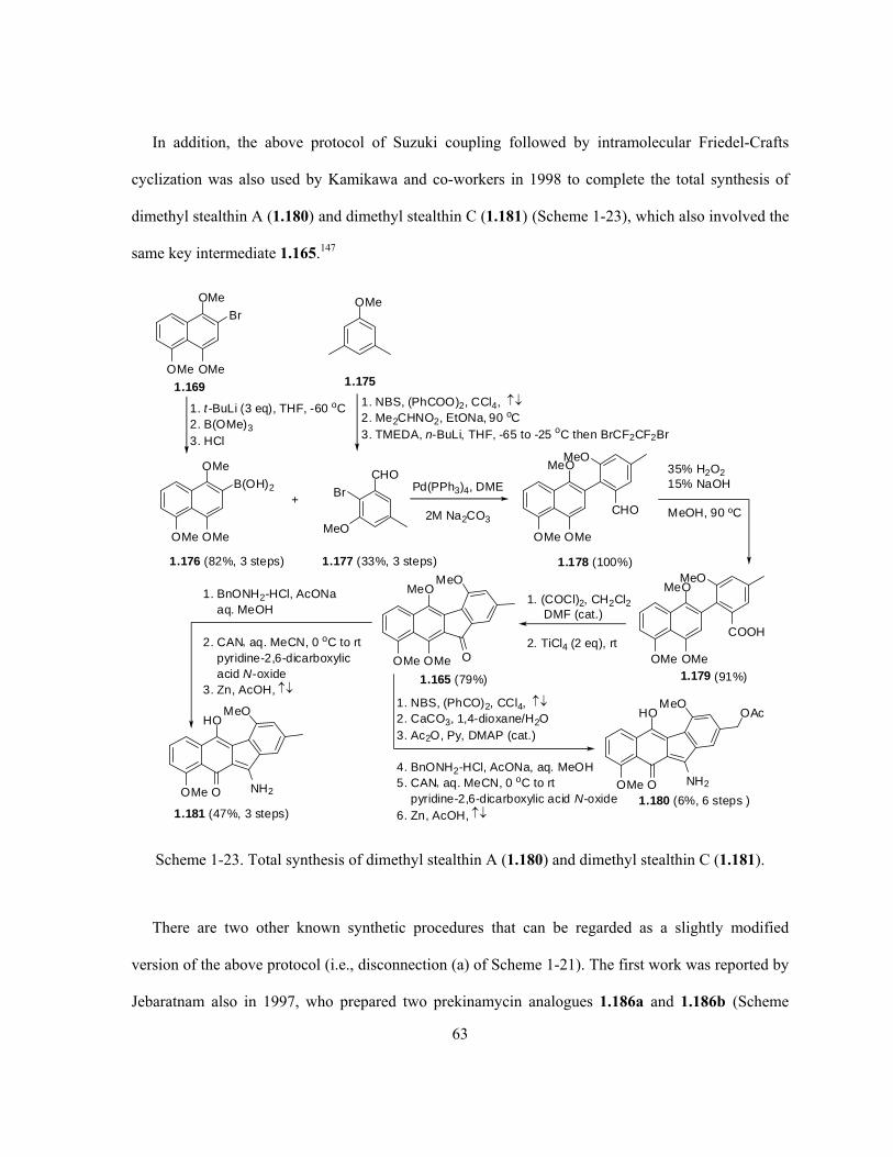
63
In addition, the above protocol of Suzuki coupling followed by intramolecular Friedel-Crafts
cyclization was also used by Kamikawa and co-workers in 1998 to complete the total synthesis of
dimethyl stealthin A (1.180) and dimethyl stealthin C (1.181) (Scheme 1-23), which also involved the
same key intermediate 1.165.147
1. (COCl)2, CH2Cl2DMF (cat.)
2. TiCl4 (2 eq), rt
35% H2O215% NaOH
MeOH, 90 ºC
OMe
OMe
1.178 (100%)
MeO
OMeOMe
B(OH)2 Pd(PPh3)4, DME
2M Na2CO3
OMe
CHO+
CHO
MeO
Br
MeO
1.179 (91%)
MeO
OMeOMe
COOH
MeO
OMe
OMeOMe
Br
1. t -BuLi (3 eq), THF, -60 oC2. B(OMe)3
3. HCl
1.169
1.176 (82%, 3 steps)
OMe
1. NBS, (PhCOO)2, CCl4, 2. Me2CHNO2, EtONa, 90 oC3. TMEDA, n-BuLi, THF, -65 to -25 oC then BrCF2CF2Br
1.175
1.177 (33%, 3 steps)
MeO
OMeOMe
MeO
O
1.165 (79%)
1. NBS, (PhCO)2, CCl4, 2. CaCO3, 1,4-dioxane/H2O3. Ac2O, Py, DMAP (cat.)
4. BnONH2-HCl, AcONa, aq. MeOH5. CAN aq. MeCN, 0 oC to rt
pyridine-2,6-dicarboxylic acid N-oxide6. Zn, AcOH,
HO
OOMe
MeO
NH2
1.180 (6%, 6 steps )
OAcHO
OOMe
MeO
NH2
1.181 (47%, 3 steps)
1. BnONH2-HCl, AcONaaq. MeOH
2. CAN aq. MeCN, 0 oC to rtpyridine-2,6-dicarboxylicacid N-oxide
3. Zn, AcOH,
Scheme 1-23. Total synthesis of dimethyl stealthin A (1.180) and dimethyl stealthin C (1.181).
There are two other known synthetic procedures that can be regarded as a slightly modified
version of the above protocol (i.e., disconnection (a) of Scheme 1-21). The first work was reported by
Jebaratnam also in 1997, who prepared two prekinamycin analogues 1.186a and 1.186b (Scheme

64
1-24a).148 The synthesis involved first an anionic addition of an aryl bromide 1.182 to acetanthranil
(1.183), after which the formed biaryl amide 1.184 underwent an intramolecular Pschorr reaction
through an in situ diazonium ion intermediate, leading to ring closure and the benzo[b]fluorenone of
1.185 followed by straight diazo transformation at C5. The second route was disclosed in 2000 by
Jones and Qabaja (Scheme 1-24b), in which the initial intermolecular addition of aryl lithium species
to substituted phenyl aldehyde followed by oxidation afforded the biaryl ketone 1.188, then a Pd(II)-
catalyzed intramolecular cross-coupling/cyclization of 1.188 under microwave conditions produced
the key intermediate 1.165 in moderate yield.149,150
1. KOH, aq. alcohol,
2. i-C5H11NO2, AcOH3. hydroquinone
OMe
OMe
O
O N2
1. BBr3, CH2Cl2, 0 ºC (for 1.185b)2. NH2NH2, EtOH,
3. Ag2CO3/celite, CH2Cl2, NEt3
1.186a (R = H, 50%)1.186b (R = OMe, 70%)
OMe
OMe
1.184a (R = H, 78%)1.184b (R = OMe, 50%)
t -BuLi, Et2O, -78 ºC
N
O
O
OMe
OMe NHAcOO
R
BrR R
R
1.182a R = H1.182b R = OMe
1.1831.185a (R = H, 34%)1.185b (R = OMe, 72%)
PdCl2(PPh3)2, NaOAc
DMA, W, 140 ºC, 1 min
1. NBS, CCl4, and thenNa2CO3, dioxane
2. n-BuLi, hexane, I2, -78 ºC3. (COCl)2, DMSO
OMe
OMe
1.187 (30%)
OMe
OMe
OMe
OMe
OMe OMe
OHC
I 1.
t-BuLi, Et2O, -78 ºC
2. PCC, CH2Cl2
BrI
OMe
1.188 (80%)
1.165 (53%)
O
(a)
(b)
1.175
1.182b
MeOMeO
OMeOMe O
Scheme 1-24. Synthesis of (a) prekinamycin analogues 1.186 and (b) kinafluorenone derivative 1.165.

65
The first attempt to construct the benzo[b]fluorenes of kinamycins via based-induced double
condensation (i.e., disconnection (b) of Scheme 1-21) was reported by Mal and co-workers in 1996
by using either the 1-indenone (1.190), generated upon flash vacuum pyrolysis (FVP) of 1.189, or
α,β-unsaturated ketone 1.194 to react with the phthalide sulfone 1.191 in the presence of a strong base
(Scheme 1-25a),151 through the well established Hauser annulation152 pathway. Shortly afterwards in
the same year, Hauser also used his very own annulation methodology152 to accomplish the first total
synthesis of prekinamycin (1.4) and its dimethyl derivative 1.4a starting from a substituted 1-
indenone 1.197 (Scheme 1-25b).76
1.190 (> 95%)
O
SO2Ar
O
1.192 (73%)
t-BuOLi, THF, -60 ºC
Ac2O
Py
FVP
O O O
OH
OH
O
O
1.189
1.191
1.193a (R = Ac, 73% from 1.192)1.193b (R = Me, from 1.195)
O
OR
OR
t-BuOLi, THF
1.191, -60 ºCthen methylation
1.194 1.195 (73%)
O
OMe
OMe
Ocheletropicelimination
aromatization
O
O
1. NaCl/AlCl3, 180 ºC2. Me2SO4, K2CO3
3. TMSOTf, Et3N4. Pd(OAc)2
OMe
O
MeO
OMe
HO
OH O
HO
OH
O
O N2
1.4 (31%, 3 steps from 1.198)
1.198 (73%)MeO
OMe
O
O N2
1. BBr3, CH2Cl2, -78 ºC2. NH2NH2, EtOH3. Ag2CO3, celite
1.196 1.197 (45%, 4 steps)
O
SO2Ar
O
t-BuOLi, THF, -78 ºC
1.191
1.4a (66%, from 1.198)(81%, from 1.4)
(a)
(b)
MeI, DMFK2CO31. NH2NH2, EtOH
2. Ag2CO3, celiteX
Scheme 1-25. Synthesis of (diazo)benzo[b]fluorenes via base-induced double condensation.

66
As more recent developments that are to be discussed in Chapter 3, the Birman lab employed
another base-induced double condensation to build the benzo[b]fluorenes and complete the second
total synthesis of prekinamycin (1.4) in 2007.153 At the same time, Estevez’s group combined an
initial base-induced aldol condensation with a subsequent intramolecular Pd(II)-catalyzed cyclization
to derive the benzo[b]fluorenone skeleton.154
The last synthetic efforts towards the benzo[b]fluorene skeleton of kinamycins to be described in
this chapter are from the Echavarren group and the Dominguez/Saa lab (1997–2001). Such syntheses
of the benzo[b]fluorenes all employed a very sophisticated thermal biradical cyclization of conjugated
polyenyne systems (i.e., disconnection (c) of Scheme 1-21) as the key step. Examples include enyne-
allene 1.203 (Scheme 1-26a)75, benzotriyne 1.207 and benzodiyne 1.211 (Scheme 1-26b)155 as well as
benzoynones 1.214 (Scheme 1-26c)156. In the last case (Scheme 1-26c), unexpected benzo[a]fluorenes
1.221 were formed concurrently with the desired benzo[b]fluorenes 1.217, possibly through an
electrocyclic ring opening-closing sequence. As unique and interesting as this chemistry appears, it is
obviously not attractive to be adopted for efficient synthesis of kinamycins and analogues.

67
OMe
MeOHC
1.199
I TMS
Pd(PPh3)4, CuIpiperidine
OMe
MeOHC
1.200 (23%)
TMS
OMe
1.201
n-BuLi
OMe
Me
TMS
OH
OMe
MeO
Me
HMeO
Ph2PCl, Et3N
1.202 (80%)
Ph2OP
OMe
MeTMS
POPh2OMe
OMe
MeMeO
1.204
POPh2
TMS
1.205 (38%)
R
1.203 (R = CC-TMS, 93%)
cyclization
[1,5]-Hshift
PhMe, 105 ºC
(a)
(b)
(excess)
TMS
O 1.215
O
TMS
O
R4
R1
R2
R3
R3
R2
R1
1.214
(R1, R2, R3 = H, Me, OMe)
180 oC
TMS
O 1.216
1.218O
TMS
O
TMS
H
HR3
R2
R1
R3
R2
R1
R3
R2
R1
R3
R2
R1
1.219
O
TMSR3
R2
R1
1.221 (8-63 %)
CHO
R1
R3Li
1.207a (R1= H, R2 = H, R3 = TMS, 90%)1.207b (R1= H, R2 = TBDMS, R3 = TMS, 92%)1.207c (R1= H, R2 = Ac, R3 = TMS, 93%)1.207d (R1= H, R2 = H, R3 = TMS)1.207e (R1= OMe, R2 = H, R3 = H)
R1
then workupor in situprotection
OR2
R3
PhMe(30 mM)
100 ºC
1.210a (56%);1.210b (46%)1.210c (80%);1.210d (60%)1.210e (21%)
R2O
R3
R1
R2O
R1
R3
R2O
R1
R3
1.208 1.209
OH
Ph
1.206
PhMe(20 mM)
170 ºC
OH
Ph
OH
+
1.211 1.212 (26%) 1.213 (70%)Ph
O
TMS HR3
R2
R1
(c)
1.220
1.217a R4 = TMS (9-30 %)1.217b R4 = H (7-45 %)
+
Scheme 1-26. Synthesis of benzo[b]fluorenes via thermal biradical cyclization of polyenynes.

68
There were no complete syntheses of the kinamycins or isoprekinamycin reported in the literature
when the present thesis project was initiated, other than the total synthesis of prekinamycin (1.4) by
Hauser and Zhou in 1996 (Scheme 1-25b).76 During the course of this work, however, there have
been a number of very recent developments in kinamycin total synthesis and model studies towards
the lomaiviticins, including the first total synthesis of kinamycin C (1.1c) by Porco and Lei (2006),23
total synthesis of (±)-methyl kinamycin C by Kumamoto and Ishikawa (2007),24 the second total
synthesis of kinamycin C (1.1c) by Nicolaou’s group (2007)16 and a most recent total synthesis of
kinamycin F (1.1f) by Herzon’s group (2010),26 as well as syntheses of model structures for the
dimeric diazobenzo[b]fluorene of lomaiviticins (1.11) by Nicolaou’s group (2006157 and 2009158) and
Shair’s group (2008)159. As for the diazobenzo[a]fluorene of isoprekinamycin (1.5), the delay in its
correct structural identification until 2000 limited the synthetic efforts towards this compound. The
Dmitrienko group is the first and only one so far that has successfully achieved the total synthesis of
isoprekinamycin (1.5) (2007),25 based on a series of continuous synthetic efforts from this lab over
the years.80,124,160,161 In addition, synthesis of the only other known naturally-occurring
benzo[a]fluorene compounds besides 1.5, i.e. the fluostatins (1.20a–e), has been reported by
Danishefsky and Yu (2008), who prepared fluostatins C (1.20c) and E (1.20e).162 Details of all this
recent synthetic progress on kinamycins and related structures are presented and discussed in Chapter
3.

69
Chapter 2
A Simple Biogenetically-Inspired Synthesis of a Ring-D Model of
Kinamycin F: Insights into the Conformation of Ring-D
2.1 Synthetic vs Natural Biopathway towards the D-Ring of Kinamycins
The highly oxygenated non-aromatic D-ring of the diazobenzo[b]fluorene type kinamycins (1.1a–
p) seems to be one of the major structural features that complicate the synthetic work towards such
“apparently simple” molecules. When the present project that aimed at total synthesis of kinamycin F
(1.1f) was undertaken, there were no reports of the total synthesis of any of the kinamycins other than
the fully aromatized prekinamycin (1.4). During the course of the project, however, four methods for
total synthesis of the kinamycins16,23,24,26 were reported. They are described and contrasted with the
present synthetic strategies towards kinamycins in Chapter 3.
Among the known synthetic methods to address the challenging kinamycin D-ring, the very first
one, which was also the only available one in the literature until the work described in this chapter
was completed in the Dmitrienko lab, was a strategy reported by Kumamoto and Ishikawa in 2000
(Scheme 2-1).163 A more detailed disclosure of this route was published a year later by the same
group.164

70
Scheme 2-1. Non-stereoselective construction of kinamycin D-ring by Kumamoto and Ishikawa.

71
The success of this rather ingenious strategy towards the kinamycin D-ring, with some significant
limitations, relies on the following key aspects. First, the desired D-ring skeleton was constructed
from a regioselective Diels-Alder reaction between indenone 2.1 and a Danishefsky-type diene 2.2.
Unfortunately the corresponding endo cycloaddition product 2.3 was obtained as a racemic mixture,
and the entire route as well as the final stereochemical outcome suffered from this intrinsic deficiency.
Second, due to the fact that the D-ring of 2.3 has no appropriate oxygen substituents as found in the
kinamycins, extensive efforts had to be made to create new unsaturation within the D-ring through
oxidation and enolization (2.3 to 2.5), followed by dihydroxylation (2.5 to 2.6) and reduction (2.6 to
2.7) to introduce the two essential oxygen substituents at C1 and C2 with the correct relative
stereochemical relationship, and the desired stereoisomer had to be isolated by recrystallization.
However, such chemical transformations also introduced an unwanted –OH (C4a of 2.7) that had to
be removed later on. Third, installation of the remaining two oxygen substituents of the D-ring was
achieved by first appropriate protection of the –OHs of 2.7 and then a (second) dihydroxylation.
Protection of the –OHs of 2.7 by small group (such as acetonide) led to undesired stereoselectivity
during the dihydroxylation, so the much bulkier TBS groups were used instead. As a result of the
significant steric hindrance introduced by the TBS ethers, the D-ring of 2.8 possessed a possible boat-
like conformation with the bulky C1 and C2-OTBS groups in diaxial orientation to minimize steric
repulsion, as indicated by NMR evidence. Thus, dihydroxylation of 2.8 would occur from the
preferred side since its concave face was severely shielded by axial C2-OTBS group and C5-H (as
shown by structure 2.9); however, the use of a stoichiometric amount of the very expensive and toxic
OsO4 became unavoidable in this case. It is at the stage of compound 2.10 that the required four
oxygen substituents on the D-ring were all installed with the relative stereochemistry matching the
natural kinamycins, as confirmed by a single crystal X-ray structure analysis of 2.11. Fourth, removal
of the unwanted –OH at C4a of 2.12 was carried out by means of an elimination reaction of the

72
corresponding xanthate 2.13 through a high temperature pyrolysis under vacuum, yielding the enone
2.14 that was transformed to the final 9-diazotetrahydrofluorene 2.16 to serve as a model compound
for the BCD-rings of the kinamycins. Overall, this route was quite lengthy and complicated,
consisting a total of 12 steps (not including the extra steps needed to prepare 2.1 and 2.2) and an
overall yield of only 0.8%. In terms of the efficiency towards oxygenation of the D-ring, it took 8
steps (from 2.1 to 2.11, ~ 12% yield) to furnish the structural requirement. In addition, some fairly
harsh conditions that were needed (e.g., 300 °C to effect β-elimination of 2.13 to 2.14) suggested that
this route might not be practical for the total synthesis of kinamycins. Eventually, however, this route
was adopted and modified, yet with no improvement of simplicity (number of steps) and overall yield,
for the total synthesis of racemic (±)-methyl kinamycin C several years later (see Chapter 3).24
On the other hand, the challenge of the kinamycin D-ring was considered in a quite different
manner by the Dmitrienko group, which has been exploring the possibility that the problem of stereo-
and regioselective construction of the kinamycin D-ring might be overcome in a relatively much
simpler way, by means of a synthesis that mimics (in part) the currently accepted biogenetic
transformation pathway to natural kinamycins (Scheme 1-4, section 1.3.3.1). As a logical extension of
such knowledge (Scheme 2-2a), a precursor that possesses a suitable aromatic D-ring such as 2.20
might be oxidized to the corresponding p-quinone 2.21, followed by (asymmetric) epoxidation and
subsequently a (stereoselective) reduction of the ketone groups to generate the epoxy diol 2.23
(Scheme 2-2b). A regioselective ring opening by an oxygen-centered nucleophile might occur at the
less hindered C2-epoxy carbon of 2.23 from the backside, under appropriate conditions, leading to the
construction of the complete D-ring with desired stereochemistry and substituents in only a few very
simple and straightforward steps (Scheme 2-2b). It was also realized that the existence of a fully
aromatized ABC-ring, or at least an aromatic C-ring for the model studies, could be essential for the
success of this proposed strategy, since a non-aromatized ABC-ring might cause unnecessary
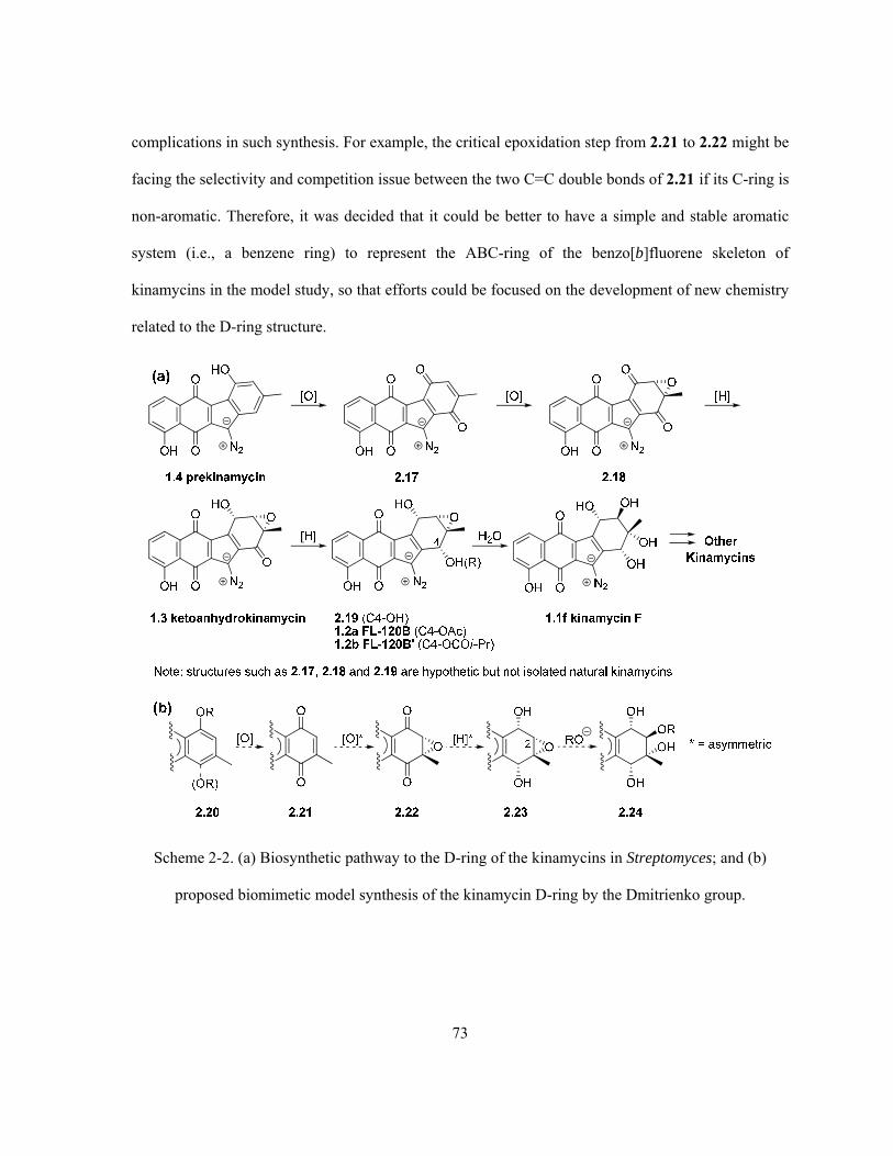
73
complications in such synthesis. For example, the critical epoxidation step from 2.21 to 2.22 might be
facing the selectivity and competition issue between the two C=C double bonds of 2.21 if its C-ring is
non-aromatic. Therefore, it was decided that it could be better to have a simple and stable aromatic
system (i.e., a benzene ring) to represent the ABC-ring of the benzo[b]fluorene skeleton of
kinamycins in the model study, so that efforts could be focused on the development of new chemistry
related to the D-ring structure.
Scheme 2-2. (a) Biosynthetic pathway to the D-ring of the kinamycins in Streptomyces; and (b)
proposed biomimetic model synthesis of the kinamycin D-ring by the Dmitrienko group.

74
2.2 A Simple Biogenetically-Inspired Synthesis of a Ring-D Model of
Kinamycin F
Based on the proposal (Scheme 2-2b) to carry out synthetic work in a manner mimicking the
extremely simple and effective biopathway of kinamycins (Scheme 2-2a), a model study for the
stereoselective elaboration of the D-ring of kinamycin in only three steps from an appropriate para-
quinone, analogous to the (possible) biogenetic intermediate 2.17, was performed successfully after
extensive attempts.165 It should be pointed out that while the chemistry discussed in this chapter was
being developed in the Dmitrienko group, the only literature work available at the time on the
synthesis of the kinamycin D-ring was that from Kumamoto and Ishikawa’s early results (Scheme 2-
1).163,164 After the completion of work described herein (but published at a much later time), Porco
and Lei (2006) released a total synthesis of natural (-)-kinamycin C (1.1c), whose D-ring (absolute)
stereochemistry was built from scratch starting from an achiral precursor.23 Later on in 2007,
Nicolaou’s group developed another total synthesis of kinamycin C (1.1c); however, the required
absolute stereochemistry of the D-ring originated from a chiral precursor this time.16 Yet none of the
above three methods was able to offer the same simplicity and efficiency to construct the kinamycin
D-ring as the Dmitrienko strategy.
The commercially available 2-methyl-1,4-naphthoquinone (2.25, Vitamin K3), which contains a
simple and stable benzene ring while possessing the essential para-quinone moiety with a methyl
substituent as required by the proposed model studies, served well for the purpose and thus was
chosen as the starting material. Nucleophilic Weitz-Scheffer epoxidation of 2.25 with 30% hydrogen
peroxide under mild basic conditions (aqueous Na2CO3 solution)166 afforded the corresponding epoxy
diketone 2.27 smoothly in 77% yield. Although various potential strategies for ensuring reduction of
2.27 with the desired stereoselectivity (i.e., the reduced diol hydroxyl groups must be syn to the epoxy
oxygen) were envisaged, NaBH4 was considered to be a good choice in this case due to the following
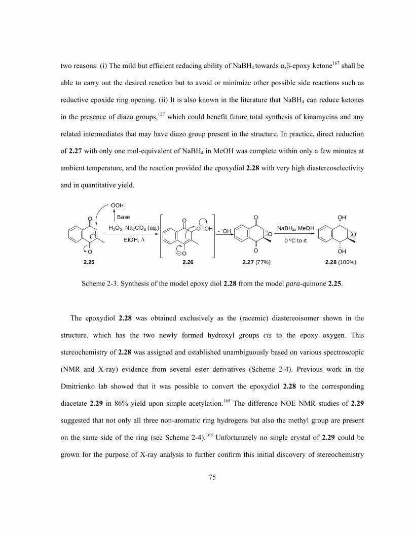
75
two reasons: (i) The mild but efficient reducing ability of NaBH4 towards α,β-epoxy ketone167 shall be
able to carry out the desired reaction but to avoid or minimize other possible side reactions such as
reductive epoxide ring opening. (ii) It is also known in the literature that NaBH4 can reduce ketones
in the presence of diazo groups,127 which could benefit future total synthesis of kinamycins and any
related intermediates that may have diazo group present in the structure. In practice, direct reduction
of 2.27 with only one mol-equivalent of NaBH4 in MeOH was complete within only a few minutes at
ambient temperature, and the reaction provided the epoxydiol 2.28 with very high diastereoselectivity
and in quantitative yield.
O
O
O
O
O
OH
OH
O
2.25 2.27 (77%)
H2O2, Na2CO3 (aq.)
EtOH,
NaBH4, MeOH
0 ºC to rt
2.28 (100%)
-OOH
BaseO
O
O OH
2.26
- -OH
Scheme 2-3. Synthesis of the model epoxy diol 2.28 from the model para-quinone 2.25.
The epoxydiol 2.28 was obtained exclusively as the (racemic) diastereoisomer shown in the
structure, which has the two newly formed hydroxyl groups cis to the epoxy oxygen. This
stereochemistry of 2.28 was assigned and established unambiguously based on various spectroscopic
(NMR and X-ray) evidence from several ester derivatives (Scheme 2-4). Previous work in the
Dmitrienko lab showed that it was possible to convert the epoxydiol 2.28 to the corresponding
diacetate 2.29 in 86% yield upon simple acetylation.168 The difference NOE NMR studies of 2.29
suggested that not only all three non-aromatic ring hydrogens but also the methyl group are present
on the same side of the ring (see Scheme 2-4).168 Unfortunately no single crystal of 2.29 could be
grown for the purpose of X-ray analysis to further confirm this initial discovery of stereochemistry

76
based on NMR evidence, since 2.29 exists as an oil at room temperature. However, the epoxydiol
2.28 was, later on in this study, transformed to the corresponding bis(3,5-dinitrobenzoate) ester 2.30
in 73% yield (Scheme 2-4), which turned out to be a very stable solid (mp > 260C). To ultimately
determine the stereochemistry of the saturated 6-membered ring within 2.28 (and 2.30), various
efforts had been made to grow single crystals of 2.30. Attempts with common pure organic solvents
or by vapor/liquid diffusion techniques all failed except in one case. Slow diffusion of Et2O vapor
into a concentrated solution of 2.30 in DMSO-d6 (a previous NMR sample) produced single crystals
of marginal quality for X-ray diffraction crystallography studies. For the first time, the single crystal
X-ray results of 2.30 (ORTEP plot in Scheme 2-4, details of X-ray are provided in Appendix B)
provided unambiguous confirmation of the stereochemistry of 2.28/2.30. However, due to the poor
quality of the single crystal used, it was noticed that one benzylic carbon atom in the obtained
ORTEP plot of 2.30 was significantly distorted (as marked in Scheme 2-4). Even though such a
severe crystal defect of 2.30 does not alter the conclusion concerning the stereochemistry of interest,
it was felt necessary to further prepare some other derivative(s) of the epoxydiol 2.28, in the hope that
better X-ray results might be achieved. In practice, the diisobutyryl ester 2.31 was prepared from 2.28
in 64% yield by acylation with i-butyryl chloride (Scheme 2-4). The ester 2.44 crystallized easily
from several solvents including Et2O, EtOAc, acetone, benzene, THF and CDCl3, all yielding good
quality single crystals suitable for X-ray studies. The corresponding ORTEP plot of 2.31 is also
shown in Scheme 2-4 (details of X-ray are provided in Appendix C), which once again confirmed the
expected stereochemistry of the epoxydiol 2.28.

77
OH
OH
Me
O
i-PrCOCl, Py, DMAP
2.28
Ac2O, Py, CH2Cl2, rt
OAc
OAc
O
2.29 (86%)
O
O
O
2.30 (73%)
O
O
O
2.31 (64%)
O
O
NO2
NO2
NO2
NO2
O
O
O
Cl
NO2
NO2
DMAP, Py
OAc
OAc
Me
O
H H
H
NMRdifference
NOE studiesof 2.29
ORTEP plot of 2.30 ORTEP plot of 2.31distorted carbon atom
due to poor crystal quality
Scheme 2-4. Synthesis of various ester derivatives of epoxy diol 2.28.
The observed high stereoselectivity in the reduction of 2.27 by NaBH4 was quite surprising but
could be explained partially by the hypothesis that solvation of the epoxy oxygen of 2.27 with MeOH
(reaction solvent) by means of hydrogen bonding interactions would cause a large steric barrier
(Scheme 2-5a), if the nucleophilic hydride approached the carbonyl groups from the side syn to the
epoxy oxygen.165 Therefore, the observed stereochemical outcome of the epoxydiol 2.28 is a direct
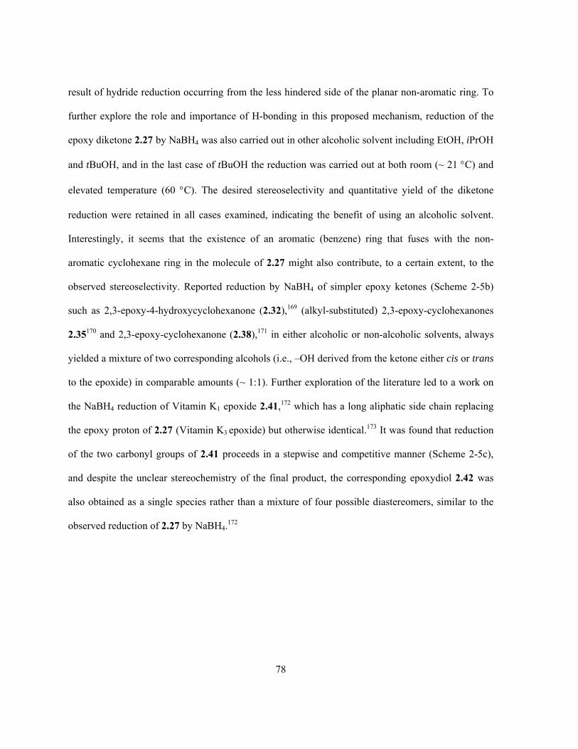
78
result of hydride reduction occurring from the less hindered side of the planar non-aromatic ring. To
further explore the role and importance of H-bonding in this proposed mechanism, reduction of the
epoxy diketone 2.27 by NaBH4 was also carried out in other alcoholic solvent including EtOH, iPrOH
and tBuOH, and in the last case of tBuOH the reduction was carried out at both room (~ 21 C) and
elevated temperature (60 C). The desired stereoselectivity and quantitative yield of the diketone
reduction were retained in all cases examined, indicating the benefit of using an alcoholic solvent.
Interestingly, it seems that the existence of an aromatic (benzene) ring that fuses with the non-
aromatic cyclohexane ring in the molecule of 2.27 might also contribute, to a certain extent, to the
observed stereoselectivity. Reported reduction by NaBH4 of simpler epoxy ketones (Scheme 2-5b)
such as 2,3-epoxy-4-hydroxycyclohexanone (2.32),169 (alkyl-substituted) 2,3-epoxy-cyclohexanones
2.35170 and 2,3-epoxy-cyclohexanone (2.38),171 in either alcoholic or non-alcoholic solvents, always
yielded a mixture of two corresponding alcohols (i.e., –OH derived from the ketone either cis or trans
to the epoxide) in comparable amounts (~ 1:1). Further exploration of the literature led to a work on
the NaBH4 reduction of Vitamin K1 epoxide 2.41,172 which has a long aliphatic side chain replacing
the epoxy proton of 2.27 (Vitamin K3 epoxide) but otherwise identical.173 It was found that reduction
of the two carbonyl groups of 2.41 proceeds in a stepwise and competitive manner (Scheme 2-5c),
and despite the unclear stereochemistry of the final product, the corresponding epoxydiol 2.42 was
also obtained as a single species rather than a mixture of four possible diastereomers, similar to the
observed reduction of 2.27 by NaBH4.172

79
O
O
Me
O
OH
OH
Me
O
2.41
2.28
3
OH
O
Me
O
HO
HOMe
O
H
OH
OH
Me
O
2.42 (single product)
3
+
2.43 (minor) 2.44 (major)
NaBH4, EtOH
(c)
(a) O
O
Me
OMeOH
NaBH4
2.27
Alcohol H-bonding with epoxide
limitedNaBH4
excessNaBH4
O
O
OH
OH
O
OH
OH
O
OH
+NaBH4
THF
45 : 552.32 2.33 2.34
(b) O
O
OH
O
OH
O+
2.35 2.36 2.37
R1 R1 R1
NaBH4
ROH
O
ONaBH4
MeOH
OH
O
OH
O+
48 : 522.38 2.39 2.40
R1 = R2 = R3 = R4 = H 42 : 58
R1 = Me; R2 = R3 = R4 = H 53.4 : 44.7
R1 = R2 = R3 = Me; R4 = H 85 : 15
R1= Me, R2 = R3 = H;
R4 = -C(CH3)=CH2 59.7 : 34.4
R4
R3
R4
R3
R4
R3R2 R2 R2
Scheme 2-5. (a) Proposed H-bonding model for the observed stereoselectivity on reduction of 2.27;
(b) literature examples of non-stereoselective reduction of cyclic epoxy ketones by NaBH4; (c)
literature example of reduction of Vitamin K1 epoxide 2.41.
With the epoxydiol 2.28 possessing the desired stereochemistry readily available in multigram
quantities at hand, the next key step was to tackle the epoxide ring opening (ERO) reaction of 2.28 or
its ester derivatives by an appropriate nucleophile (i.e., with a negatively charged or neutral oxygen
80
atom being the nucleophilic site) in a regio- (at the less hindered C2 epoxy carbon) and
stereoselective (backside attack) manner. The general strategy of the ring opening reaction of
epoxides relies heavily on the participation of a suitable protic or Lewis acid catalyst,174 particularly
in the cases requiring regio- and stereoselectivity, unless the nucleophilicity of the attacking reagent is
very strong (e.g., thiol-type nucleophiles175,176 or HO-/RO- at elevated temperature177-179). There are
many examples in the literature regarding the regioselective ERO of (cyclic) epoxides, epoxy ketones
and epoxy alcohols that possess structural similarities to compounds 2.28–2.31, and some of the
reported protic and Lewis acid catalysts for such ERO reactions include simple inorganic/organic
proton acids (such as HCl,180 HBr,181, HI182 H2SO4,183,184 HClO4,
185,186 HOAc,187,188 HOAc/alumina,189
Cl3CCO2H190), inorganic/organic metal salts (such as AlCl3,
191 Al(TFA)(iPrO)2,192 AlPO4/Al2O3,
193
BF3·Et2O,194 BiCl3,195 CeCl3,
196 CuSO4,197 CsF,198 FeCl3/SiO2,
199 Fe(ClO4)3,200 Fe(TFA)3,
201 MgSO4,202
MgBr2,166,203 MgI2,
204 NbCl5,205 TiCl3(OTf)/TiO(TFA)2,
206 Me3SiCl/DMSO207) and enzymes.208 Such
regioselective ERO might also be achieved through an one-electron oxidation process in the presence
of a nucleophile and an oxidizing reagent, such as Ce(IV) salts,209,210 DDQ211 and I2.212 Previous work
in the Dmitrienko group found that the epoxy diacetate 2.29 was unable to undergo the ERO under
several literature conditions168 and later attempts with 2.28 in this study following some other
reported procedures, which are summarized in Table 2-1, also failed. Either no reaction occurred with
mild conditions (Trial 1, 3 and 4), or extensive decomposition was observed when harsher (acidic or
basic) conditions were applied (Trial 2 and 5).

81
Table 2-1. Summary of failed attempts on ERO reactions of 2.28 and 2.29.
Trial Starting Material Reagents and ConditionsReference Obtained Product(s)
1a
OH
OH
Me
O
2.28
NaOAc, MgSO4, MeOH
↑↓202
Complete recovery of 2.28
2a
OH
OH
Me
O
2.28
NaOAc, HOAc
(90C), 4 hr187
ERO and elimination led to 2.25 as the only product
3b
OAc
OAc
Me
O
2.29
KOAc, Aliquat 336, CH3CN
rt, 72 hr213
Complete recovery of 2.29
4b
OAc
OAc
Me
O
2.29
H2O, Dioxane
(100 C), 18 hr168
Complete recovery of 2.29
5b
OAc
OAc
Me
O
2.29
NaOMe, MeOH
↑↓214
Complex mixture
Not characterized
a. This work; b. From M. B. Carriere’s MS.c. Dissertation.168
After the above unsuccessful attempts on the nucleophilic ERO of 2.28 and 2.29 to mimic the
putative biosynthetic conversion of 2.19 to kinamycin F (1.1f) (Scheme 2-2), it was eventually found
that the desired stereo- and regioselective ring opening of the epoxydiol 2.28 could be achieved in
good yield (76%) by using Me4NBH(OAc)3 in refluxing dry THF with excess NaOAc as the external
nucleophile (Scheme 2-6). Under such conditions, a mixture of two triol acetates 2.45a and 2.45b (a
pair of regioisomers) in a ratio of approximately 1:1 was obtained. The structures were assigned
based on extensive NMR spectroscopic analysis including 1H, 13C, HMQC, HMBC and difference

82
NOE (Scheme 2-6) analysis. Honda and Mizutani had previously used this boron reagent to induce
ring opening of both acyclic and cyclic 2,3-epoxy alcohols such as 2.50 (Scheme 2-6) but not of
epoxy diols.215 In the case of 2,3-epoxycyclohexanols, it was found that the ERO reaction succeeds
only if the alcohol and epoxide are syn to each other, and that ring opening occurs upon nucleophilic
attack at C3 by the acetoxy anion (released from the boron reagent) or other added external
nucleophiles.215 Interestingly, it was also noted in Honda’s original work that if the alcohol possesses
an anti correlation to the epoxide, the corresponding ERO reaction would lead to the formation of a
reduction product, in which the Me4NBH(OAc)3 acts as a nucleophilic hydride source rather than a
Lewis acid catalyst.215 This had led to the suggestion that the ERO reaction induced by
Me4NBH(OAc)3 occurs by a mechanism analogous to that proposed by Sharpless and co-workers for
ring-opening of 2,3-epoxy alcohols (2.53 in Scheme 2-6),216 2,3-epoxy acids and 2,3-epoxy amides
(2.54 in Scheme 2-6)217 with Ti(O-i-Pr)4.215
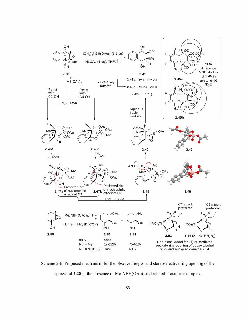
83
OH
OH
Me
O
2.28
OR
OH
OR'
OH
Me
2.45a: R= H, R'= Ac
2.45b : R= Ac, R'= H
(76%, ~ 1:1 )
2.45
(CH3)4NBH(OAc)3 (1.1 eq)
NaOAc (5 eq), THF,
Preferred siteof nucleophilicattack at C3
HB(OAc)3
OO
BOAc
OMe
AcOH
O, O-AcetylTransfer
Aqueousbasicworkup
AcO
OO
BOAc
OMe
H
Fast, - HOAc
OH
O
BOAc
OMe
H
2.47a
OAcO
OH B OAcO
Me
H
OAc
2.47b
- H2 , - OAc
2.48
2.49
1 2
34
ReactwithC1-OH
ReactwithC4-OH
4
2
3
OD
OD
OCOCH3
ODCH3
H
H
H
H
H
H
H
2.45a
OCOCH3
OD
OD
ODCH3
H
H
H
H
H
H
H
2.45b
NMRdifference
NOE studiesof 2.45 in
acetone-d6/D2O
OH
O
BOAcO
Me
H
OAc1 OAc
O
OH B OAcO
Me
H
OAc
OAc
- OAc - OAc
2.46a 2.46b 2.49
2.48
O(RO)3Ti
O
H R
HH
H
Sharpless Model for Ti(IV)-mediatedepoxide ring opening of epoxy alcohol
2.53 and epoxy acid/amide 2.54
O(RO)3Ti
X
H R
H
O
2.54 (X = O, NR1R2)2.53
C3 attackpreferred
C3 attackpreferred
OH
OMe4NBH(OAc)3, THF
Nu- (e.g. N3-, tBuCO2
-)OH
OH
OAc
+
OH
OH
Nu
2.50 2.51 2.52
no Nu- 94% -Nu- = N3
- 17-22% 75-61%Nu- = tBuCO2
- 14% 63%
1
3
Preferred siteof nucleophilicattack at C2
Scheme 2-6. Proposed mechanism for the observed regio- and stereoselective ring opening of the
epoxydiol 2.28 in the presence of Me4NBH(OAc)3 and related literature examples.

84
Essentially, the boronic reagent Me4NBH(OAc)3 is thought to react initially with the alcohol to
provide a Lewis acid, tethered to the alcohol oxygen atom. Further complexation intramolecularly
with the epoxy oxygen atom helps to induce the epoxide ring opening by a nucleophile. For
epoxydiol 2.28, either -OH group at C1 and C4 might react with Me4NBH(OAc)3 to form a
corresponding distinct adduct 2.47a or 2.47b respectively (Scheme 2-6), at the same time releasing an
acetoxy anion and hydrogen gas. The literature precedents would predict ring opening with rupture of
the epoxide C-O bond at C3 for adduct 2.47a, but with rupture of the C-O bond at C2 for adduct
2.47b. Since the accessibility of the reagent to the two alcohol groups in 2.28 is expected to be
comparable, the high degree of regioselectivity observed in this epoxide ring opening step is
somewhat surprising. It is suspected that the initial adducts, 2.47a and 2.47b, do not directly undergo
epoxide ring opening. Instead, both species may react further and quickly to form the same cyclic
complex 2.48 in which the boron serves as a Lewis acid intramolecularly and binds to all three
oxygen atoms that exist on the same side of the non-aromatic ring, to assist with the epoxide ring
opening by the nucleophile from a back-side attack at the sterically less crowded C2 position to give
2.49. The corresponding computation models of 2.48 and 2.49, generated by ab initio MO
calculations at RHF/6-31G//6-31G level, are shown in Scheme 2-6. Aqueous basic workup then
initially provides 2.45a, which equilibrates with 2.45b through a facile O, O-acetyl transfer that is
very likely to occur under the basic workup conditions involved. Such migration behavior of acyl
groups is well-known in the literature (e.g., 2.55 to 2.58, Scheme 2-7).218
OHO2C
HOHO
OHOAc
pH = 7.4 OHO2C
HOHO
OAcOH
OHO2C
HOAcO
OHOH
OHO2C
AcOHO
OHOH
2.55 2.56 2.57 2.58
Scheme 2-7. Literature example of acyl migration under mild basic conditions.

85
It should be pointed out that the ring-opening of 2.28 can also be achieved smoothly in the absence
of NaOAc but with a slightly lower yield (ca. 50–60%), and the nucleophile (i.e., the acetoxy anion
AcO-) required by the ERO reaction must be provided and released from Me4NBH(OAc)3 when
forming the boronic adducts in this case. Since Me4NBH(OAc)3 is also known to be an excellent
reducing agent for ketones,219 it was thought to be worthy to attempt a possible one-pot (double)
ketone reduction and ERO of the epoxy diketone 2.27 with Me4NBH(OAc)3. Unfortunately, the
attempted reaction in THF only afforded a very complicated mixture (not characterized) instead of the
desired product of 2.45.
The two regioisomers of the triol acetates 2.45a and 2.45b (~ 1:1) of the ring-opening product 2.45
were readily separated by flash column chromatography, even though the separation was not
necessary for the subsequent derivatization steps. The triol acetates 2.45 (as a mixture) were further
deacylated and afforded the corresponding tetrol 2.59 under several basic conditions (Scheme 2-8).
Deacetylation of the triol acetates 2.45 was initially achieved by either reduction with LiAlH4 in
anhydrous THF or a simple basic hydrolysis (aqueous NaOH solution), although low yields and
difficult isolation of the pure tetrol 2.59 were encountered in both cases (56% crude for LiAlH4
reduction and 25% crude for NaOH hydrolysis). Fortunately, a further attempt at the deacylation
reaction of 2.45 (mixture) under the Zemplen conditions (catalytic amount of NaOMe in MeOH)
followed by a workup using a proton resin instead of an aqueous acid,220 a method that is widely used
in sugar chemistry for deacylation purposes, was capable of generating 2.59 as a single product in
near quantitative (98%) yield. Moreover, peracetylation of the tetrol 2.59 was first carried out with
AcOH in the presence of Sc(OTf)3 as a Lewis acid catalyst, since literature work indicates that this is
a mild but efficient method for acetylation of all kinds of alcohols including the sterically hindered
ones.221 However, under such conditions, only partial (and random) acetylation of the tetrol 2.59
occurred at ambient temperature, while decomposition of the tetrol dominated when the acetylation
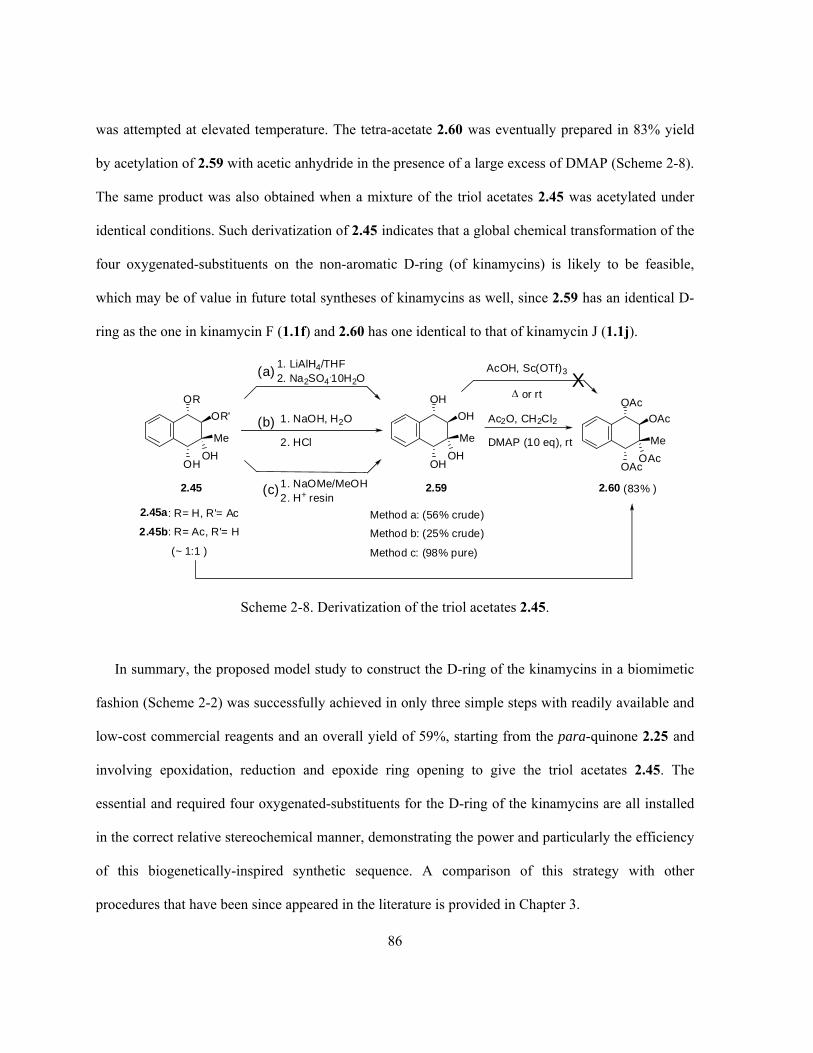
86
was attempted at elevated temperature. The tetra-acetate 2.60 was eventually prepared in 83% yield
by acetylation of 2.59 with acetic anhydride in the presence of a large excess of DMAP (Scheme 2-8).
The same product was also obtained when a mixture of the triol acetates 2.45 was acetylated under
identical conditions. Such derivatization of 2.45 indicates that a global chemical transformation of the
four oxygenated-substituents on the non-aromatic D-ring (of kinamycins) is likely to be feasible,
which may be of value in future total syntheses of kinamycins as well, since 2.59 has an identical D-
ring as the one in kinamycin F (1.1f) and 2.60 has one identical to that of kinamycin J (1.1j).
1. LiAlH4/THF2. Na2SO4
.10H2O
OR
OH
OR'
OH
Me
2.45a: R= H, R'= Ac
2.45b: R= Ac, R'= H
(~ 1:1 )
OH
OH
OH
OH
Me
Method a: (56% crude)
Method b: (25% crude)
Method c: (98% pure)
2.592.45
OAc
OAc
OAc
OAc
Me
2.60 (83% )
1. NaOH, H2O
2. HCl
1. NaOMe/MeOH2. H+ resin
Ac2O, CH2Cl2
DMAP (10 eq), rt
(a)
(b)
(c)
AcOH, Sc(OTf)3
or rtX
Scheme 2-8. Derivatization of the triol acetates 2.45.
In summary, the proposed model study to construct the D-ring of the kinamycins in a biomimetic
fashion (Scheme 2-2) was successfully achieved in only three simple steps with readily available and
low-cost commercial reagents and an overall yield of 59%, starting from the para-quinone 2.25 and
involving epoxidation, reduction and epoxide ring opening to give the triol acetates 2.45. The
essential and required four oxygenated-substituents for the D-ring of the kinamycins are all installed
in the correct relative stereochemical manner, demonstrating the power and particularly the efficiency
of this biogenetically-inspired synthetic sequence. A comparison of this strategy with other
procedures that have been since appeared in the literature is provided in Chapter 3.
87
The use of Me4NBH(OAc)3 in the ERO reaction provides more flexibility for various chemical
transformations of the epoxide functionality within the epoxydiol 2.28, since, in principle, other
nucleophiles such as halogens (e.g., Br- and I-) might also be introduced to the non-aromatic ring
through the ERO reaction. Using Me4NBH(OAc)3 as the ring-opening promoter, initial ERO attempts
on 2.28 with NaBr as the external nucleophile, however, led to a complex mixture of products (not
characterized). With the more nucleophilic I-, the corresponding iodohydrin 2.61 was generated as
expected in 90% yield (Scheme 2-9). Since coupling reagents and conditions suitable for the
dimerization of iodocyclohexane 2.63,222 2-iodocyclohexanone 2.65223 and some rather complicated
α-iodoketones 2.67224 are known in the literature (Scheme 2-9), the possibility that the obtained
iodohydrin 2.61 might be converted into the corresponding dimer 2.62 is worthy of consideration,
which is structurally closely related to lomaiviticin A (1.11a), a dimeric diazobenzo[b]fluorene.
Alternatively, proper protection of the hydroxy groups within 2.61 can lead to substrates such as 2.69,
which may undergo a possible base-induced elimination to give the corresponding enolate 2.70
followed by a hydrolysis/deprotection to yield 2.71 (Scheme 2-9). This compound possesses a
saturated cyclohexyl ring with very minor variation(s) of the ring substituents that is similar to the D-
ring of lomaiviticin A (1.11a). Such potential synthetic applications with the structure of 2.62 could
open doors to construction of simple analogues of the biologically potent dimeric
diazobenzo[b]fluorene of lomaiviticins.

88
OH
OH
Me
O
2.28
OH
OH
I
OH
Me
2.61 (90%)
(CH3)4NBH(OAc)3(1.1 eq)
NaI (5 eq), THF
OH
HOHO
OH
OHOH
2.62
OH
OH
O
O
N O
O
O
O
O
R'
H
H
R
H
Et
EtO
R
H
R'
N
O
O
OH
OH
N
N
1.11a lomaiviticin A
OOH
NMe2
R' =
O OHOMe
R =
Possibledimerization(see literatureexamples onthe right)
OAc
OAc
I
OAc
Base-inducedelimination
OAc
OAc
OAc
O
OH(Ac)
OH(Ac)
H+/-OHHydrolysis
2.69
2.70 2.71
R1
OR2
R3
2.67 (cis + trans)(R1, R2, R3 = OMe/H)
H
I
R1
OR2
R3H
R1
R2
R3
O
H
HH
In, DMF orLa, THF
2.68 (R1= R2= R3= H, In/DMF, 3-5%)(R1, R2, R3= H/OMe, La/THF, 10-20%)
I
2.64 (81%)2.63
CuCl2, Mn
H2O/THF
Ac2O, Py
Literature examples of aliphatic iodide (similar to 2.61)dimerization:
O
IO O
2.66 (53%)
In, DMF
2.65
Scheme 2-9. ERO reaction of 2.28 by halogen nucleophile in the presence of Me4NBH(OAc)3 and the
potential synthetic applications of 2.61 as a model compound.
Last but not least, there are some additional comments on the above model study that are worth
further discussion. Even though epoxidation of the quinone 2.25 was not carried out in a

89
stereoselective manner in this study, there are some literature examples regarding the asymmetric
epoxidation of such α,β-unsaturated ketones including 2.25, which normally involves the use of an
appropriate chiral catalyst to control the stereochemical outcome along with an epoxidation reagent
for the chemical transformation (or the direct use of a chiral epoxidation reagent to complete the two
tasks simultaneously). Early work in this area showed that asymmetric epoxidation of 2.25 affords the
corresponding epoxide 2.27 with fair to good yields but only with low to moderate ee (either
enantiomer is available), depending on the chiral catalysts/peroxides used, such as chiral phase-
transfer catalysts (PTC) of quaternary ammonium salts derived from quinidine (72–94% yield, 1–
34% ee),225,226 cinchonine (53–63% yield, 10–18% ee),227 and quinine (60–70% yield, ~5% ee),228,229
chiral sugar hydroperoxides (71% yield, 45% ee),230 α-/β-cyclodextrin (33–100% yield, 0–24% ee)231
and bovine serum albumin (BSA, 20–34% yield, 20–47% ee).232-234 On the other hand, if the methyl
group on the quinone moiety of 2.25 is replaced by sterically bulkier substituents such as long alkyl
chain (C8), i-Pr, cyclohexyl and aryl groups (Ph/Ar), the observed stereoselectivity of epoxidation
immediately increases to a (much) higher and reasonable level of ee (64–100% ee).225,226,235,236
Introducing substituents onto the benzene ring of 2.25 also slightly increases the ee of the obtained
epoxide under the same stereoselective epoxidation conditions.228
More interestingly, recent progress regarding the asymmetric epoxidation of 2.25 indicates that
(Scheme 2-10),237 in the presence of some readily available Cinchona alkaloids (e.g., quinidine 2.75
and quinine 2.76)-based chiral PTCs such as 2.72–2.74, and a careful choice of epoxidation
conditions (NaOCl as a mild peroxidation reagent, low reaction temperature and an appropriate
binary solvent system of chlorobenzene/water), stereoselective epoxidation of 2.25 could produce
either enantiomer of the corresponding epoxide (2.27a/2.27b in Scheme 2-10) with significantly
improved ee (67–85%) and good yields (73–92%). Note that the enantiomer 2.27a possesses the
identical absolute stereochemistry as the D-ring of the proposed kinamycin biointermediate 2.18 as

90
shown in Scheme 2-2. The stereoselectivity achieved in epoxidation of such a flat and near-symmetric
naphthoquinone was explained by the difference in steric interactions in the intermediate chlorooxy
enolate (2.77)-PTC (e.g., 2.72) ion pair depending on the enantiomer of 2.77 and the chiral PTC
involved, which was formed through possible hydrogen bonding, Coulombic interaction and -
stacking (Scheme 2-10). This new development makes the model synthetic strategy described in this
chapter more attractive and valuable towards being applied to the enantioselective total synthesis of
kinamycins and related antibiotics.

91
O
O
Me
O
O
Me
O
2.252.27b (2R, 3S)
NaOCl (aq.)2.73 or 2.74 (cat.)
PhCl, -10 ºC
O
O
Me
O
2.27a (2S, 3R)
N
CH3
N
Cl
H OH
OH
2.73
2
3
2
3
with 2.5 mol% 2.7373%, 85% ee
with 10 mol% 2.7490%, 67% ee
NaOCl (aq.), 2.72 (cat.)
PhCl, -10 ºC
with 1 mol% 2.7287%, 76% ee
with 10 mol% 2.7292%, 72% ee
N
CH2
N
Cl
H OH
OCH3
2.74
N
CH2
Cl
2.72
H OH
N
OCH3
N
CH2
H OH
N
OCH3
2.75 quinidine
N
CH2
NH OH
OCH3
2.76 quinine
stackingH-bonding
Coulombicinteraction
Left: computation model for the ion pair of the
epoxidation intermediate chlorooxy enolate (2.77)and chiral PTC (2.72). The top enantiomeric ion
pair (2.77a-2.62) is predicted to be more stable
than the bottom one (2.77b-2.72) by ca. 3.5 kJ/mol.
O
O
Me
OCl
2.77a
2.25
ClO-
- Cl-
fast
slow
O
O
Me
OCl
2.77b
O
O
Me
fast
- Cl- slow
2.27a (2S, 3R) 2.27b (2R, 3S)
Scheme 2-10. Proposed mechanism for the improved asymmetric epoxidation of 2.25.

92
2.3 Conformational Analysis of the D-Ring of Kinamycin F and Model
Compounds
While reviewing NMR spectroscopic data of the natural kinamycins presented in the literature, it
was noted that the reported D-ring H1-H2 coupling constant for kinamycin F (1.1f) was unusually
small (J = 3.4 Hz in DMSO-d6)9 for such a vicinal H-H coupling, presumablely pseudo axial-axial, as
one would normally expect (8–10 Hz).132 This coupling constant is also inconsistent with those of
other natural kinamycins and their synthetic derivatives, as listed in Table 2-2, which are all about 7–
9 Hz (in CDCl3) regardless of the various substituents on their D-rings. Therefore, it was first thought
that the significant difference in the reported coupling constants between kinamycin F (1.1f) and other
kinamycins could very likely be due to a simple error of observation or even just a typographical error
in the literature. In addition, the model compound 2.59 that possesses exactly the same (relative)
stereochemistry and substituents of the D-ring as kinamycin F (1.1f), also shows a typical vicinal H1-
H2 coupling constant of 6.9 Hz in DMSO-d6. Literature examples of some other similar cyclic
polyalcohols such as 2.78 and 2.79 give coupling constants around the same range (ca. 7.8–9.9 Hz,
Table 2-2) as well. One might also consider the fact that the use of DMSO-d6 (for kinamycin F)
instead of CDCl3 (for other kinamycins) as NMR solvent could affect the observed coupling constant
to a certain extent; however, it is generally believed that “vicinal H-H coupling constants exhibit a
negligible solvent dependence in the absence of solvent-induced changes in conformational
populations”.238 In fact, the model tetrol 2.59 served as a good example (Table 2-2, J = 6.9 Hz in
DMSO-d6 and J = 7.0 Hz in CDCl3) in support of this conclusion. Thus, it was possible that the
unusually small coupling constant of 1.1f, if it was not a mistake, would indicate that kinamycin F
(1.1f) may adopt a quite different conformation than other kinamycins.

93
Table 2-2. Literature JH-H of some natural kinamycins, derivatives and related model compounds.
Natural Kinamycin JH1–H2 (Hz)
in CDCl3
Natural Kinamycin
(* Synthetic derivatives)
JH1–H2 (Hz)
in CDCl3
HO
O
O N2
HO
OH
OAc
OAc
1 2
1.1a kinamycin A
8.59 HO
O
O N2
AcO
OH
OAc
OAc
1 2
1.1c kinamycin C
7.29,16
HO
O
O N2
HO
OH
OAc
OAc
1 2
1.1d kinamycin D
8.09
8.15
7.910 HO
O
O N2
HO
OH
OAc
OH
1 2
1.1e kinamycin E
7.95
HO
O
O N2
HO
OH
OH
OH
1 2
1.1f kinamycin F
(DMSO-d6)
3.49 HO
O
O N2
AcO
OCOiPr
OAc
OAc
1 2
1.1g kinamycin G
7.07
HO
O
O N2
AcO
OH
OCOiPr
OAc
1 2
1.1h kinamycin H
7.07 HO
O
O N2
AcO
OAc
OAc
OAc
1 2
1.1j* kinamycin J
7.59
6.616
HO
O
O N2
HO
OCOiPr
OAc
OAc
1 2
1.1m FL120-A
8.110 HO
O
O N2
HO
OH
OCOiPr
OAc
1 2
1.1n FL120-C
8.110

94
Table 2-2. (continued).
Natural Kinamycin
(* Synthetic derivatives)
JH1–H2 (Hz)
in CDCl3
Natural Kinamycin
(* Synthetic derivatives)
JH1–H2 (Hz)
in CDCl3
HO
O
O N2
HO
OCOEt
OAc
OH1 2
1.1o FL-120C'
8.110 HO
O
O N2
HO
OH
OCO iPr
OH
1 2
1.1p FL-120D'
7.810
O
O
O N2
AcO
OH
OAc
OAc
O
Br
1 2
1.1s*
7.89 HO
O
O N2
HO
O
OH
O
1.1t*
9.09,18
AcO
O
O N2
AcO
O
OH
O
1.1u*
9.09,18 AcO
O
O N2
AcO
OAc
OAc
OAc
1.1q*
6.09
Model Compound JH–H (Hz) Model Compound JH–H (Hz)
OH
OHOH
OH1 2
2.59
(CDCl3)
H1–H2: 7.0
OH
OHOH
OH1 2
2.59
(DMSO-d6)
H1–H2: 6.9
OH
OH
OH
OH1 2
34
2.78
(CD3OD)239
H1–H2: 8.0
H2–H3: 9.9
H3–H4: 3.7
OH
OH
OH1 2
34
Ha Hb
2.79
(Pyridine-d5)240
H1–H2: 3.8
H2–H3: 7.8
H3–Ha4: 6.3
H3–Hb4: 8.8
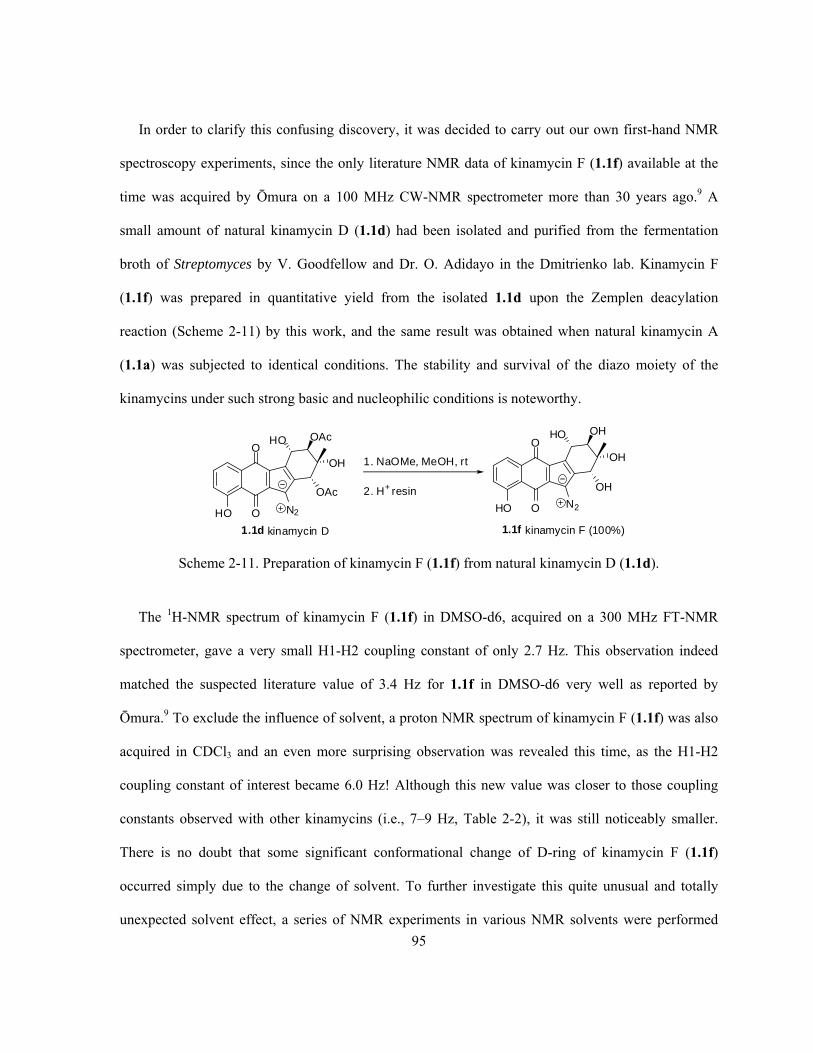
95
In order to clarify this confusing discovery, it was decided to carry out our own first-hand NMR
spectroscopy experiments, since the only literature NMR data of kinamycin F (1.1f) available at the
time was acquired by Ōmura on a 100 MHz CW-NMR spectrometer more than 30 years ago.9 A
small amount of natural kinamycin D (1.1d) had been isolated and purified from the fermentation
broth of Streptomyces by V. Goodfellow and Dr. O. Adidayo in the Dmitrienko lab. Kinamycin F
(1.1f) was prepared in quantitative yield from the isolated 1.1d upon the Zemplen deacylation
reaction (Scheme 2-11) by this work, and the same result was obtained when natural kinamycin A
(1.1a) was subjected to identical conditions. The stability and survival of the diazo moiety of the
kinamycins under such strong basic and nucleophilic conditions is noteworthy.
HO
O
O N2
HO
OH
OAc
OAc
1. NaOMe, MeOH, rt
2. H+ resin
HO
O
O N2
HO
OH
OH
OH
1.1d kinamycin D 1.1f kinamycin F (100%)
Scheme 2-11. Preparation of kinamycin F (1.1f) from natural kinamycin D (1.1d).
The 1H-NMR spectrum of kinamycin F (1.1f) in DMSO-d6, acquired on a 300 MHz FT-NMR
spectrometer, gave a very small H1-H2 coupling constant of only 2.7 Hz. This observation indeed
matched the suspected literature value of 3.4 Hz for 1.1f in DMSO-d6 very well as reported by
Ōmura.9 To exclude the influence of solvent, a proton NMR spectrum of kinamycin F (1.1f) was also
acquired in CDCl3 and an even more surprising observation was revealed this time, as the H1-H2
coupling constant of interest became 6.0 Hz! Although this new value was closer to those coupling
constants observed with other kinamycins (i.e., 7–9 Hz, Table 2-2), it was still noticeably smaller.
There is no doubt that some significant conformational change of D-ring of kinamycin F (1.1f)
occurred simply due to the change of solvent. To further investigate this quite unusual and totally
unexpected solvent effect, a series of NMR experiments in various NMR solvents were performed

96
with kinamycin F (1.1f), kinamycin D (1.1d) and some model compounds (i.e., 2.59, 2.60 and 2.45).
The corresponding results of the observed vicinal H1-H2 coupling constants are summarized in Table
2-3. Among all the compounds examined, clearly kinamycin F (1.1f) is the only one that shows a
solvent dependency of the observed coupling constant, and it is also the only species that gives a
much smaller H-H coupling constant than usual under certain circumstances.
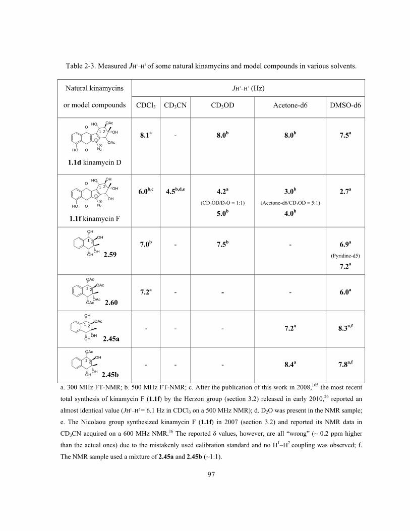
97
Table 2-3. Measured JH1–H2 of some natural kinamycins and model compounds in various solvents.
JH1–H2 (Hz) Natural kinamycins
or model compounds CDCl3 CD3CN CD3OD Acetone-d6 DMSO-d6
HO
O
O N2
HO
OH
OAc
OAc
1 2
1.1d kinamycin D
8.1a
-
8.0b
8.0b
7.5a
HO
O
O N2
HO
OH
OH
OH
1 2
1.1f kinamycin F
6.0b,c
4.5b,d,e
4.2a
(CD3OD/D2O = 1:1)
5.0b
3.0b
(Acetone-d6/CD3OD = 5:1)
4.0b
2.7a
OH
OHOH
OH1 2
2.59
7.0b
-
7.5b
-
6.9a
(Pyridine-d5)
7.2a
OAc
OAcOAc
OAc1 2
2.60
7.2a
-
-
-
6.0a
OH
OHOH
OAc1 2
2.45a
-
-
-
7.2a
8.3a,f
OAc
OHOH
OH1 2
2.45b
-
-
-
8.4a
7.8a,f
a. 300 MHz FT-NMR; b. 500 MHz FT-NMR; c. After the publication of this work in 2008,165 the most recent
total synthesis of kinamycin F (1.1f) by the Herzon group (section 3.2) released in early 2010,26 reported an
almost identical value (JH1–H2 = 6.1 Hz in CDCl3 on a 500 MHz NMR); d. D2O was present in the NMR sample;
e. The Nicolaou group synthesized kinamycin F (1.1f) in 2007 (section 3.2) and reported its NMR data in
CD3CN acquired on a 600 MHz NMR.16 The reported δ values, however, are all “wrong” (~ 0.2 ppm higher
than the actual ones) due to the mistakenly used calibration standard and no H1–H2 coupling was observed; f.
The NMR sample used a mixture of 2.45a and 2.45b (~1:1).

98
The 1H-NMR spectra of the triol acetates, 2.45a and 2.45b, and the tetra-acetate 2.60 reveal that
the observed coupling constants (JH1–H2 = 6.0–8.3 Hz in DMSO-d6) are consistent with the
predominance, in each case, of a half-chair conformation in which the C1 and C2 hydroxy or acetoxy
groups are in pseudo-equatorial orientations, so that the H1 and H2 would exist in such an orientation
(both pseudo-axial) that the dihedral angle of H1-C1-C2-H2 is large enough to give a “normal”
coupling constant. The comparable magnitude of JH1–H2 reported for those natural kinamycins as
listed in Table 2-2 except kinamycin F (1.1f) is likewise consistent with such a conformational
preference for their D-rings. X-ray crystallographic studies of the C7-O-p-bromobenzoate derivative
of kinamycin C 1.1s19 and of the C1-O-S-2-methylbutyrate derivative of kinamycin D 1.1v21 suggest
that this conformational preference persists in the solid state as well. The observed JH1–H2 for the tetrol
2.59 (6.9 Hz in DMSO-d6 and 7.0 Hz in CDCl3) and tetra-acetate 2.60 (6.0 Hz in DMSO-d6 and 7.2
Hz in CDCl3) was also consistent with a similar conformational preference regardless of the solvent.
It was surprising to find, however, that JH1–H2 for the fully deacylated kinamycin F (1.1f), in DMSO-
d6 was just 2.7 Hz. The gradual change of the coupling constant from 6 Hz to 2.7 Hz, as observed
with kinamycin F (1.1f) when gradually changing the NMR solvent from the less polar CDCl3 to
more polar DMSO-d6, would suggest that kinamycin F (1.1f), unlike the model 2.59 and other
kinamycins, exhibits a different (opposite) conformational preference for its D-ring. Kinamycin F
(1.1f), particularly in the polar solvents, has an unusual preference for a half chair conformation
where the C1-OH and C2-OH groups are pseudo-axial (Scheme 2-12, conformer A), and
consequently the H1 and H2 would exist in pseudo-equatorial orientation having a rather small
dihedral angle of H1-C1-C2-H2 and JH1–H2. This conclusion is especially interesting given the recent
suggestion that the kinamycin F (1.1f) may represent the bioactive form of the other kinamycins.241
Further study on this phenomenon provided more supportive evidence from ab initio MO calculation
of 1.1f in both gas phase and solution phase and experimental IR observations, and details of the

99
corresponding results will be discussed in Chapter 4. Although various attempts have been made to
grow single crystals of kinamycin F (1.1f) in the hope of obtaining further physical evidence, no
success has been achieved so far.
A B
1 2
Scheme 2-12. Calculated two energy minimum conformations of kinamycin F (1.1f) in which the
conformation A is preferred.
2.4 Conclusion
In summary, a new and biomimetic strategy for constructing the highly oxygenated and
stereospecific D-ring of kinamycin F (1.1f), in four steps from a D-ring quinone precursor and 57%
overall yield, has been demonstrated in a model study using 2-methyl-1,4-naphthylquinone (2.25) as
the starting material. Additionally, spectroscopic analysis as well as ab initio molecular orbital
calculations have led to the conclusion that kinamycin F (1.1f), unlike the other kinamycins, prefers a
D-ring conformation in which the C4-OH group is in a pseudo-equatorial orientation. Enhanced
diazonium ion character is predicted for this preferred conformation (see Chapter 4) and may
influence the biological activity of kinamycin F (1.1f). These observations have now appeared in
print.165

100
2.5 Experimental Details
2.5.1 General Information
Materials and Methods. All reactions were carried out in flame and oven-dried glassware under an
argon or nitrogen atmosphere, unless otherwise specified. All solvents were reagent grade. Anhydrous
tetrahydrofuran (THF) was freshly distilled from sodium/benzophenone under nitrogen prior to use.
Deionized water was supplied by a Biolab vertical series reverse osmosis system. All commercial
reagents were purchased from either Aldrich Chemical Co. or BDH Inc., and were used as received
unless otherwise indicated. Reactions were magnetically stirred and monitored by thin layer
chromatography (TLC) with Merck pre-coated silica gel plates (silica gel 60 F254 on aluminum sheet).
Flash column chromatography was carried out using the Merck silica gel (70–230 or 230–400 mesh)
or the SiliCycle silica gel (60 Å). All reported yields refer to chromatographically and
spectroscopically pure compounds, unless otherwise specified.
Characterization Methods. 1H NMR spectra were recorded on a Brüker AVANCE500 (500 MHz),
Brüker AVANCE300 (300 MHz), Brüker AC300 (300 MHz) or Brüker AC200 (200 MHz) NMR
spectrometer. 13C NMR spectra were broad band decoupled and recorded on a Brüker AVANCE500
(125.8 MHz), Brüker AVANCE300 (75.5 MHz), Brüker AC300 (75.5 MHz) or Brüker AC200 (50
MHz) NMR spectrometer using the carbon signal of the deuterated solvent as the internal standard.
HMQC, HMBC and difference NOE experiments were performed on the Brüker AVANCE500
spectrometer. The following abbreviations are used for NMR peak multiplicities: s, singlet; d,
doublet; t, triplet; q, quartet; dd, doublet of doublets; dt, doublet of triplets; m, multiplet; br, broad; w,
weak. Chemical shifts are reported in parts per million (ppm) relative to either TMS ( 0.0),
chloroform ( 7.26), DMSO ( 2.50) or acetone ( 2.05) for 1H NMR, and either chloroform (
77.16), DMSO ( 39.52) or acetone ( 29.84) for 13C NMR.242 Infrared spectra (IR) were recorded on

101
a Perkin-Elmer spectrum RX I FT-IR spectrometer as KBr discs or thin films on NaCl plates, as
indicated in each individual case. High resolution mass spectra (HRMS) and low resolution mass
spectra (LRMS), obtained via electron impact (EI) and/or chemical ionization (CI), were measured by
the McMaster Regional Centre for Mass Spectrometry (Department of Chemistry, McMaster
University, Hamilton, Ontario, Canada) or by the WATSPEC Mass Spectrometry Facility
(Department of Chemistry, University of Waterloo, Waterloo, Ontario, Canada).
Elemental/combustion analysis was performed by M-H-W Laboratories (Phoenix, Arizona, USA).
Melting points were determined on a MEL-TEMP melting point apparatus and were uncorrected.
2.5.2 Detailed Experimental Procedures
O
O
Me
O
2,3-Epoxy-2-methyl-1,4-naphthoquinone 2.27: The commercial 2-methyl-1,4-naphthoquinone 2.25
(98%, 5.005 g, 28.5 mmol) was dissolved completely in ethanol (20 mL) upon heating the solution to
about 70 C under an air atmosphere. The solution was then cooled to about 50 C and aqueous
hydrogen peroxide solution (30%, 10 mL) was added dropwise during a period of 20 min. Then
aqueous sodium carbonate solution (2.5 g of Na2CO3 dissolved in 10 mL of deionized water) was
added dropwise during a period of 15 min in order to avoid a rapid increase of the solution
temperature. A large amount of gas bubbles were generated during the addition of the Na2CO3
solution, and the yellow color of the quinone solution gradually disappeared. The almost colorless
reaction solution was kept stirring for another 5 min after the addition of base, during which some
white solid precipitated from the solution. Upon addition of deionized water (250 mL), a large
amount of white solid precipitated from the solution. The white solid was collected by vacuum

102
filtration and washed thoroughly with H2O (25 mL). The product was further dried overnight under
vacuum to yield a fine white power (4.112 g, 21.9 mmol, 77% yield) as the desired pure compound.
(Note: the epoxidation is quite exothermic so precaution to control the speed of addition of the base
and reaction temperature is necessary when working on larger scales). IR (KBr) 1701, 1595, 1337,
1298, 1250, 1194 cm-1; 1H NMR (300 MHz, CDCl3) 8.05–7.89 (m, 2 H), 7.80–7.68 (m, 2 H), 3.85
(s, 1 H), 1.72 (s, 3 H); 13C NMR (75 MHz, CDCl3) 192.0, 191.9, 134.7, 134.5, 132.2, 132.1, 127.6,
126.9, 61.6, 61.5, 14.8; HRMS (EI) m/z 188.0470 [M+; calculated for C11H8O3: 188.0473]; mp 93.5–
94.5 C (lit.166 94.5–95.5 C).
OH
OH
Me
O
2,3-Epoxy-1,4-hydroxy-2-methyl-1,2,3,4-tetrahydronaphthalene 2.28: 2,3-Epoxy-3-methyl-1,4-
naphthoquinone 2.27 (0.376 g, 2.0 mmol) was dissolved in methanol (30 mL) and the solution was
cooled to about 0 C with an ice bath. Sodium borohydride (0.0767 g, 2.0 mmol) was added to the
solution in one portion under stirring and then the ice bath was removed. The solution was stirred for
another 10 min and the temperature of the solution raised to ca. 18 C. The solvent was then removed
in vacuo at room temperature and the remaining white solid was mixed with saturated aqueous NH4Cl
solution (250 mL). The obtained aqueous solution was extracted thoroughly with ethyl acetate (6 80
mL), and the combined organic phase was dried over Na2SO4 and concentrated in vacuo. The epoxy
diol 2.28 was obtained quantitatively as a white powder (0.388 g, 2.0 mmol, 100% yield). IR (KBr)
3295, 1508, 1420, 1348, 1186, 1118, 1094, 1042, 1009 cm-1; 1H NMR (200 MHz, DMSO-d6) 7.52–
7.37 (m, 2 H), 7.30–7.18 (m, 2 H), 5.84–5.72 (m, 2 H), 4.80 (d, J = 7.0 Hz, 1 H), 4.65 (d, J = 8.5 Hz,
1 H), 3.33 (d, J = 1.5 Hz, 1 H), 1.47 (s, 3 H); 13C NMR (50 MHz, DMSO-d6/D2O) 136.5, 135.7,

103
126.89, 126.85, 125.5, 125.4, 68.7, 65.8, 61.9, 59.4, 19.2; HRMS (EI) m/z 192.0789 [M+; calculated
for C11H12O3: 192.0786]; mp 148–153 C (decomp.).
O
O
O
O
O
NO2
NO2
NO2
NO2
2,3-Epoxy-1,4-hydroxy-2-methyl-1,2,3,4-tetrahydronaphthalene bis(3,5-dinitrobenzoate ester)
2.30: The commercially available 3,5-dinitrobenzoyl chloride (98+%, 1.180 g, 5.0 mmol) was mixed
with a catalytic amount of 4-dimethylaminopyridine (DMAP, 0.0131 g, 0.11 mmol). Pyridine (10
mL) was added to the above solid mixture and the resulting solution was stirred under a N2
atmosphere in an ice bath. The epoxy diol 2.28 (0.240 g, 1.25 mmol) was dissolved in pyridine (2
mL) and the obtained orange-red solution was added dropwise to the acid chloride/pyridine solution
during a period of 5 min. The flask containing the epoxy diol-pyridine solution was further washed
with pyridine (3 1 mL), which was also transferred into the reaction flask. The clear yellow reaction
solution was stirred at ambient temperature for 10 hr, with the observation that the solution gradually
turned very turbid and the color of the reaction mixture became brown-red at the end. The reaction
was quenched by pouring the mixture into previously cooled H2O (20 mL) in an ice bath and the
obtained aqueous mixture was further stirred for 5 min, and was then extracted with CH2Cl2 (3 40
mL). The organic phases were thoroughly washed with saturated aqueous CuSO4 solution (3 60
mL), H2O (2 60 mL) and 5% aqueous NaHCO3 solution (5 60 mL). The CH2Cl2 solution was
dried over Na2SO4 and the solvent was removed in vacuo. A light brown powder was obtained as the
crude product (0.673 g). Recrystallization of the crude product in organic solvents such as ethanol,
toluene, CH2Cl2 and acetone all failed due to the low solubility of the product. However, it was found
104
that acetone could efficiently wash off the colored impurities from the crude product. Therefore, the
crude product was washed with a minimal amount of acetone under slightly reduced pressure,
yielding some pale gray powder as the pure final product (0.526 g, 0.9 mmol, 73% yield). IR (KBr)
3449, 3089, 1730, 1630, 1546, 1459, 1347, 1269, 1163, 1122, 1076 cm-1; 1H NMR (500 MHz,
DMSO-d6) 9.13 (dt, J = 2.0, 0.5 Hz, 1 H), 9.12 (dt, J = 2.0, 0.5 Hz, 1 H), 9.05 (dd, J = 2.0, ~0.5 Hz,
2 H), 9.03 (dd, J = 2.0, ~0.5 Hz, 2 H ), 7.45–7.37 (m, 3 H), 7.29 (d, J = 8.5 Hz, 1 H), 6.79 (s, 1 H),
6.75 (s, 1 H), 3.95 (d, J = 2.0 Hz, 1 H), 1.53 (s, 3 H); 13C NMR (75 MHz, DMSO-d6) 162.8, 162.6,
148.5, 148.4, 132.4, 131.8, 131.0, 130.2, 129.4, 129.2, 128.3, 125.8, 125.4, 123.0, 122.9, 73.6, 71.0,
57.9, 57.7, 18.4; HRMS (CI) m/z 580.0773 [M+; calculated for C25H16N4O13: 580.0714], 598.1039
[(M+NH4)+; calculated for C25H20N5O13: 598.1058]; mp > 260 C.
O
O
Me
O
O
O
2,3-Epoxy-1,4-hydroxy-2-methyl-1,2,3,4-tetrahydronaphthalene Diisobutyryl ester 2.31: The
epoxy diol 2.28 (0.385 g, 2.0 mmol) was mixed with a catalytic amount of DMAP (0.0506 g, 0.41
mmol), followed by the addition of pyridine (20 mL). Then isobutyryl chloride (98%, 0.86 mL, 8.0
mmol) was added dropwise through a syringe during a period of 0.5 min to the above reaction
mixture, and the resulting solution was stirred under the air atmosphere for 24 hr at ambient
temperature. The reaction was quenched by dilution with H2O (20 mL) and the obtained solution was
further stirred for 15 min, during which a large amount of white precipitate formed. The precipitate
was filtered and washed with H2O (20 mL) and it was then re-dissolved in CH2Cl2 (50 mL). The
CH2Cl2 solution was washed with saturated aqueous CuSO4 solution (1 50 mL) and H2O (1 50

105
mL). The organic solution was dried over Na2SO4 and the solvent was removed in vacuo, yielding a
white solid as the pure product (0.426 g, 1.27 mmol, 64% yield). IR (KBr) 2980, 2933, 1735, 1496,
1448, 1437, 1388, 1355, 1187, 1146, 1103 cm-1; 1H NMR (300 MHz, CDCl3) 7.40–7.20 (m, 3 H),
7.10–7.00 (m, 1 H), 6.32 (s, 1 H), 6.22 (s, 1 H), 3.57 (s, 1H), 2.90–2.70 (m, 2 H), 1.49 (s, 3 H), 1.40–
1.30 (m, 12 H); 13C NMR (75 MHz, CDCl3) 177.1, 177.0, 131.4, 130.4, 128.19, 128.12, 125.7,
125.5, 70.3, 68.3, 58.5, 57.6, 34.3, 19.26, 19.20, 18.99, 18.78; LRMS (CI) m/z 350.2 [(M+NH4)+;
calculated for C19H28NO5: 350.4]; mp 120–120.5 C.
OH
OH
OAc
OH
Me
OAc
OH
OH
OH
Me
Triol acetates 2.45: The epoxy diol 2.28 (0.963 g, 5.00 mmol) was mixed with NaOAc (99%, 2.072
g, 25.0 mmol) and the commercial Me4NBH(OAc)3 (95%, 1.525 g, 5.50 mmol) in a flame- and oven-
dried round bottom flask, followed by addition of anhydrous THF (50 mL). A large amount of gas
bubbles were generated at the beginning and the reaction mixture was heated to reflux in an oil bath
for 6 hr under an argon atmosphere. The reaction mixture was further stirred for 1 hr without heating
until the solution cooled gradually to room temperature. The reaction was quenched by the addition of
saturated aqueous NaHCO3 solution (50 mL) and the mixture was further stirred for 10 min. Another
50 mL of H2O was added to completely dissolve the remaining NaOAc powder. The aqueous solution
was then extracted thoroughly with CH2Cl2 (8 100 mL) and the organic phase was dried over
Na2SO4 and concentrated in vacuo. The crude epoxide ring-opening product was obtained as a
slightly yellow foamy solid (0.809 g). Some more crude product was successfully recovered from the
aqueous phase in the previous extraction step: the aqueous phase was concentrated to dryness and the
remaining solid residue was stirred with 2 L of CH2Cl2, and the CH2Cl2 solution was then dried over
Na2SO4, leading to another 0.180 g of the crude product upon removal of the solvent. The combined

106
crude product (0.989 g) was further purified by charcoal decolorization in CH2Cl2 and some off-white
foamy solid (0.956 g, 3.79 mmol, 76% yield) was obtained as the final pure product, which consists
of two regioisomers of the triol acetates 2.45a and 2.45b in an approximate ratio of 1:1, as determined
by NMR. This “pure” mixture is directly suitable for the next Zemplen deacylation and peracetylation
reaction. If the pure form of each of the regioisomers is desired, flash column chromatography using a
mixture of EtOAc and hexanes (3:1, v/v) as the eluent could be performed directly with the crude
product, without the need of charcoal decolorization. The separated two pure triol acetates were then
subjected to extensive NMR spectroscopy studies, including 1H, 13C, difference NOE, HMBC and
HMQC, to establish the stereochemistry assigned.
Mixture of 2.45a and 2.45b (~ 1:1): IR (KBr) 3423, 1736, 1720, 1376, 1255, 1096, 1025 cm-1; IR
(film) 3396, 2936, 1716, 1491, 1456, 1374, 1252, 1097, 1024 cm-1.
Regioisomer 2.45a: IR (film) 3392, 1719, 1375, 1251, 1094, 1037 cm-1; 1H NMR (300 MHz, acetone-
d6) 7.57 (d, J = 7.8 Hz, 1 H), 7.50–7.20 (m, 3 H), 5.45 (d, J = 7.5 Hz, 1 H), 4.72 (d, J = 6.3 Hz, 1
H), 4.70–4.55 (m, 2 H), 4.47 (d, J = 4.4 Hz, 1 H), 3.78 (s, 1 H), 2.08 (s, 3H), 1.11 (s, 3 H); 1H NMR
(500 MHz, acetone-d6/D2O) 7.56 (d, J = 8.0 Hz, 1 H), 7.40 (d, J = 7.0 Hz, 1 H), 7.36–7.27 (m, 2
H), 5.44 (d, J = 8.0 Hz, 1 H), 4.56 (d, J = 7.5 Hz, 1 H), 4.46 (s, 1 H), 2.08 (s, 3 H), 1.11 (s, 3 H); 13C
NMR (125 MHz, acetone-d6/D2O) 171.4, 138.1, 137.0, 130.5, 128.86, 128.85, 128.5, 77.7, 76.0,
73.1, 71.1, 21.2, 19.6; HRMS (EI) m/z 235.0956 [(M-OH)+; calculated for C13H15O4: 235.0970];
HRMS (CI) m/z 235.0989 [(M-OH)+; calculated for C13H15O4: 235.0970], 252.1258 [(M+NH4-H2O)+;
calculated for C13H18NO4: 252.1236], 253.1130 [(M+H)+; calculated for C13H17O5: 253.1076]; mp
153–155 C; Elemental/combustion analysis: calculated for C13H16O5: C 61.90, H 6.39; found: C
61.89, H 6.48.

107
Regioisomer 2.45b: IR (film) 3415, 2926, 1725, 1644, 1493, 1457, 1374, 1246, 1099, 1027 cm-1; 1H
NMR (300 MHz, acetone-d6) 7.50–7.10 (m, 4 H), 5.89 (d, J = 8.1 Hz, 1 H), 4.49 (s, 1 H), 4.38 (d, J
= 4.5 Hz, 1 H), 4.30–4.15 (m, 1 H), 3.91 (s, 1 H), 2.14 (s, 3 H), 1.11 (s, 3 H); 1H NMR (500 MHz,
acetone-d6/D2O) 7.43–7.38 (m, 1 H), 7.34–7.28 (m, 2 H), 7.19–7.14 (m, 1 H), 5.88 (d, J = 8.5 Hz, 1
H), 4.49 (s, 1 H), 4.20 (d, J = 8.5 Hz, 1 H), 2.14 (s, 3 H), 1.10 (s, 3 H); 13C NMR (125 MHz, acetone-
d6/D2O) 171.8, 137.9, 134.9, 131.3, 129.0, 128.9, 128.0, 76.0, 75.1, 73.6, 73.0, 21.2, 18.7; HRMS
(EI) m/z 234.0895 [(M-H2O)+; calculated for C13H14O4: 234.0892]; HRMS (CI) m/z 235.1003 [(M-
OH)+; calculated for C13H15O4: 235.0970], 252.1256 [(M+NH4-H2O)+; calculated for C13H18NO4:
252.1236], 253.1064 [(M+H)+; calculated for C13H17O5: 253.1076]; mp 45–48 C.
OH
OH
OH
OH
Me
Tetrol 2.59:
Method (a) Reductive deacylation of triol acetates 2.45 by LiAlH4:
A mixture of the two triol acetates 2.45 (0.10 g, 0.4 mmol) was dissolved in anhydrous THF (5 mL)
under argon atmosphere and the solution was cooled in an ice bath. A commercial THF solution of
LiAlH4 (1.0 M, 2 mL, 2 mmol) was added dropwise to the reaction flask through a syringe during a
period of 5 min. The reaction solution was warmed to ambient temperature by removing the ice bath
and further stirred for 0.5 h, followed by the addition of Na2SO4·10H2O (0.66 g, 2 mmol) in two
portions to quench the reaction. The reaction mixture was further stirred with ethyl acetate (200 mL)
for 2 h and then filtered under reduced pressure through a Büchner funnel preloaded with a layer of
celite (~ 2 cm thick). The solid residue in the funnel was thoroughly washed with ethyl acetate (200
mL). The combined ethyl acetate solution was concentrated in vacuo and a yellow oil was obtained as

108
the crude product (0.05 g, 59% crude yield), which solidified upon drying under vacuum overnight. A
repeated reaction with larger amounts of 2.45 (0.16 g, 0.65 mmol) produced a crude yield of 56%.
NMR spectroscopy indicated that the major component of crude product was indeed the desired
product but various purification processes turned out to be quite difficult and inefficient. See the
section of “Method (c)” for spectroscopic data of the produced tetrol 2.59.
Method (b) Deacylation of triol acetates 2.45 by simple basic hydrolysis:
A freshly prepared NaOH solution (7.6 mL, 0.1975 M, 1.5 mmol) was added in one portion to a
mixture of the two triol acetates 2.45 (0.05 g, 0.2 mmol) and the reaction mixture was stirred at room
temperature for 2 h under argon atmosphere. The reaction mixture was then cooled in an ice bath and
the excess NaOH was neutralized by adding 0.5 M HCl solution (3 mL) dropwise until the pH of the
reaction mixture reached about 6 (as indicated by pH paper). The aqueous solution was extracted with
CH2Cl2 (4 20 mL). The organic phase was dried over Na2SO4 and concentrated in vacuo, yielding a
white solid as the crude product (0.009 g, 0.04 mmol, 25% crude yield). See the section of “Method
(c)” for spectroscopic data of the produced tetrol 2.59.
Method (c) Zemplen deacylation of triol acetates 2.45
A fresh MeOH solution of NaOMe (1 M) was prepared by completely reacting sodium metal (0.23 g,
10 mmol) with small amount of methanol (~ 4 mL) first. The final volume of this NaOMe/MeOH
solution was adjusted to 10.0 mL in a volumetric flask. A mixture of the two triol acetates 2.45a and
2.45b (~1:1, 0.470 g, 1.86 mmol) was dissolved in methanol (18.8 mL). The previously prepared 1 M
NaOMe/MeOH solution (5.0 mL, 5 mmol) was added dropwise to the reaction mixture and the
solution was stirred at room temperature for 1 h under the air atmosphere. The reaction was quenched

109
by the addition of about 10 g of a weak acidic ion (H+) exchange resin (CGC-271, 200–400 mesh,
chromatographic grade, weak acid, carboxylic, methylacrylate, 2.5% XL with a total exchange
capacity of 9.6% meq/g). The mixture was stirred for 5 min until the pH of the solution reached
around 6, as indicated by wet pH paper. Vacuum filtration gave a light yellow filtrate and the ion
exchange resin was further washed with MeOH (100 mL). The combined MeOH filtrate yielded a
white (slightly yellow) foamy solid upon concentration in vacuo, which was then subjected to a
charcoal decolorization in acetone. The obtained almost colorless acetone solution of the product
gave a white foamy solid (0.385 g, 1.83 mmol, 98% yield) upon removal of the solvent, which was
proven by NMR spectroscopy to be the desired tetrol 2.59. The compound 2.59 should be stored in a
dessicator since it is slightly hydroscopic in its solid state. IR (film) 3390, 2925, 2855, 1645, 1458,
1378, 1262, 1099, 1024 cm-1; 1H NMR (300 MHz, DMSO-d6) 7.44 (d, J = 6.9 Hz, 1 H), 7.36–7.20
(m, 3 H), 5.28 (d, J = 6.6 Hz, 1 H), 5.00 (d, J = 5.1 Hz, 1 H), 4.87 (s, 1 H), 4.41 (s, 1 H), 4.29 (d, J =
3.9 Hz, 1 H), 4.21 (t, J = 6.3 Hz, 1 H), 3.77 (d, J = 6.6 Hz, 1 H), 1.00 (s, 3 H); 13C NMR (75 MHz,
DMSO-d6) 138.2, 137.1, 129.0, 128.0, 127.3, 127.0, 74.4, 74.0, 72.7, 72.0, 20.1; HRMS (EI) m/z
192.0781 [(M-H2O)+; calculated for C11H12O3: 192.0786], 174.0674 [(M-2H2O)+; calculated for
C11H10O2: 174.0681]; HRMS (CI) m/z 210.1122 [(M+NH4-H2O)+; calculated for C11H16NO3:
210.1130], 228.1214 [(M+NH4)+; calculated for C11H18NO4: 228.1236].
OAc
OAc
OAc
OAc
Me
Tetra-acetate 2.60: The tetrol 2.59 (0.053 g, 0.25 mmol) and N, N-dimethylaminopyridine (DMAP,
99%, 0.307 g, 2.5 mmol) were dissolved in CH2Cl2 (5 mL), resulting in a faint red-brown solution.
Acetic anhydride (99.4%, 0.24 mL, 2.5 mmol) was added dropwise during a period of 3 min at room
temperature under stirring, which caused the faint red-brown color of the solution to immediately turn
110
to light yellow. The yellow reaction mixture was then stirred for 12 hr at room temperature under air
atmosphere. MeOH (10 mL) was then added and the reaction mixture was further stirred for 20 min
to quench the excess acetic anhydride. Upon removal of the solvent, some yellow-orange solid was
obtained and it was then mixed with dilute HCl (2 M, 30 mL), followed by extraction with Et2O (3
50 mL). The obtained light yellow organic phase was dried over Na2SO4 and concentrated in vacuo.
A slightly yellow foamy solid was obtained as the crude product (0.085 g). The yellow color of the
crude product was completely removed by charcoal decolorization in Et2O. The pure product was
obtained as a snow-white foamy solid (0.080 g, 0.21 mmol, 83% yield). IR (film) 3474, 2992, 2939,
1750, 1374, 1256, 1227 1043, 1023 cm-1; 1H NMR (300 MHz, DMSO-d6) 7.47–7.24 (m, 4 H), 6.48
(s, 1 H), 5.96 (d, J = 6.0 Hz, 1 H), 5.85 (d, J = 6.0 Hz, 1 H), 2.13 (s, 3 H), 2.10 (s, 3 H), 2.08 (s, 3 H),
1.93 (s, 3 H), 1.50 (s, 3 H); 1H NMR (300 MHz, CDCl3) 7.55–7.50 (m, 1 H), 7.42–7.35 (m, 2 H),
7.23-7.20 (m, 1 H), 6.78 (s, 1 H), 6.14 (d, J = 7.2 Hz, 1 H), 5.94 (d, J = 7.2 Hz, 1 H), 2.16 (s, 3 H),
2.14 (s, 3 H), 2.10 (s, 3 H), 1.99 (s, 3 H), 1.58 (s, 3 H); 13C NMR (75 MHz, CDCl3) 171.0, 170.1,
169.9, 169.7, 132.9, 132.7, 130.7, 129.8, 129.4, 128.7, 128.1, 80.5, 72.7, 72.6, 71.5, 22.0, 21.2, 21.1,
16.4; HRMS (EI) m/z 319.1180 [(M-OAc)+; calculated for C17H19O6: 319.1182].
OH
OH
I
OH
Me
Iodohydrin 2.61: The epoxy diol 2.28 (0.096 g, 0.5 mmol), Me4NBH(OAc)3 (95%, 0.15 g, 0.55
mmol) and sodium iodide (98%, 0.383 g, 2.5 mmol) were mixed and dissolved in anhydrous THF (5
mL). The reaction solution was heated to reflux in an oil bath for 6 h under an argon atmosphere. A
large amount of gas bubbles was generated at the beginning and the color of the solution turned from
clear to yellow. The reaction mixture was further stirred for 1 hr without heating until it cooled
gradually to the ambient temperature, followed by the addition of saturated aqueous NaHCO3 solution

111
(10 mL) and further stirring for 10 min. The solution was then stirred with 10% aqueous Na2S2O3
solution (10 mL) for 5 min. After thorough extraction with CH2Cl2 (6 25 mL), the obtained organic
phase was dried over Na2SO4 and concentrated in vacuo. A red shiny flake was obtained as the pure
product (0.145 g, 0.45 mmol, 90% yield). IR (KBr) 3400, 1017 cm-1; 1H NMR (500 MHz, acetone-
d6) 7.59 (d, J = 7.5 Hz, 1 H), 7.41–7.29 (m, 3 H), 4.97 (d, J = 8.0 Hz, 1 H), 4.84–4.79 (m, 1 H),
4.75 (d, J = 9.0 Hz, 1 H), 4.67 (d, J = 5.5 Hz, 1 H), 4.61 (d, J = 5.0 Hz, 1 H), 4.24 (s, 1 H), 1.20 (s, 3
H); 13C NMR (125 MHz, acetone-d6) 130.8, 129.1, 128.9, 128.7, 75.9, 75.8, 74.6, 74.5, 50.6, 23.4;
HRMS (CI) m/z 320.0169 [(M+NH4-H2O)+; calculated for C11H15INO2: 320.0148], 338.0301
[(M+NH4)+; calculated for C11H17INO3: 338.0253].
HO
O
O N2
HO
OH
OH
OH
Kinamycin F 1.1f: A NaOMe-MeOH solution (1 M) was prepared by dissolving the commercial
NaOMe (95% pure, 0.5687 g, 0.01 mol) in MeOH (HPLC grade, 10.00 mL) in a volumetric flask.
Pure natural kinamycin D (1.1d) (0.0087 g, 0.0191 mmol), isolated from bacteria fermentation broth,
was dissolved in MeOH (3 mL) and the reaction flask was completely covered with aluminum foil to
avoid exposure to light. Then the previously prepared NaOMe-MeOH solution (1 M, 0.1 mL, 0.1
mmol) was added dropwise through a syringe under stirring to the bright orange yellow kinamycin D-
MeOH solution, whose color turned immediately to very dark purple. The reaction mixture was then
stirred at room temperature for 1 hr to complete the deacetylation. To a white slurry of ion (H+)
exchange resin (3 g, CGC-271, 200–400 mesh, chromatographic grade, weak acid, carboxylic,
methylacrylate, 2.5% XL with a total exchange capacity of 9.6% meq/g) in MeOH (10 mL) under
stirring, the dark purple reaction mixture was added dropwise and further stirred for 5 min, and the

112
slurry turned to dark orange red. The slurry was filtered under reduced pressure and the remaining
resin was washed with copious amount of MeOH (ca. 40 mL) until it looked snow-white. The crude
product was obtained as some a brown red solid (thin film) upon removal of solvent, which was
further purified on a small dry silica gel column (3 cm x 2.5 cm) with acetone as the eluent. Pure
kinamycin F (1.1f) was obtained as a dark brown-red fine powder quantitatively (0.0071 g, 0.0192
mmol, 100% yield). IR (KBr) 3438, 2142, 1622, 1456, 1238, 1158, 1087 cm-1; IR (MeOH film) 3468,
2137, 1620, 1458, 1087 cm-1; 1H NMR (300 MHz, DMSO-d6) 12.31 (s, disappear after D2O
exchange, 1 H), 7.68 (t, J = 8.1 Hz, 1 H), 7.58 (d, J = 6.9 Hz, 1 H), 7.26 (d, J = 8.1Hz, 1 H), 5.43 (d, J
= 9.3 Hz, disappears after D2O exchange, 1 H), 5.38 (d, J = 4.2 Hz, disappears after D2O exchange, 1
H), 5.01 (s, disappears after D2O exchange, 1 H), 4.99 (d, J = 9.0 Hz, disappears after D2O exchange,
1 H), 4.73 (br dd; after D2O exchange, d, J = 2.7 Hz; 1 H), 4.64 (d, J = 9.0 Hz; after D2O exchange, s;
1 H), 3.74 (br t; after D2O exchange, d, J = 2.1 Hz, 1 H), 1.28 (s, 3 H); HRMS (+ESI-MS, in 1:1 (v/v)
CH3CN/H2O with sucrose and LiOAc as additives) m/z 377.0962 [(M+Li)+; calculated for
C18H14N2O7Li: 377.0961; (sucrose+Li)+ with a known m/z of 349.1322 was used as the internal
standard].
113
Chapter 3
Synthetic Attempts towards the Kinamycin Antibiotics and
Analogues
3.1 Synthetic Work towards the Kinamycin Benzo[b]fluorene Skeleton
The Dmitrienko lab is among a few research groups that have been involved in synthetic studies
aimed at the kinamycins antibiotics and related structures since the late 1980s, and work in this lab
has led to several important publications regarding the various aspects of the chemistry of the
kinamycins over the years.14,22,25,73,124,165,241,243,244 Prior to the start of this research project, Laufer from
the Dmitrienko group had proposed and partially developed two synthetic strategies towards the
benzo[b]fluorene skeleton of kinamycins (Scheme 3-1a),80 which were based on even earlier results
by Mithani245 and Carrière168 also from the Dmitrienko lab. The benzo[b]fluorene structure 3.1 could
be constructed (Scheme 3-1a) via either (i) an intramolecular Jacobson-Torssell coupling of 3.2 (i.e.,
arylation/alkylation of p-quinone with free radicals derived from the corresponding carboxylic acid
under strong oxidative conditions246,247), which itself can be prepared from an intermolecular
Jacobson-Torssell coupling of the halogenated juglone 3.3 and a substituted homophthalic acid 3.4 or
(ii) a Ni(0)-catalyzed intramolecular coupling of 3.7, which could be conceivably prepared through an
intermolecular Jacobson-Torssell coupling of 3.3 and substituted o-halophenylacetic acid 3.6. In fact,
initial success to synthesize model compounds such as 3.10168 and 3.1380 (Scheme 3-1b) was achieved
by following the above retrosynthetic proposals.

114
OR O
O OR
OR O
XOR
HO2C
O
HO2C
A BD
OR O
ROO
3.3
3.13.2
3.4
11a11b
11b11a
H
3.5 (X = halogen)
OR O
Br
O
R'CO2H, (NH4)2S2O8
AgNO3, aq. MeCN,
(R' = Me, Ph, Bn,o-Br-C6H4CH2)
OR O
Br
O
R'
1. H2/Pd-C, AcOHAcONa
2. air (R' = Bn)
OAc O
H
O
3.9 (95%)
OAc O
H
O
3.10 (40%)
Ph
N2
Ph
3.8a (87%, R= Ac; R' = Me)3.8b (72%, R= Ac; R' = Ph)3.8c (96%, R= Ac; R'= Bn)3.8d (~85%, R=Ac/Me, R'=o-Br-C6H4CH2)
TsN3, Et3N
MeCN
(a)
ZnAc2OEt3N
or1. P(OEt)3, H2O
2. MeI, Ag2OOR OR'
Br
Br
OR'
3.12 (> 78%, from 3.8d,R, R' = Ac, Me)
Zn, PPh3, DMF
NiCl2(H2O)6 (2 eq)or NiBr2OR OR'
OR'
3.7a R = Ac3.7b R = Me
3.13 (14-72%,R, R' = Ac, Me)
(b)
OR OR1
XOR1 OR
11b11a
OR O
X OR
X
O
HO2C
A BD
3.3 3.6
XHO2C
A B
D
C
OAc O
Ph
O
3.11 (93%)
Ph
PhCO2H, Ag+
S2O82-,
Scheme 3-1. (a) Original retrosyntheses of the benzo[b]fluorene skeleton by the Dmitrienko lab; (b)
synthesis of kinamycin model compounds based on retrosyntheses in (a).
However, both strategies suffered from their own deficiencies at certain steps when being applied
towards the total synthesis of prekinamycin (1.4) (Scheme 3-2).80 For example, the key intramolecular
Ni(0)-catalyzed cyclization of 3.16 to form the benzo[b]fluorene skeleton experienced a low yield of
only 27% (Scheme 3-2a) even after extensive optimization. In addition, the apparently simple D-ring
precursors involved, such as 3.15 (Scheme 3-2a) and 3.20 (Scheme 3-2b), had to be prepared through
complex sequences that also encountered some difficult chemical transformations with low yields. As

115
a continuation of these available yet unpublished results, initial synthetic efforts of this thesis project
were made to repeat and attempt the incomplete synthetic route outlined (with dashed arrow) in
Scheme 3-2b, which required the preparation of 3.19 in fairly large quantities as an essential starting
material to begin with. Unfortunately, large scale preparation of 3.19 turned out to be problematic and
yields were significantly lower than the small scale ones (64–94%). Another issue with these two
synthetic paths is that, even though the desired diazo transfer reactions at C5 of benzo[b]fluorenes
was carried out successfully in the case of model compound 3.26 (Scheme 3-2c), the same chemical
derivatization at C5 of 3.17 and simpler analogues (i.e., without A-ring and/or D-ring substituents of
3.17) were found to be very difficult.80 These results led to some further reconsideration of the
original retrosynthetic plans within the Dmitrienko group. An alternative synthetic approach was
proposed in order to avoid the above challenges, and the corresponding experimental results will be
discussed next in detail.

116
(a)
(b)
O
HO Me
OAc
Me
Br
HO2C
3.7a, (NH4)2S2O8,
AgNO3, aq. CH3CNOAc
Me
Br
Br
O
OOAc
OAc
OAcOAc
OAc
Zn, Ac2O
Et3N
OAc OAc
Br
Br
OAc
3.16 (87%) 3.17 (27%)
Me
OAc
3.15 (5 steps, 30%) 3.16 (51%)
Me
O
OMe
O
Me
OH
CO2Me
CO2Me
3.21
3.20 (4 steps, < 1% from 3.21)
(6 steps, 14-25% from 3.19)
NiCl2(H2O)6 (2 eq)
Zn, PPh3, DMF
3.14
OMe
MeMe
OMe
Me Me
3.18
3.19
Ac2O
H2SO4 (cat.)
3.7a, (NH4)2S2O8
AgNO3, aq. CH3CN
3.22 (81%)
OAc
Me
CO2Me
3.23 (100%)
O
OOAc
Br
OAc
Me
CO2H
O
OOAc
AcO
MeO
OOAc
1. H2/Pd2. Me2SO4, K2CO33. H2O, EtOH, HCl4. Ac2O, H2SO45. CAN, aq. CH3CN
3.243.25
(NH4)2S2O8,
AgNO3, aq. CH3CN
CO2H
Me
OAc
CO2Me
CO2H
OMe
OMe
N2OMe
OMe
n-BuLi, THF, -78 oC
then TsN3
DDQ, HOCH2CH2OH
PhH, rt to
MeO
OMe
OO
3.273.26 3.28 (51 %, 2 steps)
(c)
Scheme 3-2. Synthetic attempts towards the benzo[b]fluorene of prekinamycin (1.4) based on
retrosynthetic plans of Scheme 3-1a.

117
3.1.1 New Retrosynthesis of the Benzo[b]fluorene System
The proposed new synthetic strategy towards the benzo[b]fluorene skeleton of the kinamycins is
based on the new retrosynthetic analysis as illustrated in Scheme 3-3. Initial disconnection still occurs
at C5 of the generic diazobenzo[b]fluorene structure 3.29 of kinamycin. Introduction of the diazo
moiety to the benzo[b]fluorenone precursor 3.30, either through a diazo transfer with 3.30a similar to
the previous model study (Scheme 3-2c) or a conversion of C5-ketone 3.30b to the corresponding
hydrazone followed by oxidation, should provide the target kinamycins. The fluorene 3.30a or the
fluorenone 3.30b might be derived from appropriate reduction or oxidation of the corresponding
alcohol (not shown), which originates from decarboxylation of the α-hydroxy carboxylic acid 3.31.
The challenge of constructing the essential but rare 6-6-5-6 benzo[b]fluorene skeleton of 3.31 (and
kinamycins), is then transformed into an easier synthetic task with many literature precedents, which
aims at a precursor possessing a much more common 6-6-6-6 aglycon core. Contraction of the C-ring
upon disconnection at the C5-C6 bond of two candidates, either the o-quinonediazide 3.32 or the
ortho-quinone 3.33, through either a Wolff rearrangement (for 3.32) or a base-induced rearrangement
(for 3.33), should be able to achieve the goal. Both 3.32 and 3.33 might be prepared from the same
precursor 3.34, except that the former requires a diazo transfer at C5 of 3.34 while the latter demands
a selective C-ring oxidation. By taking advantage of the newly developed biomimetic and highly
efficient elaboration of kinamycin D-ring (Chapter 2), the non-aromatic and oxygenated D-ring of
3.34, analogous to those found in the kinamycins, might be synthesized from the quinone precursor
3.35 through a sequence of asymmetric epoxidation, stereoselective reduction and regio-
/stereoselective epoxide ring opening. Obviously the D-ring quinone of 3.35 is a result of
regioselective oxidation of the fully aromatized 3.36, as long as appropriate ABC-ring phenol
protecting groups are present. Then the 6-6-6-6 aglycon skeleton of 3.36 would be formed via an
intramolecular Friedel-Crafts cyclization of the biaryl species 3.37, which is obviously a product from

118
the Suzuki coupling of AB-ring precursor 3.38 and D-ring precursor 3.39. In general, either of 3.38
and 3.39 can be prepared in the form of a boronate or boronic acid, while the remaining one would be
the corresponding aryl halide.
ring co
ntractio
n
to3.31b via
Wolff
rearrangement
oxidation
ring contraction
to 3.31a via
base-induced
rearrangement
diazo transfer
at C5
Scheme 3-3. New retrosynthetic analysis of the diazobenzo[b]fluorenes of kinamycins.

119
In regard to the proposed ring contraction step (i.e., 3.32/3.33 to 3.31), the century-old Wolff
rearrangement approach involving an α-diazoketone (or o-quinonediazide 3.32 in this case) has been
well documented and thoroughly reviewed,248 as the literature examples under either thermal
conditions (e.g., 3.44 to 3.45249,250), photolysis (e.g., 3.40 to 3.41251,252 and 3.44 to 3.46249,250) or even
microwave conditions (e.g., 3.42 to 3.43253) shown in Scheme 3-4a. On the other hand, the proposed
ring contraction of 3.33 through the base-induced rearrangement is strongly supported by a fairly
recent (2006) literature report from the Mal group (Scheme 3-4b),254 which was not available when
the above route was proposed in the Dmitrienko group. The α-hydroxy carboxylic acids 3.52
possessing a 6-6-5-6 benzo[b]fluorene skeleton of kinamycins were prepared in good yields by a
simple base (KOH)-induced ring contraction (a.k.a benzil-benzilic acid rearrangement255) of the
corresponding 6-6-6-6 aglycon precursors 3.51, which in turn were synthesized by using the Hauser
annulation152 of 3.47 (an equivalent to those indenones in Hauser annulation) and 3.48-3.50 (synthons
equivalent to phthalides in Hauser annulation).254
The above synthetic strategy towards kinamycin involves many well-studied reactions at various
key steps; on the other hand, it also utilizes the new biomimetic route for the elaboration of D-ring
(Chapter 2). A bonus advantage of this route is that the intermediate α-diazoketone 3.32 with the 6-6-
6-6 aglycon skeleton, if successfully prepared, is in fact a novel analogue of the 6-6-5-6
diazobenzo[b]fluorene and 6-5-6-6 diazobenzo[a]fluorene type kinamycins. Analogue 3.32 may itself
have useful biological activity and may be of value for studies on the mode-of-action and influence of
structures on the biological activities of these unusual diazo-containing antibiotics.

120
Scheme 3-4. Literature examples of ring contraction through (a) Wolff rearrangement and (b) base-
induced rearrangement related to the benzo[b]fluorene skeleton of kinamycins.
3.1.2 Synthetic Work on the Kinamycin AB-Ring Precursor
The proposed kinamycin AB-ring precursor 3.38 (Scheme 3-3), either in the form of an aryl
boronic acid/boronate or aryl halide, is a naphthol-based compound that can be easily prepared

121
through a few simple chemical transformations (Scheme 3-5), starting from the commercially
available and inexpensive 1,5-dihydroxynaphthalene (3.54). There are several literature procedures
describing the synthesis of similar compounds such as 3.56,256 3.58257 and 3.59258 (Scheme 3-5). The
naphthol 3.54 could be smoothly acetylated with acetic anhydride to the corresponding diacetoxy
derivative 3.55, which was then brominated with NBS in hot (55-65 °C) aqueous acetic acid leading
to the key intermediate of bromojuglone 3.7a for the reported several transformations. The one-pot
bromination and oxidation from 3.55 to 3.7a, which was originally developed by Grunwell and
Heinzman,259 required a minimum of 3 equivalents of NBS (relative to 3.55) to complete the reaction,
but the use of 4 equivalents of NBS offered better yields in practice. Therefore, Grunwell proposed
the mechanism as shown in Scheme 3-5b to explain such experimental observations, where the nature
of the electrophilic brominating agent was not specified.259 It was later argued by Jung and Hagenah
that the actual brominating reagent was likely the molecular bromine (Br2) derived from NBS in
situ.260 Jung and Hagenah isolated a tribromo species 3.69 as an intermediate when the reaction was
carried out at a lower temperature of 45 °C, which was converted back to the bromojuglone 3.7a
quantitatively upon further heating in aqueous acetic acid at 65 °C (Scheme 3-5c).260 The same results
were also independently observed later by Mithani from the Dmitrienko lab.245 Thus, Jung proposed a
different mechanism (Scheme 3-5c).260 An unstable 5-acetoxy-2,3,4-tribromo-3,4-
dihydronaphthalene-1(2H)-one (3.67) was believed to be the initial bromination product, whose
subsequent dehydrobromination led to the dibromonaphthol 3.68 that further reacted with NBS to
afford 3.69. Jung also suggested the possibility that a direct electrophilic bromodeacetylation of 3.55
yielding a monobromophenol followed by a rapid and second bromination leading to 3.68 might exist
as well.260

122
Scheme 3-5. (a) Several reported syntheses of compounds suitable as AB-ring precursor 3.38; (b)
proposed mechanism of NBS bromination of 3.55 by Grunwell and (c) by Jung.
This project was started with the above literature knowledge at hand as well as some previous
results within the Dmitrienko group. Initially, the bromojuglone 3.7a was prepared in large quantities
(10-20 gram/batch), and excellent yields of both large scale acetylation of 3.54 (98%) and

123
bromination/oxidation of 3.55 (85%) were smoothly achieved (Scheme 3-6) by following the
literature procedures but with modifications to accommodate the large quantities of various reagents
used. It was interesting to notice that, during the large scale bromination of 3.55, generation of Br2
(red vapour) in situ from NBS in hot aqueous acetic acid became experimentally visible, and the
presence of Br2 was further confirmed by the qualitative chemical test (i.e., KI-starch paper
immediately turned blue upon contacting the reaction vapour). This observation is supportive of the
mechanism proposed by Jung (Scheme 3-5c) for this reaction in which Br2 was considered to act as
the real brominating species, although no physical/chemical evidence was provided in that report.
Scheme 3-6. Synthesis of the bromojuglone 3.7a (this work).
The bromojuglone 3.7a was then deacetylated with dilute aqueous H2SO4 (1.5 M) in refluxing
ethanol (Scheme 3-6) by following the same literature precedent (3.7a to 3.57, Scheme 3-5a).258 This
simple acid-catalyzed hydrolysis of the ester proceeded with no problems when carried out on a small
scale (83% yield with 3 mmol of 3.7a). Larger scale reaction (20 mmol starting material), however,
only led to a moderate yield of 60% for the desired phenol 3.57. On the other hand, the literature
procedure (Scheme 3-5a) requires fairly long reaction time (i.e., 2 days) and an excessive use of the
quite expensive Ag2O for the next methylation step of 3.57 leading to 3.7b, and the latter also needs
two additional steps before being converted to the aryl bromide 3.58.258 Thus, it was felt necessary to
explore the possibility of a quicker and easier chemical transformation, from the bromojuglone 3.7a

124
to the corresponding deacetylated and reduced naphthol form of 3.70 (Scheme 3-7), which could then
be permethylated to form 3.58.
Based on the known results (Scheme 3-5a) that (i) the ester hydrolysis of 3.7a can be achieved
under an aqueous acidic condition (dilute aq. H2SO4, refluxing EtOH); and (ii) the reduction of the
bromoquinone 3.7b to the corresponding dihydroxyphenol by SnCl2·2H2O also involved a similar
acidic medium (conc. HCl, EtOH, 50 °C), it was thought that the combination of an acid (either
H2SO4 or HCl) and a reductant (such as SnCl2·2H2O) might be able to simultaneously complete the
ester hydrolysis and quinone reduction of 3.7a. In fact, an initial one-pot hydrolysis/reduction attempt
of 3.7a was made by using (excess) SnCl2•2H2O in refluxing MeOH with a catalytic amount of conc.
H2SO4 (Scheme 3-7), and satisfying results as originally expected were obtained. After 7 hours of
reaction under such conditions, the desired product 3.70 was isolated in 85% yield in fairly pure form.
Without any further purification, the naphthol bromide 3.70 was directly subjected to a
permethylation reaction using MeI and powdered KOH in DMSO, which had been reported as a very
simple and economical methylation (alkylation) method for all types of phenols, alcohols, amides and
acids, as long as other substituents within the molecule can survive such strong basic condition.261
The corresponding trimethyl-protected species 3.58 was obtained in 71% yield after only one hour of
reaction. This modification provides a more economical and efficient synthesis of 3.58 from 3.7a (2
steps, 60% yield) than the literature procedure (4 steps, 56% yield). Another significant advantage
was that the methylation conditions used could tolerate the presence of water/moisture and air fairly
well; thus no precaution had to be made to use particularly dry solvent and reagents (commercial
ACS grade DMSO and MeI were used directly). While the literature methylation procedure involving
MeI, NaH and DMF (Scheme 3-5a) required the preparation otherwise, and a trial with 3.70 under
these conditions experienced problems such as very low yield and difficult separation/purification of
the desired product 3.58. In addition, a few attempts were also made in hope to achieve the one-pot

125
deacetylation and reduction of 3.7a under basic conditions in the presence of a quinone reductant (e.g.,
KOH/Na2S2O4), followed by the methylation with MeI/KOH/DMSO, but unfortunately all efforts led
to complex and inseparable mixtures instead of the desired 3.58.
O
O
3.7aOMe
OMe
Br
OAc
Br
OMe3.58 (71%)
OH
OH
Br
OH3.70 (85%)
SnCl2-2H2O (4 eq)conc. H2SO4 (cat.)
MeOH,
CH3I (6 eq)KOH (powder, 12 eq)
DMSO, rt
Scheme 3-7. Simplified synthesis of the aryl bromide 3.58 from the bromojuglone 3.7a.
After the above simple synthesis of 3.58, it was also found that, when the reaction solvent was
switched from pure MeOH to a MeOH/H2O (1:1, v/v) mixture for the one-pot hydrolysis/reduction of
3.7a and followed by methylation (MeI/KOH/DMSO), a fully methyl-protected but debrominated
naphtholether 3.71 was obtained as the only product (75% yield, 2 steps) instead of 3.58 (Scheme 3-
8). The unexpected dehalogenation was probably due to a possible higher reducing ability of the Sn(II)
salt in the more aqueous environment. Interestingly, when the deacetylated bromojuglone 3.57 was
subjected to the SnCl2-reduction in the presence of conc. HCl in refluxing EtOH, conditions that were
very similar to the first step of the literature conversion of 3.7b to 3.58 (Scheme 3-5a), and followed
the same methylation treatment (Scheme 3-8), the expected product 3.58 was still obtained but with a
fairly low yield of only 40%. A new dimeric species 3.68, as suggested by its proton NMR, was also
isolated in comparable quantities (39% yield). It should be pointed out that, even though the
deacetylated bromojuglone 3.57 was not used to synthesize the desired aryl bromide 3.58 in this
project due to the availability of a more efficient route (i.e., 3.7a to 3.58), a compound like 3.57 could
serve as a suitable intermediate if different phenol protecting groups on A- and B-ring of the
precursor 3.38 are desired. The A-ring phenol of 3.57 can easily be protected with the first type of

126
protecting group, and the B-ring quinone could then be reduced to the hydroquinone followed by
protection with a second type of protecting group. Such flexibility may offer better chemo- and regio-
selectivity during the later chemical transformations (such as selective deprotection or oxidation)
towards kinamycins. This is clearly indicated by the literature example of 3.56 shown in Scheme 3-5a,
prepared in 4 steps from 3.7a, in which the A-ring phenol is masked as an –OBn ether while the B-
ring phenols are protected as methyl ethers.
O
O
3.7aOMe
OMe
Br
OAc OMe3.71
(75%, 2 steps)
1. SnCl2-2H2O (4 eq), conc. H2SO4 (cat.)MeOH/H2O (1:1, v/v),
2. CH3I (6 eq), KOH (powder, 12 eq)DMSO, rt
OMe
OMe
OMe3.58 (40%)
1. SnCl2-2H2O (3.5 eq), conc. HCl (cat.)EtOH,
2. CH3I (6 eq), KOH (powder, 12 eq)DMSO, rt
Br+
OMe
MeO
BrBr
MeO
MeO
MeO
OMe
O
O
3.57
Br
OH3.72 (39%)
Scheme 3-8. Attempted one-pot reduction/methylation of bromojuglones under modified conditions.
With the aryl bromide 3.58 readily available in the lab, the next step was a simple and classical
conversion of an aryl bromide to the corresponding boronic acid through a lithium-halogen exchange,
followed by quenching with trimethyl borate and an acidic hydrolysis. Even though the literature
work had already showed a good result to convert 3.58 to 3.59 (Scheme 3-5a, 82% yield), the use of
the relatively expensive and highly pyrophoric t-BuLi make the procedure less attractive. Therefore,
the desired chemical transformation was attempted by following some other literature examples and it
was eventually achieved by using the much cheaper and safer n-BuLi, and the desired boronic acid
was prepared in an adequate yield of 70% (Scheme 3-9). The boronic acid 3.59 as the AB-ring
precursor was then ready to be coupled with the D-ring precursor (section 3.1.3). One benefit of

127
making the AB-ring precursor rather than the D-ring precursor as the boronic acid is that compound
3.59 is very stable under the strong basic and nucleophilic conditions, due to its aromaticity and
simple functional groups. Conversely, the D-ring precursor might complicate the synthesis of its
corresponding boronic acid, since it contains some other (sensitive) functional groups that may not
survive the chemical transformation.
n-BuLi, THF, -78 ºC
then B(OMe)3 then HCl 3.59 (70%)AB-ring precursor
OMe
OMe
OMe
Br
OMe
OMe
OMe
B(OH)2
n-BuLi, TMEDA, THF, rt or 0 ºC
then B(OMe)3 then HCl
3.58
X
Scheme 3-9. Synthesis of the AB-ring boronic acid precursor 3.59.
3.1.3 Synthetic Work on the Kinamycin D-Ring Precursors
The kinamycin D-ring precursor having the general structure of 3.39 was proposed at the beginning
of the project (Scheme 3-3), and such a structure should be easily prepared, in principle, according to
the simple retrosynthetic analysis as shown in Scheme 3-10, starting from the commercially available
3,5-dimethylanisole (3.18).
OR"
Y
3.39D-ring precursor
(Y = halogen)
OR"
3.73
NCR'"O2C
Y
OR"
3.74
HO
OMe
3.18
Scheme 3-10. Retrosynthetic analysis of the D-ring precursor 3.39.
In practice, when 3.18 was subjected to a classical benzylic radical bromination with NBS initiated
by (PhCOO)2, the reaction produced a complex mixture including the desired product 3.75 (expected

128
radical monobromination occurred on the methyl group of 3.18) but also other unwanted side
products such as 3.76 and 3.77, as well as unreacted starting material (Scheme 3-11), even though
literature work claimed that such transformation from 3.18 to 3.75 under identical conditions could be
achieved in 68% yield.147 Careful and extensive optimization of experimental conditions carried out
in the Dmitrienko group (this work and other projects that also required 3.75 as starting material161),
such as the use of a different batch of NBS, purification of the radical initiator (PhCOO)2, switching
the reaction solvent (from CCl4 to benzene) and carrying out the bromination either thermally
(refluxing solvent) or photolytically, unfortunately all failed to provide a clean reaction with an
acceptable yield of the desired benzyl bromide 3.75. Extra difficulties were encountered during the
separation and purification of reaction products. In another bromination attempt on 3.18, NBS in
glacial acetic acid262-265 was able to smoothly afford the monobromination product 3.76 in 80% yield
(Scheme 3-11). The halogen substitution occurred on the electron rich benzene ring of 3.18 rather
than the expected methyl group, likely through an electrophilic aromatic substitution mechanism.
Despite the apparently simple but very unsatisfying bromination step, the crude mixture of
bromination products (i.e., 3.75 + 3.76 + 3.77 + 3.18) was directly used for the next hydrolysis
reaction with CaCO3 in refluxing dioxane and water (Scheme 3-11).149,150,266,267 After very tedious
column separation, two benzylic alcohols 3.78 and 3.79 were isolated as the major products of
hydrolysis but with very low yields (23% and 7%, respectively). The suitable benzylic alcohol 3.78
then underwent a directed metalation and a halogen was able to be introduced at the ortho position of
the methoxy group, leading to the aryl iodide 3.80 in 62% yield (Scheme 3-11). Even though the aryl
iodide 3.80 did not have the exact structure as the proposed D-ring precursor such as 3.81, it could in
principle undergo the Suzuki coupling with the available AB-ring boronic acid precursor 3.59. A trial
of the Suzuki coupling between 3.80 and 3.59 under the conditions reported for a similar literature
example (Suzuki coupling of 1.176 and 1.177 leading to 1.178, Scheme 1-23, section 1.5),147 however,

129
failed to give the desired biaryl product 3.82 (Scheme 3-11) and a very complex mixture was obtained
instead. The failure of the Suzuki coupling was presumably due to the steric crowdedness of the aryl
iodide 3.80, in which the iodine atom located at a concave site in between the two ortho substituents
of methoxy group and benzyl alcohol group. Such steric hindrance was expected to be even more
severe if the benzyl alcohol group (Ar-CH2OH) within 3.80 was further converted to the even bulkier
ester group (Ar-CH2CO2R) of 3.81, which had the proposed structure of D-ring precursor 3.39
(Scheme 3-11). In addition, the encountered but totally unexpected huge difficulties and low
efficiency to prepare 3.78 as described above made the D-ring precursor(s) such as 3.80 and 3.81 very
unattractive, and the proposal of a new and better D-ring precursor became an urgent task.
X
Scheme 3-11. Synthesis of the aryl iodide 3.80 and its attempted Suzuki coupling with 3.59.

130
As indicated by the retrosynthesis (Scheme 3-3), the presence of an alkoxy group (i.e., a protected
phenol) on the D-ring precursor is essential, since this functional group will be used for later
oxidation of the D-ring to form a quinone in order to elaborate the non-aromatic D-ring of kinamycins.
However, if the location of the hydroxyl (or alkoxyl) group of the original D-ring precursor 3.39 is
switched to its para site (Scheme 3-12), the corresponding structure of 3.83 would be a synthetic
equivalent to 3.39, but with the obvious benefit of leaving the halogen atom more exposed and readily
available for Suzuki coupling than that of 3.39 as a result of such structural revision. Then this new
D-ring precursor 3.83 may follow the retrosynthetic path as outlined in Scheme 3-12, and its synthetic
origin can be traced back to a very simple and readily available compound, 3-methylsalicylic acid
(3.86).
OR"
Y
3.39 (Y = halogen)Oiriginal D-ring
precursor
3.84
NCR'"O2C
Y
3.85
HOHO2C
3.86
Y
3.83 (Y = halogen)New D-ringprecursor
R'"O2C
OR" OR" OR" OH
Scheme 3-12. Proposal of the new D-ring precursor 3.83 and its retrosynthetic analysis.
Initial synthetic attempts with 3.86 were carried out by an in situ borane reduction (NaBH4,
H2SO4)268 of the carboxylic acid and the corresponding benzyl alcohol 3.87 was obtained in 87%
yield (Scheme 3-13). This newly formed benzyl alcohol group within 3.87 was used as the directing
species for a possible ortho-metalation with n-BuLi, so that an iodine atom might be introduced at
this particular position (i.e., compound 3.88). Reaction of 3.87 under such conditions, however,
produced a complex mixture that could not be clearly identified. The failure might be due to the
presence of an unprotected phenol in 3.87 that may be sensitive to the oxidative capability of I2. On

131
the other hand, the desired ortho-metalation on the benzene ring of 3.87 might also be difficult, since
3.87 had to exist as a very unstable species of a trivalent anion since its phenol and benzyl alcohol
groups would immediately deprotonate upon treatment with the strong base n-BuLi. Therefore, it was
decided to at least protect the free phenol of 3.86 in order to minimize the possible difficulties
mentioned above. A biphasic double methylation of 3.86 took place smoothly in the presence of a
quaternary ammonium phase transfer catalyst (Scheme 3-13),269 leading to 3.89 in 91% yield that had
the phenol protected as a methyl ether while the carboxylic acid was also transformed into the
corresponding methyl ester. The methyl ester 3.89 was then reduced by LiAlH4 to the corresponding
benzyl alcohol 3.90 in 82% yield, which now had a methyl-protected phenol. Directed ortho-
metalation of 3.90 was attempted under the same conditions as before; unfortunately the outcome of
the reaction was still a complex mixture instead of the desired aryl iodide 3.91. A possible
explanation to this failure was that the aryl methyl group in 3.87 (and 3.90 as well) might be more
easily deprotonated by n-BuLi than the aromatic C-H thus complicating the reaction, since the pKa of
a benzyl methyl proton is about two magnitudes more acidic in general than a typical benzene
proton.270 Such a prediction would also suggest that a milder regioselective halogenation condition
should be employed.
OH
COOH
NaBH4, H2SO4
THF, rtOH
OH
3.87 (87%)OH
OH
I
NaOH, Me2SO4PhCH2NEt3ClCH2Cl2/H2O, rt
n-BuLi, Et2O, 0 ºC
then I2/Et2O
OMe
CO2Me
3.89 (91%)
LiAlH4, Et2O
rtOMe
OH
3.90 (82%)OMe
OH
I
3.86 3.88
n-BuLi, Et2O, 0 ºC
then I2/Et2O
3.91
X
X
Scheme 3-13. Attempted syntheses of the new D-ring precursor(s) such as 3.88 and 3.91.
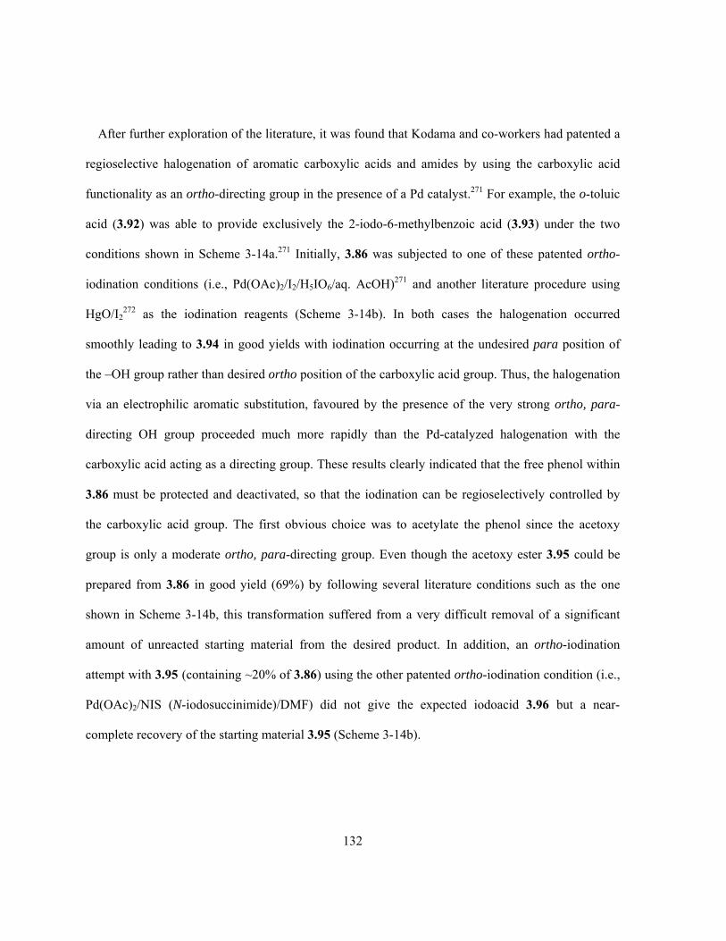
132
After further exploration of the literature, it was found that Kodama and co-workers had patented a
regioselective halogenation of aromatic carboxylic acids and amides by using the carboxylic acid
functionality as an ortho-directing group in the presence of a Pd catalyst.271 For example, the o-toluic
acid (3.92) was able to provide exclusively the 2-iodo-6-methylbenzoic acid (3.93) under the two
conditions shown in Scheme 3-14a.271 Initially, 3.86 was subjected to one of these patented ortho-
iodination conditions (i.e., Pd(OAc)2/I2/H5IO6/aq. AcOH)271 and another literature procedure using
HgO/I2272 as the iodination reagents (Scheme 3-14b). In both cases the halogenation occurred
smoothly leading to 3.94 in good yields with iodination occurring at the undesired para position of
the –OH group rather than desired ortho position of the carboxylic acid group. Thus, the halogenation
via an electrophilic aromatic substitution, favoured by the presence of the very strong ortho, para-
directing OH group proceeded much more rapidly than the Pd-catalyzed halogenation with the
carboxylic acid acting as a directing group. These results clearly indicated that the free phenol within
3.86 must be protected and deactivated, so that the iodination can be regioselectively controlled by
the carboxylic acid group. The first obvious choice was to acetylate the phenol since the acetoxy
group is only a moderate ortho, para-directing group. Even though the acetoxy ester 3.95 could be
prepared from 3.86 in good yield (69%) by following several literature conditions such as the one
shown in Scheme 3-14b, this transformation suffered from a very difficult removal of a significant
amount of unreacted starting material from the desired product. In addition, an ortho-iodination
attempt with 3.95 (containing ~20% of 3.86) using the other patented ortho-iodination condition (i.e.,
Pd(OAc)2/NIS (N-iodosuccinimide)/DMF) did not give the expected iodoacid 3.96 but a near-
complete recovery of the starting material 3.95 (Scheme 3-14b).

133
CO2H
(i) Pd(OAc)2, I2/H5IO6
AcOH/H2O, CO2H
I
3.93(50%, method i)(92%, method ii)
(ii) Pd(OAc)2,
DMF, 3.92
OH
COOH
3.94(88%, method i)(N/A, method ii,contains Hg2+)
OAc
COOH
3.95 (~ 69%)
3.86
3.96
(a)
(b)(i) Pd(OAc)2, I2/H5IO6
AcOH/H2O,
(ii) HgO, I2
CH2Cl2 or EtOH, rt
OH
COOH
I
OAc
COOH
IPd(OAc)2 (20 mol%), NIS
DMF, 100 ºC, 6 hr
H3PO4Ac2O
NO O
I
(NIS)
X
Scheme 3-14. (a) Literature example of Pd-catalyzed ortho-iodination of substituted benzoic acid; (b)
attempted Pd(II)-catalyzed ortho- iodination of 3.86 and 3.95.
After more careful consideration, it was thought that mesylation of the phenol 3.86 might be a
better solution mainly for two reasons. First the –OMs group is known to be a deactivating and meta-
directing substituent for electrophilic aromatic substitution. This characteristic would significantly
help the desired iodination to occur ortho to the carboxylic acid group, since this position would be
meta to the –OMs group. Second, the mesyl group is a very stable protecting species for phenols
under various (strong acidic, oxidative, reductive and high temperature) conditions273 yet it can be
readily removed under strong basic/nucleophilic conditions (such as warm aq. base
(NaOH/KOH/Na2CO3) solution,274-277 NaOEt/EtOH/reflux,278,279 CH3SNa/NaOH273 or LDA/THF/-78
°C280).

134
Initially, the mesylation of 3.86 was carried out with MsCl in the presence of a typical base;
however, the use of common bases including NaOH, KOH, NaOEt, pyridine and NEt3 all failed to
produce the desired mesylate 3.97 in any meaningful yields, while the use of NaH in DMF was able
to afford 3.97 but in a very poor yield of only 24% (Scheme 3-15a). Finally, mesylation of 3.86 was
accomplished in 94% yield in THF with CaO as the appropriate base (Scheme 3-15a), and addition of
a small amount of water was found to significantly accelerate the initiation of the mesylation reaction.
This success was probably due to not only the strong basicity of CaO/Ca(OH)2, but also and more
importantly the divalent capability of Ca2+ that could form a complex of 3.99 with the salicyclic acid
to ensure a complete deprotonation of the phenol (Scheme 3-15a). The mesylate 3.97 was then
subjected to the two Pd(OAc)2-catalyzed ortho-iodination conditions (Scheme 3-15a): although the
starting mesylate was essentially recovered with only about 10% conversion to the expected
monoiodination product when I2/H5IO6 in aqueous acetic acid was used as the iodination reagent, the
use of NIS in DMF afforded the desired aryl iodomesylate 3.100 in good yields. The experimental
conditions for this iodination were further optimized in various aspects and it was found that (i) an
ideal loading of the Pd(OAc)2 catalyst of about 10–15 mol% would ensure complete iodination, but
the halogenation could occur with as little as only 2% Pd catalyst loading; (ii) DMF is the solvent of
choice and no iodination occurred at all when (refluxing) THF was used, while the use of (refluxing)
AcOH also showed very limited conversion (~ 20%) of 3.97 even after extended reaction times; (iii)
the ideal reaction temperature in DMF was around 100–120 °C while refluxing DMF (bp 153°C)
would cause a severe decrease of the yield. The mechanism for this carboxylic acid-directed
regioselective iodination was not discussed in the original patent, but likely it involved a mechanism
(Scheme 3-15b) similar to the one that was proposed for the Pd(II)-catalyzed ortho halogenation of
substituted benzoic acids by IOAc, in which the carboxylate would bind and direct the Pd(II) to its
ortho position where the C-H bond was activated towards the halogenation.281

135
OH
COOH
OMs
COOH
3.97(24%, method i)(94%, method ii)
3.86 3.100 (70%)
i. NaH, DMF, rtthen MsCl
ii. CaO, THF,MsCl, rt OMs
COOH
I(i) Pd(OAc)2, I2/H5IO6
AcOH/H2O,
(ii) Pd(OAc)2 (10-15 mol%), NIS
DMF,
COOHRPd(OAc)2
R
PdIIO
O
IOAcR
PdIVO
O
I OAc
R
OPdIIOAc
O
I
3.101 3.102 3.103 3.104
(b)
(a)
OH
CO2-Ca-OH
3.98
CaO/Ca(OH)2
-O
C
3.99
O
O-
Ca2+
MsCl then H+
X
Scheme 3-15. (a) Pd(II)-catalyzed ortho-iodination of the mesylbenzoic acid 3.97; (b) proposed
literature mechanism for Pd(II)-catalyzed ortho-iodination of substituted benzoic acids.
The key intermediate 3.100 now had all the main structural features of the proposed new D-ring
precursor 3.83, except that the carboxylic acid group of 3.100 would require some further chemical
transformations (Scheme 3-16). Reduction of the carboxylic acid group within 3.100 was problematic
at the beginning and it was found that the reaction would barely occur if less than 3 equivalents of
borane, regardless of the borane source (either direct or generated in situ), were used. This was
probably due to the presence of the –OMs group in the molecule, whose multiple oxygen atoms might
bind to the Lewis-acidic borane reductant. In practice, the reduction of 3.95 was eventually carried
out with a large excess of BH3 (10 equivalents) and the corresponding benzyl alcohol 3.105 was
prepared in 77% yield (Scheme 3-16). The initial attempt on the benzylic bromination of 3.105 was
made with conc. HBr, but no substitution product was obtained and the starting material was
completely recovered. However, the desired bromo substitution of 3.105 was achieved quantitatively
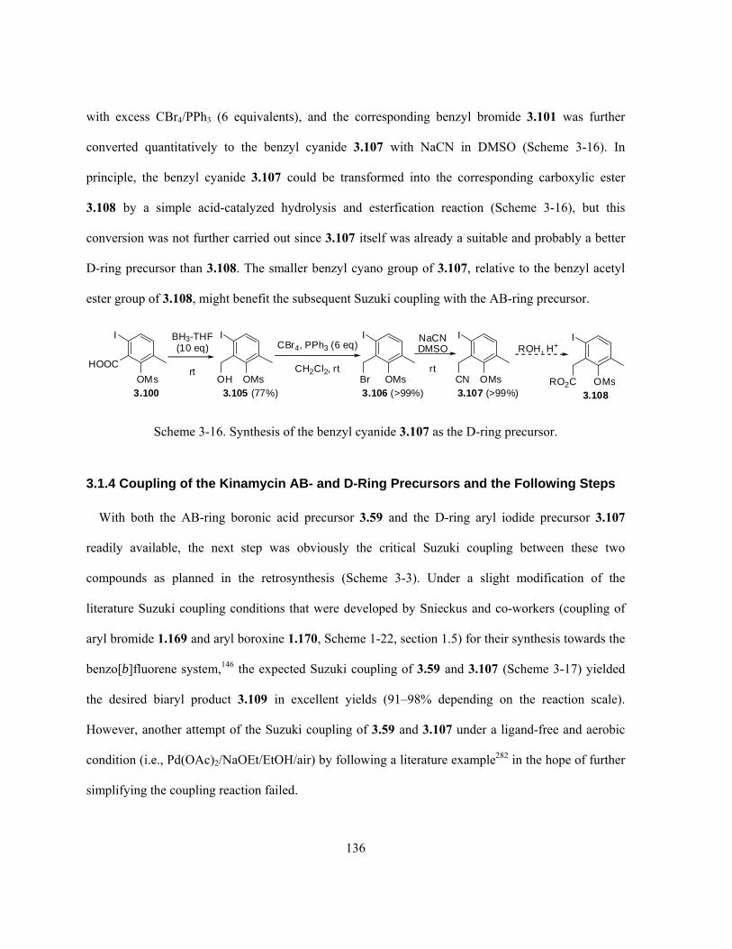
136
with excess CBr4/PPh3 (6 equivalents), and the corresponding benzyl bromide 3.101 was further
converted quantitatively to the benzyl cyanide 3.107 with NaCN in DMSO (Scheme 3-16). In
principle, the benzyl cyanide 3.107 could be transformed into the corresponding carboxylic ester
3.108 by a simple acid-catalyzed hydrolysis and esterfication reaction (Scheme 3-16), but this
conversion was not further carried out since 3.107 itself was already a suitable and probably a better
D-ring precursor than 3.108. The smaller benzyl cyano group of 3.107, relative to the benzyl acetyl
ester group of 3.108, might benefit the subsequent Suzuki coupling with the AB-ring precursor.
I
HOOC
OMs
I
OMsCN
I
OMs
BH3-THF(10 eq)
rt
I
OMsBr
NaCNDMSO
rt
3.105 (77%) 3.106 (>99%) 3.107 (>99%)OH
CBr4, PPh3 (6 eq)
CH2Cl2, rt
3.100
I
OMsRO2C3.108
ROH, H+
Scheme 3-16. Synthesis of the benzyl cyanide 3.107 as the D-ring precursor.
3.1.4 Coupling of the Kinamycin AB- and D-Ring Precursors and the Following Steps
With both the AB-ring boronic acid precursor 3.59 and the D-ring aryl iodide precursor 3.107
readily available, the next step was obviously the critical Suzuki coupling between these two
compounds as planned in the retrosynthesis (Scheme 3-3). Under a slight modification of the
literature Suzuki coupling conditions that were developed by Snieckus and co-workers (coupling of
aryl bromide 1.169 and aryl boroxine 1.170, Scheme 1-22, section 1.5) for their synthesis towards the
benzo[b]fluorene system,146 the expected Suzuki coupling of 3.59 and 3.107 (Scheme 3-17) yielded
the desired biaryl product 3.109 in excellent yields (91–98% depending on the reaction scale).
However, another attempt of the Suzuki coupling of 3.59 and 3.107 under a ligand-free and aerobic
condition (i.e., Pd(OAc)2/NaOEt/EtOH/air) by following a literature example282 in the hope of further
simplifying the coupling reaction failed.
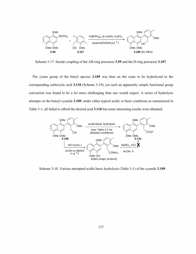
137
OMe
OMeOMe
B(OH)2 Pd(PPh3)4 (5 mol%), K3PO4
toluene/EtOH/H2O,
OMe
OMe
OMe
NC
OMs
3.109 (91-98%)
I
OMsCN
3.1073.59
+
Scheme 3-17. Suzuki coupling of the AB-ring precursor 3.59 and the D-ring precursor 3.107.
The cyano group of the biaryl species 3.109 was then on the route to be hydrolyzed to the
corresponding carboxylic acid 3.110 (Scheme 3-18); yet such an apparently simple functional group
conversion was found to be a lot more challenging than one would expect. A series of hydrolysis
attempts on the benzyl cyanide 3.109, under either typical acidic or basic conditions as summarized in
Table 3-1, all failed to afford the desired acid 3.110 but some interesting results were obtained.
OMe
OMe
OMe
OMsacidic/basic hydrolysis
(see Table 3-1 fordetailed conditions)
OMe
OMe
OMe
OMs
HCl (conc.)
AcOH or MeOH or
3.111 (major product)
NaNO2, HCl
AcOH, rt
CN CO2H
OMe
OMe
OH
OMs
CONH2
3.109 3.110
X
Scheme 3-18. Various attempted acidic/basic hydrolysis (Table 3-1) of the cyanide 3.109.

138
Table 3-1. Detailed experimental conditions and results for the hydrolysis of 3.109 (Scheme 3-18).
Trial Reagents and ConditionsReference Products and Results
1 H2SO4 (conc.)/AcOH/H2O/rt No reaction, recovery of 3.109
2 H2SO4 (conc.)/AcOH/H2O/↑↓ Complex mixture that contains no acid but small amount of cyclized product(s)
3 HCl (conc.)/MeOH/rt No reaction, recovery of 3.109
4 HCl (conc.)/AcOH (1:1)/↑↓
5 HCl (conc.)/AcOH (1:1)/70–80 °C
6 HCl (conc.)/MeOH/↑↓
3.111 (major product)
7 H3PO4 (conc.)/AcOH/H2O/rt No reaction, recovery of 3.109
8 H3PO4 (conc.)/AcOH/H2O/70–80 °C No reaction, recovery of 3.109
9 Amberlite IR-120/MeOH/H2O/↑↓283 No reaction, recovery of 3.109
10 Amberlite IR-120/AcOH/H2O/↑↓ No reaction, recovery of 3.109
11 H2O2/NaOH/↑↓284 Complex mixture: no hydrolysis of –CN at all but complete loss of –Ms
12 KOH/MeOH/↑↓285 Complex mixture: no hydrolysis of –CN at all but complete loss of –Ms
First, hydrolysis of 3.109 under mild acidic conditions (i.e., strong acids at ambient temperature or
weak acids at ambient/elevated temperature, trial 1, 3 and 7–10, Table 3-1) led only to a complete
recovery of the starting material. This was likely due to the presence of the bulky substituted naphthyl
group and mesylate group, both ortho to the benzyl cyanide moiety, which retarded the nucleophilic
attack on the cyano by H2O. Second, under some harsh acidic conditions (i.e., strong acids at elevated
temperature) but depending on the acid involved, the substrate behaved quite differently. For example,
when H2SO4 was used as the acid catalyst (trial 2, Table 3-1), although the product was obtained as a
complex mixture that contained no desired acid 3.110 at all, crude NMR indicated that there existed a
small amount of a cyclized species. This observation seemed to be consistent with the known
literature results that biaryl benzyl cyanides such as 3.112 could cyclize in conc. H2SO4 (with no

139
solvent) to give the corresponding aniline 3.113 (Scheme 3-19a).286,287 On the other hand, when conc.
HCl was used for the hydrolysis (trial 4–6, Table 3-1), the major product isolated from these reactions
was an amide 3.111 from a partial hydrolysis of the cyanide, but containing also a partially
demethylated B-ring (Scheme 3-18). The demethylation of the B-ring in these cases was unexpected,
but it could be explained by the proposed possible mechanism shown in Scheme 3-19b. Manipulation
of the solvent, reaction temperature and time could not push the amide 3.111 to be further hydrolyzed
under these acidic conditions to the desired acid 3.110 at all. Attempts to convert the amide to the
acid via an acyl-diazonium intermediate under the conditions (i.e., NaNO2/HCl/AcOH) similar to
literature examples did not succeed either and gave a complex mixture (Scheme 3-18).288,289 This was
probably due to the very electron-rich AB-ring of 3.111 that, instead of the amide moiety, could react
readily and probably preferably with the strong electrophile (NO+) generated in the reaction. Third,
hydrolysis of 3.109 under either oxidative or normal basic conditions (trial 11–12, Table 3-1) also did
not convert the cyano moiety to the desired carboxylic acid group or even to the intermediate stage of
an amide, but a complete loss of the D-ring mesyl protecting group was observed in both cases. The
in situ generated (D-ring) phenol upon demesylation was probably not stable under such conditions,
thus led to a complex mixture as the final products. Even though the last two trials did not offer the
expected chemical outcome, at least they provided some applicable conditions that might be useful
for the later deprotection/removal of the mesyl group.

140
(a)
(b) A possible mechanism for the B-ring demethylation and hydrolysis of 3.109
NC NH2
3.112 (R = H or alkyl) 3.113 (61-85%)
H2SO4 (conc.), 0 ºC
R
R
MeO
OMe
OMe
OMs
3.111
CN
OMe
OMe
OH
OMs
CONH2
3.109 H+
OMe
OMe
O
OMs
CN
3.114Me
HH
Cl-OMe
OMe
O
OMs
CN
3.115
HH
-CH3Cl
OMe
OMe
OH
OMs
CN
3.116
+ H+
MeO
OMe
OH
OMs
CN
3.117
HH
MeO
OMe
OH
OMs
C
3.119NH
H2O
MeO
OMe
OH
OMs
CNH
H
3.118
- H+
Scheme 3-19. (a) Literature example of benzyl cyanide cyclization in conc. H2SO4 and (b) a possible
mechanism for the partial demethylation and cyano hydrolysis of 3.109 under acidic (HCl) conditions.
All these failures to obtain the desired acid 3.110 from the cyanide 3.109 had led to the
consideration that, it might be necessary to prepare either the corresponding acid (or the ester) of the
D-ring precursor 3.102 upon conversion of its cyano group prior to the Suzuki coupling, which then
can couple with 3.55 to prepare the corresponding biaryl species with the required carboxylic acid (or
ester) functionality preinstalled and thus eliminate the need of a difficult cyano hydrolysis as

141
encountered in the case of 3.109. Just before efforts were about to be redirected to such an alternative
plan, a literature work using chlorotrimethylsilane (TMSCl) in alcohol solvents to convert nitriles to
the corresponding esters was discovered.290 This method involves HCl generated in situ as the acid
catalyst for the solvolysis of the nitrile. The HCl originates from the reaction of TMSCl and the first
equivalent of alcohol, and a second equivalent of the alcohol acts as the nucleophile to produce an
iminium salt from the HCl-activated nitrile that was further hydrolyzed to the ester during the
aqueous workup stage of the reaction (Scheme 3-20).290 It was decided to carry out one more trial
with the biaryl benzyl cyanide 3.109 under such conditions to see if the corresponding ester 3.120
could be obtained or not. Based on the encountered difficulties to hydrolyze the cyano group of 3.109
as described above, consideration and modification of the literature procedure in the following two
aspects were applied. First, the original literature procedure only required one equivalent of TMSCl
and two equivalents of alcohol relative to the nitrile used,290 while a large excess of both reagents that
were still kept in a 1:2 ratio were used in the case of 3.109 in the hope that the desired but difficult
conversion might be forced to a completion. Second, even though various simple alcohols such as
methanol, ethanol and isopropanol were all found to be quite effective to prepare the corresponding
esters from the nitriles (69–92% yields),290 the very crowded space environment around the benzyl
cyano group of 3.109 would probably benefit the most from the use of methanol, the smallest
alcoholic nucleophile available.
TMSCl + ROH → HCl + TMSOR
R’CN + HCl + ROH → R’-C(OR)=N+H2Cl-
R’-C(OR)=N+H2Cl- + H2O (workup) → R’COOR + NH4Cl
Scheme 3-20. Plausible literature mechanism for the conversion of nitrile to ester by TMSCl/alcohol.

142
In practice, a solution of 3.109 was heated at reflux in a large excess of TMSCl/MeOH (1:2)
mixture and followed by an aqueous workup as described in the literature work (i.e., saturated aq.
NaHCO3 solution). It was gratifying to find that the expected and desired methyl ester 3.120 was
isolated in 60% yield along with a corresponding amide 3.121 in 28% yield (Scheme 3-21 and Table
3-2), and no demethylation (of B-ring) at all occurred with either product this time. Inspired by this
initial success but not very satisfied with the product distribution (i.e., significant amount of
unwanted amide 3.121), the aqueous workup condition was further explored to see if there was any
room for improvement. In fact, it was found that the ratio between the methyl ester 3.120 and the
amide 3.121 varied significantly depending on the acidity of the workup condition used, as
summarized in Table 3-2. The best result (83% isolated yield of 3.120 and 16% isolated yield of
3.121) was achieved with 10% HCl as the workup medium. The competition between the formation
of both 3.120 and 3.121 (Scheme 3-21), starting from the same iminium salt intermediate 3.122, is
probably a reflection of the competition between the –NH2 and –OMe in their protonated forms as the
leaving groups (i.e., –N+H3 within 3.123 and –O+(H)Me within 3.124). The presence of excess acid
during the workup stage would favour keeping the intermediate in the –NH2 protonated form and thus
leading to the preferential formation of the ester 3.120. Such a result would also suggest that the B-
ring demethylation previously observed for 3.109, when it was hydrolyzed in aqueous HCl in
refluxing MeOH or heated AcOH (Scheme 3-19 and Table 3-1), might be partially due to the
presence of water during the reaction in these cases, and this conclusion was further supported by the
following experimental observations. The literature work indicated that the solvolysis of the nitriles
by the alcohols with TMSCl can be also successfully carried out in the presence of water,290 therefore
an attempt with 3.109 in a solvent mixture such as MeOH/H2O would be worthwhile if the
corresponding acid 3.110 instead of the methyl ester 3.120 could be directly prepared, since the acid
would serve as a better candidate than the ester for the subsequent intramolecular cyclization step.

143
However, a trial with 3.109 in a refluxing mixture of TMSCl/MeOH/H2O (1:1:1, in large excess
relative to 3.109) followed by an acidic workup did not yield the preferred acid 3.110 but a complex
mixture (Scheme 3-21). Even though the mixture could not be well separated and fully characterized,
NMR evidence indicated that the products in this case were still a mixture of a methyl ester 3.125 and
an amide 3.126 but both with a (partially) demethylated B-ring, along with other unidentified by-
products.
OMe
OMe
OMe
NC
OMs
OMe
OMe
OMe
OMs
TMSCl/MeOH (1:2)in large excess,
thenaqueous workup(see Table 3-2)
CO2Me
3.109 3.120OMe
OMe
OMe
OMs
CONH2
3.121
+
HCl, MeOH (2nd eq.)
TMSCl + MeOH (1st eq.)
OMe
OMe
OMe
OMs
3.122
NMeOH
H
H2O
OMe
OMe
OMe
OMs
3.124
NH2MeO
OHOMe
OMe
OMe
OMs
3.123
NH3MeO
OH H
- NH3 - MeOH
OMe
OMe
OMe
NC
OMs
TMSCl/MeOH/H2O(1:1:1, large excess)
OMsCO2Me
+
OMsCONH2
+ demethylation of B-ring occured !!!
3.109 3.125 3.126
Scheme 3-21. Products and mechanism of the solvolysis of 3.109 (refer to Table 3-2).

144
Table 3-2. Detailed workup conditions and results for the solvolysis of 3.109 (Scheme 3-21).
Workup conditions 3.120 (Isolated yield) 3.121 (Isolated yield)
saturated aq. NaHCO3 solution 60% 28%
H2O + Na2CO3 powder 68% 21%
H2O only* 66% 29%
10% HCl aq. solution 83% 16%
* This is indeed an acidic workup due to the presence of large excess HCl (generated from the large excess of
TMSCl/MeOH).
Further efforts were made to hydrolyze the methyl ester 3.120 into the corresponding carboxylic
acid 3.110 (Scheme 3-22) by following quite a few literature conditions as summarized in Table 5-3.
Even though such an ester hydrolysis could be carried out under either acidic or basic conditions, for
practical reasons all attempts were made under mild basic conditions (Table 5-3). This precaution of
using no strong acidic or basic conditions was to avoid some of the possible side reactions that may
occur with 3.120 due to its structural and electronic characters, based on the previous experience with
the very similar biaryl species such as 3.109. For example, the use of HCl may cause a very possible
B-ring demethylation; on the other hand, strong basic conditions at elevated temperature would likely
cleave the mesyl group that was to be kept as the phenol protecting group at the moment. However,
despite the various and carefully selected mild basic hydrolysis conditions attempted (Table 5-3),
some difficulties were encountered once again for such an apparently simple functional group
transformation, which was still very likely due to the enormous steric hindrance around the ester
moiety from the two neighbouring bulky groups. In most cases the methyl ester 3.120 was found to
be inert towards the mild basic hydrolysis, while in the case when ester hydrolysis did occur (trial 2),
the low conversion percentage (< 10%) and slow rate (3 days) were not acceptable from a practical
point of view, and demesylation still occurred concurrently with the ester hydrolysis, although both to
a very small extent, even at such mild conditions (trial 4).

145
mild basic hydrolysis
(see Table 5-3 for detailed conditions)
OMe
OMe
OMe
OMs
CO2Me
OMe
OMe
OMe
OMs
CO2H
3.120 3.110
Scheme 3-22. Mild basic hydrolysis (Table 3-3) of ester 3.120.
Table 3-3. Detailed experimental conditions and results for the hydrolysis of 3.120 (Scheme 3-22).
Trial Reagents and ConditionsReference Products and Results
1 Mg/MeOH/rt then H3O+291 No reaction, recovery of 3.120
2 Na2CO3/MeOH/H2O/rt/4 hr – 3 days292 Mainly recovered 3.120, only small percentage
(< 10%) of ester hydrolyzed after 3 days
3 LiOH/H2O2/THF-H2O/0 °C then H3O+ No reaction, recovery of 3.120
4 KOH/MeOH/H2O/rt then H3O+ Mainly recovered 3.120 +
OMe
OMe
OMe
OH
CO2HOMe
OMe
OMe
OH
CO2Me
+
3.127 (minor) 3.128 (minor)
The difficulties to prepare the carboxylic acid 3.110 from the methyl ester 3.120, which was the
required precursor for the next intramolecular Friedel-Crafts cyclization step as planned (Scheme 3-3),
had led to the search of possible conditions that might be suitable for the direct and intramolecular
cyclization of the ester 3.120. Generally speaking, esters are known to be much less active towards
the electrophilic aromatic substitution reactions when compared to other carbonyl compounds,
especially the acid halides and carboxylic acids. Non-aromatic esters are barely known or used as
acylation reagents. In fact, intermolecular aromatic substitution by simple esters under the typical
Friedel-Crafts conditions, i.e. AlCl3 as the Lewis acid catalyst with simple aromatic hydrocarbons
146
such as benzene as the nucleophile, has been shown to give aromatic alkylation products rather than
acylation products.293 When the ester group co-exists with other carbonyl functionalities such as an
aldehyde, the latter would preferentially undergo the intramolecular cyclization.294 Most known
literature examples of ester intramolecular aromatic cyclization involve the benzoate type of esters
(Ar-(C=O)-OR) rather than the benzyl esters (Ar-CH2-(C=O)-OR), since the conjugation of the ester
moiety with an aromatic ring would significantly enhance its reactivity yet cyclization reactions of
such activated esters would still require the use of a very strong Lewis acid catalyst.295 Unfortunately
the prepared intermediate ester 3.120 belongs to the latter and less active type. It was noticed that,
however, a literature work claimed that a styrene-methyl methacrylate (MMA) based polymer was
able to undergo an intramolecular Friedel-Crafts cyclization between the benzene ring of the styrene
unit and the ester group of the MMA unit when the polymer was heated (60 °C) in dichloroethane
with SnCl4 as the catalyst.296 However, repeated reaction of 3.120 under the same conditions, even at
a slightly higher temperature (i.e., refluxing dichloroethane, 84 °C) and extended longer reaction time
than the literature example, still recovered mainly the unreacted 3.120 instead of yielding the
expected cyclization product 3.129 (Scheme 3-23). On the other hand, a trial attempt was also made
by subjecting the ester 3.120 to methanesulfonic acid CH3SO3H (MSA) in CH2Cl2294 at ambient
temperature (Scheme 3-23) to check the (cyclization) reactivity of 3.120 under such conditions. MSA
is one of the commonly used acid catalysts for acylation of aromatic systems by carbonyl compounds
(mostly carboxylic acids), particularly for the intramolecular type, as indicated by many available
literature precedents.294,297-300 Not surprisingly, the less active ester (than acid) was once again
essentially recovered after 24 hr of reaction with no evidence for the formation of 3.129.

147
i. SnCl4, ClCH2CH2Cl, , 19 hr
or ii. CH3SO3H, CH2Cl2, rt, 24 hr
OMe
OMe
OMe
OMs
CO2Me
3.120OMe
OMe
OMe
OMs
OH3.129
X
Scheme 3-23. Attempted direct intramolecular cyclization/acylation of the ester 3.120.
Breakthrough of this apparent deadend was made after accidentally coming across an old literature
procedure developed by Loev back in the 1960s, which showed that simple alkyl or aryl (including
heterocyclic) carboxylic esters could be readily hydrolyzed to the corresponding acid by heating with
MSA in conc. formic acid (or acetic acid).301 It was felt that this condition might provide a better
chance of success on the troublesome hydrolysis of methyl ester 3.120 (Scheme 3-22). In addition, if
the carboxylic acid 3.110 could be generated from the ester 3.120 under such conditions (MSA +
HCOOH), then it was also possible that 3.110 might even undergo an in situ intramolecular
cyclization to afford 3.129 due to the presence of MSA (Scheme 3-24), which is an excellent catalyst
capable of promoting intramolecular cyclization of carboxylic acids. Therefore, an initial attempt was
made by refluxing 3.120 with excess MSA in conc. formic acid for 3 hr (Table 3-4, trial 1). Although
the obtained crude products were quite messy and still contained a large amount of unreacted 3.120,
NMR evidence did show that several chemical transformations including cyclization, partial A/B-ring
demethylation and even oxidation (reaction carried out under air atmosphere) had occurred
experimentally. When 3.120 was heated in the mixture of MSA and conc. formic acid for extended
time (12–16 hr, Table 3-4, trial 2), compound 3.130 having the structure as shown (rather than 3.129)
was surprisingly isolated as the major product from the reaction in about 44% yield. Even though the
ring closure of the in situ generated carboxylic acid (from 3.120) through an intramolecular Friedel-
Crafts cyclization had been predicted, and the formation of the B-ring quinone was very likely a

148
spontaneous air-oxidation of the corresponding phenol (from demethylation of the phenol methyl
ether) since the reaction was carried out under air atmosphere, but the complete demethylation on
both A- and B-ring (especially the former) was beyond expectation. MSA is not a typical O-
demethylation reagent273 and the very few examples where O-demethylation did occur with MSA
required the presence of a strong nucleophile such as methionine302 or NaI,303 which had no
equivalent species in this case. Hypothetically, the very strong acidity of MSA (pKa < -1) and the
electron-rich character of the AB-ring of 3.120 are two likely factors that would definitely contribute
to the observed demethylation. Formation of 3.130 was a surprising but very pleasant result since the
desired 6-6-6-6 carbon skeleton as planned in the retrosynthesis (cf. structures 3.32–3.35 in Scheme
3-3) was now achieved. However, 3.130 did have some issues that needed to be addressed. First, it
was found experimentally that the solubility of 3.130 in various common organic solvents (polar or
non-polar) was extremely low, which probably would make future chemical transformations of this
compound difficult, especially at large scales. Such physical behaviour might be due to the presence
of strong intramolecular hydrogen bonding between the A-/C-ring phenols and the B-ring quinone
carbonyl group, which is evident from the characteristic large downfield NMR chemical shifts of the
phenolic protons as a result of such H-bonding interactions, and similar phenomenon is known for
compounds such as isoprekinamycin (1.5). Second, the two phenol functionalities within 3.129 (i.e.,
the A-ring phenol from O-demethylation and C-ring phenol from ring closure), if not properly
differentiated, may compete with each other and complicate the later oxidation step that was planned
to occur (preferably) at the C-ring to generate the corresponding ortho-quinone needed for further
ring contraction reaction. Last, the relatively low yield of 44% obviously had room for improvement.
The first two trials with 3.120 in MSA/HCOOH all involved the fairly harsh condition of refluxing;
therefore, a milder attempt was explored (Table 3-4, trial 3). A large portion of 3.120 survived in the
MSA/HCOOH mixture at ambient temperature after 40 hr but partial demethylation of AB-ring did
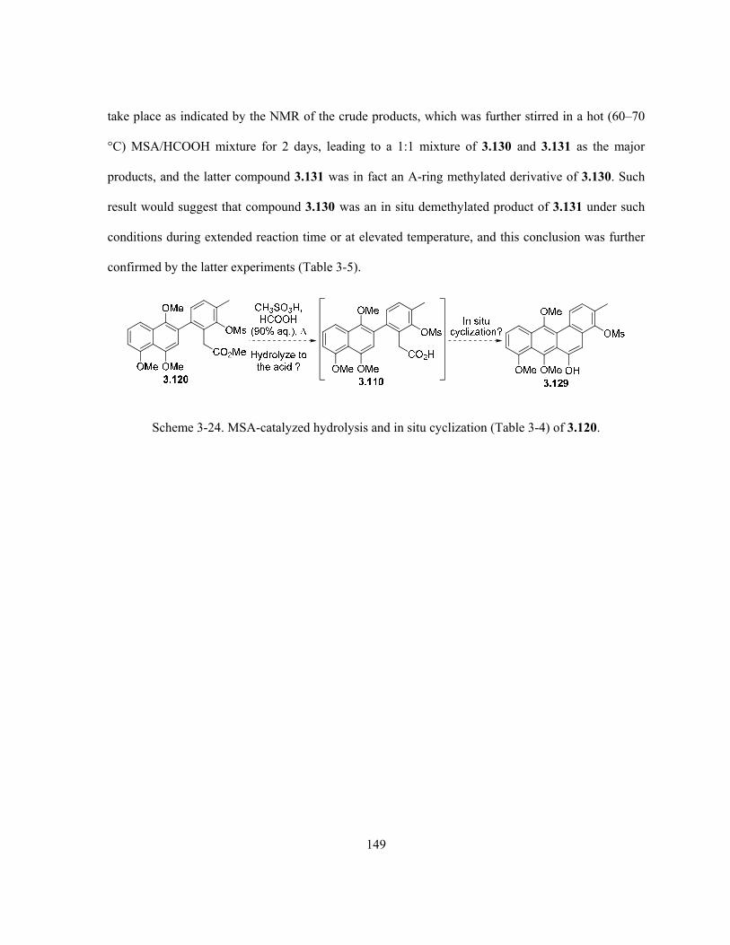
149
take place as indicated by the NMR of the crude products, which was further stirred in a hot (60–70
°C) MSA/HCOOH mixture for 2 days, leading to a 1:1 mixture of 3.130 and 3.131 as the major
products, and the latter compound 3.131 was in fact an A-ring methylated derivative of 3.130. Such
result would suggest that compound 3.130 was an in situ demethylated product of 3.131 under such
conditions during extended reaction time or at elevated temperature, and this conclusion was further
confirmed by the latter experiments (Table 3-5).
Scheme 3-24. MSA-catalyzed hydrolysis and in situ cyclization (Table 3-4) of 3.120.
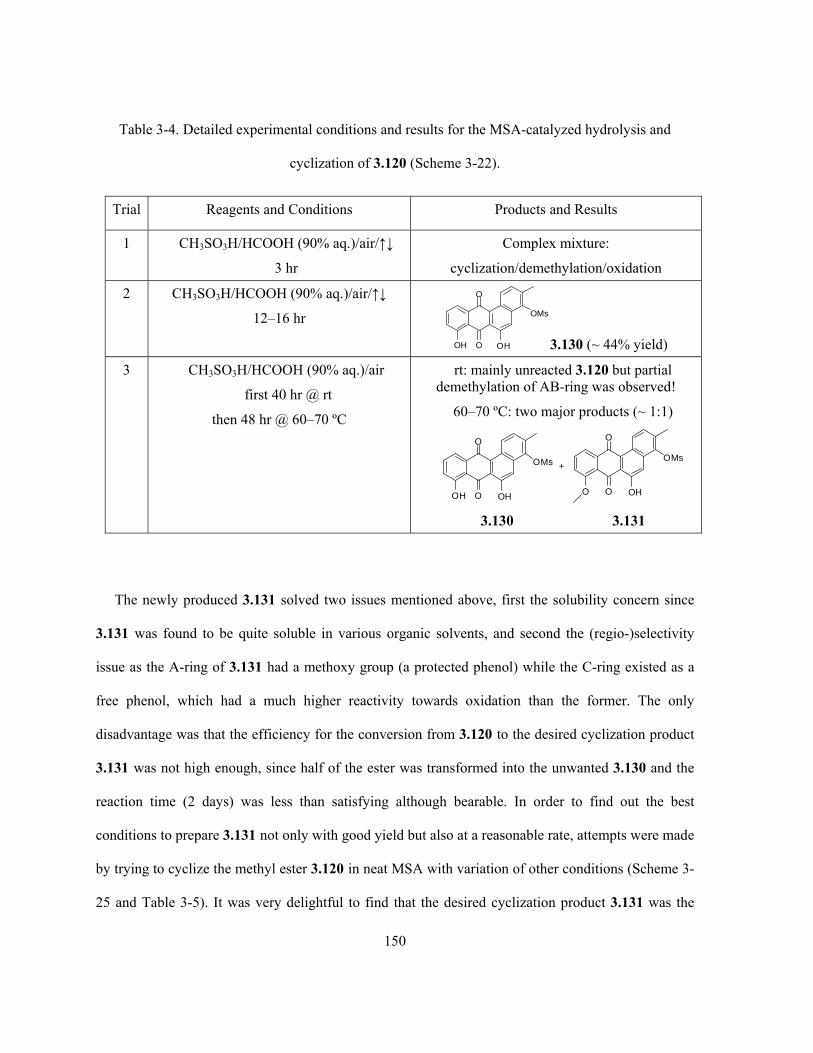
150
Table 3-4. Detailed experimental conditions and results for the MSA-catalyzed hydrolysis and
cyclization of 3.120 (Scheme 3-22).
Trial Reagents and Conditions Products and Results
1 CH3SO3H/HCOOH (90% aq.)/air/↑↓
3 hr
Complex mixture:
cyclization/demethylation/oxidation
2 CH3SO3H/HCOOH (90% aq.)/air/↑↓
12–16 hr
OH
O
O
OMs
OH 3.130 (~ 44% yield)
3 CH3SO3H/HCOOH (90% aq.)/air
first 40 hr @ rt
then 48 hr @ 60–70 ºC
rt: mainly unreacted 3.120 but partial demethylation of AB-ring was observed!
60–70 ºC: two major products (~ 1:1)
OH
O
O
OMs
OH O
O
O
OMs
OH
+
3.130 3.131
The newly produced 3.131 solved two issues mentioned above, first the solubility concern since
3.131 was found to be quite soluble in various organic solvents, and second the (regio-)selectivity
issue as the A-ring of 3.131 had a methoxy group (a protected phenol) while the C-ring existed as a
free phenol, which had a much higher reactivity towards oxidation than the former. The only
disadvantage was that the efficiency for the conversion from 3.120 to the desired cyclization product
3.131 was not high enough, since half of the ester was transformed into the unwanted 3.130 and the
reaction time (2 days) was less than satisfying although bearable. In order to find out the best
conditions to prepare 3.131 not only with good yield but also at a reasonable rate, attempts were made
by trying to cyclize the methyl ester 3.120 in neat MSA with variation of other conditions (Scheme 3-
25 and Table 3-5). It was very delightful to find that the desired cyclization product 3.131 was the

151
major species from the reaction, along with a small amount of the (A-ring) demethylated 3.130 and,
in some cases, the unreacted starting ester 3.120. To the best of our knowledge, there is no reported
example in the literature that directly and intramolecularly cyclizes a benzyl type of ester without
converting it to the corresponding carboxylic acid first. It could be clearly seen from the several trials
(Table 3-5) that reaction temperature played a significant role in determining the conversion
percentage and final product distribution, and a maximum yield of 80% of 3.131 was obtained when
the reaction was carried out at 40–45 °C in neat MSA for 6 hr (Table 3-5, trial 3). As a control
experiment (Table 3-5, trial 5), cyclization was also performed in a (1:1) mixture of MSA and CH2Cl2,
which was previously reported as a more efficient condition for such cyclization than neat MSA,294
but the result indicated otherwise.
neat CH3SO3H
OMe
O
O
OMs
OHOMe
OMe
OMe
OMs
CO2Me
3.120 3.131
Scheme 3-25. MSA-catalyzed direct cyclization of 3.120 (Table 3-5).

152
Table 3-5. Detailed experimental conditions and results for the MSA-catalyzed cyclization of 3.120
(Scheme 3-25).
Molar ratio of products Trial Reaction conditions
3.120
OMe
OMe
OMe
OMs
CO2Me
3.131
OMe
O
O
OMs
OH
3.130
OH
O
O
OMs
OH
1 neat MSA/N2
60–70 ºC/1 hr then rt/8 hr
4a 4a 1a
2 neat MSA/N2
60–65 ºC/4 hr
0 2 1
3 neat MSA/N2
40–45 ºC/6 hr
0 major
(80% isolated yield)
very minor
4 neat MSA/air
45–50 ºC/4.5 hr
0 major
(70% isolated yield)
very minor
5 MSA-CH2Cl2
(1:1), air/↑↓/8hr
majora 1 (minor)a 1 (minor)a
a. Estimated based on NMR spectra of crude product.
Due to time limitations, no further experiments to convert 3.131 into the kinamycins and their
analogues were carried out in this thesis work. However, the successful and concise preparation of
compound 3.131 had provided some quite promising results to achieve the ultimate goal. It is
expected that, by following the synthetic plan as outlined in Scheme 3-26, total synthesis of
kinamycin derivatives such as 3.140 may be accomplished if given more time. In fact, as a
continuation of this work, Janet Simons of the Dmitrienko group was able to carry on some of the
proposed synthesis as planned. Compound 3.141b bearing a protected C-ring phenol but an exposed

153
D-ring phenol for further elaboration, which corresponds to the proposed intermediate structure of
3.133, was successfully prepared in good yield from 3.131 (Scheme 3-26).304
Phenolprotection
OMe
O
O OPG
O
O
1. epoxidation2. [H]3. ERO
OMe
O
O
OMs
OH
OMe
O
O OPG
OR4
R1O
OR2
OR3
OMe
O
O O
OR4
R1O
OR2
OR3
N2OMe
O
O
OR4
R1OOR2
OR3
N2
OMe
O
O O
OR4
R1O
OR2
OR3
O
(deprotection)[O]
OMe
O
O
OR4
R1OOR2
OR3
HO CO2HOMe
O
O
OR4
R1OOR2
OR3
O
3.1313.134
3.135
3.1383.137
3.136
3.139
3.140
OMe
O
O OPG
OR
3.132 (R = Ms)
3.133 (R = H)
[O]
Base-inducedring contraction
Wolff rerrangement(ring contraction)
O
O OPNBOMe
Me
ORPNB-Br, TEBAC
acetone, K2CO3
3.141a (R = Ms, 93%)
3.141b (R = H, 73%)
NaOHMeOH
CH2O2N
PNB =OMe
O
O
OMs
OH3.131
Demesylation
(deprotection)diazo transfer
Scheme 3-26. Synthetic plan towards kinamycins from the intermediate 3.131 and some recent
progress (from 3.131 to 3.141b) made by Janet Simons according to the plan.

154
During the course of this project, there appeared a few literature reports concerning the total
synthesis of the kinamycins, particularly the diazobenzo[b]fluorene type that covers the majority of
the natural kinamycin antibiotics. These recent literature examples will be discussed and compared
with the current results and the corresponding future synthetic plan next.
3.2 Recent Literature Concerning The Total Syntheses of Kinamycins
The above synthetic strategy towards the kinamycins, i.e. section 2.2 describing a biomimetic
elaboration of the kinamycin’s D-ring and section 3.1 describing the construction towards the
benzo[b]fluorene skeleton, was generated at a time when there were no reports of successful synthesis
of these natural products. Thus, the synthetic plan was not influenced by any conscious effort to
improve upon strategies developed in other laboratories. Although the present synthetic effort
towards the kinamycins is not complete, it is worthwhile to contrast this approach with successful
synthetic methods that have been reported by other laboratories during the course of this research
project. In particular, it is of potential value to examine the synthetic approaches critically to
determine if the biogenetically inspired synthetic strategy offers any advantages over other methods
in order to decide if further work to complete the synthesis via the biomimetic method is worthwhile.
Since the biomimetic approach of this thesis work towards the kinamycins has not been
completed, it is necessary to make some educated guesses as to the total number of synthetic steps
that will be involved in this strategy, before comparison can be made with the literature approaches to
be discussed next. To summarize, thus far, the preparation of the advanced aromatic 6-6-6-6 fused
ring system of 3.131 requires a linear sequence of only three steps with an overall yield of 65%
(Scheme 3-27), starting from the corresponding AB-ring precursor 3.59 and D-ring precursor 3.107.
Either precursor can be very easily prepared, with good overall yields and in large scales (if needed),
155
from commercially available and relatively inexpensive materials/reagents. Precursor 3.59 can be
derived from 3.54 in five steps with an overall yield of 35%, while 3.107 can be obtained from 3.86
in also five steps with an overall yield of 51% (Scheme 3-27). Therefore, the longest linear sequence
to prepare the intermediate 3.131 involves eight steps, from either the naphthol 3.54 or the
methylsalicylic acid 3.86 (Scheme 3-27). The remaining steps require 3.131 to undergo O-protection
of the C-ring phenol, demesylation and the subsequent D-ring oxidation to the p-quinone of 3.134, to
which the model chemistry for ring D-elaboration might be applied (Scheme 3-26). Ideally, the ring
D-elaboration of 3.134 should require only three steps (epoxidation, reduction and epoxide ring
opening as demonstrated by the model D-ring synthesis described in Chapter 2) to yield the
intermediate 3.135 (Scheme 3-26), although it is possible that conversion of the B-ring quinone to an
O-protected hydroquinone may be necessary if the redox manipulation in the D-ring is interfered with
by the B-ring quinone. To provide the benzo[b]fluorene ring system of the kinamycins as in 3.137 via
ring contraction of 3.135 (Scheme 3-26), two possible routes are available and both involve two steps.
Conversion of 3.137 to the target diazobenzo[b]fluorene system would then be done in two more
steps using established methods for decarboxylative diazotransfer reactions (Scheme 3-26). Thus, the
present state of the biomimetic approach may be ten steps away from kinamycin F (1.1f). This would
mean that a total synthesis of the kinamycins might be achievable in approximately 18 steps from the
commercially available naphthol 3.54 and/or methylsalicylic acid 3.86.

156
O
O
3.7a (85%)
Ac2OCH2Cl2
Py, rt
NBS (4 eq)aq. AcOH
55-65 ºC
3.54 3.55 (98%)OH
OH
OAc
OAc
Br
OAc
OMe
OMe
Br
OMe3.58 (71%)
OH
OH
Br
OH3.70 (85%)
SnCl2-2H2O (4 eq)conc. H2SO4 (cat.)
MeOH,
CH3I (6 eq)KOH (powder, 12 eq)
DMSO, rt
n-BuLi, THF, -78 ºC
then B(OMe)3then HCl
3.59 (70%)OMe
OMe
OMe
B(OH)2
OH
COOH
OMs
COOH
3.97(94%, method ii)
3.86 3.100 (70%)
CaO, THF,
MsCl, rtOMs
COOH
IPd(OAc)2 (10-15 mol%)
NIS, DMF,
I
OMsCN
I
OMs
BH3-THF(10 eq)
rt
I
OMsBr
NaCNDMSO
rt
3.105 (77%) 3.106 (100%) 3.107 (100%)OH
CBr4, PPh3 (6 eq)
CH2Cl2, rt
Pd(PPh3)4 (5 mol%), K3PO4
toluene/EtOH/H2O,
OMe
OMe
OMe
NC
OMs
3.109 (98%)OMe
OMe
OMe
OMs
TMSCl/MeOH (1:2)in large excess,
thenaqueous workup
CO2Me
3.120 (83%)
neat CH3SO3H,
OMe
O
O
OMs
OH
3.131 (80%)3 steps from 3.59/3.107
overall yield 65%
AB-ring precursor5 steps from 3.5435% overall yield
D-ring precursor5 steps from 3.8651% overall yield
10 steps(Scheme 3-26)
OMe
O
O
OR4
R1OOR2
OR3
N2
3.140
direct diazo transfer
or1. C-ring bromination2. Buchwald coupling3. diazotization(refer to Scheme 3-34for literature example) OMe
O
O O
N2
3.142
OMs
Scheme 3-27. Summary of the current synthetic efforts towards the kinamycins.

157
It is important to realize that the Wolff rearrangement approach to ring contraction would involve
the formation of an o-quinodiazide intermediate 3.136 (Scheme 3-26). This intermediate diazo
species 3.136 with a 6-6-6-6 carbon skeleton is potentially very interesting in its own right, since it is
a hybrid of the isoprekinamycins (diazobenzo[a]fluorenes with 6-5-6-6 skeletons) and kinamycins
(diazobenzo[b]fluorenes with 6-6-5-6 skeletons) systems. Thus, it is in itself an interesting structural
analogue of the kinamycins that might possess useful anticancer and antibacterial activity and is, on
its own, a worthy synthetic target. This potentially bioactive compound might be accessed in eight
steps from the phenol intermediate 3.131 that has been successfully prepared in this study. In fact, if
such diazo analogues with a 6-6-6-6 skeleton are of certain values as expected, a much quicker and
easier access to a fully aromatized 3.142 from 3.131 is quite possible (Scheme 3-27), either through a
direct diazo transfer reaction of 3.131 or by taking advantage of some synthetic conditions previously
developed by the Dmitrienko group.
Although all of the synthetic work done in this project on the kinamycin D-ring has been carried
out with racemic compounds, the recent developments in regard to asymmetric epoxidation of
quinones suggests that the method could be adapted to provide highly enantiomerically enriched end
products, as discussed in Chapter 2. Thus, this approach has the potential to yield kinamycin F (1.1f)
in 18 synthetic steps in optically active form from the commercially available compounds as well as
to provide the novel kinamycin analogue 3.136 as an intermediate.
3.2.1 Literature Total Synthesis of the Diazobenzo[b]fluorene type of Kinamycins
For prekinamycin (1.4), the fully aromatized diazobenzo[b]fluorene, other than the previously
introduced total synthesis by Hauser and Zhou in 1996 (section 1.5.2, Scheme 1-25b)76 and some
even earlier and related work by Mal and co-workers (section 1.5.2, Scheme 1-25a)151, two recent
(both published in 2007) reports have appeared in the literature,153,154 which will be only very briefly

158
described here since the targets of the thesis project are diazobenzo[b]fluorenes possessing non-
aromatic D-rings. As a further development of the base-induced double condensation to build the
benzo[b]fluorenes (Scheme 1-21), Birman and co-workers synthesized prekinamycin (1.4) and its
unsubstituted analogue 3.147 via an indanone dianion 3.144 annulation with phthalate diesters 3.145
(Scheme 3-28a).153 In the case of the double condensation between 3.144b and the unsymmetrical
phthalate diester 3.145b, the observed regioselectivity of the product 3.146b matches the natural
product 1.4. The condensation was governed by the electronic nature of the indanone dianion 3.144b
(β-position is more active/electron-rich than α-position) and diester 3.145b (ester meta to the methoxy
group is more active/electron-deficient than the ortho ester). Interestingly, another work by Estevez’s
group employed the combination of an initial base-induced aldol condensation between 1-tetralone
(3.148) and an ortho-halogenated benzaldehyde 3.149, followed by a subsequent intramolecular
Pd(II)-catalyzed cyclization and spontaneous benzylic oxidation to derive the benzo[b]fluorenone
skeleton of 3.154 (Scheme 3-28b).154

159
O
Br
OHC
O
Brt -BuOK, t-BuOH
, 23 hr+
OH
Br
3.151 (70-74%)3.1503.148 3.149
Fremy's saltKH2PO4
acetone/H2Ort, 2.5 hr
O
Br
3.152 (80-92%)
O1. Na2S2O4, H2O/dioxane
rt, 5 hr
2. K2CO3, MeI, t -BuOK/THFDMF, rt, 15 hr
OMe
OMe O
OMe
Br
3.153 (75-89%)
OMe
3.154 (50-55%)
Pd(OAc)2, PPh3, NaHCO3
DMF, 100 oC, 7 hr
(a)
3.143a (R1 = R2 = H)3.143b (R1 = OMe, R2 = Me)
O
LDA (2.2 eq)for 3.143a
or LTMP (2.2 eq)for 3.143b
-78 ºC
CO2Me
CO2Me
R3
3.144a (R1 = R2 = H)3.144b (R1 = OMe, R2 = Me)
O
3.145a (R3 = H)3.145b (R3 = OMe)
R3
OH
OH O
R1
R2
R1
R2
R1
R2
3.146a (R1 = R2 = R3 = H, 80%)3.146b (R1 = R3 = OMe, R2 = Me,
67-81%)For 3.146a
1. RSO2NHNH2, EtOH, (R = p-MeC6H4 or Me)
2. NEt3, EtOH, airor 2. CAN, MeCN then
NEt3, CH2Cl2
R3
O
O N2
R1
R2
3.147 (R1 = R2= R3 = H, 70-88% from 3.146a)1.4 (R1 = R3 = OH, R2 = Me, 84% from 3.146b)
For 3.146b1. MsNHNH2, EtOH, 2. BBr3
3. NEt3, acetone, air
(b)
Scheme 3-28. (a) Total synthesis of prekinamycin (1.4) via base-induced double condensation by
Birman’s group and (b) synthesis of benzo[b]fluorenone via base-induced condensation and
intramolecular cross coupling by Estevez’s group.
For the diazobenzo[b]fluorene type of kinamycins possessing a non-aromatic D-ring, four related
total syntheses have been reported so far starting from 2006. The first is from the group of John

160
Porco, who successfully prepared the kinamycin C (1.1c) for the first time since the discovery of
these natural diazobenzofluorenes.23 As shown in Scheme 3-29, starting from an achiral
bromoquinone monoketal 3.156, the Porco approach constructed the kinamycin D-ring
stereoselectively via a hydroxyl-directed asymmetric nucleophilic epoxidation of 3.156 as the key
step to control the subsequent stereochemical outcome. The good enantioselectivity (90% ee) of the
epoxidation under a modified tartrate-mediated condition achieved with 3.157 is noteworthy. Then a
Pd-catalyzed cross coupling of the D-ring fragment 3.158 and the AB-ring precursor 3.159 led to
coupling product 3.160. A stereoselective ketone reduction, followed by a regioselective,
stereospecific and nucleophilic epoxide ring opening of 3.160 afforded an unsaturated (D-)ring within
3.161 that possesses the same absolute stereochemistry as the D-ring of kinamycins. Further chemical
transformations afforded a carboxylic acid 3.162, which underwent an intramolecular Friedel-Crafts
cyclization to yield the benzo[b]fluorenone skeleton. The subsequent introduction of the diazo group
by oxidation of a hydrazone intermeidate furnished the enantioselective total synthesis of kinamycin
C (1.1c).
The establishment of the absolute stereochemistry and substitution pattern for kinamycin D-ring in
Porco’s procedure was well designed and achieved, but it took 14 steps to complete this task (along
with other useful transformations though), i.e. 7 steps to prepare the asymmetric epoxide 3.157 from
3.155, and another 7 steps to 3.161 from 3.155. Overall, the Porco synthesis of kinamycin C (1.1c)
involves a 23 step linear sequence from 2,5-dihydroxy-6-bromobenzaldehyde (3.155), which is
referred to as a readily available starting material even though it is not commercially available.
Compound 3.155 has to be generated by a one-step bromination of commercially available 2,5-
dihydroxybenzaldehyde, an apparently very simple but expensive starting material (yet it can be
prepared through a few additional steps). In addition, the AB-ring precursor 3.159 has to be prepared
from a bromojuglone 3.188 (refer to Scheme 3-32) through three additional steps, and this simple

161
juglone itself is not commercially available and requires additional synthesis from other materials. By
contrast, the biomimetic approach of this work only requires three steps to establish the D-ring
stereochemistry/substituents and an overall 18 steps to the target molecule. Thus, in terms of the
number of synthetic operations involved, the biomimetic approach of this work has the potential to be
competitive with the Porco synthesis.
Scheme 3-29. Total synthesis of kinamycin C (1.1c) by the Porco group.
In 2007, a group of researchers led by Kumamoto and Ishikawa reported a total synthesis of
racemic (±)-methyl kinamycin C 3.176, which was based on a unique strategy of using a Diels-Alder

162
reaction between an ABC-ring (indenone type) precursor 3.169 and a Danishefsky-type diene 3.166
to form the D-ring skeleton of kinamycins (Scheme 3-30c).24 This route was developed from their
earlier synthetic model studies to construct the kinamycin BCD-ring (Scheme 3-30a)163 and ABCD-
ring (Scheme 3-30b),258 and the former model synthesis has been discussed in greater details
previously (Scheme 2-1, section 2.1). As a continuation of their previous efforts to prepare the
kinamycin ABC-ring precursor 3.169 (Scheme 3-30b), Kumamoto and Ishikawa reported some
interesting results in 2008 (Scheme 3-31).305 Regioselective Diels-Alder reaction of the quinones and
quinone ketals 3.178 with diene 3.179 (Scheme 3-31a) led to 3.180 and also an unexpected propellane
3.181, while the ring opening reaction of another Diels-Alder product 3.183 gave the unexpected
3.184 rather than 3.185 (Scheme 3-31b). However, both 3.180 and 3.184 may serve as possible
precursors for the synthesis of regioisomeric kinamycin analogues of the natural products by
employing the Kumamoto/Ishikawa’s approach.
Since, in this synthetic approach, the first chiral centres are introduced via a Diels-Alder reaction,
it is possible that the method might be modified to take advantage of effective chiral catalysts for this
process so that the method might provide an asymmetric synthesis of the kinamycins. However, the
lengthy and rather complicated 26 steps to produce the racemic O-methyl kinamycin C 3.176 from
the naphthol 3.54 (and the Danishefsky-type diene 3.166 has to be prepared by some additional
steps), which is also one of the starting material used in this study, suggest that the current
biomimetic approach, if completed, is likely to be competitive with the Kumamoto/Ishikawa’s
approach.

163
Scheme 3-30. Total synthesis of (±)-methyl kinamycin C 3.176 by the Kumamoto/Ishikawa group.

164
CH3O
CH3O
BrO
1. HOCH2CH2OHPPTS, PhH,
2. LDA (1.0 eq),-78 oC to rt3.5 hr then
O
(2.0 eq)
CH3O
CH3O
OO
O
3.182 3.183 (27%, 2 steps)
1. HCl (conc.)MeOH, 19 hr
2. HCl (conc.)MeOH/THF, 38 hr CH3O
OCH3
O
OH
3.184 (20%, 2 steps)
OCH3
OCH3
O
3.185
OH
CH3O
CH3O
XCO2H
O
XO
R R OCH3
CH2Cl2 or tolueneZnCl2 or BF3-OEt2
CH3O O
O O
H
H O
O
O
X
OCH3
+(a)
(b)
3.178(X = H or Br;
2R = O or R = OMe)
no
3.177
3.179
3.180 3.181
Scheme 3-31.Synthesis of kinamycin ABC-ring precursor analogues through Diels-Alder reactions by
the Kumamoto/Ishikawa group.
Later in 2007, Nicolaou and co-workers published the second total synthesis of kinamycin C (1.1c)
(Scheme 3-32).16 The D-ring iodo-enone precursor 3.187 was prepared from a chiral α,β-unsaturated
cyclohexanone 3.186, and the consequent modified Ullman coupling of 3.187 with a
naphthylbromoaldehyde 3.189 as the corresponding AB-ring precursor gave the coupling product
3.190, which was then cyclized through a benzoin-type reaction to form the crucial benzo[b]fluorene
skeleton found in 3.192. Further chemical transformations finished the installation of the remaining
D-ring C1-OH stereoselectively first, and then the introduction of diazo group to the
benzo[b]fluorenone structure, eventually leading to kinamycin C (1.1c), F (1.1f) and J (1.1j). Two
years later, this synthetic strategy was modified and used again by the Nicolaou group in their
synthesis of the monomeric diazobenzo[b]fluorene unit of the lomaiviticin aglycon 3.197 (Scheme 3-
32).158
Nicolaou’s procedure showed some improved efficiency in constructing the D-ring
stereochemistry/substituent (i.e., 11 steps from 3.186 to 3.193, along with other useful

165
transformations) when compared to the previous approach by the Porco group and the
Kumamoto/Ishikawa group. In the Nicolaou synthesis, the longest linear sequence involves five steps
from the chiral enone 3.186 to the iodo-enone 3.187 followed by 13 additional steps to get to
kinamycin C (1.1c) or J (1.1j). The critical chiral building block of enone 3.186, despite being
reported as readily available, requires an additional four steps for preparation from an achiral and
commercially available starting material (and surprisingly no yield is reported), while the other
precursor 3.189 also requires an additional five steps from the commercially unavailable
bromojuglone 3.188. The projected 18 step sequence for synthesis of kinamycin F (1.1f) via the
biomimetic strategy has the potential to compare relatively well in regard to efficiency with the
Nicolaou approach.

166
O
TBSO
3.186 (80% ee)
O
TBSO
O
OI
OMe
Br
CHO
OMeOBn
3.189 (47%, 5 steps)
Pd2(dba)3 (0.1 eq)CuI (0.4 eq), Cu (10 eq)
DMSO, 65 °C, 2.5 hr
3.190 (83%)
OMe
CHO
OMeOBn
O O
O
OTBS
NNN
C6F5
BF4 3.191 (0.2 eq)
Et3N, CH2Cl2, 45°C, 4 hr
3.192 (78%)
OMe
OMeOBn O
OTBS
OH OO
1. Ac2O, Et3N, DMAP, CH2Cl2, 25 °C, 20 hr2. SmI2, MeOH, THF, -78 °C, 10 min
3. Et3N, CH2Cl2, 25 °C, 1 hr4. SeO2, 1,4-dioxane, 110 °C, 9 hr
3.193 (55%, 4 steps)
OMe
OMeOBn O
OTBS
OO
HO
3.187 (50%, 98% ee, 5 steps)
O
Br
OOH
3.188
3.187 (1 eq)
+
3.189 (1.5 eq)
3.194 (88%, 3 steps)
OMe
OMeOH O
OAc
OAc
OH
AcO
1. aq. HF, MeCN2. Ac2O, Et3N
3. 10% Pd/C, H2
3.195 (38%, 3 steps)
O
OTBSO N2
OAc
OAc
OH
AcO
1. TBSCl, imidi., DMF2. TsNHNH2, aq. HCl
3. CAN, MeCNpH7 buffer, 0°C
1.1c kinamycin C (95%)
O
OOH N2
OAc
OAc
OH
AcO
1.1f kinamycin F (92%)
O
OOH N2
OH
OH
OH
HO
aq. HClMeCN
aq. LiOH
THF
1.1j kinamycin J (80%, 2 steps)
O
OOH N2
OAc
OAc
OAc
AcO
1. Ac2O, Et3N, DMAP, CH2Cl22. aq. HCl, MeCN
OMe
Br
CHO
OMeOBn
3.189
OMe
Br
CHO
OMeSEMO
3.196
SEMO
3.197 lomaiviticin monomeric unit
O
OOH N2
OH
OH
OOH
Scheme 3-32. Total synthesis of kinamycin C (1.1c) by the Nicolaou group.

167
The latest addition to the total syntheses of kinamycin was reported by Herzon’s group in early
2010 (Scheme 3-33) and offers a more concise route to the diazobenzo[b]fluorene type of kinamycin
antibiotics. The regioselective asymmetric dihydroxylation of 3.199 leading to the (protected) chiral
vicinal diol 3.200 set up the foundation to control the stereochemistry of kinamycin D-ring, although
the complete installation of necessary D-ring substituents in the correct stereochemical manner is only
achieved at the very end of the total synthesis (after the second last step).
Due to the wisely designed synthetic route, the longest linear sequence within this approach is
only 12 steps from the aromatic silyl ether 3.198 (the precursor 3.205 requires three additional steps
to prepare). The current biomimetic approach would be less effective (in terms of number of steps)
than this latest method by the Herzon group.

168
Scheme 3-33. Total synthesis of kinamycin F (1.1f) by the Herzon group.
In conclusion, the biomimetic approach developed by this project has the potential to be
competitive with the existing approaches to the total synthesis of the kinamycins. The biomimetic
approach may, however, provide access to some unique and very interesting diazo analogues for
anticancer and antibacterial screening (e.g., 3.136 in Scheme 3-26 and 3.142 in Scheme 3-27), that are

169
not accessible by any of the other methods. The current work involves neither complicated
operations/conditions nor expensive reagents/materials for chemical transformations thus far, and the
remaining steps (Scheme 3-26) are also not expected to be very different in this aspect either.
Therefore, this method might be well developed to be a practical method to prepare the kinamycins
and analogues. Thus, it is reasonable to suggest that, despite the appearance of four reported total
syntheses of the kinamycin system during the course of this research project, it is judged that the
biomimetic approach has sufficient merit to warrant further work to complete the synthesis of
kinamycin F (1.1f) via this strategy.
Finally, synthetic efforts towards some dimeric model structures for the dimeric
diazobenzo[b]fluorene of lomaiviticins (1.11) such as 3.209, 3.210 and 3.211 also appeared in the
literature lately. Such synthetic results will not be discussed here since they are of less relevance to
the synthesis of the diazobenzo[b]fluorene of the kinamycins, but details can be found from reports
by the Nicolaou group published in 2006 (for 3.209 and 3.210)157 and the Shair group released in
2008 (for 3.211)159.
O
O
OBOM
BOMO
H
H
3.210lomaiviticin B core
O
OH
H
OHHO
3.209lomaiviticin A core
HO
OH
MeOEtO
3.211lomaiviticin A core
H
O
HO Et
H
HO
OMe
O
3.2.2 Total Synthesis of the Diazobenzo[a]fluorene type of Kinamycins by the
Dmitrienko Group
Even though isoprekinamycin (1.5) was isolated in 1989,5,6 its true structure of
diazobenzo[a]fluorene was not characterized correctly until 2000.14 The delay in structural

170
identification had limited the synthetic efforts towards this compound. Apparently, the Dmitrienko
group is the only one that has succeeded in achieving the total synthesis of isoprekinamycin (1.5) and
its analogues. Although the diazobenzo[a]fluorene antibiotics were not the main focus of this thesis
project, it is necessary to briefly introduce the synthetic efforts from the Dmitrienko group towards
this molecule (by others but not this thesis work). In fact, the availability of isoprekinamycin (1.5) in
the Dmitrienko lab (initially through bacteria fermentation and later on by means of total synthesis)
and a few synthetic analogues have also led to some very intriguing discoveries and a series of
computational studies of this type of molecule by this project, which will be described and discussed
in Chapter 5.
In 2002, Laufer from the Dmitrienko group finished the synthesis of a simple analogue 1.123 of
isoprekinamycin (1.5), which possesses the exact diazobenzo[a]fluorene skeleton as the natural
product but without any substituents on either the A-ring or D-ring (Scheme 3-34a).124 The synthesis
started with a Suzuki coupling between the boronic acid 3.212 and aryl bromide 3.213 to afford the
biaryl 3.214, which was then brominated with NBS to a biaryl bromide 3.215. Anionic cyclization of
either 3.214 or 3.215 afforded 3.216 having the desired benzo[a]fluorene skeleton for the target
molecule, and demethylation of 3.216 gave 3.217. Although the skeleton was constructed fairly easily,
introducing the diazo group to 3.217 was found to be quite difficult presumably due to the presence of
the electron-withdrawing keto group on the B-ring, which prevented the desired diazo transfer or even
simple bromination to occur at C-5 of 3.217. The problem was solved by reducing the keto group of
3.216 and the corresponding alcohol from reduction was protected as a silylether 3.218. Then the
simple bromination at C-5 of 3.218 occurred smoothly with NBS in DMF, and the consequent
palladium-catalyzed amination of 3.219 with benzylamine not only introduced the essential nitrogen
substituent at C-5 but also resulted in desilylation and oxidation to give the ketone 3.220 (along with
minor amount of 3.221). Debenzylation of the amine by hydrogenolysis produced 3.221, and

171
diazotization and demethylation of 3.221 occurred simultaneously under the typical diazotization
conditions to afford the final product 1.123. Alternatively, 1.123 was also synthesized through a
shorter and more efficient route (Scheme 3-34b) by using the naphthyl methoxyether 3.222 (to
generate the boronic ester in situ) and the aryl bromide 3.223 for the Suzuki coupling, the obtained
biaryl 3.224 was brominated with NBS smoothly and then underwent a Friedel-Crafts cyclization in
high yield, which avoided the previous problems involved in the anionic cyclization route. The
produced bromoketone 3.226 was then subjected to the same amination, debenzylation and
diazotization conditions to finish the synthesis.

172
CON(i-Pr)2
B(OH)2
3.212
MeO
Br
3.213
MeO
CON(i-Pr)2
3.214 (84-96%)
O
RO3.216 R = Me (85% from 3.214)
(49% from 3.215)3.217 R = H (50%)
LDA, THF0 oCrt
5
OTBS
MeO
3.218 (95%, 2 steps)
OTBS
MeO Br
3.219 (73%)
1. NaBH4, MeOH
2. TBSOTf, 2,6-lutidineCH2Cl2
O
MeO NHBn
3.220 (72%) + 3.221 (23%)
H2 (1atm) / Pd-C
AcOH, rt(on 3.220)
O
MeO NH2
O
O N2
Pd2(dba)3·CHCl3 , (S)-BINAP,t-BuOK, BnNH2, PhMe, 80 oC
NaNO2, HCl, EtOH0-5 oC
then NaHCO3
3.221 (74%) 1.123 (64%)
Pd(PPh3)4, 2M Na2CO3
DME, MeO
CON(i-Pr)2
3.215 (93%)
Br
NBS, DMF
rt
48% HBrAcOH,
NBS, DMF
rt
(a)
(b)
MeO
3.223
MeO
CO2Et
3.224 (83%)
3. Pd(PPh3)4, 2M Na2CO3
DME, CO2Et
Br
3.222
1. n-BuLi, THF, rt2. B(OMe)3, -78 oC rt NBS, DMF
rtMeO
CO2Et
3.225 (91%)
Br
CH3SO3H, 65 oCO
MeO Br
3.226 (91%)
1.123 (3 steps, 30% from 3.226)
the same stepwiseconditions as conversionof 3.219 to 1.123
Scheme 3-34. Total synthesis of isoprekinamycin analogue 1.123 by Laufer.
Despite Laufer’s initial success, later on, Buck and Liu from the Dmitrienko group had to
unfortunately use a different approach to achieve the total synthesis of another analogue 3.236 of

173
isoprekinamycin (1.5), which possesses an A-ring methoxy group but still no D-ring substituents.25
The necessary change of the synthetic methodology was a result of some unforeseen difficulties on
the critical ring closure step by following Laufer’s procedure (Scheme 3-34) when an additional
methoxy group was introduced to the starting materials (unpublished results from this lab), which was
hoped to eventually become the A-ring methoxy group of 3.236.80 The new synthetic path towards
3.236 is given in Scheme 3-35, in which the Suzuki coupling of 3.229 (AB-ring precursor) and 3.230
(D-ring precursor) under non-basic conditions generated the crucial biaryl 3.231 in excellent yield,
which then underwent an intramolecular Dieckman-like reaction to carry out the desired ring closure
and afforded the benzo[a]fluorenenitrile 3.232. The cyano group of 3.232 was then gradually
transformed by hydrolysis (to amide 3.233), modified Hoffman rearrangement (to carbamate 3.234)
and saponification (to amine 3.235). The aryl amine 3.235 was finally diazotized to provide the
isoprekinamycin model compound 3.236.

174
O
O N2
3.236 (59%)
OMe
O
OO
OMe CO2Me
O
OMe CO2Me
Br
3.2283.229 (75%, 2 steps)
1. Br2 (2 eq)CH2Cl2, rt, 24 hr
2. DBU (1 eq)CH2Cl2, 1 hr
3.227
O
OMe CO2Me
A B D
3.231 (93%)
Pd2(dba)3·CHCl3 (5 mol %)[(t-Bu)3PH]BF4 (10 mol %)KF (3 eq), THF-H2Ort, 24 hr
CN
BOO
3.230 (0.9 eq)
CN
1. LDA (1.1 eq), THF, 1 hr
2. K2CO3. CH3I, DMF, 80 oC
3.232 R = CN (58%, 2 steps)O
MeO ROMe
3.232-3.235
HCl (conc.), NaNO2,EtOH, 0 oC, 4 hr
then NaHCO3
0 oC, 20 min
30% H2O2, K2CO3DMSO, rt, 21 hr
3.233 R = CONH2 (quant.)
PhI(OAc)2, KOHMeOH, 0 oC to r.t., 7 hr
3.234 R = NHCO2Me (70%)
3.235 R = NH2 (quant.)
LiOH, EtOH,
Scheme 3-35. Total synthesis of isoprekinamycin analogue 3.236.
The above successful strategy was then applied to the total synthesis of isoprekinamycin (1.5)
(Scheme 3-36), starting from the Suzuki coupling of an arylboronate 3.238 as the appropriate D-ring
precursor with bromide 3.239.25 However, the subsequent anionic cyclization of the biaryl 3.240
under the previously successful condition (i.e., LDA/THF/-78 °C for 3.231, Scheme 3-35) failed to
yield the desired Claisen product 3.241, instead, an aldol product 3.242 was obtained almost
exclusively. Reduction of the B-ring keto group of 3.240 with borohydride followed by cyclization
(not shown in the scheme) could suppress the undesired 3.242 to a certain extent (not completely) but
at the cost of low yield of the desired 3.241. Eventually it was found that low temperature reduction
of 3.240 with a mixture of n-butyllithium and DIBAL gave the reduced trialkylaluminate 3.243,
which was further treated with LDA in situ to perform the expected cyclization, leading to a mixture
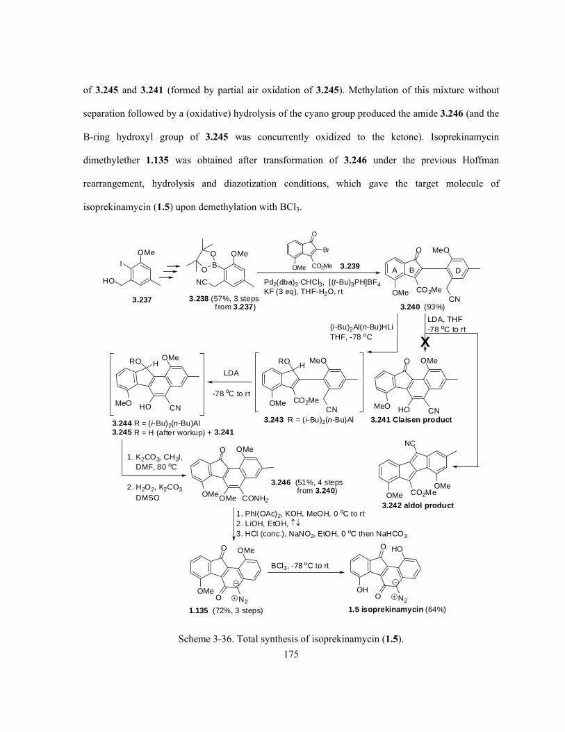
175
of 3.245 and 3.241 (formed by partial air oxidation of 3.245). Methylation of this mixture without
separation followed by a (oxidative) hydrolysis of the cyano group produced the amide 3.246 (and the
B-ring hydroxyl group of 3.245 was concurrently oxidized to the ketone). Isoprekinamycin
dimethylether 1.135 was obtained after transformation of 3.246 under the previous Hoffman
rearrangement, hydrolysis and diazotization conditions, which gave the target molecule of
isoprekinamycin (1.5) upon demethylation with BCl3.
O
O N2
1.135 (72%, 3 steps)
OMe
O
OMe CO2Me
Br
3.239
O
OMe CO2Me
MeO
A B D
3.240 (93%)
Pd2(dba)3·CHCl3, [(t-Bu)3PH]BF4KF (3 eq), THF-H2O, rt
LDA
-78 oC to rt
O
OMe CONH2OMe
1. K2CO3, CH3I,DMF, 80 oC
2. H2O2, K2CO3
DMSO
1. PhI(OAc)2, KOH, MeOH, 0 oC to rt2. LiOH, EtOH, 3. HCl (conc.), NaNO2, EtOH, 0 oC then NaHCO3
OMe
NC
B
O
O
3.237
OMe
HO
I
3.238 (57%, 3 stepsf rom 3.237)
CN
O
HO CNMeO
OMe
LDA, THF-78 oC to rt
OMeCO2Me
NC
OMe3.242 aldol product
3.241 Claisen product
(i-Bu)2Al(n-Bu)HLiTHF, -78 oC
OMe CO2Me
MeO
3.243 R = (i-Bu)2(n-Bu)AlCN
HRO
3.244 R = (i-Bu)2(n-Bu)Al3.245 R = H (after workup) + 3.241
HO CNMeO
OMeRO H
OMe
3.246 (51%, 4 stepsfrom 3.240)
OMe
BCl3, -78 oC to rt
O
O N2
1.5 isoprekinamycin (64%)
OH
HO
X
Scheme 3-36. Total synthesis of isoprekinamycin (1.5).

176
Besides the successful and first total synthesis of isoprekinamycin (1.5) by the Dmitrienko lab in
2007, the only other synthetic work towards the natural benzo[a]fluorene structures in the literature
was reported recently (2008) by Danishefsky and co-worker with their total synthesis of fluostatin C
(1.20c) and E (1.20e).162 The reported synthesis was rather complicated and of less value to be
applied as a general method for the synthesis of diazobenzo[b]fluorene analogues of kinamycin, when
compared with the above two synthetic strategies developed by the Dmitrienko group, but the
manuscript showed some interesting chemistry yet details will not be introduced here.
3.3 Conclusion
In summary, an efficient method has been developed for the synthesis of a tetraphene derivative 6-
hydroxy-8-methoxy-3-methyl-7,12-dioxo-7,12-dihydrotetraphen-4-yl methanesulfonate (3.131), a 6-
6-6-6 ring system that is to serve as a substrate for the ring-D oxygenation chemistry that is described
in Chapter 2. The method provides 3.131 in only three steps with an overall yield of 65% from the
two corresponding precursors 3.59 and 3.107, and each of the latter two intermediate can be easily
prepared from commercial starting material in five steps with an overall yield of 35% and 51%,
respectively. A strategy for the conversion of 3.131 into kinamycin F (1.1f) is described, and the
possibility arises that a key o-quinodiazide intermediate 3.136 in the process may possess interesting
biological activity similar to those of the kinamycins. Actual comparison of the formulated plan to
convert 3.131 to kinamycin F (1.1f) with total syntheses of kinamycins recently reported by other
groups suggests that this approach might be competitive with the reported synthetic procedures by the
Porco group,23 the Kumamoto/Ishikawa group24 and the Nicolaou group,16 but may not be as efficient
as that reported most recently by the Herzon group.26
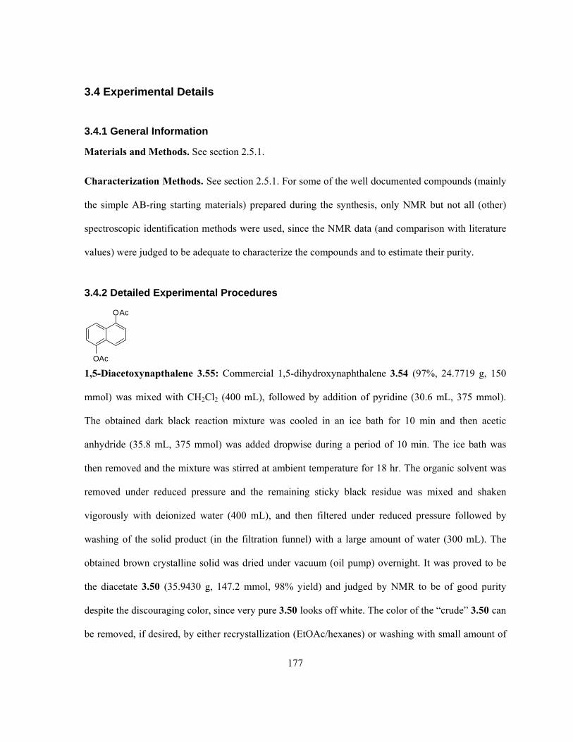
177
3.4 Experimental Details
3.4.1 General Information
Materials and Methods. See section 2.5.1.
Characterization Methods. See section 2.5.1. For some of the well documented compounds (mainly
the simple AB-ring starting materials) prepared during the synthesis, only NMR but not all (other)
spectroscopic identification methods were used, since the NMR data (and comparison with literature
values) were judged to be adequate to characterize the compounds and to estimate their purity.
3.4.2 Detailed Experimental Procedures
OAc
OAc
1,5-Diacetoxynapthalene 3.55: Commercial 1,5-dihydroxynaphthalene 3.54 (97%, 24.7719 g, 150
mmol) was mixed with CH2Cl2 (400 mL), followed by addition of pyridine (30.6 mL, 375 mmol).
The obtained dark black reaction mixture was cooled in an ice bath for 10 min and then acetic
anhydride (35.8 mL, 375 mmol) was added dropwise during a period of 10 min. The ice bath was
then removed and the mixture was stirred at ambient temperature for 18 hr. The organic solvent was
removed under reduced pressure and the remaining sticky black residue was mixed and shaken
vigorously with deionized water (400 mL), and then filtered under reduced pressure followed by
washing of the solid product (in the filtration funnel) with a large amount of water (300 mL). The
obtained brown crystalline solid was dried under vacuum (oil pump) overnight. It was proved to be
the diacetate 3.50 (35.9430 g, 147.2 mmol, 98% yield) and judged by NMR to be of good purity
despite the discouraging color, since very pure 3.50 looks off white. The color of the “crude” 3.50 can
be removed, if desired, by either recrystallization (EtOAc/hexanes) or washing with small amount of

178
MeOH under (slightly) reduced pressure. However, such treatment is not necessary for the next
reaction. 1H NMR (300 MHz, CDCl3): 7.78 (d, J = 8.4 Hz, 2 H), 7.50 (m, 2H), 7.29 (d, J = 7.5 Hz,
1H), 2.47 (s, 6H); 13C NMR (75 MHz, CDCl3) 169.2, 146.7, 128.1, 126.0, 119.3, 118.8, 21.0.
O
O
Br
OAc
2-Bromo-5-acetoxynaphthoquinone 3.7a: 1,5-Diacetoxynaphthalene 3.55 (12.2227 g, 50 mmol)
was dissolved in glacial acetic acid (150 mL) under gentle heating. The warm solution of 3.55 was
then added slowly during a period of 5 min to a mixture of commercial NBS (99%, 35.9630 g, 200
mmol), glacial acetic acid (150 mL) and deionized water (200 mL) under stirring. The obtained
aqueous mixture was then heated in an oil bath at a controlled temperature in between 65–70 °C for
90 min, during which the reaction flask was equipped with a water condenser to minimize the escape
of Br2 vapour that was generated in situ from the reaction mixture. There are several noteworthy
experimental observations. First, the color of the reaction mixture turned from the grey-yellow at the
beginning to brown-red after about 20 min of heating. Second, formation of the brown-red Br2 vapour
was physically visible during the reaction, which was further confirmed by a positive chemical test
with wet KI-starch paper. Third, the actual temperature of the reaction solution was ca. 15 °C (at the
beginning of reaction) to 5 °C (at the end of reaction) lower than the bath temperature (65–70 °C).
The oil bath was then removed and the solution was slightly cooled before it was poured into water (1
L), which produced large quantities of yellow precipitate. The precipitate was filtered and washed
with water (500 mL) under reduced pressure, and then recrystallized from EtOH*. The final product
3.7a was obtained as a bright orange solid (12.5154 g, 42.4 mmol, 85% yield). 1H NMR (300 MHz,
* The recrystallized 3.10 requires a long period of drying (at least overnight) under vaccum (oil pump) to completely remove the residual
solvent of EtOH

179
CDCl3): 8.14 (d, J = 7.8 Hz, 1H), 7.77 (t, J = 7.8 Hz, 1H), 7.42 (d, J = 7.8 Hz, 1H), 7.39 (s, 1H),
2.46 (s, 3H); 13C NMR (75 MHz, CDCl3) 181.0, 177.5, 169.3, 149.9, 141.5, 138.6, 135.1, 132.6,
130.4, 126.5, 123.2, 21.2.
OMe
OMe
Br
OMe 3.58 via OH
OH
Br
OH 3.70
2-Bromo-1,4,5-trimethoxynaphthalene 3.58: 2-Bromo-5-acetoxynaphthoquinone 3.7a (0.5915 g,
2.0 mmol) was mixed with MeOH (ACS grade, 20 mL) and stirred. Then SnCl2•2H2O powder
(1.8065 g, 8.0 mmol) was added in one portion to the 3.7a-MeOH suspension followed by dropwise
addition of conc. H2SO4 (~ 18 M, 1mL, 18 mmol). Initially, the yellow suspension quickly turned
brown upon addition of the acid, but it gradually turned again to light yellow within the next few
minutes. The reaction mixture was then heated at reflux for 7 hr. The mixture was filtered to remove
large amount of solid residue and the filtrate was then diluted with H2O (50 mL). The aqueous
solution was extracted with Et2O (100 mL x 3), and the organic extract was washed with H2O (20 mL
x 3) and brine (50 mL). The pH of the last aqueous phase (brine) after the wash was ca. 6. The Et2O
solution was then dried over Na2SO4 and concentrated, and the remaining grey-yellow solid residue
was dried under vacuum (oil pump) for a few hours. This grey-yellow solid was confirmed by 1H-
NMR (in DMSO-d6) to be compound 3.70, which was pure enough (0.4305 g, 85% yield from 3.58)
to undergo the next methylation step without any further treatment.
1H NMR (300 MHz, DMSO-d6): 10.84 (s, 1H), 10.76 (s, 1H), 9.05 (s, 1H), 7.57 (d, J = 8.4 Hz, 1H),
7.32 (m, 1H), 6.80 (d, J = 7.5 Hz, 1H), 6.80 (s, 1H). The three one proton signals at = 10.84, 10.76
and 9.05 ppm all disappeared after adding D2O to the NMR sample.

180
Powdered KOH (85%, 1.1178 g, 16.9 mmol) was mixed with DMSO (4 mL) and stirred for 5 min.
Then compound 3.70 (0.3605 g, 1.41 mmol) was added to the above KOH-DMSO suspension in one
portion followed by immediate dropwise addition of MeI (99.9%, d = 2.28, 0.53 mL, 8.43 mmol).
The mixture was stirred at room temperature under an air atmosphere for 30 min and a slight
exothermic reaction was noticed at the beginning during the addition of MeI. The reaction mixture
was filtered through a thin layer of celite to remove the remaining KOH powder and the celite was
washed with approximately 5 mL of DMSO. The dark black DMSO filtrate was then diluted with
H2O (90 mL) and neutralized by slowly adding conc. H2SO4 under stirring, until the pH of the
aqueous solution reached about 2–3. The now acidic aqueous solution was extracted with CH2Cl2
(100 mL x 4) and the combined organic phase was dried over MgSO4 and concentrated. The
remaining solid residue was further purified by flash column chromatography with hexanes:EtOAc
(10:1, v/v) as eluent. The trimethylated aryl bromide 3.58 was obtained as a yellow solid (0.2954 g,
70% yield from 3.70). 1H NMR (300 MHz, CDCl3): 7.69 (dd, J = 8.4 Hz, 0.9 Hz, 1H), 7.44 (t, J =
8.1 Hz, 1H), 6.91 (s, 1H), 6.90 (d, J = 8.1 Hz, 1H), 3.97 (s, 3H), 3.94 (s, 3H), 3.92 (s, 3H).
OMe
OMe
OMe
B(OH)2
3.59
1,4,5-Trimethoxynaphthalen-2-ylboronic acid 3.59: 2-Bromo-1,4,5-trimethoxynaphthalene 3.58
(0.4717 g, 1.59 mmol) was dissolved in anhydrous THF (8 mL) in a flame-dried flask, and the
resultant brown yellow solution was stirred in an acetone/dry ice bath (-78 °C) for 0.5 hr under N2.
Commercial n-BuLi in hexane (1.38 M, 1.21 mL, 1.67 mmol) was then added dropwise to the brown
yellow solution of 3.54 during a period of 10 min followed by further stirring at -78 °C for 0.5 hr.
Commercial B(OMe)3 (98+%, d = 0.915, 0.55 mL, 4.75 mmol) was then added dropwise by syringe
to the reaction mixture during a period of 5 min followed by a further stirring at -78 °C for 0.5 hr. The

181
dry ice bath was removed and the reaction mixture was allowed to warm to room temperature. The
mixture was then stirred with 10% HCl (2 mL) for 5 min, diluted with deionized water (20 mL) and
extracted with Et2O (40 mL x 3). The combined organic phase was dried over MgSO4 and
concentrated, leading to some brown-yellow solid as the crude product, which was further purified by
flash column chromatography on silica gel using CH2Cl2 as the eluent. Pure boronic acid 3.59 (0.1956
g, 0.75 mmol, 77% yield based on reacted starting material) was isolated along with the debrominated
compound 3.71 (0.1367 g, 0.63 mmol). 1H NMR (300 MHz, CDCl3): 7.67 (dd, J = 8.1 Hz, 0.9 Hz,
1H), 7.46 (t, J = 8.1 Hz, 1H), 7.16 (s, 1H), 6.95 (d, J = 7.8 Hz, 1H), 4.00 (s, 3H), 3.99 (s, 3H), 3.95 (s,
3H).
OMs
COOH
3.97
3-Methyl-2-(methylsulfonyloxy)benzoic acid 3.97: 3-Methylsalicylic acid 3.86 (98%, 15.5282 g,
100 mmol) was dissolved in THF (100 mL). CaO powder (14.0253g, 250 mmol) was added in one
portion to the THF solution, followed by dropwise addition of MsCl (99.5+%, d =1.474, 9.4 mL,
120.4 mmol) during a period of 10 min under stirring. Another 50 mL of THF was added to the
reaction mixture to help stir the slurry. After about 30 min of mixing all reagents, a significant and
spontaneous exothermic reaction* took place. Occasional manual stirring with a glass rod became
necessary since the slurry of the reaction mixture gradually turned thicker and stickier as the reaction
proceeded, during which another 50 mL of THF was added gradually to compensate for the loss of
solvent as a result of the exothermic reaction (a condenser was not installed so that manual stirring of
the sticky reaction mixture with a glass rod could be performed). After about 10 min of the
* The exothermic reaction can be accelerated by the addition of a small amount of water (a few drops) after mixing all reagents. If the
reaction is to be carried out at an even larger scale, extra precaution of this exothermic process is needed.

182
exothermic reaction, the white slurry of reaction mixture turned into some very sticky semi-solid.
Another 150 mL of THF was added to the reaction mixture and it was left to stand at room
temperature for another 10 hr. The reaction was worked up by slowly adding conc. HCl (45 mL) and
the resultant solution that was adjusted to pH ca. 1 was further stirred for 30 min. The aqueous
reaction mixture was then extracted with EtOAc (200 mL x 3). The combined organic phase was
dried over anhydrous Na2SO4 and concentrated to afford the crude mesylate 3.97 that contained a
small amount of unreacted 3.86. The starting material was removed by washing the crude product
with a small amount of Et2O under slightly reduced pressure until the filtrate (Et2O) looked
completely colorless. The pure mesylate 3.97 was obtained as a white solid (21.7298 g, 94.4 mmol,
94.4%). IR (CH2Cl2 film) 3028, 1691, 1606, 1586, 1466, 1412, 1337, 1299, 1207, 1190, 1171, 1144,
1088, 976, 871, 770, 700 cm-1; 1H NMR (300 MHz, CDCl3): 7.87 (d, J = 7.7 Hz, 1H), 7.50 (d, J =
7.6 Hz, 1H), 7.29 (t, J = 7.7 Hz, 1H), 3.34 (s, 3 H), 2.47 (s, 3 H); 13C NMR (75 MHz, CDCl3):
170.2, 146.2, 136.8, 134.3, 130.1, 126.9, 124.4, 39.4, 17.4; high resolution mass spectrum (EI) m/z
230.0244 [M+; calculated for C9H10O5S: 230.0249].
I
HOOC
OMs 3.100
6-Iodo-3-methyl-2-(methylsulfonyloxy)benzoic acid 3.100: 3-Methyl-2-(methylsulfonyloxy)-
benzoic acid 3.97 (11.5149 g, 50 mmol) was dissolved in DMF (ACS grade, 25 mL), followed by the
addition of Pd(OAc)2 powder (98%, 1.7181 g, 7.5 mmol, 15% catalyst loading). The resultant brown
solution was stirred for 5 min and then NIS powder (98+%, 12.6305 g, 55 mmol) was added in one
portion. The reaction mixture was heated up in an oil bath at 120 ºC for 24 hr and then filtered
through a thick layer of silica gel in a sintered glass funnel (5cm x 7cm) and washed with ether (4 L).
The ether filtrate was concentrated to ca. 700 mL and then washed with H2O (200 mL x 6). The

183
organic phase was dried over Na2SO4 and concentrated and the residue was recrystallized from THF
to afford 3.100 as a white (slightly yellow) solid (12.5274 g, 70%). IR (CH2Cl2 film): 3038, 2939,
1712, 1593, 1563, 1455, 1360, 1251, 1190, 1150, 1117, 973, 887, 816, 793, 728 cm–1;1H NMR (300
MHz, CDCl3): 11.32 (s, 1H), 7.69 (d, J = 8.1 Hz, 1H), 7.06 (d, J = 8.1 Hz, 1H), 3.29 (s, 3H), 2.39
(s, 3H); 13C NMR (75.5 MHz, CDCl3): 170.4, 144.7, 138.2, 134.7, 134.0, 133.5, 88.9, 39.6, 17.6;
high resolution mass spectrum (EI) m/z 355.9218 [M+; calculated for C9H9IO5S: 355.9215].
I
OMsOH 3.105
2-(Hydroxymethyl)-3-iodo-6-methylphenyl methanesulfonate 3.105: A fresh commercial 1M
BH3-THF solution (200 mL, 200 mmol) was added to the solid of 6-iodo-3-methyl-2-
(methylsulfonyloxy)benzoic acid 3.100 (7.1662 g, 20.1 mmol) under stirring. The first 30 mL was
added slowly as large amounts of gas was generated during the addition of BH3-THF solution. The
remaining 170 mL of BH3-THF solution was added all at once. The obtained light yellow reaction
solution, which turned completely colorless after 12 hr, was then stirred under N2 at room temperature
for 24 hr. The large excess of borane was quenched by slowly adding H2O (10 mL) to the solution
until it no longer bubbled, and the reaction mixture was washed with 10% aqueous Na2CO3 solution
(200 mL). The THF solution, which separated from the aqueous phase when washed with the basic
solution, was kept aside. The remaining aqueous (basic) phase was extracted with Et2O (200 mL x 2)
and the Et2O phases were combined with the previously separated THF solution. The combined
organic phases were washed with H2O (100 mL) and then loaded onto a layer of silica gel (5cm x
7cm) in a sintered glass funnel. The silica gel was further washed with Et2O (200 mL), the obtained
filtrates were concentrated and the remaining solid was further dried under vacuum (oil pump) for a
few hours. The benzyl alcohol 3.105 was obtained as a white solid (5.3196 g, 15.5 mmol, 77%). IR

184
(CH2Cl2 film) 3386, 3022, 2935, 1591, 1564, 1455, 1383, 1362, 1185, 1153, 1114, 1034, 1006, 974,
876, 810, 793 cm-1; 1H NMR (300 MHz, CDCl3) 7.72 (d, J = 8.1 Hz, 1 H), 6.96 (d, J = 8.1 Hz, 1 H),
4.78 (d, J = 6.9 Hz, 2 H), 3.40 (s, 3 H), 2.65 (t, J = 6.9 Hz, 1 H), 2.37 (s, 3H); 13C NMR (75 MHz,
CDCl3) 145.9, 138.6, 136.8, 133.6, 133.2, 98.3, 64.0, 38.8, 17.6; high resolution mass spectrum (EI)
m/z 341.9429 [M+; calculated for C9H11IO4S: 341.9423].
I
OMsBr 3.106
2-(Bromomethyl)-3-iodo-6-methylphenyl methanesulfonate 3.106: 2-(Hydroxymethyl)-3-iodo-6-
methylphenyl methanesulfonate 3.105 (0.2773 g, 0.81 mmol) was dissolved in CH2Cl2 (HPLC grade,
5 mL) followed by addition of CBr4 powder (98%, 1.6746 g, 5 mmol) in one portion. The resultant
colorless solution was stirred for 5 min at room temperature under an air atmosphere until all solid
dissolved. Then PPh3 pellet (99%, 1.3268 g, 5 mmol) was added in one portion but slowly to the
CH2Cl2 solution, which immediately turned to yellow and started to boil (the boiling only lasted for a
few seconds and no cooling was necessary). The reaction flask was then loosely capped and the
solution was further stirred at ambient temperature for 0.5 hr. The product was isolated from the
reaction mixture by loading the yellow CH2Cl2 solution onto a thick layer of dry silica gel (6 cm tall x
5 cm wide) in a sintered glass funnel, followed by washing the silica gel with CH2Cl2 (distilled, 300
mL) under slightly reduced pressure. The resultant colorless CH2Cl2 solution was concentrated and
the product was further dried under vacuum (oil pump) for a few more hours. The benzyl bromide
3.106 was obtained as a white solid (0.3275 g, 0.0809 mmol, 99.8% yield). IR (CH2Cl2 film) 3022,
2935, 1591, 1560, 1455, 1356, 1264, 1222, 1189, 1158, 1137, 1100 cm-1; 1H NMR (300 MHz,
CDCl3) 7.71 (d, J = 8.1 Hz, 1 H), 6.94 (d, J = 8.1 Hz, 1 H), 4.81 (s, 2 H), 3.43 (s, 3 H), 2.37 (s, 3H);

185
13C NMR (75 MHz, CDCl3) 145.7, 138.7, 134.7, 134.0, 133.6, 98.3, 39.6, 34.1, 17.8; high
resolution mass spectrum (EI) m/z 403.8584 [M+; calculated for C9H10BrIO3S: 403.8579].
I
OMsCN 3.107
2-(Cyanomethyl)-3-iodo-6-methylphenyl methanesulfonate 3.107: 2-(Bromomethyl)-3-iodo-6-
methylphenyl methanesulfonate 3.106 (0.0406 g, 0.1 mmol) was dissolved in a minimal amount of
DMSO (ACS grade, 0.5 mL) and NaCN powder (98.1%, 0.0106 g, 0.21 mmol) was added to the
DMSO solution, which immediately turned from very light yellow to dark pink (slightly reddish). The
reaction solution was further stirred at room temperature for 15 min although the reaction seemed to
be completed within 10 min as indicated by TLC. The DMSO solution was then diluted with H2O (50
mL) and the resultant white turbid aqueous solution was extracted with Et2O* (50 mL x 2). The
combined Et2O solution was dried over Na2SO4 and concentrated, and the remaining white (slightly
yellow) solid was further dried under vacuum (oil pump) for a few hours that was proved by NMR to
be the pure benzyl cyanide 3.107 (0.0353 g, 0.1 mmol, 100%). IR (CH2Cl2 film) 3039, 3030, 2981,
2940, 2249, 1726, 1593, 1565, 1457, 1410, 1351, 1263, 1200, 1185, 1155, 1114 cm-1; 1H NMR (300
MHz, CDCl3) 7.73 (d, J = 8.1 Hz, 1 H), 7.00 (d, J = 8.1 Hz, 1 H), 4.11 (s, 2 H), 3.41 (s, 3 H), 2.38
(s, 3H); 13C NMR (75 MHz, CDCl3) 145.3, 138.7, 134.4, 133.5, 128.9, 116.3, 97.4, 39.2, 26.0, 17.6;
high resolution mass spectrum (EI) m/z 350.9433 [M+; calculated for C10H10INO3S: 350.9426].
* The solubility of 3.107 in Et2O was found to be quite limited as ~ 35 mg of 3.107 was not able to completely dissolve in 3 mL of Et2O,
but CH2Cl2 was found to be a good solvent for 3.107. Therefore a larger scale of reaction could benefit from using CH2Cl2 as the extraction
solvent.

186
OMe
OMe
OMe
NC
OMs
3.109
2-(Cyanomethyl)-6-methyl-3-(1,4,5-trimethoxynaphthalen-2-yl)phenyl methanesulfonate 3.109:
A mixture of 1,4,5-trimethoxynaphthalen-2-ylboronic acid 3.59 (0.0655 g, 0.25 mmol), 2-
(cyanomethyl)-3-iodo-6-methylphenyl methanesulfonate 3.107 (0.0808 g, 0.23 mmol) and K3PO4
(0.1100 g, 0.5 mmol) was dissolved by stepwise addition of toluene (5 mL), EtOH (1.5 mL) and
deionized H2O (0.5 mL). The resultant yellow solution was then deoxygenated through three freeze-
thaw cycles under a nitrogen atmosphere. During the last freeze-thaw process and prior to the melting
of the frozen mixture, the catalyst Pd(PPh3)4 (0.0145 g, 0.0125 mmol) was added to the reaction
mixture. The reaction mixture was then stirred and heated to reflux in an oil bath under N2 for 6 hr.
The reaction solution was cooled to ambient temperature and diluted with saturated aqueous NH4Cl
solution (10 mL) and H2O (10 mL). The resultant aqueous mixture was then extracted with EtOAc
(40 mL x 3). The light yellow organic phase was dried over Na2SO4 and concentrated, and the
remaining crude product was purified by flash chromatography with EtOAc:hexanes (3:1, v/v) as the
eluent (repeat of the same column purification is necessary to achieve a better separation result with
the impurities and by-products), which appears as a light yellow foamy solid (0.0999 g, 0.226 mmol,
98% yield). When working on a larger scale of the Suzuki coupling reaction (1 mmol of each
coupling compound), the purification can be greatly simplified by carrying out the following
procedure: the crude product was dissolved in a small amount of CH2Cl2 and this solution was loaded
onto a large amount of dry silica gel (5 cm x 5 cm) in a sintered glass funnel, which was flushed
under reduced pressure first with a large amount of CH2Cl2 (400 mL, to remove grease and some non-
polar by-products) followed by acetone (400 mL). The light yellow acetone fraction was concentrated
and the remaining solid was dissolved in a minimal amount of Et2O. The Et2O solution of the product
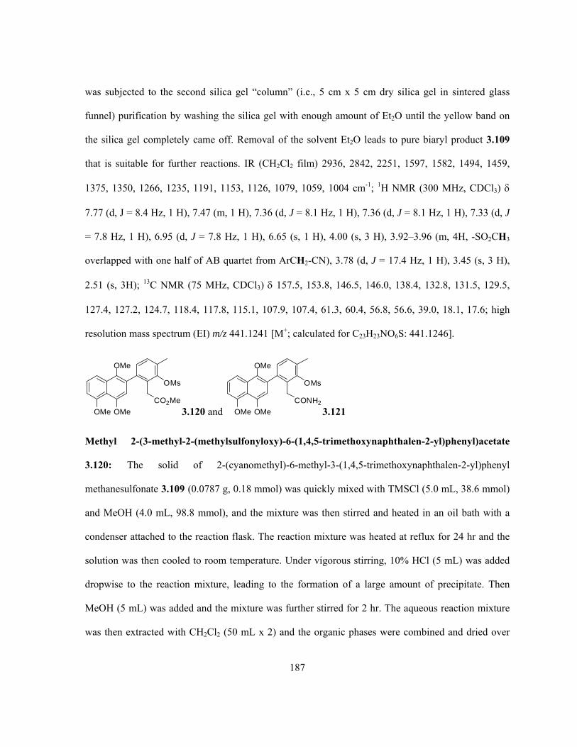
187
was subjected to the second silica gel “column” (i.e., 5 cm x 5 cm dry silica gel in sintered glass
funnel) purification by washing the silica gel with enough amount of Et2O until the yellow band on
the silica gel completely came off. Removal of the solvent Et2O leads to pure biaryl product 3.109
that is suitable for further reactions. IR (CH2Cl2 film) 2936, 2842, 2251, 1597, 1582, 1494, 1459,
1375, 1350, 1266, 1235, 1191, 1153, 1126, 1079, 1059, 1004 cm-1; 1H NMR (300 MHz, CDCl3)
7.77 (d, J = 8.4 Hz, 1 H), 7.47 (m, 1 H), 7.36 (d, J = 8.1 Hz, 1 H), 7.36 (d, J = 8.1 Hz, 1 H), 7.33 (d, J
= 7.8 Hz, 1 H), 6.95 (d, J = 7.8 Hz, 1 H), 6.65 (s, 1 H), 4.00 (s, 3 H), 3.92–3.96 (m, 4H, -SO2CH3
overlapped with one half of AB quartet from ArCH2-CN), 3.78 (d, J = 17.4 Hz, 1 H), 3.45 (s, 3 H),
2.51 (s, 3H); 13C NMR (75 MHz, CDCl3) 157.5, 153.8, 146.5, 146.0, 138.4, 132.8, 131.5, 129.5,
127.4, 127.2, 124.7, 118.4, 117.8, 115.1, 107.9, 107.4, 61.3, 60.4, 56.8, 56.6, 39.0, 18.1, 17.6; high
resolution mass spectrum (EI) m/z 441.1241 [M+; calculated for C23H23NO6S: 441.1246].
OMe
OMe
OMe
OMs
CO2Me
3.120 and OMe
OMe
OMe
OMs
CONH2
3.121
Methyl 2-(3-methyl-2-(methylsulfonyloxy)-6-(1,4,5-trimethoxynaphthalen-2-yl)phenyl)acetate
3.120: The solid of 2-(cyanomethyl)-6-methyl-3-(1,4,5-trimethoxynaphthalen-2-yl)phenyl
methanesulfonate 3.109 (0.0787 g, 0.18 mmol) was quickly mixed with TMSCl (5.0 mL, 38.6 mmol)
and MeOH (4.0 mL, 98.8 mmol), and the mixture was then stirred and heated in an oil bath with a
condenser attached to the reaction flask. The reaction mixture was heated at reflux for 24 hr and the
solution was then cooled to room temperature. Under vigorous stirring, 10% HCl (5 mL) was added
dropwise to the reaction mixture, leading to the formation of a large amount of precipitate. Then
MeOH (5 mL) was added and the mixture was further stirred for 2 hr. The aqueous reaction mixture
was then extracted with CH2Cl2 (50 mL x 2) and the organic phases were combined and dried over

188
anhydrous Na2SO4. A dark brown yellow solid was obtained as the crude product upon removal of
solvent, which contained mainly the desired methyl ester 3.120 but with small amount of unwanted
amide 3.121. Repeated flash column purification (2 times) with pure EtOAc as eluent successfully
separated 3.120 (0.0711 g, 0.15 mmol, 83% yield) from 3.121 (0.0128 g, 0.028 mmol, 16% yield).
For 3.120: 1H NMR (300 MHz, CDCl3) 7.74 (d, J = 7.6 Hz, 1H), 7.45 (dd, J = 7.8 Hz, 8.2 Hz, 1H),
7.39 (d, J = 8.1 Hz, 1H), 7.15 (d, J = 8.1 Hz, 1H), 6.91 (s, 1H), 6.64 (d, J = 8.2 Hz, 1H), 3.91 (s, 3H),
3.89 (s, 3H), 3.88 (s, 3H), 3.81 (d, J = 16.1 Hz, 1H), 3.73 (d, J = 15.7 Hz, 1H), 3.67 (s, 3H), 3.51 (s,
3H), 2.47 (s, 3H); 13C NMR (75 MHz, CDCl3) 171.6, 157.4, 153.1, 147.1, 146.7, 139.0, 131.8,
131.6, 130.6, 129.3, 128.4, 128.2, 127.1, 118.2, 115.2, 108.7, 107.2, 61.3, 60.4, 56.8, 56.6, 51.8, 39.0,
34.7.
For 3.121: 1H NMR (300 MHz, CDCl3) 7.68 (d, J = 8.1 Hz, 1 H), 7.47 (t, J = 7.8 Hz, 1 H), 7.27 (m,
2 H), 6.94 (d, J = 7.5 Hz, 1 H), 6.64 (s, 1H), 6.19 (br, 1H), 5.27 (br, 1H), 4.00 (s, 3H), 3.92 (s, 3H),
3.56 (s, 3H), 3.46 (s, 3H), 2.52 (s, 3H).
OMe
O
O
OMs
OH 3.131 and OH
O
O
OMs
OH 3.130
6-Hydroxy-8-methoxy-3-methyl-7,12-dioxo-7,12-dihydrotetraphen-4-yl methanesulfonate 3.131:
The solid of methyl 2-(3-methyl-2-(methylsulfonyloxy)-6-(1,4,5-trimethoxynaphthalen-2-
yl)phenyl)acetate 3.120 (0.0028 g, 0.0059 mmol) was mixed with neat MSA (1.0 mL), and the
resultant yellow solution was stirred under N2 atmosphere in a heated oil bath with the bath
temperature carefully controlled between 40–45 °C. The solution turned to dark brown-red (slightly
purple) after a few minutes of heating. After 6 hr of reaction, the reaction mixture was cooled and
diluted with CH2Cl2 (60 mL). Then the dark organic phase was washed with H2O (10 mL x 3), which
189
was further stirred with CaCO3 and Na2SO4 powder and then filtered. The resultant orange-yellow
filtrate was concentrated to afford a dark brown solid residue as the crude product, which was further
purified by flash column chromatography with Et2O as eluent. The desired cyclization product 3.131
was isolated as a dark red solid (0.0020 g, 0.0048 mmol, 81% yield). When the MSA-catalyzed
cyclization of 3.120 was carried out in refluxing conc. formic acid for extended time, the isolated
major product (~ 44% yield) from this harsher condition was 3.130, which is an A-ring demethylated
3.131.
For 3.131: 1H NMR (300 MHz, CDCl3): δ 9.23 (d, J = 7.9 Hz, 1H), 7.94 (dd, J = 8.2 Hz, 7.6 Hz, 1H),
7.89 (d, J = 8.2 Hz, 1H), 7.84 (s, 1H), 7.35 (d, J = 7.6 Hz, 1H), 6.99 (d, J = 7.9 Hz, 1H), 4.08 (s, 3H),
3.41 (s, 3H), 2.57 (s, 3H).
For 3.130: 1H NMR (300 MHz, CDCl3): δ 12.06 (s, 1H), 11.83 (s, 1H), 9.35 (d, J = 9.6 Hz, 1H), 7.96
(s, 1H), 7.83 (dd, J = 7.8 Hz, 1.2 Hz, 1H), 7.73 (m, 1H), 7.42 (d, J = 9 Hz, 1H), (dd, J = 8.1 Hz, 0.9
Hz, 1H), 3.43 (s, 3H), 2.58 (s, 3H).

190
Chapter 4
Chemistry of Kinamycin F: Studies on the Mode-of-Action of the
Diazobenzo[b]fluorene Type of Antibiotics
O
OH
HO
OH
O
OH
Me
OH
NN
1.1f kinamycin F
O
OH
R1O
OR3
O
OR4
Me
OR2
NN
1.1a-d kinamycin A-D
R1 R2 R3 R4
1.1a kinamycin A H Ac Ac Ac1.1b kinamycin B H H Ac H1.1c kinamycin C Ac Ac H Ac1.1d kinamycin D H Ac H Ac
Kinamycin F (1.1f), the diazobenzo[b]fluorene with a fully deacylated D-ring, received no
significant attention prior to this work, even though this substrate seems to possess better antibacterial
activity in general (Appendix A) than the other more well-studied kinamycins such as kinamycin A–
D (1.1a–d). During the course of this thesis project, the Dmitrienko group established a collaboration
with the Hasinoff group (University of Manitoba) to perform some in-depth biological studies of
these rare diazobenzofluorene antibiotics. Kinamycin F (1.1f) that had been prepared in the present
project from other natural kinamycins, and isoprekinamycin (1.5) that had been either isolated or
synthesized,25 as well as some isolated natural kinamycins A, C and D (1.1a/c/d) in the Dmitrienko
group were provided to the Hasinoff group. Some very interesting but previously unknown
bioactivities of these compounds were discovered.25,241,244 These results triggered further
consideration and significant interest to study the corresponding mechanisms for the kinamycins (this
work), as to be discussed in this Chapter 4 and later in Chapter 5. For simplification purposes, the
chemical name of “kinamycin X (X = A–F)” will be abbreviated as “kinX (X = A–F)” throughout this
chapter.

191
4.1 Kinamycin F: the Unique Diazobenzo[b]fluorene Antibiotic
The kinF (1.1f) used in the mechanistic studies was prepared quantitatively from natural kinD
(1.1d) under Zemplen deacylation conditions (NaOMe/MeOH), as introduced earlier (Scheme 2-12).
The latter along with natural kinA (1.1a) and kinC (1.1c) were isolated from the fermentation broth of
Streptomyces murayamanesis by V. Goodfellow and Dr. O. Adidayo in this laboratory. Collaborative
in vitro biological studies in the Hasinoff laboratory on these natural diazobenzo[b]fluorene type
kinamycins led to a few novel discoveries, which can be summarized as follows.241,244
First, both kinA (1.1a) and kinC (1.1c) potently inhibited cancer cell growth and the latter species
could induce a rapid apoptotic response in human Leukemia K562 cells.244 Second, kinA (1.1a)
displayed an unusual and delayed G1/S cell cycle block in synchronized Chinese hamster ovary (CHO)
cells only upon entry to a second cell cycle, which might be due to a slow deacetylation of kinA (1.1a)
to kinF (1.1f) by intracellular esterases.244 The observed similar IC50 of the kinA (1.1a), kinC (1.1c)
and kinF (1.1f) towards the Leukemia K562 cells (i.e., 0.31, 0.37 and 0.33 μM respectively) is
consistent with the suggestion that the completely-deacylated kinF (1.1f) might be the real bioactive
form of all other natural diazobenzo[b]fluorene-type kinamycins, which metabolize to kinF (1.1f)
prior to performing their biological activities.244 Third, both kinA (1.1a) and kinC (1.1c)
demonstrated inhibitory activities towards the DNA topoisomerase IIα that contains protein
sulfhydryl (-SH) groups. However, such inhibition experienced a substantial reduction when the
kinamycins were pre-incubated with a high concentration (> 1 mM) of L-glutathione (GSH, 4.1). The
GSH (4.1) is a widely-occurring thiol-containing tripeptide (γ-L-Glu-L-Cys-Gly) that exists
intracellularly in various cells at fairly high concentration (typically ~ 5 mM), and protects the cells
by scavenging harmful electrophiles (as a nucleophile) and oxidants (as a reductant).306 This

192
observation would suggest the very likely participation of the sulfhydryl groups, present in both the
DNA topoisomerase IIα and GSH (4.1), in the corresponding mechanism of action of the kinamycins.
4.2 dithiothreitol (DTT)
HS
OH
OH
SHOH
O O
NHH
H NH2
HS
O
NHOH
O
4.1 L-glutathione (GSH)
Under physiological conditions, reactions between natural kinamycins and GSH (4.1) or other
similar sulfur-based reductant/nucleophile such as the dithiothreitol (DTT, 4.2) were observed, as
indicated by the corresponding UV-vis spectra.241 In particular, in the presence of GSH (4.1) or DTT
(4.2), kinF (1.1f) was found to be capable of not only breaking the single-strand DNA (in K562 cells)
but also cleaving some double-strand DNA (i.e., nicking of pBR322 plasmid DNA) as well.241 The
involvement of the reductant GSH in the activation of kinF (1.1f) leading to significant DNA
cleavage was quite important, and this finding was in good agreement with two other studies
indicating that other natural diazoparaquinones such as lomaiviticin A (1.11a)52 and kinD (1.1d)307
are able to cleave DNA under similar reducing conditions. However, pure kinF (1.1f) itself, even in
the absence of a thiol, was found to be still capable of inducing certain amount of nicking of pBR322
plasmid DNA under physiological conditions, although only if a relatively high concentration of kinF
(1.1f) was used.241
The above observations, particularly the last two, are of significant meaning in regard to
kinamycins and related analogues (e.g., lomaiviticins) as potentially useful and novel antitumor
antibiotics, since cleavage of double-strand DNAs is generally much more lethal towards the target
cells than that of the single-strand DNAs. The only other known classes of effective double-strand
DNA scavengers as anticancer drugs in current clinical trials308,309 are the glycopeptide antibiotic

193
bleomycins 4.3310-312 and the well-known enediynes (e.g., 4.4–4.7).309,313,314 The latter are
characterized by the presence of a nine- or ten-membered carbon ring containing two triple bonds
separated by a double bond (i.e., the enediyne moiety), which is the active site for its biological
activities and commonly considered as the “warhead” against target cells. Naturally occurring
enediynes include three different types of compounds, (i) the chromoproteins315 such as the
neocarzinostatin 4.4,316 which is in fact a complex consisting of a 1:1 non-covalently bonded mixture
of a very unstable enediynes chromophore (in its neat form) that was recently synthesized,317 and a
carrier polypeptide/apoprotein318 that stabilizes and delivers the chromophore, (ii) the multisulfide-
based calicheamicins 4.5319 and esperamicins 4.6,320 which are two of the most potent antitumor
agents known to human by far, and (iii) the anthracycline-based dynemicins 4.7.321,322

194
4.5 calicheamicins
4.6b esperamicin P (n = 4, R =
4.3a bleomycin A2 (R = -NH-(CH2)3-S+(CH3)2X-)
4.3b bleomycin B2 (R = -NH-(CH2)4-NH-C-NH2)
N N
N
H2N
O
NH2O
H2N
HN
O
NH
OHO
HN
OHO
NH
O
S
N
SN
RO
O
NH
N
O O
O
CH2OHOH
O
HO
HOH2C
OH
HO
OHN
OOCH3
HO
SCH3SS
H OO OR2
OHNH
O
H3C
OH3C
S
OH
OH3C
OCH3
OCH3R1O
X
OHN
OOCH3
HO
CH3(S)n
H OO OOH
O
NH
O
H3C
NHiPrOCH3
OH3C
H3CS
OH
RO
H
OCH3
OOH
OHN
H3COOCH3
O
OCH34.6a esperamicin A1 (n = 3, R =
H2N
O
NH4.4 neocarzinostatin
apoprotein (113 amino acids)
O
OO
O
O
O
O
OCH3
H3C
OH
CH3HO
HOMeHN
enediyne chromophore
+
chromoprotein(1:1 complex)
Calicheamicin1
Br
1Br
2I
3I
1I
1I
1I
OH3CHO
H3CO OH
O NHR3OCH3
Rh = Am =
XBr
Br
II
I
II
R2
Am
Am
AmH
Am
AmAm
R1
Rh
Rh
HRh
Rh
RhRh
R3
iPr
Et
Et-
iPr
EtMe
HNOH
R
O
O HO
CO2H
OCH3
O
CH3
4.7a dynemicin A (R = OH)4.7b deoxydynemicin A (R = H)
4.6c esperamicin C (n = 3, R = H)*
* hydrolysis product of 4.6a
H

195
Regardless of the structural variation, all enediynes follow the common mechanism as shown in
Scheme 4-1a.308,309,313-315,323 The enediyne molecule first binds to the minor groove of DNA loosely,
followed by a critical activation at a specific triggering site that then allows the originally locked and
inactive enediyne warhead to adopt a new and appropriate geometry and consequently aromatize via
the Bergman reaction.324 This leads to the production of a highly active diradical species 4.9, which is
capable of abstracting hydrogen atoms from DNA to initiate a complex reaction sequence and
ultimately resulting in DNA cleavage. Binding of enediynes to DNA, however, is performed in
different ways depending on the non-enediyne part of the molecule. For example, the carbohydrate
portion within 4.5/4.6 acts as the binding and delivery mechanism, while the planar naphthoate of 4.4
and the anthraquinone of 4.7 would intercalate into the minor groove of DNA. Interestingly,
participation of thiols such as GSH (4.1) in the observed potent anticancer activity has been reported
for all three types of enediynes, but the role of GSH (4.1) in the mode-of-action depends on the
enediyne involved (Scheme 4-1b and 4-1c). In the case of the multisulfide-based and chromoprotein
type of enediynes, the GSH (4.1) acts as a nucleophilic activator (Scheme 4-1b). The GSH (4.1),
however, serves as an initial reductive activator in the case of dynemicins (Scheme 4-1c), and the
resulting hydroquinone moiety within 4.13 undergoes an intramolecular nucleophilic ERO reaction,
followed by a further nucleophilic activation by water to offer the required and activated enediyne
4.15.
Even though the cleavage of DNA by kinF (1.1f) in the presence of GSH (4.1) or DTT (4.2) had
been observed, the exact species and detailed mechanism(s) involved in these newly discovered
biological activities were not clear. It would be of great interest to examine the possibilities of thiol-
activation of kinamycins as discussed above for the enediyne system. Chemical exploration of such
reactions was expected to shed some light onto the corresponding mysterious pathway.
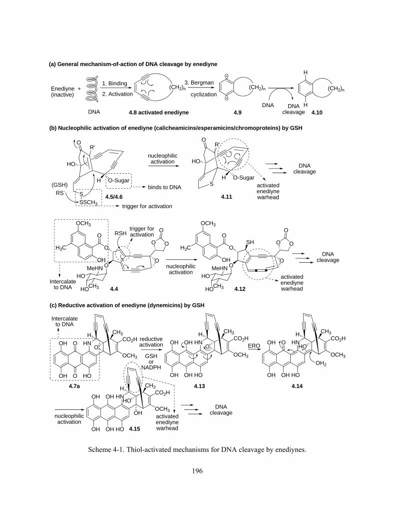
196
4.8 activated enediyne 4.9 4.10
(GSH)
OR'
HO
S
SSCH3
H O-Sugar
RS-
OR'
HO
SH O-Sugar
(CH2)n (CH2)n3. Bergman
cyclization(CH2)n
H
HDNA DNAcleavage
Enediyne +(inactive)
1. Binding
2. Activation
DNA
O
OO
O
O
O
O
OCH3
H3C
OH
CH3HO
HOMeHN
(b) Nucleophilic activation of enediyne (calicheamicins/esperamicins/chromoproteins) by GSH
4.5/4.6
HNOH
OH
O
O HO
CO2H
OCH3
O
CH3
O
OO
O
O
O
O
OCH3
H3C
OH
CH3HO
HOMeHN
SH
RSH
4.4
binds to DNA
trigger for activation
activatedenediynewarhead
activatedenediynewarhead
Intercalateto DNA
(c) Reductive activation of enediyne (dynemicins) by GSH
reductiveactivation
GSHor
NADPH
HNOH
OH
OH
OH HO
CO2H
OCH3
O
CH3H H
HNOH
OH
O
OH HO
CO2H
OCH3
HO
CH3H
(a) General mechanism-of-action of DNA cleavage by enediyne
trigger foractivation
OH2
4.7a
4.11
4.12
4.13 4.14
ERO
nucleophilicactivation
HNOH
OH
OH
OH HO
CO2H
OCH3
HO
CH3H
4.15
OH
Intercalateto DNA
DNAcleavage
DNAcleavage
DNAcleavage
nucleophilicactivation
nucleophilicactivation
activatedenediynewarhead
Scheme 4-1. Thiol-activated mechanisms for DNA cleavage by enediynes.

197
4.2 Mechanistic Study of Kinamycin F by Means of ESI-MS
4.2.1 Reactions of Thiols with Kinamycin F
When considering kinF (1.1f) from a structural point of view, the presence of a diazo group (C-
ring), a paraquinone (B-ring) and a phenolic functional group (A-ring) could all contribute
significantly to its bioactivities. On the other hand, the non-aromatic D-ring of kinF (1.1f) with the
four hydroxyl groups seems to be of less importance chemically towards the bioactivity at first glance
(see section 4.3 for further discussion). The existence of this sugar-like and highly hydrophilic D-ring,
however, would at least make the substrate more water-soluble than other kinamycins especially
under physiological aqueous conditions, which may in turn enhance the bioactivity. Then the ultimate
question regarding the mechanism-of-action of the kinamycins is obvious: the diazo moiety vs the
quinone moiety, which one (or maybe the combination of both) is the real player behind the actions?
Reactions of thiol compounds with either quinones325-327 or diazonium salts328-331 are known. In the
latter case, simple organic (both alkyl and aryl) thiols328,331 and some biologically significant
ones329,330 such as GSH (4.1), cysteine (4.16) and its N-acetyl derivative 4.17 (N-Ac-cys) could react
with aromatic diazonium salts to form the corresponding thiolethers or diazosulfides (Scheme 4-2a),
depending on the reactivity of the thiols and the stability of the corresponding diazo-thiol adducts.
The generally higher reactivity (or instability) of the diazo moiety than that of the quinone had led to
the proposal in the Dmitrienko group that, in the case of kinamycins where both functional groups co-
exist (kinF (1.1f) as an example), the sulfhydryl group of GSH (4.1) would probably undergo a
preferable nucleophilic addition to the terminal nitrogen of the diazo moiety (Scheme 4-2b). This
should generate initially a kinF-GSH adduct 4.18 that can tautomerize in between the semiquinone-
diazene form (HR–N=N–S–R’) and the corresponding quinone-diazane form (R=N–NH–S–R’). The
intermediate 4.18 in its semiquinone-diazene form should be capable of losing N2 to form a sp2

198
carbon-centred radical such as 4.19, which might lead to attack on and cleavage of DNA. One goal of
this mechanistic project was to look for evidence for this type of nucleophilic activation process, and
an ideal result would be the successful detection and characterization (or even isolation if possible) of
the proposed kinamycin-thiol adducts such as 4.18 by appropriate spectroscopic methods.
NN
HO
OH
OH
OH
O
OOH
1.1f kinF
OH
OH
O
OH
OH
HO
OH
N N
SHN
O
OH
O
HNH
O
HO
O
H2N HHSHN
O
OH
O
HNH
O
HO
O
H2N H
4.1 GSH
4.18 KinF-GSH adduct(top: semiquinone-diazene formbottom: quinone-diazane form)
Nucleophilic addition ?
OH
O
O
OH
OH
HO
OH
NNH
SHN
O
OH
O
HNH
O
HO
O
H2N H
-N2
?
DNAcleavage
OH
OH
O
OH
OH
HO
OH
DNA
4.19
(b)
(a)
Ar N2RS
+ N2
Ar'SArN=NSAr'
[H] then [O](Ar, Ar' = aromatic
or heterocyclic)
(R = alkyl)
O
HN
HO
HO
HS
4.17 N-Ac-cysteine
NH2
HO
HO
HS
4.16 cysteine
- N2 Ar S Ar'
Ar'S SAr'
Ar S R
Scheme 4-2. (a) Literature examples of nucleophilic addition of thiols to aromatic diazonium salts; (b)
proposed nucleophilic GSH-activation mechanism for DNA cleavage by kinF (1.1f).

199
As the initial attempt to verify the above proposed mechanism of action (Scheme 4-2b), N-Ac-cys
(4.17) was used as a simpler thiol analogue of GSH (4.1) to react with kinF (1.1f) in MeOD-d4 at
room temperature. The corresponding NMR spectra, when monitored as a function of time, indicated
that no reaction occurred between these two compounds even after extended time (a few days) of
mixing, and the only observed chemical change was the air oxidation of 4.17 to the corresponding
disulfide. KinF (1.1f) was apparently very stable under such conditions. The failure to observe the
expected formation of the kinamycin-thiol adduct suggests that the neutral thiol of 4.17 (pKa(-SH4.17)
= 9.52;332 pKa(-CO2H4.17) ~ 5) is not sufficiently nucleophilic to react with the diazo group of kinF
(1.1f). In the absence of a base or buffer to maintain a high enough (near neutral or weakly basic) pH,
generation of the much more nucleophilic sulfide anion (RS-) would be negligible. This observation
also suggested that, to ensure a smooth occurrence of the desired reaction between kinF (1.1f) and the
thiol such as GSH (4.1, pKa (-SHGSH) = 8.63332/8.66333), the later mechanistic studies should be
carried out at relatively high pHs.
There are several other significant challenges to be considered prior to any further mechanistic
studies. First, the supply of natural kinamycins through bacteria fermentation in the Dmitrienko
laboratory was quite limited and only a few milligrams of kinF (1.1f) were available at the time.
Therefore, the series of mechanistic studies could only be carried out on micro-scale, which would
significantly increase the difficulties to identify the products, particularly by NMR that requires
substantial amount of material. Second, the studies had to deal with a complex multi-component
system, since the reaction would involve at least two substrates (i.e., kinF and a thiol), two solvents
(both water and an appropriate organic co-solvent for kinF) and some buffer species (to control a
proper pH). The situation could become more complicated due to possible side reactions such as (air)
oxidation of the thiol to the disulfide. Even the purest commercial thiols still contain a certain amount
of the corresponding disulfides. Last but not least, the choice of the organic co-solvent would be quite

200
critical, so that both the kinamycin solubility requirement and the compatibility with the analytical
methods used for compound characterization had to be satisfied.
It was found earlier in this project (Chapter 2) that, attempts with common EI (electron impact) or
CI (chemical ionization) methods to acquire the mass spectra of kinF (1.1f) simply failed, and no
molecular ion of kinF (1.1f) [kinF]+• (C18H14N2O7+•, m/z: 370.0801) could be observed. Instead, mass
spectra of kinF (1.1f) could be obtained only by +ESI (positive electrospray ionization) mass
spectrometry in the presence of either proton or metal ion such as Li+, with 1:1 (v/v) MeCN/H2O as
the solvent. The only other literature mass data of kinF (1.1f) was later reported by Nicolaou using
exactly the same (+ESI) ionization method.16 In fact, +ESI mass was used previously in this project to
confirm the prepared kinF (1.1f) via the high-resolution mass spectra of [kinF + Li]+ in 1:1 (v/v)
MeCN/H2O. The kinF (1.1f) molecule binds with the available cations (H+ or Li+) and the
corresponding ionic species, i.e. [kinF + H]+ (C18H15N2O7, m/z: 371.0879 and [kinF + Li]+
(C18H14N2O7Li+, m/z: 377.0961), were smoothly detected in the +ESI mass spectra (Figure 4-1a and
4-1b), which further fragmented as observed in the corresponding MSMS spectra (Figure 4-1c and 4-
1d). In the meantime, formation of other non-covalent bonded ionic cluster species such as [kinF +
kinF + H]+ (C36H29N4O14+, m/z: 741.1680), [kinF + kinF + Li]+ (C36H28N4O14Li+, m/z: 747.1762) and
[kinF + Na]+ (C18H14N2O7Na+, m/z: 393.0699) were also observed (Figure 4-1a and 4-1b). The last
ionic species was formed by complexation of kinF (4.1) with Na+ ions, which most likely originate
from “environmental contamination” such as the common glassware used in the laboratory.334

201
Figure 4-1. +ESI-MS of kinF (1.1f) in 1:1 MeCN/H2O (from top to bottom, a–d): (a) MS of kinF +
LiOAc; (b) MS of kinF + 0.2% HCOOH; (c) MSMS of [kinF + Li]+; (d) MSMS of [kinF + H]+.
On the other hand, quick tests of GSH (4.1) in 1:1 (v/v) MeCN/H2O indicated that this thiol also
responds very well and similarly with +ESI-MS spectroscopy (Figure 4-2). Ionic species including
[GSH + H]+ (C10H18N3O6S+, m/z: 308.0916), [GSH + Li]+ (C10H17N3O6SLi+, m/z: 314.0998), [GSH +
Na]+ (C10H17N3O6SNa+, m/z: 330.0736), [GSH + GSH + H]+ (C20H35N6O12S2+, m/z: 615.1754), [GSH
+ GSH + Li]+ (C20H34N6O12S2Li+, m/z: 621.1836) and [GSH + GSH + Na]+ (C20H34N6O12S2Na+, m/z:
140 150 160 170 180 190 200 210 220 230 240 250 260 270 280 290 300 310 320 330 340 350 360 370 380 390m/z0
100
%
0
100
%
NOV18CN3 11 (0.404) Cm (7:52) TOF MSMS 377.10ES+ 213377.1080
271.0537219.0524
153.0708 218.0538203.0649
247.0461220.0643257.0651
285.0595
307.0641 313.0697 331.0679
378.1068
379.0926
MAR16CNKINAMYCINF3 65 (2.342) Cm (1:106) TOF MSMS 371.10ES+ 5.42e4279.0653
265.0554
251.0746223.0800
307.0665
280.0720
335.0675
308.0708355.0727
175 200 225 250 275 300 325 350 375 400 425 450 475 500 525 550 575 600 625 650 675 700 725 750 775 800m/z0
100
%
0
100
%
NOV18CN2 1 (0.045) Cm (1:35) TOF MS ES+ 1.01e3377.0866
288.2856244.2553
195.1034
304.2848376.0964
747.1413
378.0854
393.0648 746.1482481.0632
748.1552
749.1497
MAR16CNKINAMYCINF2 118 (4.224) Cm (1:120) TOF MS ES+ 1.89e4371.0853
353.0789
335.0699244.2653182.9898 279.0673313.1451
741.1727372.0972
393.0760550.6375434.1012
758.1989
764.1743

202
637.1574) were clearly observed. However, as predicted earlier, the presence of the disulfide GSSG
(oxidized dimer form of GSH) in the commercial GSH (Sigma, purity 98%) used in this study was
noticed as well. The GSSG was detected (Figure 4-2) as the corresponding protonated form [GSSG +
H]+ (C20H33N6O12S2+, m/z: 613.1598) and Na+-complex [GSSG + Na]+ (C20H32N6O12S2Na+, m/z:
635.1417).
L-Glutathione (20uL in H2O,pH7.6, 4.74e-4M) + 80uL 1:1 MeCN/H2O (no FA) +ESI
100 125 150 175 200 225 250 275 300 325 350 375 400 425 450 475 500 525 550 575 600 625 650 675 700m/z0
100
%
MAR16CNLGLUTATHIONE2 10 (0.366) Cm (1:63) TOF MS ES+ 3.09e3308.0986
179.0496
162.0270233.0605
291.0660
314.1006
613.1732
346.0571
330.0755
484.1295347.0480
490.1412
619.1688
620.1650651.1299
L-Glutathione (20uL in H2O, pH7.6, 4.74e-4M) + 80uL 1:1 MeCN/H2O+0.2%FA +ESI
100 125 150 175 200 225 250 275 300 325 350 375 400 425 450 475 500 525 550 575 600 625 650 675 700m/z0
100
%
MAR16CNLGLUTATHIONE1 33 (1.186) Cm (1:75) TOF MS ES+ 3.23e4613.1480
308.0804
179.0453
162.0229
144.0122
233.0614
180.0536
209.1562
244.2667291.0670
314.1018484.1231
346.0516
355.0827
409.0949362.0127485.1252
538.1319486.1283
614.1666
635.1482
651.1227
667.0872
Figure 4-2. +ESI-MS of GSH (4.1) in 1:1 (v/v) MeCN/H2O (pH of the aqueous GSH solution was
adjusted to 7.60 with LiOH before adding MeCN): (top) GSH alone; (bottom) GSH + 0.2% HCOOH.

203
Based on these preliminary results, +ESI-MS was considered a suitable spectroscopic method to
characterize the products from the reactions of interest between kinF (1.1f) and thiols. The high
resolution and ultra sensitivity of mass spectrometry would allow the use of very small quantities of
kinF (1.1f), and its fast response speed would be particularly beneficial if the produced kinF-thiol
adduct is an unstable and short-lived species. To mimic the biological studies of the known GSH-
promoted cleavage of DNA by kinF (1.1f) that were carried out under physiological conditions (i.e.,
37 °C, pH 7.6, 0–400 μM kinF, 5 mM GSH),241 mechanistic studies on the reactions between kinF
(1.1f) and thiols were also designed and performed under very similar conditions (Table 4-1). It
should be pointed out that the use of bases such as LiOH or NaOH provided not only the required
(near) physiological pH for the reaction, but also the metal ions (Li+ or Na+) that are essential to the
ionization of various species of interest in the +ESI-MS studies, since most of them could bind with
the metal ions. On the other hand, acidification of the mass samples with dilute formic acid (HCOOH)
also greatly promotes the +ESI mass response due to the significant protonation of the neutral species.
Acetonitrile was chosen as the organic co-solvent due to its compatibility with not only ESI-MS but
also HPLC (to be used for the separation of kinF-thiol reaction products), as well as the good
solubility of kinF (1.1f) in this polar solvent. In most cases the kinF-thiol reaction was controlled at
either ambient (23 °C) or physiological temperature (37 °C), but a few lower temperatures (i.e., 4 ºC
and -10 ºC) were also attempted. To minimize any possible photolytic process of the diazoquinone of
kinF (1.1f) or the likely kinF-thiol adducts, all reactions were carried out and examined without
exposure to light. As the control experiments and for comparison purpose, beside GSH (4.1), other
thiol and sulfur-containing species such as the N-acetyl-cysteine (4.17) and GSSG were also
employed. The mechanistic studies were carried out by following the general experimental procedure
as described in section 4.7.

204
Table 4-1. Experimental conditions for the reactions between kinF (1.1f) and thiols.
Sample No.a [kinF] (mM) : [thiol] (mM)b Basec T (C)d Reaction Time
KINFGSH0 0.027 : 0.0139 (1.94:1) LiOH 23 10/20/30/45/60/75/90/300 min
KINFGSH1 0.027 : 0.0139 (1.94:1) LiOH -10 24 hr
KINFGSH2 0.027 : 0.0139 (1.94:1) LiOH 4 24 hr
KINFGSH3 0.027 : 0.0139 (1.94:1) LiOH 23 24 hr
KINFGSH4 0.027 : 0.0139 (1.94:1) LiOH 37 24 hr
KINFGSH5 0.027 : 1.21 (1:44.8) LiOH 23 50/120/250 min, 50 hr
KINFGSH6 0.0513 : 0.121 (1:2.36) LiOH 23 10/20/30/60/90 /160 min, 44 hr
KINFGSH7 0.1323 : 0.121 (1.09:1) LiOH 23 10/90 min, 43 hr
KINFGSH8 0.054 : 0.00968 (5.58:1) LiOH 23 10 min
KINFGSH9 0.027 : 1.21 (1:44.8) LiOH 37 53 hr
KINFGSH10 0.027 : 0.02975 (1:1.1) NaOH 37 12 hr
KINFGSH11 0.027 : 0.119 (1:4.4) NaOH 37 12 hr
KINFGSH12e 0.0675 : 0.0705 (1:1.04) LiOH 23 -
KINFGSH13 0.135 : 0.141 (1:1.04) LiOH 23 15 min/2 hr
KINFGSSG1 0.027 : 1.12 (1:41.5) LiOH 23 70/230 min, 50 hr
KINFGSSG2 0.027 : 1.12 (1:41.5) LiOH 23 70/230 min, 50 hr
KINFGSSG3 0.027 : 1.12 (1:41.5) LiOH 37 53 hr
KINFCYS1 0.027 : 0.02975 (1:1.1) NaOH 37 12 hr
KINFCYS2 0.027 : 0.119 (1:4.4) NaOH 37 12 hr a. GSH = L-glutathione (4.1), GSSG = oxidized dimer form of GSH (a disulfide), KINF = kinamycin F (1.1f),
CYS= N-acetyl-L-cysteine (4.17); b. Concentration refers to the final value of the substrate in the reaction
solution (1:1 MeCN/H2O); c. pHs of the aqueous thiol solutions were adjusted to the physiological value of 7.60
with the base (either LiOH or NaOH as listed) prior to the mixing with the kinF-MeCN solution (see section 4.7
for details); d. ± 1 °C; e. Excess 0.2% HCOOH in MeCN-H2O (1:1) was added to both substrates individually
prior to the mixing, and the mixed sample was directly subjected to the +ESI-MS analysis.

205
After the quite extensive and systematic +ESI-MS studies of the reaction between kinF (1.1f) and
the thiols, some interesting results were obtained. They are summarized and interpreted as follows.
(i) Initial attempt (sample KINFGSH0, Table 4-1) was made by mixing kinF (1.1f) with GSH (4.1)
in a ratio of ~ 2:1 in 1:1 (v/v) MeCN/H2O (pH 7.6), and the reaction was monitored closely by +ESI-
MS as a function of time (10–300 min, Figure 4-3). It should be noted that observed mass peaks with
m/z around or smaller than 300 are of very little value to the mechanistic interpretation, since such
peaks, particularly those with high nominal masses (e.g., m/z ~ 288.3, 272.3, 244.3), mainly come
from the background.
100 125 150 175 200 225 250 275 300 325 350 375 400 425 450 475 500 525 550 575 600 625 650 675 700 725 750 775 800m/z0
100
%
0
100
%
0
100
%
0
100
%
0
100
%
0
100
%
0
100
%
0
100
%
MAR20CNKINFGSH0G 91 (3.260) Cm (1:140) TOF MS ES+ 2.47e3393.0748
244.2658
154.9932139.0464 175.1221203.0477
288.2982 365.1122304.2896
763.1640409.0489 747.1954635.1552613.1550
422.0768 657.1291 764.1683
MAR20CNKINFGSH0F 58 (2.081) Cm (1:140) TOF MS ES+ 853244.2630
154.9896139.0470
169.9735
288.2897
272.2936
393.0742
316.3212332.3119 763.1707434.1073 747.1716635.1592
453.1186 613.1675 657.1429 776.1759
MAR20CNKINFGSH0E 2 (0.081) Cm (2:140) TOF MS ES+ 880244.2639
169.0517139.0475 234.1413
288.2907
272.2946393.0756
316.3223332.3197 763.1532406.0856434.1012 747.1743
635.1522613.1607 657.0983 776.1581
MAR20CNKINFGSH0D 1 (0.045) Cm (1:86) TOF MS ES+ 497244.2647
169.9747154.9861200.2362
288.2917272.2955
393.0697
316.3299332.3142 763.1457394.0821 434.0951 747.1969635.1545613.1628
483.1006 657.1382 776.1711
MAR20CNKINFGSH0C 37 (1.331) Cm (2:98) TOF MS ES+ 2.34e3288.2855244.2647
113.0161176.1414 203.0511 272.2894
393.0770316.3234
332.3142 384.1040 763.1559613.1538394.0674
550.6406453.1062 483.1006 635.1360 747.1869662.5698 776.1711
MAR20CNKINFGSH0B 31 (1.116) Cm (28:140) TOF MS ES+ 668244.2665
169.0535139.0534
200.2376
393.0725288.3000272.2975 316.3191332.3166 763.1613747.1922
418.1324434.1135635.1406
613.1762 657.1429 776.1766
MAR20CNKINFGSH0A 57 (2.045) Cm (1:88) TOF MS ES+ 512244.2665
200.2376169.0535156.0768
393.0725288.2938
316.3257 377.1097763.1511747.1822394.0775
635.1406613.1581453.1094
657.1429 776.1869
MAR20CNKINFGSH0 5 (0.188) Cm (1:87) TOF MS ES+ 532393.0667244.2673
288.2885
377.0968316.3268
763.1437747.1948394.0789
635.1519613.1784422.0765
657.1076 746.1517764.1682
777.1718
[kinF]:[GSH]=1.94:1, r.t., 300min
[kinF]:[GSH]=1.94:1, r.t., 90min
[kinF]:[GSH]=1.94:1, r.t., 75min
[kinF]:[GSH]=1.94:1, r.t., 60min
[kinF]:[GSH]=1.94:1, r.t., 45min
[kinF]:[GSH]=1.94:1, r.t., 30min
[kinF]:[GSH]=1.94:1, r.t., 20min
[kinF]:[GSH]=1.94:1, r.t., 10min
Figure 4-3. Time-resolved (as labeled on each MS spectrum) +ESI-MS spectra of the reaction
mixture of kinF (1.1f) and GSH (4.1) (sample KINFGSH0, Table 4-1).
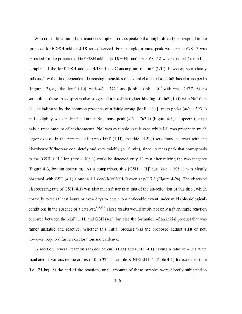
206
With no acidification of the reaction sample, no mass peak(s) that might directly correspond to the
proposed kinF-GSH adduct 4.18 was observed. For example, a mass peak with m/z ~ 678.17 was
expected for the protonated kinF-GSH adduct [4.18 + H]+ and m/z ~ 684.18 was expected for the Li+-
complex of the kinF-GSH adduct [4.18+ Li]+. Consumption of kinF (1.1f), however, was clearly
indicated by the time-dependent decreasing intensities of several characteristic kinF-based mass peaks
(Figure 4-3), e.g. the [kinF + Li]+ with m/z ~ 377.1 and [kinF + kinF + Li]+ with m/z ~ 747.2. At the
same time, these mass spectra also suggested a possible tighter binding of kinF (1.1f) with Na+ than
Li+, as indicated by the common presence of a fairly strong [kinF + Na]+ mass peaks (m/z ~ 393.1)
and a slightly weaker [kinF + kinF + Na]+ mass peak (m/z ~ 763.2) (Figure 4-3, all spectra), since
only a trace amount of environmental Na+ was available in this case while Li+ was present in much
larger excess. In the presence of excess kinF (1.1f), the thiol (GSH) was found to react with the
diazobenzo[b]fluorene completely and very quickly (< 10 min), since no mass peak that corresponds
to the [GSH + H]+ ion (m/z ~ 308.1) could be detected only 10 min after mixing the two reagents
(Figure 4-3, bottom spectrum). As a comparison, this [GSH + H]+ ion (m/z ~ 308.1) was clearly
observed with GSH (4.1) alone in 1:1 (v/v) MeCN/H2O even at pH 7.6 (Figure 4-2a). The observed
disappearing rate of GSH (4.1) was also much faster than that of the air-oxidation of this thiol, which
normally takes at least hours or even days to occur to a noticeable extent under mild (physiological)
conditions in the absence of a catalyst.335,336 These results would imply not only a fairly rapid reaction
occurred between the kinF (1.1f) and GSH (4.1), but also the formation of an initial product that was
rather unstable and reactive. Whether this initial product was the proposed adduct 4.18 or not,
however, required further exploration and evidence.
In addition, several reaction samples of kinF (1.1f) and GSH (4.1) having a ratio of ~ 2:1 were
incubated at various temperatures (-10 to 37 C, sample KINFGSH1–4, Table 4-1) for extended time
(i.e., 24 hr). At the end of the reaction, small amounts of these samples were directly subjected to

207
+ESI-MS analysis, while the remaining mixtures were further treated by HPLC separation (see
section 4.7 for experimental details) in the hope that this might lead to the isolation of either the
possible initial nucleophilic adduct such as 4.18 or any further products that may be derived from it.
Despite the many chromatographic fractions obtained and examined, +ESI-MS led to no meaningful
data that could be correlated to the proposed structure of 4.18. However, regardless of the incubation
temperature, direct MS analysis (Figure 4-4) did indicate the complete consumption of the limiting
reagent GSH (4.1), as suggested by the total absence of the characteristic peak of [GSH + H]+ (m/z ~
308.1). This observation was consistent with the earlier interpretation that a fast reaction between
kinF (1.1f) and GSH (4.1) would lead to an unstable product.
200 220 240 260 280 300 320 340 360 380 400 420 440 460 480 500 520 540 560 580 600 620 640 660 680 700 720 740 760 780 800m/z0
100
%
0
100
%
0
100
%
0
100
%
0
100
%
MAR20CNKINFGSH4ACIDIFIED 9 (0.331) Cm (1:142) TOF MS ES+ 8.85e3309.0799
244.2667
209.1562
288.2930272.2970
245.2660
333.0913
393.0763335.0706
377.0995
613.1662
484.1231394.0812 516.0940 550.6450645.1417
737.1774667.0967721.1887 763.1667
MAR20CNKINFGSH4 15 (0.545) Cm (1:144) TOF MS ES+ 50244.2675
242.3036
244.2848
288.2878
272.3161316.3318
393.0995335.1458
747.1705721.1616
605.5025437.2595 592.4755689.9323
793.5646
MAR20CNKINFGSH3 102 (3.652) Cm (1:168) TOF MS ES+ 47244.2742
242.2931
272.2989
245.2677
288.2950
288.3199 747.2032393.1154377.0951319.1598 409.0532
504.1092 733.1118703.6556763.1823
788.1167
MAR20CNKINFGSH2 57 (2.045) Cm (1:147) TOF MS ES+ 28244.2759
244.2644
242.2890
242.2491
288.2908272.3069
393.0745316.3221 377.1191335.1359 747.1784394.0796
736.2712763.1775
MAR20CNKINFGSH1 38 (1.367) Cm (2:276) TOF MS ES+ 22393.0787
244.2728242.3031
239.1761
377.1088244.2842 320.1302288.3063
288.2627377.0946
747.1964393.1006
418.1460419.1299 738.5717
635.0438583.5776573.5030543.2924 715.6179
747.2366
764.1797
775.5568
[kinF]:[GSH]=1.94:1, 37C, 24hr, acidified
[kinF]:[GSH]=1.94:1, 37C, 24hr
[kinF]:[GSH]=1.94:1, 22.5C, 24hr
[kinF]:[GSH]=1.94:1, 4C, 24hr
[kinF]:[GSH]=1.94:1, -10C, 24hr
Figure 4-4. +ESI-MS spectra of the reaction mixtures of kinF (1.1f) and GSH (4.1) incubated for
24 hr at different temperatures (as labeled on each spectrum, samples KINFGSH1–4, Table 4-1).

208
When GSH (4.1) was used in (large) excess relative to kinF (1.1f) (sample KINFGSH5, 6, 9 and
11, Table 4-1), detection of GSH- and GSSG-derived ion species dominated the obtained mass
spectra, and observation of those kinF-related ions became quite difficult (Figure 4-5). Again no
evidence for the proposed kinF-GSH adduct 4.18 was obtained, but quick (< 10 min) and complete
consumption of kinF (1.1f) under such conditions was still evident. This was indicated by the
complete absence of the characteristic peaks of [kinF + Li]+ (m/z ~ 377.1) and [kinF + Na]+ (m/z ~
393.1) only 10 min after mixing these two reagents (Figure 4-5, bottom spectrum), which matched
very well the earlier observation that the GSH (4.1) was used up within 10 min when kinF (1.1f)
presented in excess (Figure 4-3, bottom spectrum). However, it seemed that when GSH (4.1) was
used in only slightly excess (e.g., sample KINFGSH6, [kinF]:[GSH] = 1:2.36), a small amount of the
cluster ion species of kinF (1.1f) such as the [kinF + kinF + Li]+ (m/z ~ 747.2) could survive the
conditions even after extended reaction time (Figure 4-5, all spectra).
100 150 200 250 300 350 400 450 500 550 600 650 700 750 800m/z0
100
%
0
100
%
0
100
%
0
100
%
0
100
%
0
100
%
MAR23CNKINFGSH6E 17 (0.616) Cm (2:85) TOF MS ES+ 1.99e3320.0891
314.0820288.2744157.0722
141.0990 244.2500173.0508
336.0655
352.0381 418.0968 633.1566434.0689 649.1528 690.1467747.1419
MAR23CNKINFGSH6D 3 (0.116) Cm (2:85) TOF MS ES+ 2.00e3320.1121
288.2962141.1105 244.2646157.0844326.1232
418.1298 627.1970476.6496 747.1842690.1971
MAR23CNKINFGSH6C 11 (0.402) Cm (2:85) TOF MS ES+ 1.92e3320.1121
141.1105 308.0963244.2646157.0797
326.1232418.1298 633.2052476.6496 747.1942690.2067
MAR23CNKINFGSH6B 4 (0.152) Cm (2:84) TOF MS ES+ 2.59e3314.0982
308.0963141.1105 244.2704157.0844
320.1121
627.1970326.1232
418.1298 476.6576 747.1842690.2067
MAR23CNKINFGSH6A 26 (0.938) Cm (2:85) TOF MS ES+ 3.03e3314.1048
308.0963
141.1062 244.2646194.0322 418.1223326.1298627.1970476.6656 747.1942690.2067
MAR23CNKINFGSH6 20 (0.723) Cm (2:72) TOF MS ES+ 2.41e3320.1066
314.1059
288.2972244.2655141.1067326.1177
418.1313 633.2075476.6512 747.1868690.1995 789.7694
[kinF]:[GSH]=1:2.36, r.t., 160min
[kinF]:[GSH]=1:2.36, r.t., 90min
[kinF]:[GSH]=1:2.36, r.t., 60min
[kinF]:[GSH]=1:2.36, r.t., 30min
[kinF]:[GSH]=1:2.36, r.t., 20min
[kinF]:[GSH]=1:2.36, r.t., 10min
Figure 4-5. Time-resolved (as labeled on each spectrum) +ESI-MS spectra of the reaction mixture of
kinF (1.1f) and GSH (4.1) (sample KINFGSH6, Table 4-1).

209
Despite the failure to observe the mass species that correspond to the proposed kinF-GSH adduct
4.18, the above experiments did once again confirm the occurrence of a fast reaction between GSH
(4.1) and kinF (1.1f) under the near physiological conditions.
(ii) Further investigation on the interactions of kinF (1.1f) and GSH (4.1) in solution eventually
led to the observation of two different types of mass species, both of which possessed a m/z of ca.
678.18 as expected for the protonated form of the proposed kinF-GSH adduct 4.18. Despite the
identical molecular masses of these two ion species (Note: the observed m/z for these two ions did
have a small difference of ± 0.01 or less, which was due to the resolution limit and normal fluctuation
of the mass detector’s response), they showed completely different mass behaviours.
The first type of ion with an observed m/z of ca. 678.18 was a stable and non-covalent cluster
species (a complex) [kinF + GSH + H]+ (C28H32N5O13S, m/z: 678.1717), which consists of one
molecule of kinF (C18H14N2O7, Exact Mass = 370.0801), one molecule of GSH (C10H17N3O6S, Exact
Mass = 307.0838) and one proton (H+, Exact Mass = 1.0078). This cluster ion species could be easily
generated from kinF (1.1f) and GSH (4.1) when stock solutions of both substrates were diluted
individually with excess formic acid prior to the mixing (sample KINFGSH12, Table 4-1). Based on
the previous NMR finding that the reaction between kinF (1.1f) and thiol 4.17 would not occur under
weakly acidic conditions (in MeOD-d4 with no buffer, pKa (COOH4.17) ~ 5), the much stronger
formic acid (pKa = 3.75) in large excess would ensure a more complete blockage of reaction between
kinF (1.1f) and GSH (4.1) from occurring. The corresponding +ESI mass spectra of this acidified
mixture of kinF (1.1f) and GSH (4.1) in 1:1 MeCN/H2O (Figure 4-6, top two spectra, same sample
but different mass collision energy) showed a small but real mass peak with a m/z of ca. 678.18,
which synchronously diminished and eventually disappeared upon gradual dilution (up to 1:10) of the
same sample (Figure 4-6, bottom spectrum). On the contrary, other major ion species observed in the
same mass spectra were barely affected by the dilution except that their absolute signal intensities did

210
decrease as expected (Figure 4-6, middle spectrum of regular sample vs bottom spectrum of diluted
sample). The observed concentration dependence of this ion species (m/z ~ 678.18) was a good
indication that it was a loose binding complex formed by the physical interaction of kinF (1.1f), GSH
(4.1) and proton, rather than the protonated form of the expected and (chemically) covalent bonded
kinF-GSH adduct 4.18.
(25uL kinF/ACN(0.27mM)+25uL ACN/H2O-FA)+(25uL GSH/H2O(0.282mM)+25uL ACN/H2O-FA)
200 300 400 500 600 700 800 900 1000 1100 1200m/z0
100
%
0
100
%
APR19CNKINFGSH12 110 (3.935) Cm (56:113) TOF MS ES+ 3.68e4308.0824
179.0488
233.0643
747.1916309.1012
377.1004 615.1823409.0507550.6427
678.1830748.1954
764.1744 922.2817 1048.2836 1235.3773
APR19CNKINFGSH12 39 (1.401) Cm (2:55) TOF MS ES+ 2.40e4308.0759
288.2890157.0838
309.0948
747.1916615.1823371.0939393.0769 678.1830 763.1600 1048.2716
[kinF]:[GSH]= 1:1.04, with excess formic acid (no reaction)Collision Energy = 10eV
[kinF]:[GSH]= 1:1.04, with excess formic acid (no reaction)Collision Energy = 2eV
10uL APR19CNKINFGSH12 + 90uL 1:1 MeCN/H2O + 0.2% formic acid
100 150 200 250 300 350 400 450 500 550 600 650 700 750 800m/z0
100
%
APR19CNKINFGSH12DILUTION10 17 (0.616) Cm (1:57) TOF MS ES+ 7.10e3308.0930
157.0827
139.0497
203.0554
244.2678
288.2932
330.0760
393.0741371.0983
419.3272
441.3015 550.6388449.3733 763.1647
551.6384 831.6031
Figure 4-6. +ESI-MS spectra of the cluster ion [kinF + GSH +H]+ with m/z ~ 678.18 (sample
KINFGSH12, Table 4-1). Top spectrum: collision energy = 10 eV; Middle spectrum: collision energy
= 2 eV; Bottom spectrum: 1:10 dilution of the sample with collision energy = 2 eV.

211
More evidently, the corresponding MSMS spectra of the above mass peak with m/z of ca. 678.18
(Figure 4-7) clearly indicated that, upon stepwise increasing the mass collision energy from 2 eV to
35 eV, the mass signal at m/z of ca. 678.18 gradually decreased with a concurrent occurrence of a
mass peak at m/z of ca. 308.1 via the loss of a neutral fragment.
MSMS of peak 678.2 of APR19CNKINFGSH12
140 160 180 200 220 240 260 280 300 320 340 360 380 400 420 440 460 480 500 520 540 560 580 600 620 640 660 680 700m/z0
100
%
0
100
%
0
100
%
0
100
%
0
100
%
0
100
%
APR19CNKINFGSH12MSMS 279 (10.000) Cm (279:336) TOF MSMS 678.20ES+ 1.83e3179.0531
162.0260
130.0541
233.0635
215.0607
308.1006245.0668
291.0682
APR19CNKINFGSH12MSMS 274 (9.821) Cm (251:278) TOF MSMS 678.20ES+ 1.59e3308.1006
179.0531
162.0307233.0691
APR19CNKINFGSH12MSMS 229 (8.214) Cm (223:251) TOF MSMS 678.20ES+ 3.18e3308.1006
179.0531
APR19CNKINFGSH12MSMS 158 (5.678) Cm (139:167) TOF MSMS 678.20ES+ 3.17e3308.1006
APR19CNKINFGSH12MSMS 181 (6.499) Cm (168:222) TOF MSMS 678.20ES+ 4.70e3308.1006
678.1902
APR19CNKINFGSH12MSMS 114 (4.106) Cm (100:138) TOF MSMS 678.20ES+ 1.69e3308.1006
678.1902
Collision Energy = 35eV, LM/HM = 14/14
Collision Energy = 25eV, LM/HM = 14/14
Collision Energy = 15eV, LM/HM = 14/14
Collision Energy = 10eV, LM/HM = 14/14
Collision Energy = 5eV, LM/HM = 14/14
Collision Energy = 2eV, LM/HM = 14/14
Figure 4-7. +ESI-MSMS of mass peak with m/z of ca. 678.18 that corresponds to the cluster ion
species of [kinF + GSH +H]+ under different mass collision energies (as labeled on each spectrum).
The only reasonable interpretation for the above observation was that the cluster ion species [kinF
+ GSH + H]+ (m/z ~ 678.18) loses a neutral kinF (1.1f) molecule (mass ~ 370.08) and produces the
[GSH + H]+ ion 4.20 (m/z ~ 308.1). This conclusion was further confirmed by the observed major
mass fragment species with m/z of ca. 291.07, 233.06, 179.05, 162.03 and 130.05 (Figure 4-7, top
spectrum), which not only matched the previously observed mass spectra of [GSH + H]+ 4.20 (Figure
212
4-2) but also could be well explained from the structural point of view by following the fragmentation
pattern as shown in Figure 4-8a. Other observed minor mass fragments with m/z of ca. 245.07 and
215.06 under the high collision energy were more likely derived from the neutral kinF (1.1f) fragment.
The possibility that the above mass peak with m/z of ca. 678.18 corresponds to the protonated form of
the proposed kinF-GSH adduct [4.18 + H]+ was further ruled out by making analogy to the known
mass behaviour of the etoposide-GSH adduct 4.21 (Figure 4-8b).337 Under the MALDI/ESI mass
conditions, protonated 4.21 could lose various small portion(s) of the molecule, either from the
etoposide part (e.g., the carbohydrate) or the GSH part as well as the combination of both possibilities,
to produce the corresponding mass ion species. Similarly, some mass fragments with m/z values in
between 308.1 and 678.18 due to partial fragmentation of either the GSH- or kinF-structure within
[4.18 + H]+ were highly expected. However, no such ion species (308.1 < m/z < 678.18) were
observed in this case (Figure 4-7). In addition, exactly the same ion species (with m/z ~ 678.18 and
identical mass behaviour as above) were also found with some other samples studied (i.e.,
KINFGSH7 and 8), but only when these samples were acidified with 0.2% formic acid in 1:1
MeCN/H2O shortly (i.e., 10 min) after mixing kinF (1.1f) and GSH (4.1). Longer incubation time (≥
90 min) of these samples, however, led to a complete disappearance of the mass peak at m/z of ca.
678.18 even if they were acidified at the time of examination, as a result of the complete consumption
of the limiting reagent (either kinF or GSH). The observation suggested that, with no doubt, this type
of mass species with m/z of ca. 678.18 requires the mandatory co-existence of free and available kinF
(1.1f), GSH (4.1) and proton, hence consistent with the proposed cluster ion structure of [kinF + GSH
+ H]+.
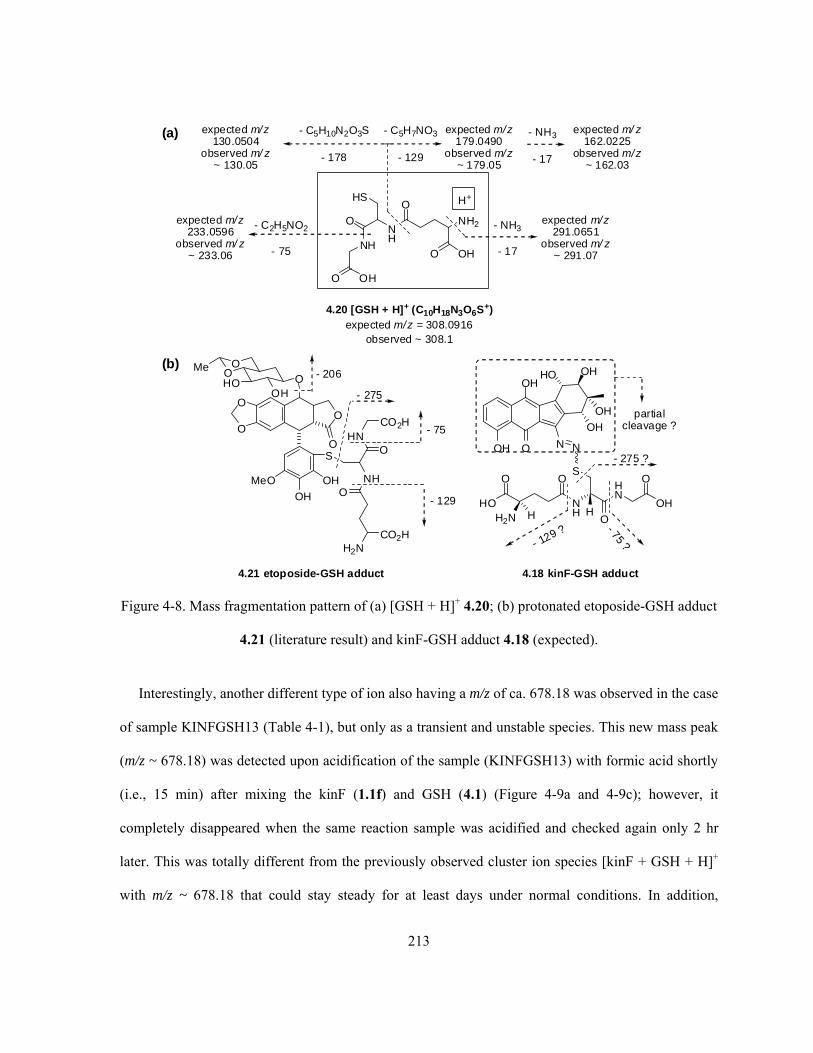
213
4.21 etoposide-GSH adduct
HS
O
NH
O OH
NH
O
NH2
O OH
4.20 [GSH + H]+ (C10H18N3O6S+)expected m/z = 308.0916
observed ~ 308.1
H+
- C5H7NO3
- 129
expected m/z179.0490
observed m/z~ 179.05
expected m/z233.0596
observed m/ z~ 233.06
- NH3
- 17
expected m/ z162.0225
observed m/z~ 162.03
S
OH
OH
MeO
O
O
NH
HNO
CO2H
O
H2NCO2H
O
O
OOH
OOMe
HO- 206
- 75
- 129
- C2H5NO2
- 75
- NH3
- 17
expected m/z291.0651
observed m/ z~ 291.07
expected m/z130.0504
observed m/ z~ 130.05
- C5H10N2O3S
- 178
(a)
(b)
OH
OH
O
OH
OH
HO
OH
N N
SHN
O
OH
O
HNH
O
HO
O
H2N H
- 129 ? - 75?
4.18 kinF-GSH adduct
partialcleavage ?
- 275
- 275 ?
Figure 4-8. Mass fragmentation pattern of (a) [GSH + H]+ 4.20; (b) protonated etoposide-GSH adduct
4.21 (literature result) and kinF-GSH adduct 4.18 (expected).
Interestingly, another different type of ion also having a m/z of ca. 678.18 was observed in the case
of sample KINFGSH13 (Table 4-1), but only as a transient and unstable species. This new mass peak
(m/z ~ 678.18) was detected upon acidification of the sample (KINFGSH13) with formic acid shortly
(i.e., 15 min) after mixing the kinF (1.1f) and GSH (4.1) (Figure 4-9a and 4-9c); however, it
completely disappeared when the same reaction sample was acidified and checked again only 2 hr
later. This was totally different from the previously observed cluster ion species [kinF + GSH + H]+
with m/z ~ 678.18 that could stay steady for at least days under normal conditions. In addition,

214
attempts by means of -ESI-MS (negative ESI) using aqueous ammonia as the base were also made
with this sample after extended time of reaction (55–57 hr). The obtained quite messy and
complicated spectra, however, showed no ion species that might be correlated to the corresponding
deprotonated form of the proposed kinF-GSH adduct [4.18 – H]-.
This new and unstable ion species with m/z of ca. 678.18 seemed to be quite sensitive to the mass
collision energy applied. For example, at 2 eV of collision energy, the mass peak at ca. 678.18 was
barely noticeable as it was buried with nearby noises having comparable intensities (Figure 4-9b and
4-9d). The use of higher collision energy of 10 eV, however, clearly showed a weak but real mass
signal at ca. 678.18 (Figure 4-9a and 4-9c). This was contrary to the previous mass peak of the cluster
ion species [kinF + GSH + H]+ (m/z ~ 678.18), which could be easily detected regardless of the
collision energy used (Figure 4-6). What really differentiated this new mass signal at 678.18 from the
previous one came from the corresponding MSMS spectra under different mass collision energies
(Figure 4-10). A series of new mass peaks with m/z in between 308.1 and 678.18 (i.e., m/z ~ 660.1,
549.1, 420.0 and 339.1), which had been predicted but not observed earlier for the expected mass
behaviour of the protonated kinF-GSH adduct [4.18 + H]+ (Figure 4-8b), started to appear in the
obtained mass spectra. Another very interesting discovery was the observation of a concurrent (and
even stronger) mass peak with a m/z of ca. 677.1 (Figure 4-9 and 4-10, see later text for detailed
discussion) along with the expected mass peak at ca. 678.18. In the mean time, protonated 4.1 [GSH
+ H]+ (m/z ~ 308.1) and the corresponding mass fragments (m/z ~ 233.1 and 179.1) were also
observed in such MS (Figure 4-9) and MSMS (Figure 4-10) spectra. This last observation would
suggest that the observed mass peak at m/z of ca. 678.18 consisted of two different species. In fact,
one is the previously observed cluster ion [kinF + GSH + H]+ (thus it gave the [GSH + H]+ (m/z ~
308.1) and the corresponding daughter fragments, cf. Figure 4-6 and 4-7) while the other one is more
likely the protonated form of the proposed kinF-GSH adduct [4.18 + H]+.

215
25uL APR19CNKINFGSH13 (20min) + 25uL 1:1 MeCN/H2O + 0.2% formic acid
100 200 300 400 500 600 700 800 900 1000 1100m/z0
100
%
0
100
%
APR19CNKINFGSH13ACIDIFIED 34 (1.222) Cm (28:83) TOF MS ES+ 2.89e4308.0855
179.0469
141.9601233.0619
747.1937309.0979377.1035
314.1068 645.1494613.1652
550.6455429.2534
677.1152 748.1974
1048.2841
APR19CNKINFGSH13ACIDIFIED 5 (0.188) Cm (2:28) TOF MS ES+ 2.93e3308.0844
244.2603182.9874
157.0816391.2903 419.3167
441.3061489.3063 550.6436
645.1377 831.5866677.1033 803.5635 859.6380
Collision Energy = 10eV
Collision Energy = 2eV
25uL APR19CNKINFGSH13 (20min) + 25uL 1:1 MeCN/H2O + 0.2% formic acid
610 620 630 640 650 660 670 680 690 700 710 720 730 740 750 760m/z0
100
%
0
100
%
APR19CNKINFGSH13ACIDIFIED 34 (1.222) Cm (28:83) TOF MS ES+ 2.89e4x4 747.1937
645.1494
613.1652
615.1848
616.1868
634.1943
677.1152
646.1567673.1951663.4776
678.1855
691.2100
679.1800692.2140
721.1949709.1010746.1906
732.1191
748.1974
763.1823749.1918
750.1869
APR19CNKINFGSH13ACIDIFIED 5 (0.188) Cm (2:28) TOF MS ES+ 2.93e3x4
645.1377613.1631
634.5526615.1826677.1033
663.4563649.4711 667.5377
691.2171 711.5933695.5282747.2010738.6366723.5997 755.5828763.5954
Collision Energy = 10eV
Collision Energy = 2eV
Figure 4-9. +ESI-MS spectra of a new (non-cluster) type of ion species with m/z of ca. 678.18
(sample KINFGSH13, Table 4-1, acidified after 20 min of mixing) under different mass collision
energies (from top to bottom, a–d): (a) 10 eV; (b) 2 eV; (c) 4x zoom-in of spectrum (a) ; (d) 4x zoom-
in of spectrum (b).

216
MSMS of peak 678.2 of APR19CNKINFGSH13ACIDIFIED
160 180 200 220 240 260 280 300 320 340 360 380 400 420 440 460 480 500 520 540 560 580 600 620 640 660 680 700 720 740m/z0
100
%
0
100
%
0
100
%
0
100
%
0
100
%
0
100
%
APR19CNKINFGSH13ACIDIFIEDMSMS 205 (7.343) Cm (196:223) TOF MSMS 678.20ES+ 980308.1038
179.0511
233.0722210.0174264.0305
339.0639
420.0311 678.1352549.0856 660.1134
APR19CNKINFGSH13ACIDIFIEDMSMS 173 (6.200) Cm (140:195) TOF MSMS 678.20ES+ 3.14e3308.1038
179.0561233.0666
678.1257549.0769339.0639 420.0311660.1134
APR19CNKINFGSH13ACIDIFIEDMSMS 422 (15.114) Cm (377:423) TOF MSMS 678.20ES+ 2.39e3308.1038
179.0561678.1257
549.0769340.0785 660.1229
APR19CNKINFGSH13ACIDIFIEDMSMS 99 (3.557) Cm (84:139) TOF MSMS 678.20ES+ 3.14e3308.1038
179.0511
678.1257
549.0941 660.1229
APR19CNKINFGSH13ACIDIFIEDMSMS 30 (1.092) Cm (28:83) TOF MSMS 678.20ES+ 4.11e3308.1038
678.1257
677.1318
APR19CNKINFGSH13ACIDIFIEDMSMS 27 (0.982) Cm (1:27) TOF MSMS 678.20ES+ 665308.0973
288.2911
678.1639
677.1128309.1098
679.1106
Coliision Energy = 25eV, LM/HM = 14/14
Coliision Energy = 20eV, LM/HM = 14/14
Coliision Energy = 17eV, LM/HM = 14.5/14.5
Coliision Energy = 15eV, LM/HM = 14/14
Coliision Energy = 10eV, LM/HM = 14/14
Coliision Energy = 2eV, LM/HM = 12/12
Figure 4-10. +ESI-MSMS of the new (non-cluster) ion species with m/z of ca. 678.18 under different
mass collision energies (as labeled on each spectrum).
It was further found that the quality of the observed MSMS spectra of this new ion species (m/z ~
678.18) was altered to a noticeable extent (Figure 4-11) by only small variation of the mass analyzer
parameters of LM (low mass) and HM (high mass) resolution. The LM/HM are control parameters
with calculated arbitrary values, which are used in the mass spectrometer controlling software that
could alter the resolution of ions at the low and high mass range of the quadrupole (i.e., the detector
of the mass spectrometer used in this study).338 If the LM/HM resolution are set too low (e.g., 5), then
isotope peaks may merge with the main peak (loss of resolving power) despite the increased signal
intensity, on the other hand, high values of LM/HM (e.g., 20) would cause the quadrupole to over-
resolve but with poor sensitivity.338 The factory default settings of both LM and HM resolution for the

217
+ESI-MS instrument were 14, and it seemed that changing these values to 15/16 (Figure 4-11, top
two spectra) could afford a slightly better resolution while maintaining a reasonable level of
sensitivity (intensity of the signals).
MSMS of peak 678.2 of APR19CNKINFGSH13ACIDIFIED
340 350 360 370 380 390 400 410 420 430 440 450 460 470 480 490 500 510 520 530 540 550m/z0
100
%
0
100
%
0
100
%
0
100
%
APR19CNKINFGSH13ACIDIFIEDMSMS 225 (8.057) Cm (224:251) TOF MSMS 678.20ES+ 202x4
340.0650549.0511
420.0386
393.0786340.1192 353.0896377.0951 397.3037418.1149
548.0882420.0687
531.0862474.0801421.0399 483.7277 547.4954549.1629
APR19CNKINFGSH13ACIDIFIEDMSMS 277 (9.928) Cm (251:307) TOF MSMS 678.20ES+ 1.16e3x4
549.0856
340.0717
420.0386
419.0236548.0711
531.0862474.0721
APR19CNKINFGSH13ACIDIFIEDMSMS 367 (13.150) Cm (308:376) TOF MSMS 678.20ES+ 2.85e3x4
549.0769339.0639
420.0311 548.0797
APR19CNKINFGSH13ACIDIFIEDMSMS 173 (6.200) Cm (140:195) TOF MSMS 678.20ES+ 3.14e3x4
549.0769339.0639
420.0311 548.0797
474.0321 531.0692
Collision Energy = 20eV, LM/HM = 16/16
Collision Energy = 20eV, LM/HM = 15/15
Collision Energy = 20eV, LM/HM = 14.5/14.5
Collision Energy = 20eV, LM/HM = 14/14
Figure 4-11. Optimization of the +ESI-MSMS spectra (only 330 < m/z < 560 were shown) of the new
(non-cluster) ion species with m/z of ca. 678.18 under fixed collision energy (20 eV) but variable
LM/HM (as labeled on each spectrum).
Further MSMS spectra with different mass collision energies and, when necessary, slightly
different LH/MH parameters were also acquired with the mass peaks at 549.1 (Figure 4-12) and 339.1
(Figure 4-13), in order to follow the evolution of these important intermediate ion species and to
determine the correlations between the observed mass fragments. In the case of the mass peak at
549.1, the MSMS spectra (Figure 4-12) were quite weak and messy, and the mass signals around

218
549.1 was found to be consist of several mass peaks with very close m/z values in fact (Figure 4-12).
However, it seemed that at least the fragments with m/z of ca. 531.1, 459.0 and 420.0 (Figure 4-12c–
4-12e) could be reasonably considered to be derived from the particular ion with m/z of ca. 549.1.
MSMS of peak 549.1 of APR19CNKINFGSH13ACIDIFIED
160 180 200 220 240 260 280 300 320 340 360 380 400 420 440 460 480 500 520 540 560 580 600m/z0
100
%
0
100
%
0
100
%
0
100
%
0
100
%
APR19CNKINFGSH13ACIDIFIEDMSMS2 84 (3.018) Cm (84:111) TOF MSMS 549.10ES+ 76548.6296
548.5954548.4664
369.0479339.0774210.0227 309.1098270.2822254.2511 393.0786 548.3718531.1624
508.0609489.3388
549.3953
549.5071
549.6276
APR19CNKINFGSH13ACIDIFIEDMSMS2 124 (4.453) Cm (112:168) TOF MSMS 549.10ES+ 157548.6212
548.4836
377.1022339.0571330.0803308.1102205.0781393.0786 548.3633
489.3225459.0551420.0311 508.0526
549.4899
549.6104
APR19CNKINFGSH13ACIDIFIEDMSMS2 49 (1.764) Cm (28:83) TOF MSMS 549.10ES+ 158549.3953
549.3522548.4836
377.1022339.0706308.0844179.0610295.2699 548.4149393.0713
490.0049420.0462
459.0316 512.9959
549.6276
APR19CNKINFGSH13ACIDIFIEDMSMS2 23 (0.835) Cm (14:27) TOF MSMS 549.10ES+ 19549.1027
549.0339
531.0777
531.0524308.1038272.3077 508.1023459.0158412.1093
398.0504
549.2145
549.5157
549.5674601.6016
APR19CNKINFGSH13ACIDIFIEDMSMS2 11 (0.407) Cm (1:13) TOF MSMS 549.10ES+ 7549.0081
548.9909548.9736548.1054
531.0270
549.1371
549.1888
600.8096593.0786570.0461
Collision Energy = 20eV, LM/HM = 14/14
Collision Energy = 17eV, LM/HM = 13.5/13.5
Collision Energy = 15eV, LM/HM = 13/13
Collision Energy = 10eV, LM/HM = 13/13
Collision Energy = 2eV, LM/HM = 13/13
Figure 4-12. +ESI-MSMS spectra of the mass peak with m/z of ca. 549.1 under different mass
collision energies and settings of LM/HM (as labeled on each spectrum, from top to bottom, a–e).
In the case of mass peak at 339.1, the corresponding MSMS spectra (Figure 4-13) looked much
better than the previous case; however, there were still a few other ions with m/z values close to 339.1
co-existing with the ion species of interest. The fairly clean MSMS spectra indicated that fragments
with m/z of ca. 322.0, 264.0, 243.1 and 210.0 were very likely the daughter species originated from
the fragmentation of the parent ion with m/z of ca. 339.1.
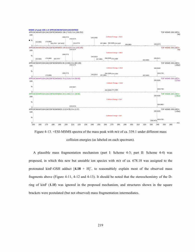
219
MSMS of peak 339.1 of APR19CNKINFGSH13ACIDIFIED
150 160 170 180 190 200 210 220 230 240 250 260 270 280 290 300 310 320 330 340 350 360m/z0
100
%
0
100
%
0
100
%
0
100
%
0
100
%
0
100
%
APR19CNKINFGSH13ACIDIFIEDMSMS3 196 (7.015) Cm (196:252) TOF MSMS 339.10ES+ 340
199.0773
173.0942153.0681181.0707 197.0470
243.0456210.0174
211.0773 257.0861 264.0305339.3073274.1037
295.0681
APR19CNKINFGSH13ACIDIFIEDMSMS3 140 (5.014) Cm (140:195) TOF MSMS 339.10ES+ 790210.0174
199.0773
173.0991153.0681 181.0707243.0513
264.0305257.0861 339.0571274.1098
322.0402
APR19CNKINFGSH13ACIDIFIEDMSMS3 85 (3.050) Cm (84:139) TOF MSMS 339.10ES+ 1.80e3210.0174
199.0773173.0991
339.0639
264.0305243.0513 257.0861
274.1158322.0402
339.2735
APR19CNKINFGSH13ACIDIFIEDMSMS3 70 (2.514) Cm (56:83) TOF MSMS 339.10ES+ 2.37e3339.0639
210.0227
274.1098264.0305339.2735
APR19CNKINFGSH13ACIDIFIEDMSMS3 29 (1.050) Cm (28:55) TOF MSMS 339.10ES+ 2.96e3339.0639
210.0174339.2802
APR19CNKINFGSH13ACIDIFIEDMSMS3 13 (0.478) Cm (1:27) TOF MSMS 339.10ES+ 1.40e3339.0639
338.3478339.2735
Collision Energy = 25eV
Collision Energy = 20eV
Collision Energy = 15eV
Collision Energy = 10eV
Collision Energy = 5eV
Collision Energy = 2eV
Figure 4-13. +ESI-MSMS spectra of the mass peak with m/z of ca. 339.1 under different mass
collision energies (as labeled on each spectrum).
A plausible mass fragmentation mechanism (part I: Scheme 4-3; part II: Scheme 4-4) was
proposed, in which this new but unstable ion species with m/z of ca. 678.18 was assigned to the
protonated kinF-GSH adduct [4.18 + H]+, to reasonablely explain most of the observed mass
fragments above (Figure 4-11, 4-12 and 4-13). It should be noted that the stereochemistry of the D-
ring of kinF (1.1f) was ignored in the proposed mechanism, and structures shown in the square
brackets were postulated (but not observed) mass fragmentation intermediates.
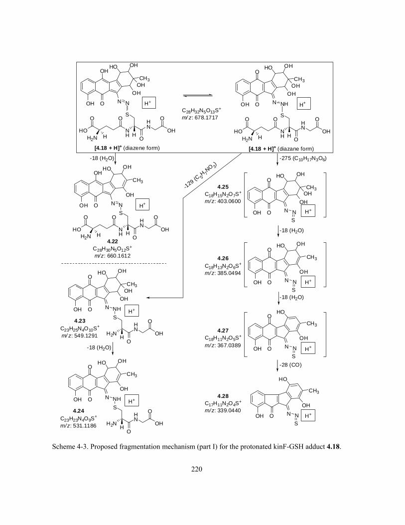
220
O
OH
OH
O
OH
CH3
OH
HO
OH
N N
SHN
O
OH
O
HNH
HO
O
H2N H
H+
C28H32N5O13S+
m/ z: 678.1717 O
OH
O
O
OH
CH3
OH
HO
OH
N NH
SHN
O
OH
O
HNH
HO
O
H2N H
H+
[4.18 + H]+ (diazene form) [4.18 + H]+ (diazane form)
OH
O
O
OH
CH3
OH
HO
OH
N N
S
H+
-275 (C10H17N3O6)
-18 (H2O)
4.25C18H15N2O7S
+
m/z: 403.0600
OH
O
O
OH
CH3
OH
HO
N N
SH+
4.26C18H13N2O6S
+
m/z: 385.0494
-18 (H2O)
OH
O
O
CH3
OH
HO
N N
SH+
4.27C18H11N2O5S
+
m/z: 367.0389
-28 (CO)
OH O
CH3
OH
HO
N N
SH+
4.28C17H11N2O4S
+
m/z: 339.0440
-129 (C 5
H 7NO 3
)
OH
O
O
OH
CH3
OH
HO
OH
N NH
SHN
O
OH
O
HH2N
4.23C23H25N4O10S+
m/ z: 549.1291
H+
-18 (H2O)
OH
O
O
OH
CH3
OH
HO
N NH
SHN
O
OH
O
HH2N
4.24C23H23N4O9S
+
m/z: 531.1186
H+
-18 (H2O)
O
OH
OH
O
OH
CH3
OH
HO
N N
SHN
O
OH
O
HNH
HO
O
H2N H
H+
4.22C28H30N5O12S
+
m/z: 660.1612
Scheme 4-3. Proposed fragmentation mechanism (part I) for the protonated kinF-GSH adduct 4.18.

221
OH
O
O
OH
CH3
OH
HO
OH
N NH
SHN
O
OH
O
HH2N
4.23C23H25N4O10S
+
m/z: 549.1291
H+
-75
(C2H5NO2) OH
O
O
OH
CH3
OH
OH
N NHH+
S
CH2N O
HO
4.29C21H20N3O8S
+
m/z: 474.0971
-18
(H2O) OH
O
O
OH
CH3
OH
OH
N NH H+
S
CH2N O
4.30C21H18N3O7S
+
m/z: 456.0865
-18 (H2O)
OH
O
O
CH3
O
OH
N NH H+
S
CH2N O
4.31C21H16N3O6S
+
m/z: 438.0760
OH
O
O
CH2
O
N NH
S
CH2N
H+
O
4.32C21H14N3O5S+
m/ z: 420.0654
-18
(H2O)OH
O
O
OH
OH
OH
N NHH+
S
CH2NO
HO
4.33
C20H17N3O8S+.
m/ z: 459.0736
- 15 (CH 3)
4.34
C17H10N2O3S+.
m/z: 322.0412
OH O
CH3
OH
HO
N N
SH+
4.28C17H11N2O4S
+
m/z: 339.0440
-17 (OH)
4.36
C16H8O4+.
m/z: 264.0423
- 32 (S) - 28 (N2) - 15 (CH3)
- 46 (N=S) - 18 (H2O) - 15 (CH3)
4.35
C16H5NO2+.
m/z: 243.0320
Scheme 4-4. Proposed fragmentation mechanism (part II) for the protonated kinF-GSH adduct 4.18.
Even though the final conclusion shall not be established solely on such mass results without
further (spectroscopic) evidence, the observed mass and chemical behaviour were very supportive of
the proposal (Scheme 4-2b) that the kinF-GSH adduct 4.18 was the initial and unstable product from
nucleophilic attack on kinF (1.1f) by GSH (4.1). Its decomposition might then initiate the cleavage of
the DNA in the biological studies. Furthermore, the observation of mass peak with m/z of ca. 677.1
(Figure 4-9 and 4-10) was surprising and worthy of some additional comments. Correlation of this
signal (m/z ~ 677.1) to the molecular ion of the proposed kinF-GSH adduct 4.18 (C28H31N5O13S+•, m/z:

222
677.1639) would be entirely consistent with the assignment of the concurrent mass peak with m/z of
ca. 678.18 to the protonated kinF-GSH adduct [4.18 + H]+. It should be pointed out that, even though
charge formation in ESI-MS occurs commonly as a result of either acid-base reaction or coordination
with metal cations (which is also the basis for this mechanistic study), formation of (radical)
molecular ions due to the direct loss of one or two electron(s) from the neutral species under the ESI-
MS conditions are not rare.339
On the other hand, it was also realized that the obtained mass results did have certain flaws that
limit the logical interpretation of the data. Due to the complexity of the multi-component reaction
system involved and the resolution limit of the instrumentation, monitoring of a pure and single ion
species on the mass spectrometer is practically impossible. Even in the case of MSMS studies on an
ion of interest with a particular m/z, the reality was that a few other ions with very close m/z (some
were due to isotopic distribution of the ion species while others may simply come from the accidental
formation of clusters/ions with very similar m/z) would normally co-exist and fragment at the same
time, causing the observed mass spectra to be more complicated than it should be. Therefore, the
obtained mass fragments were not necessarily always related with the species of interest. For example,
observation of the characteristic mass peak (m/z ~ 308.1) for the ion of [GSH + H]+ in the MSMS
spectra of the new and unstable ion species with m/z of ca. 678.18 would indicate that, the starting
source ions contains not only the protonated kinF-GSH adduct [4.18 + H]+ but also certain amount of
the cluster ion of [kinF + GSH + H]+, since only the latter species is capable of producing the ion of
[GSH + H]+.
Another issue is that the observed nominal mass(es) taking account of the mass defect does not
match perfectly with the proposed structures. For instance, the protonated kinF-GSH adduct [4.18 +
H]+ shall have a m/z of 678.1717 (C28H32N5O13S+), while the observed values for mass peaks that
were considered to be [4.18 + H]+ varied in between 678.1257 (cf. Figure 4-10) and 678.1855 (cf.

223
Figure 4-9). The apparent difference between the theoretical and experimental values (i.e., |Δ(m/z)| ~
0.014–0.046) was quite huge when judged by the typical mass spectrometry standard, since
acceptable m/z difference is generally expected to be observed only on the fourth or the third (at most)
digit after the decimal point. However, such apparently “large” mass discrepancy was mainly due to
the detection limit and fluctuation of the mass spectrometer, as well as the lack of an appropriate
internal standard to calibrate the m/z values. This kind of situation was quite common, for example, in
the case of MSMS of the mass peaks at ca. 678.1 that were regarded as the [4.18 + H]+, the same ion
was observed with several different m/z values (i.e., 678.1639, 678.1257 and 678.1352, Figure 4-10)
under almost identical mass conditions. Another two such examples are the ion species [kinF + KinF
+ Li]+ and [GSSG + H]+. Even under apparently identical mass conditions, the former ion
(C36H28N4O14Li+, expected m/z: 747.1762) was observed with several m/z values in between 747.1776
and 747.1969 (Figure 4-4, |Δ(m/z)| ~ 0.0014–0.021), and the latter one (C20H33N6O12S2+, expected m/z:
613.1598) was detected with different m/z values of 613.1732 and 613.1480 (Figure 4-2, |Δ(m/z)| ~
0.012–0.013). Therefore, the observed “large” inconsistency of m/z between the theoretical and
experimental values for mass peaks at ca. 678.18, which were considered to correspond to the [4.18 +
H]+, are still more or less within the instrumentation limit.
(iii) Mixing of kinF (1.1f) with a large excess disulfide GSSG (sample KINFGSSG1–3, Table 4-1)
under identical conditions (1:1 MeCN/H2O, pH 7.6) revealed the crucial and irreplaceable role of the
sulfhydryl (-SH) group when thiols react with kinF (1.1f). The lack of the –SH moiety within GSSG,
which is the oxidized sulfur-coupled dimeric form of GSH (4.1), led to no reaction at all with kinF
(1.1f) even after long time of incubation at ambient temperature (i.e., 50 hr @ 23 °C). Both
compounds behaved individually on the +ESI-MS as if the other one did not exist, and no obvious
mass evidence was found for the formation of possible cluster ion species such as [GSSG + kinF +
H]+, [GSSG + kinF + Li]+ or [GSSG + kinF + Na]+. However, incubation of kinF (1.1f) with large

224
excess of GSSG at higher temperature of 37 °C for 53 hr (sample KINFGSSG3, Table 4-1) led to no
detection of kinF (1.1f) or related ion species at the end. Since the commercial GSSG (Sigma , Grade
II with 5–6% EtOH) did contain a trace amount of GSH as indicated by its label of content as well as
the corresponding +ESI-MS spectra (not shown), a large excess of GSSG would imply the co-
existence of a significant amount of GSH (4.1). Therefore, it was not surprising that the very limited
amount of kinF (1.1f) was completely consumed (by GSH but not GSSG) over long time, particularly
at elevated temperature. Once again this observation confirmed the occurrence of a reaction between
the thiol (GSH) and kinF (1.1f).
(iv) When the simple thiol N-acetyl-L-cysteine 4.17 was used in excess to react with kinF (1.1f)
(sample KINFCYS1–2, Table 4-1), the experimental findings were somehow unexpected. Even after
12 hr of reaction of excess 4.17 with kinF (1.1f) (~ 4:1) at 37 °C, the corresponding mass spectra still
indicated the presence of significant amount of unreacted kinF (1.1f), as characterized by either the
[kinF + Na]+ mass peak (m/z ~ 393.1, Figure 4-14, both spectra) or the [kinF + H]+ mass peak (m/z ~
371.1, Figure 4-14, bottom spectrum from acidified reaction sample). At the same time, the acidified
mass sample (Figure 4-14, bottom spectrum) also afforded an interesting mass peak with m/z of ca.
556.1, which could be either the very likely non-covalent cluster ion of [kinF + 4.17 + Na]+ or the
other possible Na+-binding form of the kinF-4.17 adduct [kinF-4.17 + Na]+. Similar to the kinF-GSH
adduct 4.18, the possible kinF-4.17 adduct was expected to have the sulfur atom from 4.17
chemically bonded to the terminal nitrogen of the diazo moiety of kinF (1.1f).

225
kinF(0.027mM) and N-Ac-Cys(0.119mM, pH7.6) in 1:1 MeCN/H2O 12hr 37C incubation
100 150 200 250 300 350 400 450 500 550 600 650 700 750 800 850 900 950m/z0
100
%
MAR31CNKINFCYS2 42 (1.509) Cm (1:58) TOF MS ES+ 32186.0242
157.0860
150.4315
244.2730 288.2993
349.0690
316.3370489.3445349.0896
393.0898 489.3039
484.3552
489.3770652.3460
623.4492
623.4308904.1798860.5236
785.3453765.1495948.1474
25uL of MAR31CNKINFCYS2 + 25uL of 1:1 MeCN/H2O + 0.2% formic acid
100 150 200 250 300 350 400 450 500 550 600 650 700 750 800m/z0
100
%
MAR31CNKINFCYS2ACIDIFIED 23 (0.831) Cm (1:85) TOF MS ES+ 1.43e4164.0349
157.0814
135.0987
186.0192
349.0553
291.1798
244.2672187.0269
208.0066
327.0753 489.3202411.2719
371.0921393.0825 467.3399
556.1166
512.0963623.4125
557.1213 763.1606652.3554 764.1649
Figure 4-14. +ESI-MS spectra of an ion species with m/z of ca. 556.1 observed with kinF (1.1f) and
4.17 in 1:1 MeCN/H2O (sample KINFCYS2, 12 hr at 37 °C, Table 4-1). Top spectrum: reaction
sample with no formic acid; bottom spectrum: reaction sample acidified with 0.2% HCOOH.
The consequent MSMS spectra of mass peak at 556.1 under different mass collision energies (2–
35 eV, Figure 4-15) clearly indicated that this ion species only led to two major mass fragments with
m/z of ca. 393.1 and 365.1 even under fairly high collision energy. The only reasonable structural
interpretation of such mass results was that the mass peak at 393.1 is due to the [kinF + Na]+
(C18H14N2O7Na+, m/z: 393.0699) and the mass peak at 365.1 is a fragment (C17H14N2O6Na+, m/z:

226
365.0750) derived from [kinF + Na]+ upon the loss of a neutral CO or N2 (mass = 28) fragment.
Therefore, the observed mass peak at 556.1 could only correspond to the non-covalent cluster ion of
[kinF + 4.17 + Na]+ but not the Na+-binding form of the kinF-4.17 adduct [kinF-4.17 + Na]+.
MSMS of peak 556.1 of MAR31CNKINFCYS2ACIDIFIED
140 160 180 200 220 240 260 280 300 320 340 360 380 400 420 440 460 480 500 520 540 560 580m/z0
100
%
0
100
%
0
100
%
0
100
%
0
100
%
0
100
%
0
100
%
0
100
%
0
100
%
MAR31CNKINFCYS2ACIDIFIEDMSMS 248 (8.872) Cm (224:251) TOF MSMS 556.10ES+ 31208.0066
186.0292148.9718
393.0752
393.0315229.9900 365.0847307.0567291.0984
273.0671 347.0555 546.7037465.1522393.1625
539.6543596.6320
MAR31CNKINFCYS2ACIDIFIEDMSMS 197 (7.050) Cm (196:223) TOF MSMS 556.10ES+ 84393.0752
393.0533208.0119
186.0342 393.0024365.0847347.0692229.9900 301.0590
286.9546263.0428 393.3154 421.2476
MAR31CNKINFCYS2ACIDIFIEDMSMS 188 (6.729) Cm (168:195) TOF MSMS 556.10ES+ 280393.0752
208.0172186.0192 365.0776
MAR31CNKINFCYS2ACIDIFIEDMSMS 142 (5.086) Cm (140:167) TOF MSMS 556.10ES+ 863393.0825
365.0706
MAR31CNKINFCYS2ACIDIFIEDMSMS 124 (4.443) Cm (112:139) TOF MSMS 556.10ES+ 1.10e3393.0752
MAR31CNKINFCYS2ACIDIFIEDMSMS 87 (3.121) Cm (84:111) TOF MSMS 556.10ES+ 959393.0825
MAR31CNKINFCYS2ACIDIFIEDMSMS 83 (2.978) Cm (56:83) TOF MSMS 556.10ES+ 590393.0825
556.1253
MAR31CNKINFCYS2ACIDIFIEDMSMS 37 (1.335) Cm (28:55) TOF MSMS 556.10ES+ 251393.0825
556.1253
MAR31CNKINFCYS2ACIDIFIEDMSMS 19 (0.692) Cm (1:27) TOF MSMS 556.10ES+ 90556.1253
556.0734393.0752
555.9954
556.1685
Collision Energy = 35eV
Collision Energy = 2eV
Collision Energy = 30eV
Collision Energy = 25eV
Collision Energy = 20eV
Collision Energy = 15eV
Collision Energy = 10eV
Collision Energy = 8eV
Collision Energy = 5eV
Figure 4-15. +ESI-MSMS spectra of mass peak with m/z of ca. 556.1 under different mass collision
energies (as labeled on each spectrum).
The previously observed smooth reaction between GSH (4.1) and kinF (1.1f), which was quite fast
and complete (i.e., reaction accomplished within 10–20 min and limiting reagent of either kinF or
GSH would be entirely consumed by the other reagent in excess), no longer applied in the case of
4.17 and kinF (1.1f). Such observation can be explained by the known fact that, when thiols act as
potent nucleophilic agents, the reaction rates would depend on and are inversely proportional to the
acidity of the sulfhydryl moiety (i.e., its ability to dissociate to the corresponding thiolate anion).306

227
Despite the quite similar spatial and stereochemical environments for the sulfhydryl (-SH) groups
within GSH (4.1) and 4.17, their pKa values (pKa(-SH)4.17 = 9.52 vs pKa(-SH)GSH = 8.63) would
suggest that 4.17 is significantly less reactive towards the diazo moiety of kinF (1.1f) when compared
with GSH (4.1) under the conditions involved (i.e., pH 7.6), therefore leading to the observed
incomplete reaction even after long time.
For the first time, the above mechanistic studies using +ESI-MS has provided some direct and
convincing evidence to indicate that, under the (near) physiological conditions and in the presence of
an appropriate and bio-significant substrate such as GSH (4.1), nucleophilic addition to the diazo
moiety of the kinamycins is the very first key step involved in the mode-of-action of these unique
antibiotics. Therefore, exploration of other natural and synthetic diazobenzofluorene analogues with
higher diazonium ion character is a promising direction of study, which is expected to be able to
provide new candidates with even stronger biological activity.
4.2.2 Potential Significance of the Reaction of Kinamycin F with Thiols
Recent collaborative studies by the Dmitrienko and the Hasinoff groups on the biological
properties of kinF (1.1f) have revealed that treatment of human K562 leukemia cells with kinF (1.1f)
induced erythroid differentiation, a rapid apoptotic response, induction of caspase-3/7 activities and a
delayed cell cycle progression through the S and G2/M phases.340 These observed effects by kinF
(1.1f) were correlated with its selective reduction of levels of cyclin D3 protein, which plays a key
role in cell cycle progression and acts by binding primarily to cyclin-dependent kinase 6 (CDK-6).
The reduction in levels of cyclin D3 protein by kinF (1.1f) appeared to be mediated at the level of
transcription, rather than by affecting the stability of either cyclin D3 protein or mRNA, thus making
the cyclin D3 a potential target of kinF (1.1f). 340
228
There is currently a great deal of interest in the discovery of small molecules that are selective
inhibitors of specific kinases as potential anticancer agents.341 Many compounds that inhibit kinases
do so by binding to the ATP binding site, thus leading to a lack of specificity among the 500 or so
known human kinases and undesired toxic side effects. A very interesting discovery reported recently
by researchers at the Wyeth labs relates to the selective inhibition of the key human kinase called
AKT, also known as protein kinase B (PKB).342,343 AKT/PKB belongs to the AGC super family of
serine/threonine protein kinases and is a key regulator of various growth factors and hormones
leading to activation of proteins involved in proliferation, metabolism, growth and survival.344 That
work has attracted the attention of this laboratory in the context of the possible mode-of-action of
kinF (1.1f), since one of the expected consequence of inhibiting PKB/AKT is a diminution in the
level of cyclin D3.
The Wyeth group screened numerous natural products as potential selective inhibitors of
PKB/AKT and have discovered that lactoquinomycin (4.37a), a polyketide-derived pyrano-
naphthoquinone (PNQ) type antibiotic discovered in the 1970’s, is an effective and selective inhibitor
for this kinase.342 By synthesizing simplified analogues such as 4.37b–f and in vitro cytotoxicity
measurements with the breast cancer cell line MDA468, the Wyeth group has shown that neither the
amino sugar component nor the phenolic OH group of 4.37a is essential for its PKB/AKT inhibition
and anticancer activity, but the lactone functionality is vital.343 Further study of the interaction of
simpler synthetic PNQ lactone analogues 4.37f with the protein (AKT-2, an isoform of AKT)
revealed that, as evident from the corresponding LC/ESI-MS spectrometry results, such compound
covalently modify the cysteine sulfhydryl group of AKT-2 to form a mono-adduct at C297 and a bis-
adduct at C297 and C311 in a 1:1 ratio, leading to > 90% loss of enzymatic activity.343 A hypothetical
mechanism for this process involving a PNQ-based quinone methide 4.40 as the key intermediate has
been proposed (Scheme 4-5).343 It should be noted that the kinase AKT-1, shown in the mechanism
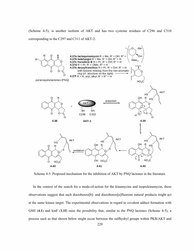
229
(Scheme 4-5), is another isoform of AKT and has two cysteine residues of C296 and C310
corresponding to the C297 and C311 of AKT-2.
O
O
O
O
R
O
AKT
SH SHC296 C310
+
4.38
reductionO
O
OH
O
R
OH
HS
HS
AKT
O
HO2C
OH
O
RHS
HS
AKT
O
HO2C
OH
OH
RHS
S
AKT
O
HO2C
O
O
RHS
S
AKT
oxidation
O
O
O
O
R
O
R'
R"4.37a lactoquinomycin R = Me; R' = OH; R" =4.37b kalafungin R = Me; R' = OH; R" = H4.37c frenolicin B R = Pr; R' = OH; R" = H4.37d R = Pr; R' = OMe; R" = H4.37e deoxyfrenolicin R = Pr; R' = OH; R" = H
with lactone missing from the non-aromaticring (cf. structure on the right)
4.37f R = H, aryl, alkyl; R' = R" = H
O Me
NMe2
OH
O
HO2C
4.39
4.404.414.42
pyranoquinolactones (PNQ)
AKT-1
Scheme 4-5. Proposed mechanism for the inhibition of AKT by PNQ lactones in the literature.
In the context of the search for a mode-of-action for the kinamycins and isoprekinamycin, these
observations suggest that such diazobenzo[b]- and diazobenzo[a]fluorene natural products might act
at the same kinase target. The experimental observations in regard to covalent adduct formation with
GSH (4.1) and kinF (1.1f) raise the possibility that, similar to the PNQ lactones (Scheme 4-5), a
process such as that shown below might occur between the sulfhydryl groups within PKB/AKT and

230
the kinamycins (Scheme 4-6): nucleophilic attack of the sulfur towards the diazo moiety leads to the
adduct 4.43, whose subsequent homolytic cleavage upon the loss of N2 followed by recombination of
the corresponding vinyl radical 4.44 and the sulfur radical 4.45 might lead to a more stable adduct
4.46 (path (a), Scheme 4-6); alternatively, another reaction mode is also worth considering (path (b),
Scheme 4-6), in which the initial adduct 4.43 might undergo an intramolecular conjugate addition of
another cysteine sulfhydryl group to the enone system followed by the expulsion of N2 and a thiolate.
However, it should be noted that an analogous pathway (b) is unlikely in the case of isoprekinamycin
(1.5), since the corresponding initial adduct 4.50 in that case would possess an aromatic C-ring that
can not act as a Michael acceptor in the second nucleophilic attack, while the diazobenzo[b]fluorene
type kinamycins could form a quinone methide (such as 4.47) in their cases to proceed further. Thus,
the mechanism involving addition to the diazo group followed by homolysis and radical
recombination (i.e., path (a) in Scheme 4-6) might be more feasible for isoprekinamycin (1.5).
Efforts to explore these possibilities with recombinant human PKB/AKT using ESI-MS
experiments, similar to those described earlier for adduct formation between kinF (1.1f) and GSH
(4.1), are currently in progress in the Dmitrienko group.

231
AKT
SH SHC296 C310
+
O
OH
R1O
OR3
O
OR4
Me
OR2
N2
1.1 kinamycin
HO
OH
R1O
OR3
O
OR4
Me
OR2
N
4.43kinamycin-AKT
adduct
N
S
AKT
SH
HO
OH
R1O
OR3
O
OR4
Me
OR2
N
N
S
AKT
SH
HO
OH
R1O
OR3
O
OR4
Me
OR2
S
AKT
SH
HO
OH
R1O
OR3
O
OR4
Me
OR2
S
AKT
SH
- N2
Path (a): homolytic cleavage followed by radical recombination
Path (b): intramolecular conjugate addition followed by elimination
HO
OH
R1O
OR3
O
OR4
Me
OR2
N
N
S
AKT
SH
- N2
HO
OH
R1O
OR3
O
OR4
Me
OR2
SH
AKT
S
HO
OH
R1O
OR3
OH
OR4
Me
OR2
S
AKT
S
HO
OH
R1O
OR3
O
OR4
Me
OR2
S
AKT
SH
HOO
OHO N2
HOO
OHHO N
N
S
AKT
SH
X
AKT
SH SH
+ X1.5 isoprekinamycin
aromatic C-ringdisfavoursconjugate addition
4.43 4.44 4.46
4.45
4.43 4.47 4.48
4.49
4.50 isoprekinamycin-AKT adduct
AKT-1
Scheme 4-6. Proposed mode-of-action for possible inhibition of PKB/AKT by the kinamycins.
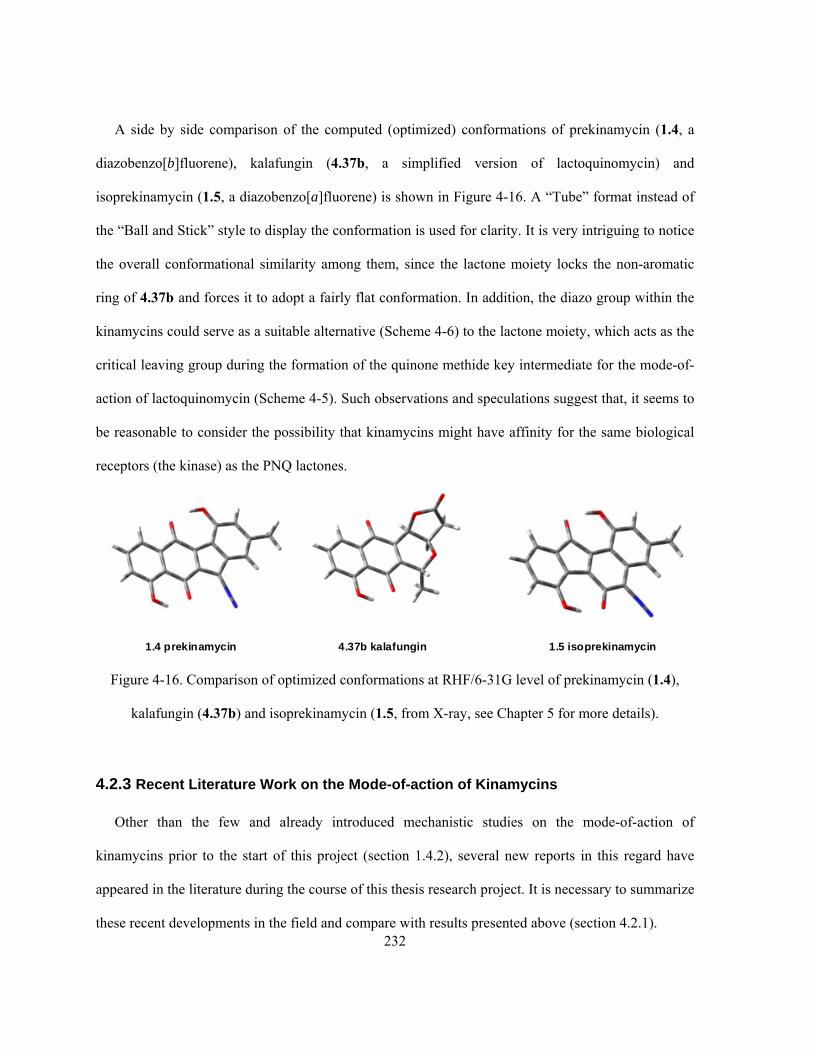
232
A side by side comparison of the computed (optimized) conformations of prekinamycin (1.4, a
diazobenzo[b]fluorene), kalafungin (4.37b, a simplified version of lactoquinomycin) and
isoprekinamycin (1.5, a diazobenzo[a]fluorene) is shown in Figure 4-16. A “Tube” format instead of
the “Ball and Stick” style to display the conformation is used for clarity. It is very intriguing to notice
the overall conformational similarity among them, since the lactone moiety locks the non-aromatic
ring of 4.37b and forces it to adopt a fairly flat conformation. In addition, the diazo group within the
kinamycins could serve as a suitable alternative (Scheme 4-6) to the lactone moiety, which acts as the
critical leaving group during the formation of the quinone methide key intermediate for the mode-of-
action of lactoquinomycin (Scheme 4-5). Such observations and speculations suggest that, it seems to
be reasonable to consider the possibility that kinamycins might have affinity for the same biological
receptors (the kinase) as the PNQ lactones.
1.4 prekinamycin 4.37b kalafungin 1.5 isoprekinamycin
Figure 4-16. Comparison of optimized conformations at RHF/6-31G level of prekinamycin (1.4),
kalafungin (4.37b) and isoprekinamycin (1.5, from X-ray, see Chapter 5 for more details).
4.2.3 Recent Literature Work on the Mode-of-action of Kinamycins
Other than the few and already introduced mechanistic studies on the mode-of-action of
kinamycins prior to the start of this project (section 1.4.2), several new reports in this regard have
appeared in the literature during the course of this thesis research project. It is necessary to summarize
these recent developments in the field and compare with results presented above (section 4.2.1).

233
In late 2005, Feldman and Eastman published some preliminary results on the possible mode-of-
action of prekinamycin (1.4),345 a diazobenzo[b]fluorene type kinamycin (considered as a
diazoparaquinone by Feldman), followed by a more detailed disclosure in the following year.346 By
using the literature observation that cleavage of double stranded DNA by lomaiviticin A (1.11a) was
performed under reducing conditions as an argument,52 Feldman proposed a mechanism involving
one-electron reduction of the quinone moiety of kinamycins to a reactive semi-quinone followed by
non-obligatory protonation as the initial key step (Scheme 4-7).345,346 The radical (anion) species
4.52a, generated upon reduction of the generic kinamycin-type diazoparaquinone structure 4.51, may
also be represented by the corresponding resonance forms 4.52b and 4.52c. Feldman further assumed
that C-5 in 4.52 is “pyramidalized sufficiently to permit adequate overlap between the enol (enolate)
orbitals and σ*C-N ”345 to allow the consequent rapid loss (β-elimination) of N2 and afford the sp2 C-5
radical 4.53, which then may perform hydrogen abstraction from DNA if the radical stays in the close
proximity of DNA. In addition, Feldman also proposed that the diazoparaquinone/DNA interaction
may even go beyond simple hydrogen abstraction to damage DNA further (Scheme 4-7).346 The
possible quinone methide 4.54, derived from 4.53 upon hydrogen abstraction from DNA, was
considered to have a hydroxymethylacylfulvene (4.55) subunit. This may act as the key electrophilic
pharmacophore upon nucleophilic attack by the DNA bases such as guanosine unit 4.56 with its N-7
as the nucleophilic site, resulting in DNA damage. Alternatively, radical addition of DNA• with
quinone methide 4.54 could afford a covalent adduct 4.58 that eventually leads to the same result of
DNA damage.
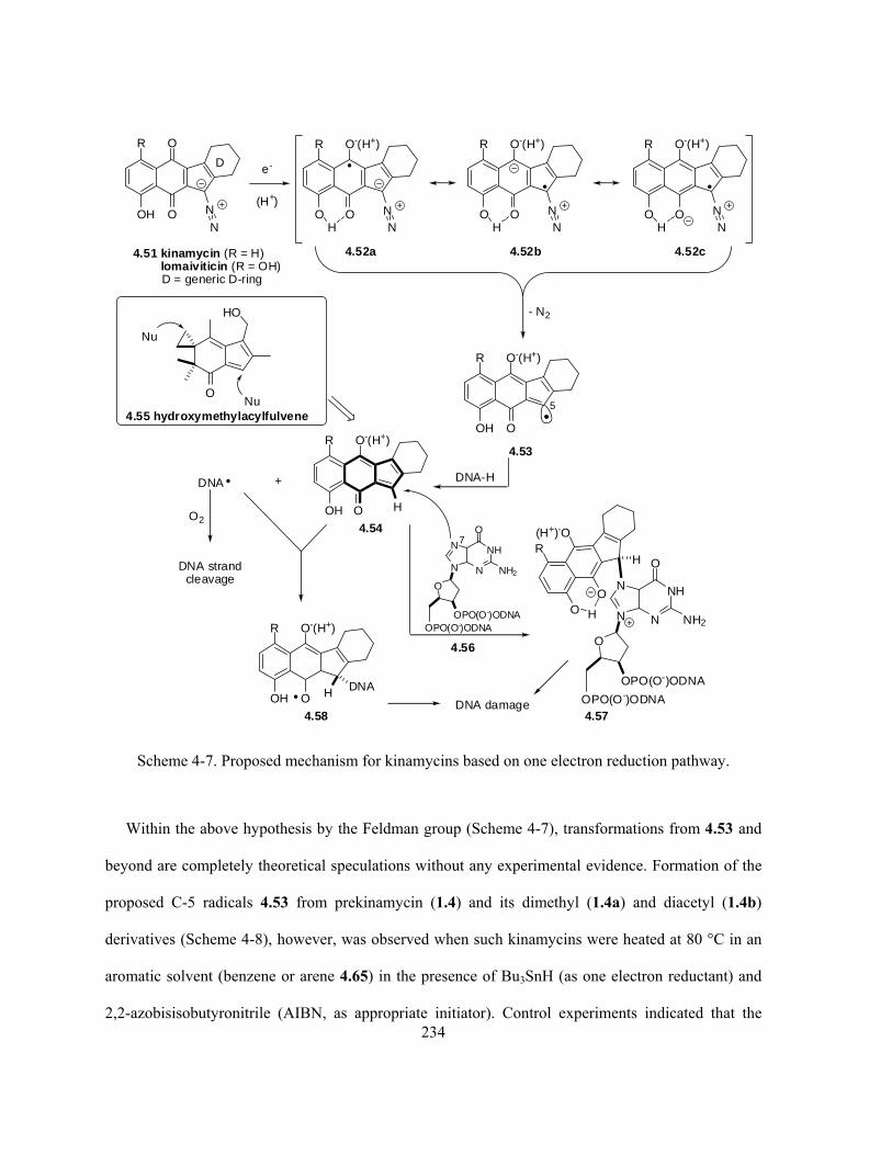
234
O
OOH N
N
R
4.51 kinamycin (R = H)lomaiviticin (R = OH)D = generic D-ring
D e-
(H+)
O-(H+)
OO NN
R
4.52a
H
O-(H+)
OO NN
R
4.52b
H
O-(H+)
OO NN
R
4.52c
H
- N2
O-(H+)
OOH
R
4.53
5
DNA-H
O-(H+)
OOH
R
4.54
DNA +
O2
DNA strandcleavage
H
HO
O
Nu
Nu4.55 hydroxymethylacylfulvene
N
NHN
N
O
NH2
O
OPO(O-)ODNAOPO(O-)ODNA
4.56
N
NHN
N
O
NH2
O
OPO(O-)ODNA
OPO(O-)ODNA
O-(H+)
OOH
R
4.58
H DNA
(H+)-O
OO
R
4.57
H
H
7
DNA damage
Scheme 4-7. Proposed mechanism for kinamycins based on one electron reduction pathway.
Within the above hypothesis by the Feldman group (Scheme 4-7), transformations from 4.53 and
beyond are completely theoretical speculations without any experimental evidence. Formation of the
proposed C-5 radicals 4.53 from prekinamycin (1.4) and its dimethyl (1.4a) and diacetyl (1.4b)
derivatives (Scheme 4-8), however, was observed when such kinamycins were heated at 80 °C in an
aromatic solvent (benzene or arene 4.65) in the presence of Bu3SnH (as one electron reductant) and
2,2-azobisisobutyronitrile (AIBN, as appropriate initiator). Control experiments indicated that the
235
smooth occurrence of this reaction required the mandatory co-existence of heat, the tinhydride and
AIBN, but light was found to be not necessary. On the other hand, non-aromatic solvents including
THF, 1,4-dioxane, CCl4, CH3CN, EtOH and CHCl3 only led to unreacted starting kinamycins even if
other essential conditions were complied with, suggesting that the sp2 radical 4.53 is either promoted
to form or stabilized by π-complexation in aromatic solvents. Formation of the critical radical species
4.62 from the prekinamycin dimethylether 1.4a (Scheme 4-8), which is the same type as 4.53 in
Scheme 4-7, was evident from the obtained final products of 4.60a–c, 4.61, 4.67, 4.69, 4.70 and 4.71,
which all had a new substituent (e.g., -Ph, -Ar, -SePH, -SBn and -D) introduced at C-5 depending on
the available reagent(s) (i.e., benzene, substituted arenes, thiols and tinhydride) in the system that
participated in the radical reaction. The concurrent isolation of the minor product of dimer 4.64,
whose formation might undergo several possible pathways such as direct radical dimerization of 4.62,
radical addition between 4.62 and 4.63 followed by oxidation, or hetero Diels-Alder dimerization of
4.63 followed by oxidation, seemed to be difficult to explain if a radical mechanism was not involved
(Scheme 4-8). The proposed radical mechanism also fit with the observed kinetic data, product
distributions and Hammett correlation when substituted arenes 4.65 and isotope-labeled
tinhydride/thiols were used in such reactions (Scheme 4-8).345,346
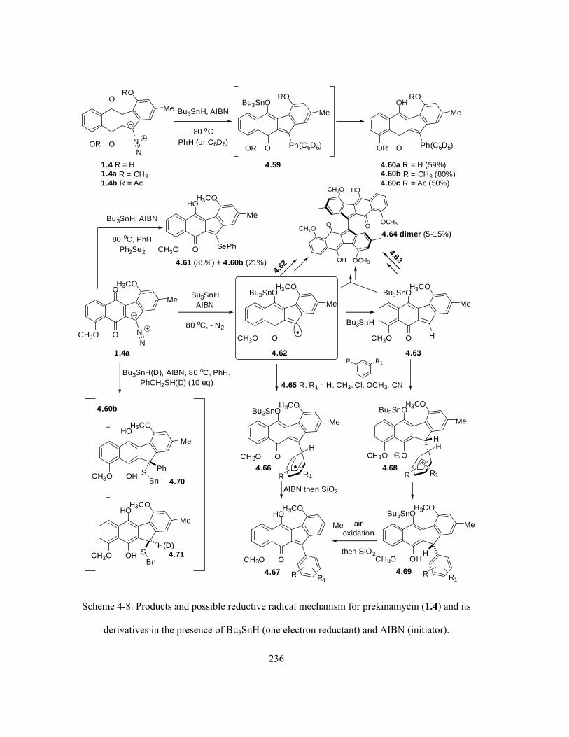
236
O
OOR NN
Me
RO
1.4 R = H1.4a R = CH31.4b R = Ac
Bu3SnH, AIBN
80 oCPhH (or C6D6)
Bu3SnO
OOR Ph(C6D5)
Me
ROOH
OOR Ph(C6D5)
Me
RO
4.59 4.60a R = H (59%)4.60b R = CH3 (80%)4.60c R = Ac (50%)
O
OCH3O NN
Me
H3CO
1.4a
Bu3SnHAIBN
80 oC, - N2
Bu3SnO
OCH3O
Me
H3CO
4.62
Bu3SnH
Bu3SnO
OCH3O
Me
H3CO
4.63
H
R R1
4.65 R, R1 = H, CH3, Cl, OCH3, CN
Bu3SnO
OCH3O
Me
H3CO
4.66
H
R R1
Bu3SnO
OCH3O
Me
H3CO
4.68
H
R R1
AIBN then SiO2
HO
OCH3O
Me
H3CO
4.67 R R1
Bu3SnO
OHCH3O
Me
H3CO
4.69 R R1
airoxidation
then SiO2
HO
OCH3O
Me
H3CO
4.61 (35%) + 4.60b (21%)
SePh
4.62
Bu3SnH, AIBN
80 oC, PhHPh2Se2
CH3OO
OH OCH3
OCH3O
HOCH3O
4.64 dimer (5-15%)
Bu3SnH(D), AIBN, 80 oC, PhH,PhCH2SH(D) (10 eq)
HO
OHCH3O
Me
H3CO
HO
OHCH3O
Me
H3CO
+
+
4.60b
PhSBn 4.70
4.71SH(D)
Bn
4.63
H
H
Scheme 4-8. Products and possible reductive radical mechanism for prekinamycin (1.4) and its
derivatives in the presence of Bu3SnH (one electron reductant) and AIBN (initiator).
237
Feldman’s work provided observations of some interesting chemical behavior of the
diazobenzo[b]fluorene type kinamycins under a (rare) reducing condition, and an alternative
possibility for the mode-of-action of kinamycins. However, even Feldman himself admitted that the
conditions involved in his studies were far from the physiological conditions under which the
bioactivities of kinamycins were observed and measured.346 The use of tinhydride as a reducing agent
in the presence of radical initiator such as AIBN is by no means to have any bio-significance or even
chemical-significance related to the known in vitro and in vivo bioactivity studies of the kinamycins,
not to mention the unrealistically high temperature and non-aqueous aromatic solvent involved that
are essential for the observed chemistry and proposed mechanism. Therefore, the breadth of
Feldman’s conclusion is rather limited, and its validity for the actual mechanism of kinamycins is
questionable.
Around the same time when Feldman’s mechanism was published, Arya reviewed the known
mechanistic studies on diazo and diazonium DNA cleavage agents at the time (prior to 2006 but not
including Feldman’s results), with a focus on kinamycins.347 In this review, Arya proposed two other
theoretically possible mechanisms for the kinamycins but with no experimental evidence to support
the proposals. The first one (Scheme 4-9a) involves an initial protonation of the kinamycin at C-5 of
the diazobenzo[b]fluorene skeleton as the triggering step, perhaps after its binding with DNA, and the
obtained kinamycin diazonium ion 4.72 can lose N2 either heterolytically to produce the
corresponding carbocation 4.73, or homolytically and reductively to give the resonance stabilized
radical (4.74 ↔ 4.75). Then the vinyloxy radical 4.75 can react further to cause DNA cleavage.
Protonation at C-5 of kinamycin is obviously the core of this proposed mechanism, but one can easily
argue that, at least in theory, such C-5 protonation should be strongly disfavoured or easily prohibited
since it would destroy the aromaticity of the C-ring. It is also true that the kinamycins exhibit a high
degree of stability towards acids experimentally, and hence it seems highly unlikely that protonation

238
is a key step in the mode-of-action as suggested by Ayra. For example, the Dmitrienko group recently
isolated the kinamycins in very good yield from bacteria fermentation broth that were adjusted to pH
2 (unpublished results by V. Goodfellow), and it was also found that kinamycins are very stable even
in the presence of excess trifluoroacetic acid (unpublished results by Dr. A. Ghavami). Reported
derivatization and synthesis of the kinamycins in the literature also show that these compounds,
particularly the C5-diazo moiety, could tolerate and survive various strong acids such as PTSA (p-
toluenesulfonic acid) (Scheme 1-1),9 sulfuric acid (Scheme 1-1)5 and HCl (Scheme 3-32 and 3-
33).16,26
Ayra’s second mechanism involves a photolytic decomposition of the kinamycin, which shall
produce the corresponding carbene 4.77 that may induce DNA cleavage (Scheme 4-9a). In addition,
4.77 may also rearrange to the vinylic carbon radical 4.78 that might be capable of DNA cleavage as
well. Until now there are no direct reports in the literature regarding the photochemical behaviour of
kinamycins. However, in accordance to Arya’s photolytic proposal, the Zaleski group recently carried
out some photochemistry work with (2-diazo-3-oxo-tetraphenylchorinato)M(II) (4.80, M = Ni, Zn,
Cu) that were claimed as model compounds for natural kinamycins and to be potentially useful in
photodynamic therapy (PDT) for tumors. Photolysis of 4.80 was found to involve singlet carbenes as
the reactive intermediates to form the corresponding Wolff rearrangement products 4.81 and dimers
4.82 (Scheme 4-9b).348 The photochemical mechanism by Arya and the related work by Zaleski are
also of very little value to clarify the real mode-of-action of these antibiotics, since the known
biological activities of kinamycins require no light at all. As an added support for this argument,
experimental observation by the mechanistic study of this thesis work has found that, exposure of a
mixture of kinF (1.1f) and GSH (4.1) in aqueous acetonitrile to normal room/day light, even for hours,
does not alter the reaction outcome when compared with the same sample that has been stored in the
dark (see section 4.7 for experimental details).

239
O
OH
AcO
OH
O
OAc
Me
OAc
N2
1.1c kinamycin C
O
OH
AcO
OH
O
OAc
Me
OAc
4.77
O
OH
AcO
OH
O
OAc
Me
OAc
4.78
OH
OH
AcO
OH
O
OAc
Me
OAc
4.79DNA DNAcleavage
h
O
OH
AcO
OH
O
OAc
Me
OAc
N2
4.72
DNA binding
H+
H
O
OH
AcO
OH
O
OAc
Me
OAc
4.73
H
heterolytic
O
OH
AcO
OH
O
OAc
Me
OAc
4.74
H
hom
olyt
ic+
e,- N
2
O
OH
AcO
OH
O
OAc
Me
OAc
4.75
H
OH
OH
AcO
OH
O
OAc
Me
OAc
4.76
H
DNADNAcleavage
DNA DNAcleavage
N
N
N
N
Ph
N2
O
PhPh
Ph
M
4.80 M = Ni2+, Zn2+, Cu2+
h - N2
CH2Cl2/MeOHN
N
N
N
Ph
PhPh
Ph
M
4.81
OMe
O
H
4.82
N
NN
N
O
Ph
Ph
Ph M
N N
NN
Ph
Ph
Ph
Ph
M
O
H+
(b)
(a)
- N2
Scheme 4-9. (a) Two possible mechanisms proposed by Ayra for the mode-of-action of kinamycins;
(b) photochemical reaction of diazo-oxochlorin (as kinamycin model compounds) involving a carbene.
Also in 2006, Melander and co-workers synthesized some simple analogues of 9-
diazofluorene (1.109), including 1-substituted (4.83a–d), 4-substituted (4.84a–d) and 1,4-

240
disubstituted-9-diazofluorenes (4.85a–b), as model compounds for natural kinamycins.349 Biological
tests to cleave supercoiled DNA with these diazofluorenes and natural kinD (1.1d) (Scheme 4-10a)
indicated that, under physiological conditions, DNA cleavage was only observed with such diazo
compounds in the presence of an appropriate promoter (in most cases) such as dithiothreitol (DTT,
4.2), and apparently the DNA cleavage was fairly slow (2 days). Among the analogues tested, 1-
methoxy-9-diazofluorene (4.83b) was found to be the most active one and its activity towards DNA
cleavage was comparable to (in fact slightly higher than) the natural kinD (1.1d).349 However, no
mechanism was provided for the observed bioactivities of these diazofluorene model compounds at
the time. Two year later, the Melander group released some additional results regarding the DNA
cleavage by natural kinD (1.1d) in the presence of either DTT (4.2) or GSH (4.1) as a promoter, and
increasing in temperature was found to significantly accelerate the DNA cleavage by the kinamycin
(accomplished within 24 hr at physiological temperature vs 2 days at room temperature).307 Since
sulfides such as DTT (4.2) are well-known sources for 2e- reduction, Melander proposed a
corresponding mechanism in which the first key step is a two electron reduction of kinD (1.1d) by the
thiol (DTT or GSH) leading to the formation of a diazodihydroxyquinone 4.86 (Scheme 4-10b).307
Further conversion of 4.86 leading to the ultimate DNA cleavage could undergo two different
pathways as proposed by Melander (Scheme 4-10b). The first route borrowed an idea from the
published Dmitrienko mechanism (Scheme 1-28) involving a nucleophilic attack on the diazo group
of 4.86, then the corresponding azo adduct 4.87 underwent a homolytic cleavage to generate a carbon
radical 4.88 that is responsible for DNA cleavage; the second route showed certain similarities to
Arya’s mechanism (Scheme 4-9a) with protonation at C-5 of 4.86 as the key step, followed by the
spontaneous decomposition of the diazo group of 4.89 to generate an ortho-quinone methide 4.90,
which could also act as an alkylating agent for DNA. This two electron reductive mechanism for
kinamycins seemed to be supported (only) by some very limited MO calculations on a simplified

241
diazobenzo[b]fluorene model structure (i.e., 1.1d without all D-ring substituents) and its reduced
dihydroxyquinone form.307 Despite the physiological conditions used in Melander’s study, no
chemical or biological evidence was reported in this work for the existence or involvement of 4.86 or
any other derivatives within the proposed mechanism, thus leaving its credibility in doubt.307

242
N2R1
4.83a-d R1 = NH2, MeO, Cl, NO2; R2 = H4.84a-d R1 = H; R2 = NH2, MeO, Cl, NO24.85a R1 = R2 = MeO
N2
4.85b
R2
O
O
O
OOH N2
HO
OAc
OAc
OH
1.1d kinamycin D
4.2 DTT
HS
OH
OH
SH
DNA, 2 daysDNA cleavage
(a)
(b) O
OOH N2
HO
OAc
OAc
OH
1.1d kinamycin D
4.2 DTT or 4.1 GSH
2e-/2H+
OH
OHOH N2
HO
OAc
OAc
OH
4.86
OH
O O
NHH
H NH2
HS
O
NHOH
O
4.1 GSH
Nu
OH
OHOH N
HO
OAc
OAc
OH
4.87
NNu
OH
OHOH
HO
OAc
OAc
OH
4.88
OH
OHOHN2
HO
OAc
OAc
OH
4.89
H+
H
OH
OOH
HO
OAc
OAc
OH
4.90
DNA cleavage
Scheme 4-10. (a) Observed DNA cleavage by diazofluorenes in the presence of DTT (4.2); (b)
proposed two electron reductive DNA cleavage mechanisms for kinamycins by Melander.

243
Very recently (2009), Skibo and Khdour carried out physical organic chemistry experiments
aimed at identifying the possible mode-of-action of prekinamycin (1.4), a diazobenzo[b]fluorene with
a fully aromatized D-ring, via the two electron reduction that might occur in vivo (Scheme 4-11).90 It
was proposed that, reduction of the B-ring quinone of 1.4 to the corresponding dihydroxyquinone of
4.91 followed by protonation and dediazoniation, both at C-5, leads to the formation of 4.93, an
ortho-quinone methide intermediate. Compound 4.93 could equilibrate with its keto tautomer 4.94,
which is a deazotized quinone, but only the former species is capable of trapping a nucleophile and
thus considered to be responsible for the biological activity of kinamycins. In fact, one can easily
realize that this mode-of-action is essentially the same as the previous mechanistic proposal by
Melander for kinD (1.1d) (Scheme 4-10b, 4.86 to 4.90 via 4.89), a diazobenzo[b]fluorene with a non-
aromatic D-ring.
O
OOH N2
HO
1.4 prekinamycin
+ 2e-
+ 2H+
OH
OHOH N2
HO
4.91
OH
OOH N2
HO
4.92
HH+
+ H+
- H+
H
O
OOH
HO
4.93 quinone methide
- N2
H
O
OOH
HO
4.94 keto tautomer
HH
HH+
H+
OH
OOH
HO
H
4.93
Nu
H+
OH
OHOH
HO
HNu
4.95
Scheme 4-11. Proposed two electron reduction mechanism for prekinamycin (1.4) by Skibo.

244
In particular, the Skibo group prepared a number of prekinamycin analogues including 1.4a and
4.96a–d with the C-5 diazo carbon atom enriched with 13C, and subjected these compounds to
catalytic hydrogenation (H2/Pd-C in anaerobic and aqueous DMSO buffer, pH 7.2–9.4) to mimic the
possible two electron reduction in vivo (Scheme 4-12).90
O
OOH N2
4.96a
OH
OHOH N2
4.97
+ H+ then - N2
averagedk' = 2.6 x 10⁻² min-1
OH
OOH
HO
H
4.98
O
OR3N2
R1 Synthetic prekinamycin analogues (* = 13C labelled)1.4a R1 = R3 = OMe; R2 = Me4.96a R1 = R2 = H; R3 = OH4.96b R1 = R2 = H; R3 = OMe4.96c R1 = OH; R2 = Me; R3 = H4.96d R1 = OMe; R2 = Me; R3 = H
R2
H2/Pd-C (anaerobic)DMSO/buffer(pH = 7.2-9.4)
averagedk = 2.5 x 10⁻² min-1
quinonemethideproducts
*
4.96aH2/Pd-C (anaerobic)
then aerobic workup
OH
OOH*
OH
OOH
*OH
OH
OHOH
*O
OH
OOH*
OH
O OH*
++
+
4.99 4.100 4.101
4.102
1.4aH2/Pd-C (anaerobic)
then aerobic workup
O
OO
*
OH
OO
*OH
OH
OHO
*O
++
4.103 4.104 4.105
OOO
H H
productscharacterizedby 13C-NMR
and/or1H-NMR,
MS
Scheme 4-12. Experimental results by Skibo and co-worker in support of the proposed two
electron reduction mechanism for the kinamycins.

245
The above evidence from kinetic data, UV-vis spectroscopy and product analysis allowed Skibo
and Khdour to detect certain intermediates that are reactive and possibly electrophilic, and such
observations were suggested to be consistent with the proposed mechanism as shown in Scheme 4-11.
It was postulated further, based on some rough MO calculations (only at the HF/3-21G level), that
those prekinamycin analogues that favour the quinone methide tautomer over the keto tautomer are
more likely to act as electrophiles with DNA bases as nucleophilic partners in conjugate addition
reactions, thus having higher bioactivity. Such alkylated DNA was then suggested to be unstable with
respect to an elimination reaction that ultimately effects cleavage of the DNA-backbone, thus
rationalizing anticancer and antimicrobial activity on the basis of bioreductive activation of the
prekinamycin system in vivo to create electrophilic species that can alkylate DNA leading eventually
to sufficient damage to induce cell death.
There are a number of interesting, but somewhat troubling, aspects to the proposal by the Skibo
group in regard to the mode-of-action of prekinamycin (1.4) and, by implication, of the kinamycins
(1.1–1.3), the lomaiviticins (1.11) and isoprekinamycin (1.5). In the case of isoprekinamycin (1.5),
hydrodediazoniation following Skibo’s pathway would yield an aromatic system (cf. structure 4.50 in
Scheme 4-6) rather than an ortho-quinone methide so that such a mechanism can not be operative.
Thus, to accept the Skibo proposal for biological activity, one needs to assume that, isoprekinamycin
(1.5) and prekinamycin (1.4) must act by different mechanisms despite their high structural similarity.
The Skibo mechanism also invoked an unusually high basicity for the oxygen atom of the ketone
carbonyl group (pKa of approximately 6–7), wherein the non-protonated ortho-quinone methide such
as 4.106 was argued to be unreactive as an electrophile and must first be O-protonated to form a
cation 4.107 (Scheme 4-13a), which was proposed to be the reactive alkylating agent for DNA or
other biological targets. On the other hand, Skibo also predicted a pKa > 8 for the enolic OH to

246
explain the lack of reactivity of the quinone methide 4.106 observed at pH > 9, and the corresponding
enolate 4.108 was considered electron-rich enough to resist nucleophilic attack (Scheme 4-13a). Why
such a carbonyl group would be so basic is not only unclear (Skibo offered no explanation at all) but
also confusing, since protonated carbonyl species (>C=O+–H) typically have negative pKa values
suggesting such carbonyl oxygens are generally very weak bases. Interestingly, in the case of the
natural product momofulvenones 1.14 incorporating an ortho-quinone methide system of the same
type invoked by the Skibo group, the enolic OH is so acidic that the compounds actually exist in the
corresponding conjugated base form at physiological pHs (Scheme 4-13b).54 Such known
experimental observations disfavour Skibo’s argument of the unusually high basicity of the carbonyl
oxygen and the relatively low acidity of the enol for the quinone methide. Likewise, the presence of
the keto group in the D-ring of the lomaiviticin A (1.11a) may cause the corresponding ortho-quinone
methide 4.109, generated from 1.11a by following the Skibo’s mechanism, to exist in its deprotonated
form (i.e., 4.110) at pH 7 (Scheme 4-13c), similar to the momofulvenones (Scheme 4-13b). This
enolate 4.110 would also make such system unable to function as an electrophile, if following Skibo’s
judgment for the same type of species of 4.108. Therefore, the Skibo mechanism for the mode-of-
action of prekinamycin (1.4) is not suitable to be the mode-of-action for the lomaiviticins, which are
known to be very potent anticancer agents.

247
OH
OO
*H
4.106quinone methide
O
OOH N2
4.96a
*
H
OH
OO
*H
4.107quinone methide cation
(electrophilic)
HH
carbonyl
pK a= 6-7
(a) DNAdamage
enolpKa > 8
O
OO
*H
4.108quinone methide enolate
(electron rich, nucleophilic-resistant)
H
(b)
OH
O
O
O
CH3
OH
RO OH
1.14a momofulvenone A (R = H)1.14b momofulvenone B (R = Ac)
OH
HO
O
O
CH3
OH
RO OH
H2O (M+)
actual isolated stable formof 1.14 (M = Na, K, Mg, Ca)
OH
OH
O
O
N O
O
O
R'
H
H
REt
N
1.11a lomaiviticin A
OOH
NMe2
R' =O OH
OMeR =
2
(c)OH
OH
O
OH
O
O
O
R'
H
H
REt
2
4.109 quinone methide
Skibo mechanism*:[H] then H+
followed by -N2
H
* cf. Scheme (a) andScheme 4-11
M
OH
OH
O
O
O
O
O
R'
H
H
REt
2
4.110 quinone methide enolate(non-electrophilic)
H
Enol ionization is expected tobe preferred, cf. Scheme (b)
Scheme 4-13. (a) Skibo’s argument on the pKa of carbonyl oxygen and phenolic OH of the quinone
methide; (b) known acid-base equilibrium of momofulvenones (1.14) favouring the enolate; and (c)
possible acid-base equilibrium of the quinone methide generated from lomaiviticin A (1.11a).

248
Finally, Skibo’s study used H2/Pd-C in aqueous (DMSO) buffer as the reducing agent for
kinamycins in the absence of oxygen (air). Strictly speaking, even though such experimental
conditions are probably better choices than Feldman’s totally abiological conditions
(tinhydride/AIBN/aromatic solvent/high temperature/anaerobic), they are still not ideal imitations of
the biological studies of kinamycins that were carried out under not only physiological but also
(mostly) aerobic conditions. The limited reducing power of H2, as judged by its redox potential (i.e.,
Eh(H2/H+, STP) = 0 V), is significantly weaker than bio-significant reducing agents such as thiols or
NADPH. The common aerobic condition, on the other hand, is in fact rather oxidative due to the
presence of O2 (i.e., Eh(O2/H2O, STP, pH 7) = 0.82 V350). This would cause the water that is in contact
with air, such as the tap water, to have an Eh in the range of ca. 0.4–0.5 V,351 which may retard or
even prohibit the occurrence of the observed reductive process by Skibo to generate the kinamycin-
derived quinone methides. Even if such kinamycin-based quinone methides might form under the
aerobic condition in the presence of the reductant and catalyst (H2/Pd-C), their resistance towards air
oxidation is not clear. Therefore, the aerobic condition in the real world poses a serious challenge to
Skibo’s theory.
A common feature among the above three mechanisms, which were proposed by Feldman,
Melander and Skibo respectively, is that an initial but essential one-electron or two-electron reduction
of the kinamycin’s quinone moiety must occur first prior to any further actions. As pointed out earlier,
conclusions under both Feldman and Skibo’s reductive conditions have certain flaws to be reasonably
extended to explain the mode-of-action of kinamycins in real biological systems. On the other hand,
Melander did use mild aerobic and physiological conditions (thiols in aqueous buffers at
physiological temperatures and pH) in his study, but the work did not provide any chemical evidence
to support the mechanistic proposal. In addition, DTT (4.2) and GSH (4.1) are not only good
reductants as Melander realized (i.e., Eh(DTT/DTT-disulfide, pH 7–10) = -0.4 V;352 Eh(GSH/GSSG,
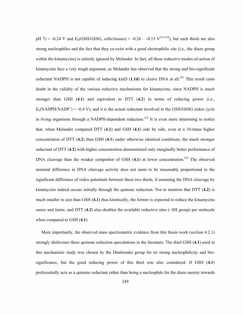
249
pH 7) = -0.24 V and Eh(GSH/GSSG, cells/tissues) = -0.26 – -0.15 V353,354), but such thiols are also
strong nucleophiles and the fact that they co-exist with a good electrophilic site (i.e., the diazo group
within the kinamycins) is entirely ignored by Melander. In fact, all these reductive modes-of-action of
kinamycins face a very tough argument, as Melander has observed that the strong and bio-significant
reductant NADPH is not capable of inducing kinD (1.1d) to cleave DNA at all.307 This result casts
doubt in the validity of the various reductive mechanisms for kinamycins, since NADPH is much
stronger than GSH (4.1) and equivalent to DTT (4.2) in terms of reducing power (i.e.,
Eh(NADPH/NADP+) = -0.4 V), and it is the actual reductant involved in the GSH/GSSG redox cycle
in living organisms through a NADPH-dependent reductase.353 It is even more interesting to notice
that, when Melander compared DTT (4.1) and GSH (4.1) side by side, even at a 10-times higher
concentration of DTT (4.2) than GSH (4.1) under otherwise identical conditions, the much stronger
reductant of DTT (4.2) with higher concentration demonstrated only marginally better performance of
DNA cleavage than the weaker competitor of GSH (4.1) at lower concentration.307 The observed
minimal difference in DNA cleavage activity does not seem to be reasonably proportional to the
significant difference of redox potentials between these two thiols, if assuming the DNA cleavage by
kinamycins indeed occurs initially through the quinone reduction. Not to mention that DTT (4.2) is
much smaller in size than GSH (4.1) thus kinetically, the former is expected to reduce the kinamycins
easier and faster, and DTT (4.2) also doubles the available reductive sites (–SH group) per molecule
when compared to GSH (4.1).
More importantly, the observed mass spectrometric evidence from this thesis work (section 4.2.1)
strongly disfavours these quinone reduction speculations in the literature. The thiol GSH (4.1) used in
this mechanistic study was chosen by the Dmitrienko group for its strong nucleophilicity and bio-
significance, but the good reducing power of this thiol was also considered. If GSH (4.1)
preferentially acts as a quinone reductant rather than being a nucleophile for the diazo moiety towards

250
the kinamycins (Scheme 4-14), then the B-ring quinone of kinF (1.1f) might be reduced to the
corresponding semi-quinone radical form 4.111 (by following Feldman’s one-electron reduction
pathway) or dihydroxyquinone form 4.112 (by following Melander/Skibo’s two-electron reduction
pathway). It is not unreasonable to expect that, based on the structural similarity, these possible initial
products derived from kinF (1.1f) should be observed as either the protonated or Li+-complexed ion
species under the +ESI-MS conditions involved. However, mass peaks with the expected m/z for such
ion species (Scheme 4-14) were never observed in this thesis work. Instead, covalently bonded kinF-
GSH adduct 4.18 was spectrometrically observed as the corresponding protonated form. This clearly
suggests that the first and key step, prior to any later actions of kinamycins, is a nucleophilic attack at
the diazo moiety by the thiols rather than quinone reduction despite their good reducing power. Since
the (in vitro) mechanistic studies of this thesis work were carried out under aerobic and physiological
conditions (see section 4.7 for details) in the presence of a bio-significant thiol, the chemical
behaviour of kinamycins observed under such control should be very close to the actual mode-of-
action of kinamycins in the real biological systems (in vivo).

251
Scheme 4-14. Possible products and +ESI-MS ion species derived from kinF (1.1f) by following two
quinone reductive activation processes for kinamycins proposed in the literature.
Even though this thesis work does not exclude the possibility that a quinone reductive activation
might be involved, the results did suggest that the kinamycin quinone reduction needs some very
strong reductant under the physiological and aerobic conditions. This is quite different from
observations of kinamycin quinone reduction by certain non-bio-significant/bio-relevant chemical
reductants under the abiological/anaerobic conditions, such as the Feldman’s and Skibo’s discovery.
Under the physiological and aerobic conditions, the required power to smoothly reduce the quinone
moiety of the kinamycins is definitely beyond the capability of GSH (4.1, Eh(cells/tissues) = -0.26 –
-0.15 V353,354), or even NADPH (Eh = -0.4 V353) and DTT (4.2, Eh(pH 7–10) = -0.4 V352). From a
practical point of view, such redox requirement (Eh < -0.4 V) is also beyond the reducing power of
many biological systems. For example, blood under low oxygen pressure (55–32 mm Hg, cf.

252
Poxygen(air) ~ 160 mm Hg) has an Eh of ca. -0.062 – -0.102 V, and even in the complete absence of
oxygen, Eh of blood would drop to -0.202 – -0.416 V (depending on the time of equilibrium after
deoxygenation),355 which is at most comparable to the redox potential of NADPH and DTT (4.2).
Another example is bacteria, some of which are important targets of the kinamycin antibiotics, and
the bacteria’s range of Eh generally varies from -0.4 V for anaerobes to 0.3 V for aerobes.356 Thus, it
is reasonable to predict that the quinone of kinamycin is unlikely to be reduced in vivo either,
particularly under aerobic conditions. Such prediction is also consistent with the literature report that
GSH (4.1) can not reduce the α-tocopherol quinone 4.113, under the aerobic and physiological
conditions, both in aqueous buffer (pH 7.4) and in rat liver microsome.357 This quinone 4.113 is in
fact the oxidized form of Vitamin E and has a fairly high Eh of -0.48 V at pH 7.358 It could serve as a
model for the B-ring quinone moiety of kinamycin, whose redox potential is not known. In case if
one argues that the quinone moiety of kinamycin is fused with two aromatic rings so the tetra-
alkylated quinone model 4.113 may not be electronically appropriate, anthraquinones such as 4.114
can be used as an alternative model, which also has a comparable high Eh of -0.527 V at pH 7.352
Either way, such literature examples are in support of the proposal that reduction of kinamycin
quinone does require very strong reducing agents, which are very likely not available in real
biological systems. Thus, the reductive modes-of-action for kinamycins are likely not involved in the
action of their bioactivity under the physiological conditions.

253
4.3 NMR Studies on the Interaction of Kinamycin F and Double-stranded DNA
Upon the collaboration between the Dmitrienko group and the Hasinoff group, weak binding of
kinF (1.1f) to DNAs had been confirmed by evidence from UV-vis spectroscopy, DNA melting
experiments and fluorescence spectroscopy.241 It was further proposed by Dmitrienko and Hasinoff
that the fairly planar structure of kinF (1.1f), similar to that of anthracycline antitumor drugs such as
doxorubicin and daunomycin, might also be able to intercalate with the DNA base pairs.241 This
hypothesis was supported by some molecular modeling results, as calculations with the docking
program GOLD (Genetic Optimisation for Ligand Docking) predicted that kinF (1.1f) could
intercalate with model DNA at least from a steric point of view (Figure 4-17).241 The model DNA was
deduced from a doxorubicin-DNA X-ray structure (PDB ID: 1DA9) having two doxorubicin
molecules bound to the self-complementary DNA of d(TGGCCA), and one doxorubicin was removed
to make the space available for docking kinF (1.1f) computationally. The D-ring of kinF (1.1f) could
adopt two different conformations (see section 2.3 for details), and both conformations dock well in
the model DNA but in opposite orientations (ca. 180 degree rotation along the long axis of kinF).
Figure 4-17 illustrated the simulated result of docking conformer A of kinF (1.1f) having an
equatorial C4-OH into the model DNA, and such intercalation would be stabilized through hydrogen
bonding of the C1-OH and C2-OH on the six-membered D-ring of kinF (1.1f) to DNA as well as
base(π)-stacking. However, the conformer A of kinF (1.1f) having an equatorial C4-OH, which
happens to be the more stable one and also higher in diazonium ion character, scored better than the
corresponding C4-axial conformer B in the docking simulations.241
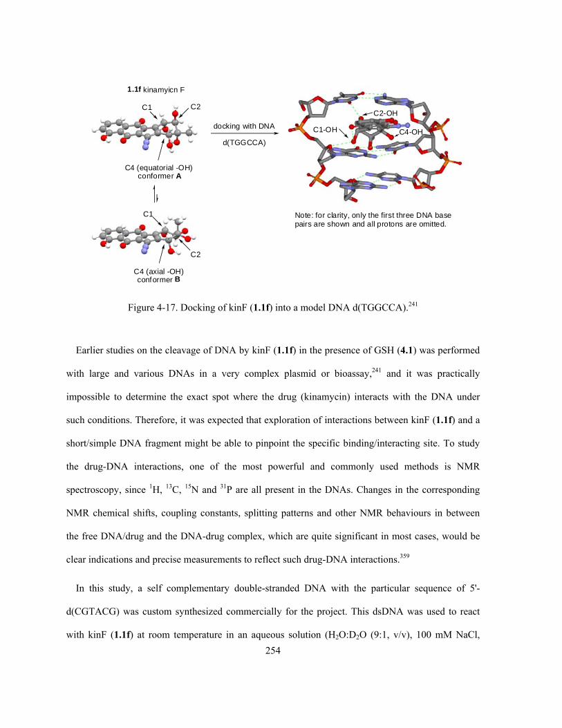
254
C1-OH
C2-OHC1 C2
C4 (equatorial -OH)conformer A
C1
C2
C4 (axial -OH)conformer B
1.1f kinamyicn F
docking with DNA
d(TGGCCA)C4-OH
Note: for clarity, only the first three DNA basepairs are shown and all protons are omitted.
Figure 4-17. Docking of kinF (1.1f) into a model DNA d(TGGCCA).241
Earlier studies on the cleavage of DNA by kinF (1.1f) in the presence of GSH (4.1) was performed
with large and various DNAs in a very complex plasmid or bioassay,241 and it was practically
impossible to determine the exact spot where the drug (kinamycin) interacts with the DNA under
such conditions. Therefore, it was expected that exploration of interactions between kinF (1.1f) and a
short/simple DNA fragment might be able to pinpoint the specific binding/interacting site. To study
the drug-DNA interactions, one of the most powerful and commonly used methods is NMR
spectroscopy, since 1H, 13C, 15N and 31P are all present in the DNAs. Changes in the corresponding
NMR chemical shifts, coupling constants, splitting patterns and other NMR behaviours in between
the free DNA/drug and the DNA-drug complex, which are quite significant in most cases, would be
clear indications and precise measurements to reflect such drug-DNA interactions.359
In this study, a self complementary double-stranded DNA with the particular sequence of 5'-
d(CGTACG) was custom synthesized commercially for the project. This dsDNA was used to react
with kinF (1.1f) at room temperature in an aqueous solution (H2O:D2O (9:1, v/v), 100 mM NaCl,

255
sodium phosphate buffer, pH 6.2), with a small amount of MeOD-d4 (5% by volume) as the co-
solvent. Without the presence of GSH (4.1), when kinF (1.1f) was mixed with the above dsDNA in a
molar ratio of 1:2, neither immediate binding nor slow interaction between the kinF (1.1f) and the
dsDNA could be observed on the corresponding 1H-NMR spectra (Figure 4-18) when the sample was
monitored up to 11 days, since the entire proton signals for both the DNA and kinF (1.1f) showed
essentially no change at all during the whole process. Upon addition of one equivalent of GSH (4.1)
to the above aqueous NMR sample ([kinF]:[GSH]:[dsDNA]=1:1:2), a small change in chemical shifts
was observed in the 1H-NMR region of 12.5–13.5 ppm (Figure 4-18, bottom spectrum), which
corresponded to the hydrogen-bonded imino protons of the DNA bases of guanine (G) and thymine
(T). The magnitude of the observed change in chemical shifts (only ca. 0.05 ppm), however, was way
less than what would be expected if significant or strong interaction between kinF (1.1f) and the
dsDNA indeed occur. As a comparison, literature results indicated that binding of various antitumor
drugs with DNA even at lower drug to DNA ratio from 1:50 to 1:3 (relative to the 1:2 ratio between
kinF (1.1f) and DNA used in this study) would cause a significant changes in the chemical shifts for
those DNA-imino protons, typically in between 0.1–1 ppm.360 In addition, the proton signals in the
aromatic region for both the kinF (1.1f) and DNA bases showed practically no change even after the
addition of GSH (4.1), which would also suggest that no specific interaction occurred in between
kinF (1.1f) and this particular DNA. Otherwise some noticeable changes in the chemical shifts of the
aromatic protons were very likely to be observed, since intercalation of daunomycin with DNA could
lead to about 0.1–0.2 ppm shift for some aromatic protons of both daunomycin and DNA.361,362

256
7891011121314 ppm
No.1 dsDNA only in H2O:D2O (9:1) pH6.2 700MHz
No.2 KinF+dsDNA (0.5:1) in H2O:D2O (9:1) pH6.2 700MHz
No.3 KinF+dsDNA (0.5:1) +5% MeOD-d4 in H2O:D2O (9:1) pH6.2 700MHz
No.4 KinF+dsDNA (0.5:1) +5% MeOD-d4 in H2O:D2O (9:1) pH6.2 600MHz 3-days, room temperature, no light
No.5 KinF+dsDNA (0.5:1) +5% MeOD-d4 in H2O:D2O (9:1) pH6.2 600MHz 11-days, room temperature, no light
No.6 KinF+GSH+dsDNA (0.5:0.5:1) +5% MeOD-d4 in H2O:D2O (9:1) pH6.2 600MHz
Figure 4-18. Time-resolved NMR spectra of kinF (1.1f) and a self complementary dsDNA 5'-
d(CGTACG) in aqueous buffer (conditions as marked on each spectrum; for clarity, only the NMR
region 6–14 ppm that corresponds to the aromatic and DNA-imino protons is shown).
The above NMR observation, although disappointing, does not necessarily mean that no further
experimental and computational attempts should be made. The double-stranded DNA fragments
involved in this study (Figure 4-19) such as 4.115 d(TGGCCA) (for modeling) and 4.116
d(CGTACG) (for NMR) did not provide all the possible combinations of having each of the four
available DNA-bases (i.e., A, T, C and G) bonded to all other available DNA bases, by which a total
of eight different types of spatial cavities in between the two layers of DNA base pairs could be
generated. They are marked as (1) to (8) in Figure 4-19. Failure of kinF (1.1f) to intercalate/bind with

257
4.116 would only imply that half of the possible cavities, i.e. (1) to (4) in Figure 4-19, were not
suitable for the expected interaction even though some situation such as (2) had been predicted to be
feasible by docking simulation. However, double-stranded DNA containing fragments of d(AAG) and
d(CCT) such as 4.117 could provide four other possible binding sites (i.e., (5) to (8) in Figure 4-19)
that are untested, which may allow the real physical intercalation of DNA by kinF (1.1f) to occur. The
proposed dsDNA fragment structure such as 4.117 d(AAG···CCT) is not self complementary, which
could make its preparation relatively more complicated and difficult. However, simpler and self
complementary DNA fragments that can be readily prepared, such as d(AAGCTT), d(CTTAAG),
d(GAATTC) and d(TTCGAA) to provide the possible binding site of (5) and (6) as well as
d(CCTAGG), d(TCCGGA), d(AGGCCT) and d(GGATCC) to provide the possible binding site of (7)
and (8), shall be able to serve as better alternative DNAs to study the interactions with kinF (1.1f).
C G
4.116 dsDNA: d(CGTACG)
T A C G
G C A T G C
4.115 dsDNA: d(TGGCCA)
T G G C C A
A C C G G T
Intercalation ofkinF (1.1f)
Intercalation ofdoxorubicin
XIntercalation of kinF (1.1f)
not observed with four possiblebinding sites (1) to (4)
Docking/Modeling prediction
A A
4.117 dsDNA containgingd(AAG) and d(CCT)
G C C T
T T C G G A
Other four possible DNA-intercalation sites (5) to (8)
for kinF (1.1f)
?
(1) (2) (3) (4) (1) (5) (6) (7) (8)(2)
Figure 4-19. Possible binding sites within dsDNA for kinF (1.1f).
4.4 Ab initio MO Calculations and Solution IR Studies of Kinamycin F
As described earlier in the thesis (section 2.3), based on NMR spectroscopic observations, the D-
ring of kinF (1.1f) was found experimentally to adopt two different conformations, and the

258
corresponding equilibrium showed a clear solvent-dependence, i.e. polar solvent favours the
conformation of kinF (1.1f) that gives a small NMR coupling constant for the D-ring H1-H2 (Scheme
2-12, section 2.3). However, similar NMR behaviour was never encountered with other kinamycins
(either literature results or this work). Therefore, quite extensive computational analyses were also
carried out in the Dmitrienko group (this work) in hope that such efforts may revealed the origin of
this unusual conformational behavior of kinF (1.1f). Since kinF (1.1f) possesses the most breadth of
antibacterial activity of the kinamycin family, and it may be generated from the other kinamycins by
esterase action in vivo to function as the actual bioactive growth inhibitory form of these anticancer
agents, it is very interesting and essential to understand the influence of such conformational change
on the various properties of the molecule, as this may in turn affect their biological activities to a
certain extent.
The kinamycins are quite rigid and flat molecules due to their aromatic ABC-rings, and the only
flexible part is the non-aromatic D-ring with four oxygen substituents and a (common) methyl group,
which can be considered as a fully substituted cyclohexene ring by itself. Similar to the well-known
cyclohexane ring system, the unsubstituted cyclohexene ring would also equilibrate in between two
half-chair conformations that interconvert via a boat form, but with a much lower free energy barrier
of activation (ΔG‡) of about 5.6 kcal/mol (cf. the corresponding ΔG‡ for cyclohexane is ~ 10.1solution
or 10.4gas-phase kcal/mol).363 For substituted cyclohexenes, the two conformations would normally have
certain energy difference depending on the orientation of the substituent(s), i.e. ax. (axial) vs eq.
(equatorial). However, the values of the free energy difference between these two conformations
(ΔG0, also known as the A-value) are significantly smaller (in general) for the cyclohexene system
than those found with the cyclohexane system (Table 4-2).

259
Table 4-2. A-Values for some substituted cyclohexenes and cyclohexanes.
ΔG0 (axial-equatorial) (kcal/mol)*
Substituent 4-substituted cyclohexene 4-substituted 1,2-di-methyl cyclohexene
Mono substituted cyclohexane
F 0.014a,b - 0.276a,c, 0.25d
Cl 0.20a,b, 0.31a,e, -0.02a,f, 0.19c,g, 0.23c,h 0.38a,e , 0.06a,f 0.528a,c, 0.52d
Br 0.077a,b, 0.2a,e 0.30a,e 0.476a,c, 0.48-0.62d
I -0.016a,b, 0.16a,e 0.20a,e 0.468a,c, 0.46d
-C≡N 0.15a,e, 0.02a,f, -0.15i,j, 0.1i,g 0.14a,e, 0.14a,f 0.24a,c, 0.20i,k, 0.44i,g
-OH 0.22a,e 0.70a,e 1.04a,l, 0.92-0.97d
-OTMS 0.31a,e - -
-OAc - - 0.71d
-OCH3 - - 0.75d
-CH3 ~ 1m - 1.74d
-CH2CH3 - - ~1.75d
-CH=CH2 > 1.0m,n - 1.7d
-CHO 0.5m,o, 0.26m,p, 0.44m,q - -
-CH2• 0.17a, 0a (3-substituted) - - *For a given substrate, the ΔG0 generally shows small (but noticeable in some cases) dependence on the
solvent and temperature; a. Taken from reference364 (and references therein); b. -160 °C in CD2=CDCl; c. -80
°C in CS2; d. Taken from reference365 (and references therein); e. -145 °C in CF2Cl2; f. 145 °C in CHF2Cl; g.
30 °C in CS2; h. -60 °C in CS2; i. Taken from reference366; j. 75 °C in DMSO-d6; k. 0 °C in CS2; l. -83 °C in
MeOD-d4; m. Taken from reference367; n. -133 °C in CF2Cl2-CHF2Cl (1:1); o. room temperature in CHCl3; p.
-167 °C in CHF2Cl; q. -165 °C in CF2Cl2.
There is a lack of literature data of A-values for the cyclohexene ring system. It is then necessary
to estimate a few such values based on available data and their correlations (Table 4-2), so that a
semi-quantitative conformational analysis could be carried out with the kinamycins. For instance,
when comparing kinF (1.1f) and kinE (1.1e) (Figure 4-20), A-values of 0.70 kcal/mol for -OH, 0.71
kcal/mol for -OAc and 1 kcal/mol for -CH3 are used to estimate the approximate free energy
difference between the two D-ring conformer A (with an equatorial C4-substituent) and B (with an
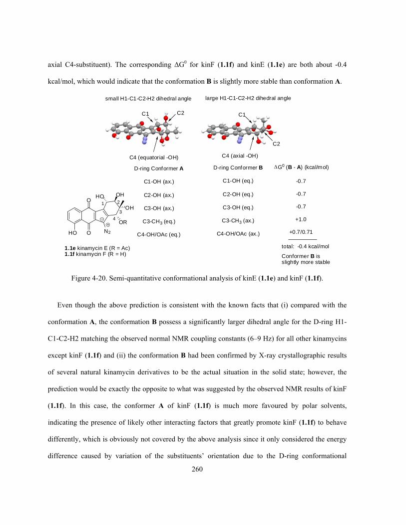
260
axial C4-substituent). The corresponding ΔG0 for kinF (1.1f) and kinE (1.1e) are both about -0.4
kcal/mol, which would indicate that the conformation B is slightly more stable than conformation A.
HO
O
O N2
HO
OH
OR
OH
1 2
3
4
D-ring Conformer A
C1-OH (ax.)
C2-OH (ax.)
C3-OH (ax.)
C3-CH3 (eq.)
C4-OH/OAc (eq.)
D-ring Conformer B
C1-OH (eq.)
C2-OH (eq.)
C3-OH (eq.)
C3-CH3 (ax.)
C4-OH/OAc (ax.)
-0.7
-0.7
-0.7
+1.0
+0.7/0.71
total: -0.4 kcal/mol
G0 (B - A) (kcal/mol)
1.1e kinamycin E (R = Ac)1.1f kinamycin F (R = H)
C1 C2
C4 (equatorial -OH)
C1
C2
C4 (axial -OH)
small H1-C1-C2-H2 dihedral angle large H1-C1-C2-H2 dihedral angle
Conformer B isslightly more stable
Figure 4-20. Semi-quantitative conformational analysis of kinE (1.1e) and kinF (1.1f).
Even though the above prediction is consistent with the known facts that (i) compared with the
conformation A, the conformation B possess a significantly larger dihedral angle for the D-ring H1-
C1-C2-H2 matching the observed normal NMR coupling constants (6–9 Hz) for all other kinamycins
except kinF (1.1f) and (ii) the conformation B had been confirmed by X-ray crystallographic results
of several natural kinamycin derivatives to be the actual situation in the solid state; however, the
prediction would be exactly the opposite to what was suggested by the observed NMR results of kinF
(1.1f). In this case, the conformer A of kinF (1.1f) is much more favoured by polar solvents,
indicating the presence of likely other interacting factors that greatly promote kinF (1.1f) to behave
differently, which is obviously not covered by the above analysis since it only considered the energy
difference caused by variation of the substituents’ orientation due to the D-ring conformational

261
change. The additional interacting factors may include intramolecular hydrogen bonding and possible
interactions from the C4-substituent towards the nearby C5-diazo group, both of which can be
affected by the D-ring conformation.
Ab initio molecular orbital (MO) calculations, on the other hand, would be able to afford a
quantitative and much more precise prediction of the thermodynamics (stability), geometry (bond
length and bond angle) and spectroscopic behaviour of the compounds of interest. Gas phase MO
calculations (geometry optimization and frequency calculation) of kinF (1.1f) carried out using the
Gaussian03® program at the RHF/6-31G//6-31G level predict that the corresponding A-type
conformer (eq. C4-OH) is favored by 0.4 kcal/mol (Table 4-3). A similar computational study of kinE
(1.1e) reveals an opposite but substantial preference of the B-type conformation (ax. C4-OAc) that is
favoured by 4.8 kcal/mol (Table 4-3), which is in parallel with the experimental data for this and
other kinamycins but in contrast with the conformational preference found for kinF (1.1f).

262
Table 4-3. Gas phase ab initio MO calculations of kinE (1.1e) and kinF (1.1f).a
E (RHF) (a.u.)b
Conformation A Conformation B E (B - A)
kinE (1.1e)
HO
O
O N2
OH
OH
OAc
OH
1 2
-1473.433862
-1473.441514
-0.007652
(-4.8 kcal/mol)
Conformer B is more stable
kinF (1.1f)
HO
O
O N2
OH
OH
OH
OH
1 2
-1321.773871
-1321.773242
0.000629
(0.4 kcal/mol)
Conformer A is more stable
a. Ab initio MO geometry optimization and calculation of vibrational frequencies were performed with
Gaussian03® at the RHF/6-31G//6-31G level; b. The E(RHF) (restricted Hartree-Fork energy) was corrected
with ZPE (zero-point energy), 1 a.u. = 627.5095 kcal/mol.
Further solution phase MO calculations, employing the PCM (polarizable continuum model)
solvation model, showed a preference for the A-type conformer of kinF (1.1f) increases substantially
with the increasing solvent polarity. This is clearly indicated by the concurrent increasing change in
free energy of solvation (Gsolvation = 3.4, 5.3 and 12.4 kcal/mol for CHCl3, acetone and DMSO
respectively, Table 4-4) between the two conformers, which takes into account the contributions from
both the electrostatic and non-electrostatic factors. Unfortunately, similar calculations with kinE
(1.1e) in identical solvents failed unexpectedly, particularly with its less-favoured conformation A,
and led to no useful data for comparison (Table 4-4). In addition, the calculated dipole moments for
the two conformers of kinF (1.1f), in both gas phase and solutions (Table 4-4), also showed a

263
consistent increasing tendency along with the increasing medium/solvent polarity (as indicated by the
corresponding dielectric constant ε). The (much) higher dipole moment was always associated with
the conformer A, which would also suggest its preference in more polar environment. On the
contrary, for kinE (1.1e) in gas phase, the more stable conformer B (0.7316 Debye) is also much more
polar than its competitor conformer A (0.2925 Debye).
Table 4-4. Ab initio MO calculations of kinF (1.1f) and kinE (1.1e) in various solvents.a
kinF (1.1f): conformation A is more stable than B by 0.4 kcal/mol (gas phase)
Gsolvation (kcal/mol) Solvent
Conformation A Conformation B
Gsolvation (B - A)b
(kcal/mol)
CHCl3 -13.53 -10.13 3.4
Acetone -25.9 -20.62 5.3
DMSO -30.62 -18.26 12.4
Dipole Moment μ (Debye) Solvent (ε)
Conformation A Conformation B
Gas phase (0) 3.1963 2.1963
CHCl3 (5) 4.6083 2.7730
Acetone (21) 5.0711 3.2696
DMSO (49) 7.9513 3.4780
kinE (1.1e): conformation B is more stable than A by 4.8 kcal/mol (gas phase)
Gsolvation (kcal/mol) Solvent
Conformation A Conformation B
Gsolvation (B - A)
(kcal/mol)
CHCl3 -9.81 -c -
Acetone -c -17.99 -
DMSO -c -14.44 - a. Ab initio MO calculations were performed with Gaussian03® at the RHF/6-31G//6-31G level, PCM
(polarizable continuum model) solvation model was used for solution phase modeling; b. Values are rounded to
one decimal point; c. MO calculations failed even after several attempts.
Furthermore, it appears that there is some influence from the conformational preferences for D-
ring of kinF (1.1f) on the diazonium ion character of the diazo group, which can be characterized by

264
its corresponding IR stretching frequency (and diazo bond length). Ab initio MO calculations
overestimate the absolute values for the diazo (and other) stretching frequencies, even after these
calculated raw frequency values had already been corrected with an empirical scaling factor (i.e.,
0.8929) to account for systematic errors originated from the neglect of electron correlation. However,
the significant difference in calculated diazo IR frequency for the two conformers of kinF (1.1f), i.e.
νN≡N(conformer A) = 2206 cm-1 and νN≡N(conformer B) = 2183 cm-1, is quite surprising.
A solution IR study of kinF (1.1f) was carried out in various solvents and the corresponding
results, as summarized in Table 4-5, revealed more physical evidence to support the above theoretical
predictions. The diazo stretch in the solution IR spectra of kinF (1.1f) was observed experimentally
(Table 4-5, part II) as two clearly distinguished bands in the very polar and H-bonding solvents such
as DMSO (2142 cm-1 and 2165 cm-1, ∆ν = 23 cm-1) and acetone (2137 cm-1 and 2174 cm-1, ∆ν = 37
cm-1), which are essentially non-concentration dependent. This is consistent with the proposed co-
existence of two conformers of kinF (1.1f) having different diazonium ion character in these two
solvents. However, in much less polar and non-H-bonding solvents such as MeCN, CHCl3 and CCl4
(Table 4-5, part I), the corresponding diazo stretching IR signal of kinF (1.1f) appeared as essentially
one single peak, and the observed frequencies (i.e., 2138–2140 cm-1 in MeCN, 2132–2140 cm-1 in
CHCl3 and 2130 cm-1 in CCl4) are in better agreement with the diazo IR results of kinF (1.1f)
obtained from the solid-state (i.e., 2142 cm-1 in KBr; 2134–2140 cm-1 as film on NaCl plate
depending on the solvent used to prepare the film), suggesting that the less polar B-type conformer of
kinF (1.1f) with a lower diazo IR frequency predominates in non-polar solvents. The predicted
significant preference in polar solvents for conformer A of kinF (1.1f) is also entirely consistent with
the previous NMR findings (i.e., the observed D-ring H1-H2 coupling constant of kinF (1.1f) is
inversely proportional to the solvent polarity involved, Table 2-4, section 2.3). The more polar
conformer A of kinF (1.1f) has a calculated small dihedral angle of 75° for H1-C1-C2-H2 (close to

265
90°), which should give a rather small NMR coupling constant as estimated by the Karplus equation.
As a comparison, the corresponding dihedral angle is 171° (close to 180°) within the conformer B,
which shall lead to a normal (large) proton coupling constant.
It is very interesting to notice that in the case of using MeOH as the solvent (Table 4-5, part II), a
gradual shift of the diazo IR signal of kinF (1.1f) from 2142 cm-1 to 2170 cm-1 (∆ν = 28 cm-1) was
observed upon dilution of the kinF-MeOH solution, which appeared as a broad peak (an intermediate
situation in between a single peak and two bands) during the entire shifting process. It is very likely
that the two conformers of kinF (1.1f) also equilibrate within MeOH, similar to the situation observed
with DMSO and acetone. In the mean time, the strong and bidirectional hydrogen bonding capability
of MeOH and kinF (1.1f), i.e. both species can act inter- and intramolecularly as a H-bond donor
and/or acceptor while DMSO/acetone can only behave as a hydrogen bond acceptor, would
complicate the various interactions between the solvent (MeOH) and solute (kinF) as well as the
appearance of the IR spectra, hence the observed broad diazo IR peak and the concentration-
dependent diazo IR shift.
The predicted ∆ν of 23 cm-1 (in gas phase) with the calculated higher stretching frequency being
associated with the conformer A, agrees pretty well with the difference between the two solution IR
bands observed for the diazo group of kinF (1.1f) in solutions (∆ν = 23 cm-1 in DMSO and ∆ν = 37
cm-1 in acetone, Table 4-5). It is also noteworthy that the calculated diazo IR frequencies for the two
conformers of kinF (1.1f) in DMSO and acetone (Table 4-6), even though giving a smaller calculated
∆ν between the two conformers when compared with the experimental values in each case, do show a
consistency with the experimental observation that acetone induces larger change in diazo IR
frequency than DMSO.

266
Table 4-5 (part I). Diazo IR frequency of kinF (1.1f) in organic solvents.a
IR sample No.b [kinF]c Solvent NN (cm-1)d
CNIR0037 C0
Saturated stock solutione
CCl4 2130 (w)
CNIR0041 C1
Saturated stock solution
CHCl3 2132 (w)
CNIR0042 0.2C1
5x dilution of stock
CHCl3 2139 (w, br)
CNIR0043 0.1C1
10x dilution of stock
CHCl3 2137 (w)
CNIR0044 0.04C1
5x dilution of CNIR0042
CHCl3 2136 (w, noisy)
CNIR0045 0.02C1
5x dilution of CNIR0043
CHCl3 2135–2140
(very w, noisy)
CNIR0032 C2
Saturated stock solution
MeCN 2140
CNIR0033 0.2C2
5x dilution of stock
MeCN 2138
CNIR0034 0.04C2
25x dilution of stock
MeCN 2139 (w)
a. See section 4.7 for experimental details; b. IR spectra were recorded consecutively in the numeric
order as indicated by the sample’s No., however, IR results were tabulated and presented in the
increasing order of solvent polarity, and for each individual solvent, data were listed in the descending
order of substrate concentration; c. The concentration of kinF (1.1f) in solutions had no actual values
since each stock solution was prepared with a very small amount of kinF (ca. 0.1–0.2 mg or less). In
case a saturated stock solution was made, excess kinF (1.1f) was mixed with a small volume of
solvent and quickly sonicated to ensure complete saturation of the solvent by kinF (1.1f). The
saturated stock solution was then filtered to remove the undissolved substrate; d. All IR wavenumbers
were rounded to the closest integer. w = weak, br = broad; e. solubility of kinF (1.1f) in CCl4 was
extremely low and there were still visible and very fine solid particles of kinF (1.1f) in the solution
even after sonication, which was used directly for the IR study without filtration.

267
Table 4-5 (part II). Diazo IR frequency of kinF (1.1f) in organic solvents.a
IR sample No.b [kinF]c Solvent NN (cm-1)d Δν (cm-1)
CNIR0023 3.0C3
3x concentration of stock
MeOH 2142 -
CNIR0020 C3 (Stock solution) MeOH 2142 -
CNIR0021
0.2C3
5x dilution of stock
MeOH 2148
(w, br)
-
CNIR0027 0.1C3
concentration of CNIR0025
MeOH 2159
(very br)
-
CNIR0025 0.04C3
concentration of CNIR0024
MeOH 2169
(br)
-
CNIR0024 0.02C3
50x dilution of stock
MeOH 2170
(w, br)
-
CNIR0071 C4
Saturated stock solution
Acetone 2138
2173
35
CNIR0063 0.2C5
5x dilution of stock
Acetone 2136
2174
38
CNIR0065 0.1C5
10x dilution of stock
Acetone 2137
2174
37
CNIR0067 0.04C5
5x dilution of CNIR0063
Acetone 2135
2174
39
CNIR0069 0.02C5
5x dilution of CNIR0065
Acetone 2136
2173
37
CNIR0049 C5 (stock solution) DMSO 2145 (br) -
CNIR0050 0.2C5
5x dilution of stock
DMSO 2142
2165
23
CNIR0052 0.1C5
10x dilution of stock
DMSO 2142
2165
23
CNIR0054 0.04C5
5x dilution of CNIR0050
DMSO 2142
2165
23
CNIR0056 0.02C5
5x dilution of CNIR0052
DMSO 2142
2165
23

268
Table 4-6. Calculated diazo IR frequency and bond length of kinF (1.1f) in gas phase and solutions.a
Solvent Conformer A of kinF (1.1f) Conformer B of kinF (1.1f) ΔνN≡N (cm-1)
Gas phase νN≡Nb: 2206 cm-1; dN≡N: 1.098 Å νN≡N
b: 2183 cm-1; dN≡N:1.100 Å 23
CHCl3 νN≡Nb: 2225 cm-1; dN≡N: 1.095 Å νN≡N
b: 2197 cm-1; dN≡N:1.098 Å 28 (not observed)
Acetone νN≡Nb: 2229 cm-1; dN≡N: 1.095 Å νN≡N
b: 2206 cm-1; dN≡N:1.097 Å 23 (observed 37)
DMSO νN≡Nb: 2216 cm-1; dN≡N: 1.096 Å νN≡N
b: 2210 cm-1; dN≡N:1.096 Å 6 (observed 23)
a. Ab initio MO geometry optimization and vibrational frequency calculation were performed with Gaussian03®
at the RHF/6-31G//6-31G level, PCM (polarizable continuum model) solvation model was used for solution
phase modeling; b. Scaled by the scaling factor of 0.8929.
These observations suggest that some specific interaction of the diazo group, which is absent in
the D-ring model compound of kinF (1.1f) such as the simple aromatic tetrol 2.59 that showed no
conformational change in different (NMR) solvents (Table 2-3, section 2.3), might be responsible for
the preference for the equatorial orientation of the C4-OH group in kinF (1.1f) as observed in its
calculated conformation A. Further ab initio molecular orbital calculations on simpler models 4.118
(Figure 4-21) led to the initial suggestion that the effect on diazonium ion character may be a
consequence of a favorable through space interaction of the C4-O bond dipole with the oppositely
polarized C5-N bond dipole of the diazo group, which is enhanced when the two bond dipoles
(C4→O/C5←N) are aligned. The triatomic link –C-–N+≡N of kinamycins exists within the flat plane
as defined by the aromatic ABC-rings, as confirmed by several X-ray works and many MO
calculation results from this work. A good alignment between the two nearby polar bonds of C4-O
and C5-N would only be achieved if the C4-OH occupies the equatorial orientation, while the axial
C4-OH conformer would almost completely lose such energetically beneficial interaction due to the
near perpendicular correlation of the C4-substituent with the ring plane and C5-N bond. This is
clearly indicated by the calculated small dihedral angle of ca. 36.7° for O-C4-C5-N within conformer
A of kinF (1.1f), relative to a much larger dihedral angle of ca. 60.2° for O-C4-C5-N within
conformer B of kinF (1.1f).

269
O
O N
X
X (equatorial) = H OAc F OH
N-N (cm-1) 2104 2123 2158 2173dN-N (Å) 1.107 1.105 1.102 1.100
N
4
5
4.118
Figure 4-21. Calculated diazo IR stretching frequencies and N≡N bond lengths (RHF/6-31G//6-31G)
for the proposed simple kinamycin analogue 4.118 with different equatorial C4-substituents.
However, subsequent MO calculations on isoprekinamycin analogues, which are to be described in
Chapter 5, have led to the conclusion that a through-space interaction of an electron-rich heteroatom
at C4 with the C5-diazo group might be the major cause of the increase in diazonium ion character.
Further discussion is provided in Chapter 5.
The significant influence from a simple D-ring conformational change on the diazonium character
of kinF (1.1f) might play some important role in the corresponding biological activities of such
natural antibiotics, especially when considering the earlier mechanistic studies (section 4.2) indicating
the crucial reaction between the bio-significant thiols and kinF (1.1f) to involve the very likely attack
of the diazo moiety by the sulfur nucleophile. Therefore, the conformer of kinF (1.1f) having a diazo
group with a higher diazonium ion character and hence higher reactivity, which is favoured by polar
solvents, may be the bioactive form.
4.5 Investigation of Intramolecular H-bonds within Diazobenzo[b]fluorenes
The common presence of strong intramolecular H-bonds within the diazobenzo[b]fluorene type of
kinamycins in between their C7-OH and C6-O as well as C1-OH and C11-O is well known. This is
characterized by the unusually high NMR chemical shifts of such H-bonded phenolic and alcoholic
protons9 and is supported by some unambiguous evidence from X-ray crystallographic studies.21

270
O
O
O
OR3
O
OR4
Me
OR2
N2
1
5
7
113
10 2
4
68
9
A B CD11a
11b
5a
4a
6a
10a
H
H
Previous MO calculations carried out by Laufer in the Dmitrienko group (unpublished results)
indicate that such intramolecular H-bonding interactions within a diazobenzo[b]fluorene structure
will increase the diazonium ion character but only to a rather small extent (Table 4-7).80
Table 4-7. Calculated diazo IR frequencies and bond lengths of some diazobenzo[b]fluorenes.a
Compound NN (cm-1)b dN≡N (Å) Compound NN (cm-1)b dN≡N (Å) O
O N2
HO
4.119
2120 1.105(9)
O
OOH N2
Me
HO
1.4
2058 1.111
O
O N2
OH
4.120
2116 1.106(3)
O
O N2
Me
HO
4.123
2060 1.111(6)
O
O N2OH 4.121
2113 1.106(5)
O
O N2
Me
OH 4.124
2050 1.112(8)
O
O N2 4.122
2104 1.107
O
O N2
Me
4.125
2044 1.113
a. From R. Laufer’s thesis,80 ab initio MO geometry optimization and IR frequencies of the compounds were
performed with Gaussian98® at RHF/6-31G//6-31G level; b. Scaled by a scaling factor of 0.8929.
However, one can easily realize that, the two series of model diazobenzo[b]fluorene compounds in
Laufer’s result (Table 4-7) experience the loss of intramolecular H-bonding interaction upon the
gradual removal/relocation of hydroxy group(s) (e.g., from 4.119 to 4.122 and from 1.4 to 4.123–
4.125). Thus, the concurrent presence of a substituent effect (i.e., replacement of –OHs by –Hs) is
unavoidable, and consequently the corresponding change in the calculated diazo IR frequencies
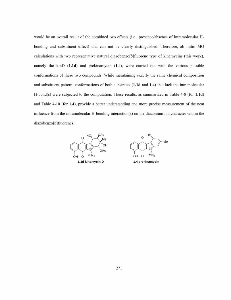
271
would be an overall result of the combined two effects (i.e., presence/absence of intramolecular H-
bonding and substituent effect) that can not be clearly distinguished. Therefore, ab initio MO
calculations with two representative natural diazobenzo[b]fluorene type of kinamycins (this work),
namely the kinD (1.1d) and prekinamycin (1.4), were carried out with the various possible
conformations of these two compounds. While maintaining exactly the same chemical composition
and substituent pattern, conformations of both substrates (1.1d and 1.4) that lack the intramolecular
H-bond(s) were subjected to the computation. These results, as summarized in Table 4-8 (for 1.1d)
and Table 4-10 (for 1.4), provide a better understanding and more precise measurement of the neat
influence from the intramolecular H-bonding interaction(s) on the diazonium ion character within the
diazobenzo[b]fluorenes.
O
OH
HO
O
Me
N2
1.4 prekinamycin
O
OH
HO
OH
O
OAc
Me
OAc
N2
1.1d kinamycin D

272
Table 4-8. Gas phase MO calculations of kinD (1.1d) with different conformations.a
Starting geometry of 1.1d
used for MO calculationb
Ending geometry of 1.1d
obtained from MO calculation
NN (cm-1)c
dN≡N (Å)
E(RHF) (a.u.)d
Starting conformation I
Starting conformation II
Starting conformation III
Starting conformation IV
Ending conformation I
Ending conformation II
Ending conformation III
(identical to I)
Ending conformation IV
(identical to II)
2235
1.095(6)
-1625.113881
2230
1.096(0)
-1625.093600
2235
1.095(6)
-1625.113881
2230
1.096(0)
-1625.093601
a. Ab initio MO geometry optimization and calculation of vibrational frequencies were performed with Gaussian03® at the RHF/6-31G//6-31G level; b. For kinD (1.1d), starting geometry I was modified from the corresponding X-ray results of (+)-α-methylbutryryl kinD (1.1v) by adding or removing necessary substituents. Other starting geometries of 1.1d with intramolecular H-bond(s) missing were obtained upon manual rotation of the phenol or alcohol hydroxy group(s) within the optimized conformation I to “break” the H-bond(s), which were then subjected to MO calculation; c. Scaled by a scaling factor of 0.8929; d. The E(RHF) was corrected with ZPE. 1 a.u. = 627.5095 kcal/mol.

273
In theory, the C5 partial negative charge of 1.1d could delocalize, as shown in Scheme 4-15, to
either the C6-O (resonance form I) or C11-O (resonance form II) to enhance the corresponding
intramolecular A-B ring or B-D ring H-bonding interactions, and the diazonium ion character is
simultaneously enhanced due to the decrease of nearby negative charge. However, it can be clearly
seen from the above calculation results that the two intramolecular H-bonds within the
diazobenzo[b]fluorenes are quite different in terms of their effects on the stability and the diazonium
ion character of the compound. Relative to the most stable conformation of kinD (1.1d) that possesses
two intramolecular H-bonds (i.e., conformer I/III), loss of the A-B ring H-bonding interaction
between C7-OH and C6-O as in conformation II/IV significantly destabilizes the molecule by 12.7
kcal/mol, but the corresponding diazonium ion character only drops slightly, which is indicated by a
concurrent decrease of 5 cm-1 in diazo IR frequency. On the other hand, various attempts to “break”
the intramolecular B-D ring H-bond between the C1-OH and C11-O all failed (cf. starting and ending
conformation I, III and IV, Table 4-8), and the optimized geometry of 1.1d always ends up with the
B-D ring H-bond presenting within the final optimized conformation, regardless of the starting
conformation or the status of the A-B ring H-bond. Such observation would suggest that the overall
energetic benefit from having a B-D ring H-bond is even larger than the already very strong A-B ring
H-bond.
O
O
O
OH
O
OAc
Me
OAc
N
N
H
H
O
O
O
OH
O
OAc
Me
OAc
N
N
H
H
1.1d resonance form II
567
5
111
1.1d
O
O
O
OH
O
OAc
Me
OAc
N
N
H
H
567
111
1.1d resonance form I
A BD
Scheme 4-15. Possible resonance delocalization of C5 negative charge within kinD (1.1d).

274
Although the energy contribution from the B-D ring H-bonding interaction within kinD (1.1d) can
not be obtained quantitatively, the corresponding influence on the diazonium ion character can be
estimated. Obviously, the B-D ring H-bonding interaction within the structure of kinD (1.1d) would
disappear if the C1-OH group is methylated (i.e., C1-OMe-kinD 4.126) or acetylated (i.e., C1-OAc-
kinD, which is kinC (1.1c) indeed). Compounds 4.126 and 1.1c may represent the situation of kinD
(1.1d) missing the B-D ring H-bonding interaction and offer some meaningful results for comparison;
however, it shall be noted that estimation by this manner would contain some contribution from a
substituent effect (i.e., C1-O-Ac of 1.1c vs C1-O-H of 1.1d vs C1-O-Me of 4.126).
MO calculations on the diazo IR frequencies of 1.1c, 1.1d, 4.126 and another closely related
synthetic analogue 3.17624 are summarized in Table 4-9. Even though the calculated absolute values
of these diazo IR frequencies are much higher than the corresponding experimental observations, the
calculated relative differences between them are in very good agreement with the experimental ones.
For example, diazo IR frequencies of 1.1d and 1.1c have a calculated difference of 8 cm-1 while the
experimental difference is 5–7 cm-1 (Table 4-9). Comparison between the calculated diazo IR
frequencies of 1.1d (with H-bonded C1-OH, 2235 cm-1), 4.126 (with C1-OMe, 2216 cm-1) and 1.1c
(with C1-OAc, 2227 cm-1) indicates that the influence on the diazonium ion character from the B-D
ring H-bond of 1.1d should be within the range of ca. 8–19 cm-1, since the acetyl group is electron-
withdrawing while the methyl is electron-donating when compared to proton. This effect is slightly
larger than the corresponding one from the A-B ring H-bond, which reduces the diazo IR frequency
by 5 cm-1 when the intramolecular A-B ring H-bond is absent (Table 4-8). The noticeable decrease of
diazo IR frequency by 11 cm-1 from 1.1c (C1-OAc) to 4.126 (C1-OMe) would suggest a moderate
benefit of having some electron-withdrawing factor at the C1 position to enhance the diazonium ion
character, presumably through inductive effect that could help to delocalize the C5 negative charge.

275
Table 4-9. Calculated and experimental diazo IR frequencies of some diazobenzo[b]fluorenes.a
Compound NN (cm-1)b dN≡N (Å) Compound NN (cm-1)b dN≡N (Å) O
O
MeO
OH
O
OAc
Me
OAc
NNH
4.126
2216 1.097(1)
O
O
AcO
OH
O
OAc
Me
OAc
NNH
1.1c
2227 Expt. 2150c
Expt. 2148d 1.096(3)
O
O
AcO
OH
O
OAc
Me
OAc
NNMe
3.176
2219 Expt. 2146e
1.096(9)
O
O
O
OH
O
OAc
Me
OAc
N
N
H
H 1.1d (double
H-bonds)
2235 Expt. 2155f
1.095(6)
O
O
O
OH
O
OAc
Me
OAc
N
NH
H
1.1d (No A-B ring H-bond)
2230 1.096(0)
O
O
OH
OH
O
OAc
Me
OAc
NNH
1.1d (No B-D ring H-bond)
N/A N/A
O
O
OO
O
OH
Me
O
NNH
H
1.1t
Expt. 2160g N/A
O
OAc
AcOO
O
OH
Me
O
N
N 1.1u
Expt. 2150g N/A
a. Ab initio MO geometry optimization and vibrational frequencies calculation were performed with
Gaussian03® at the RHF/6-31G//6-31G level; b. Scaled by a scaling factor of 0.8929, Expt. refers to
experimental IR values; c. Film, from reference23; d. Film, from reference16; e. Racemic mixture (oil) prepared
by total synthesis, IR was measured by means of ATR (attenuated total reflection), from reference24; f. In KBr,
from reference78; g. In KBr, from reference9.
In addition, compound 3.176 (with C1-OAc and C7-OMe) serves as another good and practical
example to demonstrate the effect on the diazonium ion character, either from the double
intramolecular H-bonding interactions when compared with 1.1d (with C1-OH and C7-OH), or from
the A-B ring H-bond alone when compared with 1.1c (with C1-OAc and C7-OH). The calculated
decrease of diazo IR frequency are 16 cm-1 (3.176 vs 1.1d) and 8 cm-1 (3.176 vs 1.1c) respectively,
which are in good agreement with the corresponding experimental values of 9 cm-1 (3.176 vs 1.1d)
and 2–4 cm-1 (3.176 vs 1.1c). This suggests that the overall combined effect from the double

276
intramolecular H-bonding interactions on the diazonium ion character within 1.1d should fall in the
range of ca. 8–16 cm-1 (theoretical prediction) or 2–9 cm-1 (experimental observation). This
conclusion is also entirely consistent with (i) the estimated individual H-bonding effect within 1.1d
on its diazonium ion character (i.e., 5 cm-1 from the A-B ring H-bond and ~ 8–19 cm-1 from the B-D
ring H-bond); and (ii) the experimental difference in diazo IR frequencies of 10 cm-1 observed for
isopropylidenekinamycin C (1.1t) and its C1/C7-diacetate 1.1u (Table 4-9).9
When the same MO calculations were carried out with prekinamycin (1.4), a
diazobenzo[a]fluorene but having an aromatic D-ring, the corresponding results (Table 4-10) are even
more interesting. The most stable conformation I of 1.4 possessing the double intramolecular H-
bonds was used as the starting point, and a gradual loss/breaking of H-bond(s), first with the A-B ring
H-bond (conformation II) then the B-D ring H-bond (conformation III) and finally at both H-bonded
sites (conformation IV) provided three additional and relatively stable conformations of 1.4 after
computation. The stabilities of these conformations decrease in the same consecutive order and the
corresponding increase in energy are 12.4, 12.5 and 25.1 kcal/mol, respectively. The last value (25.1
kcal/mol) is very close to the sum of the first two (24.9 kcal/mol), indicating the self-consistency of
such MO calculations. The energy contribution of 12.4 kcal/mol due to the presence of the
intramolecular A-B ring H-bond of 1.4 is almost identical to the calculated corresponding value for
1.1d (i.e., 12.7 kcal/mol for its A-B ring H-bond); however, the B-D ring H-bond of 1.4 seems to be
much easier to break than that of 1.1d.

277
Table 4-10. Gas phase MO calculations of prekinamycin (1.4) with different conformations.a
Starting geometry of 1.4
used for MO calculationb
Ending geometry of 1.4
obtained from MO calculation
NN (cm-1)c
dN≡N (Å)
E(RHF) (a.u.)d
Starting conformation I
Starting conformation II
Starting conformation III
Starting conformation IV
Ending conformation I
Ending conformation II
Ending conformation III
Ending conformation IV
2065
1.111
-1095.054362
2071
1.110(5)
-1095.034643
2057
1.112
-1095.034486
2061
1.111(7)
-1095.014427
a. Ab initio MO geometry optimization and vibrational frequency calculation were performed with Gaussian03®
at the RHF/6-31G//6-31G level; b. For prekinamycin 1.4, starting geometries were obtained by modification of
other optimized kinamycin conformations upon adding or removing necessary substituents, and its missing of
H-bonding was carried out in the same manner as in the case of 1.1d (see footnote b of Table 4-8); c. The
E(RHF) was corrected with ZPE. 1 a.u. = 627.5095 kcal/mol; d. Scaled by a scaling factor of 0.8929.
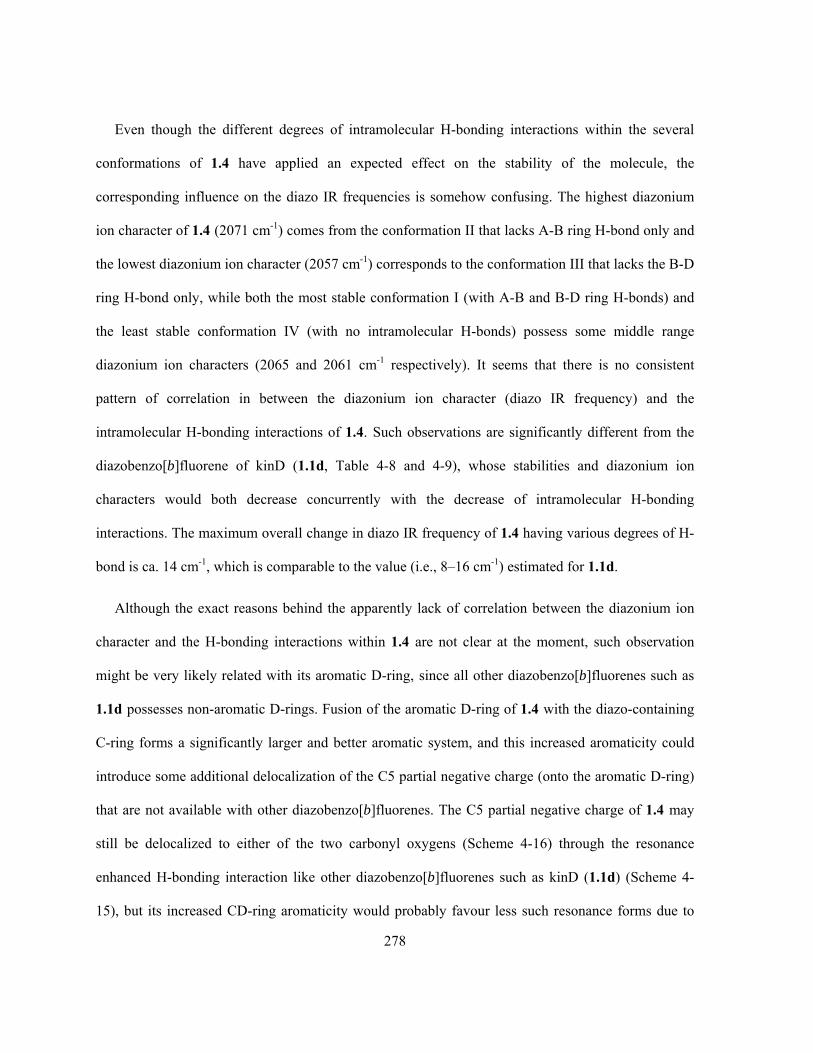
278
Even though the different degrees of intramolecular H-bonding interactions within the several
conformations of 1.4 have applied an expected effect on the stability of the molecule, the
corresponding influence on the diazo IR frequencies is somehow confusing. The highest diazonium
ion character of 1.4 (2071 cm-1) comes from the conformation II that lacks A-B ring H-bond only and
the lowest diazonium ion character (2057 cm-1) corresponds to the conformation III that lacks the B-D
ring H-bond only, while both the most stable conformation I (with A-B and B-D ring H-bonds) and
the least stable conformation IV (with no intramolecular H-bonds) possess some middle range
diazonium ion characters (2065 and 2061 cm-1 respectively). It seems that there is no consistent
pattern of correlation in between the diazonium ion character (diazo IR frequency) and the
intramolecular H-bonding interactions of 1.4. Such observations are significantly different from the
diazobenzo[b]fluorene of kinD (1.1d, Table 4-8 and 4-9), whose stabilities and diazonium ion
characters would both decrease concurrently with the decrease of intramolecular H-bonding
interactions. The maximum overall change in diazo IR frequency of 1.4 having various degrees of H-
bond is ca. 14 cm-1, which is comparable to the value (i.e., 8–16 cm-1) estimated for 1.1d.
Although the exact reasons behind the apparently lack of correlation between the diazonium ion
character and the H-bonding interactions within 1.4 are not clear at the moment, such observation
might be very likely related with its aromatic D-ring, since all other diazobenzo[b]fluorenes such as
1.1d possesses non-aromatic D-rings. Fusion of the aromatic D-ring of 1.4 with the diazo-containing
C-ring forms a significantly larger and better aromatic system, and this increased aromaticity could
introduce some additional delocalization of the C5 partial negative charge (onto the aromatic D-ring)
that are not available with other diazobenzo[b]fluorenes. The C5 partial negative charge of 1.4 may
still be delocalized to either of the two carbonyl oxygens (Scheme 4-16) through the resonance
enhanced H-bonding interaction like other diazobenzo[b]fluorenes such as kinD (1.1d) (Scheme 4-
15), but its increased CD-ring aromaticity would probably favour less such resonance forms due to

279
their sabotage of the stable aromatic structure. This situation is similar to the example of pyrrole vs
indole. The indole can be considered as a benzene ring fused with the pyrrole that is also aromatic.
The increased aromaticity of indole would require a greater conjugation of the nitrogen’s lone pair of
electrons and made it less available. Therefore, indole (pKb ~ 17.5) is an even weaker base than
pyrrole (pKb ~ 13.6). Back to the case of prekinamycin (1.4), the higher aromaticity of its CD-rings
could further offer a few more resonance forms (Scheme 4-16) that are not available in other
diazobenzo[b]fluorenes having non-aromatic D-rings. These new resonance forms may contribute
quite differently depending on the presence/absence of intramolecular H-bonding interaction(s),
leading to the observed rather random pattern of the H-bonding effect on the diazonium ion character.
O
O
O
O N
N
H
H
O
O
O
O N
N
H
H
1.4 resonance form II
567
5
11
1
1.4 prekinamycin
O
O
O
O N
N
H
H
567
11
1
1.4 resonance form I
A BDMe Me
C
O
O
O
O N
N
H
H
567
11
1
MeO
O
O
O N
N
H
H
567
11
1
Me
O
O
O
O N
N
H
H
MeO
O
O
O N
N
H
H
Me
567
11
1
567
11
1
New resonance forms (boxed) of 1.4 due to the presence of aromatic C- and D-rings
Scheme 4-16. Resonance forms of prekinamycin (1.4) to delocalize the C5 negative charge.

280
A brief conclusion from the above studies of the intramolecular H-bonding interaction within the
diazobenzo[b]fluorenes is that such interaction is crucial for the stability of the molecule, but the
corresponding influence to enhance the diazonium ion character is rather small or even negligible in
some cases. It is highly possible that simple diazobenzo[b]fluorene analogues of kinamycins and
lomaiviticins, which are as (or even more) biologically active as the natural antibiotics, may not
require the mandatory presence of such H-bonding interactions.
4.6 Conclusion
In summary, the formation of a covalent adduct between kinF (1.1f) and glutathione (GSH, 4.1) in
experiments that approximate physiological conditions has been demonstrated using ESI-MS
methods. The MS/MSMS fragmentation is consistent with the assumption that the kinF-GSH adduct
is formed by nucleophilic attack of the thiol group of GSH (4.1) on the terminal nitrogen of the diazo
group of kinF (1.1f). However, the structure of this kinF-GSH adduct still awaits more rigorous
analysis by complementary and more powerful spectroscopic methods such as 1H and 13C NMR,
which require somewhat larger quantities of the adduct than that could be generated in the present
work. Although only a few milligrams of kinF (1.1f) were available for this study, recent dramatic
improvement in the fermentation method in the Dmitrienko group now provide access to gram
quantities of kinF (1.1f) and other kinamycins so that further characterization of the adduct will soon
be possible.
Whether the kinF-GSH adduct represents a key intermediate in DNA cleavage remains to be
established by further experimentation. The possibility that the interaction of the diazo group of kinF
with thiol groups in other possible biological targets such as topoisomerase IIα and the
serine/threonine kinases such as Akt/PKB is being actively pursued in this group and that of
collaborators at the University of Manitoba.

281
The previously observed D-ring conformational preference of kinF (1.1f) in solution by means of
NMR, as determined by the solvent polarity, is further confirmed by molecular orbital calculations in
solution phase. The apparently simple D-ring conformational change of kinF (1.1f) in solution is
found, as suggested by consistent results from both experimental IR studies and computational
analysis, to be able to significantly alter the diazonium ion character. The conformer of kinF (1.1f)
having a diazo group with a higher diazonium ion character and hence higher reactivity, which should
possess a D-ring with a pseudo-equatorial C4-OH group that is favoured by polar solvents, may be
the actual bioactive form. On the other hand, the intramolecular H-bonds, which are commonly
present in the diazobenzo[b]fluorenes of kinamycins, are found to be crucial for the stability of the
molecule but to be much less influential on the corresponding diazonium ion character. Such finding
suggests that simple diazobenzo[b]fluorene analogues of kinamycins with comparable (or even higher)
biological activity as the natural antibiotics may not require the mandatory presence of such
intramolecular H-bonds.
4.7 Experimental Details
+ESI-MS studies of kinF reacting with thiols
+ESI-MS studies were performed on a Micromass Q-TOF Ultima Global spectrometer equipped
with an atmospheric pressure ionization (API) source (Department of Chemistry, University of
Waterloo, Waterloo, Ontario, Canada). HPLC separation was carried out using a Waters HPLC
system (Waters 600 controller, Waters Millenium® software) equipped with a Waters 996 photodiode
array detector on a reverse phase column (NovaPak® C18 column: 60 Å, 4 μm, 3.9 x 150 mm; Sigma-
Aldrich® Vydac 214TP C4 column: 5 μm, 4.6 x 250 mm). A linear gradient of HPLC-eluents
specifically developed by Val Goodfellow in the Dmitrienko group for the separation of kinamycin

282
antibiotics, known as either the “Val Method” (on C18 and C4 column) or “Val Slow Method” (on
C18 column) and listed in Table 4-11, was used as the separation conditions.
Table 4-11. HPLC gradient conditions for chromatographic separation of kinamycin antibiotics.
“Val” Method
Time
(min)
Flow rate
(mL/min)
Solvent A%
(H2O)
Solvent B%
(MeCN)
Solvent C%
(10% HOAc)
Solvent D%
0 1.50 94 5 1 0
20 1.50 5 94 1 0
21 1.50 94 5 1 0
“Val Slow” Method
Time
(min)
Flow rate
(mL/min)
Solvent A%
(H2O)
Solvent B%
(MeCN)
Solvent C%
(10% HOAc)
Solvent D%
0 0.25 94 5 1 0
10 0.25 88 11 1 0
11 1.50 88 11 1 0
20 1.50 5 94 1 0
21 1.50 94 5 1 0
A stock MeCN solution of kinF (1.1f) and a stock aqueous solution of the thiol with comparable
concentrations were prepared separately. KinF (1.1f) was prepared from natural kinD (1.1d) under the
Zemplen deacylation conditions (Chapter 2), and its purity was verified by both NMR and +ESI-MS
spectroscopy. Commercial optical grade MeCN (Optima® by EMD) was used directly to prepare the
kinF-MeCN solution. Fresh ultra-pure water with a conductivity 18 Mcm, obtained from a Milli-
Q® ultra-pure water system by passing deionized water through a Millipore QPAK filter, was used
to prepare the aqueous thiol solutions and the LiOH/NaOH solutions (for pH adjustment). To
minimize the air oxidation of the thiol, the corresponding stock solution was always freshly prepared
right before the start of the reaction with kinF (1.1f). The LiOH/NaOH solution was added dropwise

283
under stirring to the aqueous thiol solution, whose pH was monitored with a pH meter until the pH
reached the desired value of 7.60. The final concentration of the thiol stock solution was calculated by
taking into account the volume of the base solution added for pH adjustment. The mechanistic studies
were carried out under aerobic conditions by following the general procedure as shown in Scheme 4-
17.
Stock MeCNsolution of
kinamycin F
Stock aqueoussolution ofthiol (GSH)
adjust pH withLiOH/NaOH
aqueous thiol (GSH)solution (pH = 7.60)containing Li+or Na+
Reaction Mixture:
Kinamycin F + thiol (GSH) in1:1 MeCN/H2O (with Li+ / Na+)
incubate and reactTemperature = -10, 4, 23 or 37 oCTime = 0 - 53 hr
+ESI MS Analytical Sample
mix
Directinjection
acidif y with0.2% HCOOHin 1:1 MeCN/H2O
+ESI MS or MSMSspectra
HPLCseparation
HPLCf ractions acidif y with 0.2%
HCOOH in 1:1MeCN/H2O
Scheme 4-17. Experimental procedure to study reactions between kinF (1.1f) and thiols by +ESI-MS.
Certain volumes of both stock solutions, i.e. the kinF-MeCN solution and the (pH-adjusted)
aqueous thiol solution, were pipetted into an Eppendorf® tube. If necessary, an appropriate amount of
either MeCN or water was added to the kinF-thiol mixture to maintain a final 1:1 (v/v) ratio of MeCN
and H2O. The Eppendorf® tube containing the reaction mixture was capped and then shaken on an

284
IKA MS2 minishaker for at least 10 sec at ca. 500–1000 rpm to ensure a thorough mix, which was
then allowed to incubate and react at the desired temperature. The Eppendorf® tube containing the
reaction mixture was covered with aluminum foil during the entire course of the reaction to minimize
any possible photolytic (side) reactions, however, quick tests indicated that even direct exposure of
the reaction sample to normal room/day light for several hours applied no observable effect on the
obtained mass spectra. After certain time intervals, a sample of the reaction solution was either
directly subjected to the +ESI mass spectrometer, or in some cases it was first diluted with excess
0.2% formic acid in 1:1 (v/v) MeCN/H2O prior to the mass studies. Some reaction samples were also
separated first by HPLC and the obtained fractions were acidified with 0.2% formic acid and then
subjected the +ESI-MS studies. To ensure a precise analysis of the mass samples without any
contamination and undesired interference, prior to the loading of a new sample, the +ESI-MS
spectrometer was flushed thoroughly with blank solvent (1:1 MeCN/H2O) until the obtained mass
spectra for the background showed no presence of any left-over kinF (1.1f) or thiol from the previous
experiment.
IR studies of kinamycin F
All IR spectra of kinF (1.1f) in both solution and solid phase were recorded on a Perkin Elmer RX
I FT-IR spectrometer without calibration. However, the reading consistency of the IR wavenumber
was monitored and verified by periodically checking the instrument with a standard polystyrene film
(0.038 mm thick). An excellent matching of the observed IR peaks with the expected standard IR
values (± 0.1 cm-1 at most) for polystyrene was found consistently. Solution IR spectra were
measured with each individual solution sample in a demountable liquid IR cell kit (Aldrich catalog
No. Z112003-1KT) with two CaF2 windows (diameter x thickness: 32 mm x 3 mm) that have a light
path of 0.1 mm. All solution IR spectra were recorded with an automatic deduction of both solvent
and air background. All solid IR spectra were recorded with an automatic deduction of only the air

285
background. All IR spectra were recorded with a default resolution of 4.0 cm-1 except sample
CNIR0046, which was recorded with a resolution of 1.0 cm-1. Most IR spectra were recorded in the
default IR range of 4000 to 400 cm-1 but some were observed in narrower ranges (2300–2000 or
2200–2000 cm-1) to achieve slightly better quality spectra. Within experimental errors and
instrumental resolution, such change in scanning range did not affect the accuracy or consistency of
the observed wavenumbers at all. All solvents used were either HPLC grade (MeOH, MeCN, CHCl3
and acetone), Omni® grade (DMSO) or Optima® grade (CH2Cl2) expect CCl4, which was ACS grade.
Ab initio MO calculations of kinamycins
Ab initio MO calculations were carried out with the Gaussian03® (G03) program on either Microsoft
Windows®-based PCs (G03 revision B04)368 or RedHat Enterprise Linux-based workstations (G03
revision C02),369 and a basis set of RHF/6-31G//6-31G was used for geometry optimization and
vibrational frequency calculation. The PCM (Polarizable Continuum Model) solvation model was
used for solution phase calculations.

286
Chapter 5
Chemistry of the Diazobenzo[a]fluorene of Isoprekinamycin
HO
O OH
Me
N2O
1.5 isoprekinamycin
In the Dmitrienko lab, the availability of isoprekinamycin (1.5), initially through bacterial
fermentation and later on by means of total synthesis, led to some important discoveries of this
molecule that may help to further understand the mode-of-action of the kinamycins. During the
course of the present project, the development of synthetic methodology for construction of
isoprekinamycin (1.5) and other diazobenzo[a]fluorene analogues in the Dmitrienko group was
underway in parallel with the work related to the diazobenzo[b]fluorene types of kinamycins (Chapter
2 and 3). Since interesting results as presented in Chapter 4 were obtained both experimentally (IR)
and computationally (MO calculations) for the diazobenzo[b]fluorene of kinamycin F (1.1f), it was
decided that similar studies with the diazobenzo[a]fluorene ring system of isoprekinamycin (1.5)
might be helpful not only in predicting the effect of structure on the reactivity, but also in designing
promising analogues.
For simplification purposes, the name “isoprekinamycin” will be abbreviated as “IPK” throughout
this chapter and accordingly, the name “prekinamycin” will be abbreviated as “PK”. The elemental
van der Waals (vdW) radii used in this chapter for discussion are values suggested by CCDC
(Cambridge Crystallographic Data Centre, Cambridge, UK).370 The typical bond lengths used in this
chapter for discussion are all taken from March’s “Advanced Organic Chemistry”.365 Where a

287
literature model compound that is used for discussion is taken from the CSD (Cambridge Structural
Database, by CCDC), then the corresponding Refcode is given and the original reference can be
retrieved accordingly from the CSD.
5.1 Structural Properties of Isoprekinamycin
Previous attempts in both the Gould and the Dmitrienko laboratories over the past many years to
crystallize IPK (1.5), ever since its discovery in 19895,6 (although it was mistakenly considered as the
isomeric PK (1.4) at the time until the structure was revised in 200014), failed to produce the desired
single crystals. However, during the course of this project, an NMR sample of some fairly pure
natural IPK (1.5), which was isolated from the bacterial fermentation broth by this work, very slowly
deposited a dark purple crystalline solid inside the NMR tube. This crystal of IPK (1.5) was then
subjected to X-ray crystallographic studies and the corresponding results (Figure 5-1, see Appendix D
for details),25 for the very first time, provided not only the ultimate and unambiguous spectroscopic
evidence for the assigned structure of IPK (1.5)14 but also some very important geometrical
information of this compound that helps to elucidate the diazo activity. At the same time when IPK
single crystals became available, synthetic work carried out by Wei Liu in the Dmitrienko group
accomplished the total synthesis of a simple IPK-analogue 3.236, which had an unsubstituted D-ring
and an A-ring with methoxy group. Contrary to the problematic IPK (1.5), crystallization of 3.236
was very smooth and the corresponding single crystal was also examined by X-ray (Figure 5-1, see
Appendix E for details).25
288
O
O
OH
HO
N O
O
ON
N N
H-bonding
H-bonding 1.116 Å1.104 Å
1.334 Å
1.457 Å1.227 Å
1.424 Å
1.362 Å
1.246 Å
1.5 IPK 3.236 IPK analogue
56 56
1.666 Å
1.637 Å
O
O
OH
HO
N
Nmore double bondcharacter due toresonance (shorterbond length)
more single bondcharacter due toresonance (longerbond length)
Figure 5-1. ORTEP plots of single crystals of natural IPK (1.5) and its synthetic analogue 3.236.
Comparison of these two kinamycins, both having a diazobenzo[a]fluorene skeleton, clearly
indicates a significantly increased diazonium ion character in IPK (1.5) relative to 3.236 (the
experimental diazo IR stretching frequencies are NN (1.5) =2160 cm-1 and NN (3.236) = 2096 cm-1),
as evidenced by the alternating shorter N-N (1.104 vs 1.116 Å), longer C5-N (1.362 vs 1.334 Å),
shorter C5-C6 (1.424 vs 1.457 Å), and longer C6-O (1.246 vs 1.227 Å) bond length. This observation
provides solid physical evidence for the previous proposal by the Dmitrienko group, which was solely
based on computational prediction at the time, that an increased diazonium ion contribution to the
resonance hybrid representing the o-quinodiazide functionality exist in IPK (1.5) (Figure 5-1).124 The

289
presence of strong intramolecular hydrogen bonding interactions within IPK (1.5), between the A-
ring phenol and C-ring carbonyl group (dC=O•••HO= 1.637 Å) as well as the D-ring phenol and the B-
ring carbonyl group (dC=O•••HO= 1.666 Å), were also confirmed by the corresponding X-ray results
(Figure 5-1). It should be noted that, although hydrogen atom portions are typically not determined by
X-ray crystallographic methods, in this case of IPK (1.5), the presence of electron density connected
with the placement of the phenolic H atoms in the indicated H-bonding arrangement was determined.
This intramolecular H-bonding interaction is considered one of the primary factors that significantly
enhance the diazonium ion character, which does not apply in the case of IPK-analogue 3.236 due to
the lack of appropriate functionalities.
In addition, comparison of the crystal structure of the diazobenzo[b]fluorene ring system of IPK
(1.5) with the diazobenzo[a]fluorene skeleton of PK (1.4), which was obtained from MO
computations since no crystal structure of 1.4 was available, offered some interesting observations.
The superimposition of these two isomeric species (Figure 5-2) revealed that, despite the rearranged
carbon skeletons, the carbonyl oxygens and diazo nitrogen atoms on the periphery of these two
different types of kinamycins align well. This finding would suggest that the diazobenzo[a]fluorenes
and diazobenzo[b]fluorenes might be two variants of the same pharmacophore.25

290
56
NN
O
NN
O6 5
A B
CD
AB
CD
1.5 IPK (X-ray)
Superimposition of 1.5 and 1.4
1.4 PK (MO modeling)
Figure 5-2. Superimposition of the diazobenzo[a]fluorene of IPK (1.5) and the diazobenzo[b]fluorene
of PK (1.4).
5.2 Computation Studies of the Influence of Structure on the Diazonium Ion
Character of Isoprekinamycin Analogues
The successful total synthesis of the diazobenzo[a]fluorene of IPK (1.5)25 and some of its simpler
analogues,25,124 using the synthetic procedures developed by the Dmitrienko group (introduced in
section 3.2.2), has provided the feasibility of generating various analogues and derivatives of the rare
diazobenzo[a]fluorene antibiotics. Such analogues may possess better biological activities and drug-
like properties than the original natural products. However, the choice of which IPK-analogues should
be prepared is not an easy decision to make, since very little is known about the influence on
molecular properties when introducing different substituents onto the diazobenzo[a]fluorene skeleton.
Therefore, it would be valuable if some molecular modeling work could be performed to predict
useful and effective IPK-similar structures that are worthy of the effort required for total synthesis.
291
Previous MO calculations on diazobenzo[a]fluorenes in the Dmitrienko group mainly focused on
IPK (1.5), with a strong interest in the influence on diazonium ion character from intramolecular H-
bonding interactions.80,124 This diazonium ion character of kinamycins has been considered by the
Dmitrienko group as a potentially important factor for the observed biological activities of these
diazobenzofluorene antibiotics, which is supported by evidence from early work of Laufer124 in this
group and the current work of this thesis (section 4.2). The discovery that different conformations of
the diazobenzo[b]fluorene kinamycin F (1.1f) have different diazonium ion character, which is due to
simple D-ring conformational change leading to different orientation of its C4 polar substituent
relative to the nearby C5-diazo moiety (section 4.4), also triggered the interest in studying the similar
case with the diazobenzo[a]fluorenes.
Experimental results obtained in this laboratory have demonstrated that the diazo group of
kinamycins can serve as an electrophile with a suitable nucleophilic species such as electron-rich
aromatics (Scheme 1-18) or thiols (Scheme 4-2b and 4-3). Such observations have also led to the
proposal by this group that nucleophilic addition to the diazo moiety of kinamycins by the
DNA/DNA bases (Scheme 1-20a) might be the ultimate mode-of-action of these antibiotics, which is
supported by evidence from this thesis work (Chapter 4) under the (near) physiological conditions.
However, the possibility of involving a reductive activation of such diazoquinones through single
electron transfer can not be completely ruled out (Dmitrienko’s proposal, Scheme 1-20b; Feldman’s
proposal, Scheme 4-7; Melander’s proposal, Scheme 4-10; Skibo’s proposal, Scheme 4-11). The
observation that DNA cleavage by lomaiviticins occurred under reducing conditions seems to
partially support the latter idea.52 Unlike the nucleophilic diazo-addition mechanism, the reductive
activation by electron transfer does not require close contact of the reductant with the kinamycins, as
long as electron(s) can be freely transferred in between them. Even if the mode-of-action of
kinamycins occurs via the electron-reductive pathway, a high diazonium ion character would still

292
enhance the bioactivities of such diazoquinones. The high electron-demanding nature of the diazo
moiety would make the compound more readily to accept electrons and be reduced, especially in the
case of kinamycins in which the diazo group is in conjugation with the quinone moiety. It should be
noted that, although the Dmitrienko nucleophilic addition mechanism for the kinamycins (section 4.2)
would suggest a positive correlation between the diazonium ion character and the biological activity,
this does not necessarily mean that kinamycin analogues having diazonium ion character that is much
higher than those of the natural products would be practically better drug candidates. Excessively
high diazonium ion character of such possible kinamycin analogues, even if successfully synthesized,
might cause unwanted biological deactivation or cell damage upon competitive reactions with other
available nucleophiles (or reductants) in the system, instead of the occurrence of the desired selective
reactions between such highly active kinamycins and the intended biotargets (e.g., DNA of
cancer/bacterial cells). In other words, it is crucial to balance the diazo reactivity to achieve a higher
biological activity than the natural kinamycins but probably not over-active as those typical aryl
diazonium salts. Furthermore, the structural complexity of such kinamycin analogues should be
controlled at a reasonable level from the synthetic point of view, when various substituents are to be
introduced into (or removed from) the skeletons of benzo[a]fluorenes or benzo[b]fluorenes to
modulate the corresponding diazonium ion character.
The entirely aromatic diazobenzo[a]fluorene core of IPK (1.5) is significantly flatter and more rigid
than the carbon skeleton of the diazobenzo[b]fluorene-type kinamycins, such as in kinamycin F (1.1f).
This flatter geometry is expected to provide a better alignment of its C4-substituent with the C5-diazo
group than in the diazobenzo[b]fluorenes having an equatorial C4-substituent, thus leading to a
possibly stronger interaction between the C5-N2 and C4-X groups and perhaps a higher diazonium
ion character. On the other hand, the expected better alignment between the C4-substituent and the
C5-diazo group within the IPK-C4-substituted analogues may be compromised if the C4-substituent

293
is bulky enough, and unfavourable steric interactions between this C4-substituent with either the C5-
diazo group or the C3-methyl group may further affect the corresponding diazonium ion character
significantly. In addition, the aromatic D-ring of the diazobenzo[a]fluorene would also make the
inductive and the resonance effect from the C4-substituent more effective than the C4-substituent on
the non-aromatic D-ring of diazobenzo[b]fluorene, both of which delocalize the C5 negative charge
and consequently enhance the diazonium ion character as well. Last but not least, this C4-position of
IPK (1.5) is the closest available spot to the C5-diazo group within the molecule, which is expected to
be able to apply the maximum influence on the diazonium ion character upon appropriate substitution.
Therefore, a series of IPK-C4-substituted analogues such as 5.1–5.6 and an IPK-D-ring-quinone
analogue 5.7 as shown below were proposed for the study, which cover the common and possible
substituents including (saturated and unsaturated) aliphatic groups, halogens, heteroatom-based (S, N
and O) functional groups and carbonyl derivatives.
HO
O OH
Me
N2O
5.1-5.6 IPK-C4-substitutedanalogues
X
5.4a X = NH25.4b X = NHCOMe5.4c X = NO5.4d X = NO2
5.5a X = SH5.5b X = SMe5.5c X = OH5.5d X = OMe5.5e X = O-
5.5f X = OAc5.5g X = OSO2Me5.5h X = OSO2PhMe
5.1a X = Me5.1b X = Et5.1c X = n-Pr5.1d X = n-Bu5.2a X = vinyl5.2b X = ethynyl5.2c X = CN
5.3a X = F5.3b X = Cl5.3c X = Br 5.6a X = CHO
5.6b X = COMe5.6c X = CONH2
5.6d X = CO2H5.6e X = CO2Me5.6f X = CO2
-
4
3
5HO
O O
Me
N2O
O
5.7 IPK-D-ring-quinoneanalogue
Before discussing the modeling results of these proposed IPK-C4-substituted analogues, it is useful
to examine a few key structural and electronic features of IPK (1.5) and its analogue 3.236 from
physical (X-ray) and theoretical (MO calculations) evidence (Table 5-1), which are closely related
with their diazonium ion characters. X-ray results of 1.5 and 3.236 (Appendix D and E) revealed that,
both C5-diazo groups adopt an almost linear geometry, as indicated by the corresponding bond angles

294
of C5-N1-N2 (177.6° and 176.3° respectively). Both C5-diazo groups also possess near-perfect
coplanarity with each individual aromatic ring plane defined by the benzo[a]fluorene carbon skeleton,
as indicated by the very small dihedral angle between the diazo moiety and the corresponding
aromatic ring plane (< 3° for 1.5 and < 6° for 3.236). Such experimental observations are consistent
with literature X-ray results of many other diazo compounds such as aryl diazonium salts and
(aromatic) α-diazoketones.371 In addition, the computed geometry and the relative values of the diazo
IR stretching frequency for these two diazobenzo[b]fluorenes (Table 5-1) are also in very good
agreement with the experimental findings.
O
OHO N
HO
CH3
N
O
OO N
N
1.5 3.236

295
Table 5-1. Ab initio MO calculations and experimental measurements of IPK (1.5) and its simple
synthetic analogue 3.236.
Structure Optimized geometry NN (cm-1)a dN≡N (Å) <C-N-N(°) O
OHO N
HO
CH3
N 1.5
from X-ray
Ib IIc
from MO calculation
ΔE(II - I) = 17.4 kcal/mol
Expt.:
2160
MO:
I. 2139
II. 2084
X-ray:
1.104
MO:
I. 1.103
II. 1.108
X-ray:
177.6
MO:
I. 176.2
II. 176.1
O
OO N
N 3.236
from X-ray
Id IIe
from MO calculation
ΔE(II - I) = 2.6 kcal/mol
Expt.:
2099
MO:
I. 2081
II. 2068
X-ray:
1.116
MO:
I. 1.109
II. 1.110
X-ray:
176.3
MO:
I. 176.4
II. 176.2
a. Scaled by a scaling factor of 0.8929 (MO results); b. This geometry was obtained from MO optimization
starting from the X-ray structure of 1.5; c. This geometry was obtained from MO optimization starting from the
X-ray structure of 1.5 but the intramolecular A-C ring H-bond was manually broken; d. This geometry was
obtained from MO optimization starting from the X-ray structure of 3.236 with no restrictions; e. This geometry
was obtained from MO optimization starting from the X-ray structure of 3.236, but the relative orientation of
the A-ring methoxy towards the aromatic ring plane (i.e., dihedral angle between the methoxy group and the flat
aromatic A-ring) was fixed, which would otherwise be optimized to conformation I.

296
Initial MO computation on IPK (1.5) provided an intramolecular double H-bonded conformation
(conformer I, Table 5-1) as expected, which entirely agrees with the X-ray findings. Similar to the
previous MO studies on PK (1.4) (section 4.5), various further attempts were also made to “break”
the intramolecular H-bonding interactions between the A-C rings and/or B-D rings within IPK (1.5).
However, only one additional conformation (conformer II, Table 5-1) in which the A-C ring H-bond
is missing, was obtained. Failure to find a stable conformation in which the intramolecular B-D ring
H-bond is missing would suggest that, the energy benefit from this interaction is even larger than the
corresponding contribution from the “breakable” A-C ring H-bond, whose absence in conformer II of
1.5 already destabilizes the molecule dramatically by 17.4 kcal/mol. The absence of A-C ring H-bond
within IPK (1.5) also causes a large decrease of the diazonium ion character (ΔNN = 55 cm-1),
suggesting that this intramolecular H-bond is essential for the delocalization of the C5 negative
charge and the consequent and significant enhancement of the diazonium ion character (Figure 5-1).
This theoretical prediction in fact agrees fairly well with some experimental observations. When the
two phenols of IPK (1.5) are either acetylated (i.e., IPK-diacetyl ester, 1.5a) or methylated (i.e., IPK-
dimethyl ether, 1.135) so that both intramolecular H-bonds no longer exist, the observed experimental
diazo IR frequencies of these two substrates (i.e., NN = 2113–2124 cm-1/2119 cm-1 for 1.5a; NN =
2111–2114 cm-1 for 1.135)* are about 40–50 cm-1 lower than that of IPK (1.5, NN = 2160 cm-1). This
comparison also suggests that, even thought the B-D ring H-bonding is very important for the
* Diazo IR frequencies for 1.5a, which was prepared by acetylation of natural IPK (1.5) in the Dmitrienko laboratory, were determined in
this thesis work to be 2114 cm-1 (CH2Cl2 solution), 2113 cm-1 (CHCl3 solution) and 2124 cm-1 (film). The cause for this observed difference
in diazo IR frequency of 1.5a between solution and solid state is not clear at the moment. Literature work by Gould gave a value of 2119
cm-1 (film) for 1.5a. (5) Seaton, P. J.; Gould, S. J. J. Antibiot. 1989, 42, 189-197. Diazo IR frequencies for 1.135, which was
prepared by methylation of natural IPK (1.5) in the Dmitrienko laborotary, were determined in this thesis work to be 2111 cm-1 (CH2Cl2
solution), 2114 cm-1 (CHCl3 solution) and 2112 cm-1 (film). These two substrates could serve as suitable models to examine the influence
from intramolecular H-bonding interactions on the diazonium ion character of IPK (1.5), even though they do possess small but very likely
negligible substituent effect (i.e. –OMe or –OAc vs –OH) on the diazonium ion character.

297
stability of IPK (1.5) molecule, its influence on the diazonium ion character is likely to be rather
limited when compared to the A-C ring H-bonding interaction.
Interestingly, in the case of 3.236, gas phase MO calculations also predicted two possible
conformations (Table 5-1) that are apparently only different in the orientation of its A-ring methoxy
group. Conformation II of 3.236 with its methoxy group being parallel (coplanar) to the aromatic
skeleton was found experimentally by X-ray to be the real situation in its solid state (single crystals),
despite its higher energy of 2.6 kcal/mol than that of the conformation I, whose methoxy group is
almost perpendicular to the aromatic ring plane. The lower stability of conformation II (in the gas
phase) seems to be derived from a unfavourable lone pair electron repulsion between the C7-O and
C6-O, which is minimized in conformation I upon simple C7-O bond rotation (Figure 5-3a). In
addition, the C7-O/C6-O lone pair electron repulsion within conformation II is expected to disfavour
the delocalization of C5 negative charge onto the C6 carbonyl oxygen and thus decreases the
diazonium ion character accordingly, which is consistent with the observed decrease in diazo IR
frequency from conformation I (NN = 2081 cm-1) to II (NN = 2068 cm-1). On the other hand, the
real physical packing of 3.236 that employs the more energetic conformation II instead of the more
stable conformation I is still explainable. Conformation II is capable of providing closer distance and
hence stronger π-π stacking interactions between the aromatic planes of molecules within the crystal
(Figure 5-3b), while conformation I’s methoxy group being perpendicular to the aromatic ring plane
would significantly decrease such beneficial packing interaction.

298
O
O
Me
N2O
3.236 conformation I
5
H3C
O
O
Me
N2O 5
H3C
67
67
Physical packing within the single crystals of 3.236 (conformation I):significant and benifical stacking requires the A-ring methoxy groupto be coplanar rather than perpendicular with the aromatic ring plane
3.236 conformation II
Lone pair repulsion
(a)
(b)
lone pairs belowaromatic ring plane
lone pairs withinaromatic ring plane
Figure 5-3. (a) Repulsion of lone pair of electrons within IPK analogue 3.236 (conformation II) and
(b) π-π stacking of 3.236 (conformation I) in its single crystals.
Within these two diazobenzo[a]fluorenes, there seems to exist a through-space and beneficial
interaction between the lone pair of electrons of the C6 carbonyl oxygen and the nearby C5-diazo
nitrogens. This proposal is necessary in order to explain the small but preferential bending of the C5-
N1-N2 bond towards the apparently more crowded C6-carbonyl site instead of the unsubstituted and
less hindered C4-H site, which have been observed for both IPK (1.5) and 3.236 from the X-ray and
computational results (Figure 5-4). One might have expected that the steric repulsion alone between

299
the C6-oxygen and C5-diazo, when no favourable electronic interactions between them are involved,
would have forced the diazo moiety to bend towards the less hindered C4-H site. However, such
prediction is opposite to both experimental and theoretical observations for 1.5 and 3.236.
Interestingly, in the optimized conformation of PK (1.4), the diazo group is entirely linear with a very
slight bending (0.2°) towards the less hindered C4-H site (Figure 5-4).
R"O
O
R'
N1O 5
67
R
H4
O
ON1
HO
Me
OH
N2N2
1.5 IPK (R = OH, R' = Me, R' = H)bond angle (C5-N1-N2) = 177.6 o (X-ray)
176.2 o (MO)3.236 (R = R' = H, R' = Me)bond angle (C5-N1-N2) = 176.3 o (X-ray)
176.4 o (MO)
H567
4
1.4 PKbond angle (C5-N1-N2) = 179.8 o (MO)
Preferencialdiazo bendingtowards C6-O
Figure 5-4. Different preference of diazo bending within diazobenzo[a]- and diazobenzo[b]fluorenes.
The difference in diazo bending between IPK (1.5) and PK (1.4) would suggest a lack of such lone
pair electron-diazo interaction within the diazobenzo[b]fluorenes in the case of PK (1.4). Despite the
isomeric and nicely superimposed skeletons of diazobenzo[a]- and diazobenzo[b]fluorenes (Figure 5-
2), the significantly different carbonyl oxygen-diazo distances within these two types of structures are
likely the cause of such difference in this intramolecular lone pair-diazo interaction. The computed
distances between the C6 carbonyl oxygen and the two diazo nitrogens for PK (1.4, dC6=O•••N1 = 2.904
Å, dC6=O•••N2 = 3.300 Å) are much longer than the corresponding distances within IPK (1.5, dC6=O•••N1 =
2.635 Å, dC6=O•••N2 = 3.241 Å) and its analogue (3.236, dC6=O•••N1 =2.615 Å, dC6=O•••N2 = 3.220 Å,

300
conformation II).* Judging by the vdW radius of oxygen (rO = 1.52 Å) and nitrogen (rN = 1.55 Å), it is
reasonable to predict that the diazobenzo[a]fluorene system should possess relatively much stronger
(C6 carbonyl oxygen) lone pair-diazo interactions than the isomeric diazobenzo[b]fluorene system.
This through-space lone pair-diazo interaction is expected to partially neutralize the partial positive
charge of the diazo moiety and stabilize the molecule. On the other hand, the influence from this lone
pair-diazo interaction on the diazonium ion character was not clear initially when this study began.
However, later studies (section 5.2.3) revealed that such lone pair-diazo interaction actually favours a
higher diazonium ion character.
With the above knowledge in mind, gas phase ab initio MO calculations were carried out with the
proposed IPK-C4-substitued analogues 5.1–5.6 and the IPK-D-ring-quinone analogue 5.7. The
calculated diazo IR stretching frequencies (NN), N≡N bond lengths (dN≡N), C5-N1-N2 bond angles
(<C-N-N) and the optimized geometries are summarized in the following sections. There are some
very interesting predictions that could be made based on these MO calculation results. A C4-
substituent on the aromatic D-ring within the flat and rigid diazobenzo[a]fluorene skeleton of IPK
(1.5) is found to apply a quite complex and combined (overall) substitution effect on the
corresponding diazonium ion character, which includes inductive effect, resonance effect, dipole-
dipole interaction, steric effect and some other unique through-space electronic interactions with the
diazo moiety as well, depending on the electronic and steric nature of the C4-substituent involved.
* These computed values are in good agreement with the (C6 carbonyl) oxygen-diazo distances measured from X-ray data of IPK (1.5,
dC6=O•••N1 = 2.675 Å, dC6=O•••N2 = 3.312 Å) and analogue 3.236 (dC6=O•••N1 =2.636 Å, dC6=O•••N2 = 3.251 Å, conformation II). Unfortunately, no
X-ray data is available for PK (1.4).

301
5.2.1 MO Calculations of IPK-C4-aliphatic Analogues
Introducing an alkyl group at the sp2-hybridized C4 position of IPK (1.5), as summarized in Table
5-2, provides no significant inductive effect, resonance effect or dipole-dipole interaction in favour of
the diazonium ion character. Even if these factors were to be considered, polarity and dipole moment
of the C4–C bond would oppose a higher diazonium ion character due to the stronger electron-
withdrawing ability of the sp2-C4 than that of the sp3-carbon of the alkyl substituent (Figure 5-5). The
electron-donating nature of the alkyl group (relative to hydrogen) towards the aromatic ring would
also counteract the resonance contribution towards a higher diazonium ion character (Figure 5-5).
Table 5-2. Ab initio MO calculations of IPK-C4-alkyl analogues 5.1a–5.1d.
Structure Optimized geometry NN (cm-1)a dN≡N (Å) <C-N-N(°) O
OHO N
HO
CH3
N 1.5
2139
1.103
176.2
O
OHO N
HO
CH3
N
CH3
5.1a
2129
1.103
170.6
O
OHO N
HO
CH3
N
CH2CH3
5.1b
2129
1.103
168.7
O
OHO N
HO
CH3
N
(CH2)2CH3
5.1c
2128
1.103
168.5
O
OHO N
HO
CH3
N
(CH2)3CH3
5.1d
2127
1.103
168.5
a. Scaled by a scaling factor of 0.8929.
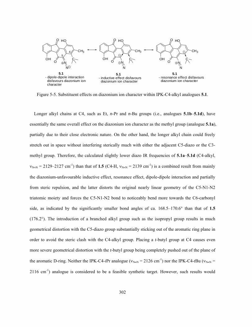
302
O
OHO N
HO
CH3
N
45
C
O
OHO N
HO
CH3
N
C
5.1- inductive effect disfavours
diazonium ion character
O
OHO N
HO
CH3
N
C
5.1- resonance effect disfavours
diazonium ion character
5.1- dipole-dipole interactiondisfavours diazonium ioncharacter
Figure 5-5. Substituent effects on diazonium ion character within IPK-C4-alkyl analogues 5.1.
Longer alkyl chains at C4, such as Et, n-Pr and n-Bu groups (i.e., analogues 5.1b–5.1d), have
essentially the same overall effect on the diazonium ion character as the methyl group (analogue 5.1a),
partially due to their close electronic nature. On the other hand, the longer alkyl chain could freely
stretch out in space without interfering sterically much with either the adjacent C5-diazo or the C3-
methyl group. Therefore, the calculated slightly lower diazo IR frequencies of 5.1a–5.1d (C4-alkyl,
NN = 2129–2127 cm-1) than that of 1.5 (C4-H, NN = 2139 cm-1) is a combined result from mainly
the diazonium-unfavourable inductive effect, resonance effect, dipole-dipole interaction and partially
from steric repulsion, and the latter distorts the original nearly linear geometry of the C5-N1-N2
triatomic moiety and forces the C5-N1-N2 bond to noticeably bend more towards the C6-carbonyl
side, as indicated by the significantly smaller bond angles of ca. 168.5–170.6° than that of 1.5
(176.2°). The introduction of a branched alkyl group such as the isopropyl group results in much
geometrical distortion with the C5-diazo group substantially sticking out of the aromatic ring plane in
order to avoid the steric clash with the C4-alkyl group. Placing a t-butyl group at C4 causes even
more severe geometrical distortion with the t-butyl group being completely pushed out of the plane of
the aromatic D-ring. Neither the IPK-C4-iPr analogue (NN = 2126 cm-1) nor the IPK-C4-tBu (NN =
2116 cm-1) analogue is considered to be a feasible synthetic target. However, such results would

303
suggest that steric repulsion alone from the C4-substituent towards the C5-diazo group would
decrease the diazonium ion character, but only in a very limited manner.
Even though the IPK-C4-alkyl substituted analogues 5.1a–5.1d are predicted to have lower
diazonium ion character and hence to be less active than the natural IPK (1.5), such compounds may
serve as useful candidates for mechanistic studies. If the mode-of-action of the kinamycins indeed
involves the nucleophilic diazo-addition mechanism (section 4.2) as the Dmitrienko group has
proposed and (partially) proved, such IPK analogues are expected to be more difficult to undergo the
nucleophilic attack at the C5-diazo group due to the presence of the bulky C4-substituents.
When unsaturated groups such as vinyl, alkynyl and cyano (i.e., sp/sp2-hybridized carbon atoms)
are introduced at the sp2-C4 position (analogues 5.2a–5.2c), a significant increase of the diazo IR
frequency and hence diazonium ion character relative to that of IPK (1.5) is observed (Table 5-3).
Table 5-3. Ab initio MO calculations of IPK-C4-carbon (unsaturated) analogues 5.2a–5.2c.
Structure Optimized geometry NN (cm-1)a dN≡N (Å) <C-N-N(°) O
OHO N
HO
CH3
N
CH2CH3
5.1b
2129
1.103
168.7
O
OHO N
HO
CH3
N 5.2a
2159
1.101
171.5
O
OHO N
HO
CH3
N 5.2b
2180
1.099
169.8
O
OHO N
HO
CH3
N
CN
5.2c
2189
(C≡N: 2254)
1.098
(dC≡N:1.149)
169.8
a. Scaled by a scaling factor of 0.8929.

304
The vinyl substituent (-CH=CH2) at the C4 position as in the optimized conformation of 5.2a is
found to be oriented with the plane of the C=C bond nearly orthogonal to the aromatic ring plane
(Table 5-3 and Figure 5-6). Thus, resonance delocalization of the C5 negative charge by the vinyl
group is expected to be minimal, since coplanarity of the C=C bond with the aromatic system is not
allowed by steric factors (Figure 5-6). The sp2 carbon of the vinyl group would be (slightly) more
inductively electron-withdrawing when compared with the sp3 carbon in 5.1b, and hence (slightly) in
favour of a higher diazonium ion character than 5.1b. However, the favourable influence on
diazonium ion character from either the inductive effect or the dipole-dipole interaction should be
very small, since the C4-C bond within 5.2a is not very polar (dipole ~ 0 as both carbons are sp2-
hybridized). Therefore, in order to explain the substantially higher diazo IR frequency of 5.2a (C4-
CH=CH2, NN = 2159 cm-1) than that of not only 5.1b (C4-CH2CH3, NN = 2129 cm-1) but also 1.5
(C4-H, NN = 2139 cm-1), an intramolecular and through-space interaction between the C4-vinyl
group and the C5-diazo moiety seems to be the only other option. This possible vinyl-diazo
interaction within 5.2a is geometrically allowed by the space proximity of its C4-vinyl group to the
C5-diazo group (Figure 5-6), since the closest distance between them (dC1’•••N1 = 2.771 Å) is much
shorter than the sum of the corresponding vdW radii (rN = 1.55 Å, rC·= 1.70 Å). More importantly, the
vinyl-diazo interaction would also be greatly favoured by the electronic nature of the two
participating groups, as the vinyl moiety is fairly rich in π electron density while the diazo group is
somewhat electron deficient. The reason(s) why such vinyl-diazo interaction would enhance the
diazonium ion character, however, is not very clear at first glance.

305
5.2a- essentially no inductive effect or dipole-dipole
interaction in favour of diazonium ion character- vinyl-diazo interaction favours diazonium
ion character (see optimized geometry below)
O
OHO N
HO
CH3
N
CH
CH2
O
OHO N
HO
CH3
N
CH
CH2
O
OHO N
HO
CH3
N
C R'
R
5.2a(R = CH2
-; R' = H; R = H; R' = CH2-)
- orthogonal geometry of C=C bond does notallow such resonance forms that are in favourof higher diazonium ion character
X
N1
N2C1' C2'
5.2a partial optimized geometry(Note: AB-rings are omitted for clarity)
dN1...C2' = 3.182 Å, dN2...C2' = 3.411 ÅdN1...C1' = 2.771 Å, dN2...C1' = 3.180 Å
Figure 5-6. Substituent effects on diazonium ion character within IPK-C4-vinyl analogue 5.2a.
The increase in diazonium ion character is much more pronounced for the IPK-C4-acetylenic
structure 5.2b (C4-C≡CH, NN = 2180 cm-1). In this case, the slightly higher electronegativity of the
sp-hybridized alkynyl carbon than that of the sp2-hybridized C4-carbon would introduce some
inductive effect and dipole-dipole interaction (C4(sp2)→C(sp)/C5←N1), both in favour of a higher
diazonium ion character. At the same time, delocalization of electron density onto the acetylenic
linkage to increase the diazonium ion character also becomes feasible (Figure 5-7). The calculated
bond length of 1.197 Å for the carbon-carbon triple bond within 5.2b is significantly longer (more
double-bond like) than the typical bond length of ca. 1.18 Å for typical alkynes, consistent with the
prediction of resonance delocalization of the C5 partial negative charge favouring the diazonium ion
character. In addition, some through-space interaction is also possible between the C4-alkynyl and the

306
C5-diazo groups, similar to the previous case of 5.2a. It is expected that, due to the higher electron
density on the C4-alkyne (than the C4-alkene of 5.2a) and the even closer distances between the two
linear groups that are now coplanar (Figure 5-7), the acetylene-diazo interaction within 5.2b should
be stronger than the vinyl-diazo interaction within 5.2a. Switching from the C4-vinyl (5.2a) to C4-
alkynyl (5.2b) group applies positive influence on all the diazonium-affecting interactions (i.e.,
inductive effect, resonance effect, dipole-dipole interaction and through-space C4/C5 interaction),
consistent with the substantial increase of diazo IR frequency from 5.2a (NN = 2159 cm-1) to 5.2b
(NN = 2180 cm-1).
O
OHO N
HO
CH3
N
45
CH
O
OHO N
HO
CH3
NCH
O
OHO N
HO
CH3
NCH
O
OHO N
HO
CH3
N
CCH
5.2b- dipole-dipole interaction favours
diazonium ion character- acetylene-diazo interaction favours
diaoznium ion character(see optimized geometry below)
5.2b- inductive effect favours
diazonium ion character
5.2b- resonance effect favoursdiazonium ion character
N1N2
C1'C2'
5.2b partial optimized geometry(Note: AB-rings are omitted for clarity)
dN1...C2' = 3.207 Å, dN2...C2' = 3.252 Å
dN1...C1' = 2.757 Å, dN2...C1' = 3.207 Å
Figure 5-7. Substituent effects on diazonium ion character within IPK-C4-acetylene analogue 5.2b.
The diazonium ion character increases even more with a cyano substituent at C4 as in structure
5.2c, which possesses the highest diazo IR frequency of 2189 cm-1 among this series of analogues.

307
Qualitatively, this is consistent with the strong inductive and resonance effects expected from the
cyano group to enhance the diazonium ion character (Figure 5-8). The latter effect is evident from the
calculated C≡N bond length of 1.149 Å, which is longer than the typical cyano bond length of ca.
1.14 Å as a result of such resonance contribution. In addition, the strong electron-withdrawing
capability of the cyano group would cause a highly polar C4-C bond that benefits a strong dipole-
dipole interaction (C4(sp2)→C(sp3)/C5←N1) to enhance the diazonium ion character (Figure 5-8).
Finally, a through-space interaction between the cyano and diazo groups in favour of a higher
diazonium ion character that is very similar to such interaction between the alkynyl and the diazo
moiety within 5.2b, is also applicable in the case of 5.2c (Figure 5-8). Since the sp3-hybridized C1’
within 5.2b (C4-C≡N) is expected to be significantly lower in electron density than that of 5.2a (C4-
C≡CH), the cyano-diazo interaction is likely to be weaker than the acetylene-diazo interaction.
O
OHO N
HO
CH3
N
45
N
O
OHO N
HO
CH3
NN
O
OHO N
HO
CH3
NN
O
OHO N
HO
CH3
N
CN
5.2c- dipole-dipole interaction favoursdiazonium ion character
- cyano-diazo interaction favoursdiazonium ion character(see optimized geometry below)
5.2c- inductive effect favours
diazonium ion character
5.2c- resonance effect favoursdiazonium ion character
N1N2
C1'
N1'
5.2c partial optimized geometry(Note: AB-rings are omitted for clarity)
dN1...N1' = 3.166 Å, dN2...C2' = 3.113 Å
dN1...C1' = 2.735 Å, dN2...C1' = 3.186 Å
Figure 5-8. Substituent effects on diazonium ion character within IPK-C4-cyano analogue 5.2c.

308
Interestingly, despite the much stronger electronegativity of the -C≡N group than that of the -
C≡CH group, analogue 5.2c (C4-C≡N, NN = 2189 cm-1) is only slightly higher in diazonium ion
character than 5.2b (C4-C≡CH, NN = 2180 cm-1). This seems to suggest that the inductive effect,
dipole-dipole interaction and resonance effect, which would all enhance the diazonium ion character
when the C4-substituent is electron-withdrawing, are likely to play a less influencing role on the
diazonium ion character than the intramolecular through-space interaction between the C4-π-
substituent and the C5-diazo group.
5.2.2 MO Calculations of IPK-C4-halogen Analogues
When halogens such as F, Cl and Br are present at the C4 position (analogues 5.3a–5.3c, Table 5-
4), the corresponding diazonium ion characters of such analogues are also greatly enhanced to
approximately the same extent as those IPK-C4-carbon (unsaturated) analogues such as 5.2b (C4-
C≡CH) and 5.2c (C4-C≡N). Within this series of analogues, the diazonium ion character decreases
slightly with decreasing electronegativity of the halogen involved.

309
Table 5-4. Ab initio MO calculations of IPK-C4-halogen analogues 5.3a–5.3c.*
Structure Optimized geometry NN (cm-1)a dN≡N (Å) <C-N-N(°) O
OHO N
HO
CH3
N 1.5
2139
1.103
176.2
O
OHO N
HO
CH3
N
F
5.3a
2193
1.09837
174.6
O
OHO N
HO
CH3
N
Cl
5.3b
2188
1.09839
169.0
O
OHO N
HO
CH3
N
Br
5.3c
2180
1.099
167.3
* I (iodine) was not examined since it is not covered by the basis set of 6-31G used for MO calculations; a.
Scaled by a scaling factor of 0.8929.
The observed influence and tendency of diazonium ion character can be reasonably explained by
the strong electron-withdrawing capability of the halogen, which decreases with the increasing atomic
number, leading to inductive, resonance and dipole-dipole interactions all favouring a higher
diazonium ion character than the unsubstituted parent compound IPK (1.5). In addition, the observed
clear tendency of decreasing bond angle of C5-N1-N2 in the order of 5.3a > 5.3b > 5.3c is consistent
with the increasing size of the C4-halogen atom in the order of F < Cl < Br and hence increasing
steric interaction.
Interestingly, the diazonium ion character of 5.3a (C4-F, NN = 2193 cm-1) is only modestly higher
than the cases of 5.3b (C4-Cl, NN = 2188 cm-1) and 5.3c (C4-Br, NN = 2180 cm-1), despite the

310
substantially higher electronegativity of the fluorine atom. One possibility* is that, when compared
with the previous IPK-C4-aliphatic analogues (5.1a–5.1d and 5.2a–5.2c), the enhancement of
diazonium ion character in the IPK-C4-halogen compounds may rely more significantly on a
favourable dipole-dipole (C4→X/C5←N1) interaction due to the highly polarized C4-halogen bond.
The C-F bond has more covalent character than the C-Cl and C-Br bonds, and thus the C-Cl and C-Br
bond have higher bond polarity. Therefore, compared to the C4-Cl and C4-Br, the C4-F substituent
would enhance the corresponding diazonium ion character significantly more from the inductive and
resonance effects but much less from the dipole-dipole interaction. The combined overall result is
then the observed (only) slightly higher diazo IR frequency for 5.3a (C4-F) than that of 5.3b (C4-Cl)
and 5.3c (C4-Br).
5.2.3 MO Calculations of IPK-C4-nitrogen Analogues
When some nitrogen-based substituents are introduced at the C4 position of IPK (1.5), the
calculated diazo IR frequencies of such analogues are overall much higher than the parent compound
with some more interesting discoveries (Table 5-5).
* Later studies indicate that there is another possibility to explain the rather small difference in diazo IR frequency for the IPK-C4-
halogen analogues, which relies more on the very likely (halogen) lone pair-diazo interaction. See section 5.2.3 for detailed discussion.
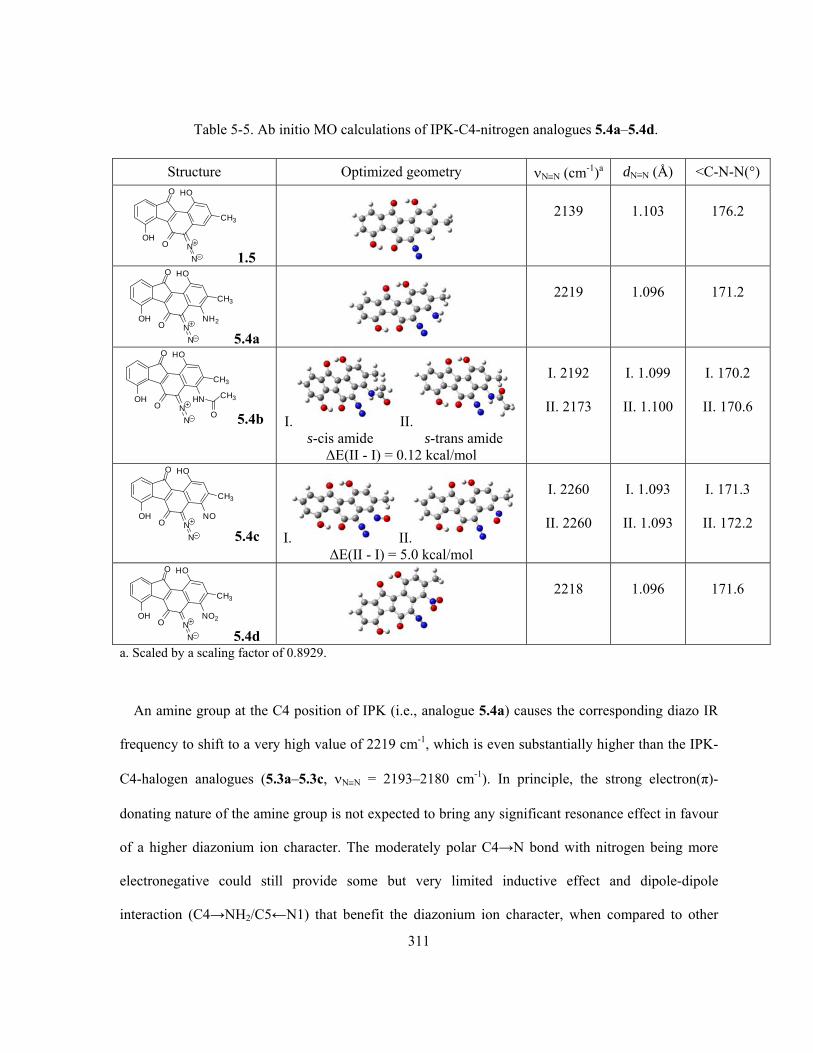
311
Table 5-5. Ab initio MO calculations of IPK-C4-nitrogen analogues 5.4a–5.4d.
Structure Optimized geometry NN (cm-1)a dN≡N (Å) <C-N-N(°) O
OHO N
HO
CH3
N 1.5
2139
1.103
176.2
O
OHO N
HO
CH3
N
NH2
5.4a
2219
1.096
171.2
O
OHO N
HO
CH3
N
HN
O
CH3
5.4b I. II. s-cis amide s-trans amide
ΔE(II - I) = 0.12 kcal/mol
I. 2192
II. 2173
I. 1.099
II. 1.100
I. 170.2
II. 170.6
O
OHO N
HO
CH3
N
NO
5.4c I. II. ΔE(II - I) = 5.0 kcal/mol
I. 2260
II. 2260
I. 1.093
II. 1.093
I. 171.3
II. 172.2
O
OHO N
HO
CH3
N
NO2
5.4d
2218
1.096
171.6
a. Scaled by a scaling factor of 0.8929.
An amine group at the C4 position of IPK (i.e., analogue 5.4a) causes the corresponding diazo IR
frequency to shift to a very high value of 2219 cm-1, which is even substantially higher than the IPK-
C4-halogen analogues (5.3a–5.3c, NN = 2193–2180 cm-1). In principle, the strong electron(π)-
donating nature of the amine group is not expected to bring any significant resonance effect in favour
of a higher diazonium ion character. The moderately polar C4→N bond with nitrogen being more
electronegative could still provide some but very limited inductive effect and dipole-dipole
interaction (C4→NH2/C5←N1) that benefit the diazonium ion character, when compared to other
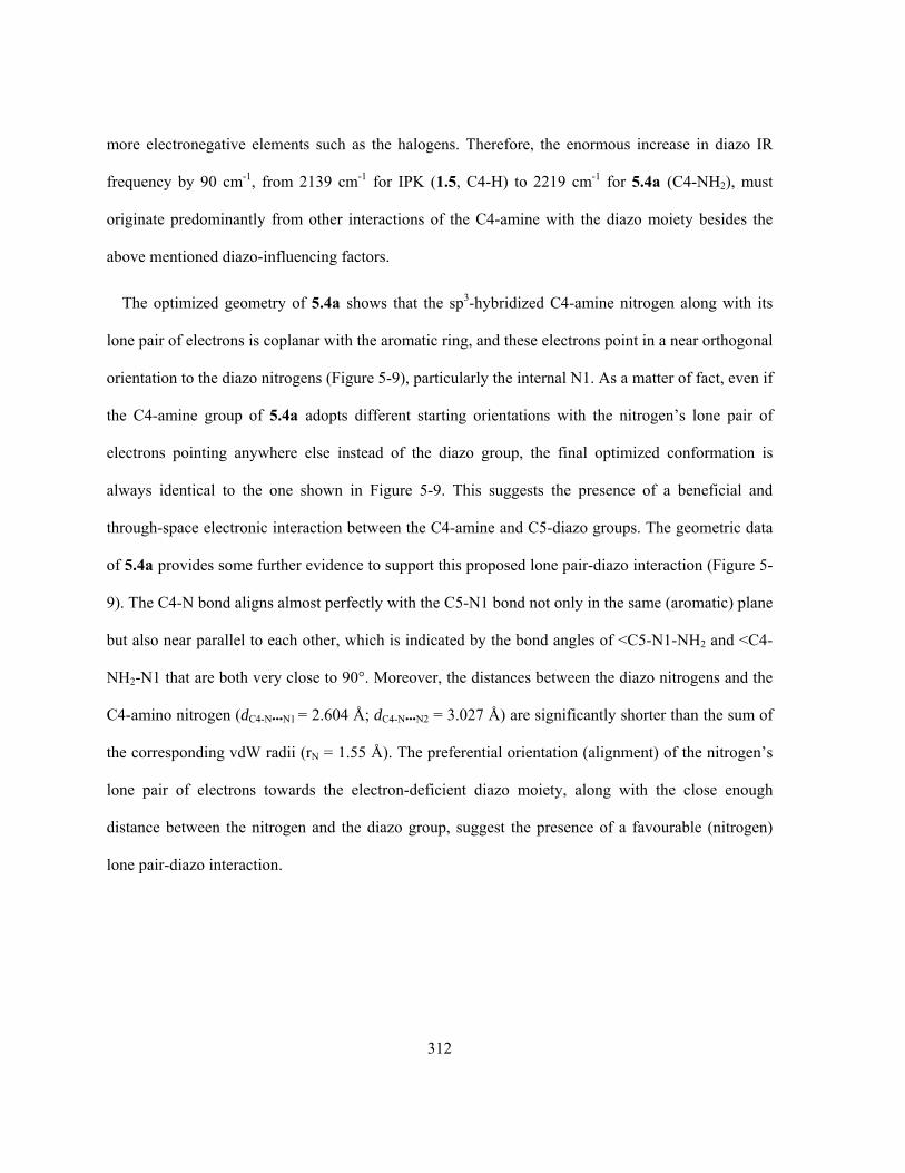
312
more electronegative elements such as the halogens. Therefore, the enormous increase in diazo IR
frequency by 90 cm-1, from 2139 cm-1 for IPK (1.5, C4-H) to 2219 cm-1 for 5.4a (C4-NH2), must
originate predominantly from other interactions of the C4-amine with the diazo moiety besides the
above mentioned diazo-influencing factors.
The optimized geometry of 5.4a shows that the sp3-hybridized C4-amine nitrogen along with its
lone pair of electrons is coplanar with the aromatic ring, and these electrons point in a near orthogonal
orientation to the diazo nitrogens (Figure 5-9), particularly the internal N1. As a matter of fact, even if
the C4-amine group of 5.4a adopts different starting orientations with the nitrogen’s lone pair of
electrons pointing anywhere else instead of the diazo group, the final optimized conformation is
always identical to the one shown in Figure 5-9. This suggests the presence of a beneficial and
through-space electronic interaction between the C4-amine and C5-diazo groups. The geometric data
of 5.4a provides some further evidence to support this proposed lone pair-diazo interaction (Figure 5-
9). The C4-N bond aligns almost perfectly with the C5-N1 bond not only in the same (aromatic) plane
but also near parallel to each other, which is indicated by the bond angles of <C5-N1-NH2 and <C4-
NH2-N1 that are both very close to 90°. Moreover, the distances between the diazo nitrogens and the
C4-amino nitrogen (dC4-N•••N1 = 2.604 Å; dC4-N•••N2 = 3.027 Å) are significantly shorter than the sum of
the corresponding vdW radii (rN = 1.55 Å). The preferential orientation (alignment) of the nitrogen’s
lone pair of electrons towards the electron-deficient diazo moiety, along with the close enough
distance between the nitrogen and the diazo group, suggest the presence of a favourable (nitrogen)
lone pair-diazo interaction.

313
N
N
NH2
CH3
1.402 Å1.3
52Å
1.4
28
Å
1.447 Å
88.6o
86.9o
2.604 Å
3.027 Å
5.4a Optimized geometry withnitrogen (lone pair)-diazo interaction
Figure 5-9. Optimized geometry of IPK-C4-NH2 analogue 5.4a.
In addition, the optimized conformation of 5.4a was further subjected to some single point energy
(SPE) calculations, in which the entire optimized conformation was kept unaltered except that the
dihedral angle between the C4-amine and the aromatic ring plane was manually set and fixed at 90°,
180° and 270° that deviates from the optimized C4-amine orientation (Figure 5-10). Such
manipulation would force the amine-nitrogen’s lone pair of electrons to point away and stay far
enough from the diazo group, so that the lone pair-diazo interaction should either decrease
significantly or even totally vanish. It is found that the optimized conformation of 5.4a having the
nitrogen lone pair-diazo interaction is much more stable than all the rest, which lack such interaction,
by at least 8.3 kcal/mol. Therefore, the energy contribution associated with this amine lone pair-diazo
interaction should be slightly less than 8.3 kcal/mol, since the optimized conformation of 5.4a with
the 180°-rotated C4-amine (Figure 5-10) might be accompanied with some additional but likely small
steric repulsion due to the close distance between the two amine-hydrogens and the diazo group.

314
5.4a Optimized geometry withnitrogen (lone pair)-diazo interaction
E = 14.7 kcal/mol
E = 8.3 kcal/mol
E =14.7 kcal/mol
C4-NH2 rotate 90o clockwiseC4-NH2 rotate 180o clockwise
C4-NH2 rotate 270o clockwise
Note: All conformations are entirely identical except their C4-amine orientations.
Figure 5-10. SPE calculations of IPK-C4-NH2 analogue 5.4a.
In fact, the observation that the C4-amine acts as an electron donor and interacts with the C5-diazo
group is consistent with and resembles some similarity to two previous discoveries. First, within 1.5
and 3.236 (section 5.1), the C6 carbonyl oxygen with available lone pair of electrons does interact
with the C5-diazo group through space and induce a preferential diazo bending towards the carbonyl
oxygen. Second, in the case of 5.2a–c (section 5.2.1), through-space interaction between the C4-
vinyl/acetylenic/cyano and C5-diazo group is considered an important factor to be responsible for the
corresponding (high) diazonium ion character, while such C4-π-substituents can also be regarded as
an electron donor. The nature of this electron donor-diazo interaction is intriguing but not intuitively
obvious (see section 5.3 for further discussion). Such an intramolecular lone pair-diazo interaction
does, however, lead to a very strongly increased diazonium ion character.
In addition, later studies that are to be discussed next further confirmed the common presence of
such lone pair-diazo interaction and its dramatic capability to enhance the diazonium ion character.

315
Exploring the real physical nature of such electron donor-diazo interaction would be of at least
academic interest by itself (section 5.3). On the other hand, and probably more importantly from a
practical point of view, this finding may be very useful in the design of diazobenzo[a]fluorene type of
kinamycin analogues. Apparently, the introduction of an appropriate electron donor at the C4 position
is a powerful yet simple strategy to achieve a high diazonium ion character, which may lead to
structurally simpler diazobenzo[a]fluorene analogues with comparable or even higher bioactivity than
the natural IPK (1.5).
Moreover, correlation of this finding of lone pair-diazo interaction with the IPK-C4-halogen
analogues 5.3a–c (section 5.2.2) would suggest that, the calculated high diazonium ion character of
these IPK-C4-halogen analogues are very likely to also have some contribution from the halogen’s
lone pair electrons interacting with the diazo group. The availability of the multiple lone pairs of
electrons on the C4-halogen, along with the halogen’s close distance to the diazo group (Figure 5-11)
that is significantly shorter than the sum of the corresponding vdW radii (rF = 1.47 Å, rCl = 1.75 Å, rBr
= 1.85 Å and rN = 1.55 Å), does permit such beneficial halogen lone pair-diazo interaction
electronically and geometrically. In addition, this newly discovered interaction offers a second
explanation to the rather small difference in diazo IR frequency for 5.3a–c (section 5.2.2), which does
not match well with the significant difference in the halogens’ electronegativity. Previously, it was
presumed that the dipole-dipole interaction may play an important role that is significant than other
interactions in affecting the diazonium ion character of 5.3a–c (section 5.2.2), so that the less polar
C4-F bond than C4-Cl and C4-Br bonds was thought to be responsible for its less-than-expected
increase (based on electronegativity) in diazo IR frequency. Alternatively, if the halogen lone pair-
diazo interaction to enhance the diazonium ion character plays the important role in the case of 5.3a–c,
then the very high electronegativity of F is expected to make the fluorine to maintain its lone pair of
electrons much better than Cl and Br and hence a smaller increase in diazonium ion character from its
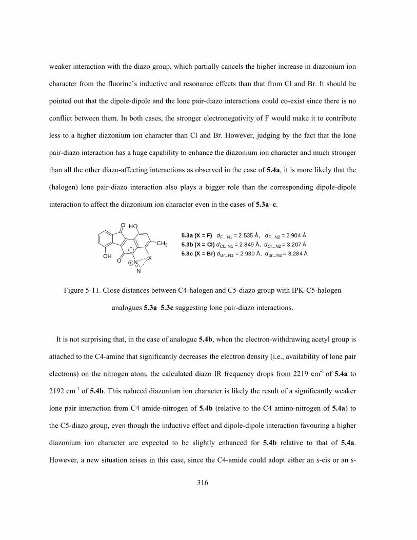
316
weaker interaction with the diazo group, which partially cancels the higher increase in diazonium ion
character from the fluorine’s inductive and resonance effects than that from Cl and Br. It should be
pointed out that the dipole-dipole and the lone pair-diazo interactions could co-exist since there is no
conflict between them. In both cases, the stronger electronegativity of F would make it to contribute
less to a higher diazonium ion character than Cl and Br. However, judging by the fact that the lone
pair-diazo interaction has a huge capability to enhance the diazonium ion character and much stronger
than all the other diazo-affecting interactions as observed in the case of 5.4a, it is more likely that the
(halogen) lone pair-diazo interaction also plays a bigger role than the corresponding dipole-dipole
interaction to affect the diazonium ion character even in the cases of 5.3a–c.
O
OHO N
HO
CH3
N
X
5.3b (X = Cl) dCl...N1 = 2.849 Å, dCl...N2 = 3.207 Å
5.3a (X = F) dF...N1 = 2.535 Å, dF...N2 = 2.904 Å
5.3c (X = Br) dBr...N1 = 2.930 Å, dBr...N2 = 3.284 Å
Figure 5-11. Close distances between C4-halogen and C5-diazo group with IPK-C5-halogen
analogues 5.3a–5.3c suggesting lone pair-diazo interactions.
It is not surprising that, in the case of analogue 5.4b, when the electron-withdrawing acetyl group is
attached to the C4-amine that significantly decreases the electron density (i.e., availability of lone pair
electrons) on the nitrogen atom, the calculated diazo IR frequency drops from 2219 cm-1 of 5.4a to
2192 cm-1 of 5.4b. This reduced diazonium ion character is likely the result of a significantly weaker
lone pair interaction from C4 amide-nitrogen of 5.4b (relative to the C4 amino-nitrogen of 5.4a) to
the C5-diazo group, even though the inductive effect and dipole-dipole interaction favouring a higher
diazonium ion character are expected to be slightly enhanced for 5.4b relative to that of 5.4a.
However, a new situation arises in this case, since the C4-amide could adopt either an s-cis or an s-
317
trans conformation that are interconvertible upon a simple rotation of the acetyl group (Table 5-5).
Despite the very small energy difference of only 0.12 kcal/mol between these two possible
conformations of 5.4b, the corresponding diazo IR frequency of the s-trans species (conformer II,
NN = 2173 cm-1) is significantly lower than the s-cis species (conformer I, NN = 2192 cm-1).
Apparently the diazonium ion character is very sensitive to the change in the surrounding electronic
environment, even though such variation may not cause any noticeable alteration of the molecule’s
energy or steric congestion.
The calculations on IPK-C4-nitroso analogue 5.4c present some other interesting results and two
conformers are found as energy minima (Table 5-5). Conformer I, in which the nitroso-nitrogen is
close to the diazo group and the nitroso group is coplanar with the aromatic ring, is lower in energy
by 5 kcal/mol than conformer II, in which the nitroso-oxygen is close to the diazo group and the
nitroso group is skewed to the aromatic ring (Figure 5-12). There is no doubt that the fairly strong
electron-withdrawing capability of the nitroso group would introduce inductive, resonance and
dipole-dipole interactions in favour of a (significantly) higher diazonium ion character than IPK (1.5).
It should be noted that the resonance contribution to delocalize the C5 partial negative charge onto the
nitroso group is expected to be less significant in conformer II than I, which is due to the skewed
geometry of the nitroso group towards the aromatic ring (a torsion of ca. 20º) within the former
conformation. The enormous increase of 120 cm-1 for the diazo IR frequency (5.4c vs 1.5), however,
is well beyond the known influencing capability of the several electronic effects mentioned above. In
the case of 5.4c, the presence of some additional and very strong interactions that enhance and
dominate the influence on the diazonium ion character is mandatory. This concern makes even more
sense when considering the later result that, introducing the stronger electron-withdrawing nitro group
at C4 (i.e., analogue 5.4d, to be discussed next) in fact induces less increase in diazonium ion
character than the nitroso group. Once again the required additional interaction to dramatically

318
increase the diazonium ion character seems to be a through-space interaction from the nitroso group
towards the diazo group, which is clearly different in the two conformers (Figure 5-12).
Figure 5-12. Lone pair-diazo interactions within the two conformers of IPK-C4-nitroso analogue 5.4c.
Within conformer I of 5.4c (Figure 5-12), a strong lone pair effect from the sp2-hybridized nitroso-
nitrogen (C4-N=O) towards the diazo moiety is evident from the following two observations. First,
the nitroso-nitrogen stays in very close distance from the two diazo nitrogens (dO=N•••N1 = 2.542 Å;
dO=N···N2 = 2.996 Å), which are even significantly shorter than such distances observed for 5.4a (C4-
NH2, dC4-N···N1 = 2.604 Å; dC4-N···N2 = 3.027 Å) that is found to already have strong (nitrogen) lone pair-
diazo interactions. Second, the lone pair of electrons on the nitroso-nitrogen does point to the two
diazo nitrogens in an orientation that looks suitable for some orbital interactions. For conformer II of
5.4c (Figure 5-12), rotation of the C4-nitroso group (from conformer I) leads to a lack of nitroso-
nitrogen lone pair-diazo interaction, which is then compensated by a strong through-space nitroso-
oxygen lone pair-diazo interaction. The close distance between the nitroso-oxygen and the diazo
moiety within conformer II (dN=O•••N1 = 2.434 Å; dN=O···N2 = 2.720 Å), which is also much shorter than
the sum of the corresponding vdW radii (rO = 1.52 Å, rN = 1.55 Å), not only makes the oxygen lone
pair-diazo interaction feasible but also explains the higher energy associated with this conformation

319
(i.e., from steric repulsion between nitroso-oxygen and the diazo group). Interestingly, the predicted
diazonium ion character of the two conformers, as measured by the diazo IR stretching frequency and
N≡N bond length, are coincidentally identical. Yet it might be argued that the overall strength of
interactions from the nitroso-nitrogen within conformer I and nitroso-oxygen within conformer II
towards the diazo group are fortuitously comparable. This coincidentally identical effect is also
consistent with a qualitative analysis that the nitroso-nitrogen is a better electron donor but interacting
at a longer distance with the diazo moiety than the nitroso-oxygen, both of which are sp2-hydridized
with oxygen being more electronegative.
Although the nitro group is one of the strongest electron-withdrawing substituents available, its
presence at the C4 position of IPK (i.e., analogue 5.4d) has less influence on the diazonium ion
character than does the nitroso group. The diazo IR frequency of 5.4d (C4-NO2, NN = 2218 cm-1) is
still much higher than IPK (C4-H, NN = 2139 cm-1) as expected, but the significantly lower effect of
the nitro group on diazonium ion character when compared with the nitroso group (5.4c, C4-NO, NN
= 2260 cm-1) was at first surprising. Consideration of the various interactions that might influence the
diazonium ion character does provide a possible rationalization of the result. On one hand, one would
predict that the nitro group would have a stronger inductive and dipole-dipole interactions favouring
higher diazonium ion character than the nitroso group. On the other hand, the nitroso group within
5.4c is able to interact more favourably in a resonance fashion because the plane of the C4-N=O
(within either conformer) is much less skewed from planarity than the C4-NO2 plane within 5.4d,
which possesses a torsion angle of ca. 50º relative to the corresponding aromatic ring plane (Figure 5-
13). Also, the through-space interaction with the diazo group of 5.4d from the lone pair of electrons
of one of two nitro-oxygen atoms is evident (Figure 5-13), as the nitro plane tilts so that one nitro-
oxygen does get close enough to the diazo moiety. Since the electron density on the oxygen atom of
the -NO2 group is lower than on the oxygen of the –N=O group, and the orientation of the oxygen’s

320
lone pair of electrons towards the diazo moiety within 5.4d (Figure 5-13) to achieve the lone pair-
diazo interaction seems to be less perfect than 5.4c (Figure 5-12), the through-space interaction to
enhance the diazonium ion character is expected to be significantly weaker in the case of 5.4d. Thus,
the difference between the overall effect of the -NO and -NO2 groups on diazonium ion character may
be an indication of the importance of both resonance stabilization and through-space lone pair-diazo
interaction.
5.4d partial optimized geometry(Note: AB-ring are omitted for clarity)
dN-O = 1.234 Å
dN-O = 1.223 ÅdN1...O = 2.569 Å
dN2...O = 2.858 Å
N1N2
Figure 5-13. Optimized (partial) geometry of the IPK-C4-nitro analogue 5.4d.
There is one more interesting discovery with 5.4d that is worthy of discussion, which seems to be
further supportive to the presence of a through-space interaction from one of the nitro-oxygens to the
diazo moiety. The resonance forms of the nitro group would suggest that the two oxygen atoms are
equivalent (Figure 5-14), therefore the geometry of the nitro group (especially the two N-O bonds) is
not affected significantly by the nature of the moiety to which the nitro group is attached.372 As
indicated by examples in a literature review372 and structural survey on simple aromatic nitro-
substituted compounds (this work) within CSD (version 5.30), bond lengths of the two N-O bonds
within the nitro group are in general identical or at least very close to each other. It is noticed that, in
the case of 5.4d, the two N-O bond lengths are in fact quite different (Figure 5-14). The N-O bond
(dN-O = 1.234 Å), which is closer to the diazo group with its oxygen that is presumed to interact with
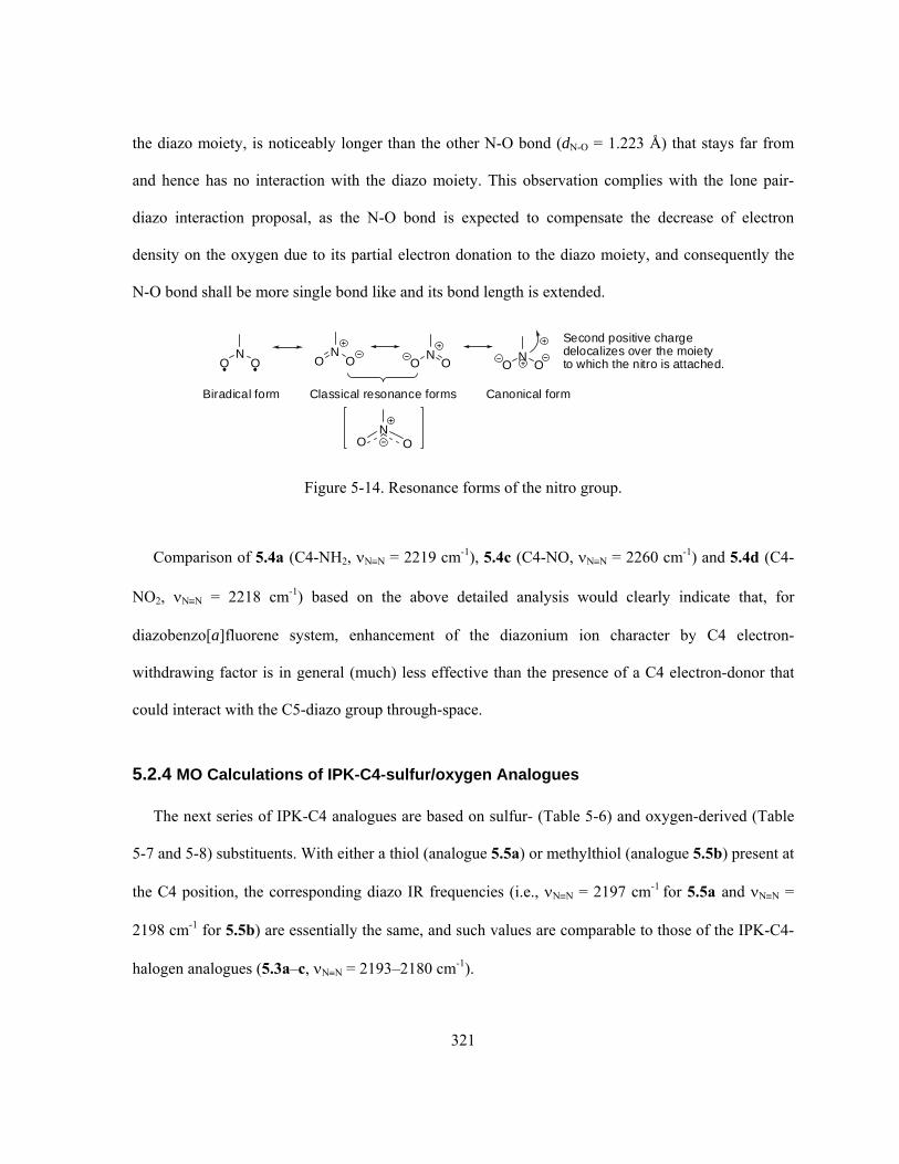
321
the diazo moiety, is noticeably longer than the other N-O bond (dN-O = 1.223 Å) that stays far from
and hence has no interaction with the diazo moiety. This observation complies with the lone pair-
diazo interaction proposal, as the N-O bond is expected to compensate the decrease of electron
density on the oxygen due to its partial electron donation to the diazo moiety, and consequently the
N-O bond shall be more single bond like and its bond length is extended.
NOO N
OO
NOO
NOO
Classical resonance forms Canonical form
NOO
Biradical form
Second positive chargedelocalizes over the moietyto which the nitro is attached.
Figure 5-14. Resonance forms of the nitro group.
Comparison of 5.4a (C4-NH2, NN = 2219 cm-1), 5.4c (C4-NO, NN = 2260 cm-1) and 5.4d (C4-
NO2, NN = 2218 cm-1) based on the above detailed analysis would clearly indicate that, for
diazobenzo[a]fluorene system, enhancement of the diazonium ion character by C4 electron-
withdrawing factor is in general (much) less effective than the presence of a C4 electron-donor that
could interact with the C5-diazo group through-space.
5.2.4 MO Calculations of IPK-C4-sulfur/oxygen Analogues
The next series of IPK-C4 analogues are based on sulfur- (Table 5-6) and oxygen-derived (Table
5-7 and 5-8) substituents. With either a thiol (analogue 5.5a) or methylthiol (analogue 5.5b) present at
the C4 position, the corresponding diazo IR frequencies (i.e., NN = 2197 cm-1 for 5.5a and NN =
2198 cm-1 for 5.5b) are essentially the same, and such values are comparable to those of the IPK-C4-
halogen analogues (5.3a–c, NN = 2193–2180 cm-1).

322
Table 5-6. Ab initio MO calculations of IPK-C4-sulfur analogues 5.5a–b.
Structure Optimized geometry NN (cm-1)a dN≡N (Å) <C-N-N(°) O
OHO N
HO
CH3
N 1.5
2139
1.103
176.2
O
OHO N
HO
CH3
N
SH
5.5a
2197
1.0978
168.8
O
OHO N
HO
CH3
N
SMe
5.5b
2196
1.0979
169.1
a. Scaled by a scaling factor of 0.8929.
Since sulfur is a much weaker electronegative element than the halogens such as fluorine and
chlorine, the observed high level of diazonium ion character of 5.5a (and 5.5b) is likely not a result of
diazonium-favourable inductive, resonance or dipole-dipole interactions. This would suggest the
possibility of the lone pair interaction from sulfur to the diazo to account for the fairly high calculated
diazo IR frequencies. Similar to the previous case of 5.4a (C4-NH2), optimization of both 5.5a (C4-
SH) and 5.5b (C4-SMe) with different starting orientations of the sulfur atom always ends up with
one of the sulfur’s two lone pairs of electrons pointing towards the diazo nitrogens (Figure 5-15).
Such geometries would suggest again the presence of beneficial electronic interactions between the
sulfur lone pair of electrons and the diazo group. Partial donation of a lone pair of electrons to the
diazo group from sulfur would also benefit from its low electronegativity. The high diazonium ion
character of 5.5a and 5.5b is likely the consequence of such lone pair-diazo interaction. This sulfur
lone pair-diazo interaction is further evident from the close distances in between the C4-S and two
diazo nitrogens (Figure 5-15), which are significantly shorter than the sum of the corresponding vdW

323
radii (rN= 1.55 Å, rS= 1.80 Å). The long C4-S bonds in both cases (1.849 Å for 5.5a and 1.842 Å for
5.5b) would align the sulfur atom approximately to the middle point of the N≡N bond (Figure 5-15),
since distances between C5 and the middle point of the N≡N bond are ca. 1.90 Å for both 5.5a and
5.5b. Such alignment would probably allow a stronger interaction between the sulfur (lone pair) and
the terminal N2 atom than the case of 5.4a (C4-NH2), in which the amine aligns mainly with the
middle N1 atom (Figure 5-9) due to the shorter C4-N bond (vs the C4-S bond). Similar geometry was
in fact observed in the previous case of 5.3c (C4-Br), in which the extended C4-Br bond (dC4–Br =
1.945 Å) also aligns the bromine atom approximately to the middle point of the corresponding diazo
moiety, while distance between C5 and the middle point of the N≡N bond is also ca. 1.90 Å for 5.3c.
N
NS
CH3
1.407 Å1.353
Å
1.8
49Å
1.452
Å
2.908 Å
5.5a
3.272 ÅH
N
N
S
CH3
1.408 Å1.3
53Å
1.84
2Å
1.453
Å
2.916 Å
5.5b
3.284Å
CH3
~ 1.90 Å ~ 1.90 Å
Figure 5-15. Optimized geometries of IPK-C4-sulfur analogues 5.5a (C4-SH) and 5.5b (C4-SMe).
When substituents such as –OH (analogue 5.5c) and –OMe (analogue 5.5d) are introduced at the
C4 position (Table 5-7), the corresponding diazo IR frequencies are expected and found indeed to be
higher than those of 5.5a (C4-SH) and 5.5b (C4-SMe), partially due to the much higher
electronegativity of oxygen than sulfur that should strengthen the inductive effect and dipole-dipole

324
interaction favouring a high diazonium ion character. On the other hand, the significantly smaller size
of oxygen than sulfur allows the –OH/–OMe to adopt better orientations than the –SH/–SMe to offer
stronger lone pair-diazo interactions in favour of higher diazonium ion character, even though oxygen
is a weaker electron donor than sulfur.
Table 5-7. Ab initio MO calculations of IPK-C4-oxygen analogues 5.5c–e.
Structure Optimized geometry NN (cm-1)a dN≡N (Å) <C-N-N(°) O
OHO N
HO
CH3
N 1.5
2139
1.103
176.2
O
OHO N
HO
CH3
N
OH
5.5c
2210
1.097
173.5
O
OHO N
HO
CH3
N
OMe
5.5d
2198
1.098
173.6
O
OHO N
HO
CH3
N
O
5.5e
2226
1.095
170.8
a. Scaled by a scaling factor of 0.8929.
Similar to the previous amine/amide (5.4a/5.4b) and thiol/methylthiol (5.5a/5.5b) cases, geometry
optimization of 5.5c and 5.5d also leads to conformations (Table 5-7 and Figure 5-16) with their C4-
oxygen’s lone pair(s) of electrons pointing towards the diazo moiety regardless of the starting
orientations. Once again a lone pair interaction between the C4-oxygen and the C5-diazo group is
evident from the optimized geometries of 5.5c and 5.5b (Figure 5-16), as the distances between the
C4-oxygen and the two diazo nitrogens are significantly shorter than the sum of the corresponding
vdW radii (rN = 1.55 Å, rO = 1.52 Å) in both cases. There are some interesting and noticeable

325
difference between the 5.5c (C4-OH) and 5.5d (C4-OMe) as to how the corresponding lone pair of
electrons of oxygen interact with the diazo moiety. The C4-OH within 5.5c is coplanar with the
aromatic ring in the optimized geometry (Figure 5-16), which makes the oxygen’s two lone pairs of
electrons both intersect and interact with the diazo moiety. Yet the C4-OMe within 5.5d can not make
the oxygen’s both lone pairs of electrons interact with the diazo group simultaneously (Figure 5-16),
due to the steric interaction between the C4-methoxy methyl group and the C3-methyl group that
forces the methoxy group to adopt a conformation orthogonal to the aromatic ring plane. As a result
of this geometry, only one of the two lone pairs of electrons on the methoxy-oxygen could interact
with the diazo moiety. The significantly stronger lone pair-diazo interaction within 5.5c (C4-OH)
than 5.5d (C4-OMe), as determined by the geometry, explains the noticeably lower diazonium ion
character of the latter structure (5.5c, NN = 2210 cm-1; 5.5d, NN = 2198 cm-1).
N
N
O
CH3
1.393 Å1.349
Å
1.3
89Å
1.446 Å
2.498 Å
5.5c
2.894 Å
H
N
N
O
CH3
1.396 Å1.3
47Å
1.39
2Å
1.446
Å
2.549 Å
5.5d
2.940 ÅCH3
Figure 5-16. Optimized geometries of IPK-C4-oxygen analogues 5.5c (C4-OH) and 5.5d (C4-OMe).

326
On the other hand, since methyl is an electron-donating group when compared to hydrogen, the
methoxy-oxygen is higher in electron density than the hydroxy-oxygen. If the C3-methyl group
within 5.5c (C4-OH, C3-Me) and 5.5d (C4-OMe, C3-Me) is replaced by the smallest “substituent” of
hydrogen so that the steric interaction between C3/C4 substituents no longer prohibits the free
rotation of the C4-moiety to achieve a maximum lone pair-diazo interaction, then a higher diazonium
ion character induced by the C4-OMe than C4-OH is expected. In fact, computation of such simpler
analogues (i.e., IPK-C4-OH-C3-H 5.8a and IPK-C4-OMe-C3-H 5.8b, Figure 5-17) did provide
results that are entirely consistent with such a prediction. Without the steric restriction from the
nearby C3 group, the C4-OMe within 5.8b (not including the methyl hydrogens though) is now
coplanar with the aromatic ring, similar to the case of 5.5c (C4-OH, C3-Me) but not possible within
5.5d (C4-OMe, C3-Me) for steric reasons. Similarly, the C4-OH within 5.8a, not surprisingly, adopts
the beneficial coplanarity with the aromatic ring in the even less crowded environment. Such
conformations of 5.8a and 5.8b are capable of providing a maximum (oxygen) lone pair-diazo
interaction in which both lone pairs of electrons on the oxygen participate. The higher electron
density on the methoxy-oxygen within 5.8b than the hydroxy-oxygen within 5.8a does lead to a
slightly higher diazo IR frequency for the former structure as predicted (5.8b, NN = 2213 cm-1; 5.8a,
NN = 2211 cm-1). Stronger lone pair-diazo interaction within 5.8b (C4-OMe, C3-H) than 5.8a (C4-
OH, C3-H) is also evident from the shorter O-N interacting distances that associate with 5.8b (Figure
5-17), which are correlated in an opposite manner (Figure 5-16) in the case of 5.5d (C4-OMe, C3-Me)
and 5.5c (C4-OH, C3-Me).

327
N
N
O
H1.395 Å1.35
0Å
1.3
86Å
1.445 Å
2.510 Å
5.8a
2.900 ÅH
N
N
O
H1.400 Å1.3
50Å
1.38
1Å
1.445 Å
2.489 Å
5.8b
2.893 ÅC
HO
OHN2
O
O
OH
H
HO
OCH3N2
O
O
OH
H
minimalstericinteraction
minimalstericinteraction
1.09679 Å 1.09673 Å
H
H
H
Figure 5-17. Optimized geometries of IPK-C4-OH-C3-H 5.8a and IPK-C4-OMe-C3-H 5.8b.
Similar to the previous work on 5.4a (C4-NH2) as shown in Figure 5-10, SPE calculations were
also performed with 5.5c (C4-OH) in order to estimate the energy contribution from the (oxygen)
lone pair-diazo interaction. Rotation of dihedral angle between the C4-OH group and the aromatic
ring plane by 90°, 180° and 270°, while keeping the rest of the optimized conformation unaltered,
results in noticeable increase in energy (Figure 5-18). An estimated moderate energy contribution of
ca. 4.3 kcal/mol from the (oxygen) lone pair-diazo interaction is obtained when the C4-O is deviated
by either 90° or 270° from the optimized orientation (Figure 5-18), however, one needs to realize that,
within these two conformations, some lone pair-diazo interaction from one of the two lone pairs of
electrons is still feasible due to the geometry. On the other hand, the much higher energy of 26.5
kcal/mol of the conformation having the C4-O deviated by 180° from the optimized orientation

328
(Figure 5-18) is mainly due to the unfavourable steric repulsion between the hydrogen and diazo
moiety even though in this case the beneficial lone pair-diazo interaction completely disappears.
Therefore, the energy contribution from the oxygen lone pair-diazo interaction should be in the range
of 26.5 kcal/mol >> E > 4.3 kcal/mol. Considering the higher electronegativity of oxygen than that of
nitrogen and thus less electron donation and weaker lone pair interaction with the diazo, this
electronic energy from oxygen is likely to be lower than the corresponding value of 5.4a (C4-NH2, ~
8.3 kcal/mol). This lone pair effect would still dominate but probably to a lesser extent to affect the
diazonium ion character in the case of 5.5c (C4-OH) than 5.4a (C4-NH2), which seems to be
consistent with the observed slightly lower diazo IR frequency of 5.5c (NN = 2210 cm-1) than that of
5.4a (NN = 2219 cm-1).
5.5c Optimized geometry- oxygen (lone pairs)-diazo interaction- C4-OH is coplanar with the aromatic plane
E = 4.3 kcal/mol
E = 26.5 kcal/mol
E =4.3 kcal/mol
Note: All conformations are entirely identical except their C4-OH orientations.
C4-OH rotate 90o clockwiseC4-OH rotate 180o clockwise
C4-OH rotate 270o clockwise
Figure 5-18. SPE calculations of IPK-C4-OH analogue 5.5c.

329
The phenolate analogue 5.5e (C4-O-) is in fact a variant of 5.5c (C4-OH) upon simple C4-phenol
deprotonation (ionization). This ionization is possible under the physiologically important pHs (7.4 ±
1) due to the moderate acidity of the phenols, whose typical pKa is about 7–10. The resulting even
higher diazo IR frequency of 5.5e (C4-O-, NN = 2226 cm-1) than the corresponding neutral form 5.5c
(C4-OH, NN = 2210 cm-1) is not surprising by now. The negatively charged C4-O- would definitely
provide neither favourable inductive nor resonance effect to delocalize the C5 negative charge to
enhance the diazonium ion character. Dipole-dipole interaction in favour of a higher diazonium ion
character, however, should still be applicable in this case yet it is expected to be very limited and
weaker than the case of 5.5c. This would once again make the lone pair interaction from the C4-O-
towards the C5-diazo group as the major contributing factor to be responsible for the very high
diazonium ion character of 5.5e. Such lone pair-diazo interaction is further supported by the very
close O-N distances (dC4-O-···N1 = 2.447 Å, dC4-O-···N2 = 2.875 Å) and shall benefit significantly from the
very high electron density on the negative C4-oxygen.
Furthermore, the typical bond lengths for C(sp2)–O and C(sp2)=O are 1.34 Å and 1.21 Å
respectively, and literature X-ray data of model compounds such as 5.9 (CSD Refcode: TALGOD)
and 5.10 (CSD Refcode: MENAQU10) also give similar values (Figure 5-19). Thus, the short C4-O-
bond length of 1.299 Å within 5.5e would indicate that the resonance delocalization of the phenolic
negative charge onto the aromatic D-ring (Figure 5-19) is a significant contribution to the stability of
the anion. Consequently the C4-O bond of 5.5e possesses substantial double-bond character and
hence a short bond length. This result is consistent with a corresponding value from the computed
results of the anion of 5.10 (Figure 5-19), which possesses significant delocalization of the phenolic
negative charge onto the aromatic ring.

330
Figure 5-19. Model compounds and resonance forms of IPK-C4-O- analogue 5.5e.
Interestingly, when electron-withdrawing groups such as acetyl (analogue 5.5f), mesyl (analogue
5.5g) and tosyl (analogue 5.5h) are attached to the C4-oxygen of 5.5c, the corresponding diazo IR
frequencies show some consistency with the electron-withdrawing ability of such ester groups (Table
5-8).
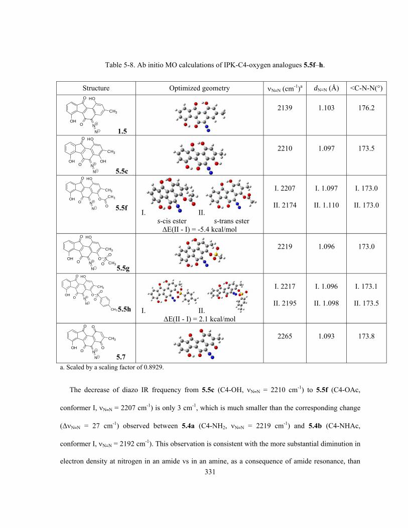
331
Table 5-8. Ab initio MO calculations of IPK-C4-oxygen analogues 5.5f–h.
Structure Optimized geometry NN (cm-1)a dN≡N (Å) <C-N-N(°) O
OHO N
HO
CH3
N 1.5
2139
1.103
176.2
O
OHO N
HO
CH3
N
OH
5.5c
2210
1.097
173.5
O
OHO N
HO
CH3
N
O
O
CH3
5.5f I. II.
s-cis ester s-trans ester ΔE(II - I) = -5.4 kcal/mol
I. 2207
II. 2174
I. 1.097
II. 1.110
I. 173.0
II. 173.0
O
OHO N
HO
CH3
N
O SCH3O
O
5.5g
2219
1.096
173.0
O
OHO N
HO
CH3
N
O SO
O
CH35.5h I. II. ΔE(II - I) = 2.1 kcal/mol
I. 2217
II. 2195
I. 1.096
II. 1.098
I. 173.1
II. 173.5
O
OHO N
O
CH3
N
O
5.7
2265
1.093
173.8
a. Scaled by a scaling factor of 0.8929.
The decrease of diazo IR frequency from 5.5c (C4-OH, NN = 2210 cm-1) to 5.5f (C4-OAc,
conformer I, NN = 2207 cm-1) is only 3 cm-1, which is much smaller than the corresponding change
(ΔNN = 27 cm-1) observed between 5.4a (C4-NH2, NN = 2219 cm-1) and 5.4b (C4-NHAc,
conformer I, NN = 2192 cm-1). This observation is consistent with the more substantial diminution in
electron density at nitrogen in an amide vs in an amine, as a consequence of amide resonance, than

332
that observed at oxygen in an ester vs an alcohol. Not surprisingly, the acetyl ester 5.5f also exists as
either s-cis or s-trans conformers that are quite different in energy (|ΔE| = 5.4 kcal/mol, Table 5-8),
similar to the previous case of 5.4b (C4-NHAc) but its s-cis and s-trans conformers are comparably
stable (|ΔE| = 0.12 kcal/mol, Table 5-5). An even larger difference in diazo IR frequencies between
the two conformers of 5.5f (ΔNN = 33 cm-1) than the case of 5.4b (ΔNN = 19 cm-1) is observed.
There is no good explanation at the moment to rationalize such diazo IR difference between the s-
cis/s-trans conformation for both 5.5f (C4-OAc) and 5.4b (C4-NHAc); however, it is interesting to
notice that in both cases, the s-cis conformer is always associated with a higher diazonium ion
character.
When much stronger electron-withdrawing groups such as mesyl (Ms) and tosyl (Ts) are present
(analogue 5.5g and 5.5h), the (C4-oxygen) lone pair-diazo interaction is expected to decrease more
noticeablely. There are, however, two additional and beneficial influencing factors for the diazonium
ion character to be considered at the same time. The first one, of less importance, is the enhanced
inductive and dipole-dipole interaction from the more electron-withdrawing ester groups (–OMs/–
OTs vs –OH) in favour of a higher diazonium ion character. Second, very likely a (much) stronger
influencing factor, is the common presence of an electron-rich sp2-hybridized sulfonyl-oxygen (O=S-)
within 5.5g (C4-OMs) and 5.5h (C4-OTs), which is also located in close proximity to the diazo group
besides the C4-oxygen but slightly further away (Table 5-8). For example, within the optimized
conformation of 5.5g (C4-OMs), distances between one of the sulfonyl-oxygens and the diazo moiety
(dS=O•••N1 = 2.985 Å; dS=O•••N2 =3.029 Å) are longer than the corresponding values for the C4-
oxygen/diazo (dC4-O•••N1 = 2.637Å; dC4-O•••N2 =3.042 Å) but still shorter than the sum of the
corresponding vdW radii (rO = 1.52 Å, rN = 1.55 Å). Similar to the C4-oxygen, the sulfonyl-oxygen “-
S=O” is also capable of donating partially its lone pair of electrons to the diazo moiety to enhance the
diazonium ion character when geometry allows. The strong lone pair interaction from the C4-X: (X =

333
N, O, S) towards the diazo group due to the close interacting distance can be regarded as a primary
effect, while the further but weaker lone pair interaction from the “-S=O” towards the diazo moiety
(due to the longer interacting distance) is more like a secondary effect. This secondary lone pair-diazo
interaction, along with the enhanced inductive effect and dipole-dipole interaction, apparently could
override the decreasing influence on the diazonium ion character due to the weakening of the primary
lone pair effect from the strong electron-withdrawing –Ms or –Ts group. Therefore, the observed
slightly higher diazo IR frequencies for 5.5g (C4-OMs, NN = 2219 cm-1) and 5.5h (C4-OTs,
conformer I, NN = 2217 cm-1) than that of 5.5c (C4-OH, NN = 2210 cm-1) are the overall results
from the combined influence as discussed above.
In the case of 5.5h (C4-OTs), it is interesting to find a slightly less stable conformer II than
conformer I by ca. 2.1 kcal/mol (Table 5-8 and Figure 5-20). More surprisingly, distances between
the C4-oxygen and the diazo nitrogens within conformer II (dC4-O•••N1 = 2.581 Å, dC4-O•••N2 =2.986 Å)
are shorter than the corresponding values from conformer I (dC4-O•••N1 = 2.632 Å, dC4-O•••N2 = 3.033 Å),
yet the former is significantly lower in diazonium ion character. This observation seems to be
contradictory to the earlier results at first glance, since for the IPK-C4-X: analogues, shorter distances
between C4-X: and diazo nitrogens would (in principle) correspond to stronger lone pair-diazo
interactions and consequently higher diazonium ion character. However, careful examination of the
optimized geometries of both conformers of 5.5h (Figure 5-20) indicates that, the lone pair of
electrons on the sp3-hybridized C4-oxygen within conformer I possesses a much better lone pair-
diazo alignment than conformer II. To achieve a maximum of the beneficial electronic interaction,
alignment of the interacting orbitals (in an appropriate orientation to overlap) should be of the
importance as well as the interaction distance. Therefore, despite the slightly longer C4-oxygen/diazo
distances, conformer I is expected to possess a stronger primary lone pair-diazo interaction in favour
of a higher diazonium ion character due to the right orientation of the lone pair of electrons. In

334
addition, conformer I also has one of its sulfonyl-oxygen “-S=O” to stay close enough to the diazo
moiety to offer additional secondary lone pair-diazo interaction to further enhance the diazonium ion
character, while conformer II totally lacks such interaction from either of its two “-S=O” oxygens due
to their far distance from the diazo group (Figure 5-20). Combination of these two effects might be
the cause of the significant difference in diazonium ion character between the two conformers of 5.5h.
1.399 Å1.348Å
1.41
0Å
1.443 Å
3.033 Å
1.395 Å1.346Å 1.
408
Å
1.445 Å
Figure 5-20. Optimized geometries of the two conformers of IPK-C4-tosylate analogue 5.5h.

335
Strictly speaking, the IPK-D-ring-quinone analogue 5.7 is no longer an IPK-C4-analogue since it
loses the D-ring’s aromaticity when two conjugated carbonyl groups are present at C1 and C4. This
D-ring quinone system presents a strong resonance stabilization effect owing to the presence of the
C1-carbonyl group, which is expected to enhance the diazonium ion character over and above that
induced by strong inductive effect and dipole-dipole interaction from the C4-carbonyl group, as
described for the IPK-C4 analogues previously (Figure 5-21). However, the very high diazo IR
frequency of 5.7 (NN = 2265 cm-1), which is by far the largest value obtained, might suggest the
presence of some other strong interactions to be capable of enhancing the diazonium ion character to
such a high level. This can be traced back again to a strong oxygen lone pair-diazo interaction (Figure
5-21), in which the sp2-hybridized and electron-rich C4 carbonyl oxygen locates close enough to the
diazo moiety (dO•••N1 = 2.579 Å, dO•••N2 = 2.985 Å, cf. rN = 1.55 Å, rO = 1.52 Å) with one of its two
lone pairs of electrons pointing towards the electron-deficient diazo nitrogens.
O
OHO N
O
CH3
N
O
5.7- inductive effect favours
diazonium ion character- dipole-dipole interaction favoursdiazonium ion character
O
OHO N
O
CH3
N
O
5.7- resonance delocalization
of C5 negative charge alsofavours diazonium ion character
N
N
O
CH3
1.487 Å
1.363
Å
1.2
24Å
1.421 Å
2.579 Å
2.98
5Å
Figure 5-21. Substitution effects within IPK-D-ring-quinone analogue 5.7 on diazonium ion character.
The molecule 5.7 is of particular interest not only because of its unusually high diazonium ion
character but also due to its structural simplicity. In principle, 5.7 might be easier to prepare than the
other proposed IPK-C4-analogues. For example, a direct and selective D-ring phenol oxidation of

336
IPK (1.5), which is now readily available through either fermentation or synthesis, is apparently the
easiest way to achieve the goal. Alternatively, modification of the Dmitrienko synthetic procedure of
1.5 (Scheme 3-36, section 3.2.2) is also feasible. Careful selection of appropriate D-ring phenol
protecting group, which should be different from and more easily cleaved than the A-ring phenol
protecting group later on, may provide access to an intermediate having a free D-ring phenol while
the A-ring phenol is still protected as an ether or ester. It should be possible to selectively oxidize the
D-ring of such a compound to the corresponding quinone (as in 5.7), prior to the introduction of the
remaining diazo moiety and deprotection of the A-ring phenol ether or ester.
5.2.5 MO Calculations of IPK-C4-carbonyl Analogues
The last series of IPK-C4-analogues are based on some carbonyl-derived substituents present at the
C4 position of IPK (1.5). Although the results (Table 5-9 and 5-10) may seem a little bit more
complicated than the previous cases, all calculated diazo IR frequencies can be reasonably explained
by using the concepts discussed earlier.

337
Table 5-9. Ab initio MO calculations of IPK-C4-carbonyl analogues 5.6a–c.
Structure Optimized geometry NN (cm-1)a dN≡N (Å) <C-N-N(°) O
OHO N
HO
CH3
N 1.5
2139
1.103
176.2
O
OHO N
HO
CH3
NH
O
5.6a I. II. ΔE(II - I) = 3.6 kcal/mol
I. 2248
II. 2147
I. 1.094
II. 1.102
I. 172.1
II. 171.9
O
OHO N
HO
CH3
NCH3
O
5.6b
2142
1.102
171.4
O
OHO N
HO
CH3
NNH2
O
5.6c I. II. ΔE(II - I) = 0.06 kcal/mol
I. 2186
II. 2173
I. 1.099
II. 1.100
I. 170.8
II. 171.3
a. Scaled by a scaling factor of 0.8929.
With the simplest carbonyl-based substituent, an aldehyde, introduced at the C4 position (analogue
5.6a), two C4-conformers of 5.6a with a significant energy difference of 3.6 kcal/mol are found
through geometry optimization (Table 5-9). The major difference between the two conformers is the
orientation of the C4-aldehyde group, and the geometry of the aldehyde’s C=O double bond of
conformer I resembles the previously observed one for 5.2a (C4-CH=CH2) and 5.4c (C4-NO,
conformer II). Interestingly, the corresponding diazonium ion character is found to be dramatically
affected by the orientation of the aldehyde carbonyl group. When the C=O double bond of the
aldehyde is closer to the diazo moiety as in conformer I (Figure 5-22), the presence of a very strong
lone pair interaction from the carbonyl oxygen with the diazo moiety is indicated by the geometry
data (dHC=O•••N1 = 2.470 Å, dHC=O•••N2 = 2.722 Å), while such an effect is completely absent in the case

338
of conformer II since its aldehyde carbonyl oxygen is too far from the diazo moiety (dHC=O···N1 > 3.68
Å) to interact through-space. The structure of 5.6a would still have inductive and dipole-dipole
interactions from the electron-withdrawing C4-CHO group in favour of a higher diazonium ion
character, and such effects are expected to be very limited but identical for the two conformers.
Contribution to higher diazonium ion character from resonance delocalization is also expected to be
small but still comparable between conformer I and II, since the CH=O plane is skewed to the
corresponding aromatic ring plane in approximately the same dihedral angle within the two
conformers (Figure 5-22). Therefore, the observed enormous difference in diazonium ion character
(ΔNN = 101 cm-1) between these two conformers of 5.6a must originate predominantly from the
different level of (carbonyl oxygen) lone pair-diazo interactions. Another noteworthy observation is
that in the case of conformer I of 5.6a in which the lone pair-diazo interaction is quite obvious, the
interacting lone pair of electrons from oxygen points towards the N1-C5 site rather than the N1-N2
site (Figure 5-22), while all previously observed lone pair-diazo interactions have the interacting lone
pair pointing towards the N1-N2 site.
5.6a conformer I 5.6a conformer II
Note: AB-rings are omitted for clarity.
Figure 5-22. Optimized (partial) geometries of the two conformers of IPK-C4-CHO analogue 5.6a.
In addition, the calculated diazo IR frequency for conformer II of 5.6a (C4-CHO, NN = 2147
cm-1), which has no intramolecular lone pair-diazo interaction to greatly enhance the diazonium ion

339
character, is only slightly higher than the corresponding value of IPK (C4-H, NN = 2139 cm-1). This
small and rather limited increase in diazo IR frequency would clearly suggest (again) that,
contributions towards a higher diazonium ion character by inductive effect, resonance effect and
dipole-dipole interaction from the C4-substituent are much weaker than the through-space lone pair-
diazo interaction, if the latter is present.
It is not surprising to find that, within the lowest energy conformation of IPK-C4-acetyl analogue
5.6b, the C4-acetyl plane is almost orthogonal to the aromatic skeleton (Table 5-9). The much larger
acetyl methyl group within 5.6b than that of aldehyde hydrogen within 5.6a would greatly limit the
freedom of the C4-substituetnt. The best and probably the only solution for 5.6b is to adopt the
geometry as shown. For 5.6b, adopting a conformation similar to either of the conformer I or II of
5.6a is unfavourable, since this would introduce significant steric repulsion between the C4 acetyl
methyl group with either the C3-methyl group or the C5-diazo group. Within the optimized
conformation of 5.6b, the carbonyl oxygen not only stays far from the diazo moiety (dMeC=O•••N > 3.22
Å), but also the orientation of the carbonyl oxygen is such that its lone pair of electrons can not
interact at all with the diazo moiety. The absence of the lone pair-diazo interaction to enhance the
diazonium ion character within 5.6b is similar to the situation observed for conformer II of 5.6a. The
slightly smaller diazo IR frequency of 5.6b (NN = 2142 cm-1) than that of 5.6a (conformer II, NN =
2147 cm-1) is consistent with the prediction that the C4-carbonyl of 5.6b (C4-COMe) is less electron-
deficient than that of 5.6a (C4-CHO) since methyl is more electron-donating than hydrogen, which
corresponds to slightly weaker inductive, resonance and dipole-dipole interactions and hence a lower
diazonium ion character.
The IPK-C4- CONH2 analogue 5.6c adopts two conformations that are very close in energy (Table
5-9), both of which are very similar to the optimized conformation of 5.6b, with the carbamide plane
nearly orthogonal to the aromatic ring plane (Figure 5-23). Such conformations would obviously

340
minimize the steric interactions between the carbamide group with both the C5-diazo and C3-methyl
groups. The significantly higher diazo IR frequency of 5.6c (C4-CONH2, NN = 2173/2186 cm-1) than
that of 5.6b (C4-COCH3, NN = 2142 cm-1) is expected; initially and apparently it was thought that
the carbamide nitrogen within 5.6c is able to provide a lone pair-diazo interaction that is absent
within 5.6b. However, the existence of a (carbamide nitrogen) lone pair-diazo interaction within 5.6c
is very questionable upon a more detailed examination of the geometry (Figure 5-23), since the
distances between the carbamide nitrogen and the two diazo nitrogens (conformer I, dO=C–N•••N1/N2 >
3.31 Å; conformer II, dO=C–N•••N1/N2 > 3.11 Å) are in fact larger than the sum of the corresponding vdW
radii (rN·= 1.55 Å). In addition, the calculated higher diazo IR frequency for conformer I (NN = 2186
cm-1) than that of conformer II (NN = 2173 cm-1) of 5.6c also does not match well with the observed
much longer distances between the carbamide nitrogen and the two diazo nitrogens for conformer I
than those of conformer II (Figure 5-23), even if such (nitrogen) lone pair-diazo interaction exists.
3.318 Å
3.522 Å
3.111 Å
Note: AB-rings are omitted for clarity.
3.361 Å
5.6c conformer I 5.6c conformer II
Figure 5-23. Optimized (partial) geometries of the two conformers of IPK-C4-carbamide analogue
5.6c suggesting no (carbamide-nitrogen) lone pair-diazo interaction.

341
Instead, the C4 carbamide-carbon actually stays in very close proximity to the diazo nitrogens in
both conformers of 5.6c, and the corresponding distances (Figure 5-24) are significantly shorter than
the sum of the vdW radii (rN·= 1.55 Å, rC·= 1.70 Å). It is also found that the lone pair of the
carbamide-nitrogen partially delocalizes to the carbonyl group through resonance, which is indicated
by the partial double bond character of the carbamide’s C-N bond in the optimized geometry (Figure
5-24). This lone pair delocalization would significantly increase the electron density on the
carbamide-carbon, which may in turn act as an electron donor to interact with the diazo (particularly
the N1) and consequently enhances the diazonium ion character. Such diazonium-enhancing
electronic interaction is expected to be noticeablely stronger in conformer I, due to its much better
overall alignment between the C5-diazo and C4-carbamide groups than conformer II (Figure 5-24),
despite the slightly longer interacting distances. In addition, the good alignment between the C5→N1
and C4←C in conformer I, when compared to the distorted alignment in conformer II, would offer a
better dipole-dipole interaction in favour of a higher diazonium ion character.
good spatialalignment between
diazo and carbamidedistorted spatial
alignment betweendiazo and carbamide
5.6c conformer I
good bondalignment betweenC5-N1 and C4-C
distorted bondalignment betweenC5-N1 and C4-C
5.6c conformer II
lone pairdelocalizationpartial double
bond character
lone pairdelocalizationpartial double
bond character
C3C4
C5 C5
C4 C3
Note: C3-methyl group and uncessary skeleton atoms are omitted for clarity. Viewing from the directionperpendicular to the C4-carbamide plane from the C3-methyl side.
2.731 Å
3.141 Å
2.728 Å
3.129 Å
Figure 5-24. Optimized (partial) geometries of the two conformers of IPK-C4-carbamide analogue
5.6c suggesting interactions between carbamide-carbon and the diazo moiety.

342
The IPK-C4-carboxylic acid analogue 5.6d and IPK-C4-acetoxy analogue 5.6e (Table 5-10) are
the most complicated examples among all IPK-C4-analogues examined, since the C4-
COOH/COOMe group possesses two geometric variables concurrently, and consequently a maximum
of four different but possible conformations may arise from their combination. First, such C4-
substituents may possess different orientations as a result of C4-C rotation leading to two possible
C4-rotamers. Second, such C4-substituents may also independently adopt two different
conformations (i.e., s-trans vs s-cis acid/ester) due to their C(carbonyl)-O rotation. In fact, geometry
optimization of 5.6d (C4-COOH) does provide all four possible conformers (Table 5-10) with a
maximum energy difference of ca. 6.2 kcal/mol among them, which includes two rotamers for each of
the s-trans and s-cis acid as predicted. S-cis acids (conformation III and IV of 5.6d) are obviously
higher in energy due to the unfavourable steric repulsion between the hydrogen and the aromatic
plane, which is minimized in the corresponding s-trans conformations (I and II of 5.6d). The same but
more severe situation also applies to 5.6e (C4-COOMe) when the small carboxylic proton of 5.6d
(C4-COOH) is replaced by a much bulkier methyl group, and it makes more logical and chemical
sense that the observed energy difference among the corresponding conformers of 5.6e is noticeablely
larger (i.e., 8.4 kcal/mol) than that of 5.6d (6.2 kcal/mol). However, only one stable conformer IV is
found with the C4-acetoxy group of 5.6e being s-cis, and absence of another theoretically possible s-
cis conformer III is not surprising at all. Such a conformer would be strongly disfavoured due to the
predictable large steric interaction between its C4-ester methyl group and C3-methyl group, which is
expected to be even higher in energy than the already quite unstable conformer IV.
It can be easily recognized that 5.6e (C4-COOMe) and 5.5f (C4-OCOMe) are indeed regioisomers
that only differ in the order of connectivity of their C4-substituents. However, they behave quite
differently from each other in terms of the C4-substitution effect on the diazonium ion character.

343
Within 5.5f (C4-OCOMe), the sp3-hybridized C4-oxygen is always much closer to the diazo group
than the sp2-hybridized carbonyl-oxygen, regardless of orientation of the C4-ester group (Table 5-8).
For 5.6e (C4-COOMe), the sp3-hybridized methoxy-oxygen and the sp2-hybridized carbonyl-oxygen
could stay at almost identical distances from the diazo moiety if the C4-carbonyl plane is orthogonal
to the aromatic ring plane, and the same situation also applies to 5.6d (C4-COOH) as well. Therefore,
rotation of C4-C bond of 5.6e (and 5.6d) could lead to an electron-donor competition between these
two available but different types of oxygens as to which one preferentially interacts with the diazo
moiety. On the other hand, the s-trans and s-cis conformation could also significantly alter the
orientation of the oxygen’s lone pair to the diazo group. All these geometrical variations of 5.6d and
5.6e are expected and observed to have significant effects on the corresponding diazonium ion
characters.

344
Table 5-10. Ab initio MO calculations of IPK-C4-carbonyl analogues 5.6d–f.
Structure Optimized geometry NN (cm-1)a dN≡N (Å) <C-N-N(°) O
OHO N
HO
CH3
N 1.5
2139
1.103
176.2
O
OHO N
HO
CH3
NOH
O
5.6d
s-trans acid
I. II.� C=O closer to –N2
+ -OH closer to –N2+
ΔE(II - I) = 0.06 kcal/mol s-cis acid
III. IV. C=O closer to –N2
+ -OH closer to –N2+
ΔE(IV - III) = 0.6 kcal/mol ΔE(IV - I) = 6.2 kcal/mol Stability: I ~ II > III > IV
I. 2221
II. 2182
III. 2209
IV. 2190
I. 1.096
II. 1.099
III. 1.097
IV. 1.098
I. 171.6
II. 172.4
III. 170.6
IV. 171.8
O
OHO N
HO
CH3
NOMe
O
5.6e
s-trans ester
I. II.� C=O closer to –N2
+ -OMe closer to –N2+
ΔE(II - I) = -0.17 kcal/mol s-cis ester
III. IV. C=O closer to –N2
+ -OMe closer to –N2+
ΔE(IV - I) = 8.4 kcal/mol Stability: II ~ I > IV (> III)
I. 2214
II. 2178
III. N/A
IV. 2200
I. 1.096
II. 1.099
III. N/A
IV. 1.098
I. 171.6
II. 172.2
III. N/A
IV. 171.6
O
OHO N
HO
CH3
NO
O
5.6f
2275
1.092
171.0
a. Scaled by a scaling factor of 0.8929.
345
The observed difference in diazo IR frequencies among the conformers for either 5.6d or 5.6e can
be very well explained solely by the different level of lone pair-diazo interaction involved in each
conformation. Other factors such as inductive effect, resonance effect and dipole-dipole interaction
have already been shown to have rather limited influence on the diazonium ion character, not to
mention that for the conformers of the same compound, such interactions are essentially identical.
The oxygen lone pair-diazo interaction within 5.6d and 5.6e is determined by the combination of
three individual but closely related aspects: (i) the distance between the interacting oxygen atom and
the diazo group (i.e., strength of the lone pair-diazo interaction is inversely proportional to the
interacting distance); (ii) the orientation of the involved oxygen’s lone pair with respect to the diazo
group (i.e., poor alignment of the donor’s lone pair of electrons with the diazo moiety would
significantly decrease the lone pair-diazo interaction even if the interacting distance maybe short
enough); and (iii) the choice of the preferential diazo-interacting donor from the concurrent presence
of two different types of oxygens both having available lone pairs of electrons. There is something
noteworthy in regard to the very last aspect. Generally speaking, for the same element, the availability
of the donor’s lone pair of electrons is inversely proportional to the electronegativity of the atom as
determined by its hybridization state. Therefore, the sp3-hybridized oxygen should be a better electron
source than the sp2-hybridized oxygen, if they exist individually. However, in the case of a carboxylic
acid or ester group when an sp3-hybridized oxygen and an sp2-hybridized oxygen are connected
simultaneously to an sp2-hybridized carbon, the situation becomes a little bit complicated. It is well
known in organic chemistry that, for carboxylic acids and esters, protonation373 and formation of
intermolecular hydrogen bond374,375 predominantly occur at the sp2-hybridized carbonyl oxygen rather
than the sp3-hybridized hydroxy or alkoxy oxygen. This would suggest that, contrary to the
hybridization trend, the sp2-hybridized carbonyl oxygen is a stronger base and better electron donor
than the sp3-hybridized competitor in the solution phase. However, it has been reported that, based on

346
evidence of (experimental) molecular ionization potentials, protonation of esters in the gas phase
should occur at the alkoxy oxygen rather than the carbonyl oxygen yet protonation of carboxylic acids
in the gas phase would still occur in the same manner as in the solution phase, i.e. preferentially at the
carbonyl oxygen.376 Since all MO calculations in this study were carried out in the gas phase, it is
important for the following discussion to keep in mind that, in the case of carboxylic acid (analogue
5.6d), the carbonyl-oxygen is a better electron donor than the hydroxy-oxygen; while in the case of
the ester (analogue 5.6e), the alkoxy-oxygen is a stronger donor than the corresponding carbonyl-
oxygen.
It can be clearly seen from the optimized four different conformations of 5.6d (Figure 5-25) that
rotation of the C4-C bond would suit only one oxygen at a time from the carboxylic acid to act as an
electron donor staying close enough to the diazo moiety, while the remaining oxygen of the
carboxylic acid then has to stay far away from the diazo group due to the planar geometry of the
carboxylic group. Such conformations could achieve a strong lone pair-diazo interaction that should
dominate the influence on the diazonium ion character. Among all four conformers of 5.6d, even the
shortest distances between the “remaining” carboxylic oxygen and the diazo nitrogens (i.e., 3.349 Å
and 3.448 Å, conformer IV, Figure 5-25) are still significantly larger than the sum of their vdW radii
(rO= 1.52 Å; rN= 1.55 Å), thus it is safe to conclude that only the donor oxygen, which is much closer
to the diazo moiety, is able to interact through space.
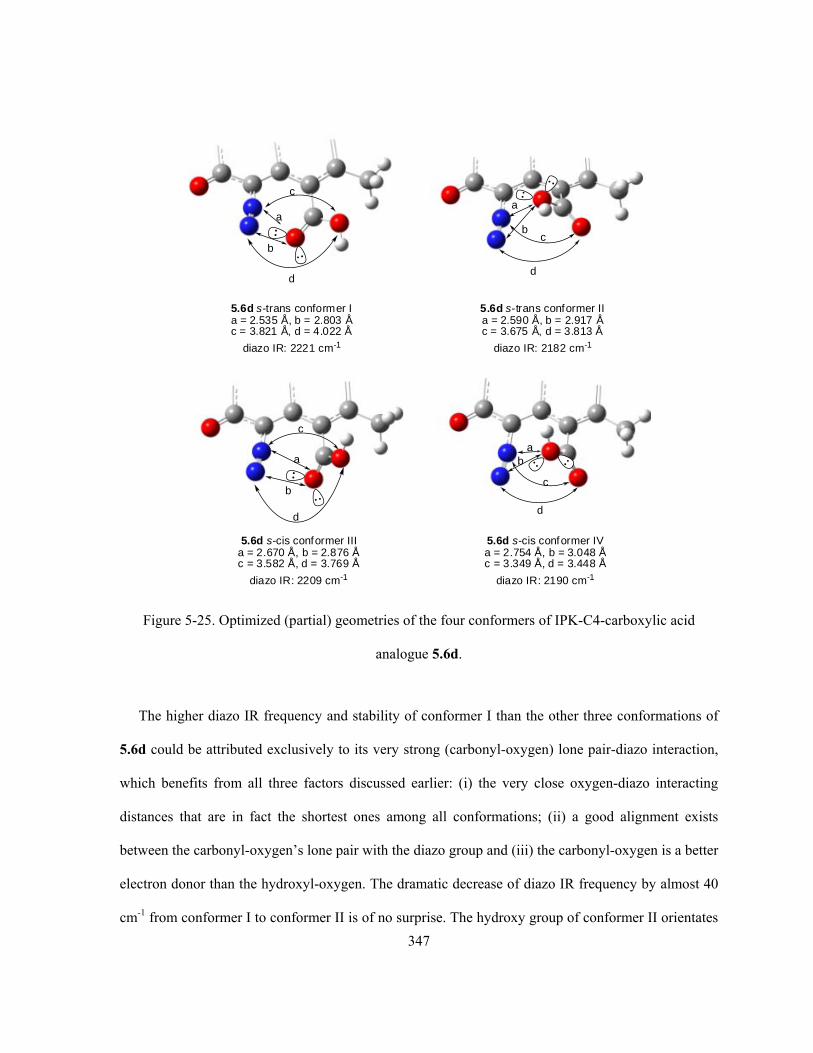
347
5.6d s-trans conformer Ia = 2.535 Å, b = 2.803 Åc = 3.821 Å, d = 4.022 Å
a
b
c
d
5.6d s-trans conformer IIa = 2.590 Å, b = 2.917 Åc = 3.675 Å, d = 3.813 Å
b
a
c
d
5.6d s-cis conformer IIIa = 2.670 Å, b = 2.876 Åc = 3.582 Å, d = 3.769 Å
a
b
c
d
5.6d s-cis conformer IVa = 2.754 Å, b = 3.048 Åc = 3.349 Å, d = 3.448 Å
ba
c
d
diazo IR: 2221 cm-1 diazo IR: 2182 cm-1
diazo IR: 2209 cm-1 diazo IR: 2190 cm-1
Figure 5-25. Optimized (partial) geometries of the four conformers of IPK-C4-carboxylic acid
analogue 5.6d.
The higher diazo IR frequency and stability of conformer I than the other three conformations of
5.6d could be attributed exclusively to its very strong (carbonyl-oxygen) lone pair-diazo interaction,
which benefits from all three factors discussed earlier: (i) the very close oxygen-diazo interacting
distances that are in fact the shortest ones among all conformations; (ii) a good alignment exists
between the carbonyl-oxygen’s lone pair with the diazo group and (iii) the carbonyl-oxygen is a better
electron donor than the hydroxyl-oxygen. The dramatic decrease of diazo IR frequency by almost 40
cm-1 from conformer I to conformer II is of no surprise. The hydroxy group of conformer II orientates

348
in such a manner that its diazo-interacting lone pair of electrons are indeed pointing towards the C5-
N1 bond rather than the N1-N2 diazo site. This situation represents a severely compromised diazo-
lone pair alignment, which is the worst among all four conformers. In addition, the hydroxyl-oxygen
is a weaker donor when compared to the carbonyl-oxygen. Therefore, despite its apparently fairly
close O-N interacting distances, which are only slightly longer than those of conformer I but
significantly shorter than those of conformer III and IV, conformer II still ends up with the lowest
diazo IR frequency. It is also interesting to notice that, in the case of conformer II, the polar O←H
bond within the carboxylic acid aligns quite well in parallel with the polar diazo N1→N2 bond at
some distances (~ 2.6–3.2 Å). Such observation resembles the known dipole-dipole interaction
between C5←N1 and C4→X, suggesting the presence of some additional and energetically beneficial
dipole-dipole interaction. However, such interaction itself might be weak as a result of the long
distance and its influence on the diazonium ion character is expected to be negligible, since even the
much closer and hence stronger dipole-dipole interaction (C5←N1/C4→X) is not able to affect it
much.
One can easily identify the geometric similarity between conformer III and I (Figure 5-25) with
both of their carbonyl-oxygens as electron donor to stay close to the diazo moiety at comparable
distances and lone pair orientations, other than the different orientations of their hydroxy groups (s-
cis vs s-trans). The slightly longer O-N interacting distances within conformer III (dC=O•••N1 = 2.670 Å,
dC=O•••N2 =2.876 Å) than I (dC=O•••N1 = 2.535 Å, dC=O•••N2 = 2.803 Å) are consistent with the
corresponding slightly smaller diazo IR frequency of III (NN = 2209 cm-1) than I (NN = 2221 cm-1).
Even though a shorter O-N interacting distance within conformer III to match that of conformer I may
be apparently achievable by a simple and small C4-C rotation clockwise (when viewing from the C5-
diazo/C4-acid side), such action would cause large unfavourable steric interaction between the

349
hydroxy group of this s-cis acid and the C3-methyl group that are already close enough and thus
prohibited.
For conformer IV, although the corresponding O-N interacting distances (dHO•••N1 = 2.754 Å,
dHO•••N2 = 3.048 Å) are significantly longer than the other three cases, its diazo IR frequency is
surprisingly not the lowest one. Even though the orientation and alignment of its hydroxy-oxygen’s
lone pair towards the diazo moiety is not as good as those found within conformer I and III, the
situation is obviously much better than that of conformer II (Figure 5-25). Since both conformer IV
and II have the hydroxyl-oxygen to act as the electron donor and the conformer II even possesses a
significantly shorter O-N interacting distances than conformer IV, then the much better lone pair-
diazo alignment seems to be the only reason that the conformer IV is still higher in diazonium ion
character than conformer II.
Two key conclusions can be made based on the above observations. First, for the same type of
lone pair donor (same element having identical hybridization state), orientation of the lone pair
electrons and its alignment with the diazo moiety are more important than the interacting distance
(within certain limits), as evident from the above comparison between conformer II and IV. Second,
if the orientations and alignments of the donor’s lone pair towards the diazo are very similar, then the
interacting distance becomes the determining factor, as evident from the comparison between
conformer I and III. It should be noted that the O-N interacting distance is largely determined by the
steric interaction from the C4-substituent with respect to the nearby C3-methyl group (and C5-diazo
group yet to a smaller extent), but the contribution to stability from such lone pair-diazo effect can not
be ignored at the same time. In other words, the C4-substituent having available electrons to interact
with the diazo moiety through space would try to adopt a conformation to simultaneously minimize
the steric repulsion but maximize the electron-diazo interaction.
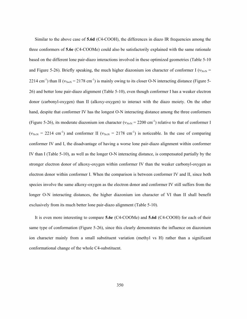
350
Similar to the above case of 5.6d (C4-COOH), the differences in diazo IR frequencies among the
three conformers of 5.6e (C4-COOMe) could also be satisfactorily explained with the same rationale
based on the different lone pair-diazo interactions involved in these optimized geometries (Table 5-10
and Figure 5-26). Briefly speaking, the much higher diazonium ion character of conformer I (NN =
2214 cm-1) than II (NN = 2178 cm-1) is mainly owing to its closer O-N interacting distance (Figure 5-
26) and better lone pair-diazo alignment (Table 5-10), even though conformer I has a weaker electron
donor (carbonyl-oxygen) than II (alkoxy-oxygen) to interact with the diazo moiety. On the other
hand, despite that conformer IV has the longest O-N interacting distance among the three conformers
(Figure 5-26), its moderate diazonium ion character (NN = 2200 cm-1) relative to that of conformer I
(NN = 2214 cm-1) and conformer II (NN = 2178 cm-1) is noticeable. In the case of comparing
conformer IV and I, the disadvantage of having a worse lone pair-diazo alignment within conformer
IV than I (Table 5-10), as well as the longer O-N interacting distance, is compensated partially by the
stronger electron donor of alkoxy-oxygen within conformer IV than the weaker carbonyl-oxygen as
electron donor within conformer I. When the comparison is between conformer IV and II, since both
species involve the same alkoxy-oxygen as the electron donor and conformer IV still suffers from the
longer O-N interacting distances, the higher diazonium ion character of VI than II shall benefit
exclusively from its much better lone pair-diazo alignment (Table 5-10).
It is even more interesting to compare 5.6e (C4-COOMe) and 5.6d (C4-COOH) for each of their
same type of conformation (Figure 5-26), since this clearly demonstrates the influence on diazonium
ion character mainly from a small substituent variation (methyl vs H) rather than a significant
conformational change of the whole C4-substituent.
351
N
N
C
CH3
5.6d conformer I
2.803 Å
2.535 Å
O O
HIR: 2221 cm-1
N
N
C
CH3
5.6d conformer II
2.917 Å
2. 590 Å
O
HIR: 2182 cm-1
O
N
N
C
CH3
5.6d conformer IV
3.048 Å
2. 754 Å
O
H
IR: 2190 cm-1O
5.6e conformer I
N
N
C
CH3
2.816 Å
2.558 Å
O O
CH3
IR: 2214 cm-1
5.6e conformer II
N
N
C
CH3
2.926 Å
2.600 ÅOO
CH3
IR: 2178 cm-1
5.6e conformer IV
N
N
C
CH3
2.965 Å
2.698 ÅOO
H3C
IR: 2200 cm-1
Larger stericinteractionthan 5.6d
conformer I
Larger stericinteractionthan 5.6d
conformer II
Figure 5-26. Comparison of geometries and diazo IR frequency between similar conformers of IPK-
C4-carboxylic acid 5.6d and IPK-C4-ester 5.6e.
Since both conformer I of 5.6e and 5.6d involve the same type of carbonyl-oxygen as electron
donor with very similar lone pair-diazo alignment (Table 5-10), the lower diazo IR frequency for
conformer I of 5.6e (NN = 2214 cm-1) than that of 5.6d (NN = 2221 cm-1) is due to the slightly
longer O-N interacting distance within 5.6e (Figure 5-26), which originates from the larger steric
352
interaction between its carboxylic-methoxy/C3-methyl groups than the carboxylic-hydroxy/C3-
methyl groups within 5.6d. Similar steric interaction between the C5-diazo and the ester methoxy
group causing larger O-N interacting distance is able to explain the smaller diazo IR frequency for
conformer II of 5.6e than 5.6d (Figure 5-26). The most interesting result comes from the comparison
of conformer IV of both structures. In this case, both the ester methyl group of 5.6e and the hydroxy
group of 5.6d points up to the open space above the aromatic ring (see optimized geometries in Table
5-10 and Figure 5-25), and the steric interaction from the C4-substituent with the C5-diazo moiety
and the C3-methyl group is no longer a significant issue. Therefore, the observed diazo IR frequency
difference should be mainly a result of change in electron-donating capability of the sp3-oxygen
(alkoxy-oxygen vs hydroxyl-oxygen). The ester methyl group of 5.6e is expected to increase the
electron density of the alkoxy-oxygen to be higher than the hydroxy-oxygen within 5.6d. The
calculated higher diazo IR frequency for conformer IV of 5.6e (NN = 2200 cm-1) than 5.6d (NN =
2190 cm-1) is completely consistent with the predicted increase of electron density on the sp3-oxygen
due to the presence of ester methyl group within 5.6e, which shall offer a noticeablely stronger lone
pair-diazo interaction and hence a higher diazonium ion character than 5.6d. This stronger lone pair-
diazo interaction is further reflected by the closer O-N interacting distances within 5.6e (dMeO•••N1 =
2.698 Å, dMeO•••N2 = 2.965 Å) than those within 5.6d (dHO•••N1 = 2.754 Å, dHO•••N2 = 3.048 Å), even
though the methyl is much bulkier than the proton.
As the last compound within this series, the IPK-C4-carboxylate 5.6f (C4-COO-) is just the
deprotonated form of the corresponding carboxylic acid 5.6d (C4-COOH), which at physiological
pHs would be in fact in the form of the carboxylate anion rather than the neutral acid. A practical
advantage of 5.6d/5.6f, if synthetically available, is that their solubility in aqueous solutions
(particularly the bioassay and cells) and common polar organic solvents is expected to be

353
significantly improved relative to that of IPK (1.5).* It can be predicted that, due to the much higher
electron density (negative charge) of the carboxylate anion than the neutral acid, 5.6f (C4-COO-) shall
possess some very strong diazonium ion character that is likely even higher than 5.6d (C4-COOH,
NN = 2182–2221 cm-1). Indeed, the calculated diazo IR frequency of 5.6f (NN = 2275 cm-1) is the
highest value among the entire series of IPK-C4-analogues studied.
Delocalization of the carboxylate negative charge evenly in between two identical resonance
forms would make the two oxygen atoms equivalent in electron density and hybridization (Figure 5-
27). Unlike the previous cases of the carboxylic acid and ester (i.e., 5.6d and 5.6e), there is neither a
competition of electron donor between the two oxygen atoms within the carboxylic group, nor a
choice of an s-cis vs s-trans conformation any more. Despite the various possible starting geometries
attempted, optimization only led to one single conformation as given in Figure 5-27. The optimized
conformation for 5.6f shows several interesting observations. First of all, the O-N interacting
distances within 5.6f (dCOO•••N1 =2.491 Å, dCOO•••N2 = 2.751 Å) are found to be closer than any of the
previous examples having (oxygen) lone pair-diazo interactions, not to mention that such distances
are a lot shorter than the sum of the corresponding vdW radii (rO = 1.52 Å, rN = 1.55 Å). This is
entirely consistent with the highest diazo IR frequency associated with the structure of 5.6f. Second,
delocalization of the negative charge within the carboxylate is also evident from the computed two
close C-O bond lengths (i.e., 1.263 Å and 1.252 Å), but more interestingly, it is noticed that the C-O
bond having its oxygen interacting with the diazo moiety is slightly longer than the other C-O bond
that has no interaction with the diazo moiety (Figure 5-27). The same observation was previously
encountered with the two N-O bonds within 5.4d (C4-NO2) as given in Figure 5-13 (section 5.2.3),
and the earlier rationale also applies in the case of 5.6f. Last but not least, it is quite obvious that
* Solubility of IPK (1.5) in all common organic solvents (except CH2Cl2) and water is very low, and this physical property greatly limits
the research and practial application of IPK (1.5) as a potential drug candidate.

354
rotation of the C4-C bond (Figure 5-27) would further shorten the O-N interacting distances and
enhance the lone pair-diazo interaction and consequently the diazonium ion character, although one
needs to realize that there is also some unfavourable steric interaction between the carboxylic group
and the diazo moiety that associates and increases with such C4-C rotation. On the other hand, the
C4-C rotation shall be prohibited by the increasing steric repulsion from the C3-methyl group towards
the carboxylic oxygen, which does not interact with the diazo moiety but stays close to the C3-methyl
group. Therefore, it is expected that replacement of the C3-methyl group by hydrogen shall remove
the steric limit on C4-C rotation and an even stronger lone pair-diazo interaction might be achieved.
This prediction is in fact confirmed later on (see section 5.2.6 for details).
O
OAr
O
OAr
O
OAr
(a) resonance forms of carboxylate (b) partial optimized geometry of 5.6f
dC-O = 1.263 Å
dC-O = 1.252 ÅdN1...O = 2.491 Å
dN2...O = 2.751 Å
Note: AB-rings are omitted for clarity
Figure 5-27. (a) Resonance of (aromatic) carboxylate anion and (b) optimized (partial) geometry of
IPK-C4-carboxylate analogue 5.6f.
5.2.6 Design of Simple Diazobenzo[a]fluorene Analogues
All of the above computational results (section 5.2.1–5.2.5) reveal the potential importance of the
C4-substituents on the diazonium ion character of IPK, or diazobenzo[a]fluorene in general. The key
discovery is that the through-space electronic interaction with the C5-diazo group from the C4-
substituent, which partially donates electrons (if available) to the diazo moiety when it stays at close

355
enough distance with appropriate orientation, applies dramatic influence on the corresponding
diazonium ion character. Future research in the area of kinamycin could benefit significantly from
such a finding. With the predicted much higher diazonium ion character than the natural antibiotic of
IPK (calc. νN≡N = 2139 cm-1; expt. νN≡N = 2160 cm-1), IPK-C4-analogues such as 5.2c (C4-CN, νN≡N =
2189 cm-1), 5.4a (C4-NH2, νN≡N = 2219 cm-1), 5.5c/5.5e (C4-OH/O-, νN≡N = 2210/2226 cm-1), 5.6a
(C4-CHO, νN≡N = 2248/2147 cm-1), 5.7 (IPK-D-ring-quinone, νN≡N = 2265 cm-1) and 5.6d/5.6f (C4-
COOH/COO-, νN≡N = 2182–2221/2275 cm-1) are some promising target molecules to prepare to
explore the influence of diazonium ion character on biological activity.
From a synthetic point of view, the above mentioned several analogues are also closely related in a
general chemical sense. For example, interconversion in between IPK (1.5), 5.5c (C4-OH) and 5.7
(IPK-D-ring-quinone) through oxidation/reduction, and functional group transformations in between
5.2c (C4-CN), 5.6d (C4-COOH, from 5.2c upon hydrolysis of C4-CN) and 4.66 (C4-CHO, from 5.6d
upon reduction of C4-COOH) are all possible. Nevertheless, compounds such as 5.2c (C4-CN) and
5.7 (IPK-D-ring-quinone) are worthy of some particular attention since they have the potential to be
easily transformed into other IPK-C4-analogues while their own syntheses do not seem to be very
difficult. Of course the real synthetic efforts towards such IPK-C4-analogues, by modifying the
Dmitrienko synthetic route, are probably more complicated than stated above, since the involvement
of extra protection/deprotection steps, unforeseen difficulties in preparing the corresponding starting
materials, and managing the high reactivities of (diazo-containing) advanced intermediates and final
target molecules are possible.
The power of having appropriate C4-substituents to enhance the diazonium ion characters offers
some further flexibility to design and prepare (much) simpler diazobenzo[a]fluorene analogues,
which can still possess high enough diazonium ion character. Simplifying the structure of
diazobenzo[a]fluorenes would make the corresponding synthetic work more concise and efficient.
356
For example, judging by its electronic nature, the C3-methyl group on the D-ring of these
diazobenzo[a]fluorenes is expected to be non-critical to the diazo group’s properties by itself, and it is
probably better to be removed since the electron-donating nature of the methyl group is not expected
to favour a higher diazonium ion character. However, it has already been shown that the C3-methyl
group can apply significant steric restraints on the C4-substituent, and consequently may affect the
diazonium ion character to a significant extent indirectly. Another example is the intramolecular H-
bonding between the C1-OH and C10-O as well as the C7-OH and C6-O within such IPK-C4-
analogues. It has been found experimentally (undisclosed results from this work) and computationally
(by earlier work of Laufer from the Dmitrienko group124) that these intramolecular H-bonding
interactions are essential for the high diazonium ion character of IPK (1.5). However, these H-bonds
also seem to be apparently the origin for IPK’s (1.5) poor solubility,* which is a practical issue that
would significantly decrease the efficiency and value of such IPK-C4-analogues to be used as drug
candidates. Therefore, removal of these functional groups (i.e., C3-methyl, C1-OH or C7-OH) from
the diazobenzo[a]fluorenes would cause certain (decreasing) effects on the diazonium ion character,
which is hoped to be counteracted to a greater extent by the installation of an appropriate C4-
substituent that interacts electronically with the C5-diazo moiety. This hypothesis of designing
simpler diazobenzo[a]fluorene analogues with high enough diazonium ion character can be clearly
illustrated by the results to be discussed next. Table 5-11 presents the calculated diazo IR frequencies
of some simpler diazobenzo[a]fluorene analogues derived from IPK (1.5), and results for those
simpler structures derived from or 5.6f (IPK-C4-COO-) are given in Table 5-12.
* IPK analogue 3.236 without the double intramolecular H-bonds, which is due to the lack of appropriate functional groups, has much
better solubility than IPK (1.5) in all common organic solvents and even aqueous solutions.

357
Table 5-11. Ab initio MO calculations of some simpler IPK-based diazobenzo[a]fluorene analogues.
Structure Optimized geometry NN (cm-1)a dNN (Å) <C-N-N(°) O
OHO N
HO
CH3
N 1.5
Expt. IR:
2160
MO:
2139
X-ray:
1.104
MO:
1.103
X-ray:
177.6
MO:
176.2 O
OHO N
HO
N 5.11
2138
1.103
176.3
O
OHO N
N 5.12
2124
1.105
176.4
O
O N
N
HO
5.13
2100
1.107
176.2
O
O N
N 1.123b
Expt. IRc: 2105 MO: 2086
MO: 1.108
MO: 176.4
a. Scaled by a scaling factor of 0.8929 (MO results); b. This compound was synthesized by Laufer from the
Dmitrienko group124; c. Experimental IR frequency observed with 1.123 as film.124

358
Table 5-12. Ab initio MO calculations of some simpler 5.6f-based diazobenzo[a]fluorene analogues.
Structure Optimized geometry NN (cm-1)a dN≡N (Å) <C-N-N(°) O
OHO N
HO
CH3
N 1.5
2139
1.103
176.2
O
OHO N
HO
CH3
NO
O
5.6f
2275
1.092
171.0
O
OHO N
HO
NO
O
5.14
2291
1.0908
169.8
O
OHO N
NO
O
5.15
2285
1.0912
169.9
O
O N
NO
O
HO
5.16
2254
1.093
170.1
O
O N
NO
O
5.17
2247
1.094
170.2
a. Scaled by a scaling factor of 0.8929.
In the series of IPK(1.5)-based simpler analogues (Table 5-11), initial replacement of C3-methyl
group of IPK (1.5) by hydrogen (i.e., analogue 5.11) causes a decrease of only 1 cm-1 in the diazo IR
frequency, consistent with the earlier prediction that this C3-methyl group is of very little importance
towards the diazonium ion character when diazo-interacting C4-substituent is not present. Further
substituent replacement within the structure of 5.11, first the C1-OH replaced by hydrogen (analogue
5.12), then C7-OH (analogue 5.13) and eventually both C1/C7-OHs (analogue 1.123) replaced by
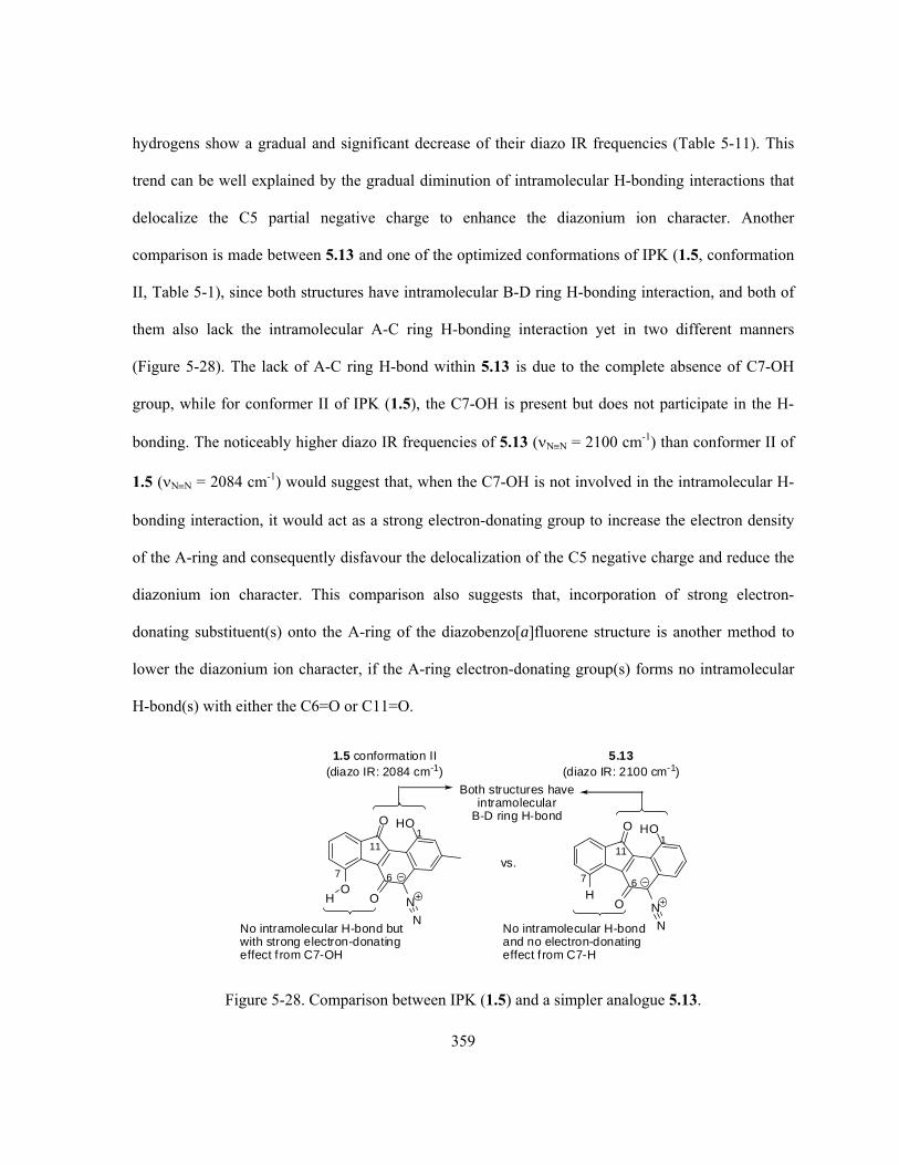
359
hydrogens show a gradual and significant decrease of their diazo IR frequencies (Table 5-11). This
trend can be well explained by the gradual diminution of intramolecular H-bonding interactions that
delocalize the C5 partial negative charge to enhance the diazonium ion character. Another
comparison is made between 5.13 and one of the optimized conformations of IPK (1.5, conformation
II, Table 5-1), since both structures have intramolecular B-D ring H-bonding interaction, and both of
them also lack the intramolecular A-C ring H-bonding interaction yet in two different manners
(Figure 5-28). The lack of A-C ring H-bond within 5.13 is due to the complete absence of C7-OH
group, while for conformer II of IPK (1.5), the C7-OH is present but does not participate in the H-
bonding. The noticeably higher diazo IR frequencies of 5.13 (NN = 2100 cm-1) than conformer II of
1.5 (NN = 2084 cm-1) would suggest that, when the C7-OH is not involved in the intramolecular H-
bonding interaction, it would act as a strong electron-donating group to increase the electron density
of the A-ring and consequently disfavour the delocalization of the C5 negative charge and reduce the
diazonium ion character. This comparison also suggests that, incorporation of strong electron-
donating substituent(s) onto the A-ring of the diazobenzo[a]fluorene structure is another method to
lower the diazonium ion character, if the A-ring electron-donating group(s) forms no intramolecular
H-bond(s) with either the C6=O or C11=O.
O
O N
N
5.13(diazo IR: 2100 cm-1)
HOO
O N
N
1.5 conformation II(diazo IR: 2084 cm-1)
HO
OH
No intramolecular H-bond butwith strong electron-donatingeffect f rom C7-OH
Both structures haveintramolecular
B-D ring H-bond
67
11
67
11
H
11
No intramolecular H-bondand no electron-donatingeffect f rom C7-H
vs.
Figure 5-28. Comparison between IPK (1.5) and a simpler analogue 5.13.

360
The results become even more interesting in the case of 5.6f (C4-COO-), when its C3-methyl, C1-
OH and C7-OH groups are replaced by hydrogen(s) stepwise (Table 5-12). The absence of the C3-
methyl group from the structure of 5.6f (i.e., analogue 5.14, NN = 2291 cm-1) causes a fairly large
increase in diazonium ion character (ΔNN = 16 cm-1) from the starting structure 5.6f (NN = 2275
cm-1). This observation is contrary to the previous situation of almost identical diazo IR frequencies
for IPK (1.5) and its simpler methyl-missing analogue 5.11 (IPK-C3-H), but entirely consistent with
the significantly shorter O-N interacting distances within 5.14 (dCOO•••N1 = 2.386 Å, dCOO•••N2 = 2.670
Å) than those within 5.6f (dCOO•••N1 = 2.491 Å, dCOO•••N2 = 2.751 Å) as shown in Figure 5-29. Such
positive influence on diazonium ion character has been previously predicted (see Figure 5-27 and
related text in section 5.2.5), since the C3-methyl group is an obvious steric obstacle for the C4-
carboxylate when the latter tries to get closer to the C5-diazo group as much as possible to achieve
the maximum lone pair-diazo interaction. Therefore, only after the replacement of C3-methyl group
by hydrogen, the C4-carboxylate is then free from the steric limitation by the C3-substituent and
rotation of C4-C bond to achieve the maximum lone pair-diazo interaction becomes possible. This is
clearly indicated by the observation that, the large torsion angle of ca. 51º between the C4-carboxylic
plane and the aromatic ring plane within 5.6f becomes a much smaller torsion angle of only ca. 24º
within 5.14 (Figure 5-29). It should be noted that the diazo IR frequency of 2291 cm-1 for 5.14 is the
most diazonium-like diazobenzo[a]fluorene analogue so far (Note: Such a high diazonium ion
character of 5.14 is comparable to the typical aryl diazonium salts, which have experimental diazo IR
frequencies of ca. 2230–2300 cm-1.377,378) Similar to the IPK (1.5) and its simpler analogues (Table 5-
11), the stepwise change in substituent (Table 5-12) in the order of 5.6f, 5.14 (C3-Me of 5.6f replaced
by H), 5.15 (C1-OH of 5.14 replaced by H), 5.16 (C7-OH of 5.14 replaced by H) and 5.17 (both C1-
OH and C7-OH of 5.14 replaced by Hs) shows the same tendency of gradual decreasing diazo IR
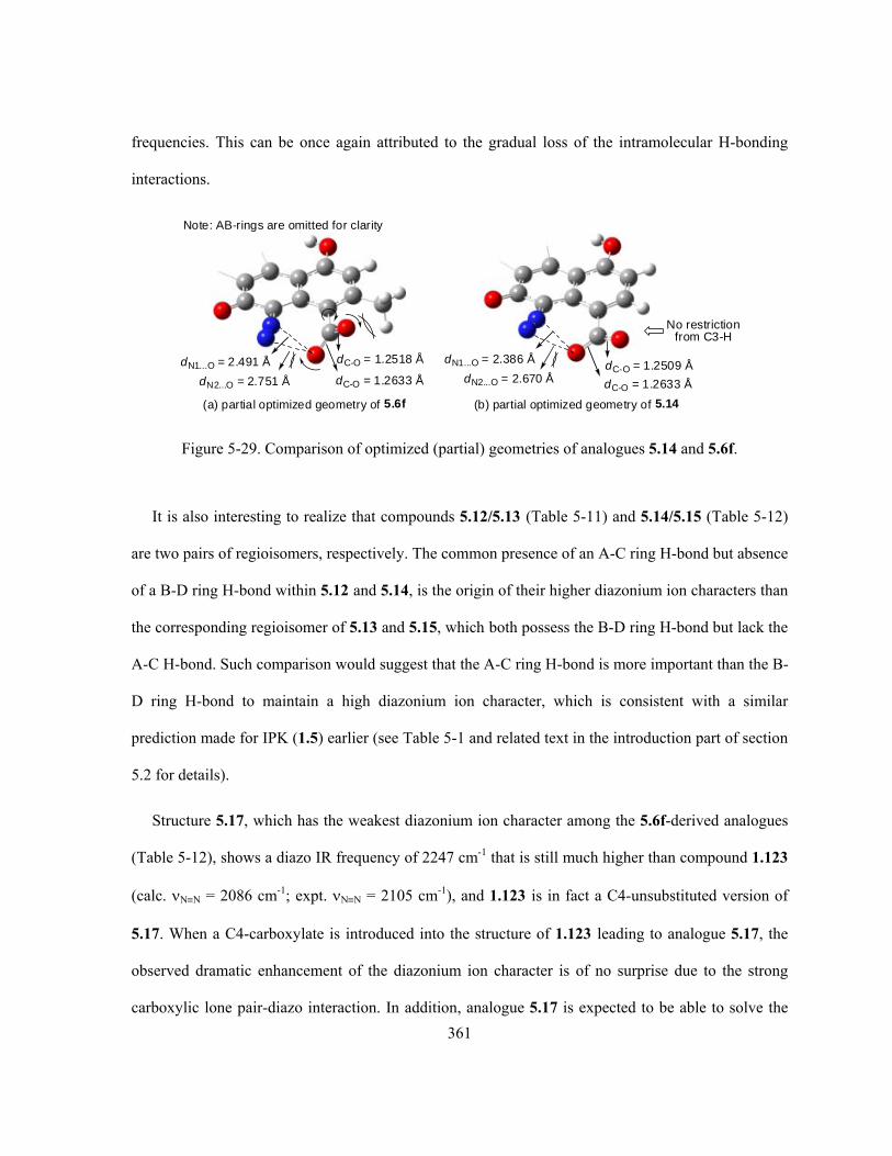
361
frequencies. This can be once again attributed to the gradual loss of the intramolecular H-bonding
interactions.
(a) partial optimized geometry of 5.6f
dC-O = 1.2633 Å
dC-O = 1.2518 ÅdN1...O = 2.491 Å
dN2...O = 2.751 Å
Note: AB-rings are omitted for clarity
(b) partial optimized geometry of 5.14
dC-O = 1.2633 Å
dC-O = 1.2509 ÅdN1...O = 2.386 Å
dN2...O = 2.670 Å
No restrictionfrom C3-H
Figure 5-29. Comparison of optimized (partial) geometries of analogues 5.14 and 5.6f.
It is also interesting to realize that compounds 5.12/5.13 (Table 5-11) and 5.14/5.15 (Table 5-12)
are two pairs of regioisomers, respectively. The common presence of an A-C ring H-bond but absence
of a B-D ring H-bond within 5.12 and 5.14, is the origin of their higher diazonium ion characters than
the corresponding regioisomer of 5.13 and 5.15, which both possess the B-D ring H-bond but lack the
A-C H-bond. Such comparison would suggest that the A-C ring H-bond is more important than the B-
D ring H-bond to maintain a high diazonium ion character, which is consistent with a similar
prediction made for IPK (1.5) earlier (see Table 5-1 and related text in the introduction part of section
5.2 for details).
Structure 5.17, which has the weakest diazonium ion character among the 5.6f-derived analogues
(Table 5-12), shows a diazo IR frequency of 2247 cm-1 that is still much higher than compound 1.123
(calc. NN = 2086 cm-1; expt. NN = 2105 cm-1), and 1.123 is in fact a C4-unsubstituted version of
5.17. When a C4-carboxylate is introduced into the structure of 1.123 leading to analogue 5.17, the
observed dramatic enhancement of the diazonium ion character is of no surprise due to the strong
carboxylic lone pair-diazo interaction. In addition, analogue 5.17 is expected to be able to solve the

362
known low solubility issue of compound 1.123 in both aqueous solutions and organic solvents.80,124
More importantly, structure 5.17 is a good example to support the hypothesis for designing simpler
diazobenzo[a]fluorene analogues as mentioned at the beginning of this section (section 5.2.6). The
appropriate selection of C4-substituent, which strongly and electronically interacts with the diazo
moiety, can override the decreasing effect on the diazonium ion character from the (complete)
removal of other substituents within the diazobenzo[a]fluorene. Such structural simplification of
diazobenzo[a]fluorene while maintaining a high diazonium ion character shall greatly simplify the
corresponding total synthesis. The fairly high diazo IR frequency of 5.17 further triggered some
interests in modeling the complete stripped-down diazobenzo[a]fluorene structure 1.123 with
minimal presence of diazo-influencing substituent, i.e. a single substituent at either the C4 or C2
position, in hoping to find very simple diazobenzo[a]fluorene structures with high enough diazonium
ion character as suitable synthetic targets. Choices of the substituent at C4- or C2-position of 1.123
were kept essentially the same as those IPK-C4-analogues but with small variations. The
corresponding results (Table 5-13) of these two series of compounds, i.e. 1.123-C4-analogues and
1.123-C2-analogues are summarized below and reveal some more interesting discoveries.

363
O
N2O
1.123 unsubstituteddiazobenzo[a]fluorene
4
3
5
21 O
N2O
X4
5
O
N2O
2
5
X
1.123-C4-analogue 1.123-C2-analogue
Table 5-13. Calculated diazo IR stretching frequency of C4- and C2- analogues of 1.123.
C4 substituent X NN (cm-1)a C2 substituent X NN (cm-1)a
X = CH3 2080 X = O- 1977
X = H (1.123) 2086 X = COO- 2056
X = Br 2130 X = NH2 2060
X = Cl 2136.9 X = OMe 2067
X = CN 2137.4 X = OH 2070
X = SH 2141b X = CH3 2081
X = F 2143 X = H (1.123) 2086.2
X = OH 2157.2 X = F 2086.4
X = OMe 2157.6 X = Br 2092.5
X = CF3 2160 X = Cl 2092.9
X = OCF3 2161 X = SH 2095
X = NH2 2166 X = OCF3 2098
X = O- 2177 X = COOMe 2104
X = COCF3 2183 X = CN 2105
X = COMe 2192.2 X = COOH 2106.2
X = COOMe 2192.5b X = COMe 2106.6
X = NO 2192.8b X = CF3 2108
X = NO2 2192.9 X = NO 2115
X = COOH 2194b X = COCF3 2117.2
X = COO- (5.17) 2247
Lower diazonium ion
character ↑ ↑ ↑
IPK (1.5) ← 2139 cm-1
↓ ↓ ↓ ↓ ↓ ↓ ↓ ↓ ↓ ↓ ↓ ↓ ↓ ↓ ↓ ↓ ↓ ↓ ↓
Higher diazonium ion
character X = NO2 2117.9 a. Scaled by a scaling factor of 0.8929. The IR frequencies are listed in ascending order; b. Multiple diazo IR
frequencies are available due to the multiple possible conformations; however, only the diazo IR frequency
of the most stable conformation is used in this table for ranking.

364
First of all, judging by the calculated diazo IR stretching frequencies (Table 5-13), there are a
number of 1.123-C4-analogues that possess higher diazonium ion character than the natural antibiotic
of IPK (1.5, NN = 2139 cm-1) and are suitable as potential synthetic targets. On the other hand, even
the highest diazo IR frequency of 1.123-C2-analogues (C2-NO2, NN = 2117.9 cm-1) is still
significantly lower than that of IPK (1.5, NN = 2139 cm-1), which makes none of this C2-series
structures a promising synthetic candidate. However, it is very interesting to realize the totally
different trends of change in diazo IR frequency as a function of the substituent observed for the
1.123-C4-analogues and 1.123-C2-analogues (Table 5-13). The 1.123-C4-analogues demonstrate
very similar substitution effects as the IPK-C4-analogues, and lone pair-diazo interaction dominates
the influence on the diazonium ion character whenever available. Therefore, both (strong) electron-
donating and electron-withdrawing substituents, as long as this C4-moiety contains heteroatom(s)
with available lone pair(s) that could stay close enough to the C5-diazo group, lead to high diazonium
ion character. However, diazo IR frequencies of the 1.123-C2-analogues indicate that their diazonium
ion characters are proportional to the electron-withdrawing capability of the C2-substituent. Since the
C2-substituent is far from the C5-diazo group, it can no longer provide a dipole-dipole interaction or
lone pair-diazo interaction to affect the diazonium ion character yet such interactions must be
considered in the cases of C4-substitution. Instead, the C2-substituent is only able to apply inductive
and resonance effects through the aromatic D-ring to influence the diazonium ion character, but at a
much further distance than the C4-substituent and hence weaker (in general). Obviously, an electron-
withdrawing C2-substituent would favour the delocalization of the C5 partial negative charge, either
inductively or through resonance, to enhance the diazonium ion character. Another noticeable
difference, as one can predict, is that variations of the substituent’s conformation no longer affects the
diazonium ion character in the cases of the 1.123-C2-analogues, but it is still applicable to some

365
1.123-C4-analogues that are very similar to the corresponding IPK-C4-analogues (see footnote b of
Table 5-13).
The current work offers a systematic computation and analysis of various diazo-affecting
interactions within the diazobenzo[a]fluorenes upon possible substitution. Structures of some IPK-
C4-analogues and simpler diazobenzo[a]fluorene analogues, with predicted diazo IR frequencies
(much) higher than the natural IPK (1.5), are proposed for future synthetic projects. Whether or not
such proposed target molecules are higher in biological activity than the natural IPK (1.5), as a result
of their higher diazonium ion character, remains to be established. It is also hoped that such target
molecules involve smoother and easier syntheses, especially for those rather simple structures such as
the 1.123-C4-analogues, by taking advantage of the two established synthetic procedures towards the
diazobenzo[a]fluorenes from the Dmitrienko group (section 3.2) that have already led to the
successful total synthesis of IPK (1.5) 25and 1.123.124.
5.3 Theoretical Analysis of Interaction of Proximal Electron Rich Heteroatoms
with Diazonium Groups
The mechanistic studies of kinamycin F (1.1f), from chemical evidence for nucleophilic thiol
addition to diazo and spectroscopic/computational evidence of diazo IR frequency as described in
Chapter 4, have demonstrated that the diazo group in diazobenzo[b]fluorenes is indeed diazonium-
like. This is consistent with an earlier suggestion by the Dmitrienko group that the chemical
properties of the diazo group in the diazobenzo[a]fluorene of IPK (1.5) are also more like those of
aryl diazonium ions rather than those of typical diazo groups, based on chemical evidence for
nucleophilic naphthol addition to diazo and computational evidence of diazo IR frequency.124
Therefore, it is appropriate to compare the discussion presented above (section 5.2), particularly the

366
discovered and unexpected lone pair-diazo interactions, with discussions in the literature concerning
the interactions of aryl diazonium groups with proximal electron rich functional groups.
In the early 1990s, X-ray crystallographic studies by Wallis, Easton and Dunitz revealed that,
within the 8-substituted-naphthalene-1-diazonium cations 5.18 (Figure 5-30), the presence of a
proximal group such as (N+–)O-, MeS, Me2N and NO2 to the diazonium moiety causes a deviation of
the diazo group geometry from C-N-N linearity such that the terminal nitrogen (Nβ) is bent away
from the proximal group, yet the distances between the proximal group’s electron-rich heteroatom (O,
S or N) and the internal nitrogen (Nα) of the diazonium group are close enough (i.e., shorter than the
sum of the corresponding vdW radii) to warrant some (apparently) attractive interactions.379 They
suggested that this observed phenomenon of the diazonium moiety distortion by the proximal group is
reminiscent of related interactions of proximal electron rich groups with carbonyl groups, which was
likened to the generally accepted Bürgi-Dunitz model for the interaction of nucleophiles with ketones
and aldehydes where the intramolecular interaction is considered to be an “incipient nucleophilic
attack” (INA) as shown in the example 5.19 (Figure 5-30) that leads to distortion of the C=O group
involving partial pyramidalization of the carbonyl group carbon atom.380
N
ORO O
R RNu-
-
+X
NY
Nu-
N
N
N
R
X
N+
C
C
C
Y
O-
MeS:
Me2N:
ONO:
5.18 5.19 Burgi-Dunitz modelINA: incipient
nucleophilic attack
Figure 5-30. Intramolecular electron-rich proximal group interacting with the diazonium moiety
within the 8-substituted-naphthalene-1-diazonium cations 5.18 and the corresponding Bürgi-
Dunitz/INA model of 5.19.
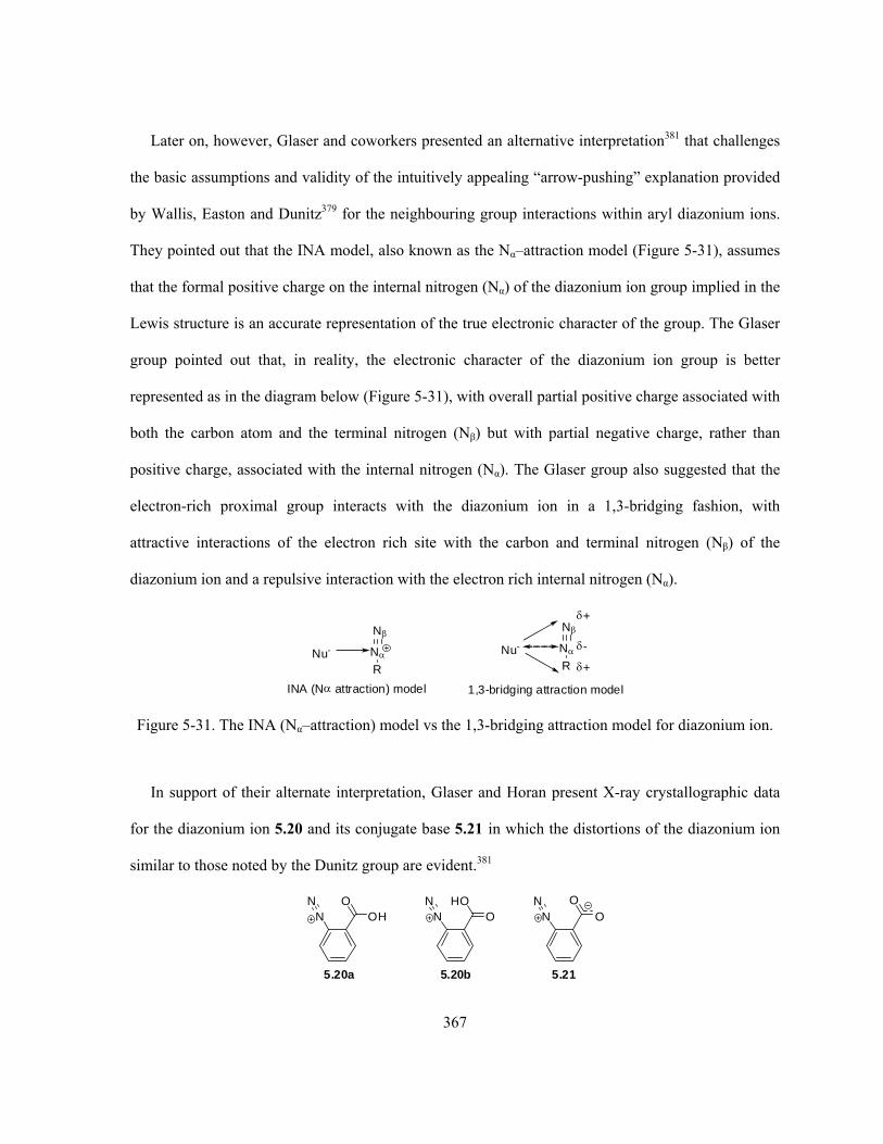
367
Later on, however, Glaser and coworkers presented an alternative interpretation381 that challenges
the basic assumptions and validity of the intuitively appealing “arrow-pushing” explanation provided
by Wallis, Easton and Dunitz379 for the neighbouring group interactions within aryl diazonium ions.
They pointed out that the INA model, also known as the Nα–attraction model (Figure 5-31), assumes
that the formal positive charge on the internal nitrogen (Nα) of the diazonium ion group implied in the
Lewis structure is an accurate representation of the true electronic character of the group. The Glaser
group pointed out that, in reality, the electronic character of the diazonium ion group is better
represented as in the diagram below (Figure 5-31), with overall partial positive charge associated with
both the carbon atom and the terminal nitrogen (Nβ) but with partial negative charge, rather than
positive charge, associated with the internal nitrogen (Nα). The Glaser group also suggested that the
electron-rich proximal group interacts with the diazonium ion in a 1,3-bridging fashion, with
attractive interactions of the electron rich site with the carbon and terminal nitrogen (Nβ) of the
diazonium ion and a repulsive interaction with the electron rich internal nitrogen (Nα).
Nu-
N
N
R
INA (N attraction) model
+
+
-Nu-
N
N
R
1,3-bridging attraction model
Figure 5-31. The INA (Nα–attraction) model vs the 1,3-bridging attraction model for diazonium ion.
In support of their alternate interpretation, Glaser and Horan present X-ray crystallographic data
for the diazonium ion 5.20 and its conjugate base 5.21 in which the distortions of the diazonium ion
similar to those noted by the Dunitz group are evident.381
N OHON
N OHON
N OON
5.20a 5.20b 5.21

368
A computational analysis of the two rotamers of the acid, 5.20a and 5.20b, and of the conjugate
base 5.21 at the RHF/6-31G* level reproduced the geometry observed experimentally relatively well
and also provided insight into the nature of the electronic interactions of the proximal groups with the
diazonium ion.381 The ab initio MO analysis results were then employed as input into a calculation of
the electrostatic interaction energy between the atoms in the diazonium ion and the proximal electron
rich groups. The analysis involved a model in which the electrostatic interaction energy between two
atoms (ESIij) was considered to be the sum of a Coulomb energy (CCij) arising from interaction
between the atomic charges, an energy term (CDij) arising from interaction of the atomic charge on
each atom with the atomic dipole at each other atom, a dipole-dipole interaction (DDij), a quadrupole-
atomic charge interaction (QCij), a quadrupole-dipole interaction (QDij) and a quadrupole-quadrupole
interaction (QQij), therefore, ESIij = CCij + CDij + DDij + QCij + QDij + QQij. This analysis leads to a
clear demonstration that the interaction of the proximal group with the diazonium ion cannot be
interpreted via the INA model that depends on the assumption of partial positive charge on the
internal nitrogen (Nα) of the diazonium ion group. The computed model for 5.20a (Figure 5-32) is
entirely consistent with the 1,3-bridging attraction model (Figure 5-31), and brings to light an
attractive interaction of the electron rich internal nitrogen (Nα) of the diazonium ion group with the
partially positively charged carbonyl carbon atom bearing the electron rich carbonyl oxygen atom of
the proximal group. This attractive interaction contributes to the tendency of the diazonium ion to be
distorted from linearity so as to increase the favourable interaction of the internal nitrogen (Nα) with
this carbonyl carbon atom, but at the same time increasing somewhat the interaction of this nitrogen
atom (Nα) to the electron rich site of the proximal group as suggested in Figure 5-32.
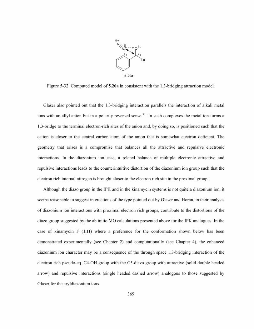
369
NN
OH
O
+
+
+
- -
5.20a
Figure 5-32. Computed model of 5.20a in consistent with the 1,3-bridging attraction model.
Glaser also pointed out that the 1,3-bridging interaction parallels the interaction of alkali metal
ions with an allyl anion but in a polarity reversed sense.381 In such complexes the metal ion forms a
1,3-bridge to the terminal electron-rich sites of the anion and, by doing so, is positioned such that the
cation is closer to the central carbon atom of the anion that is somewhat electron deficient. The
geometry that arises is a compromise that balances all the attractive and repulsive electronic
interactions. In the diazonium ion case, a related balance of multiple electronic attractive and
repulsive interactions leads to the counterintuitive distortion of the diazonium ion group such that the
electron rich internal nitrogen is brought closer to the electron rich site in the proximal group.
Although the diazo group in the IPK and in the kinamycin systems is not quite a diazonium ion, it
seems reasonable to suggest interactions of the type pointed out by Glaser and Horan, in their analysis
of diazonium ion interactions with proximal electron rich groups, contribute to the distortions of the
diazo group suggested by the ab initio MO calculations presented above for the IPK analogues. In the
case of kinamycin F (1.1f) where a preference for the conformation shown below has been
demonstrated experimentally (see Chapter 2) and computationally (see Chapter 4), the enhanced
diazonium ion character may be a consequence of the through space 1,3-bridging interaction of the
electron rich pseudo-eq. C4-OH group with the C5-diazo group with attractive (solid double headed
arrow) and repulsive interactions (single headed dashed arrow) analogous to those suggested by
Glaser for the aryldiazonium ions.

370
N
N
O
H
CH3
OH
OH
HH
OH
H
Figure 5-33. Analysis of the kinamycin F (1.1f) conformer having pseudo-eq. C4-OH group by using
the 1,3-bridging attraction model of Glaser.
Application of Glaser’s electrostatic interaction analysis method to the IPK systems should be
explored in future to evaluate the importance of the 1,3-attractive interaction in determining the
properties of the diazo group in the substituted IPK analogues.
5.4 Conclusion
In summary, this study has provided a number of insights into the potential influence of
substituents on the diazonium ion character of the diazo group within the diazobenzo[a]fluorene ring
system found in isoprekinamycin (IPK, 1.5). X-ray crystallographic analysis of IPK (1.5) and a
synthetic analogue 3.236 lacking intramolecular H-bonding clearly shows that the diazonium ion
character in IPK (1.5) is substantially enhanced by the presence of such intramolecular H-bonds.
Ab initio molecular orbital calculations indicate that, within the diazobenzo[a]fluorenes, the C4-
substituent can interact with the C5-diazo group so as to substantially increase the diazonium ion
character. Particularly effective in this regard are C4-functional group with heteroatom(s) bearing
lone pairs of electrons (e.g., –NH2), which must locate geometrically close enough and in an
appropriate orientation toward the C5-diazo moiety. Although the precise nature of such lone pair
interaction with the diazo group that results in a substantial increase in diazonium ion character

371
remains to be elucidated, there is good reason to believe that a through-space bonding interaction of
the lone pair orbitals with the π-bonding orbital of the diazo group, which is orthogonal to the rest of
the π-system, might contribute significantly to the discovered effect. This can be clearly demonstrated
by one of the molecular orbitals for the IPK-C4-NH2 analogue 5.4a as shown in Figure 5-34. This
predicted effect, which is by no means intuitively obvious, forms the basis for synthetic experiments
being pursued by others in this group. It is hoped that such efforts may be able to not only test the
predictions presented in this chapter experimentally but also to determine if such alteration of
diazonium ion character influences the biological activity of these compounds in a beneficial way.
Figure 5-34. Computed MO orbital of IPK-C4-NH2 analogue 5.4a suggesting possible lone pair-
diazo interactions.
5.5 Experimental Details
Refer to section 4.7 for details regarding the ab initio MO calculations of the diazobenzo[a]fluorene
type of kinamycins and related analogues.
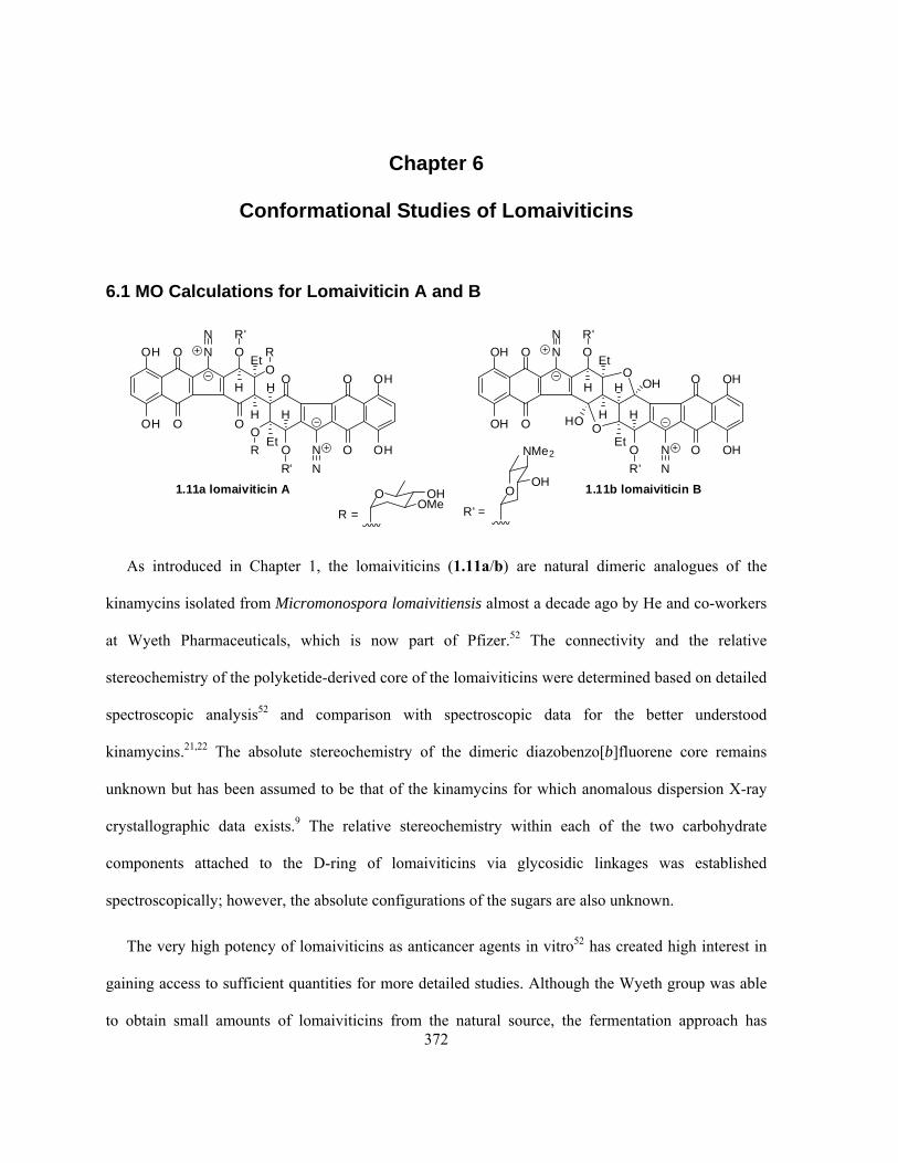
372
Chapter 6
Conformational Studies of Lomaiviticins
6.1 MO Calculations for Lomaiviticin A and B
OH
OH
O
O
N O
O
O
O
O
R'
H
H
R
H
Et
EtO
R
H
R'
N
O
O
OH
OH
N
N
OH
OH
O
O
N O
O
O
R'
H
H
H
Et
EtO
H
R'
N
O
O
OH
OH
N
N
HO
OH
1.11a lomaiviticin A 1.11b lomaiviticin BOOH
NMe2
R' =
O OHOMe
R =
As introduced in Chapter 1, the lomaiviticins (1.11a/b) are natural dimeric analogues of the
kinamycins isolated from Micromonospora lomaivitiensis almost a decade ago by He and co-workers
at Wyeth Pharmaceuticals, which is now part of Pfizer.52 The connectivity and the relative
stereochemistry of the polyketide-derived core of the lomaiviticins were determined based on detailed
spectroscopic analysis52 and comparison with spectroscopic data for the better understood
kinamycins.21,22 The absolute stereochemistry of the dimeric diazobenzo[b]fluorene core remains
unknown but has been assumed to be that of the kinamycins for which anomalous dispersion X-ray
crystallographic data exists.9 The relative stereochemistry within each of the two carbohydrate
components attached to the D-ring of lomaiviticins via glycosidic linkages was established
spectroscopically; however, the absolute configurations of the sugars are also unknown.
The very high potency of lomaiviticins as anticancer agents in vitro52 has created high interest in
gaining access to sufficient quantities for more detailed studies. Although the Wyeth group was able
to obtain small amounts of lomaiviticins from the natural source, the fermentation approach has

373
proven to be highly variable such that only milligram quantities of these natural products have ever
been isolated. As a consequence of the inability to have access to multigram quantities of these
compounds, the Wyeth group has not mounted a full medicinal chemistry program around these
compounds and, in fact, disclosed their structures without filing a patent.382 The Wyeth group has
made efforts to isolate all of the genes for the biosynthetic pathway with the hope that these might be
used for more efficient heterologous expression in another bacterium and that the substrate
specificities of the enzymes of the biosynthetic pathway might be altered to allow for biosynthesis of
structural analogues.382 Elsewhere, much interest has arisen in synthetic organic chemistry groups in
total synthesis of these structurally complex compounds.157-159
In the Dmitrienko group, it was felt that a better understanding and some valuable insight of the
three dimensional structure and the diazonium ion character of the lomaiviticins might be obtained by
computational molecular modeling, even if there is no direct access to these natural products at the
moment. In addition, results from such modeling studies might help to design simpler analogues with
anticancer activities comparable to those of the lomaiviticins. Due to the structural complexity of the
lomaiviticins, it was decided that the compounds would be analyzed computationally in a step-wise
fashion as follows:
1) Conformational analysis of the two carbohydrate components of lomaiviticins, namely the
methyl glycoside form of α-L-oleandrose (6.1) and the β-D-N,N-dimethylpyrrolosamine (6.2).
The methyl group (bolded in the structures shown below) represents the aglycon skeleton
within the lomaiviticins to which these sugars are attached.
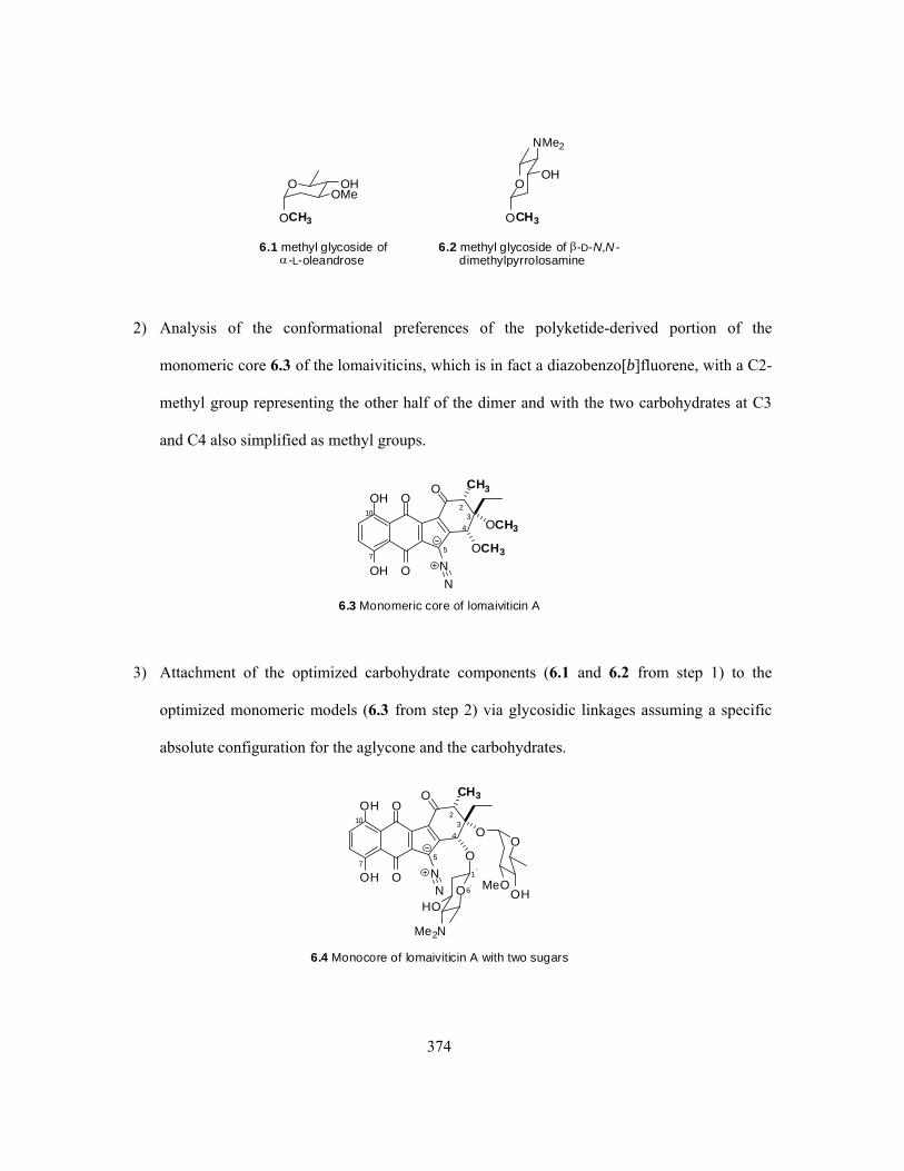
374
OCH3OCH3
OOH
O OHOMe
NMe2
6.1 methyl glycoside of-L-oleandrose
6.2 methyl glycoside of -D-N,N -dimethylpyrrolosamine
2) Analysis of the conformational preferences of the polyketide-derived portion of the
monomeric core 6.3 of the lomaiviticins, which is in fact a diazobenzo[b]fluorene, with a C2-
methyl group representing the other half of the dimer and with the two carbohydrates at C3
and C4 also simplified as methyl groups.
O
O
OH
OH
O
N
N
CH3
OCH3
OCH3
6.3 Monomeric core of lomaiviticin A
23
4
7
10
5
3) Attachment of the optimized carbohydrate components (6.1 and 6.2 from step 1) to the
optimized monomeric models (6.3 from step 2) via glycosidic linkages assuming a specific
absolute configuration for the aglycone and the carbohydrates.
O
O
OH
OH
O
NN
CH3
O
O
6.4 Monocore of lomaiviticin A with two sugars
23
4
7
10
5
O
OHMeOO
HO
Me2N
1'
6'

375
4) Exploration of the conformational preferences of the aglycone of lomaiviticin A 6.5 (i.e., a
dimeric diazobenzo[b]fluorene core) with methyls representing the carbohydrates, by
attachment of the various possible conformations of two monomers (i.e., 6.4).
O
O
OH
OH
O
NN
OCH3
OCH3
6.5 Dimeric core of lomaiviticin A
23
4
7
10
5
O
O
OH
OH
O
NN
H3CO
H3CO2'
3'4'
1
1'
5'7'
10'
4a
4'a
5) Conformational study of the full structures of lomaiviticin A (1.11a) and lomaiviticin B
(1.11b), which can be constructed through either linking of two monomers containing the
carbohydrates (6.4 from step 3) or attaching appropriate carbohydrates (6.1 and 6.2 from step
1) to the dimeric core (6.5 from step 4).
This series of analyses constitutes a complex computational problem which is not as yet complete.
However, the progress that has been made in pursuit of a full computational description of the
lomaiviticins has revealed some interesting intramolecular interactions between the diazo group and
the carbohydrate component at C4, which may influence the reactivity of the diazo group and in turn
impact the behaviour of these natural products with biological targets in vivo.
The progress made in each of the five sequential analyses listed above is described below.

376
6.1.1 Conformational Analysis of the Carbohydrates of Lomaiviticins
OCH3OCH3
OOH
O OHOMe
NMe2
6.1 methyl glycoside of-L-oleandrose
6.2 methyl glycoside of -D-N,N -dimethylpyrrolosamine
The two types of carbohydrates within lomaiviticin A (1.11a) were concluded to be C3/C3’α-
oleandrose and C4/C4’ β-N,N-dimethylpyrrolosamine, respectively.52 The relative stereochemistry
within each of these two sugars was established solely from the observed NMR homonuclear
coupling constants and comparison with literature results for the corresponding carbohydrates (i.e., α-
oleandrose and β-pyrrolosamine), yet their absolute configurations were not determined.52 The
existence of the glycosidic linkages between the carbohydrates and the lomaiviticin core of carbon
skeleton, either in the α- or β-manner, was determined exclusively by the NMR data with no further
need of other spectroscopic evidence.
When this computational work started, however, it was noticed that there are a few obvious but
previously ignored flaws with the reported structures of the carbohydrates within lomaiviticins. The
two sugar units of either the C3/C3’ α-oleandrose or the C4/C4’ β-N,N-dimethylpyrrolosamine were
concluded to be entirely identical, since only one set of NMR signals (both 1H and 13C) was observed
for each type of carbohydrate.52 Within the published chemical drawing of lomaiviticin A (Figure 6-
1),52 however, the two supposedly identical sugar units are illustrated with apparently opposite
absolute stereochemical configurations, in which the oleandrose at C3 is shown as an α-L-sugar while
the one at C3’ is given as an α-D-sugar, and similarly the carbohydrate at C4 is shown as a β-D-sugar
while the corresponding one at C4’ is demonstrated as a β-L-sugar. The same stereochemical problem
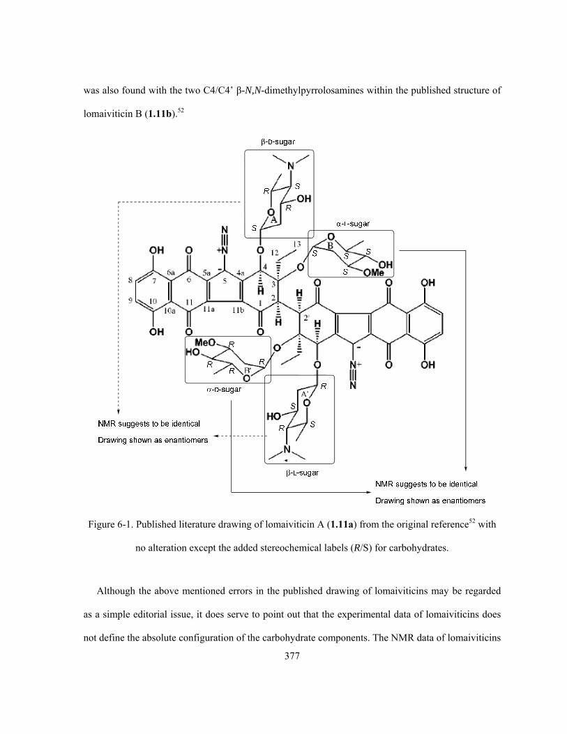
377
was also found with the two C4/C4’ β-N,N-dimethylpyrrolosamines within the published structure of
lomaiviticin B (1.11b).52
Figure 6-1. Published literature drawing of lomaiviticin A (1.11a) from the original reference52 with
no alteration except the added stereochemical labels (R/S) for carbohydrates.
Although the above mentioned errors in the published drawing of lomaiviticins may be regarded
as a simple editorial issue, it does serve to point out that the experimental data of lomaiviticins does
not define the absolute configuration of the carbohydrate components. The NMR data of lomaiviticins

378
requires symmetry in the dimer and, if assuming that the kinamycin-like core structure has the same
absolute configuration in the two halves of the dimer, the symmetry of lomaiviticins must be C2 (at
least on average). Thus, the absolute configurations of the two C3/C3’ oleandrose-derived
components must either both be of the L- or both be of the D-configuration. Likewise the two C4/C4’
pyrrolosamine-derived components must either both be of the D- or both be of the L-configuration.
The uncertainty of the absolute stereochemistry of these carbohydrates would complicate the situation,
since the experimental data can be reasonably accommodated by any of the four stereoisomeric
possibilities for lomaiviticin A (1.11a) as follows: C3/C3’-α-L-sugars and C4/C4’-β-D-sugars,
C3/C3’-α-D-sugars and C4/C4’-β-L-sugars, C3/C3’-α-L-sugars and C4/C4’-β-L-sugars or C3/C3’-α-
D-sugars and C4/C4’-β-D-sugars. For lomaiviticin B (1.11b), the absence of carbohydrates at C3/C3’
simplifies its stereoisomeric possibilities to two options, either C4/C4’-β-D-sugars or C4/C4’-β-L-
sugars.
Ideally, the absolute configuration of the carbohydrates in the lomaiviticins should be determined
through rigorous experimental methods. In the absence of such information, however, some
consideration of the structures of biosynthetically related polyketide-derived antibiotics from other
Actinomycetes/Streptomyces species is instructive. For example, oleandomycin (6.6, Scheme 6-1a) is
an antibiotic produced by Streptomyces antibioticus, whose polyketide-derived macrolide core
contains two carbohydrate components attached by glycosidic linkages. One of these sugar units is L-
oleandrose (cf. dTDP-activated L-oleandrose 6.14, Scheme 6-1a), which is proposed to be also a
component of lomaiviticins. The other carbohydrate component in oleandomycin is D-desosamine (cf.
dTDP-activated D-desosamine 6.19, Scheme 6-1a), which is in fact isomeric with the N,N-
dimethylpyrrolosamine of lomaiviticins. A very interesting biosynthetic pathway (Scheme 6-1a) has
been proposed whereby both the D-desosamine component and the L-oleandrose component of
oleandomycin (6.6) are derived from the same bioprecursor D-glucose-6-phosphate (6.7).383 The key

379
to the ability of producing both L-series and D-series carbohydrate derivatives from the same D-series
precursor in the case of oleandomycin (6.6) is the existence of a common C4-keto intermediate 6.9.
This D-sugar is then converted enzymatically to either 6.19 (a D-sugar) or 6.14 (a L-sugar), and
epimerization at C3 and C5 catalyzed by an appropriate enzyme (from 6.11 to 6.12) plays a critical
role in the D- to L- transformation. It is reasonable to suggest that a related and similar biosynthetic
pathway (Scheme 6-1b) may exist in Micromonospora lamaivitiensis, the Actinomycete species that
produces lomaiviticins, providing on one hand a L-oleandrose intermediate 6.14, on the other hand, a
D-N,N-dimethylpyrrolosamine derivative 6.23, both of which are activated by dTDP for O-
glycosylation of the hydroxyl groups on the D-ring of lomaiviticins.

380
O
O
O O
O
OH
O
OOCH3
OH
O
N
HO
6.6 oleandomycin
O
CH2OH
OOH
HOOH
PO3
Synthase
O
CH2OH
OOH
HOOH
dTDP
4,6-DehyrataseOleE
O
CH3
OOH
OH
dTDP
2,3-DehyrataseOleV
1
3
4
6
O
2
5
O
CH3
O dTDPO
O
6.7
6.8
6.9
6.10
Isomerase
OleP1
O
CH3
O
OH
dTDP
6.15HO
O
O
CH3
O
OH
dTDP
6.16HO
AminotransferaseOleN2
NH2
3,4-De-hyratase
OleNI O
CH3
O
OH
dTDP
6.17H2N3,4-Re-ductase
OleT O
CH3
O
OH
dTDP
6.18
NH2
N-methyltransferaseOleM1
O
CH3
O
OH
dTDP
6.19 dTDP-D-desosamine
NMe2
O
CH3
O dTDPO
6.11
OH
3,5-Epimerase
OleL
3-Ketoreductase
OleW
OCH3 O dTDPO
6.12OH4-Keto-
reductase
OleU OCH3 O dTDP
6.13OH
HO
OCH3 O dTDP
6.14 dTDP-L-oleandrose
OCH3
HO
O-methyltransferaseOleY
OleS
(b)
(a)
O
CH2OH
OOH
HOOH
PO3
O
CH3
O dTDPO
O 6.10
O
CH3
O dTDPO
6.11
OH
3,5-Epimerase3-KetoreductaseO
CH3 O dTDPO
6.12OH4-Keto-
reductase
OCH3 O dTDP
6.13OH
HO
OCH3 O dTDP
6.14 dTDP-L-oleandrose
OCH3
HO
O-methyltransferase
O
CH3
OOH
OH
dTDPO
6.9
Amino-transferase
O
CH3
OOH
OH
dTDP
6.20
H2N
2,3-De-hyratase
Keto-reductase
O
CH3
O dTDP
6.21
H2N
O
O
CH3
O dTDP
6.22
H2N
O
CH3
O dTDP
6.23 dTDP-D-N,N-dimethyl-pyrrolsamine
Me2N
OH
OH
6.7
2,3-Dehyratase
OH
OH
O
O
N O
O
O
H
H
Et
N
OOH
NMe2
O OHOMe
21.1a lomaiviticin A
Scheme 6-1. Proposed (a) literature biosynthetic pathways of L-oleandrose and D-desosamine
component of oleandomycin (6.6) and (b) hypothetic pathways of biosynthesis for L-oleandrose and
D-N,N-dimethylpyrrolosamine of lomaiviticins by this work.

381
Thus, the computational analysis of the conformational properties of lomaiviticin A (1.11a) were
carried out based on the assumption that the carbohydrate components consist of L-oleandrose,
attached via a glycosidic linkage to the oxygen at C3 and C3’, and D-N,N-dimethylpyrrolosamine,
attached via a glycosidic linkage to the oxygen at C4 and C4’. MO calculations at the RHF/6-31G
level on both methyl glycoside forms of carbohydrates α-L-oleandrose (6.1) and β-D-N,N-
dimethylpyrrolosamine (6.2), produced the optimized geometries as shown in Figure 6-2. The
obtained results are used directly in the later construction of the lomaiviticin-related structures. Not
surprisingly, both species have an obvious and beneficial intramolecular H-bonding interaction
(between the C-OH and adjacent C-OMe within 6.1, dO-H•••O = 2.423 Å, <O-H-O = 102.1º; and
between the C-OH and adjacent C-NMe2 within 6.2, dO-H•••N = 2.377 Å, <O-H-N = 108.4º).
optimized geometry ofthe methyl glycoside of-L-oleandrose 6.1
optimized geometry ofthe methyl glycoside of-D-pyrrolosamine 6.2
Methyl group represents thebenzo[b]fluorene skeleton.
IntramolecularO-H...O
H-bonding
IntramolecularO-H...N
H-bonding
Figure 6-2. Optimized geometries of the methyl glycoside of α-L-oleandrose 6.1 and the methyl
glycoside of β-D-N,N-dimethylpyrrolosamine 6.2.

382
6.1.2 Conformational Preferences of the Monomeric Diazobenzo[b]fluorene Core of
Lomaiviticins
O
O
OH
OH
O
N
N
CH3
OCH3
OCH3
6.3 Monomeric core of lomaiviticin A
23
4
7
10
5
The structure of the monomeric core of lomaiviticin A (1.11a) was initially approximated, with the
C3 and C4 sugar units being simplified as two methyl groups and another methyl placed at C2 to
represent the other diazobenzo[b]fluorene core of the dimer. This simplified monomeric core model
6.3 is essentially a diazobenzo[b]fluorene that matches the same type of skeletons within the
kinamycins, yet with three distinctive differences. The first one is the presence of an extra hydroxy
group at C10 in the lomaiviticins, besides the common C7-OH that exists in all natural kinamycins
and lomaiviticins. In principle, this additional C10-OH should lead to a strong intramolecular H-
bonding interaction with the nearby C11 carbonyl oxygen, which may further help to delocalize the
C5 partial negative charge and enhance the diazonium ion character. Unfortunately, no direct
spectroscopic evidence for such intramolecular H-bonding interactions (i.e., significant downfield
chemical shifts of the phenolic proton due to H-bonding that are commonly observed with
kinamycins) is available, since the 1H-NMR spectra of lomaiviticins was acquired in MeOD-d4.52 The
second difference lies in the non-aromatic D-ring, in which an sp2-hydridized ketone carbonyl carbon
is located at the C1 position of 6.3 (and 1.11a) while all other natural diazobenzo[b]fluorene of
kinamycins contain an sp3-hydridized C1-carbon with either a hydroxy or ester substituent. The
flatness of the C1-carbonyl within 6.3 (and 1.11a) may restrain the conformation of its six-membered

383
cyclohexenyl D-ring to a greater extent when compared with the kinamycins, but the D-ring is still
capable of adopting at least two possible conformations (i.e., two half chairs). On the other hand, the
strong electron-withdrawing capability of the C1-carbonyl group is predicted to be beneficial towards
a higher diazonium ion character. The last difference also originates from the D-ring but at its C3
position, 6.3 possesses an ethyl group while all other diazobenzo[b]fluorenes of kinamycins have a
slightly smaller methyl substituent. Even though the difference in electronic effect between the C3-Et
and the C3-Me group is expected to be negligible, the steric interaction is probably going to play a
much bigger role, especially in the case of lomaiviticins, since their dimeric structures would provide
significantly less spacial flexibility.
The non-aromatic D-ring of 6.3 is capable of adopting two different half chair conformations,
similar to those of other diazobenzo[b]fluorene type of kinamycins (section 2.3 and 4.4). Between the
two possible D-ring conformations of 6.3, the one that matches the D-ring of most kinamycins (i.e.,
C4-OMe in a pseudo-axial orientation) will be called the “normal D-ring”, while the other
conformation (i.e., C4-OMe in a pseudo-equatorial orientation) will be named as the “flipped D-ring”
since it can be obtained from the former upon a simple ring flip. In addition, both the C7-OH and
C10-OH of 6.3 adopt a conformation favouring the intramolecular H-bonding interaction, since
previous experience on simple diazobenzo[b]fluorene type of kinamycins has already demonstrated
the significant importance of such H-bonds towards the stability of the molecule and their (limited)
benefit towards a higher diazonium ion character (section 4.5). The structure of 6.3 possessing the
two different D-ring conformations was then subjected to geometry optimization and frequency
calculation, and the corresponding results are listed in Table 6-1. The MO calculations predict that,
for the monomeric core 6.3, the conformer having a “flipped D-ring” is not only slightly more stable
than the one with a “normal D-ring” by 0.92 kcal/mol, but it also has a significantly higher diazo IR
frequency (Δν = 19 cm-1) and hence higher diazonium ion character than the competitor.

384
Table 6-1. Comparison of the two D-ring conformers of the monocore model 6.3.a
Optimized conformation of 6.3b E(RHF) (a.u.)c νN≡Nd (cm-1)
normal D-ring (pseudo-ax. C4-OMe)
-1476.528437
2174
flipped D-ring (pseudo-eq. C4-OMe)
-1476.529905
2193
a. Ab initio MO geometry optimization and vibrational frequency calculation were performed with Gaussian03®
at the RHF/6-31G//6-31G level; b. Protons on the D-ring ethyl and methyl groups are omitted for clarity; c. The
E(RHF) was corrected with ZPE, 1 a.u. = 627.5095 kcal/mol; d. Scaled with a scaling factor of 0.8929.
This calculated energetic preference and higher diazonium ion character of 6.3 with “flipped D-
ring” than the “normal D-ring” can be explained by using the previous arguments to rationalize the
similar observations with kinamycin F (1.1f) (section 4.4). The free energy difference between the
two conformers of 6.3, when estimated by using the corresponding A-values of the D-ring
substituents (i.e., 0.75 kcal/mol for -OCH3, 1 kcal/mol for -CH3 and ~ 1 kcal/mol for -CH2CH3 from
Table 4-2), is ca. 2 kcal/mol. This empirical value is quite consistent with the MO predicted energy
difference of ca. 0.92 kcal/mol, suggesting that the major influence leading to the slightly different
stabilities of the two conformers of 6.3 should come from the simple conformational change of its D-
ring. As in the case of kinamycin F (1.1f) (section 4.4), 6.3 with a “flipped D-ring” present a dipole-

385
dipole interaction between the C4-O bond and the C5-N bond that is expected to enhance the
diazonium ion character. At this stage, it is not clear if the simplification of the lomaiviticin structure
(i.e., replacing half of the very bulky dimeric core of lomaiviticin A and the sugar units with the much
smaller methyl groups) is responsible for the calculated rather small energy difference between the
two conformers of 6.3. It is possible that more complicated lomaiviticin-related structure with
dramatically increased steric factors introduced onto the D-ring might experience much greater
energy difference and conformational preference in between their corresponding D-ring conformers.
6.1.3 Conformational Preference of the Lomaiviticin Monocore Containing Sugars
O
O
OH
OH
O
NN
CH3
O
O
6.4 Monocore of lomaiviticin A with two sugars
23
4
7
10
5
O
OHMeOO
HO
Me2N
1'
6'
A B CD
With the optimized geometries of both the C3/C4-carbohydrates (section 6.1.1) and the
monomeric diazobenzo[b]fluorene core of lomaiviticin A 6.3 (section 6.1.2) available, the next
logical step was to attach the carbohydrate units to each of the two conformers of the monocore 6.3
(normal vs flipped D-ring), via the corresponding α- or β-glycosidic linkage as suggested by the
NMR data of lomaiviticin A (1.11a). To construct this bigger monocore of lomaiviticin A 6.4 that
now contains the two sugar components as in the natural product, it was obvious that a few necessary
methyl groups from the optimized structures of 6.1/6.2 and 6.3, which were used to represent the
bigger substituents, had to be chopped off and the remaining pieces were to be merged with minimal

386
modification on the optimized bond length and bond angle of the two C-O-C glycosidic linkages;
however, there were some additional factors that had to be carefully considered.
The C3-α-oleandrose within 6.4 is in a less-hindered environment than the C4-β-pyrrolosamine and
can rotate rather freely. The predictable influence on the conformation of the core from this C3-sugar
is not major but some obvious steric repulsion with the nearby C2-methyl, C3-ethyl and C4-sugar.
However, the C4-β-pyrrolosamine is expected to have much less conformational freedom than its C3
neighbor since it has to stay in a crowded and concave space, whose boundary is defined by the C2-
methyl, C3-ethyl, C3-sugar and C5-diazo groups, as well as the planar C-ring. A reasonable starting
geometry of 6.4 can be constructed by attaching the optimized sugar 6.2 at the C4 position with the
sugar’s ring plane either orthogonal (diagram (a) in Figure 6-3) or parallel (diagram (b) in Figure 6-3)
to the aromatic diazobenzo[b]fluorene ring plane, which are the two extreme situations of possible
conformations. Interestingly, MO optimization always forced a rotation, if necessary, of the C4-
carbohydrate so that its ring plane ends up with a near orthogonal orientation relative to the aromatic
ring plane, which is expected as this should minimize the steric repulsion of the C4-sugar from at
least the C5-diazo and C3-sugar. In addition, this preferred orthogonal correlation between the two
planes would further create two possibilities of conformation, since the cyclic C4-sugar ring is
unsymmetric along the C1’-O (the one attached to C4) bond/axis and thus the oxygen that attaches to
the anomeric carbon (i.e., O6’) may be close (diagram (c) in Figure 6-3) or far (diagram (d) in Figure
6-3) from the C5-diazo group. If O6’ and the diazo group stay in close enough proximity, the
available lone pair of electrons on the former may provide some favourable intramolecular through-
space interactions towards the diazo moiety, similar to the earlier observations with the
diazobenzo[a]fluorenes of some IPK-C4-analogues (Chapter 5). This possible oxygen-diazo
interaction is also encountered previously in the optimized geometries of some other diazobenzo[b]-
fluorene type of kinamycins such as kinamycin E (1.1e, Table 4-3, section 4.4) and kinamycin D

387
(1.1d, Table 4-8, section 4.5), in which the carbonyl oxygens of their C4-acetoxy groups (i.e., -C4-O-
(C=O)-CH3) seem to always preferentially point towards the C5-diazo nitrogens at some rather short
distances (ca. 2.7–2.9 Å, shorter than the sum of the corresponding vdW radii). The structural
similarity between the -C4-O-C1’(H)-O6’- of 6.4 and the -C4-O-C(=O)- of 1.1d/1.1e would imply
that such oxygen-diazo interaction may well exist in the case of lomaiviticins and related structures.
Similar to kinamycins and compound 6.3, the non-aromatic D-ring of 6.4 is also capable of adopting
either the normal (pseudo-ax. C4-O-sugar) or flipped (pseudo-eq. C4-O-sugar) conformation, which
has to be considered together. In the end, MO optimization provided a total of four stable conformers
of 6.4 as summarized in Table 6-2.
O
O
OH
OH
O
N2
CH3
O
O
6.4 Monocore of lomaiviticin Awith two sugars
23
4O
OHMeOO
HO
Me2N
1'
6'
aromatic ring plane
C4-sugarring plane
C4
aromatic ringplane
sugarring plane
C4
aromatic ringplane
sugarring plane
two ring planesorthogonal to each other
two ring planesparallel to each other
5
C4
aromatic ringplane
orthogonal ring planeswith possible O(6')-diazo
interaction
O 6'
C
N
5
Nsugarring
planepossible O(6')-diazointeraction
if close enough
C4
aromatic ringplane
O6'
C
N
5
Nsugarring
planetoo far tointeract
orthogonal ring planeswith no O(6')-diazo
interaction
(a) (b)
(c) (d)
O O1'
1'
Figure 6-3. Possible conformations of 6.4 based on its D-ring conformation and C4-sugar orientation.

388
Table 6-2. Optimized geometries of the monocore of lomaiviticin A with two sugars 6.4.a
Optimized conformations of 6.4b E(RHF) (a.u)c νN≡N (cm-1)d dO6’•••N1/dO6’•••N2 (Å)
conformer I (normal D-ring)
-2410.138023
2150
4.085/4.135
conformer II (normal D-ring)
-2410.138045
ΔEe = -0.014
kcal/mol
2175
3.211/3.219
conformer III (flipped D-ring)
-2410.148218
ΔEe = -6.40
kcal/mole
2162
4.568/4.695
conformer IV (flipped D-ring)
-2410.149376
ΔEe = -7.12
kcal/mole
2198
2.921/2.985
a. Ab initio MO geometry optimization and calculation of vibrational frequencies were performed with
Gaussian03® at the RHF/6-31G//6-31G level; b. Unimportant protons of D-ring substituents were omitted for
clarity; c. All E(RHF) were corrected with ZPE, 1 a.u. = 627.5095 kcal/mol; d. Scaled by a scaling factor of
0.8929; e. Relative to conformer I, the least stable conformation.

389
For both the normal and the flipped D-ring conformation of 6.4, two conformations with different
C4-sugar orientation towards the corresponding C5-diazo group were obtained. Comparison of the
energies, geometries and diazo IR frequencies of these conformers (Table 6-2) has revealed several
important aspects of the molecule. First, the stability of the four conformers of 6.4 increases in the
order of I < II < III < IV, in which the two conformers I and II having normal D-rings are
significantly less stable than the other two conformers III and IV having flipped D-rings. This
tendency of change in energy due to variation of D-ring conformation is entirely consistent with the
previous example of 6.3 but the absolute value is much higher (i.e., ~ 6–7 kcal/mol for 6.4 vs 0.92
kcal/mol for 6.3), indicating that the much larger sugar units of 6.4 (than the corresponding methyl
groups of 6.3) have applied significantly larger steric effect on the stability of the molecule,
depending on the axial or equatorial orientation of the carbon-sugar bonds. However, when the D-ring
of 6.4 adopts the particular “normal” or “flipped” conformation, the energy differences between the
two conformers that are only slightly different with the orientation of their C4-sugars (i.e., I vs II or
III vs IV, Table 6-2), which can be judged by the distance between the C4-sugar’s ring-oxygen (O6’)
and the diazo moiety, are indeed very small (~ 0.01–0.7 kcal/mol). In fact, the distances between the
O6’ and the two diazo nitrogens vary significantly among the four conformers (i.e., ~ 2.9–4.7 Å). The
vdW radius of oxygen (rO = 1.52 Å) and nitrogen (rN = 1.55 Å), along with the optimized geometries
(Table 6-2), would suggest that conformer II and IV (particularly the latter one) possess some fairly
strong intramolecular oxygen-diazo interactions as predicted earlier, since their O6’-N distances
(dO6’•••N1/N2 = 2.9–3.2 Å) are shorter or at least comparable to the sum of the corresponding vdW radii.
Conformer I and III, however, completely lack such an interaction since their O6’ is too far away
from the corresponding diazo group (dO6’•••N1/N2 = 4.1–4.7 Å). The presence of this oxygen-diazo
interaction, which is very similar to the previous observations of some IPK-C4-analogues having
diazonium-favouring electron donor-diazo interactions (Chapter 5), shall be the main reason for the

390
much higher diazonium ion characters of conformer II (NN = 2175 cm-1) and IV (NN = 2198 cm-1)
than that of conformer I (NN =2150 cm-1). Of course in the case of both conformer III and IV having
flipped D-rings, their diazonium ion characters shall also be partially attributed to the presence of the
diazonium-favouring dipole-dipole interactions between their C4-O and C5-N bonds, which are not
available within conformer I and II having normal D-rings. However, the enhancement of diazonium
ion character by dipole-dipole interaction is significantly smaller than the oxygen-diazo interaction.
This conclusion is strongly supported by the following comparisons: (i) oxygen-diazo interaction
causes a large increase of diazo IR frequency by 25cm-1 from conformer I (NN = 2150 cm-1) having
long O6’-diazo distance to conformer II (NN = 2175 cm-1) having short O6’-diazo distance, both of
which have no C4→O/C5←N dipole-dipole interactions; (ii) the observed even larger diazo IR
frequency increase of 36 cm-1 from conformer III (NN = 2162 cm-1) to conformer IV (NN = 2198
cm-1) shall be mainly due to the oxygen-diazo interaction that presents in conformer IV but not III,
since these two conformers have almost identical C4→O/C5←N dipole-dipole interactions (dipole-
related bond lengths, distance between bonds and dihedral angles are essentially the same for
conformer III and IV); and (iii) the moderate diazo IR frequency increase of 12 cm-1 from conformer I
(NN = 2150 cm-1) to conformer III (NN = 2162 cm-1) is the result of a C4→O/C5←N dipole-dipole
interaction, which presents in conformer III (“flipped” D-ring) but not conformer I (“normal” D-ring),
since neither conformation possesses the oxygen-diazo interaction due to the large distance of their
O6’ from the corresponding diazo group.
Within the above optimized four conformations of 6.4 (Table 6-2), the C4-sugar ring plane always
adopts an orthogonal correlation with the aromatic diazobenzo[b]fluorene plane (near perpendicular
in cases of conformer I, II and III). It is quite obvious that even a small variation of the C4-sugar’s
orientation in the space may significantly affect its interactions with the nearby C5-diazo group and
C3-sugar as a result of changes in interacting distances, both electronically (e.g., the possible oxygen-

391
diazo interaction) and sterically (e.g., steric repulsion with C3-sugar). However, such (small)
conformational deviation may not necessarily cause significant increase in the overall energy of the
molecule (e.g., conformer I vs II or conformer III vs IV). To ensure that the obtained geometries are
indeed the (local) energy minimum and no other possible and stable conformations are missing, a
torsion scan was performed with each of the four conformers of 6.4 along the dihedral angle of C4-O-
C1’-O6’. Starting with each of the optimized conformers of 6.4, the corresponding dihedral angle of
C4-O-C1’-O6’ was rotated 10° stepwise until a full circle (360°) is achieved while the remaining
(optimized) part of the molecule was kept intact. For each conformation generated during the torsion
scan, the corresponding single point energy (SPE) was calculated and plotted as a function of the
dihedral angle (Figure 6-4). This obtained dependence of SPE on the orientation of the C4-sugar
clearly indicates that the four geometries obtained (Table 6-2) are indeed the energy minima. Despite
its strong enhancement of the diazonium ion character, energetically, the oxygen-diazo interaction
within 6.4 is only slightly beneficial (< 1 kcal/mol). This observation is similar to the previous
findings with the diazobenzo[a]fluorenes of some IPK-C4-analogues (such as IPK-C4-NH2 5.4a and
IPK-C4-OH 5.5c, section 5.2), but the latter ones have significantly higher energy contribution from
the corresponding electron donor-diazo interactions (i.e., 8.3 and > 4.3 kcal/mol for 5.4a and 5.5c
respectively).
The above discovered interaction between the C4-sugar and C5-diazo group, and the
corresponding effect on the diazonium ion character within the simple model compound 6.4, is
expected to be present also in the more complicated structures of lomaiviticins. This might influence
the reactivity of the diazo group and in turn impact the biological behaviour of such natural products.

392
Figure 6-4. SPE of each conformer of 6.4 as a function of the dihedral angle of C4-O-C1’-O6’.

393
6.1.4 Conformational Preference of the Dimeric Core of Lomaiviticins
O
O
OH
OH
O
NN
OCH3
OCH3
6.5 Dimeric core of lomaiviticin A
23
4
7
10
5
O
O
OH
OH
O
NN
H3CO
H3CO2'
3'4'
1
1'
5'7'
10'
4a
4'a
Within the previous simple monocore model compounds such as 6.3 (section 6.1.2) and 6.4
(section 6.1.3), half of the dimeric diazobenzo[b]fluorene core of lomaiviticin A (1.11a) is simplified
as a C2-methyl group. A significant drawback of such a simplification is that the C2-methyl group
does not provide a close imitation of the steric situation within the lomaiviticins. The C2-C2’ single
bond linkage between the two D-rings of diazobenzo[b]fluorenes within lomaiviticins is one of the
unique structural features of these natural products, and initial studies of the corresponding influence
on the conformation was thought to be better performed starting with the simplified dimeric species
6.5, which is in fact a trimmed lomaiviticin A (1.11a) with its four sugar units substituted by methyl
groups.
To construct the appropriate starting geometries of 6.5, the optimized conformation of 6.3 was
used as the essential building block. Even though the C2-methyl group of 6.3 could be easily chopped
off and two such units then joined to afford 6.5, there were two additional conformational factors that
needed to be considered. The first one is obviously the D-ring conformation, since each D-ring within
6.5 may adopt the one of the two possible conformations (i.e., “normal” vs “flipped”) and the two D-
rings could behave independently. Therefore, three different conformational possibilities could arise

394
from such combinations, i.e. “normal-normal”, “normal-flipped” and “flipped-flipped” D-rings. The
second concern is that when attaching two units of the optimized 6.3 (with its C2-methyl group
removed) together, the C2-C2’ bond linkage has to be orientated in an appropriate manner to
minimize obvious and unfavourable steric interactions. The basic organic principle of preferring a
staggered rather than eclipsed correlation in between the (six) substituents attached to the two vicinal
carbons >C-C< (as in the simplest example of such, e.g., ethane) was used as a logical criterion to
produce some possible starting geometries of 6.5. Since the three substituents on C2 (and C2’) of 6.5
are all different from each other, three staggered conformations along the >C2-C2’< bond had to be
considered. Therefore, combination of the various possible conformations of the D-rings and along
the C2-C2’ linkage would lead to at least nine (3 x 3) different starting geometries of 6.5 to be
subjected to the MO calculations (Figure 6-5). It can be realized from these simplified drawing of 6.5
that, some of the conformations (e.g., conformer I and VII) might in principle possess a C2 symmetry,
which is indicated by the experimental observation for natural lomaiviticins.
C1
HC3
C3' H
C1'
normalD-ring
normalD-ring
C1
HC3
C3' H
C1'
normalD-ring
flippedD-ring
C1
HC3
C3' H
C1'
flippedD-ring
flippedD-ring
C1
HC3
H C1'
C3'
normalD-ring C1
HC3
C1' C3'
H
normalD-ring
normalD-ring
C1
HC3
H C1'
C3'f lippedD-ring
C1
HC3
H C1'
C3'
C1
HC3
C1' C3'
H
normalD-ring
f lippedD-ring
C1
HC3
C1' C3'
H
flippedD-ring
f lippedD-ring
normalD-ring
normalD-ring
flippedD-ring
f lippedD-ring
conformer I conformer II conformer III
conformer IV conformer V conformer VI
conformer VII conformer VIII conformer IX
Figure 6-5. Schematic diagram of the nine possible starting geometries for 6.5.

395
The geometry optimization and IR frequency calculations of the above nine different starting
conformations of 6.5 were carried out at the RHF/6-31G//6-31G level as usual. The corresponding
optimized conformations are given in Figure 6-6. The calculated Hartree-Fock energy, diazo IR
stretching frequencies and some geometric data for the nine conformers are summarized in Table 6-3,
which are tabulated in the descending order of the conformers’ stability.

396
conformer I
Conformer II(two parrallel ring planes)
Conformer III
Figure 6-6 (part I). Optimized conformer I–IX of lomaiviticin model compound 6.5 (Refer to
Appendix F (Figure F1–9) for a more detailed conformation diagram displayed in multiple viewing
angles. Due to the structural complexity, the molecule of 6.5 is displayed in “Tube” format instead of
the previous “Ball and Stick” format. For further clarity, all hydrogens on the D-ring C4/C4’-OMe
methyl groups and C3/C3’-Et groups are omitted).

397
Conformer VI Conformer VII
Conformer VIIIConformer IX
Conformer IV Conformer V
Figure 6-6 (part II). Optimized conformer I–IX of lomaiviticin model compound 6.5 (Refer to
Appendix F (Figure F1–9) for a more detailed conformation diagram).

398
Table 6-3. Summary of some calculated physical properties of the nine possible conformers of 6.5.
Conformer of 6.5
(D-rings)a
E (RHF) (a.u.)b
ΔE (kcal/mol)c
νN≡N (cm-1)d
<O-C4-C4a-C5 (°)e
C2-C2’
conformationf
dC2-C2’ (Å)
Conformer VII
(flipped-flipped)
-2873.923413
0
2207/2208
21.8/21.8 near Eclipsed 1.575
Conformer VIII
(flipped-flipped)
-2873.919512
2.45
2199/2200
26.8/26.8 Eclipsed 1.572
Conformer II
(normal-normal)
-2873.915437
5.01
2168/2171
62.2/62.2 Eclipsed 1.587
Conformer VI
(normal-flipped)
-2873.910056
8.38
2167/2177
68.4/34.2 Staggered 1.558
Conformer III
(normal-normal)
-2873.906928
10.34
2182/2184
61.6/61.6 Staggered 1.577
Conformer V
(normal-flipped)
-2873.905350
11.33
2159/2200
85.1/29.5 near Eclipsed 1.571
Conformer IV
(normal-flipped)
-2873.904530
11.85
2181/2206
70.8/21.5 Staggered 1.575
Conformer IX
(flipped-flipped)
-2873.904206
12.05
2192/2193
30.9/30.9 Staggered 1.554
Conformer I
(twist-boat-twist-boat)g
-2873.881362
26.39
2143/2145
86.8/86.8 Staggered 1.570
a. All final optimized geometries except conformer I (see footnote g) adopt the same type of D-ring
conformations as in the corresponding starting geometry, i.e. a starting conformation with “normal-normal” D-
rings would give an optimized one also having “normal-normal D-rings”, and so on; b. All E(RHF) were
corrected with ZPE, 1 a.u. = 627.5095 kcal/mol; c. Relative to the most stable conformer (i.e., conformer VII);
d. Scaled by a scaling factor of 0.8929; e. Each conformer has two of such dihedral angles (i.e., <O-C4-C4a-C5
and <O-C4’-C4a’-C5’); f. Refer to Appendix F (Figure F1–9) for details regarding the C2-C2’ conformation;
g. Conformer I started with “normal-normal” D-rings but ended with “twist-boat-twist-boat” D-rings.

399
There are several important and interesting findings based on the above modeling results. First,
each of the nine starting conformations of 6.5 led to a corresponding optimized geometry after MO
calculations (Figure 6-6 and Appendix F). Since previous experience with 6.3, 6.4 and kinamycin F
(1.1f) has already indicated the energetic benefit when their D-rings adopt the “flipped”
conformation, it is not surprising that the final two most stable (preferred) conformations of 6.5 are
derived from conformer VII and VIII, which both have “flipped-flipped” D-rings. However, the third
(conformer II) and the fourth (conformer VI) most stable conformers of 6.5 have “normal-normal” D-
rings and “normal-flipped” D-rings respectively, suggesting that the conformation of the D-rings is
not the sole factor that dominates the stability of the molecule. On the other hand, the particular
starting conformation I with “normal-normal” D-rings was twisted by the Gaussian03® program
during the geometry optimization, and the corresponding two final D-rings were found to both exist
in twist-boat conformation (Figure 6-6 and Figure F-1) rather than either the “normal” or “flipped”
half-chair conformation. Since the twist-boat conformation is an intermediate in the transition
between two half-chairs and thus much higher in energy, it is not surprising that conformer I with its
two twist-boat D-rings, which is indeed a true local energy minimum, is the least stable one among
the nine possible conformations. The energy difference of conformer I relative to the most stable one
(conformer VII) is so high (i.e., 26.39 kcal/mol) that, with no hesitation, this very unstable species
can be excluded from the possible conformations of 6.5. Yet conformer I may serve as a theoretically
possible intermediate during the conformational change of 6.5, particularly when its D-ring(s) is
interconverting between the “normal” and “flipped” half-chair conformations. In theory,
thermodynamic interconversions between these conformers (except conformer I) is possible, since
some of such conformational equilibria only require a small change in overall energy through either a
simple/small rotation along the C2-C2’ bond (e.g., VII↔VIII, ΔE = 2.45 kcal/mol; V↔IV, ΔE = 0.52
kcal/mol), or a combination of the C2-C2’ bond rotation along with a single/double cyclohexene ring

400
inversion (e.g., VI↔III↔V, ΔEIII-VI = 1.96 kcal/mol, ΔEV-III = 0.99 kcal/mol). However, the very
unstable conformer I, if indeed acting as the actual conformational intermediate, would suggest that
such conformational changes are kinetically impossible under standard (room temperature)
conditions. More importantly, when judging by the calculated energy difference, the ca. 5 kcal/mol
between the conformer II (3rd most stable conformation) and the most stable conformer VII would
already indicate that contributions from any conformers with even higher energy are completely
negligible, and conformation VII and VIII of 6.5 shall dominate. One could also easily realize that,
many of the above eight conformations of 6.5 (Figure 6-5 and Appendix F), such as conformations
III, V and VI, are not physically possible for lomaiviticin A (1.11a), since the four D-ring methoxy
groups within 6.5 are already very close to each other (as well as the two ethyl groups) and leave no
further room to accommodate the much bulkier sugar units of 1.11a.
Another interesting observation from these optimized geometries of 6.5 is the relatively long C2-
C2’ bond. The calculated bond lengths for this dimeric linkage within the two most stable conformers
of 6.5 are 1.575 and 1.572 Å (Table 6-3), while the typical C-C single bond length is only ca. 1.54 Å.
As a further comparison with some other simple models for the C2-C2’ linkage of lomaiviticin A
(1.11a), the computed (at the RHF/6-31G level) C-C bond lengths for the dimeric linkage of 2,3-
dimethylbutane (CH3)2>CH–CH<(CH3)2 and 1,1’-dicyclohexane C6H11–C6H11 are 1.552 Å and 1.554
Å respectively, which are longer than the typical C-C bond length but still significantly shorter than
the C2-C2’ bond of 6.5. The significantly extended C2-C2’ bond within 6.5 is a natural response of
the molecule to minimize the repulsive interactions (steric congestion) between the six substituents
attached to the carbon linkage. In fact, literature results indicate that extra long single carbon-carbon
bond (> 1.6 Å) have been experimentally observed when large enough steric congestion exists,384
therefore, the calculated C2-C2’ bond length of ca. 1.57 Å of 6.5 seems to be fairly reasonable given
the surrounding steric congestion. It is expected that, in the case of lomaiviticins when the much

401
bulkier D-ring sugar units are present, the corresponding C2-C2’ bond length might be comparable to
(if not longer than) the current calculated values of 6.5. On the other hand, the proposed preference of
staggered conformation along the C2-C2’ linkage is only retained in five cases (out of nine), and
apparently some near eclipsed conformations along the C2-C2’ linkage are even lower in energy than
those similar but staggered conformers (note that the three most stable conformers of 6.5 all have
eclipsed or near eclipsed conformations along their C2-C2’ bonds, refer to Appendix F for the
detailed illustration of the staggered/eclipsed C2-C2’ correlation), which could benefit significantly
from this extended C2-C2’ bond.
In addition, conformer II (Figure 6-6 and Figure F-2) is found to be unique since its two flat
aromatic planes of diazobenzo[b]fluorene skeleton are parallel to each other, and the distance
between them is ca. 3.15 Å (Figure F-2). Such a distance is comparable to the rise of base pair (ca.
3.3–3.4 Å) along the double-helix axis within B-DNA, which is the most common DNA
conformation in vivo. This coincidental distance similarity would suggest that a double-intercalation
between the conformer II of 6.5 and DNA might be geometrically possible, and some docking
simulations in this regard would be intriguing. It is also very interesting to notice the likely presence
of some π-π interactions in the case of conformer III (Figure 6-6 and Figure F-3), whose two aromatic
ring planes stay fairly close to each other and its two aromatic C-rings within the two
diazobenzo[b]fluorenes align well (one on top of the other when viewed from the C2-C2’ axis).
The symmetric dimer structure of 6.5 is in principle expected to adopt a C2 symmetry as do the
lomaiviticins. The rotation flexibility and freedom of the substituents on the two D-rings of 6.5, even
though quite limited, have made none of the optimized conformation of 6.5 to truly possess a C2
symmetry (by strict definition). Examination of the optimized conformers having the same type of D-
rings (e.g., conformer I having “normal-normal” D-rings and conformer VII having “flipped-flipped”
D-rings), however, indicates that they are very close to C2 symmetric species.

402
The conformation of the two D-rings within 6.5 is found to have quite a dramatic influence on the
diazonium ion character of the corresponding diazo groups (Table 6-3). When both D-rings adopt the
same type of conformation, the individual diazonium ion character of the two diazo groups are
essentially the same. However, when the two D-rings adopt different conformation (i.e., one D-ring is
“normal” while the other D-ring is “flipped”), the two diazo groups show substantial differences
between their diazonium ion characters. In all cases, the “flipped” D-ring having a C4(C4’)-OMe
group in pseudo-equatorial position is found to always associate with a (much) higher diazo IR
frequency than the “normal” D-ring having a pseudo-axial C4(C4’)-OMe (Table 6-3), and a clear
trend is that the diazo IR frequency is inversely related to the corresponding dihedral angle in
between the C4/C4’-OMe and the attached aromatic ring plane (i.e., <O-C4-C4a-C5 or <O-C4’-C4a’-
C5’), which also serves as a good indication for the D-ring conformation (i.e., the D-ring is a
“flipped” one if this dihedral angle is < 35° but a “normal” one if the dihedral angle is > 60°). The
flipped D-ring(s) of 6.5 offers a much better alignment between the polar C4-O (C4’-O) bond and the
C5-N (C5’-N) bond and consequently a stronger dipole-dipole interaction favouring a higher
diazonium ion character, which is entirely consistent with the earlier findings with kinamycin F (1.1f,
section 4.4) and other simple lomaiviticin model compounds such as 6.3 (section 6.1.2) and 6.4
(section 6.1.3).
Finally, the calculated dipole moments of the nine conformers of 6.5 vary significantly between
5.9–13.3 Debye depending on the conformation involved, yet it is not expected to play any significant
role at the moment since all MO calculations were carried out in the gas phase. However, the different
polarities of this molecule based on its different conformations may be of certain importance when
the equilibria between these conformations are to be considered in solution phase.

403
6.1.5 Conformational Preference of Lomaiviticin A and B
OH
OH
O
O
N O
O
O
O
O
R'
H
H
R
H
Et
EtO
R
H
R'
N
O
O
OH
OH
N
N
OH
OH
O
O
N O
O
O
R'
H
H
H
Et
EtO
H
R'
N
O
O
OH
OH
N
N
HO
OH
1.11a lomaiviticin A 1.11b lomaiviticin BOOH
NMe2
R' =
O OHOMe
R =
The last step in this series of computational studies is to build and optimize the complete structure
of lomaiviticin A (1.11a) and B (1.11b). Obviously there are two different construction pathways to
achieve the same goal: (i) by taking advantage of the optimized conformers of the lomaiviticin core
model compound 6.5 (section 6.1.4), and replacement of methyl groups with appropriate sugar units
such as 6.1 and 6.2 (section 6.1.1) affords some starting conformations of the natural product; (ii) by
using the previously obtained conformations of simpler lomaiviticin monocore model compound 6.4
(section 6.1.3) that contains the carbohydrate components, and combination of two such molecules
will also produce the desired starting geometry.
As indicated earlier, despite the higher structural/chemical similarity between 6.5 and 1.11a (the
only difference between them is their D-ring oxygenated substituents, i.e. methyls of 6.5 vs sugar
units of 1.11a), practical building of the appropriate and reasonable starting conformations of 1.11a
from optimized 6.5 (pathway i) within GaussView®, the graphic control interface for Gaussian03®,
and the consequent MO calculations were found to be very problematic, mainly due to the rather
limited space around the dimeric D-ring structure that restricts the orientation of the much larger
sugars. These initial unsuccessful attempts, however, suggested that the real possible conformations
of the natural lomaiviticin A (1.11a) should be fairly limited when compared to its simpler model 6.5.
404
Later, the modeling work on 1.11a was performed more smoothly by joining two molecules of 6.4 at
their C2 positions (pathway ii), originally occupied by a methyl group to represent the bigger C2-
fluorene substituent. Since the D-ring of 6.4 is also capable of adopting either the “normal” or
“flipped” conformation, the starting geometry of 1.11a was then constructed having either “normal-
normal” D-rings, “normal-flipped” D-rings or “flipped-flipped” D-rings by using the previously
optimized 6.4 with either type of D-ring. Furthermore, within each starting conformation of 1.11a, the
C4/C4’ amino sugar was kept in the same orientation as in the optimized 6.4 so that the sugar’s ring
oxygen O6’, the one that is bonded to the sugar’s anomeric carbon, stays in close proximity to the
C5/C5’ diazo group, since modeling results of 6.4 had already suggested the significant benefits on
molecular energy (stability) and the corresponding diazonium ion character by doing so. While the
C3/C3’ sugars within the starting conformations of 1.11a were given more flexibility during the
construction of the molecule to better accommodate the very crowded environment. The same
modeling practice was also carried out with lomaiviticin B (1.11b), whose starting conformations
having three different situations of D-rings were also built from the previously optimized structures of
6.4.
Even though this is still an ongoing investigation that requires some further work to complete, the
present results are enough to draw some significant conclusions. For lomaiviticin A (1.11a), four
conformations having either the “normal-normal” or “flipped-flipped” D-rings (in fact two for each
type of D-rings) were obtained after extensive attempts of conformational optimization. However, all
efforts to achieve conformations of 1.11a containing the “normal-flipped” D-rings failed. Such failure
should not be a major concern since literature spectroscopic data (NMR) suggest the natural
lomaiviticin A (1.11a) to exist as a C2 symmetric dimer,52 which would obviously require the
molecule to possess two (identical) D-rings of the same type. In other words, both D-rings within a
stable conformation of 1.11a shall be either “normal” or “flipped” but not a mixed “normal-flipped”

405
situation. Interestingly and similar to the previous model compound 6.5 (section 6.1.4), an unusual
conformation of 1.11a having the “twist-boat-flipped” D-rings was also obtained from the
optimization, which might serve as the other possible key intermediate (besides a conformation
containing the “normal-flipped” D-rings) for the conformational changes of this molecule. These
modeling results of 1.11a are given in Figure 6-7 and summarized in Table 6-4 (tabulated in
descending order of conformers’ stability).

406
Figure 6-7a. Optimized conformer I of lomaiviticin A (1.11a). For further clarity, all hydrogens
within the molecule are omitted. The partial dimeric D-ring structures, however, show some critical
hydrogens including the C2/C2’ and C4/C4’-Hs, and this “display rule” also applies to the rest.
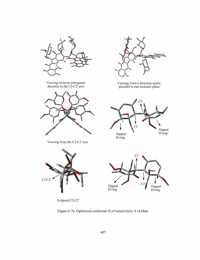
407
Viewing from a direction nearlyparrallel to one aromatic plane
Viewing from an orthogonaldirection to the C2-C2' axis
Viewing from the C2-C2' axis
C2
C2'
f lippedD-ring
Eclipsed C2-C2'
f lippedD-ring
C2
C2'C2/C2'
f lippedD-ring
f lippedD-ring
Figure 6-7b. Optimized conformer II of lomaiviticin A (1.11a).

408
Viewing from an orthogonaldirection to the C2-C2' axis
Viewing from a direction nearlyparrallel to one aromatic plane
Viewing from the C2-C2' axis
C2
C2'
f lippedD-ring
Eclipsed C2-C2'
f lippedD-ring
C2
C2'C2/C2'
f lippedD-ring
flippedD-ring
Figure 6-7c. Optimized conformer III of lomaiviticin A (1.11a).

409
Viewing from an orthogonaldirection to the C2-C2' axis
Viewing from a direction nearlyparrallel to one aromatic plane
Viewing from the C2-C2' axis
C2C2' normal
D-ring
near Eclipsed C2-C2'
normalD-ring
normalD-ring
C2
C2'
C2/C2'
Figure 6-7d. Optimized conformer IV of lomaiviticin A (1.11a).
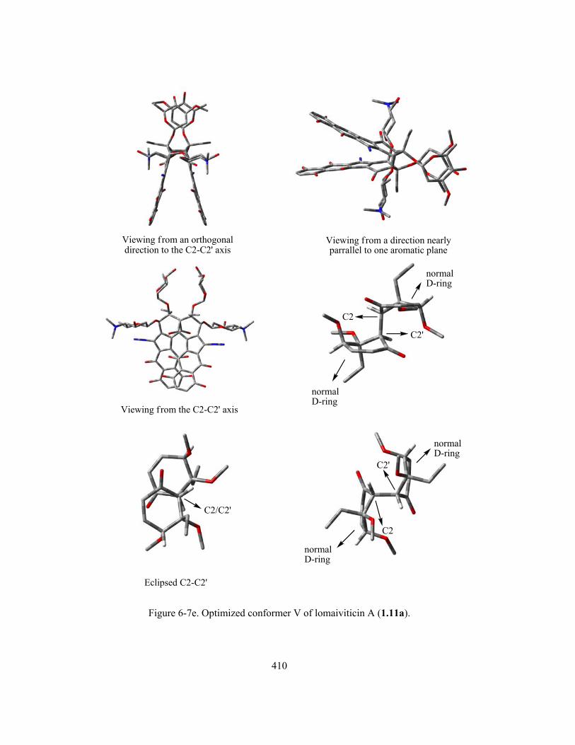
410
Viewing from an orthogonaldirection to the C2-C2' axis
Viewing from a direction nearlyparrallel to one aromatic plane
Viewing from the C2-C2' axis
C2
C2'
Eclipsed C2-C2'
normalD-ring
normalD-ring
normalD-ring
normalD-ring
C2
C2'
C2/C2'
Figure 6-7e. Optimized conformer V of lomaiviticin A (1.11a).

411
Table 6-4. Summary of some calculated physical properties of the possible conformations of 1.11a.
Conformer of 1.11a
(D-rings)a
E (RHF) (a.u.)b
ΔE (kcal/mol)c
νN≡N (cm-1)d
<O-C4-C4a-C5 (°)e
C2-C2’
conformation
dC2-C2’ (Å)
Conformer V
(normal-normal)
-4742.72165161
0
N/A
86.9/86.7 Eclipsed 1.572
Conformer I
(twist-boat-flipped)f
-4742.71652412
3.22
failed
17.2 (twist-boat) /37.3 (flipped)
near Eclipsed 1.566
Conformer II
(flipped-flipped)
-4742.69837153
14.39
N/A
27.4/27.4 Eclipsed 1.578
Conformer IV
(normal-normal)
-4742.69763559
15.07
2186/2186
83.7/83.7 near Eclipsed 1.575
Conformer III
(flipped-flipped)
-4742.69015339
19.77
N/A
31.1/25.5 Eclipsed 1.582
a. All final geometries except conformer I (see footnote f) adopt the same type of D-ring conformations as in the
corresponding starting geometry, i.e. a starting conformation with “normal-normal” D-rings would give an
optimized one also having “normal-normal” D-rings, and so on; b. All E(RHF) were not corrected with ZPE
since it was not available from MO calculations due to the lack/failure of frequency calculations, 1 a.u. =
627.5095 kcal/mol; c. Relative to the most stable conformer (i.e., conformer V); d. Scaled by a scaling factor of
0.8929. “N/A” means that no frequency calculation was performed while “failed” indicates that the frequency
calculation was carried out but terminated due to errors; e. Similar to the cases of 6.5, each conformer of
lomaiviticin A (1.11a) also has two of such dihedral angles (i.e., <O-C4-C4a-C5 and <O-C4’-C4a’-C5’) due to
the symmetric dimer structure, which is a good indication for the D-ring conformation (dihedral angle < 40°
indicates “flipped” D-ring and dihedral angle > 80° corresponds to “normal” D-ring); f. Conformer I started
with “flipped-flipped” D-rings but ended with “twist-boat-flipped” D-rings.
Some very interesting observations and useful conclusions could be made based on these
computational results of lomaiviticin A (1.11a). First of all, the chances to find other stable
conformations of 1.11a besides the above five (Figure 6-7 and Table 6-4) are judged to be extremely
small. The availablity of fewer conformations of lomaiviticin A (1.11a) than the model compound 6.5
(nine conformations) is expected, since the four bulky sugars within 1.11a would definitely take more

412
space along the D-rings, which is already very crowded even in the case of 6.5. This dramatically
limits the conformational freedom of the dimeric skeleton as well as the sugars themselves. Second,
the obtained conformations of 1.11a resemble some characteristics of those previously observed for
6.5 (section 6.1.4). For example, all conformers of 1.11a along the C2-C2’ axis possess a (near)
eclipsed rather than staggered conformation for the six C2/C2’ substituents (Figure 6-7 and Table 6-
4), as previously observed with the three most stable conformations of 6.5 (Figure 6-6 and Appendix
F). The C2-C2’ bond lengths within 1.11a (i.e., 1.566–1.582 Å), as predicted earlier, are also closely
comparable to the ones found within 6.5 (i.e., ~ 1.57 Å). Third, to satisfy the C2 symmetry
requirement of lomaiviticin A (1.11a) as suggested by its spectroscopic data, among the five
conformational candidates, the obviously unsymmetric conformer I (Figure 6-7a) and III (Figure 6-7c)
can be immediately ruled out. The remaining conformers II (Figure 6-7b), IV (Figure 6-7d) and V
(Figure 6-7e) are all essentially near C2 symmetric, especially when the dimeric
diazobenzo[b]fluorene skeletons (including the skeletons of the corresponding four carbohydrates) of
these conformations are examined for symmetry. However, the much higher energy of conformer II
and IV than V (i.e., ~ 14–15 kcal/mol) would suggest that the last species is the only reasonable
conformation of 1.11a to be considered.
Interestingly, the most stable and near C2 symmetric conformation V of 1.11a adopts the “normal-
normal” D-rings. This is in contrast to the structurally very similar dimer 6.5 and other simpler model
species (i.e., 6.3 and 6.4) studied earlier, which all have the “flipped” D-ring(s) within their most
stable conformations. This apparent preference for “normal-normal” D-ring of lomaiviticin A (1.11a)
can be traced to some unique structural features and beneficial interactions within the conformation V
as illustrated in Figure 6-8.

413
Figure 6-8. Structural features within conformer V of lomaiviticin A (1.11a) favouring the “normal-
normal” D-rings.
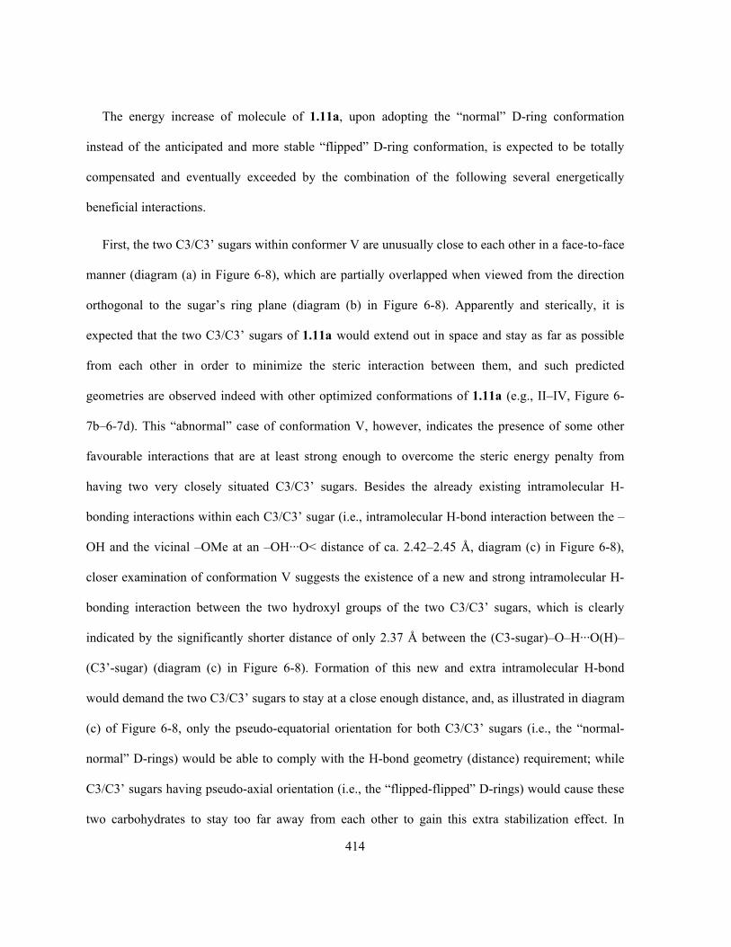
414
The energy increase of molecule of 1.11a, upon adopting the “normal” D-ring conformation
instead of the anticipated and more stable “flipped” D-ring conformation, is expected to be totally
compensated and eventually exceeded by the combination of the following several energetically
beneficial interactions.
First, the two C3/C3’ sugars within conformer V are unusually close to each other in a face-to-face
manner (diagram (a) in Figure 6-8), which are partially overlapped when viewed from the direction
orthogonal to the sugar’s ring plane (diagram (b) in Figure 6-8). Apparently and sterically, it is
expected that the two C3/C3’ sugars of 1.11a would extend out in space and stay as far as possible
from each other in order to minimize the steric interaction between them, and such predicted
geometries are observed indeed with other optimized conformations of 1.11a (e.g., II–IV, Figure 6-
7b–6-7d). This “abnormal” case of conformation V, however, indicates the presence of some other
favourable interactions that are at least strong enough to overcome the steric energy penalty from
having two very closely situated C3/C3’ sugars. Besides the already existing intramolecular H-
bonding interactions within each C3/C3’ sugar (i.e., intramolecular H-bond interaction between the –
OH and the vicinal –OMe at an –OH···O< distance of ca. 2.42–2.45 Å, diagram (c) in Figure 6-8),
closer examination of conformation V suggests the existence of a new and strong intramolecular H-
bonding interaction between the two hydroxyl groups of the two C3/C3’ sugars, which is clearly
indicated by the significantly shorter distance of only 2.37 Å between the (C3-sugar)–O–H···O(H)–
(C3’-sugar) (diagram (c) in Figure 6-8). Formation of this new and extra intramolecular H-bond
would demand the two C3/C3’ sugars to stay at a close enough distance, and, as illustrated in diagram
(c) of Figure 6-8, only the pseudo-equatorial orientation for both C3/C3’ sugars (i.e., the “normal-
normal” D-rings) would be able to comply with the H-bond geometry (distance) requirement; while
C3/C3’ sugars having pseudo-axial orientation (i.e., the “flipped-flipped” D-rings) would cause these
two carbohydrates to stay too far away from each other to gain this extra stabilization effect. In

415
addition, this intramolecular H-bond situation around the C3/C3’ sugars within the conformer V of
1.11a shall now be considered as a three-centered (bifurcated) hydrogen bond (diagram (c) in Figure
6-8), which are rather common both intermolecularly and intramolecularly (to a lesser extent) among
crystal structures of simple sugars and amino acids.385,386 The three-centered hydrogen bond has been
estimated, based on a structural survey, to be energetically comparable to the most common two-
centered hydrogen bonds (-O–H···O-).386 Another literature work on 6.24, a diaryl amide with two
nearby interacting methoxy groups has found, both experimentally and computationally, that an
intramolecular three-centered hydrogen bond in this partially rigidified structure significantly
enhances its stability due to a positive cooperativity between the two two-centered components of the
three-centered hydrogen bonding interaction.387,388 Therefore, it is likely that the three-centered
hydrogen bond interaction observed within the conformation V of 1.11a is one of the significant
energetic contributors to the preference for this geometry. On the other hand, the presence of multiple
oxygen atoms and nearby hydrogen atoms (attached to carbon) in close proximity within these two
very close C3/C3’ sugars might also lead to weak but possible and multiple C-H···O hydrogen
bonding interactions,389 which could further stabilize the conformation.
Second, as an added bonus for stability when compared with others, the conformation V of 1.11a
should also benefit decreased steric repulsion between the two adjacent sugars at C3 and C4 (as well
C3’ and C4’), as a result of to the now larger space and further distance between them caused by the
closer correlation between the C3 and C3’ sugars.

416
Assuming that the conformer V is indeed not only the most stable conformation for lomaiviticin A
(1.11a) in the gas phase but also in solution phase, it is necessary to consider the very likely
protonation of the dimethylamino group(s) of the C4/C4’-sugars under (near) physiological pH (~
7.4), in which its biological activities were studied. The pKa values of the two dimethylamino groups
within the two C4/C4’-sugars of lomaiviticin A (1.11a) are unknown, but they are expected to be
comparable to a typical tertiary organic amine such as NMe3 (pKa ~ 9.8). Despite the many measured
pKa and proposed models for predicting pKa of various simple aminosugars, literature data on the
basicity of 4-aminosugars is rather limited.390-392 The only known example, to the best of the author’s
knowledge, is a UDP-4-aminosugar 6.25 that has a measured pKa of 9.5 ± 0.4 for its C4 amino
group.393 In addition, estimation of the pKa of the methylated model aminosugar 6.2 by using the
(online) program “ALOGPS 2.1”394,395 also gave a predicted pKa value of 8.70, which is fairly
consistent with the above analysis and literature example.
6.25 UDP-4-aminosugar
O
OUDP
AcHNHO
NH2
Since the two amino nitrogens of lomaiviticin A (1.11a) stay very far away from each other even
though they co-exist within the same molecule, it is reasonable to assume that each of them would
behave independently. Therefore, both dimethylamino groups of 1.11a should be almost fully
protonated under physiological pH (~ 7.4) when considering the estimated range of pKa (~ 8.70–
9.80). It is of considerable practical meaning to determine whether such double protonation would
significantly alter the conformation of the molecule or not. The study was carried out by manually
adding a proton at each of the two amino nitrogen sites of the most stable conformation V of
lomaiviticin A (1.11a). The double protonated structure [1.11a + H + H]2+ was then subjected to the

417
same geometry optimization as before. The final results, as given in Figure 6-9 (diagram (a) vs (b),
neutral 1.11a vs double protonated 1.11a), clearly indicate that such protonation does not apply any
significant alteration to the overall conformation of the molecule. The most obvious change caused by
the protonation, as one would expect, is that the intramolecular hydrogen bonding interactions within
the C4/C4’-sugar vary from the –Me2N···H–O– in the neutral molecule to the –Me2N+H···O(H)– in
the double protonated species, since hydrogen bonding of the latter type is significantly stronger than
the former one. Of course, it would be of even greater interest to examine the influence of such
protonation on the diazonium ion character (diazo IR frequency) of the molecule, which is currently
in progress in the Dmitrienko group. However, the diazonium ion character of 1.11a is not expected
to be affected dramatically upon such protonation, since the protonated sites are too far away from the
corresponding diazo moiety to apply any significant electronic effects, either inductively or through
space, or steric effects.

418
C2-C2'axis
(a) Optimized neutral lomaiviticin A (1.11a)Conformer V (the most stable one)
(b) Optimized double protonated Conformer V oflomaiviticin A (1.11a)
C2-C2'axisalmost identical
protonationsite
protonationsite
intramolecular -Me2N H-O- hydrogen bond
C2
C2'
(c) Partial structure from (a)*
intramolecular -Me2NH O(H)- hydrogen bond
C2
C2'
(d) Partial structure from (b)*
neutralamine
neutralamine
protonatedamine
protonatedamine
* For clarity, only the dimeric D-rings with C4/C4'-sugars are shown and C3/C3'-sugars are omitted.All hydrogens except C2/C2'-H and those involved in the intramolecular hydrogen bonding are omitted.
C4
C4'
C4
C4'N
H O
N
HO
N+
HO
N+H
O
Figure 6-9. Comparison of the optimized conformations of the double protonated and neutral
lomaiviticin A (1.11a).

419
For lomaiviticin B (1.11b), after extensive optimization of various possible conformations, only
one single conformation having the “flipped-flipped” D-rings (Figure 6-10) was obtained. All efforts
to achieve stable conformations of 1.11b containing either the “normal-flipped” D-rings or the
“normal-normal” D-rings failed. The former situation is once again of very little importance, since
just like lomaiviticin A (1.11a), spectroscopic evidence suggest this slightly simpler derivative 1.11b
also exists as a symmetric dimer with two identical D-rings.52 Although the obtained conformation of
1.11b (Figure 6-10) is not considered to possess a C2 symmetry by the Gaussian03® program, one can
easily recognize the near C2 symmetry that lies in the main skeleton of this conformation. The “C2
symmetry axis” would be in the middle point of and orthogonal to the C2-C2’ bond within the first
structure shown in Figure 6-10, which is also perpendicular to the paper plane that the C2-C2’ bond
lies in.

420
Figure 6-10. Optimized conformation of the lomaiviticin B (1.11b) having “flipped-flipped” D-rings.

421
The preference of “flipped-flipped” D-rings rather than “normal-normal” D-rings for lomaiviticin
B (1.11b), which is different from the lomaiviticin A (1.11a) but consistent with other model
compounds (i.e., 6.3–6.5), can be explained by the following rationale. Formation of the double
intramolecular hemiketals within lomaiviticin B (1.11b) requires not only the loss of two C3/C3’-
sugars (very likely through hydrolysis) leading to C3/C3’ free hydroxyl groups, but also a close
distance between this tertiary alcohol at C3’ (or C3) with the corresponding C1 (or C1’) carbonyl
carbon. Only the “flipped” D-ring would be able to offer a close enough distance since its C3-OH (or
C3’-OH) exists in an axial orientation, which seems to be the best possible position to facilitate the
formation of the saturated furan ring and produce the hemiketal. As labeled in Figure 6-11, the
significantly longer C-O-C bond lengths within the two 5-membered furan rings than the other similar
C-O-C bonds within the molecule (i.e., 1.425/1.442 Å vs 1.411/1.438 Å) clearly indicate the
difficulty to form such a concave and cyclic structure. If the C3/C3’-OH is part of a “normal” D-ring
instead, the corresponding equatorial orientation of this tertiary alcohol as well as the corresponding
axial C1/C1’-O bond (which exists in equatorial orientation in a “flipped” D-ring) would stay much
further away from each other and make it practically impossible to carry out the same construction of
the hemiketal. In addition, this “flipped-flipped” D-rings of lomaiviticin B (1.11b) would provide the
added benefit of having the equatorial C4/C4’-O(sugar) bond to align much better (than the axial
C4/C4’-O bond within a “normal” D-ring) with the C5/C5’-diazo bond, leading to favourable dipole-
dipole interactions that lower the energy and enhance the diazonium ion character. As a supportive
evidence, the unusual and possible conformation of lomaiviticin A (1.11a) having the “twist-boat-
flipped” D-rings is only ca. 3 kcal/mol higher in energy than its most stable conformation with the
“normal-normal” D-rings, suggesting that switching from normal D-ring (of 1.11a) to flipped D-ring
(of 1.11b) may not be difficult at least thermodynamically, especially when such conformational

422
conversion is accompanied and partially driven by the chemical transformation (loss of C3/C3’-
sugars and formation of hemiketals) at the mean time.
C1
C1'
C2
C2'C4'
C3
1.556Å1.442 Å
1.425 Å
1.425 Å
1.442 Åax.
ax.
C3'
C4
1.438 Å
1.438 Å
1.411 Å
1.411 Åeq.
eq.
"f lipped" D-ring
"f lipped" D-ring
Note: All hydrogens except the C2/C2'-H and C4/C4'-H are omitted.
Figure 6-11. Detailed geometric information of partial structure from the optimized conformation of
lomaiviticin B (1.11b) with “flipped-flipped” D-rings.
Even though it is slightly premature at the moment to completely exclude the possibility of
lomaiviticin B (1.11b) to adopt the “normal-normal” D-rings, the above analysis would suggest that
such a possibility is extremely low. In addition, fusion of the two saturated furan rings with the two
adjacent D-rings, which are part of the rigid diazobenzo[b]fluorene system, completely locks the
entire skeleton of the molecule (not including the C4/C4’-sugars though) and restrains the orientation

423
of the D-ring substituents greatly in space. Therefore, if there exist other possible/stable
conformations of lomaiviticin B (1.11b), they are very likely dealing with mainly (if not only) small
variation of the two C4-sugars’ position when compared to the stable conformation shown in Figure
6-10.
Finally, the uncertainty of the absolute stereochemistry of the carbohydrate components of
lomaiviticins, as discussed in section 6.1.1, is worthy of some further consideration. The lack of both
C3/C3’ sugars within lomaiviticin B (1.11b) due to the formation of intramolecular bishemiketals
seems to affect the biological activities to a minimal extent, since lomaiviticin A (1.11a) and B (1.11b)
possess almost identical DNA cleavage capability.52 This observation would suggest that the C3/C3’
sugars are not essential components of the molecule to account for its biological activity, thus the
sugar’s absolute stereochemistry would be of even less importance. On the other hand, the C4/C4’
sugars are probably more involved in the biological activities of lomaiviticins, partially due to the fact
that this carbohydrate stays in the close proximity of the C5/C5’-diazo group and could significantly
enhance the diazonium ion character upon taking up an appropriate conformation (section 6.1.3). The
mode-of-action of the lomaiviticins is presumably similar to the kinamycins, as suggested by the
Dmitrienko group based on the evidence discussed in Chapter 4. Yet the absolute stereochemistry of
these C4/C4’-sugars is probably of less importance as well, since both enantiomers of the C4/C4’-
sugar are expected to have the same type and same degree of influence on the nearby linear and
achiral diazo group. In the mean time, one should not underestimate the very possible and potential
usefulness of these sugar units from some other points of view. The presence of the sugar moiety
within the lomaiviticins could, in principle, improve the solubility of the compounds in the aqueous
biological systems and help the molecule to diffuse through the cell membrane, which may be
practically crucial to their potent activities. Therefore, it is believed that the absolute stereochemistry

424
of these sugars is not as important as the mere presence of such carbohydrates within lomaiviticins
towards their biological activities.
With no further available information on the absolute stereochemistry of the carbohydrates within
lomaiviticins, current MO calculations assuming the C3/C3’ sugars as α-L-oleandrose and the C4/C4’
sugars as β-D-pyrrolosamine, which are the same as what had been shown in the original work from
the Wyeth group, should be a reasonable starting point. Even if later spectroscopic evidence may
prove the absolute stereochemistry of the carbohydrate component of lomaiviticins to be otherwise,
the computed conformations of lomaiviticins can be easily modified to reflect such possible changes,
which are not expected to pose significant influence on the overall conformation and diazonium ion
character of these natural antibiotics. It has been noticed that two very recent works regarding the
total synthesis of the lomaiviticin carbohydrate components (or similar analogues) have been reported
by the Herzon’s group396 and Shair’s group,397 yet none of these results have realized or offered any
further information to clarify the absolute stereochemistry problem of such sugars.
In summary, the above successful computation of stable conformation(s) of lomaiviticins has
provided the first study of its kind in this area of kinamycins and related diazobenzo[b]fluorenes. The
results could be the important basis for future in-depth mechanistic studies regarding these natural
products and their biological activities, and may be of great help in designing simpler but efficient
analogues of kinamycins and lomaiviticins.
6.2 Vibrational Circular Dichroism Spectra of Kinamycins
The vibrational optical activity (VOA) is the response of a molecule to circularly polarized
radiation, and VOA in the IR region is more commonly known as the vibrational circular dichroism
(VCD).398,399 A chiral molecule would behave differently toward the left and right circularly polarized
(IR) radiation, which is in fact an electromagnetic wave with chirality. Thus, the VCD technique is
425
very effective to study chirality (absolute configuration) and distinguish between
enantiomers/diastereomers in a non-destructive manner,400 yet it receives much less attention from
most organic chemists than it really deserves. As an added bonus feature and particularly related to
this study, VCD is also extremely useful in determining conformation in solution, since even a rather
small change in conformation of the same molecule would lead to an apparent and significant
difference of the corresponding VCD spectrum.401 When compared with other traditional methods for
studying stereochemistry, chirality and conformation, VCD requires neither physical separation of
stereoisomers/conformers (vs chiral chromatography), nor very complicated/expensive equipment (vs
X-ray or NMR), additional reagents (e.g., chiral NMR shift reagent) or procedures (e.g., growth of
single crystal for X-ray). Most VCD cases simply involve an acquisition of the spectrum, which is
very similar to a routine IR experiment, along with an electronic structure calculation that is
performed through computational methods with a software package such as Gaussian®. Then a
comparison between the experimental and the simulated VCD spectra should be able to determine the
stereochemistry of interest. Such practice offers a further and unique advantage when dealing with an
inseparable mixture of two or more stereoisomers/conformers. Theoretical VCD spectrum of each
individual stereoisomer/conformer can be calculated rather accurately, and mathematical treatment
with the experimental spectrum based on these predicted individual VCD spectrum can provide, in
principle, a good estimation of the distribution of each species presents. The less common availability
of commercial VCD instruments and the lack of practical molecular modeling study among the
general organic community, however, are probably the two main reasons for the rather limited
applications of VCD in organic chemistry so far, although they have been growing in recent years.
With no direct access to a VCD spectrometer by the Dmitrienko group at the moment, studies were
focused on the VCD calculations of the kinamycins and lomaiviticins. Previous NMR, IR and
theoretical studies on kinamycin F (1.1f) in different solvents (section 2.3 and 4.4) have already

426
suggested an equilibrium between its two D-ring conformers, which is affected dramatically by the
solvent polarity. The simulated VCD spectra of kinamycin F (1.1f) in various media (gas phase and
solutions) showed that the corresponding two conformers, with the only structural difference in their
D-ring conformation (i.e., “normal” D-ring vs “flipped” D-ring), would lead to distinctively different
VCD spectra as illustrated in Figure 6-12a (1.1f with a “normal” D-ring) and Figure 6-12b (1.1f with
a “flipped” D-ring). On the other hand, for each individual conformer, the media (gas phase vs non-
polar solvent CHCl3 vs polar solvent acetone) also shows some (solvent) effects on the calculated
VCD spectra, which involves mainly the shift of peaks and variation of signal intensities due to the
change in environmental polarity, but the major spectrum pattern persists fairly well. Since kinamycin
F (1.1f) is readily available in the Dmitrienko laboratory through chemical transformation of other
and more abundant natural kinamycins produced by bacterial fermentation, possible collaborations
with other research laboratories having a VCD spectrometer in the near future would definitely lead
to a quick and hopefully quantitative analysis of the conformational preference of kinamycin F (1.1f)
in solution.

427
Figure 6-12a. Computed VCD spectra of kinamycin F (1.1f) with a “normal” D-ring at RHF/6-31G
level in different medium. (Note: 1. IR frequencies were corrected with a scaling factor of 0.8929; 2.
PCM model was used for solution calculations.)

428
Figure 6-12b. Computed VCD spectra of kinamycin F (1.1f) with a flipped D-ring at RHF/6-31G
level in different medium. (Note: 1. IR frequencies were corrected with a scaling factor of 0.8929; 2.
PCM model was used for solution calculations; 3. Calculation in acetone failed.)
The benefit of using the VCD technique to determine the unknown absolute stereochemistry and
conformation of lomaiviticins would be very significant, especially when considering the
computational results of lomaiviticins discussed in the previous section of this chapter. There are only
very few possible (reasonable) conformations for these complex natural products (section 6.1.5), and
the corresponding VCD spectrum for every single one of them can be calculated in principle. If the
experimental VCD spectra of lomaiviticins become available in the future (the Wyeth group who
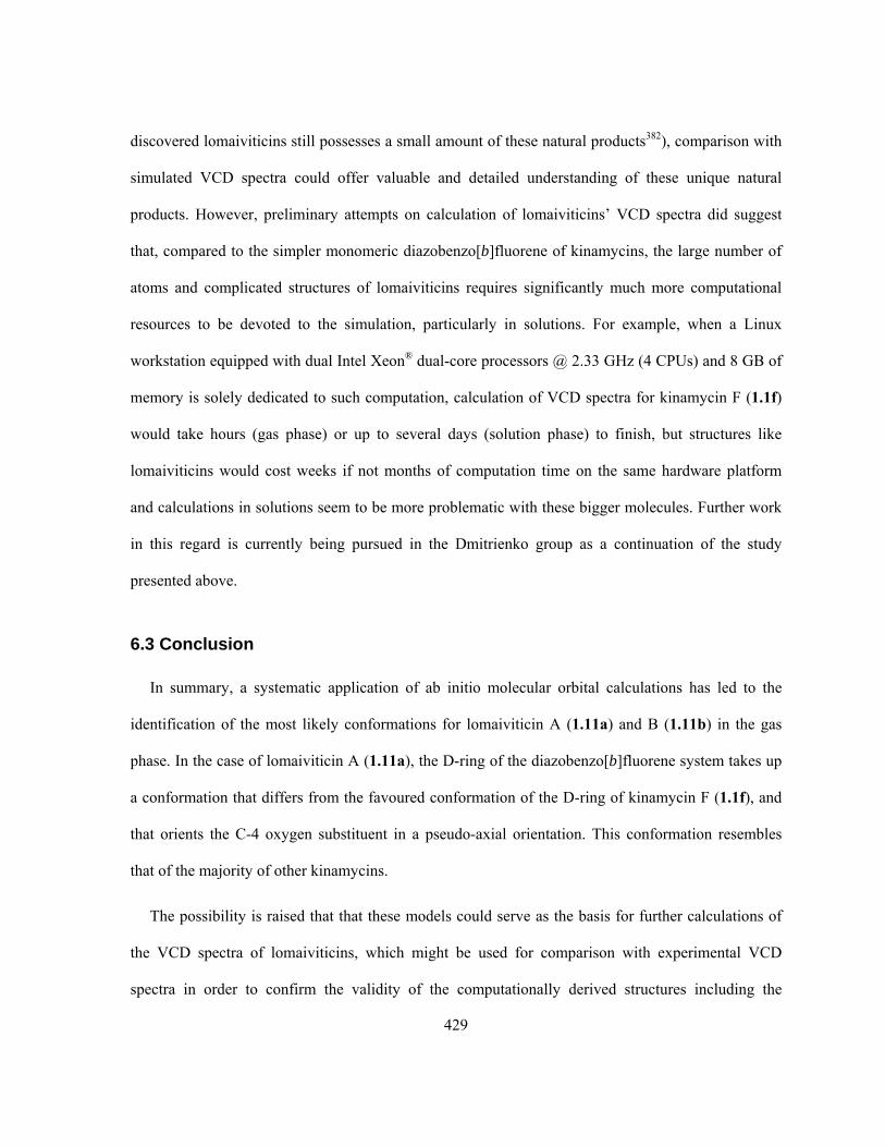
429
discovered lomaiviticins still possesses a small amount of these natural products382), comparison with
simulated VCD spectra could offer valuable and detailed understanding of these unique natural
products. However, preliminary attempts on calculation of lomaiviticins’ VCD spectra did suggest
that, compared to the simpler monomeric diazobenzo[b]fluorene of kinamycins, the large number of
atoms and complicated structures of lomaiviticins requires significantly much more computational
resources to be devoted to the simulation, particularly in solutions. For example, when a Linux
workstation equipped with dual Intel Xeon® dual-core processors @ 2.33 GHz (4 CPUs) and 8 GB of
memory is solely dedicated to such computation, calculation of VCD spectra for kinamycin F (1.1f)
would take hours (gas phase) or up to several days (solution phase) to finish, but structures like
lomaiviticins would cost weeks if not months of computation time on the same hardware platform
and calculations in solutions seem to be more problematic with these bigger molecules. Further work
in this regard is currently being pursued in the Dmitrienko group as a continuation of the study
presented above.
6.3 Conclusion
In summary, a systematic application of ab initio molecular orbital calculations has led to the
identification of the most likely conformations for lomaiviticin A (1.11a) and B (1.11b) in the gas
phase. In the case of lomaiviticin A (1.11a), the D-ring of the diazobenzo[b]fluorene system takes up
a conformation that differs from the favoured conformation of the D-ring of kinamycin F (1.1f), and
that orients the C-4 oxygen substituent in a pseudo-axial orientation. This conformation resembles
that of the majority of other kinamycins.
The possibility is raised that that these models could serve as the basis for further calculations of
the VCD spectra of lomaiviticins, which might be used for comparison with experimental VCD
spectra in order to confirm the validity of the computationally derived structures including the

430
assumed absolute configuration, if sufficient quantities of the lomaiviticins are made available by the
Wyeth (now Pfizer) research group. It is hoped that these computed three dimensional structures will
serve to guide the design of simpler, more accessible molecules with biological activities comparable
to those of the natural lomaiviticins.

431
Appendix A. Summary of Antibiotic Activities of Natural
Kinamycins in The Literature4,6-10,18,52
432

433

434

435

436

437

438

439

440

441
Appendix B. X-ray Crystallographic Structure of Epoxy Diester 2.30
Table B-1. Crystal data and structure refinement for epoxy diester 2.30
Empirical formula C25H16N4O13
Formula weight 580.42
Cell measurement temperature 200(1) K
Wavelength 0.71073 Å
Crystal system, space group Monoclinic, C2
Unit cell dimensions a = 29.621(4) Å alpha = 90.00 deg. b = 5.5479(7) Å beta = 95.328(4) deg. c = 15.0615(18) Å gamma = 90.00 deg.
Volume, Z 2464.4(5) Å3, 4
Density 1.564 g/cm3
Absorption coefficient 0.130 mm-1
F(000) 1192
Crystal size 0.50 mm × 0.052 mm × 0.026 mm
2 range for data collection 2.74 to 24.84
Limiting indices -31 < h < 31, -5 < k < 5, -16 < l < 15
Reflections collected / unique 4779 / 2961 [R(int) = 0.0750]
Completeness to = 24.84 99.8 %
Absorption correction None
Refinement method Full-matrix least-squares on F2
Data / restraints / parameters 2961 / 0 / 376
Goodness-of-fit on F2 1.591
Final R indices [I>2(I)] R1 = 0.0983, wR2 = 0.1555
R indices (all data) R1 = 0.1217, wR2 = 0.1618
Largest diff. peak and hole 0.356 and -0.285 e-.Å-3

442
Table B-2. Atomic coordinates (x 104) and equivalent isotropic displacement parameters (Å2 x 10³) for 2.30. Ueq is defined as one third of the trace of the orthogonalized Uij tensor.
x y z Ueq
C(1) 1704(3) 200(20) 6577(5) 44(3) C(2) 1902 -721 5780 68(3) C(3) 1564(4) 690(20) 4940(7) 62(3) C(4) 1094(4) 30(20) 4988(7) 65(3)
C(4A) 1025(3) 1910(20) 5689(6) 45(2) C(5) 678(3) 3580(30) 5613(6) 57(3) C(6) 597(3) 5140(30) 6237(6) 61(3) C(7) 908(3) 5230(30) 7035(7) 78(4) C(8) 1255(3) 3670(20) 7112(6) 51(3)
C(8A) 1329(3) 1970(20) 6446(6) 42(3) C(10) 2226(3) 170(20) 7913(6) 48(3) C(12) 2619(3) 1510(20) 8395(6) 35(2) C(13) 2782(3) 510(20) 9214(5) 40(2) C(14) 3135(3) 1650(20) 9671(5) 36(2) C(15) 3333(3) 3800(20) 9383(6) 41(3) C(16) 3152(3) 4634(19) 8579(5) 30(2) C(17) 2808(3) 3560(20) 8069(6) 39(2) C(25) 1712(5) -2480(30) 4245(9) 143(6) C(27) 541(3) 260(20) 3688(6) 47(3) C(29) 393(3) 1740(20) 2906(6) 44(3) C(30) -9(3) 1040(20) 2405(6) 48(3) C(31) -138(3) 2320(20) 1632(6) 46(3) C(32) 78(3) 4370(20) 1377(6) 43(3) C(33) 467(3) 5020(20) 1905(6) 43(3) C(34) 616(3) 3720(20) 2664(6) 52(3) N(18) 3348(3) 390(20) 10488(4) 45(2) N(21) 3362(3) 6788(18) 8205(5) 45(2) N(35) -563(4) 1600(20) 1072(8) 90(4) N(38) 710(3) 7158(19) 1673(6) 54(2) O(9) 2079(2) 1303(15) 7148(4) 48(2)
O(11) 2090(2) -1748(17) 8125(4) 68(2) O(19) 3169(2) -1391(16) 10729(4) 47(2) O(20) 3685(3) 1300(19) 10876(4) 82(3) O(22) 3645(2) 7854(15) 8733(4) 44(2) O(23) 3252(2) 7398(16) 7457(4) 54(2) O(24) 1909(2) 1185(17) 5073(4) 68(2) O(26) 919(2) 1162 4121(4) 62(2) O(28) 346(2) -1470(20) 3949(4) 78(3)
443
Table B-2 (continued). Atomic coordinates (x 104) and equivalent isotropic displacement parameters (Å2 x 10³) for 2.30. Ueq is defined as one third of the trace of the orthogonalized Uij tensor.
x y z Ueq
O(36) -765(4) 70(20) 1338(7) 191(7) O(37) -648(3) 2550(20) 371(5) 103(3) O(39) 599(3) 8069(18) 941(5) 83(3) O(40) 1018(2) 7928(17) 2185(5) 63(2) H(1) 1591 -1211 6905 53 H(2) 2147 -1966 5846 81 H(4) 907 -1426 5097 78 H(5) 485 3608 5073 68 H(6) 341 6173 6166 73 H(7) 867 6360 7494 93 H(8) 1460 3724 7635 61
H(13) 2652 -904 9438 48 H(15) 3573 4603 9725 50 H(17) 2702 4196 7502 47
H(25X) 1540 -3987 4278 215 H(25Y) 2037 -2819 4365 215 H(25Z) 1653 -1788 3647 215 H(30) -185 -270 2589 57 H(32) -33 5286 870 51 H(34) 880 4240 3020 62

444
Table B-3. Bond lengths and bond angles for 2.30.
Length [Å] Length [Å]
C1-O9 1.472(9) C32-C33 1.385(12) C1-C8A 1.478(12) C33-C34 1.386(12) C1-C2 1.478(9) C33-N38 1.449(13)
C2-O24 1.502(9) N35-O37 1.187(11) C2-C3 1.539(11) N35-O36 1.191(13) C3-C4 1.458(15) N38-O40 1.216(9)
C3-O24 1.461(13) N38-O39 1.230(9) C3-C25 1.536(17) C4-O26 1.497(11) Angle [] C4-C4A 1.511(13) O9-C1-C8A 108.7(7) C4A-C5 1.382(13) O9-C1-C2 106.4(6)
C4A-C8A 1.386(11) C8A-C1-C2 118.1(6) C5-C6 1.315(14) C1-C2-O24 111.4(6) C6-C7 1.446(12) C1-C2-C3 112.7(5) C7-C8 1.338(13) O24-C2-C3 57.4(5)
C8-C8A 1.412(12) C4-C3-O24 117.2(9) O9-C10 1.349(10) C4-C3-C25 123.1(11)
C10-O11 1.192(11) O24-C3-C25 108.4(11) C10-C12 1.510(12) C4-C3-C2 120.9(8) C12-C17 1.377(12) O24-C3-C2 60.0(6) C12-C13 1.398(11) C25-C3-C2 110.4(10) C13-C14 1.353(11) C3-C4-O26 109.0(9) C14-C15 1.417(12) C3-C4-C4A 114.6(9)
C14-N18 1.502(10) O26-C4-C4A 105.2(9)
C15-C16 1.358(11) C5-C4A-C8A 118.3(9)
C16-C17 1.355(11) C5-C4A-C4 123.6(9)
C16-N21 1.482(11) C8A-C4A-C4 118.1(9)
N18-O19 1.193(10) C6-C5-C4A 124.4(9)
N18-O20 1.217(10) C5-C6-C7 118.2(10)
N21-O23 1.193(8) C8-C7-C6 118.3(11)
N21-O22 1.249(8) C7-C8-C8A 122.7(9)
O26-C27 1.339(10) C4A-C8A-C8 117.9(9)
C27-O28 1.205(12) C4A-C8A-C1 121.2(9)
C27-C29 1.467(13) C8-C8A-C1 120.9(7)
C29-C34 1.351(12) C10-O9-C1 118.2(7)
C29-C30 1.405(11) O11-C10-O9 123.6(9)
C30-C31 1.388(12) O11-C10-C12 125.1(9)
C31-C32 1.378(13) O9-C10-C12 110.8(9)
C31-N35 1.502(12) C17-C12-C13 121.6(8)

445
Table B-3 (continued). Bond lengths and bond angles for 2.30.
Angle [] Angle []
C17-C12-C10 123.8(8) O26-C27 C29 110.9(10)
C13-C12-C10 114.6(8) C34-C29 C30 119.4(9) C14-C13-C12 116.8(8) C34-C29-C27 123.7(9) C13-C14-C15 124.0(8) C30-C29-C27 116.8(9) C13-C14-N18 116.5(9) C31-C30-C29 117.6(9) C15-C14-N18 119.3(8) C32-C31-C30 123.8(9) C16-C15-C14 114.8(8) C32-C31-N35 116.8(9) C17-C16-C15 124.7(8) C30-C31-N35 119.1(9) C17-C16-N21 117.3(8) C31-C32-C33 116.0(9) C15-C16-N21 117.8(8) C34-C33-C32 121.5(9) C16-C17-C12 118.0(8) C34-C33-N38 119.7(10) O19-N18-O20 124.4(8) C32-C33-N38 118.7(9) O19-N18-C14 117.9(8) C29-C34-C33 121.3(10) O20-N18-C14 117.6(9) O37-N35-O36 124.9(12) O23-N21-O22 125.1(8) O37-N35-C31 118.6(11) O23-N21-C16 120.0(8) O36-N35-C31 116.1(11) O22-N21-C16 114.9(7) O40-N38-O39 123.2(9)
C3-O24-C2 62.6(6) O40-N38-C33 119.7(9)
C27-O26-C4 118.3(8) O39-N38-C33 117.0(10)
O28-C27 O26 122.9(9)
O28-C27-C29 126.1(9)

446
Appendix C. X-ray Crystallographic Structure of Epoxy Diester 2.31
Table C-1. Crystal data and structure refinement for epoxy diester 2.31.
Empirical formula C19H24O5
Formula weight 332.38
Cell measurement temperature 200(2) K
Wavelength 0.71073 Å
Crystal system, space group Triclinic, P1
Unit cell dimensions a = 9.2389(8) Å alpha = 110.302(2) deg. b = 10.0480(9) Å beta = 97.268(2) deg. c = 10.7755(9) Å gamma = 106.443(2) deg.
Volume, Z 871.27(13) Å3, 2
Density 1.267 g/cm3
Absorption coefficient 0.091 mm-1
F(000) 356
Crystal size 0.38 mm × 0.28 mm × 0.20 mm
2 range for data collection 2.08 to 27.88
Limiting indices -12 < h < 12, -13 < k < 13, -14 < l < 14
Reflections collected / unique 9304 / 4163 [R(int) = 0.0351]
Completeness to = 27.88 99.9 %
Absorption correction None
Refinement method Full-matrix least-squares on F2
Data / restraints / parameters 4163 / 0 / 223
Goodness-of-fit on F2 1.164
Final R indices [I>2(I)] R1 = 0.0426, wR2 = 0.0963
R indices (all data) R1 = 0.0490, wR2 = 0.0986
Largest diff. peak and hole 0.337 and -0.205 e-.Å-3

447
Table C-2. Atomic coordinates (x 104) and equivalent isotropic displacement parameters (Å2 x 10³) for 2.31. Ueq is defined as one third of the trace of the orthogonalized Uij tensor.
x y z Ueq
C(1) 5155(1) 3009(1) 3380(1) 32(0) C(2) 5374(1) 1942(1) 4020(1) 33(0) C(3) 4525(1) 1789(1) 5049(1) 33(0) C(4) 3460(1) 2658(1) 5440(1) 30(0)
C(4A) 2813(1) 3091(1) 4341(1) 30(0) C(5) 1431(1) 3396(1) 4336(1) 36(0) C(6) 863(2) 3878(2) 3388(2) 42(0) C(7) 1668(2) 4056(2) 2426(2) 47(0) C(8) 3031(2) 3749(2) 2415(1) 41(0)
C(8A) 3622(1) 3263(1) 3366(1) 32(0) C(10) 6426(1) 3302(1) 1633(1) 32(0) C(12) 6364(2) 2553(2) 142(1) 39(0) C(13) 7091(2) 1331(2) -58(2) 48(0) C(14) 7173(2) 3720(2) -377(2) 57(0) C(16) 6899(2) 1684(2) 4056(2) 47(0) C(18) 2296(2) 1895(2) 7061(1) 35(0) C(20) 753(2) 1086(2) 7249(1) 42(0) C(21) -284(2) 2009(2) 7267(2) 68(1) C(22) 928(2) 765(3) 8515(2) 77(1) O(9) 5294(1) 2420(1) 1984(1) 37(0)
O(11) 7356(1) 4498(1) 2432(1) 48(0) O(15) 4029(1) 608(1) 3688(1) 37(0) O(17) 2145(1) 1757(1) 5751(1) 34(0) O(19) 3496(1) 2591(1) 7917(1) 54(0) H(1) 6013 4011 3896 38 H(3) 5049 1564 5783 39 H(4) 4047 3604 6276 36 H(5) 873 3271 4994 43 H(6) -78 4086 3397 51 H(7) 1283 4389 1772 56 H(8) 3576 3872 1748 50
H(12) 5246 2056 -383 47 H(13X) 8189 1799 450 72 H(13Y) 7018 812 -1032 72 H(13Z) 6533 596 279 72 H(14X) 6705 4508 -196 85 H(14Y) 7053 3224 -1363 85 H(14Z) 8284 4181 93 85
448
Table C-2 (continued). Atomic coordinates (x 104) and equivalent isotropic displacement parameters (Å2 x 10³) for 2.31. Ueq is defined as one third of the trace of the orthogonalized Uij tensor.
x y z Ueq
H(16X) 6967 1054 4565 71 H(16Y) 7759 2661 4505 71 H(16Z) 6966 1168 3120 71 H(20) 252 94 6442 50
H(21X) 166 2975 8068 102 H(21Y) -1321 1449 7309 102 H(21Z) -371 2201 6434 102 H(22X) 1616 175 8478 115 H(22Y) -96 188 8566 115 H(22Z) 1380 1724 9325 115
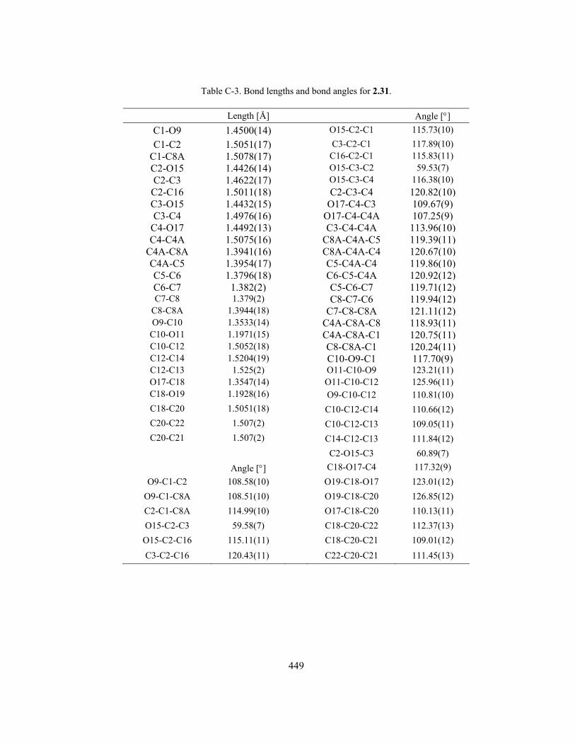
449
Table C-3. Bond lengths and bond angles for 2.31.
Length [Å] Angle []
C1-O9 1.4500(14) O15-C2-C1 115.73(10)
C1-C2 1.5051(17) C3-C2-C1 117.89(10) C1-C8A 1.5078(17) C16-C2-C1 115.83(11) C2-O15 1.4426(14) O15-C3-C2 59.53(7) C2-C3 1.4622(17) O15-C3-C4 116.38(10)
C2-C16 1.5011(18) C2-C3-C4 120.82(10) C3-O15 1.4432(15) O17-C4-C3 109.67(9) C3-C4 1.4976(16) O17-C4-C4A 107.25(9)
C4-O17 1.4492(13) C3-C4-C4A 113.96(10) C4-C4A 1.5075(16) C8A-C4A-C5 119.39(11)
C4A-C8A 1.3941(16) C8A-C4A-C4 120.67(10) C4A-C5 1.3954(17) C5-C4A-C4 119.86(10) C5-C6 1.3796(18) C6-C5-C4A 120.92(12) C6-C7 1.382(2) C5-C6-C7 119.71(12) C7-C8 1.379(2) C8-C7-C6 119.94(12)
C8-C8A 1.3944(18) C7-C8-C8A 121.11(12) O9-C10 1.3533(14) C4A-C8A-C8 118.93(11)
C10-O11 1.1971(15) C4A-C8A-C1 120.75(11) C10-C12 1.5052(18) C8-C8A-C1 120.24(11) C12-C14 1.5204(19) C10-O9-C1 117.70(9) C12-C13 1.525(2) O11-C10-O9 123.21(11) O17-C18 1.3547(14) O11-C10-C12 125.96(11) C18-O19 1.1928(16) O9-C10-C12 110.81(10)
C18-C20 1.5051(18) C10-C12-C14 110.66(12)
C20-C22 1.507(2) C10-C12-C13 109.05(11)
C20-C21 1.507(2) C14-C12-C13 111.84(12)
C2-O15-C3 60.89(7)
Angle [] C18-O17-C4 117.32(9)
O9-C1-C2 108.58(10) O19-C18-O17 123.01(12)
O9-C1-C8A 108.51(10) O19-C18-C20 126.85(12)
C2-C1-C8A 114.99(10) O17-C18-C20 110.13(11)
O15-C2-C3 59.58(7) C18-C20-C22 112.37(13)
O15-C2-C16 115.11(11) C18-C20-C21 109.01(12)
C3-C2-C16 120.43(11) C22-C20-C21 111.45(13)

450
Appendix D. X-ray Crystallographic Structure of Isoprekinamycin
(1.5)
Table D-1. Crystal data and structure refinement for isoprekinamycin 1.5
Empirical formula C18H10N2O4
Formula weight 318.28
Cell measurement temperature 180(2) K
Wavelength 0.71073 Å
Crystal system, space group Monoclinic, P2_1/c
Unit cell dimensions
a = 4.8053(5) Å alpha = 90.00 deg.
b = 14.6913(16) Å beta = 91.020(3) deg.
c = 18.927(2) Å gamma = 90.00 deg. Volume 1336.0(2) Å3
Z, Calculated density 4, 1.582 g/cm3
Absorption coefficient 0.115 mm-1
F(000) 656
Crystal size 0.66 mm × 0.056 mm × 0.034 mm
2 range for data collection 1.75 to 25.02
Limiting indices -5 < h < 5, -17 < k < 17, -22 < l < 21
Reflections collected / unique 7057 / 2344 [R(int) = 0.0664]
Completeness to = 25.02 99.2 %
Absorption correction None
Refinement method Full-matrix least-squares on F2
Data / restraints / parameters 2344 / 0 / 226
Goodness-of-fit on F2 1.186
Final R indices [I>2(I)] R1 = 0.0598, wR2 = 0.0993
R indices (all data) R1 = 0.0847, wR2 = 0.1051
Largest diff. peak and hole 0.304 and -0.194 e-.Å-3

451
Table D-2. Atomic coordinates (x 104) and equivalent isotropic displacement parameters (Å2 x 10³) for 1.5. Ueq is defined as one third of the trace of the orthogonalized Uij tensor.
x y z Ueq
C(1) 7070(6) 7872(2) 2655(2) 30(1) C(2) 9012(6) 7503(2) 2212(2) 32(1) C(3) 9753(5) 6579(2) 2247(1) 28(1) C(4) 8528(5) 6038(2) 2745(1) 28(1)
C(4A) 6527(5) 6390(2) 3197(1) 25(1) C(5) 5147(6) 5832(2) 3711(2) 27(1) C(6) 2987(6) 6092(2) 4176(2) 28(1)
C(6A) 2321(5) 7048(2) 4121(1) 26(1) C(6B) 238(5) 7538(2) 4545(1) 25(1) C(7) -1521(5) 7274(2) 5082(1) 28(1) C(8) -3223(6) 7949(2) 5378(2) 32(1) C(9) -3213(6) 8833(2) 5146(2) 34(1)
C(10) -1471(6) 9098(2) 4597(2) 31(1) C(10A) 219(5) 8444(2) 4319(1) 27(1) C(11) 2286(5) 8548(2) 3750(1) 28(1)
C(11A) 3580(5) 7621(2) 3645(1) 25(1) C(11B) 5715(5) 7329(2) 3162(1) 25(1) O(12) 6582(5) 8778(1) 2570(1) 39(1) C(13) 11847(6) 6194(2) 1746(2) 35(1) N(14) 5897(4) 4941(2) 3765(1) 28(1) N(15) 6449(5) 4215(2) 3827(1) 44(1) O(16) 1798(4) 5550(1) 4577(1) 37(1) O(17) -1746(4) 6431(1) 5347(1) 38(1) O(18) 2816(4) 9261(1) 3447(1) 37(1) H(2) 9874 7883 1873 38 H(4) 9050 5416 2784 33 H(8) -4421 7786 5752 39 H(9) -4394 9268 5359 41
H(10) -1464 9705 4425 37 H(13X) 11992 5535 1816 53 H(13Y) 13665 6477 1838 53 H(13Z) 11247 6321 1258 53 H(12) 5260(60) 8983(19 2872(16) 44(10) H(17) -520(80) 6010(30) 5090(20) 93(14)

452
Table D-3. Bond lengths and bond angles for 1.5.
Length [Å] Angle [] C(1)-O(12) 1.361(3) C(2)-C(3)-C(13) 120.3(2) C(1)-C(2) 1.377(4) C(3)-C(4)-C(4A) 120.9(3)
C(1)-C(11B) 1.416(4) C(4)-C(4A)-C(11B) 121.2(2) C(2)-C(3) 1.404(4) C(4)-C(4A)-C(5) 122.1(2) C(3)-C(4) 1.374(4) C(11B)-C(4A)-C(5) 116.7(2)
C(3)-C(13) 1.505(4) N(14)-C(5)-C(6) 114.0(2) C(4)-C(4A) 1.397(4) N(14)-C(5)-C(4A) 118.2(2)
C(4A)-C(11B) 1.435(3) C(6)-C(5)-C(4A) 127.8(2) C(4A)-C(5) 1.444(4) O(16)-C(6)-C(5) 123.6(2) C(5)-N(14) 1.362(3) O(16)-C(6)-C(6A) 124.2(2) C(5)-C(6) 1.424(4) C(5)-C(6)-C(6A) 112.3(2)
C(6)-O(16) 1.246(3) C(11A)-C(6A)-C(6) 122.8(2) C(6)-C(6A) 1.445(3) C(11A)-C(6A)-C(6B) 111.4(2)
C(6A)-C(11A) 1.379(4) C(6)-C(6A)-C(6B) 125.8(2) C(6A)-C(6B) 1.481(4) C(7)-C(6B)-C(10A) 119.3(2) C(6B)-C(7) 1.390(4) C(7)-C(6B)-C(6A) 133.4(2)
C(6B)-C(10A) 1.398(4) C(10A)-C(6B)-C(6A) 107.4(2) C(7)-O(17) 1.340(3) O(17)-C(7)-C(6B) 125.9(2) C(7)-C(8) 1.408(4) O(17)-C(7)-C(8) 116.7(2) C(8)-C(9) 1.371(4) C(6B)-C(7)-C(8) 117.4(2)
C(9)-C(10) 1.401(4) C(9)-C(8)-C(7) 122.3(3) C(10)-C(10A) 1.369(4) C(8)-C(9)-C(10) 120.4(3) C(10A)-C(11) 1.486(4) C(10A)-C(10)-C(9) 117.2(3) C(11)-O(18) 1.222(3) C(10)-C(10A)-C(6B) 123.4(3)
C(11)-C(11A) 1.512(3) C(10)-C(10A)-C(11) 128.0(2) C(11A)-C(11B) 1.451(3) C(6B)-C(10A)-C(11) 108.6(2)
N(14)-N(15) 1.104(3) O(18)-C(11)-C(10A) 125.1(2) O(18)-C(11)-C(11A) 128.4(3) C(10A)-C(11)-C(11A) 106.5(2)
Angle [] C(6A)-C(11A)-C(11B) 123.7(2)
O(12)-C(1)-C(2) 115.5(2) C(6A)-C(11A)-C(11) 106.1(2)
O(12)-C(1)-C(11B) 123.4(2) C(11B)-C(11A)-C(11) 130.2(2)
C(2)-C(1)-C(11B) 121.1(2) C(1)-C(11B)-C(4A) 116.4(2)
C(1)-C(2)-C(3) 121.7(2) C(1)-C(11B)-C(11A) 126.9(2)
C(4)-C(3)-C(2) 118.7(2) C(4A)-C(11B)-C(11A) 116.7(2)
C(4)-C(3)-C(13) 121.0(2) N(15)-N(14)-C(5) 177.6(3)

453
Appendix E. X-ray Crystallographic Structure of Isoprekinamycin
Analogue 3.236
Table E-1. Crystal data and structure refinement for isoprekinamycin analog 3.236.
Empirical formula C18H10N2O3
Formula weight 302.28
Cell measurement temperature 180(2) K
Wavelength 0.71073 Å
Crystal system, space group Orthorhombic, Pna21
Unit cell dimensions a = 19.6350(16) Å alpha = 90.00 deg. b = 4.6545(4) Å beta = 90.00 deg. c = 14.8606(12) Å gamma = 90.00 deg.
Volume, Z 1358.13(19) Å3, 4
Density 1.478 g/cm3
Absorption coefficient 0.103 mm-1
F(000) 624
Crystal size 0.24 mm × 0.11 mm × 0.11 mm
2 range for data collection 2.07 to 28.02
Limiting indices -24 < h < 25, -6 < k < 6, -19 < l < 19
Reflections collected / unique 9029 / 1707 [R(int) = 0.0464]
Completeness to = 28.02 100.0 %
Absorption correction None
Refinement method Full-matrix least-squares on F2
Data / restraints / parameters 1707 / 1 / 210
Goodness-of-fit on F2 1.044
Final R indices [I>2(I)] R1 = 0.0344, wR2 = 0.0656
R indices (all data) R1 = 0.0414, wR2 = 0.0673
Largest diff. peak and hole 0.158 and -0.162 e-.Å-3

454
Table E-2. Atomic coordinates (x 104) and equivalent isotropic displacement parameters (Å2 x 10³) for 3.236. Ueq is defined as one third of the trace of the orthogonalized Uij tensor.
x y z Ueq
C(1) -246(1) 805(4) 2184(2) 32(1)
C(2) -620(1) -1272(4) 1748(2) 38(1)
C(3) -470(1) -2009(4) 866(2) 39(1)
C(4) 51(1) -672(4) 410(2) 36(1)
C(4A) 438(1) 1458(4) 840(2) 30(1)
C(5) 984(1) 2979(4) 413(1) 33(1)
C(6) 1386(1) 5326(4) 785(1) 33(1)
C(6A) 1236(1) 5799(4) 1740(2) 29(1)
C(6B) 1573(1) 7761(4) 2400(2) 33(1)
C(7) 2131(1) 9627(5) 2362(2) 41(1)
C(8) 2317(1) 11128(5) 3144(2) 49(1)
C(9) 1964(1) 10809(5) 3934(2) 52(1)
C(10) 1406(1) 8988(5) 3984(2) 44(1)
C(10A) 1225(1) 7525(4) 3214(2) 34(1)
C(11) 658(1) 5458(4) 3104(1) 32(1)
C(11A) 712(1) 4365(4) 2154(1) 28(1)
C(11B) 289(1) 2214(4) 1741(1) 28(1)
N(12) 1154(1) 2327(4) -432(1) 41(1)
N(13) 1330(1) 1849(5) -1130(2) 60(1)
O(14) 1790(1) 6697(3) 323(1) 48(1)
O(15) 2470(1) 9880(4) 1567(1) 60(1)
C(16) 2976(1) 12049(6) 1477(2) 65(1)
O(17) 232(1) 4848(3) 3667(1) 43(1)
H(1) -350 1287 2790 39
H(2) -984 -2204 2053 45
H(3) -730 -3454 573 46
H(4) 148 -1189 -194 43
H(8) 2696 12396 3125 59
H(9) 2104 11844 4454 62
H(10) 1157 8760 4528 53
H(16X) 2781 13917 1639 97
H(16Y) 3136 12108 852 97
H(16Z) 3360 11615 1876 97

455
Table E-3. Bond lengths and bond angles for 3.236.
Length [Å] Angle []
C1-C2 1.377(3) C2-C1-C11B 120.5(2) C1-C11B 1.403(3) C1-C2-C3 120.3(2)
C2-C3 1.387(3) C4-C3-C2 120.8(2) C3-C4 1.375(3) C3-C4-C4A 119.9(2)
C4-C4A 1.403(3) C4-C4A-C11B 119.68(19) C4A-C11B 1.414(3) C4-C4A-C5 123.5(2)
C4A-C5 1.434(3) C11B-C4A-C5 116.77(18) C5-N12 1.334(3) N12-C5-C4A 119.46(19) C5-C6 1.457(3) N12-C5-C6 113.06(19)
C6-O14 1.227(2) C4A-C5-C6 127.44(18) C6-C6A 1.466(3) O14-C6-C5 121.83(19)
C6A-C11A 1.374(3) O14-C6-C6A 126.4(2) C6A-C6B 1.495(3) C5-C6-C6A 111.77(18) C6B-C10A 1.392(3) C11A-C6A-C6 120.79(19)
C6B-C7 1.400(3) C11A-C6A-C6B 109.49(18) C7-O15 1.360(3) C6-C6A-C6B 129.72(19) C7-C8 1.403(4) C10A-C6B-C7 117.9(2) C8-C9 1.372(4) C10A-C6B-C6A 107.80(18)
C9-C10 1.389(4) C7-C6B-C6A 134.3(2) C10-C10A 1.378(3) O15-C7-C6B 118.1(2) C10A-C11 1.481(3) O15-C7-C8 123.3(2) C11-O17 1.216(2) C6B-C7-C8 118.6(2)
C11-C11A 1.504(3) C9-C8-C7 121.6(2)
C11A-C11B 1.438(3) C8-C9-C10 120.7(2)
N12-N13 1.116(3) C10A-C10-C9 117.4(2)
O15-C16 1.423(3) C10-C10A-C6B 123.8(2)
C10-C10A-C11 127.3(2)
C6B-C10A-C11 108.93(19)
O17-C11-C10A 126.4(2)
O17-C11-C11A 128.0(2)
C10A-C11-C11A 105.68(19)
C6A-C11A-C11B 125.40(19)
C6A-C11A-C11 108.00(18)
C11B-C11A-C11 126.60(19)
C1-C11B-C4A 118.88(19)
C1-C11B-C11A 123.80(19)
C4A-C11B-C11A 117.31(18)
N13-N12-C5 176.3(2)
C7-O15-C16 119.0(2)

456
Appendix F. Optimized and Possible Conformations of Lomaiviticin
Model Compound 6.5
Staggered C2-C2'
C2'
C2twist-boat D-ring
twist-boatD-ringC2'
C2
C2 C2'
Figure F-1. Optimized conformer I of lomaiviticin model compound 6.5.

457
Viewing from the C2-C2' axis Viewing from an orthogonal directionto the C2-C2' axis (parrellel aromatic planes)
Viewing from another orthogonal directionto the C2-C2' axis (parrellel aromatic planes)
C2
C2'
ax. C4-OMenormal D-ring
ax. C4-OMenormal D-ring
Eclipsed C2-C2'
C2'(front)
C2C2'
normalD-ring
normalD-ring
~ 3.15 A
~ 3.15 A
Figure F-2. Optimized conformer II of lomaiviticin model compound 6.5.

458
Figure F-3. Optimized conformer III of lomaiviticin model compound 6.5.

459
Viewing from an orthogonaldirection to the C2-C2' axis
Viewing from another orthogonaldirection to the C2-C2' axis
Viewing from the C2-C2' axis
C2 C2'
C2' (front)
Staggered C2-C2'
ax. C4'-OMenormal D-ring
eq. C4-OMeflipped D-ring
C2
C2'
Figure F-4. Optimized conformer IV of lomaiviticin model compound 6.5.

460
Figure F-5. Optimized conformer V of lomaiviticin model compound 6.5.

461
Viewing from an orthogonaldirection to the C2-C2' axis
Viewing from another orthogonaldirection to the C2-C2' axis
Viewing from the C2-C2' axis
C2
C2'
ax. C4-OMenormal D-ring
eq. C4-OMeflipped D-ring
Staggered C2-C2'
C2 (front)
C2 C2'
Figure F-6. Optimized conformer VI of lomaiviticin model compound 6.5.

462
Figure F-7. Optimized conformer VII of lomaiviticin model compound 6.5.
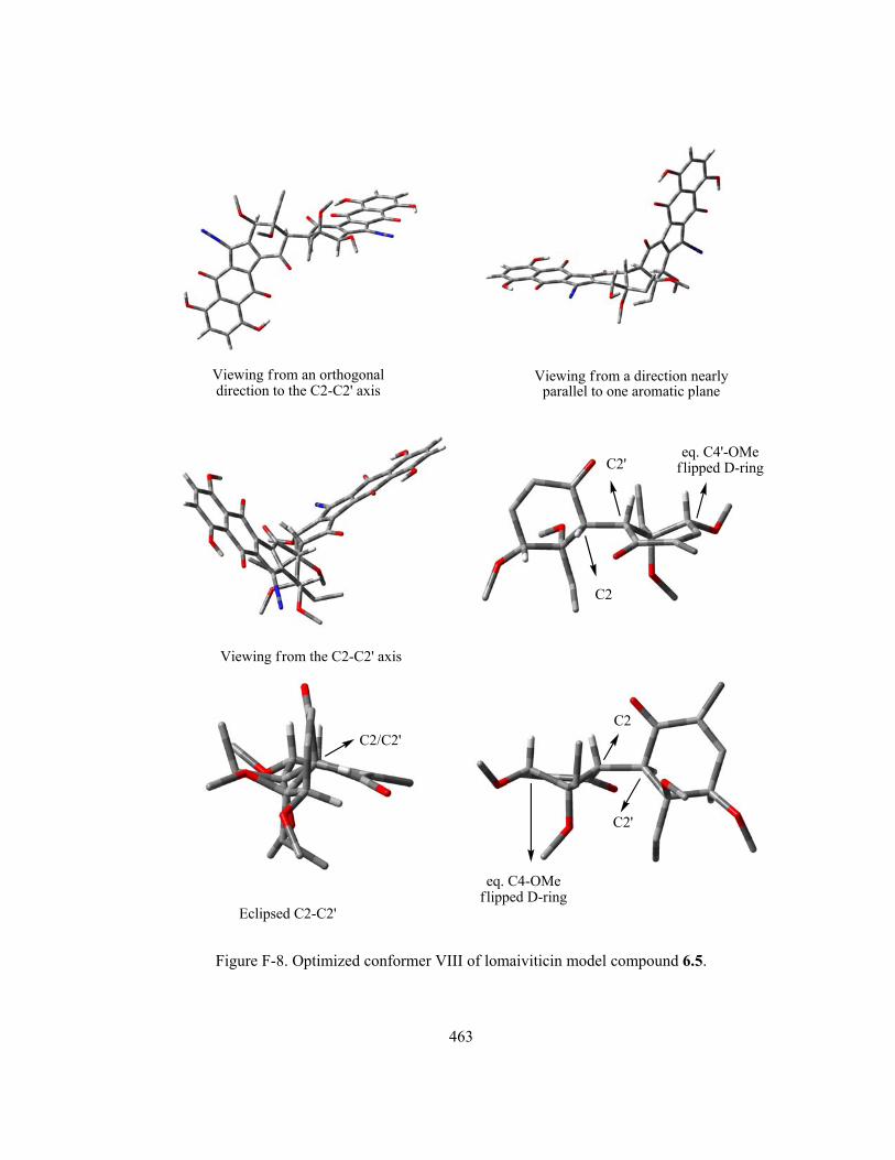
463
Viewing from an orthogonaldirection to the C2-C2' axis
Viewing from a direction nearlyparallel to one aromatic plane
Viewing from the C2-C2' axis
C2
C2'
Eclipsed C2-C2'
C2/C2'C2
C2'
eq. C4'-OMeflipped D-ring
eq. C4-OMeflipped D-ring
Figure F-8. Optimized conformer VIII of lomaiviticin model compound 6.5.
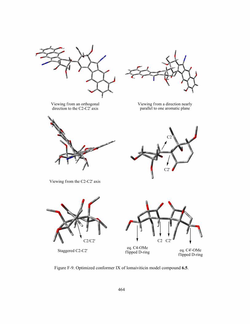
464
Viewing from an orthogonaldirection to the C2-C2' axis
Viewing from a direction nearlyparallel to one aromatic plane
Viewing from the C2-C2' axis
C2
C2'
eq. C4-OMeflipped D-ring
C2/C2'
Staggered C2-C2' eq. C4'-OMeflipped D-ring
C2 C2'
Figure F-9. Optimized conformer IX of lomaiviticin model compound 6.5.

465
References
(1) Sarker, S. D.; Nahar, L. In Chemistry For Pharmacy Students; John Wiley & Sons Ltd.: Sussex, 2007, p 283-370.
(2) Borders, D. B. In Kirk-Othmer Encylopedia of Chemical Technology; John Wiely & Sons, Inc.: 2007.
(3) Hopwood, D. A. Streptomyces in Nature and Medicine. The Antibiotic Makers; Oxford University Press Inc.: New York, 2007.
(4) Ito, S.; Matsuya, T.; Omura, S.; Otani, M.; Nakagawa, A.; Takeshima, H.; Iwai, Y.; Ohtani, M.; Hata, T. J. Antibiot. 1970, 23, 315-317.
(5) Seaton, P. J.; Gould, S. J. J. Antibiot. 1989, 42, 189-197. (6) Cone, M.; Seaton, P. J.; Halley, K. A.; Gould, S. J. J. Antibiot. 1989, 42, 179-188. (7) Isshiki, K.; Sawa, T.; Naganawa, H.; Matsuda, N.; Hattori, S.; Hamada, M.; Takeuchi, T.;
Oosono, M.; Ishizuka, M.; Yang, Z.; Zhu, B.; Xu, W. J. Antibiot. 1989, 42, 467-468. (8) Smitka, T. A.; Bonjouklian, R.; Perun, T. J. J.; Hunt, A. H.; Foster, R. S.; Mynderse, J. S.;
Yao, R. C. J. Antibiot. 1992, 45, 581. (9) Omura, S.; Nakagawa, A.; Yamada, H.; Hata, T.; Furusaki, A.; Watanabe, T. Chem. Pharm.
Bull. 1973, 21, 931-940. (10) Young, J.-J.; Ho, S.-N.; Ju, W.-M.; Chang, L.-R. J. Antibiot. 1994, 47, 681-687. (11) Lin, H.-C.; Chang, S.-C.; Nan-Li, W.; Chang, L.-R. J. Antibiot. 1994, 47, 675-680. (12) Cone, M. C.; Hassan, A. M.; Gore, M. P.; Gould, S. J.; Borden, D. B.; Alluri, M. R. J. Org.
Chem. 1994, 59, 1923-1924. (13) Gould, S. J.; Chen, J.; Cone, M. C.; Gore, M. P.; Melville, C. R.; Tamayo, N. J. Org. Chem.
1996, 61, 5720-5721. (14) Proteau, P. J.; Li, Y.; Chen, J.; Willianson, R. T.; Gould, S. J.; Laufer, R. S.; Dmitrienko, G.
I. J. Am. Chem. Soc. 2000, 122, 8325-8326. (15) Gould, S. J. Chem. Rev. 1997, 97, 2499-2510. (16) Nicolaou, K. C.; Li, H.; Nold, A. L.; Pappo, D.; Lenzen, A. J. Am. Chem. Soc. 2007, 129,
10356-10357. (17) Gould, S. J.; Melville, C. R.; Cone, M. C.; Chen, J.; Carney, J. R. J. Org. Chem. 1997, 62,
320-324. (18) Omura, S.; Nakagawa, A.; Yamada, H.; Hata, T.; Furusaki, A.; Watanabe, T. Chem. Pharm.
Bull. 1971, 19, 2428-2430. (19) Furusaki, A.; Matsui, M.; Watanabe, T.; Omura, S.; Nakagawa, A.; Hata, T. Isr. J. Chem.
1972, 10, 173-187. (20) Hata, T.; Omura, S.; Iwai, Y.; Nakagawa, A.; Otani, M.; Ito, S.; Matsuya, T. J. Antibiot.
1971, 24, 353-359. (21) Gould, S. J.; Tamayo, N.; Melville, C. R.; Cone, M. C. J. Am. Chem. Soc. 1994, 116, 2207-
2208. (22) Mithani, S.; Weeratunga, G.; Taylor, N. J.; Dmitrienko, G. I. J. Am. Chem. Soc. 1994, 116,
2209-2210. (23) Lei, X.; Porco, J. A. J. J. Am. Chem. Soc. 2006, 128, 14790-14791. (24) Kumamoto, T.; Kitani, Y.; Tsuchiya, H.; Yamaguchi, K.; Seki, H.; Ishikawa, T. Tetrahedron
2007, 63, 5189-5199. (25) Liu, W.; Buck, M.; Chen, N.; Shang, M.; Taylor, N. J.; Asoud, J.; Wu, X.; Hasinoff, B. B.;
Dmitrienko, G. I. Org. Lett. 2007, 9, 2915-2918. (26) Woo, C. M.; Lu, L.; Gholap, S. L.; Smith, D. R.; Herzon, S. B. J. Am. Chem. Soc. 2010, 132,
2540-2541.

466
(27) Ehrlich, J.; Coffey, G. L.; Fisher, M. W.; Hillegas, A. B.; Kohberger, D. L.; Machamer, H. E.; Rightsel, W. A.; Roegner, F. R. Antibiotics & Chemotherapy 1956, 6, 487-497.
(28) Dion, H. W.; Fusari, S. A.; Jakubowski, Z. L.; Zora, J. G.; Bartz, Q. R. J. Am. Chem. Soc. 1956, 78, 3075-3077.
(29) Stock, C. C.; reilly, H. C.; Buckley, S. M.; Clarke, D. A.; Rhoads, C. P. Nature 1954, 173, 71-72.
(30) Ehrlich, J.; Anderson, L. E.; Coffey, G. L.; Hillegas, A. B.; Knudsen, M. P.; Koepsell, H. J.; Kohberger, D. L.; Oyaas, J. E. Nature 1954, 173, 72.
(31) Bartz, Q. R.; Elder, C. C.; Frohardt, R. P.; Fusari, S. A.; Haskell, T. H.; Johannessen, D. W.; Ryder, A. Nature 1954, 173, 72-73.
(32) Fusari, S. A.; Frohardt, R. P.; Ryder, A.; Haskell, T. H.; Johannessen, D. W.; Elder, C. C.; Bartz, Q. R. J. Am. Chem. Soc. 1954, 76, 2878-2881.
(33) Fusari, S. A.; Haskell, T. H.; Frohardt, R. P.; Bartz, Q. R. J. Am. Chem. Soc. 1954, 76, 2881-2883.
(34) Moore, J. A.; Dice, J. R.; Nicolaides, E. D.; Westland, R. D.; Wittle, E. L. J. Am. Chem. Soc. 1954, 76, 2884-2887.
(35) Nicolaides, E. D.; Westland, R. D.; Wittle, E. L. J. Am. Chem. Soc. 1954, 76, 2887-2891. (36) Hata, T.; Umezawa, I.; Iwai, Y.; Katagiri, M.; Awaya, J.; Komiyama, K.; Oiwa, R.; Atsumi,
K. J. Antibiot. 1973, 26, 181-183. (37) Satoh, K.; Komiyama, K.; Kitao, C.; Iwai, Y.; Atsumi, K.; Oiwa, R.; Katagiri, M.; Umezawa,
I.; Omura, S.; Hata, T. J. Antibiot. 1974, 27, 620-625. (38) Pittillo, R. F.; Hunt, D. E. In Antibiotics I. Mechanism of Action; Gottlieb, D., Shaw, P. D.,
Eds. New York, 1967, p 481-493. (39) Lee, M. D.; Fantini, A. A.; Kuck, N. A.; Greenstein, M.; Testa, R. T.; Borders, D. B. J.
Antibiot. 1987, 40, 1657-1663. (40) Kameyama, T.; Takahashi, A.; Matsumoto, H.; Kurasawa, S.; Hamada, M.; Okami, Y.;
Ishizuka, M.; Takeuchi, T. J. Antibiot. 1988, 41, 1561-1567. (41) Takahashi, A.; Nakamura, H.; Daishiro Ikeda; Naganawa, H.; Toshiyuki Kameyama;
Kurasawa, S.; Okami, Y.; Takeuchi, T.; Iitaka, Y. J. Antibiot. 1988, 41, 1568-1574. (42) Nishimura, M.; Nakada, H.; Nakajima, H.; Hori, Y.; Ezaki, M.; Goto, T.; Okuhara, M. J.
Antibiot. 1989, 42, 542-548. (43) Nishimura, M.; Nakada, H.; Takase, S.; Akira Katayama; Goto, T.; Tanaka, H.; Hashimoto,
M. J. Antibiot. 1989, 42, 549-552. (44) Nishimura, M.; Nakada, H.; Kawamura, I.; Mizota, T.; Shimomura, K.; Nakahara, K.; Goto,
T.; Yamaguchi, I.; Okuhara, M. J. Antibiot. 1989, 42, 553-557. (45) McGuire, J. N.; Wilson, S. R.; Rinehart, K. L. J. Antibiot. 1995, 48, 516-519. (46) Varley, L. M.; Moody, C. J. Synthesis 2008, 3601-3604. (47) Singh, P. D.; Johnson, J. H.; Aklonis, C. A.; O'Sullivan, J. J. Antibiot. 1986, 39, 1054-1058. (48) Nihei, Y.; Hasegawa, M.; Suzuki, K.; Yamamoto, S.; Hanada, M.; Furumai, T.; Fukagawa,
Y.; Oki, T. J. Antibiot. 1993, 46, 900-907. (49) Imae, K.; Nihei, Y.; Oka, M.; Yamasaki, T.; Konishi, M.; Oki, T. J. Antibiot. 1993, 46, 1031-
1033. (50) Shomura, T.; Gomi, S.; Ito, M.; Yoshida, J.; Tanaka, E.; Amano, S.; Watabe, H.-o.; Ohuchi,
S.; Itoh, J.; Sezaki, M. J. Antibiot. 1987, 40, 732-739. (51) Gomi, S.; Ohuchi, S.; Sasaki, T.; Itoh, J.; Sezaki, M. J. Antibiot. 1987, 40, 740-748. (52) He, H.; Ding, W.; Bernan, V. S.; Richardson, A. D.; Ireland, C. M.; Greenstein, M.; Ellestad,
G. A.; Carter, G. T. J. Am. Chem. Soc. 2001, 123, 5362-5363. (53) Seaton, P. J.; Gould, S. J. J. Am. Chem. Soc. 1987, 109, 5282-5284.

467
(54) Volkmann, C.; Rossner, E.; Metzler, M.; Zahner, H.; Zeeck, A. Liebigs Ann. 1995, 1169-1172.
(55) Shin-ya, K.; Furihata, K.; Teshima, Y.; Hayakawa, Y.; Seto, H. Tetrahedron Lett. 1992, 33, 7025-7028.
(56) Gould, S. J.; Melville, C. R. Bioorg. Med. Chem. Lett. 1995, 5, 51-54. (57) Cone, M. C.; Melville, C. R.; Gore, M. P.; Gould, S. J. J. Org. Chem. 1993, 58, 1058-1061. (58) Gould, S. J.; Hong, S.-T.; Carney, J. R. J. Antibiot. 1998, 51, 50-57. (59) Carney, J. R.; Hong, S.; Gould, S. J. Tetrahedron Lett. 1997, 38, 3139. (60) Aoyama, T.; Zhao, W.; Kojima, F.; Muraoka, Y.; Naganawa, H.; Takeuchi, T. J. Antibiot.
1993, 46, 1471-1474. (61) Meselhy, M. R.; Kadota, S.; Tsubono, K.; Hattori, M.; Nambaa, T. Tetrahedron 1994, 50,
3081-3098. (62) Meselhy, M. R.; Nishimoto, E.; Akao, T.; Hattori, M. J. Tradit. Med. 2001, 18, 58-63. (63) Akiyama, T.; Harada, S.; Kojima, F.; Takahashi, Y.; Imada, C.; Okami, Y.; Muraoka, Y.;
Aoyagi, T.; Takeuchi, T. J. Antibiot. 1998, 51, 553-559. (64) Schneider, K.; Nicholson, G.; Ströbele, M.; Baur, S.; Niehaus, J.; Fiedler, H.-P.; Süssmuth, R.
D. J. Antibiot. 2006, 59, 105-109. (65) Baur, S.; Niehaus, J.; Karagouni, A. D.; Katsifas, E. A.; Chalkou, K.; Meintanis, C.; Jones, A.
L.; Goodfellow, M.; Ward, A. C.; Beil, W.; Schneider, K.; Süssmuth, R. D.; Fiedler, H.-P. J. Antibiot. 2006, 59, 293-297.
(66) Gould, S. J.; Melville, C. R. Tetrahedron Lett. 1997, 38, 1473. (67) Iwai, Y.; Omura, S. J. Antibiot. 1982, 35, 123-141. (68) Short, J. M.; Paradkar, A.; Varoglu, M.; Mathur, E. J.; Organization, W. I. P., Ed.; Diversa
Co. (CA, USA): USA, 2003, p 47. (69) Sato, Y.; Geckle, M.; Gould, S. J. Tetrahedron Lett. 1985, 26, 4019-4022. (70) Sato, Y.; Gould, S. J. Tetrahedron Lett. 1985, 26, 4023-4026. (71) Seaton, P. J.; Gould, S. J. J. Am. Chem. Soc. 1988, 110, 5912-5914. (72) Duthaler, R. O.; Fonster, H. G.; Roberts, J. D. J. Am. Chem. Soc. 1978, 100, 4974-4979. (73) Dmitrienko, G. I.; Nielsen, K. E.; Steingart, C.; Ming, N. S.; Willson, J. M.; Weeratunga, G.
Tetrahedron Lett. 1990, 31, 3681. (74) Echavarren, A. M.; Tamayo, N.; Paredes, M. C. Tetrahedron Lett. 1993, 34, 4713-4716. (75) Echavarren, A. M.; Frutos, O. d. Tetrahedron Lett. 1997, 38, 7941-7942. (76) Hauser, F. M.; Zhou, M. J. Org. Chem. 1996, 61, 5722. (77) Sato, Y.; Gould, S. J. J. Am. Chem. Soc. 1986, 108, 4625-4631. (78) Ajisaka, K.; Takeshima, H.; Omura, S. J. Chem. Soc., Chem. Commun. 1976, 571-572. (79) Gould, S. J.; O'Hare, T.; Seaton, P.; Soodsma, J.; Tang, Z. Bioorg. Med. Chem. 1996, 4, 987. (80) Laufer, R., Ph.D. thesis, University of Waterloo, 2002. (81) LaRue, T. A. Lloydia 1977, 40, 307-321. (82) Walton, K.; Coombs, M. M.; Walker, R.; Ioannides, C. Mutat. Res., Fundam. Mol. Mech.
Mutagen. 1997, 381, 131-139. (83) Walton, K.; Coombs, M. M.; Catterall, F. S.; Walker, R.; Ioannides, C. Carcinogenesis 1997,
18, 1603-1608. (84) Parry, R. J.; Li, W. J. Chem. Soc., Chem. Commun. 1994, 995-996. (85) Walton, K.; Coombs, M. M.; King, L. J.; Walker, R.; Ioannides, C. Nutri. Can. 2000, 37, 55-
64. (86) Gonzalez-Lopez, J.; Martinez-Toledo, M. V.; Pozo, C.; Rodelas, B.; Gomez, M. A.; Hontoria,
E. Trends Soil Sci. 2004, 3, 91-97. (87) Suzuki, T. Genes Environ. 2006, 28, 48-55.

468
(88) Omura, S.; Tanaka, H.; Oiwa, R.; Nagai, R.; Koyama, Y.; Takahashi, Y. J. Antibiot. 1979, 32, 978-984.
(89) Tanaka, Y.; Sugoh, M.; Ji, W.; Iwabuchi, J.; Yoshida, H.; Omura, S. J. Antibiot. 1995, 48, 720-724.
(90) Khdour, O.; Skibo, E. B. Org. Biomol. Chem. 2009, 7, 2140-2154. (91) Moore, H. W. Science 1977, 197, 527-532. (92) Moore, H. W.; Czerniak, R. Med. Res. Rev. 1981, 1, 249-280. (93) Rajski, S. R.; Williams, R. M. Chem. Rev. 1998, 98, 2723-2795. (94) Griffiths, J.; Murphy, J. A. J. Chem. Soc., Chem. Commun. 1992, 24-26. (95) Kizil, M.; Yilmaz, E. I.; Pirinccioglu, N.; Aytekin, C. Turk. J. Chem. 2003, 27, 539-544. (96) Augusto, O. Free Rad. Bio. Med. 1993, 15, 329-336. (97) Zollinger, H. Diazo Chemistry I: Aromatic and Heteroaromatic Compounds; VCH: New
York, 1994. (98) Reszka, K. J.; Chignell, C. F. Chem.-Biol. Interact. 1995, 96, 223-234. (99) Gannett, P. M.; Lawson, T.; MIller, M.; Thakkar, D. D.; Lord, J. W.; Yau, W.-M.; Toth, B.
Chem.-Biol. Interact. 1996, 101, 149-164. (100) Chin, A.; Hung, M.-H.; Stock, L. M. J. Org. Chem. 1981, 46, 2203-2207. (101) Hung, M.-H.; Stock, L. M. J. Org. Chem. 1982, 47, 448-453. (102) McLick, J.; Bauer, P. I.; Hakam, A.; Kun, E. Biochemistry 1987, 26, 2226-2231. (103) Koepke, S. R.; Kroeger-Koepke, M. B.; Michejda, C. J. Chem. Res. Toxicol. 1990, 3, 17-20. (104) Lawson, T.; Gannett, P. M.; Yau, W.-M.; Dalal, N. S.; Toth, B. J. Agric. Food Chem. 1995,
43, 2627-2635. (105) Gannett, P. M.; Powell, J. H.; Rao, R.; Shi, X.; Lawson, T.; Kolar, C.; Toth, B. Chem. Res.
Toxicol. 1999, 12, 297-304. (106) Gannett, P. M.; Ye, J.; Ding, M.; Powell, J.; Zhang, Y.; Darian, E.; Daft, J.; Shi, X. Chem.
Res. Toxicol. 2000, 13, 1020-1027. (107) Kikugawa, K.; Kato, T.; Takeda, Y. Mutat. Res. 1987, 177, 35-43. (108) Furihata, C.; Yamakoshi, A.; Matsushima, T.; Kato, T.; Kikugawa, K. Mutagenesis 1988, 3,
299-301. (109) Kikugawa, K.; Kato, T.; Takeda, Y. Chem. Pharm. Bull. 1989, 37, 1600-1603. (110) Kikugawa, K.; Kato, T. Food Chem. Toxic. 1988, 26, 209-214. (111) Kikugawa, K.; Kato, T.; Kojima, K. Mutat. Res. 1992, 1, 65-75. (112) Kato, T.; Kojima, K.; Hiramoto, K.; Kikugawa, K. Mutat. Res. 1992, 268, 105-114. (113) Kato, T.; Tadokoro, N.; Tsutsui, M.; Kikugawa, K. Mutat. Res. 1991, 249, 243-254. (114) Hiramoto, K.; Inoue, M.; Maeda, K.; Kato, T.; Kikugawa, K. Jpn. J. Toxicol. Environ. Health
1994, 40, 414-421. (115) Hiramoto, K.; Kato, T.; Kikugawa, K. Mutat. Res., Fundam. Mol. Mech. Mutagen. 1994, 306,
153-163. (116) Hiramoto, K.; Kaku, M.; Sueyoshi, A.; Fujise, M.; Kikugawa, K. Chem. Res. Toxicol. 1995,
8, 356-362. (117) Hiramoto, K.; Kaku, M.; Kato, T.; Kikugawa, K. Chem.-Biol. Interact. 1995, 94, 21-36. (118) Hiramoto, K.; Fujino, T.; Kikugawa, K. Mutat. Res., Environ. Mutagen. 1996, 360, 95-100. (119) Kato, T.; Kikugawa, K. Food Chem. Toxic. 1992, 30, 617-626. (120) Hiramoto, K.; Yasuhara, Y.; Sako, K.-i.; Aoki, K.; Kikugawa, K. Biol. Pharm. Bull. 2003, 26,
1129-1134. (121) Arya, D. P.; Jebaratnam, D. J. J. Org. Chem. 1995, 60, 3268-3269. (122) Eppley, H. J.; Lato, S. M.; Ellington, A. D.; Zaleski, J. M. Chem. Commun. 1999, 2405-2406.

469
(123) Kraft, B. J.; Eppley, H. J.; Huffman, J. C.; Zaleski, J. M. J. Am. Chem. Soc. 2002, 124, 272-280.
(124) Laufer, R. S.; Dmitrienko, G. I. J. Am. Chem. Soc. 2002, 124, 1854-1855. (125) Cain, J. C. The Chemistry And Technology of The Diazo-compounds; 2nd ed.; Oxford:
London, 1920. (126) Porai-Koshits, B. A. Russ. Chem. Rev. 1970, 39, 283-289. (127) Regitz, M.; Mass, G. Diazo Compounds: Properties and Synthesis Academic Press: New
York, 1986. (128) Zollinger, H. Diazo Chemistry II: Aliphatic, Inorganic and Organometallic Compounds;
VCH: New York, 1995. (129) Kostuch, A.; Urban, A.; Tomasik, P. ARKIVOC 2000, 181-185 and references therein. (130) McDowell, L. J.; Khodae, M. M.; Bethell, D. Org. Biomol. Chem. 2003, 1, 995. (131) Pearson, R. G. Chemical Hardness; WILEY-VCH: New York, 1997. (132) Silverstein, R. M.; Webster, F. X. Spectrometric Identification of Organic Compounds; 6th
ed.; John Wiley & Sons: New York, 1998. (133) Kobayashi, K.; Takeuchi, H.; Seko, S.; Suginome, H. Helv. Chim. Acta 1991, 74, 1091-1094. (134) O'Sullivan, P. J.; Moreno, R.; Murphy, W. S. Tetrahedron Lett. 1992, 33, 535-538. (135) Hu, H.-Y.; Liu, Y.; Ye, M.; Xu, J.-H. Synlett 2006, 1913-1917. (136) Poumaroux, A.; Bouaziz, Z.; Fillion, H.; Domard, M.; Giraud, J.; Petavy, A.-F. Chem.
Pharm. Bull. 1999, 47, 643-646. (137) Itoigawa, M.; Kashiwada, Y.; Ito, C.; Furukawa, H.; Tachibana, Y.; Bastow, K. F.; Lee, K.-
H. J. Nat. Prod. 2000, 63, 893-897. (138) Bernardo, P. H.; Chai, C. L. L.; Guen, M. L.; Smith, G. D.; Waring, P. Bio. Med. Chem. Lett.
2007, 17, 82-85. (139) Hopkingson, A. C.; Lee-Ruff, E.; Maleki, M. Synthesis 1986, 366-371. (140) Trost, B. M.; Walchli, R. J. Am. Chem. Soc. 1987, 109, 3487-3488. (141) Bestmann, H. J. Pure & Appl. Chern. 1980, 52, 771-788. (142) Streitwieser-Jr., A.; Brown, S. M. J. Org. Chem. 1988, 53, 904-906. (143) Krogh, E.; Wan, P. Can. J. Chem. 1990, 68, 1725-1731. (144) Gore, M. P.; Gould, S. J.; Weller, D. D. J. Org. Chem. 1991, 56, 2289-2291. (145) Gore, M. P.; Gould, S. J.; Weller, D. D. J. Org. Chem. 1992, 57, 2774-2783. (146) Mohri, S.-i.; Stefinovic, M.; Snieckus, V. J. Org. Chem. 1997, 62, 7072-7073. (147) Koyama, H.; Kamikawa, T. J. Chem. Soc., Perkin Trans. 1 1998, 203-209. (148) Williams, W.; Sun, X.; Jebaratnam, D. J. Org. Chem. 1997, 62, 4364-4369. (149) Qabaja, G.; Jones, G. B. Tetrahedron Lett. 2000, 41, 5317-5320. (150) Qabaja, G.; Jones, G. B. J. Org. Chem. 2000, 65, 7187-7194. (151) Mal, D.; Hazra, N. K. Tetrahedron Lett. 1996, 37, 2641-2642. (152) Hauser, F. M.; Rhee, R. P. J. Org. Chem. 1978, 43, 178-180. (153) Birman, V. B.; Zhao, Z.; Guo, L. Org. Lett. 2007, 9, 1223-1225. (154) Ana Martinez, J. C.; Barcia, A. M. E.; Fernandez, F.; Gonzalez, L.; Estevez, J. C.; Estevez, R.
J. Tetrahedron Lett. 2007, 48, 2147-2149. (155) Rodríguez, D.; Castedo, L.; Domínguez, D.; Saá, C. Tetrahedron Lett. 1999, 40, 7701-7704. (156) Atienza, C.; Mateo, C.; Frutos, O. d.; Echavarren, A. M. Org. Lett. 2001, 3, 153-155. (157) Nicolaou, K. C.; Denton, R. M.; Lenzen, A.; Edmonds, D. J.; Li, A.; Milburn, R. R.;
Harrison, S. T. Angew. Chem. Int. Ed. 2006, 45, 2076-2081. (158) Nicolaou, K. C.; Nold, A. L.; Li, H. Angew. Chem. Int. Ed. 2009, 48, 1-5. (159) Krygowski, E. S.; Murphy-Benenato, K.; Shair, M. D. Angew. Chem. Int. Ed. 2008, 47, 1680-
1684.

470
(160) Buck, M., M.Sc. thesis, University of Waterloo, 2003. (161) Liu, W., Ph.D. thesis, University of Waterloo, 2009. (162) Yu, M.; Danishefsky, S. J. J. Am. Chem. Soc. 2008, 130, 2783-2785. (163) Kumamoto, T.; Tabe, N.; Yamaguchi, K.; Ishikawa, T. Tetrahedron Lett. 2000, 41, 5693-
5697. (164) Kumamoto, T.; Tabe, N.; Yamaguchi, K.; Yagishita, H.; Iwasa, H.; Ishikawa, T. Tetrahedron
2001, 57, 2717-2728. (165) Chen, N.; Carriere, M. B.; Laufer, R. S.; Taylor, N. J.; Dmitrienko, G. I. Org. Lett. 2008, 10,
381-384. (166) Fieser, L. F.; Campbell, W. P.; Fry, E. M.; Marshall D. Gates, J. J. Am. Chem. Soc. 1939, 61,
3216-3223. (167) Masahiko, T.; Fujii, H.; Oshima, K.; Utimoto, K. Tetrahedron 1995, 51, 679-686. (168) Carriere, M. B., M.Sc. thesis, University of Waterloo, 1998. (169) Akbulut, N.; Balci, M. J. Org. Chem. 1988, 53, 3338. (170) Rucker, G.; Horster, H.; Gajewski, W. Synth. Commun. 1980, 10, 623. (171) Masahiko, T.; Hideaki, F.; Oshima, K.; Utimoto, K. Tetrahedron 1995, 51, 679-686. (172) Preusch, P. C.; Smalley, D. M. Free Rad. Res. Comms. 1990, 8, 401-415. (173) Preusch, P. C.; Suttie, J. W. J. Org. Chem. 1983, 48, 2291-2293. (174) Schneider, C. Synthesis 2006, 3919-3944. (175) Cavelier, F.; Yim, A.-M. Org. Prep. Proced. Int. 1998, 30, 103-106. (176) O'Brien, D. F. J. Org. Chem. 1968, 33, 262-265. (177) Nan, F.; Chen, X.; Xiong, Z.; Li, T.; Li, Y. Synth. Commun. 1994, 24, 2319-2324. (178) Kido, F.; Abe, T.; Yoshikoshi, A. J. Chem. Soc., Chem. Commun. 1986, 590. (179) Isobe, M.; Kitamura, M.; Goto, T. Tetrahedron Lett. 1981, 22, 239-242. (180) Phillips, R. E.; Soulen, R. L. J. Chem. Ed. 1995, 72, 624-625. (181) Loken, B.; Kaufmann, S.; Rosenkranz, G.; Sondheimer, F. J. Am. Chem. Soc. 1956, 78, 1738-
1741. (182) Kamano, Y. Chem. Pharm. Bull. 1969, 17, 1711-1719. (183) Baraldi, P. G.; Barco, A.; Benetti, S.; Ferretti, V.; Pollini, G. P.; Polo, E.; Zanirato, V.
Tetrahedron 1989, 45, 1517-1532. (184) Naruta, Y.; Nishigaichi, Y.; Maruyama, K. J. Org. Chem. 1988, 53, 1192-1199. (185) Urones, J. G.; Basabe, P.; Lithgow, A. M.; Marcos, I. S.; Jimenez, A.; Diez, D.; Gomez, A.;
White, A. J. P.; Williams, D. J.; Simmonds, M. S. J.; Blaney, W. M. Tetrahedron 1995, 51, 2117-2128.
(186) Urones, J. G.; Basabe, P.; Marcos, I. S.; Jimenez, A.; Lithgow, A. M.; Lopez, M.; Moro, R. F.; Gomez, A. Tetrahedron 1994, 50, 10791-10802.
(187) Confalone, P. N.; Pizzolato, G. J. Org. Chem. 1990, 55, 5520-5525. (188) Isaacs, N. S.; Neelakantan, K. Can. J. Chem. 1968, 46, 1043-1046. (189) Ponser, G.; Rogers, D. Z. J. Am. Chem. Soc. 1977, 99, 8208-8214. (190) Kakiuchi, H.; Iijima, T. Tetrahedron 1980, 36, 1011-1016. (191) Girdhar, N. K.; Ishar, M. P. S.; Kumar, R.; Singh, R.; Singh, G. Tetrahedron 2001, 57, 7199-
7204. (192) Rampalli, S.; Chaudhari, S. S.; Akamanchi, K. G. Synthesis 2000, 78-80. (193) Riego, J.; Costa, A.; SAA, J. M. Chem. Lett. 1986, 1565-1568. (194) Liu, Y.-j.; Chu, T.-y.; Engel, R. Synth. Commun. 1992, 22, 2367-2371. (195) Mohammadpoor-Baltork, I.; Tangestaninejad, S.; Mirkhani, V. Synth. Commun. 2000, 30,
2365-2374. (196) Montalban, A. G.; Wittenberg, L.-O.; McKillop, A. Tetrahedron Lett. 1999, 40, 5893-5896.

471
(197) Hanzlik, R. P.; Leinwetter, M. J. Org. Chem. 1978, 43, 438-440. (198) Nambu, Y.; Endo, T. Tetrahedron Lett. 1990, 31, 1723-1726. (199) Iranpoor, N.; Tarrian, T.; Movahedi, Z. Synthesis 1996, 1473-1476. (200) Salehi, P.; Seddighi, B.; Irandoost, M.; Behbahani, F. K. Synth. Commun. 2000, 30, 2967-
2973. (201) Iranpoor, N.; Adibi, H. Bull. Chem. Soc. Jpn. 2000, 73, 675-680. (202) Kalita, B.; Barua, N. C.; Bezbarua, M. S.; Bez, G. Synlett 2001, 1411-1414. (203) Righi, G.; Pescatore, G.; Bonadies, F.; Bonini, C. Tetrahedron 2001, 57, 5649-5656. (204) Bonini, C.; Righi, G. Tetrahedron 1992, 48, 1531-1538. (205) Constantino, M. G.; Jr., V. L.; Aragão, V. Molecules 2001, 6, 770-776. (206) Iranpoor, N.; Zeynizadeh, B. Synth. Commun. 1999, 29, 1017-1024. (207) Ghelfi, F.; Grandi, R.; Pagnoni, U. M. J. Chem. Res. (S) 1988, 200-201. (208) Armstrong, R. N. In Comprehensive Natural Products Chemistry; Poulter, C. D., Ed.;
Elsevier Science: New York, 1999; Vol. 5, p 51-70. (209) Iranpoor, N.; Baltork, I. M. Synth. Commun. 1990, 20, 2789-2797. (210) Iranpoor, N.; Zardaloo, F. S. Synth. Commun. 1994, 24, 1959-1969. (211) Iranpoor, N.; Baltork, I. M. Tetrahedron Lett. 1990, 31, 735-738. (212) Iranpoor, N.; Tamami, B.; Niknam, K. Can. J. Chem. 1997, 75, 1913-1919. (213) Zwan, M. C. V.; Hartner, F. W. J. Org. Chem. 1978, 43, 2655-2657. (214) Mall, T.; Stamm, H. J. Org. Chem. 1987, 52, 4812-4817. (215) Honda, T.; Mizutani, H. Heterocycles 1998, 48, 1753-1757. (216) Caron, M.; Sharpless, K. B. J. Org. Chem. 1985, 50, 1557-1560. (217) Chong, J. M.; Sharpless, K. B. J. Org. Chem. 1985, 50, 1560-1563. (218) Akira, K.; Taira, T.; Hasegawa, H.; Sakuma, C.; Shinohara, Y. Drug Metab. Dispos. 1998,
26, 457-464. (219) Evans, D. A.; Chapman, K. T.; Carreira, E. M. J. Am. Chem. Soc. 1988, 110, 3560-3578. (220) Thompson, H.; Wolfrom, M. L.; Whistler, R. L., Wolfrom, M. L., BeMiller, J. N., Eds.;
Academic Press: New York, 1963; Vol. 2, p 215-220. (221) Barrett, A. G. M.; Braddock, D. C. Chem. Commun. 1997, 351-352. (222) Ranu, B. C.; Dutta, P.; Sarkar, A. Tetrahedron Lett. 1998, 39, 9557. (223) Ma, J.; Chan, T. H. Tetrahedron Lett. 1998, 39, 2499. (224) Li, W.-D. Z.; Ma, B.-C. Org. Lett. 2005, 7, 271-274. (225) Arai, S.; Oku, M.; Miura, M.; Shioiri, T. Synlett 1998, 1201-1202. (226) Arai, S.; Tsuge, H.; Oku, M.; Miura, M.; Shioiri, T. Tetrahedron 2002, 58, 1623-1630. (227) Baba, N.; Oda, J. i.; Kawaguchi, M. Agric. Biol. Chem. 1986, 50, 3113-3117. (228) Pluim, H.; Wynberg, H. J. Org. Chem. 1980, 45, 2498-2502. (229) Wynberg, H. Chimia 1976, 30, 445-451. (230) Dwyer, C. L.; Gill, C. D.; Ichihara, O.; Taylor, R. J. K. Synlett 2000, 704-705. (231) Colonna, S.; Manfredi, A.; Annunziata, R.; Gaggero, N.; Casella, L. J. Org. Chem. 1990, 55,
5862-5866. (232) Colonna, S.; Manfredi, A. Tetrahedron Lett. 1986, 27, 387-390. (233) Colonna, S.; Gaggero, N.; Manfredi, A.; Spadoni, M.; Casella, L.; Carrea, G.; Pasta, P.
Tetrahedron 1988, 44, 5169-5178. (234) Colonna, S.; Manfredi, A.; Annunziata, R.; Spadoni, M. Tetrahedron 1987, 43, 2157-2164. (235) Harigaya, Y.; Yamaguch, H.; Onda, M. Heterocycles 1981, 15, 183-185. (236) Porter, M. J.; Skidmore, J. Chem. Commun. 2000, 1215--1225. (237) Berkessel, A.; Guix, M.; Schmidt, F.; Neudorfl, J. M.; Lex, J. Chem. Eur. J. 2007, 13, 4483-
4498.

472
(238) Barfield, M.; Johnston, M. D. J. Chem. Rev. 1972, 73, 53-73. (239) Desjardins, M.; Lallemand, M.-C.; Freeman, S.; Hudlicky, T.; Abboud, K. A. J. Chem. Soc.,
Perkin Trans. 1 1999, 621-628. (240) Orsini, F.; Pelizzoni, F. Tetrahedron: Asymmetry 1996, 7, 1033. (241) O'Hara, K. A.; Wu, X.; Patel, D.; Liang, H.; Yalowich, J. C.; Chen, N.; Goodfellow, V.;
Adedayo, O.; Dmitrienko, G. I.; Hasinoff, B. B. Free Rad. Bio. Med. 2007, 43, 1132-1144. (242) Gottlieb, H. E.; Kotlyar, V.; Nudelman, A. J. Org. Chem. 1997, 62, 7512-7515. (243) Weeratunga, G.; Prasad, G. K. B.; Dilley, J.; Taylor, N. J.; Dmitrienko, G. I. Tetrahedron
Lett. 1990, 31, 5713-5716. (244) Hasinoff, B. B.; Wu, X.; Yalowich, J. C.; Goodfellow, V.; R., L.; Adedayo , O.; Dmitrienko,
G. I. Anti-Cancer Drugs 2006, 17, 825-837. (245) Mithani, S., Ph.D. thesis, University of Waterloo, 1996. (246) Jacobsen, N.; Torssell, K. Liebigs Ann. Chem. 1972, 763, 135-147. (247) Jacobsen, N.; Torssell, K. Acta Chem. Scand. 1973, 27, 3211-3216. (248) Kirmse, W. Eur. J. Org. Chem. 2002, 2193-2256. (249) Baumbach, B.; Bendig, J.; Nagel, T.; Dubsky, B. J. Prakt. Chem. 1991, 333, 625-635. (250) Bendig, J.; Sauer, E.; Polz, K.; Schopf, G. Tetrahedron 1992, 48, 9207-9216. (251) Sus, O.; Steppan, H.; Dietrich, R. Justus Liebigs Ann. Chem. 1958, 20-25. (252) Trost, B. M.; Kinson, P. L. J. Am. Chem. Soc. 1975, 97, 2438-2449. (253) Presset, M.; Coquerel, Y.; Rodriguez, J. J. Org. Chem. 2009, 74, 415-418. (254) Patra, A.; Ghorai, S. K.; De, S. R.; Mal, D. Synthesis 2006, 2556-2562. (255) Acheson, R. M. Acc. Chem. Res. 1971, 4, 177-186 and references therein. (256) Jung, M. E.; Jung, Y. H. Tetrahedron Lett. 1988, 29, 2517. (257) Rizzacasa, M. A.; Sargent, M. V. J. Chem. Soc. Perkin Trans. 1 1991, 1991, 841-844. (258) Kitani, Y.; Morita, A.; Kumamoto, T.; Ishikawa, T. Helv. Chim. Acta 2002, 85, 1186-1195. (259) Heinzman, S. W.; Grunwell, J. R. Tetrahedron Lett. 1980, 21, 4305-4308. (260) Jung, M. E.; Hagenah, J. A. J. Org. Chem. 1983, 48, 5359-5361. (261) Johnstone, R. W.; Rose, M. Tetrahedron 1979, 35, 2169-2173. (262) Tschesche, R.; Korte, F. Chem. Ber. 1951, 84, 77-83. (263) Bateman, L.; Cunneen, J. I.; Koch, H. F. J. Chem. Soc. 1950, 3045-3051. (264) Bateman, L.; Cunneen, J. I.; Koch, H. F. J. Chem. Soc. 1950, 3051-3056. (265) Dupont, M. G.; Dulou, R.; Defay, M. N. Bull. Soc. Chim. Fr. 1943, 16, 310-314. (266) Smith, J. G.; Dibble, P. W.; Sandborn, R. E. J. Org. Chem. 1986, 51, 3762-3768. (267) Bringmann, G.; Pabst, T.; Henschel, P.; Michel, M. Tetrahedron 2001, 57, 1269-1275. (268) Abike, A.; Mesamune, S. Tetrahedron Lett. 1992, 33, 5517-5518. (269) Cannon, J. G.; Koble, D. L. J. Med. Chem. 1980, 23, 750-754. (270) Ripin, D. H.; Evans, D. A.; http://evans.harvard.edu/pdf/evans_pKa_table.pdf: 2005. (271) Kodama, H.; Katsuhira, T.; Nishida, T.; Hino, T.; Tsubata, K.; WIPO, Ed. Japan, 2001; Vol.
WO 01/83421 A1. (272) Orito, K.; Hatakeyama, T.; Takeo, M.; Suginome, H. Synthesis 1995, 1273-1277. (273) Greene, T. W.; Wuts, P. G. M. Protective Groups in Organic Synthesis; 3rd ed.; John Wiley
& Sons: New York, 1999. (274) Sorrell, T. N.; Ellis, D. J. J. Org. Chem. 1985, 50, 5765-5769. (275) Majewski, M.; Bantle, G. Tetrahedron Lett. 1989, 30, 6653-6656. (276) Majewski, M.; Irvine, N. M.; Bantle, G. W. J. Org. Chem. 1994, 59, 6697-6702. (277) Yonezawa, S.; Komurasaki, T.; Kawada, K.; Tsuri, T.; Fuji, M.; Kugimiya, A.; Haga, N.;
Mitsumori, S.; Inagaki, M.; Nakatani, T.; Tamura, Y.; Takechi, S.; Taishi, T.; Ohtani, M. J. Org. Chem. 1998, 63, 5831-5837.

473
(278) Ali, Y.; Hussain, M. Carbohydrate Res. 1979, 69, 311-315. (279) Webster, K. T.; Eby, R.; Schuerch, C. Carbohydrate Res. 1983, 123, 335-340. (280) Ritter, T.; Stanek, K.; Larrosa, I.; Carreira, E. M. Org. Lett. 2004, 6, 1513-1514. (281) Mei, T.-S.; Giri, R.; Maugel, N.; Yu, J.-Q. Angew. Chem. Int. Ed. 2008, 47, 5215-5219. (282) Deng, C.-L.; Guo, S.-M.; Xie, Y.-X.; Li, J.-H. Eur. J. Org. Chem. 2007, 1457-1462. (283) Suau, R.; Najera, F.; Rico, R. Tetrahedron 2000, 56, 9713-9723. (284) Davis, F. A.; Reddy, G. V.; Chen, B.-C.; Kumar, A.; Haque, M. S. J. Org. Chem. 1995, 60,
6148-6153. (285) Lampkins, A. J.; Abdul-Rahim, O.; Li, H.; Castellano, R. K. Org. Lett. 2005, 7, 4471-4474. (286) Bradsher, C. K.; Beavers, D. J.; Little, E. D. J. Am. Chem. Soc. 1954, 76, 948. (287) Bradsher, C. K.; Little, E. D.; Beavers, D. J. J. Am. Chem. Soc. 1956, 78, 2153-2156. (288) Sperber, N.; Papa, D.; Schwenk, E. J. Am. Chem. Soc. 1948, 70, 3091-3094. (289) Moss, R. A.; Bracken, K.; Emge, T. J. J. Org. Chem. 1995, 60, 7739-7746. (290) Luo, F.-T.; Jeevanandam, A. Tetrahedron Lett. 1998, 39, 9455-9456. (291) Xu, Y.-C.; Lebeau, E.; Walker, C. Tetrahedron Lett. 1994, 35, 6207-6210. (292) Kaestle, K. L.; Anwer, M. K.; Audhya, T. K.; Goldstein, G. Tetrahedron Lett. 1991, 32, 327-
330. (293) Browden, E. J. Am. Chem. Soc. 1938, 60, 645-647. (294) Leon, A. A.; Daub, G.; Silverman, I. R. J. Org. Chem. 1984, 49, 4544-4545. (295) Hwang, J. P.; Prakash, G. K. S.; Olah, G. A. Tetrahedron 2000, 56, 7199-7203 and references
therein. (296) Grassie, N. J. Polym. Sci.:Poly. Chem. Ed. 1973, 11, 2287-2294. (297) Premasagar, V.; Palaniswamy, V. A.; Eisenbraun, E. J. J. Org. Chem. 1981, 46, 2974-2976. (298) Cho, H.; Matsuki, S. Heterocycles 1996, 43, 127-131. (299) Sharma, A. K.; Amin, S.; Kumar, S. Polycyclic Aromatic Compounds 2002, 22, 277-288. (300) Seganish, W. M.; DeShong, P. Org. Lett. 2006, 8, 3951-3954. (301) Loev, B. Chem. Ind. 1964, 193-194. (302) Berenyi, S.; Csutoras, C.; Gyulai, S.; Rusznyak, G. Synth. Commun. 2001, 31, 1987-1992. (303) Kamal, A.; Laxman, N.; Ramesh, G. Bio. Med. Chem. Lett. 2000, 10, 2059-2062. (304) Simons, J., B.Sc. thesis, University of Waterloo, 2009. (305) Etomi, N.; Kumamoto, T.; Nakanishi, W.; Ishikawa, T. Beils. J. Org. Chem. 2008, 4,
http://www.beilstein-journals.org/bjoc/content/pdf/1860-5397-4-15.pdf. (306) Simplicio, P. D.; Franconi, F.; Frosalı, S.; Giuseppe, D. D. Amino Acids 2003, 25, 323-339. (307) Ballard, T. E.; Melander, C. Tetrahedron Lett. 2008, 49, 3157-3161. (308) Galm, U.; Hager, M. H.; Lanen, S. G. V.; Ju, J.; Thorson, J. S.; Shen, B. Chem. Rev. 2005,
105, 739-758. (309) Gredicak, M.; Jeric, I. Acta Pharm. 2007, 57, 133-150. (310) Takita, T.; Muraoka, Y.; Yoshika, T.; Fujii, A.; Maeda, K.; Umezawa, H. J. Antibiot. 1972,
25, 755-758. (311) Lloyd, R. S.; Haidle, C. W.; Robberson, D. L.; Dodsom-Jr., M. L. Current Microbio. 1978, 1,
45-50. (312) Mirabelli, C. K.; Huang, C.-H.; Fenwick, R. G.; Crooke, S. T. Antimicrob. Agents
Chemother. 1985, 27, 460-467. (313) Nicolaou, K. C.; Smith, A. L.; Yue, E. W. Proc. Natl. Acad. Sci. 1993, 90, 5881-5888. (314) Smith, A. L.; Nicolaou, K. C. J. Med. Chem. 1996, 39, 2103-2117. (315) Baker, J. R.; Woolfson, D. N.; Muskett, F. W.; Stoneman, R. G.; Urbaniak, M. D.; Caddick,
S. Chembiochem 2007, 8, 704-717.

474
(316) Edo, K.; Saito, K.; Mtsuda, Y.; Akiyama-murai, Y.; Mizugaki, M.; Koide, Y.; Ishida, N. Chem. Pharm. Bull. 1991, 39, 170-176.
(317) Kobayashi, S.; Hori, M.; Wang, G. X.; Hirama, M. J. Org. Chem. 2006, 71, 636-644. (318) Teplyakov, A.; Obmolova, G.; Wilson, K.; Kuromizu, K. Eur. J. Biochem. 1993, 213, 737-
741. (319) Lee, M. D.; Manning, J. K.; Williams, D. R.; Kuck, N. A.; Testa, R. T.; Borders, D. B. J.
Antibiot. 1989, 42, 1070-1087. (320) Golik, J.; Dubay, G.; Groenewold, G.; Kawaguchi, H.; Konishi, M.; Krishnan, B.; Ohkuma,
H.; Saitoh, K.-i.; Doyle, T. W. J. Am. Chem. Soc. 1987, 109, 3464-3466. (321) Konishi, M.; Ohkuma, H.; Tsuno, T.; Oki, T. J. Am. Chem. Soc. 1990, 112, 3715-3716. (322) Shiomi, K.; Iinuma, H.; Naganawa, H.; Hamada, M.; Hattori, S.; Nakamura, H.; Takeuchi, T.
J. Antibiot. 1990, 43, 1000-1005. (323) Zhang, Y.-c.; Abula, A.-g.; Yang, X.-p. World Notes on Antibiotics 2008, 29, 20-23 and
references therein. (324) Jones, R. R.; Bergman, R. G. J. Am. Chem. Soc. 1972, 94, 66-67. (325) Snell, J. M.; Weissbeger, A. J. Am. Chem. Soc. 1939, 61, 450-453. (326) Lyons, J. M.; Thomson, R. H. J. Chem. Soc. 1953, 2910-2915. (327) Bolton, J. L.; Trush, M. A.; Penning, T. M.; Dryhurst, G.; Monks, T. J. Chem. Res. Toxicol.
2000, 13, 135-160. (328) Price, C. C.; Tsunawaki, S. J. Org. Chem. 1963, 28, 1867-1867. (329) Chan, J. K.; Anderson, B. M. J. Biol. Chem. 1975, 250, 67-72. (330) Paulson, G. D.; Feil, V. J. Xenobiotica 1990, 20, 81-89. (331) Barbero, M.; Degani, I.; Diulgheroff, N.; Dughera, S.; Fochi, R.; Migliaccio, M. J. Org.
Chem. 2000, 65, 5600-5608. (332) Podhradsky, D.; Drobnica, L.; Kristina, P. Cell Mol. Life Sci. 1979, 35, 154-155. (333) Pirie, N. W.; Pinhey, K. G. J. Biol. Chem. 1929, 84, 321-333. (334) Nesbett, F. B.; Ames, A. Anal. Biochem. 1963, 5, 452-457. (335) Bagiyan, G. A.; Koroleva, I. K.; Soroka, N. V.; Ufimtsev, A. V. Russ. Chem. Bull. Ind. Ed.
2003, 52, 1135-1141. (336) Wang, S. S.-S.; Hung, Y.-T.; Lin, Y.-C. Bioprocess Biosyst. Eng. 2009, 32, Epub ahead of
print. (337) Fan, Y.; Schreiber, E. M.; Giorgianni, A.; Yalowich, J. C.; Day, B. W. Chem. Res. Toxicol.
2006, 19, 937-943. (338) Mallet, C.; Waters Corporation: 2004, p 82. (339) Vessecchi, R.; M. Crotti, A. E.; Guaratini, T.; Colepicolo, P.; Galembeck, S. E.; Lopes, N. P.
Mini-Rev. Org. Chem. 2007, 4, 75-87. (340) O'Hara, K. A.; Dmitrienko, G. I.; Hasinoff, B. B. Chem.-Biol. Interact. 2010, Publication
Date (Web): January 15 2010. DOI: 10.1016/j.cbi.2010.01.013 (341) Heerding, D. A.; Safonov, I. G.; Verma, S. K. In Ann. Rep. Med. Chem.; Macor, J. E., Ed.
New York, 2007; Vol. 42, p 365-373. (342) Toral-Barza, L.; Zhang, W.-G.; Huang, X.; McDonald, L. A.; Salaski, E. J.; Barbieri, L. R.;
Ding, W.-D.; Krishnamurthy, G.; Hu, Y. B.; Lucas, J.; Bernan, V. S.; Cai, P.; Levin, J. I.; Mansour, T. S.; Gibbons, J. J.; Abraham, R. T.; Yu, K. Mol. Cancer Ther. 2007, 6, 3028-3038.
(343) Salaski, E. J.; Krishnamurthy, G.; Ding, W.-D.; Yu, K.; Insaf, S. S.; Eid, C.; Shim, J.; Levin, J. I.; Tabei, K.; Toral-Barza, L.; Zhang, W.-G.; McDonald, L. A.; Honores, E.; Hanna, C.; Yamashita, A.; Johnson, B.; Li, Z.; Laakso, L.; Powell, D.; Mansour, T. S. J. Med. Chem. 2009, 52, 2181-2184.

475
(344) Hanks, S. K.; Hunter, T. FASEB J. 1995, 9, 576-596. (345) Feldman, K. S.; Eastman, K. J. J. Am. Chem. Soc. 2005, 127, 15344-15345. (346) Feldman, K. S.; Eastman, K. J. J. Am. Chem. Soc. 2006, 128, 12562-12573. (347) Arya, D. P. In Topics in Heterocyclic Chemistry 02 Heterocyclic Antitumor Antibiotics; Lee,
M., Ed.; Springer: 2006; Vol. 2, p 129-152. (348) Kopke, T.; Zaleski, J. M. Anti-Cancer Agents in Med. Chem. 2008, 8, 292-304. (349) Zeng, W.; Ballard, T. E.; Tkachenko, A. G.; Burns, V. A.; Feldheim, D. L.; Melander, C.
Bioorg. Med. Chem. Lett. 2006, 16, 5148-5151. (350) Sawyer, D. T. In Oxygen Chemistry; Oxford University Press: New York, 1991, p 19-50. (351) Cvetković, V. S.; Purenović, J. M.; Jovićević, J. N. Appl. Clay Sci. 2008, 38, 268-278. (352) Wardman, P. J. Phys. Chem. Ref. Data 1989, 18, 1637-1755. (353) Jones, D. P. In Methods in Enzymology; Sies, H., Packer, L., Eds.; Academic Press: New
York, 2002; Vol. 348, p 93-112. (354) Nkabyo, Y. S.; Gu, L. H.; Jones, D. P.; Ziegler, T. R. J. Nutr. 2006, 136, 1242-1248. (355) Grosz, H. J.; Farmer, B. B. Nature 1967, 213, 717-718. (356) Brown, H. M.; Emberger, O. In Microbial Ecology of Foods; I.C.M.S.F., Ed.; Academic
Press Inc.: New York, 1980; Vol. 1, p 112-125. (357) Haaften, R. I. M. v.; Evelo, C. T. A.; Haenen, G. R. M. M.; Bast, A. Biochem. Pharmco.
2001, 61, 715-719. (358) Lien, E. J.; Ren, S.; Bui, H.-h.; Wang, R. Free Rad. Bio. Med. 1999, 26, 285-294. (359) Lane, A. N. In Methods in Enzymology. Drug-Nucleic Acid Interactions; Chaires, J. B.,
Waring, M. J., Eds.; Academic Press: New York, 2001; Vol. 340, p 252-281. (360) Feigon, J.; Denny, W. A.; Leupin, W.; Kearns, D. R. J. Med. Chem. 1984, 27, 450-465. (361) Barthwal, R.; Sharma, U.; Srivastava, N.; Jain, M.; Awasthi, P.; Kaur, M.; Barthwal, S. K.;
Govil, G. Eur. J. Med. Chem. 2006, 41, 27-39. (362) Evstigneev, M. P.; Khomich, V. V.; Davies, D. B. Eur. Biophys. J. 2006, 36, 1-11. (363) Anet, F. A. L.; Freedberg, D. I.; Storer, J. W.; Houk, K. N. J. Am. Chem. Soc. 1992, 114,
10969-10971. (364) Anet, F. A. L. In Conformational Analysis of Cyclohexenes, Cyclohexadienes, and Related
Hydroaromatic Compounds; Rabideau, P. W., Ed.; VCH: New York, 1989, p 1-45. (365) Smith, M. B.; March, J. March's Advanced Organic Chemistry Reactions, Mechanisms, and
Structure; 5th ed.; John Wiley & Sons Inc.: New York, 2001. (366) Aycard, J.-P.; Bodot, H.; Garnier, R.; Lauricella, R.; Pouzard, G. Org. Mag. Res. 1970, 2, 7-
18. (367) Concepcion, R. V.; Breeyear, J. J.; Jewett, J. G.; Bushweller, C. H. J. Phys. Org. Chem. 1998,
11, 84-90. (368) Frisch, M. J.; Trucks, G. W.; Schlegel, H. B.; Scuseria, G. E.; Robb, M. A.; Cheeseman, J. R.;
J. A. Montgomery, J.; Vreven, T.; Kudin, K. N.; Burant, J. C.; Millam, J. M.; Iyengar, S. S.; Tomasi, J.; Barone, V.; Mennucci, B.; Cossi, M.; Scalmani, G.; Rega, N.; Petersson, G. A.; Nakatsuji, H.; Hada, M.; Ehara, M.; Toyota, K.; Fukuda, R.; Hasegawa, J.; Ishida, M.; Nakajima, T.; Honda, Y.; Kitao, O.; Nakai, H.; Klene, M.; Li, X.; Knox, J. E.; Hratchian, H. P.; J. B. Cross; Adamo, C.; Jaramillo, J.; Gomperts, R.; Stratmann, R. E.; Yazyev, O.; Austin, A. J.; Cammi, R.; Pomelli, C.; Ochterski, J. W.; Ayala, P. Y.; Morokuma, K.; Voth, G. A.; Salvador, P.; J. J. Dannenberg; Zakrzewski, V. G.; Dapprich, S.; Daniels, A. D.; Strain, M. C.; Farkas, O.; Malick, D. K.; Rabuck, A. D.; Raghavachari, K.; Foresman, J. B.; Ortiz, J. V.; Cui, Q.; Baboul, A. G.; Clifford, S.; Cioslowski, J.; Stefanov, B. B.; Liu, G.; Liashenko, A.; Piskorz, P.; Komaromi, I.; Martin, R. L.; Fox, D. J.; Keith, T.; Al-Laham, M. A.; Peng, C. Y.;

476
Nanayakkara, A.; Challacombe, M.; Gill, P. M. W.; Johnson, B.; Chen, W.; Wong, M. W.; Gonzalez, C.; Pople, J. A.; Gaussian, Inc.: Pittsburgh PA, 2003.
(369) Frisch, M. J.; Trucks, G. W.; Schlegel, H. B.; Scuseria, G. E.; Robb, M. A.; Cheeseman, J. R.; J. A. Montgomery, J.; Vreven, T.; Kudin, K. N.; Burant, J. C.; Millam, J. M.; Iyengar, S. S.; Tomasi, J.; Barone, V.; Mennucci, B.; Cossi, M.; Scalmani, G.; Rega, N.; Petersson, G. A.; Nakatsuji, H.; Hada, M.; Ehara, M.; Toyota, K.; Fukuda, R.; Hasegawa, J.; Ishida, M.; Nakajima, T.; Honda, Y.; Kitao, O.; Nakai, H.; Klene, M.; Li, X.; Knox, J. E.; Hratchian, H. P.; Cross, J. B.; Adamo, C.; Jaramillo, J.; Gomperts, R.; Stratmann, R. E.; Yazyev, O.; Austin, A. J.; Cammi, R.; Pomelli, C.; Ochterski, J. W.; Ayala, P. Y.; Morokuma, K.; Voth, G. A.; Salvador, P.; Dannenberg, J. J.; Zakrzewski, V. G.; Dapprich, S.; Daniels, A. D.; Strain, M. C.; Farkas, O.; Malick, D. K.; Rabuck, A. D.; Raghavachari, K.; Foresman, J. B.; Ortiz, J. V.; Cui, Q.; Baboul, A. G.; Clifford, S.; Cioslowski, J.; Stefanov, B. B.; Liu, G.; Liashenko, A.; Piskorz, P.; Komaromi, I.; Martin, R. L.; Fox, D. J.; Keith, T.; Al-Laham, M. A.; Peng, C. Y.; Nanayakkara, A.; Challacombe, M.; Gill, P. M. W.; Johnson, B.; Chen, W.; Wong, M. W.; Gonzalez, C.; Pople, J. A.; Gaussian, Inc.: Wallingford CT, 2004.
(370) http://www.ccdc.cam.ac.uk/products/csd/radii/; CCDC (Cambridge Crystallographic Data Centre): and references therein, Cambridge, UK, 2010.
(371) Allen, F. H. Acta Cryst. 1995, B51, 378-381. (372) Exner, O.; Krygowski, T. M. Chem. Soc. Rev. 1996, 71-75. (373) Scudder, P. H. In Electron Flow in Organic Chemistry; John Wiley & Sons: New York,
1992, p 77-88. (374) Levchenko, A. I.; Sukach, Y. A.; Panasenko, B. F.; Levchenko, T. A. J. Appl. Spectrosc.
1977, 26, 71-74. (375) Besseau, F.; Laurence, C.; Berthelot, M. J. Chem. Soc. Perkin Trans. 2 1994, 485-489. (376) Levitt, L. S.; Widing, H. F.; Levitt, B. W. Can. J. Chem. 1973, 51, 3963-3965 and references
therein. (377) Aroney, M.; Fevre, R. J. W. L.; Werner, R. L. J. Chem. Soc. 1954, 276. (378) Tabei, K.; Ito, C. Bull. Chem. Soc. Jpn. 1968, 41, 514-515. (379) Wallis, J. D.; Easton, R. J. C.; Dunitz, J. D. Helv. Chim. Acta. 1993, 76, 1411-1424. (380) Burgi, H. B.; Dunitz, J. D.; Shefter, E. J. Am. Chem. Soc. 1973, 95, 565-5067. (381) Glaser, R.; Horan, C. J. Can. J. Chem. 1996, 74, 1200-1214. (382) Personal communication from Dr. Guy Carter (Wyeth) to Dr. Gary I. Dmitrienko. (383) Aguirrezabalaga, I.; Olano, C.; Allende, N.; Rodriguez, L.; Brana, A. F.; Mendez, C.; Salas,
J. A. Antimicrob. Agents Chemother. 2000, 44, 1266-1275. (384) Kaupp, G.; Boy, J. Angew. Chem. Int. Ed. 1997, 36, 48-49. (385) Jeffrey, G. A.; Maluszynska, H. Int. J. Quantum Chem.: Quantum Bio. Symp. 1981, 8, 231-
239. (386) Jeffrey, G. A.; Mitra, J. Acta Cryst. 1983, B39, 469-480. (387) Parra, R. D.; Zeng, H.; Zhu, J.; Zheng, C.; Zeng, X. C.; Gong[, B. Chem. Eur. J. 2001, 7,
4352-4357. (388) Parra, R. D.; Gong, B.; Zeng, X. C. J. Chem. Phys. 2001, 115, 6036-6041. (389) Desiraju, G. R. Acc. Chem. Res. 1996, 29, 441-449. (390) Inouye, S. Chem. Pharm. Bull. 1968, 16, 1134-1137. (391) Barlow, C. B.; Gutheie, R. D.; Prior, A. M. Carbohydrate Res. 1969, 10, 481-485. (392) Blasko, A.; Bunton, C. A.; Bunel, S.; Ibarra, C.; Moraga, E. Carbohydrate Res. 1997, 298,
163-172. (393) Olivier, N. B.; Imperiali, B. J. Biol. Chem. 2008, 283, 27937-27946.

477
(394) Tetko, I. V.; Gasteiger, J.; Todeschini, R.; Mauri, A.; Livingstone, D.; Ertl, P.; Palyulin, V. A.; Radchenko, E. V.; Zefirov, N. S.; Makarenko, A. S.; Tanchuk, V. Y.; Prokopenko, V. V. J. Comput. Aid. Mol. Des. 2005, 19, 453-463.
(395) VCCLAB Virtual Computational Chemistry Laboratory, http://www.vcclab.org, 2005. (396) Gholap, S. L.; Woo, C. M.; Ravikumar, P. C.; Herzon, S. B. Org. Lett. 2009, 11, 4322-4325. (397) Morris, W. J.; Shair, M. D. Org. Lett. 2009, 11, 9-12. (398) Dukor, R. K.; Nafie, L. A. In Encyclopedia of Analytical Chemistry: Instrumentation and
Applications; Meyers, R. A., Ed.; Wiley: Chichester, 2000, p 662-676. (399) Vargek, M.; Freedman, T. B.; Nafie, L. A. Raman Spectrosc. 1997, 28, 627. (400) Freedman, T. B.; Cao, X.; Nafie, L. A.; Solladie-Cavallo, A.; Jierry, L.; Bouerat, L. Chirality
2004, 16, 467. (401) Stephens, P. J.; Devlin, F. J. Chirality 2000, 12, 172-179.