Embed Size (px)
Citation preview

Syncope
A Diagnostic and
Treatment Strategy

The Significance of Syncope
The only difference between
syncope and sudden death
is that in one you wake up.1
1 Engel GL. Psychologic stress, vasodepressor syncope, and sudden death. Ann Intern Med 1978; 89: 403-412.

The Significance of Syncope
1 National Disease and Therapeutic Index on Syncope and Collapse, ICD-9-CM 780.2, IMS America, 1997
2 Blanc J-J, L’her C, Touiza A, et al. Eur Heart J, 2002; 23: 815-820.
3 Day SC, et al, AM J of Med 1982
4 Kapoor W. Evaluation and outcome of patients with syncope. Medicine 1990;69:160-175

Individuals <18 yrs
Military Population 17- 46 yrs
Individuals 40-59 yrs*
Individuals >70 yrs*
15%
20-25%
16-19%
23%
Syncope
Reported Frequency
*during a 10-year period Brignole M, Alboni P, Benditt DG, et al. Eur Heart J, 2001; 22: 1256-1306.

The Significance of Syncope
500,000 new syncope patients each year 5
170,000 have recurrent syncope 6
70,000 have recurrent, infrequent, unexplained
syncope 1-4
explained: 53% to 62%
infrequent,
unexplained:
38% to 47% 1-4
1 Kapoor W, Med. 1990;69:160-175.
2 Silverstein M, et al. JAMA. 1982;248:1185-1189.
3 Martin G, et al. Ann Emerg. Med. 1984;12:499-504.
4 Kapoor W, et al. N Eng J Med. 1983;309:197-204.
5 National Disease and Therapeutic Index, IMS America, Syncope and Collapse #780.2; Jan 1997-Dec 1997.
6 Kapoor W, et al. Am J Med. 1987;83:700-708.

1 Day SC, et al. Am J of Med 1982;73:15-23.
2 Kapoor W. Medicine 1990;69:160-175. 3 Silverstein M, Sager D, Mulley A. JAMA. 1982;248:1185-1189. 4 Martin G, Adams S, Martin H. Ann Emerg Med. 1984;13:499-504.
Some causes of syncope are potentially fatal
Cardiac causes of syncope have the highest mortality rates
The Significance of Syncope
0%
5%
10%
15%
20%
25%
Syn
co
pe M
ort
ali
ty
Overall Due to Cardiac Causes

Syncope:
A Symptom…Not a Diagnosis
Self-limited loss of consciousness and
postural tone
Relatively rapid onset
Variable warning symptoms
Spontaneous complete recovery

Syncope: Etiology
Orthostatic Cardiac
Arrhythmia
Structural
Cardio-
Pulmonary
*
1
• Vasovagal
• Carotid
Sinus
• Situational Cough
Post-
micturition
2
• Drug
Induced
• ANS
Failure Primary
Secondary
3
• Brady Sick sinus
AV block
• Tachy VT
SVT
• Long QT
Syndrome
4
• Aortic
Stenosis
• HOCM
• Pulmonary
Hypertension
5
• Psychogenic
• Metabolic
e.g. hyper-
ventilation
• Neurological
Non-
Cardio-
vascular
Neurally-
Mediated
Unknown Cause = 34%
24% 11% 14% 4% 12%
DG Benditt, UM Cardiac Arrhythmia Center

Causes of Syncope-like States
Migraine*
Acute hypoxemia*
Hyperventilation*
Somatization disorder (psychogenic syncope)
Acute Intoxication (e.g., alcohol)
Seizures
Hypoglycemia
Sleep disorders
* may cause ‘true’ syncope

Initial Evaluation (Clinic/Emergency Dept.)
Detailed history
Physical examination
12-lead ECG
Echocardiogram (as available)

Conventional Diagnostic Methods/Yield Test/Procedure Yield
(based on mean time to diagnosis of 5.1 months7
History and Physical
(including carotid sinus massage)
49-85% 1, 2
ECG 2-11% 2
Electrophysiology Study without SHD* 11% 3
Electrophysiology Study with SHD 49% 3
Tilt Table Test (without SHD) 11-87% 4, 5
Ambulatory ECG Monitors:
Holter 2% 7
External Loop Recorder
(2-3 weeks duration)
20% 7
Insertable Loop Recorder
(up to 14 months duration)
65-88% 6, 7
Neurological †
(Head CT Scan, Carotid Doppler)
0-4% 4,5,8,9,10
* Structural Heart Disease † MRI not studied
1 Kapoor, et al N Eng J Med, 1983.
2 Kapoor, Am J Med, 1991.
3 Linzer, et al. Ann Int. Med, 1997.
4 Kapoor, Medicine, 1990.
5 Kapoor, JAMA, 1992
6 Krahn, Circulation, 1995
7 Krahn, Cardiology Clinics, 1997.
8 Eagle K,, et al. The Yale J Biol and Medicine. 1983; 56: 1-8.
9 Day S, et al. Am J Med. 1982; 73: 15-23.
10 Stetson P, et al. PACE. 1999; 22 (part II): 782.

Syncope Evaluation and Differential Diagnosis
Complete Description
From patient and observers
Type of Onset
Duration of Attacks
Posture
Associated Symptoms
Sequelae
History – What to Look for

12-Lead ECG
Normal or Abnormal?
Acute MI
Severe Sinus Bradycardia/pause
AV Block
Tachyarrhythmia (SVT, VT)
Preexcitation (WPW), Long QT, Brugada
Short sampling window (approx. 12 sec)

Carotid Sinus Massage
Site:
Carotid arterial pulse just below thyroid cartilage
Method:
Right followed by left, pause between
Massage, NOT occlusion
Duration: 5-10 sec
Posture – supine & erect

Carotid Sinus Massage
Outcome:
3 sec asystole and/or 50 mmHg fall in systolic blood
pressure with reproduction of symptoms =
Carotid Sinus Syndrome (CSS)
Contraindications
Carotid bruit, known significant carotid arterial disease,
previous CVA, MI last 3 months
Risks
1 in 5000 massages complicated by TIA

Head-up Tilt Test (HUT)
Unmasks VVS susceptibility
Reproduces symptoms
Patient learns VVS warning symptoms
Physician is better able to give prognostic / treatment advice
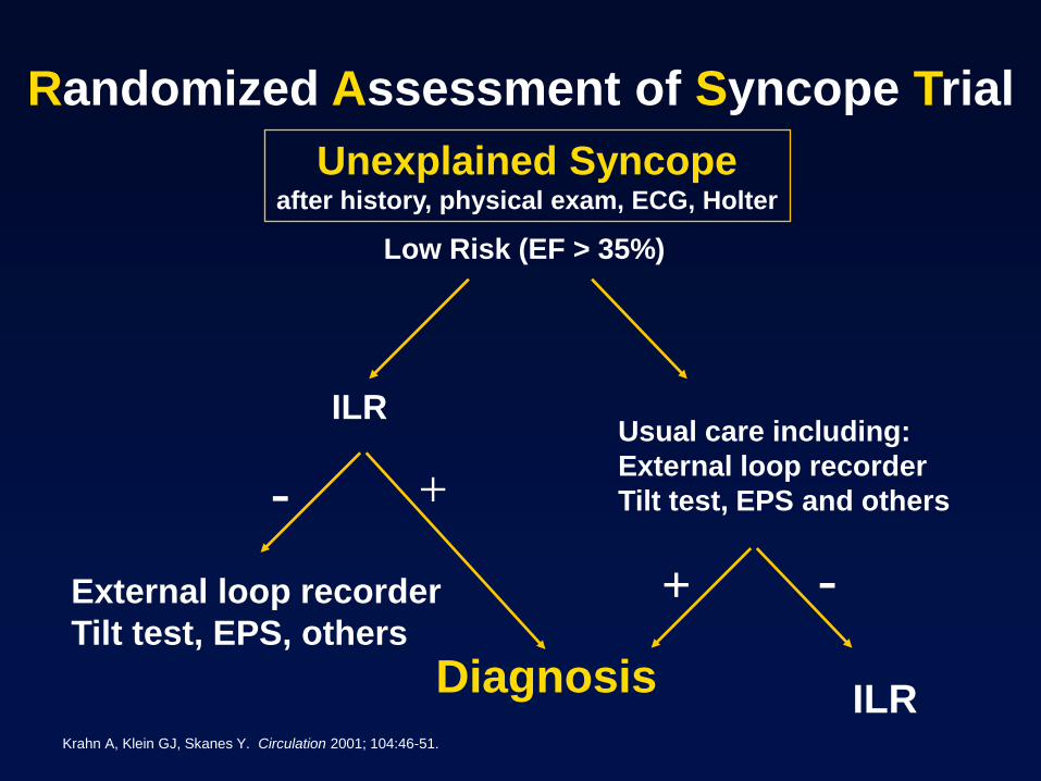
Randomized Assessment of Syncope Trial
Usual care including:
External loop recorder
Tilt test, EPS and others
Unexplained Syncope after history, physical exam, ECG, Holter
Low Risk (EF > 35%)
ILR
Diagnosis
+
+ -
-
ILR
External loop recorder
Tilt test, EPS, others
Krahn A, Klein GJ, Skanes Y. Circulation 2001; 104:46-51.

Unexplained Syncope Diagnosis
History and Physical Exam Surface ECG
Neurological
Testing
• Head CT Scan
• Carotid Doppler
• MRI
• Skull Films
• Brain Scan
• EEG
CV Syncope Workup
• Holter
• ELR or ILR
• Tilt Table
• Echo
• EPS
Other CV
Testing
• Angiogram
• Exercise Test
• SAECG
Psychological Evaluation
ENT Evaluation Endocrine Evaluation
Adapted from: W.Kapoor.An overview of the evaluation
and management of syncope. From Grubb B, Olshansky B (eds)
Syncope: Mechanisms and Management.
Armonk, NY: Futura Publishing Co., Inc.1998.

Typical Cardiovascular Diagnostic Pathway
History and Physical, ECG
Syncope
Known
SHD No
SHD
Echo
EPS
+
Treat
> 30 days;
> 2 Events
Tilt
ILR
Tilt
Holter/ ELR
ILR
Tilt/ILR
< 30 days
-
Adapted from:
Linzer M, et al. Annals of Int Med, 1997. 127:76-86.
Syncope: Mechanisms and Management. Grubb B, Olshansky B (eds) Futura Publishing 1999
Zimetbaum P, Josephson M. Annals of Int Med, 1999. 130:848-856.
Krahn A et al. ACC Current Journal Review,1999. Jan/Feb:80-84.

Neurally-Mediated Reflex Syncope (NMS)
Vasovagal syncope (VVS)
Carotid sinus syndrome (CSS)
Situational syncope
post-micturition
cough
swallow
defecation
blood drawing
etc.

Neurally Mediated Physiologic Reflex Mechanism with two Components:
Cardioinhibitory ( HR )
Vasodepressor ( BP )
Both components are usually present
Vasovagal Syncope (VVS):
Clinical Pathophysiology

Prevalence of VVS
Prevalence is poorly known
Various studies report 8% to 37% (mean 18%) of cases of syncope (Linzer 1997)
In general:
VVS patients younger than CSS patients
Ages range from adolescence to elderly (median 43 years)
Pallor, nausea, sweating, palpitations are common
Amnesia for warning symptoms in older patients

Management Strategies for VVS
Optimal management strategies for VVS are a source of debate
Patient education, reassurance, instruction
Fluids, salt, diet
Tilt Training
Support hose
Drug therapies
Pacing
Class II indication for VVS patients with positive HUT and cardioinhibitory or mixed reflex

Carotid Sinus Syndrome (CSS)
Syncope clearly associated with carotid sinus stimulation is rare (≤1% of syncope)
CSS may be an important cause of unexplained syncope / falls in older individuals

Carotid Sinus Hypersensitivity(CSH)
Abnormal response to CSM
Absence of symptoms attributable to CSS
CSH reported frequent in ‘fallers’ (Kenny)
CSH CSS

VVS: Pharmacologic Rx
Salt /Volume
Salt tablets, ‘sport’ drinks, fludrocortisone
Beta-adrenergic blockers
1 positive controlled trial (atenolol),
1 on-going RCT (POST)
Disopyramide
SSRIs
1 controlled trial
Vasoconstrictors (e.g., midodrine)
1 negative controlled trial (etilephrine)
Midodrine for Neurocardiogenic Syncope
Journal of Cardiovascular Electrophysiology Vol. 12, No. 8, Perez-Lugones, et al.
Months
p < 0.001
Sym
pto
m –
Fre
e In
terv
al
180 160 140 120 100 80 60 40 20 0
100
80
60
40
20
0
Fluid
Midodrine

Principal Causes of
Orthostatic Syncope
Drug-induced (very common)
diuretics
vasodilators
Primary autonomic failure multiple system atrophy
Parkinsonism
Secondary autonomic failure diabetes
alcohol
amyloid
Alcohol orthostatic intolerance apart from neuropathy

Syncope Due to Arrhythmia or
Structural CV Disease:
General Rules
Often life-threatening and/or exposes patient to high risk of injury
May be warning of critical CV disease
Aortic stenosis, Myocardial ischemia, Pulmonary hypertension
Assess culprit arrhythmia / structural abnormality aggressively
Initiate treatment promptly

Principal Causes of Syncope due to
Structural Cardiovascular Disease
Acute MI / Ischemia Acquired coronary artery disease
Congenital coronary artery anomalies
HOCM
Acute aortic dissection
Pericardial disease / tamponade
Pulmonary embolus / pulmonary hypertension
Valvular abnormalities Aortic stenosis, Atrial myxoma

Syncope Due to Cardiac Arrhythmias
Bradyarrhythmias
Sinus arrest, exit block
High grade or acute complete AV block
Tachyarrhythmias
Atrial fibrillation / flutter with rapid ventricular rate (e.g. WPW syndrome)
Paroxysmal SVT or VT
Torsades de pointes

Syncope: Torsades
From the files of DG Benditt, UM Cardiac Arrhythmia Center

83 yo woman
Bradycardia: Pacemaker
implanted
28 yo man in the ER multiple
times after falls resulting in
trauma
VT: ablated and medicated
Reveal ® ILR recordings; Medtronic data on file.

Drug-Induced QT Prolongation
Antiarrhythmics Class IA ...Quinidine, Procainamide, Disopyramide
Class III…Sotalol, Ibutilide, Dofetilide, Amiodarone, (NAPA)
Antianginal Agents (Bepridil)
Psychoactive Agents
Phenothiazines, Amitriptyline, Imipramine, Ziprasidone
Antibiotics Erythromycin, Pentamidine, Fluconazole
Nonsedating antihistamines (Terfenadine), Astemizole
Others (Cisapride), Droperidol

Treatment of Syncope Due to
Bradyarrhythmia
Class I indication for pacing using dual-
chamber system wherever adequate
atrial rhythm is available
Ventricular pacing in atrial fibrillation
with slow ventricular response
Treatment of Syncope Due to
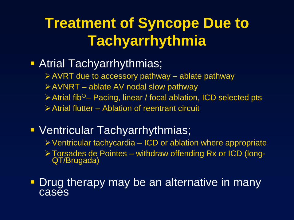
Tachyarrhythmia
Atrial Tachyarrhythmias; AVRT due to accessory pathway – ablate pathway
AVNRT – ablate AV nodal slow pathway
Atrial fib– Pacing, linear / focal ablation, ICD selected pts
Atrial flutter – Ablation of reentrant circuit
Ventricular Tachyarrhythmias; Ventricular tachycardia – ICD or ablation where appropriate
Torsades de Pointes – withdraw offending Rx or ICD (long-QT/Brugada)
Drug therapy may be an alternative in many cases

Conclusion
Syncope is a common symptom,
often with dramatic consequences,
which deserves thorough investigation
and appropriate treatment of its cause.

Sudden Cardiac Death in Young
Athletes

Athlete SCD– why is it important
Family physicians see a lot of athletes for sports related issues
Family physicians are involved in school health and pre-participation examinations
It is important to know the clinical characteristics of SCD and identify athletes who are at risk
It is important to know the current recommendations for pre-participation examinations
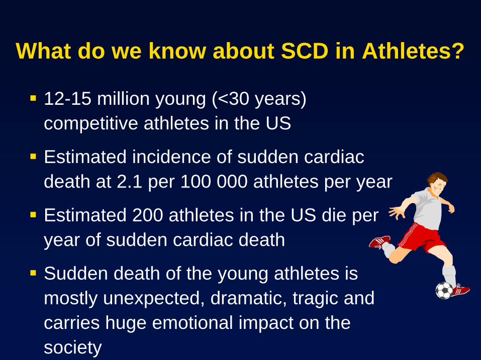
What do we know about SCD in Athletes?
12-15 million young (<30 years)
competitive athletes in the US
Estimated incidence of sudden cardiac
death at 2.1 per 100 000 athletes per year
Estimated 200 athletes in the US die per
year of sudden cardiac death
Sudden death of the young athletes is
mostly unexpected, dramatic, tragic and
carries huge emotional impact on the
society

Epidemiology Based on Autopsy Series
Age--
9% in middle school
62% in high school
22% in college
7% in professional
Sex—90% male, 10% female
Caucasians at highest risk
Sudden Cardiac Death of Athletes
Maron BJ et al, JAMA 1996 ; 276 : 199 - 203

Sports engaged in at the time
of sudden death
0
10
20
30
40
50
Ba
sk
etb
all
Fo
otb
all
Tra
ck
So
cc
er
Ba
se
ba
ll
Sw
imm
ing
No of athletes
Maron BJ et al, JAMA 1996 ; 276 : 199 - 203
Sudden Cardiac Death of Athletes

Causes of Sudden Cardiac Deaths
in Young Athletes
Sudden Cardiac Death of Athletes
Maron BJ et al, JAMA 1996 ; 276 : 199 - 203
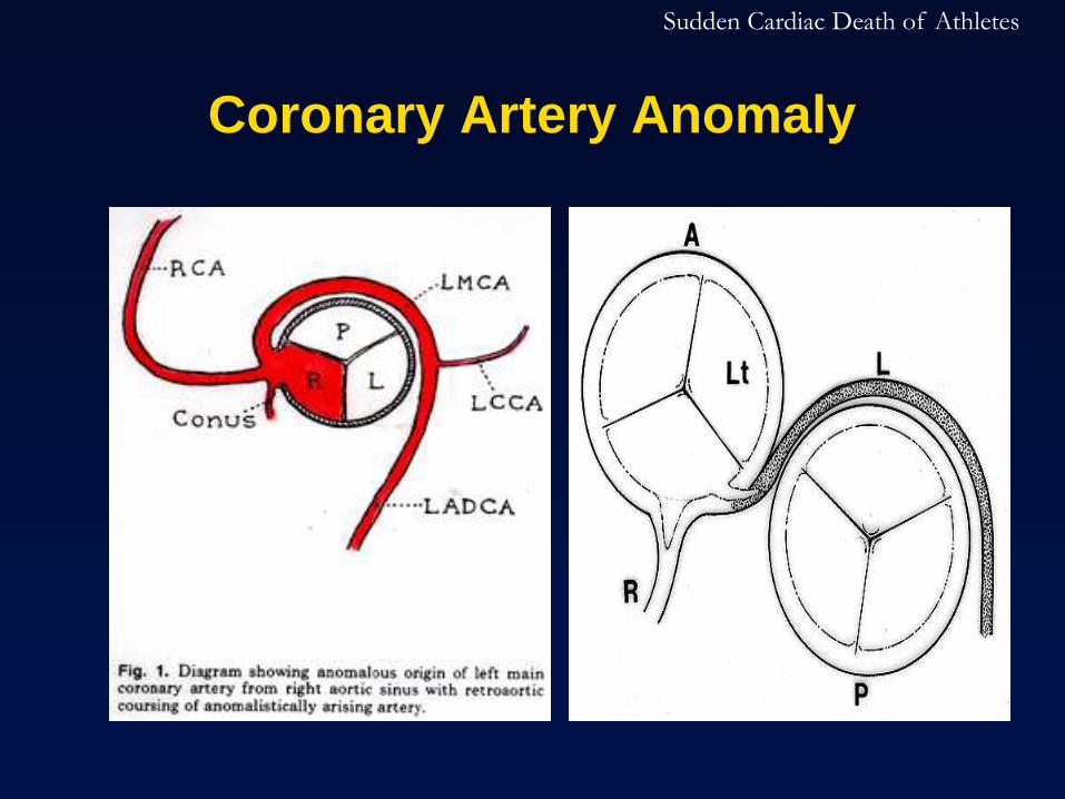
Coronary Artery Anomaly
Sudden Cardiac Death of Athletes

Coronary Artery Anomaly
With increased stroke volume during exercise
ascending aorta expands and the take-off angle is
further exaggerated
LMCA may also be compressed against root of
pulmonary trunk during exercise
ECG is likely normal
Very difficult to screen or diagnose even with
echocardiogram
Treatment: Surgery for coronary reimplantation
Sudden Cardiac Death of Athletes

Commotio Cordis Sudden disturbance of heart rhythm as the result
of a blunt, non-penetrating impact to the
precordial region
Impact occurring within a specific 10-20
millisecond portion of the cardiac cycle in the
ascending phase of the T wave, when the
ventricular myocardium is repolarizing, moving
from systole to diastole
Most effective preventions are:
Chest shield
Automatic External Defibrillator
(AED)
Sudden Cardiac Death of Athletes

European Guidelines for PPE
In 2005, The European Society of Cardiology issued official recommendation for PPE, including 12-lead ECG

Circulation 2007;115;1643-1655
Sudden Cardiac Death of Athletes

Sudden Cardiac Death of Athletes