Embed Size (px)
Citation preview
AbstractBrachial Plexus Birth Palsy (BPBP) is defined as a flaccid paralysis of the upper limb that occurs as a result of traction injury to the brachial plexus during the process of birth.The incidence of BPBP has been estimated between 0.4% to 5.1% in various studies worldwide.A precise clinical examination is the key to ascertain the type of injury, prognosticate the outcome and forecast the probable need of surgical intervention. A detailed clinical examination methodology and important signs directing to the intervention are described in this paper. Importance of regular clinical follow up has been emphasised. Early rehabilitation of infants with BPBP and physiotherapy protocols are discussed.
Keywords: Brachial Plexus Birth Palsy (BPBP); Active Movement Scale (AMS); Early rehabilitation; Erb’s Palsy; Narakas.
IntroductionBrachial Plexus Birth Palsy (BPBP) is defined as a flaccid paralysis of the upper limb that occurs as a result of traction injury to the brachial plexus during the process of birth. While high percentage of infants improve spontaneously, about 25-30% patients have permanent deficit requiring either nerve surgery or secondary salvage surgeries [1].Many imaging modalities support the diagnosis of BPBP but clinical examination is the mainstay of assessing severity and the extent of injury. Decisions about surgical interventions are broadly based on a precise clinical examination. This article will discuss in detail the methodology of clinical examination of a BPBP child and its utility in decision making.
Epidemiology and EtiologyThe incidence of BPBP has been estimated between 0.4% to 5.1% in various studies worldwide [2-7]. This variation reflects differences in availability of heath care, reporting methods, referral bias and population dissimilarity in different parts of the world [8]. About 5% of all BPBP cases can be bilateral and are more frequently seen with breech deliveries [9]. Multiple studies have shown steady decrease in the incidence of BPBP cases which may be due to an increase in the rate of caesarean sections [7, 10]. Another probable
Symposium International Journal of Paediatric Orthopaedics | January-April 2021; 7(1): Page 28-36
@ 2021 by International Journal of Paediatric Orthopaedics| Available on www.ijpoonline.com | DOI- 10.13107/ijpo.2021.v07i01.099This is an Open Access article distributed under the terms of the Creative Commons Attribution Non-Commercial License (http://creativecommons.org/licenses/by-nc/3.0) which permits unrestricted noncommercial use, distribution, and reproduction in any medium, provided the original work is properly cited.
28 International Journal of Paediatric Orthopaedics Volume 7 Issue 1 January-April 2021 Page 28-36 | | | | |
POSIiJPO
Clinical Examination and Early Management of Brachial Plexus Birth Palsy
(BPBP)
Dr. Gaurav Gupta Dr. Tejas Patel
Dr. Chasanal Rathod Dr. Amila S. Ratnayake
1 2Gaurav Gupta MBBS, MS Ortho. , Tejas Patel PT, C/NDT, SI. , 3 4Chasanal Rathod MBBS, MS Ortho. , Amila S. Ratnayake MBBS, MS MRCS(Ed) ,
5 6Maulin M. Shah MBBS, MS Ortho. , Bharat K. Kadadi MBBS, MS Ortho.
Address of CorrespondenceDr. Maulin M Shah,
Consultant, Paediatric Orthopaedic Surgeon, OrthoKids Clinic, Ahmedabad, Gujrat, India.
E-mail: [email protected]
1Consultant, Paediatric Orthopaedic Surgeon, Child Ortho Clinic, New Delhi
2Consultant, Paediatric Physiotherapist, Sparsh Paediatric Rehabilitation Clinic, Ahmedabad, Gujrat,
India.3Consultant Pediatric Orthopedic Surgeon, NHSRCC
Children's Hospital, Mumbai, Maharashtra, India.4Plastic & Reconstructive Surgeon, National Hospital
Kandy, Sri Lanka5Consultant, Paediatric Orthopaedic Surgeon, OrthoKids Clinic, Ahmedabad, Gujrat, India.
6Bengaluru Hand Centre & Manipal Hospitals, Bangalore, Karnataka, India.
Dr. Maulin M. Shah Dr. Bharat K. Kadadi
www.ijpoonline.comGupta G et alcause for this decrease could be simulation-based training courses for obstetricians leading to better management of shoulder dystocia [10].
Risk factorsAlthough several risk factors have been identified to be associated with BPBP, more than 50% patients do not have any risk factor [2]. Shoulder dystocia was found to be the strongest risk factor associated with BPBP [10, 11]. Others include macrosomia (child birth weight > 3.99 kg), breech presentation, gestational diabetes, cephalo-pelvic disproportion, multiparity, prolonged second stage of labour, duration and use of vacuum assisted deliveries, and use of fundal pressure [8, 11, 12]. In recent studies, hypotonia and hypoxia have also been found to be independent risk factors [7, 10]. Caesarean section has been shown to be protective for BPBP [12, 13].
Mechanism of InjuryThe exact mechanism of injury is unknown but the most probable, in cases associated with shoulder dystocia, is traction to the child’s neck by manual pull, vacuum or forceps. This stretch causes a strain to the brachial plexus leading to varying degrees of brachial plexus injury [8]. Other mechanisms described in the literature include intrauterine maladaptation, intrauterine stretch of brachial plexus in early stage of labour, excessive fundal pressure impacting the anterior shoulder behind the pubic symphysis and bicornuate uterus [11, 14, 15].
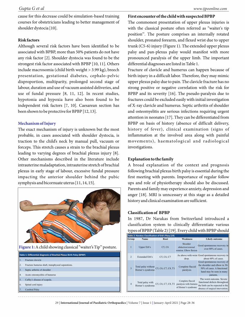
First encounter of the child with suspected BPBPThe commonest presentation of upper plexus injuries is with the classical posture often referred as “waiter's tip position”. The posture comprises an internally rotated shoulder, pronated forearm, and flexed wrist due to upper trunk (C5-6) injury (Figure 1). The extended upper plexus palsy and pan-plexus palsy would manifest with more pronounced paralysis of the upper limb. The important differential diagnoses are listed in Table 1.Fracture of clavicle and humerus can happen because of birth injury in a difficult labor. Therefore, they may mimic upper plexus palsy due to pain. The clavicle fracture has no strong positive or negative correlation with the risk for BPBP and its severity [16]. The pseudo-paralysis due to fractures could be excluded easily with initial investigation of X-ray clavicle and humerus. Septic arthritis of shoulder and osteomyelitis are serious infections requiring urgent attention in neonates [17]. They can be differentiated from BPBP on basis of history (absence of difficult delivery, history of fever), clinical examination (signs of inflammation at the involved area along with painful movements) , haematolog ica l and radiolog ica l investigations.
Explanation to the familyA broad explanation of the context and prognosis following brachial plexus birth palsy is essential during the first meeting with parents. Importance of regular follow ups and role of physiotherapy should also be discussed. Parents and family may experience anxiety, depression and anger [18]. MRI is unncessary at this stage as a detailed history and clinical examination are sufficient.
Classification of BPBPIn 1987, Dr Narakas from Switzerland introduced a classification system to clinically differentiate various types of BPBP (Table 2) [19]. Every child with BPBP should
29 International Journal of Paediatric Orthopaedics Volume 7 Issue 1 January-April 2021 Page 28-36 | | | | |
Table 1: Differential diagnosis of Brachial Plexus Birth Palsy (BPBP)
1.
Fracture clavicle
2.
Fracture humerus shaft, transphyseal separations.
3.
Septic arthritis of shoulder
4.
Acute osteomyelitis of humerus
5.
Caffey’s disease of scapula.
6.
Spinal cord injury
7.
Cerebral Palsy
Figure 1: A child showing classical “waiter’s Tip” posture.
Group Name Root Weakness Likely outcome
1 Upper Erb’s C5, C6
Shoulder
abduction/external
rotation. Elbow flexion
Good spontaneous recovery in
over 80% of cases.
2 Extended Erb’s C5, C6, C7As above with wrist
drop.
Good spontaneous recovery in
about 60% of cases.
3Total palsy without
Horner’s syndrome.C5, C6, C7, C8, T1
Complete flaccid
paralysis.
Good spontaneous recovery of
the shoulder and elbow in 30-
50% of cases. A functional
hand may be seen in many
patients.
4Total palsy with
Horner’s syndrome.C5, C6, C7, C8, T1
Complete flaccid
paralysis with features
of Horner’s syndrome
The worst outcome. Severe functional defects throughout
the limb can be expected in the
absence of surgical intervention.
Table 2: Narakas Classification of Erb’s Plasy [19]
be classified as per Narakas classification after 2 months of age. The initial clinical presentation might change due to resolving edema surrounding the brachial plexus which usually takes 3-4 weeks. There are 4 grades on the basis of increasing severity and involvement of nerve roots. Lower grades of injury are associated with better prognosis and lesser probability of primary or secondary surgical intervention.Al Qattan proposed further division of Group 2 patients into 2a and 2b. 2a included those children who recover wrist extension within 2 months. 2b comprised those who do not recover wrist extension within 2 months and carries a poorer prognosis [20].
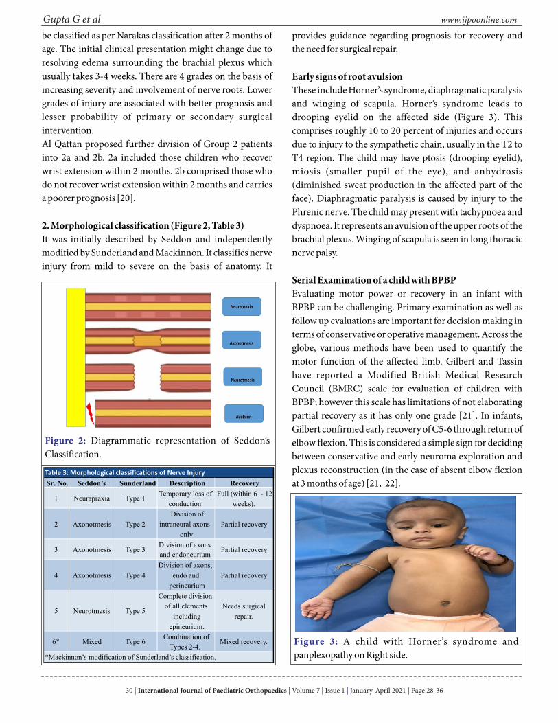
2. Morphological classification (Figure 2, Table 3)It was initially described by Seddon and independently modified by Sunderland and Mackinnon. It classifies nerve injury from mild to severe on the basis of anatomy. It
provides guidance regarding prognosis for recovery and the need for surgical repair.
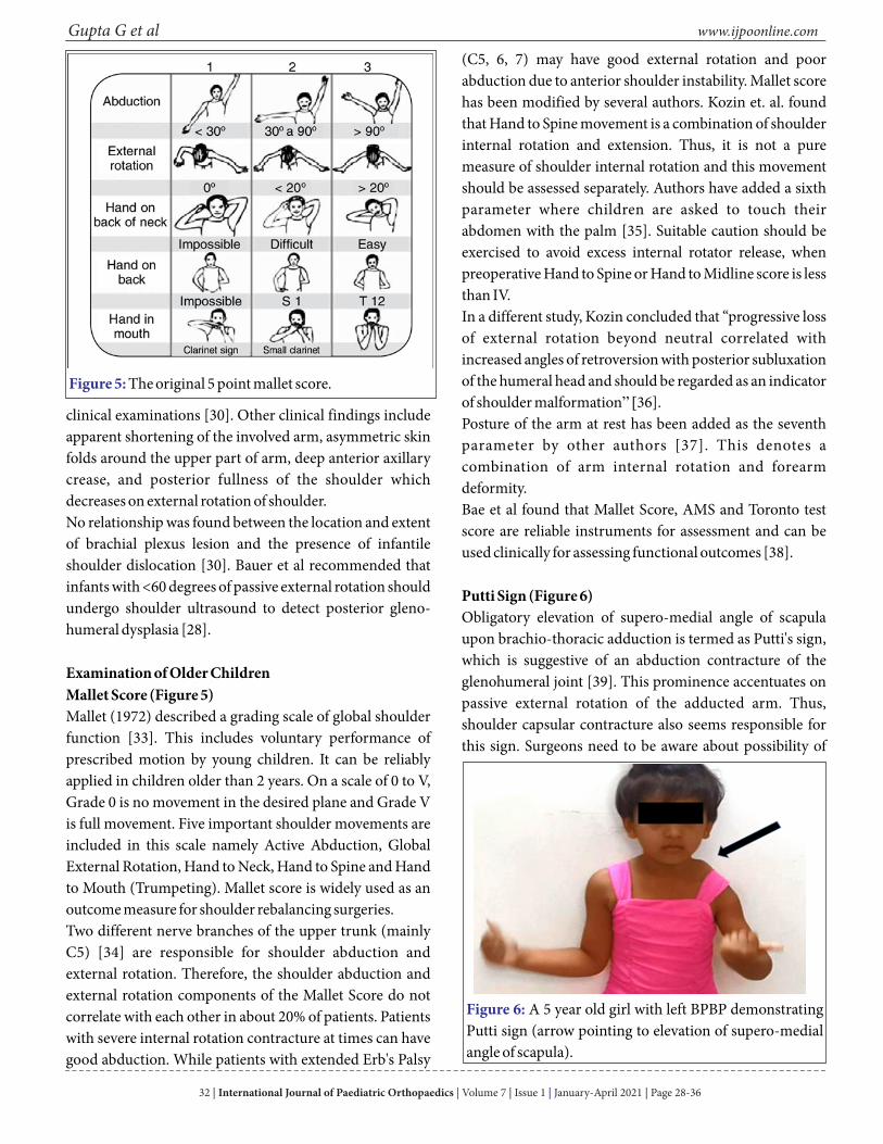
Early signs of root avulsionThese include Horner’s syndrome, diaphragmatic paralysis and winging of scapula. Horner’s syndrome leads to drooping eyelid on the affected side (Figure 3). This comprises roughly 10 to 20 percent of injuries and occurs due to injury to the sympathetic chain, usually in the T2 to T4 region. The child may have ptosis (drooping eyelid), miosis (smaller pupil of the eye), and anhydrosis (diminished sweat production in the affected part of the face). Diaphragmatic paralysis is caused by injury to the Phrenic nerve. The child may present with tachypnoea and dyspnoea. It represents an avulsion of the upper roots of the brachial plexus. Winging of scapula is seen in long thoracic nerve palsy.
Serial Examination of a child with BPBPEvaluating motor power or recovery in an infant with BPBP can be challenging. Primary examination as well as follow up evaluations are important for decision making in terms of conservative or operative management. Across the globe, various methods have been used to quantify the motor function of the affected limb. Gilbert and Tassin have reported a Modified British Medical Research Council (BMRC) scale for evaluation of children with BPBP; however this scale has limitations of not elaborating partial recovery as it has only one grade [21]. In infants, Gilbert confirmed early recovery of C5-6 through return of elbow flexion. This is considered a simple sign for deciding between conservative and early neuroma exploration and plexus reconstruction (in the case of absent elbow flexion at 3 months of age) [21, 22].
www.ijpoonline.comGupta G et al
Figure 2: Diagrammatic representation of Seddon’s Classification.
Figure 3: A child with Horner’s syndrome and panplexopathy on Right side.
Sr. No. Seddon’s Sunderland Description Recovery
1 Neurapraxia Type 1Temporary loss of
conduction.
Full (within 6 - 12
weeks).
2 Axonotmesis Type 2
Division of
intraneural axons
only
Partial recovery
3 Axonotmesis Type 3Division of axons
and endoneuriumPartial recovery
4 Axonotmesis Type 4
Division of axons,
endo and
perineurium
Partial recovery
5 Neurotmesis Type 5
Complete division
of all elements
including
epineurium.
Needs surgical
repair.
6* Mixed Type 6Combination of
Types 2-4.Mixed recovery.
*Mackinnon’s modification of Sunderland’s classification.
Table 3: Morphological classifications of Nerve Injury
30 International Journal of Paediatric Orthopaedics Volume 7 Issue 1 January-April 2021 Page 28-36 | | | | |

www.ijpoonline.comGupta G et al
Clark and Curtis developed the Hospital for Sick Children Active Movement Scale (HSC AMS) [23, 24]. This scale has seven grades and involves detailed evaluation of 15 movements across the affected upper extremity. It is applicable to infants as well as older children. Small improvements in movement can be detected by the HSC AMS. The child is evaluated in supine, side lying and sitting positions and joint movements are assessed as gravity eliminated or against gravity and accordingly the scores can be less than 4 or higher than 4 respectively (Table 4) [23, 24].Toronto Test score is derived from AMS and is used to predict recovery in infants with BPBP. It evaluates elbow flexion and extension, wrist, finger and thumb extension as per AMS. Each AMS grade is then converted to a
numerical score ranging from 0 (no motion) to 10 (full motion). A score less than 3.5 at 3 months of age is suggestive of poor recovery and greater than 3.5 indicates fair recovery [25, 26]. Absence of improvement of HSC AMS score or Toronto test score suggests a need for surgical intervention. At 9 months of age, the assessment includes a Cookie test wherein the infant is offered a cookie and assessed if adequate elbow flexion is present to bring the cookie to the mouth without flexing the trunk or neck >45 degrees. Ability to get the cookie to the mouth excludes the need for surgical intervention [24]. Another test performed between 6-9 months of age is the Towel test also known as Eye cover test or Hand to face test [27]. Towel test is performed by covering face of the child with a towel and assessing child’s ability to reach out to the towel; the ability to do so suggests that surgical intervention may not be necessary.
Infantile shoulder dislocation (Figure 4)Infantile shoulder dislocation is developmental dysplasia of the glenohumeral joint where the glenoid and humeral head develop in a posteriorly oriented alignment [28]. It is caused by an imbalance of internal and external shoulder rotators. It was acknowledged as early as 1905 by Whitman [29]. Its incidence varies from 8% to 29% in different studies on infants with BPBP [28, 30]. Due to better awareness and diagnostic modalities, infantile shoulder dislocation is being increasingly reported [28, 30, 31, 32].The hallmark of infantile shoulder dislocation is progressively decreasing external rotation on successive
Observation Muscle Grade
No contraction 0
Contraction, No motion 1
Motion<1/2 range 2
Motion>1/2 range 3
Full motion 4
Motion<1/2 range 5
Motion>1/2 range 6
Full motion 7
Gravity eliminated
Against Gravity
Table 4: Active Movement Scale
Observation Muscle Grade Numerical Score
No Contraction 0 0
Contraction, no motion 1 0.3
Motion < 1/2 range 2 0.3
Motion > 1/2 range 3 0.6
Full motion 4 0.6
Motion<1/2 range 5 0.6
Motion>1/2 range 6 1.3
Full motion 7 2
Gravity eliminated
Against Gravity
Table 5: The Toronto Score
Figure 4: A child with infantile shoulder dislocation. There is relative shortening of the arm, asymmetric skin folds and a deep axillary crease (arrow). Horner' Syndrome on the affected side is also evident.
31 International Journal of Paediatric Orthopaedics Volume 7 Issue 1 January-April 2021 Page 28-36 | | | | |
www.ijpoonline.comGupta G et al
clinical examinations [30]. Other clinical findings include apparent shortening of the involved arm, asymmetric skin folds around the upper part of arm, deep anterior axillary crease, and posterior fullness of the shoulder which decreases on external rotation of shoulder.No relationship was found between the location and extent of brachial plexus lesion and the presence of infantile shoulder dislocation [30]. Bauer et al recommended that infants with <60 degrees of passive external rotation should undergo shoulder ultrasound to detect posterior gleno-humeral dysplasia [28].
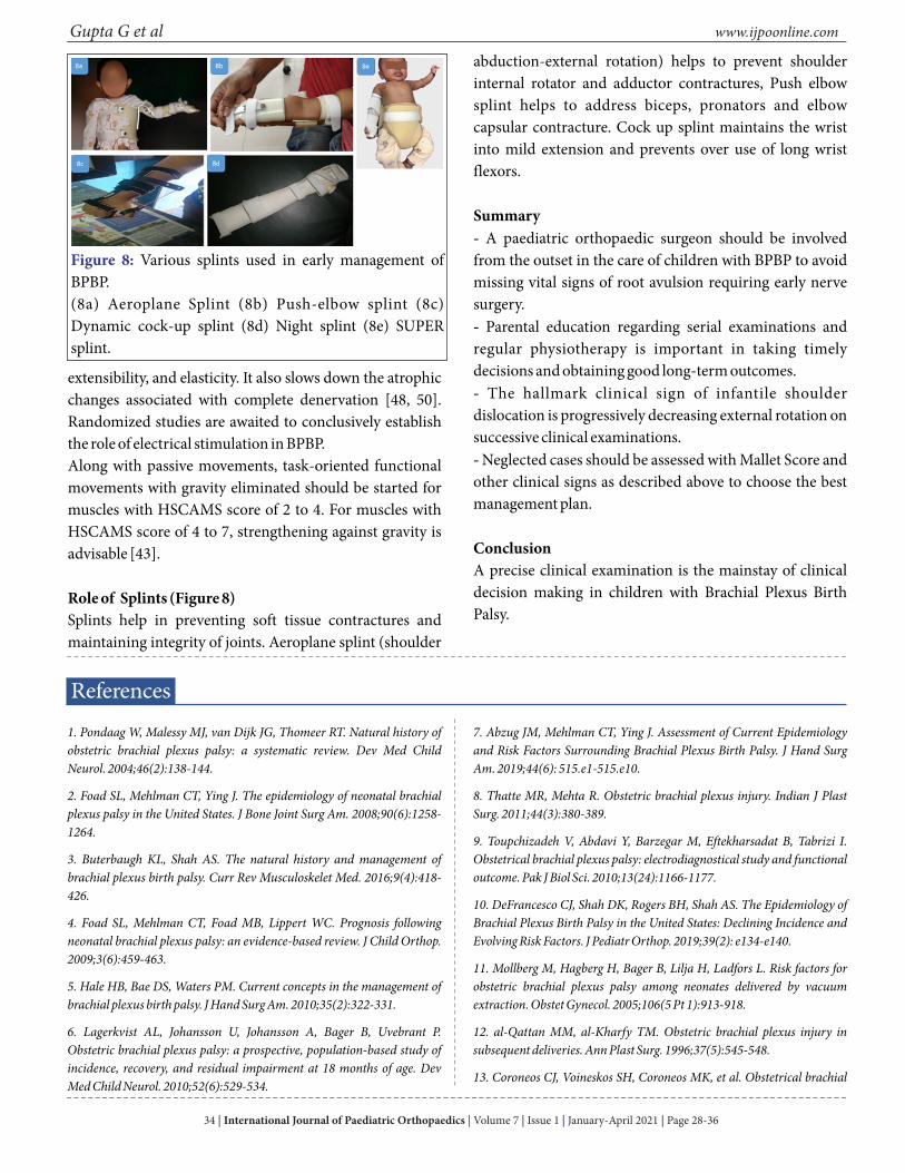
Examination of Older Children Mallet Score (Figure 5)Mallet (1972) described a grading scale of global shoulder function [33]. This includes voluntary performance of prescribed motion by young children. It can be reliably applied in children older than 2 years. On a scale of 0 to V, Grade 0 is no movement in the desired plane and Grade V is full movement. Five important shoulder movements are included in this scale namely Active Abduction, Global External Rotation, Hand to Neck, Hand to Spine and Hand to Mouth (Trumpeting). Mallet score is widely used as an outcome measure for shoulder rebalancing surgeries. Two different nerve branches of the upper trunk (mainly C5) [34] are responsible for shoulder abduction and external rotation. Therefore, the shoulder abduction and external rotation components of the Mallet Score do not correlate with each other in about 20% of patients. Patients with severe internal rotation contracture at times can have good abduction. While patients with extended Erb's Palsy
(C5, 6, 7) may have good external rotation and poor abduction due to anterior shoulder instability. Mallet score has been modified by several authors. Kozin et. al. found that Hand to Spine movement is a combination of shoulder internal rotation and extension. Thus, it is not a pure measure of shoulder internal rotation and this movement should be assessed separately. Authors have added a sixth parameter where children are asked to touch their abdomen with the palm [35]. Suitable caution should be exercised to avoid excess internal rotator release, when preoperative Hand to Spine or Hand to Midline score is less than IV.In a different study, Kozin concluded that “progressive loss of external rotation beyond neutral correlated with increased angles of retroversion with posterior subluxation of the humeral head and should be regarded as an indicator of shoulder malformation” [36].Posture of the arm at rest has been added as the seventh parameter by other authors [37]. This denotes a combination of arm internal rotation and forearm deformity. Bae et al found that Mallet Score, AMS and Toronto test score are reliable instruments for assessment and can be used clinically for assessing functional outcomes [38].
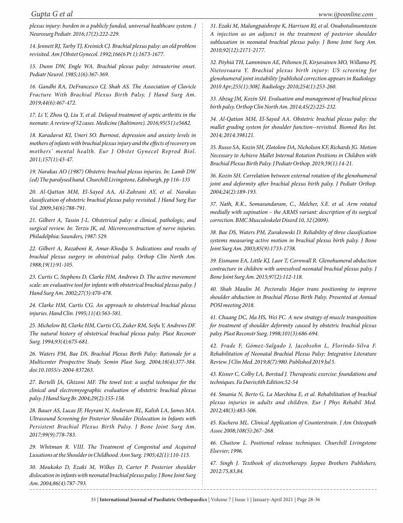
Putti Sign (Figure 6)Obligatory elevation of supero-medial angle of scapula upon brachio-thoracic adduction is termed as Putti's sign, which is suggestive of an abduction contracture of the glenohumeral joint [39]. This prominence accentuates on passive external rotation of the adducted arm. Thus, shoulder capsular contracture also seems responsible for this sign. Surgeons need to be aware about possibility of
Figure 5: The original 5 point mallet score.
Figure 6: A 5 year old girl with left BPBP demonstrating Putti sign (arrow pointing to elevation of supero-medial angle of scapula).
32 International Journal of Paediatric Orthopaedics Volume 7 Issue 1 January-April 2021 Page 28-36 | | | | |

www.ijpoonline.comGupta G et alworsening of Putti Sign after tendon transfer for achieving better external rotation. This needs to be discussed with parents at the outset. Supraspinatus slide or resection of supero-medial angle of scapula is sometimes performed to address this cosmetic concern.
Pediwrape Test (Figure 7)CŸhildren with Extended Erb's palsy (C5, 6, 7) may have limited shoulder abduction in the presence of good active external rotation. In addition, a certain degree of Triceps weakness and occasionally elbow flexion deformity may coexist. Varying degrees of internal rotation weakness or external rotation contracture is observed in these patients, where they are unable to touch the midline with the palm. Upon application of an elbow extension splint (Pediwrape),
shoulder abduction typically improves to beyond 90°. Improved sidewise elevation after application of a Pediwrape is termed as a “Positive Test". It indicates that the lack of abduction is due to weak internal rotators and resultant anterior shoulder instability [40].Ÿ
Co-contractions in BPBPCross innervation by misdirection of regenerating axons leads to impaired movements and it is termed as co-contraction. When agonist and antagonist groups contract together, the resultant movements are insignificant. Cheung et. al. found four distinct types of co-contractions in BPBP [41]. Co-contractions between shoulder adductors and abductors lead to limited elevation, while between elbow flexors and extensors leads to inability of
child to reach the mouth. In a suspected co-contraction, antagonists are relaxed temporarily by Botulinum toxin injection which unmasks the agonist movement. Co-contractions between shoulder abductors and elbow flexors leads to "Trumpeting" (arm elevation on attempted hand-to-mouth movement).
Early Treatment of BPBP patientsIn infants with BPBP, early rehabilitation is advocated to maintain normal muscle length and prevent muscle atrophy and soft tissue contractures.
Role of Passive Range of Motion In the first 2 months of life, care givers are taught home-based gentle and graded passive range of motion exercises for fingers, wrist, elbow, forearm and shoulder joint. They are typically advised to perform these exercises before every feed. Passive range of motion exercises should be delayed for two weeks in patients with associated fractures [42, 43, 44].In case of an established soft tissue contracture, deeper shortening (based upon Total Motion Release principles) of tight fascia sets a better platform for further course of therapy before elongating shortened tissues and strengthening the weaker group of muscles [45, 46]. To encourage orientation of the affected limb, patient should be exposed to hand-to-hand, hand-to-arm and hand-to-mouth tactile exploration.
Role of Electrical StimulationAfter 2 months, the denervation process completes and severity/type of injury is established. Low frequency electrical stimulation for partial or completely denervated muscles should be initiated [47]. Interrupted galvanic (IG) current (Impulse with pulse duration 100 microseconds (ms) and frequency of 1-2 hertz (Hz)) is used for denervated muscles and muscles with Hospital for sick children active movement scale (HSCAMS) score of 0 to 4 [48, 49, 50]. On the basis of strength duration curve findings and HSCAMS score of 3 to 5, current settings can be changed from Interrupted galvanic to Surged Faradic (SF) (pulse duration of less than 0.1 to 1 ms and frequency of 50-100 hz) [47, 50].There is evidence in favour of electrical stimulation for nerve regeneration in animals, but not in humans. Electrical stimulation helps in maintaining the normal properties of muscle like irritability, contractility,
Figure 7: A 7 year girl with Right BPBP demonstrating Pediwrape test. The test is considered positive when shoulder abduction improves beyond 90degrees upon application of an elbow extension splint.
33 International Journal of Paediatric Orthopaedics Volume 7 Issue 1 January-April 2021 Page 28-36 | | | | |
www.ijpoonline.comGupta G et al
1. Pondaag W, Malessy MJ, van Dijk JG, Thomeer RT. Natural history of obstetric brachial plexus palsy: a systematic review. Dev Med Child Neurol. 2004;46(2):138-144.
2. Foad SL, Mehlman CT, Ying J. The epidemiology of neonatal brachial plexus palsy in the United States. J Bone Joint Surg Am. 2008;90(6):1258-1264.
3. Buterbaugh KL, Shah AS. The natural history and management of brachial plexus birth palsy. Curr Rev Musculoskelet Med. 2016;9(4):418-426.
4. Foad SL, Mehlman CT, Foad MB, Lippert WC. Prognosis following neonatal brachial plexus palsy: an evidence-based review. J Child Orthop. 2009;3(6):459-463.
5. Hale HB, Bae DS, Waters PM. Current concepts in the management of brachial plexus birth palsy. J Hand Surg Am. 2010;35(2):322-331.
6. Lagerkvist AL, Johansson U, Johansson A, Bager B, Uvebrant P. Obstetric brachial plexus palsy: a prospective, population-based study of incidence, recovery, and residual impairment at 18 months of age. Dev Med Child Neurol. 2010;52(6):529-534.
7. Abzug JM, Mehlman CT, Ying J. Assessment of Current Epidemiology and Risk Factors Surrounding Brachial Plexus Birth Palsy. J Hand Surg Am. 2019;44(6): 515.e1-515.e10.
8. Thatte MR, Mehta R. Obstetric brachial plexus injury. Indian J Plast Surg. 2011;44(3):380-389.
9. Toupchizadeh V, Abdavi Y, Barzegar M, Eftekharsadat B, Tabrizi I. Obstetrical brachial plexus palsy: electrodiagnostical study and functional outcome. Pak J Biol Sci. 2010;13(24):1166-1177.
10. DeFrancesco CJ, Shah DK, Rogers BH, Shah AS. The Epidemiology of Brachial Plexus Birth Palsy in the United States: Declining Incidence and Evolving Risk Factors. J Pediatr Orthop. 2019;39(2): e134-e140.
11. Mollberg M, Hagberg H, Bager B, Lilja H, Ladfors L. Risk factors for obstetric brachial plexus palsy among neonates delivered by vacuum extraction. Obstet Gynecol. 2005;106(5 Pt 1):913-918.
12. al-Qattan MM, al-Kharfy TM. Obstetric brachial plexus injury in subsequent deliveries. Ann Plast Surg. 1996;37(5):545-548.
13. Coroneos CJ, Voineskos SH, Coroneos MK, et al. Obstetrical brachial
extensibility, and elasticity. It also slows down the atrophic changes associated with complete denervation [48, 50]. Randomized studies are awaited to conclusively establish the role of electrical stimulation in BPBP.Along with passive movements, task-oriented functional movements with gravity eliminated should be started for muscles with HSCAMS score of 2 to 4. For muscles with HSCAMS score of 4 to 7, strengthening against gravity is advisable [43].
Role of Splints (Figure 8)Splints help in preventing soft tissue contractures and maintaining integrity of joints. Aeroplane splint (shoulder
abduction-external rotation) helps to prevent shoulder internal rotator and adductor contractures, Push elbow splint helps to address biceps, pronators and elbow capsular contracture. Cock up splint maintains the wrist into mild extension and prevents over use of long wrist flexors.
Summary- A paediatric orthopaedic surgeon should be involved from the outset in the care of children with BPBP to avoid missing vital signs of root avulsion requiring early nerve surgery.- Parental education regarding serial examinations and regular physiotherapy is important in taking timely decisions and obtaining good long-term outcomes.- The hallmark clinical sign of infantile shoulder dislocation is progressively decreasing external rotation on successive clinical examinations.- Neglected cases should be assessed with Mallet Score and other clinical signs as described above to choose the best management plan.
ConclusionA precise clinical examination is the mainstay of clinical decision making in children with Brachial Plexus Birth Palsy.
Figure 8: Various splints used in early management of BPBP. (8a) Aeroplane Splint (8b) Push-elbow splint (8c) Dynamic cock-up splint (8d) Night splint (8e) SUPER splint.
34 International Journal of Paediatric Orthopaedics Volume 7 Issue 1 January-April 2021 Page 28-36 | | | | |
www.ijpoonline.comGupta G et alplexus injury: burden in a publicly funded, universal healthcare system. J Neurosurg Pediatr. 2016;17(2):222-229.
14. Jennett RJ, Tarby TJ, Kreinick CJ. Brachial plexus palsy: an old problem revisited. Am J Obstet Gynecol. 1992;166(6 Pt 1):1673-1677.
15. Dunn DW, Engle WA. Brachial plexus palsy: intrauterine onset. Pediatr Neurol. 1985;1(6):367-369.
16. Gandhi RA, DeFrancesco CJ, Shah AS. The Association of Clavicle Fracture With Brachial Plexus Birth Palsy. J Hand Surg Am. 2019;44(6):467-472.
17. Li Y, Zhou Q, Liu Y, et al. Delayed treatment of septic arthritis in the neonate: A review of 52 cases. Medicine (Baltimore). 2016;95(51):e5682.
18. Karadavut KI, Uneri SO. Burnout, depression and anxiety levels in mothers of infants with brachial plexus injury and the effects of recovery on mothers' mental health. Eur J Obstet Gynecol Reprod Biol. 2011;157(1):43-47.
19. Narakas AO (1987) Obstetric brachial plexus injuries. In: Lamb DW (ed) The paralysed hand. Churchill Livingstone, Edinburgh, pp 116–135
20. Al-Qattan MM, El-Sayed AA, Al-Zahrani AY, et al. Narakas classification of obstetric brachial plexus palsy revisited. J Hand Surg Eur Vol. 2009;34(6):788-791.
21. Gilbert A, Tassin J-L. Obstetrical palsy: a clinical, pathologic, and surgical review. In: Terzis JK, ed. Microreconstruction of nerve injuries. Philadelphia: Saunders, 1987: 529.
22. Gilbert A, Razaboni R, Amar-Khodja S. Indications and results of brachial plexus surgery in obstetrical palsy. Orthop Clin North Am. 1988;19(1):91-105.
23. Curtis C, Stephens D, Clarke HM, Andrews D. The active movement scale: an evaluative tool for infants with obstetrical brachial plexus palsy. J Hand Surg Am. 2002;27(3):470-478.
24. Clarke HM, Curtis CG. An approach to obstetrical brachial plexus injuries. Hand Clin. 1995;11(4):563-581.
25. Michelow BJ, Clarke HM, Curtis CG, Zuker RM, Seifu Y, Andrews DF. The natural history of obstetrical brachial plexus palsy. Plast Reconstr Surg. 1994;93(4):675-681.
26. Waters PM, Bae DS. Brachial Plexus Birth Palsy: Rationale for a Multicenter Prospective Study. Semin Plast Surg. 2004;18(4):377-384. doi:10.1055/s-2004-837263.
27. Bertelli JA, Ghizoni MF. The towel test: a useful technique for the clinical and electromyographic evaluation of obstetric brachial plexus palsy. J Hand Surg Br. 2004;29(2):155-158.
28. Bauer AS, Lucas JF, Heyrani N, Anderson RL, Kalish LA, James MA. Ultrasound Screening for Posterior Shoulder Dislocation in Infants with Persistent Brachial Plexus Birth Palsy. J Bone Joint Surg Am. 2017;99(9):778-783.
29. Whitman R. VIII. The Treatment of Congenital and Acquired Luxations at the Shoulder in Childhood. Ann Surg. 1905;42(1):110-115.
30. Moukoko D, Ezaki M, Wilkes D, Carter P. Posterior shoulder dislocation in infants with neonatal brachial plexus palsy. J Bone Joint Surg Am. 2004;86(4):787-793.
31. Ezaki M, Malungpaishrope K, Harrison RJ, et al. Onabotulinumtoxin A injection as an adjunct in the treatment of posterior shoulder subluxation in neonatal brachial plexus palsy. J Bone Joint Surg Am. 2010;92(12):2171-2177.
32. Pöyhiä TH, Lamminen AE, Peltonen JI, Kirjavainen MO, Willamo PJ, Nietosvaara Y. Brachial plexus birth injury: US screening for glenohumeral joint instability [published correction appears in Radiology. 2010 Apr;255(1):308]. Radiology. 2010;254(1):253-260.
33. Abzug JM, Kozin SH. Evaluation and management of brachial plexus birth palsy. Orthop Clin North Am. 2014;45(2):225-232.
34. Al-Qattan MM, El-Sayed AA. Obstetric brachial plexus palsy: the mallet grading system for shoulder function--revisited. Biomed Res Int. 2014; 2014:398121.
35. Russo SA, Kozin SH, Zlotolow DA, Nicholson KF, Richards JG. Motion Necessary to Achieve Mallet Internal Rotation Positions in Children with Brachial Plexus Birth Palsy. J Pediatr Orthop. 2019;39(1):14-21.
36. Kozin SH. Correlation between external rotation of the glenohumeral joint and deformity after brachial plexus birth palsy. J Pediatr Orthop. 2004;24(2):189-193.
37. Nath, R.K., Somasundaram, C., Melcher, S.E. et al. Arm rotated medially with supination – the ARMS variant: description of its surgical correction. BMC Musculoskelet Disord 10, 32 (2009).
38. Bae DS, Waters PM, Zurakowski D. Reliability of three classification systems measuring active motion in brachial plexus birth palsy. J Bone Joint Surg Am. 2003;85(9):1733-1738.
39. Eismann EA, Little KJ, Laor T, Cornwall R. Glenohumeral abduction contracture in children with unresolved neonatal brachial plexus palsy. J Bone Joint Surg Am. 2015;97(2):112-118.
40. Shah Maulin M. Pectoralis Major trans positioning to improve shoulder abduction in Brachial Plexus Birth Palsy. Presented at Annual POSI meeting 2018.
41. Chuang DC, Ma HS, Wei FC. A new strategy of muscle transposition for treatment of shoulder deformity caused by obstetric brachial plexus palsy. Plast Reconstr Surg. 1998;101(3):686-694.
42. Frade F, Gómez-Salgado J, Jacobsohn L, Florindo-Silva F. Rehabilitation of Neonatal Brachial Plexus Palsy: Integrative Literature Review. J Clin Med. 2019;8(7):980. Published 2019 Jul 5.
43. Kisner C, Colby LA, Borstad J. Therapeutic exercise: foundations and techniques. Fa Davis;6th Edition:52-54
44. Smania N, Berto G, La Marchina E, et al. Rehabilitation of brachial plexus injuries in adults and children. Eur J Phys Rehabil Med. 2012;48(3):483-506.
45. Kuchera ML. Clinical Application of Counterstrain. J Am Osteopath Assoc 2008;108(5):267–268.
46. Chaitow L. Positional release techniques. Churchill Livingstone Elsevier; 1996.
47. Singh J. Textbook of electrotherapy. Jaypee Brothers Publishers; 2012:75,83,84.
35 International Journal of Paediatric Orthopaedics Volume 7 Issue 1 January-April 2021 Page 28-36 | | | | |
www.ijpoonline.comGupta G et al
How to Cite this ArticleGupta G, Patel T, Rathod C, Ratnayake AS, Shah MM, Kadadi BK Clinical |examination and Early Management of Brachial Plexus Birth Palsy (BPBP) |International Journal of Paediatric Orthopaedics January-April 2021; 7(1): 28-36.|
Conflict of Interest: NILSource of Support: NIL
48. Low JL, Reed A. Electrotherapy explained: principles and practice. Elsevier Health Sciences; 2000;71-75.
49. Cameron MH. Physical agents in rehabilitation: from research to practice. Elsevier Health Sciences; 2012 Oct 12; 243,246.
50. Angela F, Nigel P. Clayton’s electrotherapy: Theory and practical (9th edn). AITBS Publishers: New Delhi, 2000;55-94.
36 International Journal of Paediatric Orthopaedics Volume 7 Issue 1 January-April 2021 Page 28-36 | | | | |