Embed Size (px)
Citation preview
*Ass
Oral an
of Ce
Maxillo
Surger
yAsUniver
Federa
zAsUniver
xAs
Surgical Treatment, OralRehabilitation, and Orthognathic
Surgery After Failure of PharmacologicTreatment of Central Giant Cell Lesion:
A Case ReportRenato Luiz Maia Nogueira, DDS, MSc, PhD,* Rafael Lima Verde Osterne, DDS, MSc,y
Roberta Barroso Cavalcante, DDS, MSc, PhD,z andRicardo Teixeira Abreu, DDS, MSc, PhDx
Although pharmacologic treatments for central giant cell lesions have gained much emphasis, these treat-
ment modalities do not always have successful outcomes, and surgical treatment may be necessary. The
purpose of the present study was to report a case of aggressive central giant cell lesion initially treated
by nonsurgical methods without satisfactory results, necessitating segmental mandibular resection for
definitive treatment and oral rehabilitation. A 20-year-old womanwas diagnosed with an aggressive central
giant cell lesion in the mandible. The patient was first treated with intralesional corticosteroid injections.Subsequently, the lesion increased in size. Therefore, a second pharmacologic treatment was proposed
with salmon calcitonin nasal spray, but no signs of a treatment response were noted. Because of the
lack of response, surgical excision was performed, and a mandibular reconstruction plate was installed.
At 12 months after surgical resection, the patient underwent mandibular reconstruction with bone grafts.
After 6 months, 7 dental implants were installed, and fixed prostheses were made. After installation of the
prostheses, the patient experienced persistent mandibular laterognathism, and a mandibular orthognathic
surgery was performed to correct the laterognathia. The follow-up examination 4 years after orthognathic
surgery showed no signs of recurrence and good facial symmetry.� 2016 American Association of Oral and Maxillofacial Surgeons
J Oral Maxillofac Surg 74:2567.e1-2567.e10, 2016
Central giant cell lesion (CGCL) is a benign, intraoss-
eous proliferative lesion composed of multinucleated
giant cells (MGCs) in a cellular background composed
of mononucleated stromal cells with ovoid or spindle-
shaped nuclei.1 This tumor-like lesion occurs in both
jaws, but the mandible is more often involved than
ociate Professor, Department of Dental Clinic, Discipline of
d Maxillofacial Surgery and Stomatology, Federal University
ara School of Dentistry, Fortaleza, Brazil; Oral and
facial Surgeon, Department of Oral and Maxillofacial
y, Memorial Batista Hospital, Fortaleza, Brazil.
sistant Professor, Department of Pathology, Fortaleza
sity School of Medicine, Fortaleza, Brazil; PhD Student,
l University of Ceara School of Dentistry, Fortaleza, Brazil.
sociate Professor, Department of Oral Pathology, Fortaleza
sity School of Dentistry, Fortaleza, Brazil.
sociate Professor, S~ao Leopoldo Mandic, Fortaleza, Brazil.
2567.e
the maxilla, and most cases appear before the age
of 30 years.1,2
The etiology of CGCL remains uncertain. Aggressive
cases with rapid growth, tissue destruction, and high
rates of recurrence suggest a neoplastic origin, but
cases reported after tooth extraction suggest a reactive
Address correspondence and reprint requests to Dr Osterne:
Department of Pathology, Universidade de Fortaleza, Ave Washing-
ton Soares, 1321, PO Box 1258, Edson Queiroz, Fortaleza, Cear�a
60811-905, Brazil; e-mail: [email protected]
Received July 28 2016
Accepted August 22 2016
� 2016 American Association of Oral and Maxillofacial Surgeons
0278-2391/16/30772-8
http://dx.doi.org/10.1016/j.joms.2016.08.038
1
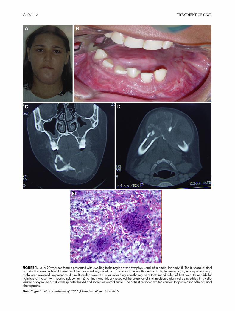
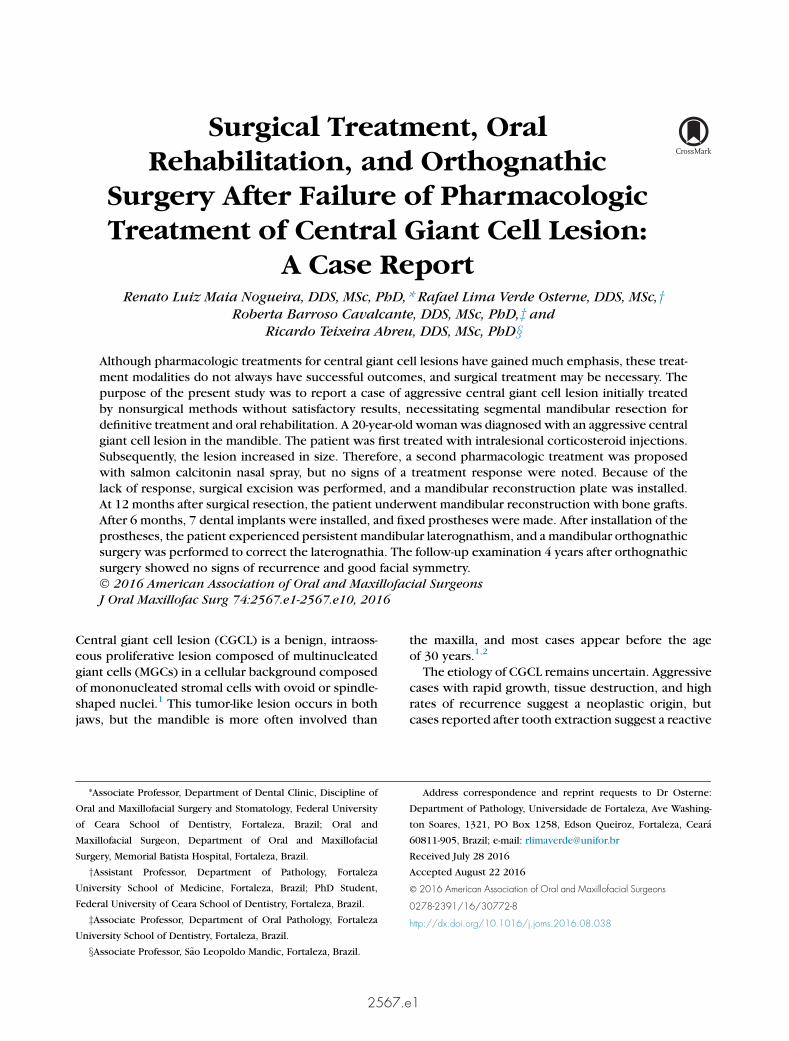
FIGURE 1. A, A 20-year-old female presented with swelling in the region of the symphysis and left mandibular body. B, The intraoral clinicalexamination revealed an obliteration of the buccal sulcus, elevation of the floor of the mouth, and tooth displacement.C, D, A computed tomog-raphy scan revealed the presence of a multilocular osteolytic lesion extending from the region of teeth mandibular left first molar to mandibularright lateral incisor, with tooth displacement. E, An incisional biopsy revealed the presence of multinucleated giant cells embedded in a cellu-larized background of cells with spindle-shaped and sometimes ovoid nuclei. The patient provided written consent for publication of her clinicalphotographs.
Maia Nogueira et al. Treatment of CGCL. J Oral Maxillofac Surg 2016.
2567.e2 TREATMENT OF CGCL
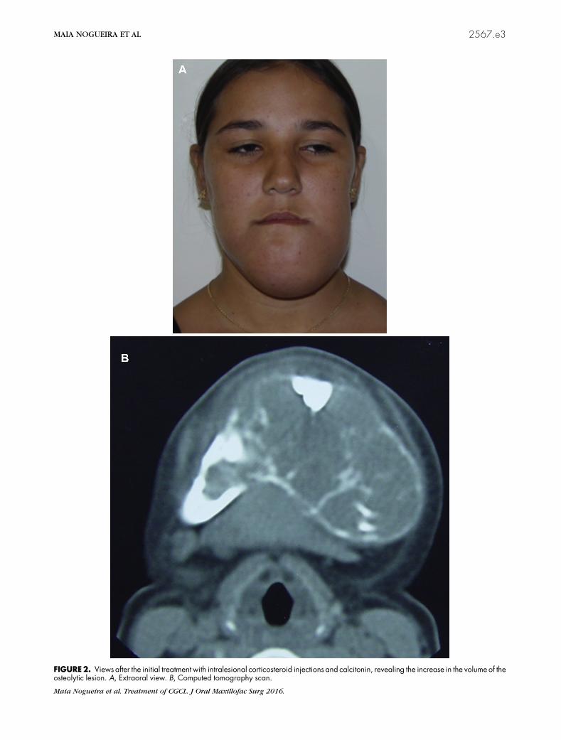
FIGURE2. Views after the initial treatment with intralesional corticosteroid injections and calcitonin, revealing the increase in the volume of theosteolytic lesion. A, Extraoral view. B, Computed tomography scan.
Maia Nogueira et al. Treatment of CGCL. J Oral Maxillofac Surg 2016.
MAIA NOGUEIRA ET AL 2567.e3
FIGURE3. A, Surgical excision of central giant cell lesion with the patient under general anesthesia. B, Installation of a mandibular reconstruc-tion plate. C, Postoperative panoramic radiograph.
Maia Nogueira et al. Treatment of CGCL. J Oral Maxillofac Surg 2016.
2567.e4 TREATMENT OF CGCL
lesion.3 Chuong et al2 classified CGCL into aggressive
and nonaggressive lesions. Nonaggressive lesions pre-
sent as slow and asymptomatic growths without
cortical perforation. Aggressive lesions are character-ized by swellings of rapid growth, which can be asso-
ciated with pain, tooth mobility, root resorption, and
cortical perforation.
Surgery is still considered the first therapeutic op-
tion for CGCL; however, new nonsurgical approaches
have gained much emphasis, including calcitonin,4 in-
tralesional corticosteroid injections,5 antiangiogenic
agents,6-8 and novel receptor activator of nuclearfactor kappa-B ligand (RANKL) inhibitors.9-11
The purpose of the present study was to report a
case of aggressive CGCL in which the initial treatment
was nonoperative but without satisfactory results,
necessitating segmental mandibular resection for
definitive treatment. The oral rehabilitation is
also reported.
Case Report
In July 2003, a 20-year-old female patient presented
to the oral and maxillofacial surgery department with
complaints of painful swelling in the mandible with
rapid growth. Her medical and family histories were
noncontributory. The extraoral clinical examination
revealed considerable swelling in the region of the
symphysis and left mandibular body. No associated
lymphadenopathy was present. The intraoral clinicalexamination revealed an obliteration of the buccal sul-
cus, elevation of the floor of the mouth, and tooth
displacement. Imaging studies revealed the presence
of a multilocular osteolytic lesion extending from the
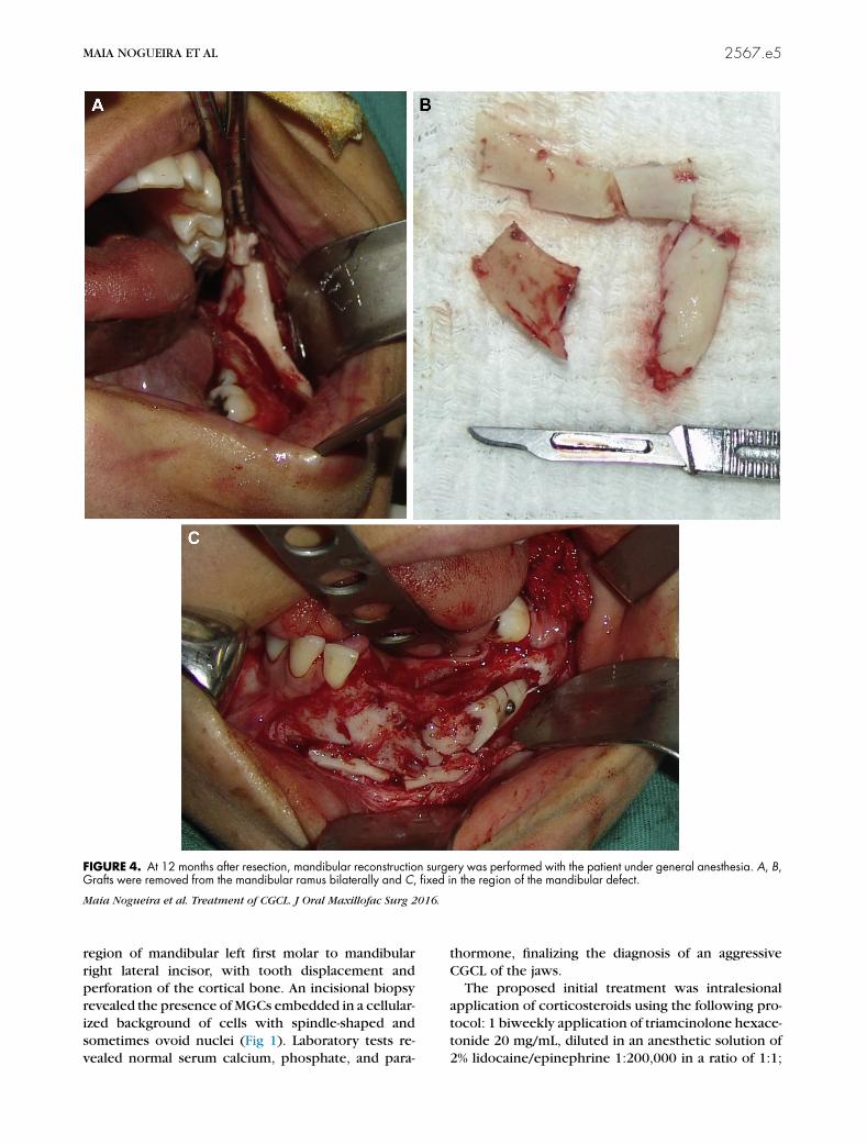
FIGURE 4. At 12 months after resection, mandibular reconstruction surgery was performed with the patient under general anesthesia. A, B,Grafts were removed from the mandibular ramus bilaterally and C, fixed in the region of the mandibular defect.
Maia Nogueira et al. Treatment of CGCL. J Oral Maxillofac Surg 2016.
MAIA NOGUEIRA ET AL 2567.e5
region of mandibular left first molar to mandibular
right lateral incisor, with tooth displacement andperforation of the cortical bone. An incisional biopsy
revealed the presence of MGCs embedded in a cellular-
ized background of cells with spindle-shaped and
sometimes ovoid nuclei (Fig 1). Laboratory tests re-
vealed normal serum calcium, phosphate, and para-
thormone, finalizing the diagnosis of an aggressive
CGCL of the jaws.The proposed initial treatment was intralesional
application of corticosteroids using the following pro-
tocol: 1 biweekly application of triamcinolone hexace-
tonide 20 mg/mL, diluted in an anesthetic solution of
2% lidocaine/epinephrine 1:200,000 in a ratio of 1:1;
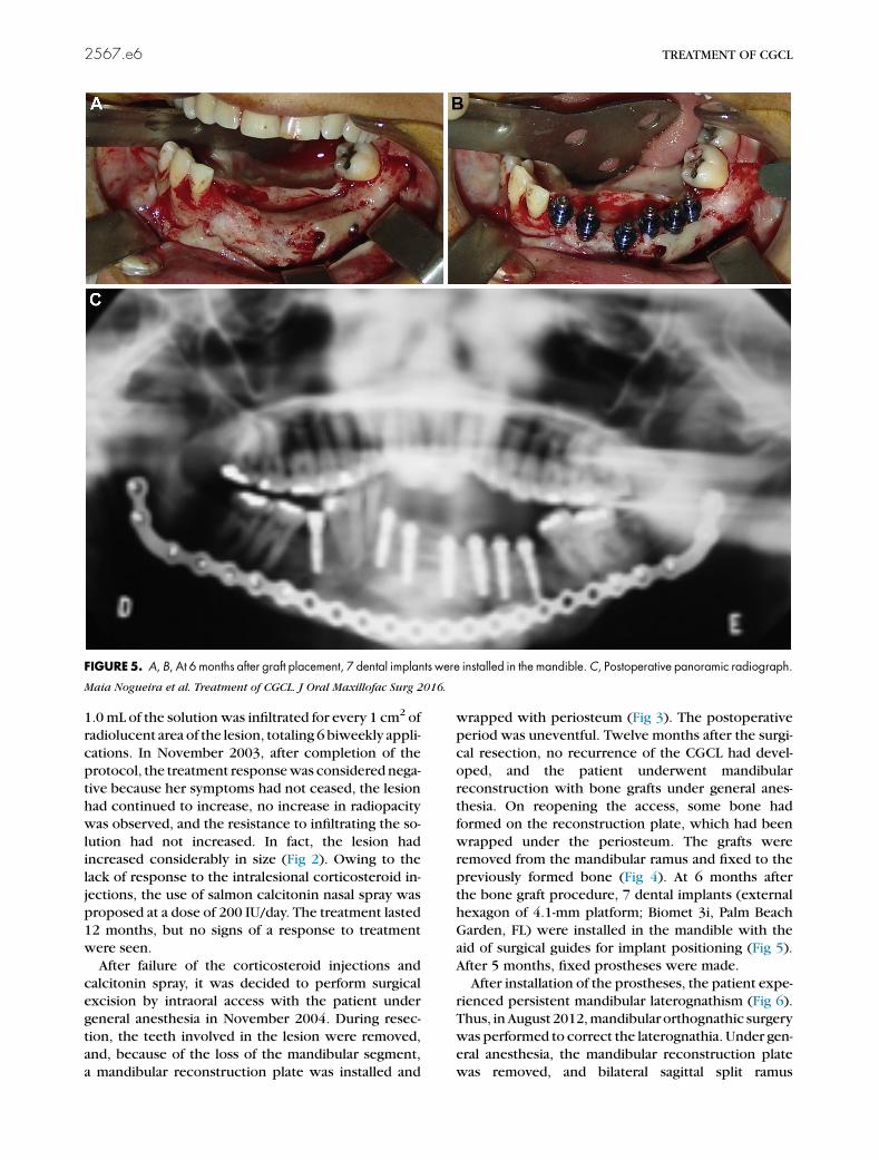
FIGURE5. A, B, At 6 months after graft placement, 7 dental implants were installed in the mandible.C, Postoperative panoramic radiograph.
Maia Nogueira et al. Treatment of CGCL. J Oral Maxillofac Surg 2016.
2567.e6 TREATMENT OF CGCL
1.0mL of the solution was infiltrated for every 1 cm2 of
radiolucent area of the lesion, totaling 6biweekly appli-
cations. In November 2003, after completion of the
protocol, the treatment responsewas considered nega-
tive because her symptoms had not ceased, the lesion
had continued to increase, no increase in radiopacity
was observed, and the resistance to infiltrating the so-
lution had not increased. In fact, the lesion hadincreased considerably in size (Fig 2). Owing to the
lack of response to the intralesional corticosteroid in-
jections, the use of salmon calcitonin nasal spray was
proposed at a dose of 200 IU/day. The treatment lasted
12 months, but no signs of a response to treatment
were seen.
After failure of the corticosteroid injections and
calcitonin spray, it was decided to perform surgicalexcision by intraoral access with the patient under
general anesthesia in November 2004. During resec-
tion, the teeth involved in the lesion were removed,
and, because of the loss of the mandibular segment,
a mandibular reconstruction plate was installed and
wrapped with periosteum (Fig 3). The postoperative
period was uneventful. Twelve months after the surgi-
cal resection, no recurrence of the CGCL had devel-
oped, and the patient underwent mandibular
reconstruction with bone grafts under general anes-
thesia. On reopening the access, some bone had
formed on the reconstruction plate, which had been
wrapped under the periosteum. The grafts wereremoved from the mandibular ramus and fixed to the
previously formed bone (Fig 4). At 6 months after
the bone graft procedure, 7 dental implants (external
hexagon of 4.1-mm platform; Biomet 3i, Palm Beach
Garden, FL) were installed in the mandible with the
aid of surgical guides for implant positioning (Fig 5).
After 5 months, fixed prostheses were made.
After installation of the prostheses, the patient expe-rienced persistent mandibular laterognathism (Fig 6).
Thus, inAugust 2012,mandibular orthognathic surgery
was performed to correct the laterognathia. Under gen-
eral anesthesia, the mandibular reconstruction plate
was removed, and bilateral sagittal split ramus
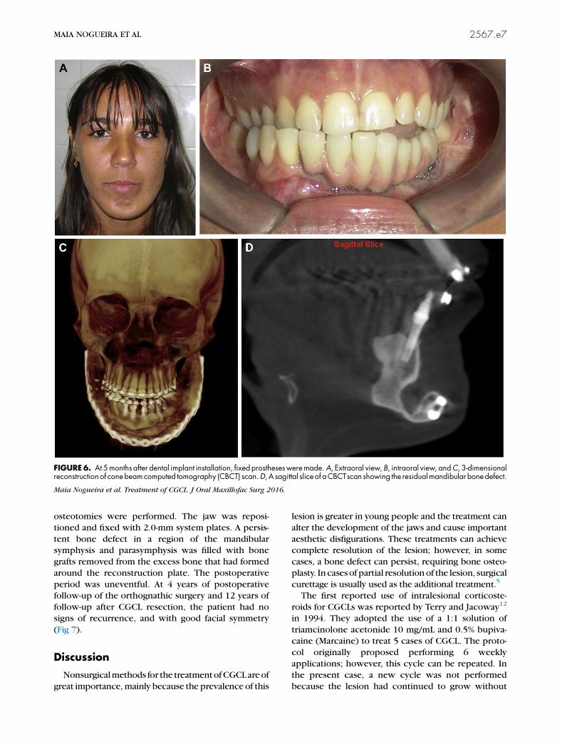
FIGURE6. At 5months after dental implant installation, fixed prosthesesweremade.A, Extraoral view, B, intraoral view, andC, 3-dimensionalreconstructionof conebeamcomputed tomography (CBCT) scan.D,A sagittal sliceof aCBCTscan showing the residualmandibular bonedefect.
Maia Nogueira et al. Treatment of CGCL. J Oral Maxillofac Surg 2016.
MAIA NOGUEIRA ET AL 2567.e7
osteotomies were performed. The jaw was reposi-
tioned and fixed with 2.0-mm system plates. A persis-
tent bone defect in a region of the mandibularsymphysis and parasymphysis was filled with bone
grafts removed from the excess bone that had formed
around the reconstruction plate. The postoperative
period was uneventful. At 4 years of postoperative
follow-up of the orthognathic surgery and 12 years of
follow-up after CGCL resection, the patient had no
signs of recurrence, and with good facial symmetry
(Fig 7).
Discussion
Nonsurgicalmethods for the treatmentofCGCLareof
great importance,mainly because the prevalence of this
lesion is greater in young people and the treatment can
alter the development of the jaws and cause important
aesthetic disfigurations. These treatments can achievecomplete resolution of the lesion; however, in some
cases, a bone defect can persist, requiring bone osteo-
plasty. In cases of partial resolutionof the lesion, surgical
curettage is usually used as the additional treatment.5
The first reported use of intralesional corticoste-
roids for CGCLs was reported by Terry and Jacoway12
in 1994. They adopted the use of a 1:1 solution of
triamcinolone acetonide 10 mg/mL and 0.5% bupiva-caine (Marcaine) to treat 5 cases of CGCL. The proto-
col originally proposed performing 6 weekly
applications; however, this cycle can be repeated. In
the present case, a new cycle was not performed
because the lesion had continued to grow without
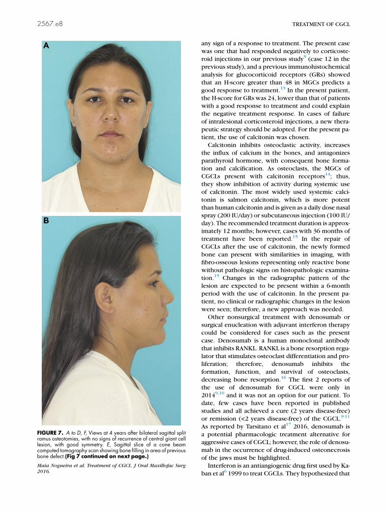
FIGURE 7. A to D, F, Views at 4 years after bilateral sagittal splitramus osteotomies, with no signs of recurrence of central giant celllesion, with good symmetry. E, Sagittal slice of a cone beamcomputed tomography scan showing bone filling in area of previousbone defect.(Fig 7 continued on next page.)
Maia Nogueira et al. Treatment of CGCL. J Oral Maxillofac Surg
2016.
2567.e8 TREATMENT OF CGCL
any sign of a response to treatment. The present case
was one that had responded negatively to corticoste-
roid injections in our previous study5 (case 12 in the
previous study), and a previous immunohistochemical
analysis for glucocorticoid receptors (GRs) showed
that an H-score greater than 48 in MGCs predicts a
good response to treatment.13 In the present patient,
the H-score for GRs was 24, lower than that of patientswith a good response to treatment and could explain
the negative treatment response. In cases of failure
of intralesional corticosteroid injections, a new thera-
peutic strategy should be adopted. For the present pa-
tient, the use of calcitonin was chosen.
Calcitonin inhibits osteoclastic activity, increases
the influx of calcium in the bones, and antagonizes
parathyroid hormone, with consequent bone forma-tion and calcification. As osteoclasts, the MGCs of
CGCLs present with calcitonin receptors14; thus,
they show inhibition of activity during systemic use
of calcitonin. The most widely used systemic calci-
tonin is salmon calcitonin, which is more potent
than human calcitonin and is given as a daily dose nasal
spray (200 IU/day) or subcutaneous injection (100 IU/
day). The recommended treatment duration is approx-imately 12 months; however, cases with 36 months of
treatment have been reported.15 In the repair of
CGCLs after the use of calcitonin, the newly formed
bone can present with similarities in imaging, with
fibro-osseous lesions representing only reactive bone
without pathologic signs on histopathologic examina-
tion.15 Changes in the radiographic pattern of the
lesion are expected to be present within a 6-monthperiod with the use of calcitonin. In the present pa-
tient, no clinical or radiographic changes in the lesion
were seen; therefore, a new approach was needed.
Other nonsurgical treatment with denosumab or
surgical enucleation with adjuvant interferon therapy
could be considered for cases such as the present
case. Denosumab is a human monoclonal antibody
that inhibits RANKL. RANKL is a bone resorption regu-lator that stimulates osteoclast differentiation and pro-
liferation; therefore, denosumab inhibits the
formation, function, and survival of osteoclasts,
decreasing bone resorption.16 The first 2 reports of
the use of denosumab for CGCL were only in
20149,10 and it was not an option for our patient. To
date, few cases have been reported in published
studies and all achieved a cure (2 years disease-free)or remission (<2 years disease-free) of the CGCL.9-11
As reported by Tarsitano et al17 2016, denosumab is
a potential pharmacologic treatment alternative for
aggressive cases of CGCL; however, the role of denosu-
mab in the occurrence of drug-induced osteonecrosis
of the jaws must be highlighted.
Interferon is an antiangiogenic drug first used by Ka-
ban et al6 1999 to treat CGCLs. They hypothesized that
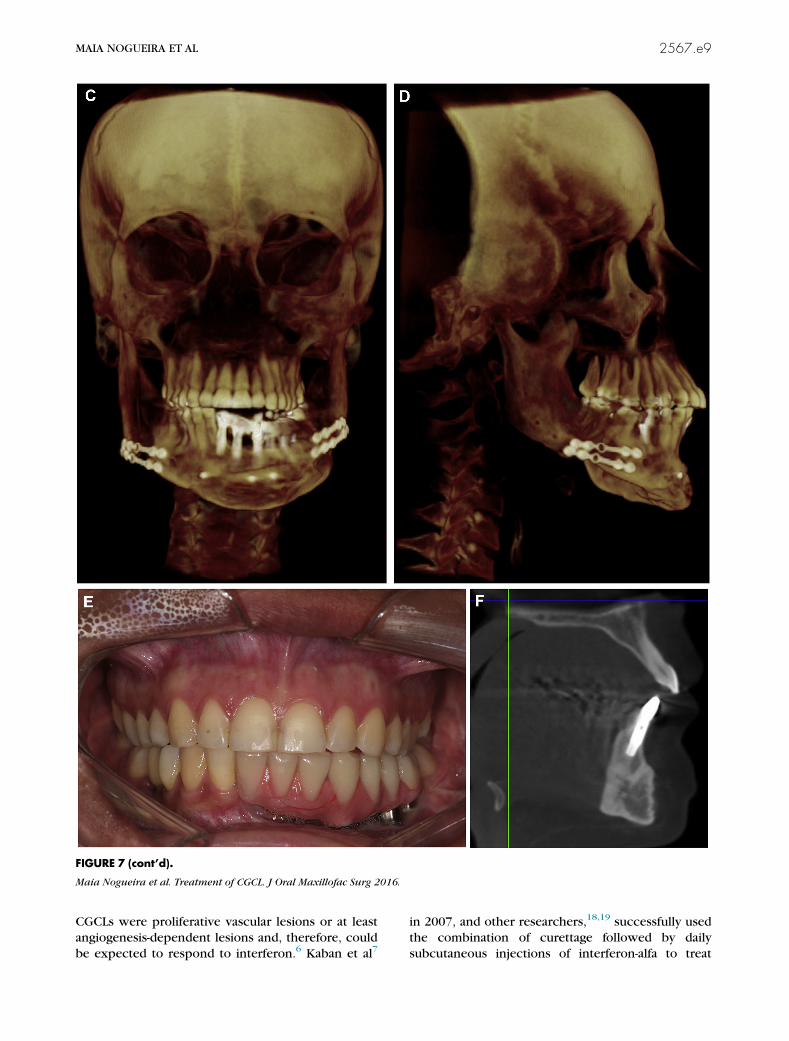
FIGURE 7 (cont’d).
Maia Nogueira et al. Treatment of CGCL. J Oral Maxillofac Surg 2016.
MAIA NOGUEIRA ET AL 2567.e9
CGCLs were proliferative vascular lesions or at least
angiogenesis-dependent lesions and, therefore, could
be expected to respond to interferon.6 Kaban et al7
in 2007, and other researchers,18,19 successfully used
the combination of curettage followed by daily
subcutaneous injections of interferon-alfa to treat
2567.e10 TREATMENT OF CGCL
aggressive CGCLs. The combined treatment seemed to
be more successful than corticosteroid injections for
aggressive CGCLs. Kaban et al7 showed that all cases
treated by surgery combined with interferon were
cured or in remission.
By the time of failure of calcitonin in the present
patient, the remaining options were surgical
resection or surgical curettage combined with inter-feron. Because both options included surgery, en
bloc resection, as the reference standard treatment,
was chosen.
Surgical treatment can range from simple curettage
to en bloc resection with loss of a mandibular
segment; the recurrence rates differ depending on
the surgical technique used. Nonaggressive small le-
sions can be treated by curettage with low recurrencerates; however, multilocular lesions with limited surgi-
cal access, cortical bone perforation, and the presence
of teeth can make curettage difficult and increase the
risk of recurrence. Surgical resection has the lowest
recurrence rate but the greatest morbidity. It usually
causes significant facial disfigurement, requiring
extensive and time-consuming bone reconstruction20
and incurs high costs for rehabilitation. In the presentcase, because of the lack of a response to nonoperative
treatments, surgical resection was performed, with
mandibular reconstruction performed later.
Small alveolar reconstruction can be performed us-
ing the techniques of guided bone regeneration. How-
ever, because of the extent of the bone defect in the
present case, guided bone regeneration was not
used, and autogenous bone grafting was elected.Autogenous bone graft is considered the reference
standard, but some amount of bone resorption is still
expected after grafting. Some cases could require a
second reconstructive technique such as distraction
osteogenesis to obtain a satisfactory bony height
before implant placement.20 In the present patient,
the first bone graft was performed with the aim of ob-
taining sufficient bone for implant placement in theproper position. The second graft was performed to
fill the bone defect of the symphysis and parasymphy-
sis. Finally, orthognathic surgery was performed to
correct the laterognathia; this procedure is a well-
accepted treatment modality and is used for the reso-
lution of dentoskeletal deformities, such as in the
present patient.
In conclusion, treatment of CGCL using pharmaco-logic techniques can be considered the first therapeu-
tic option, especially for extensive cases in which
surgical treatment would cause significant tissue dam-
age. In cases of pharmacologic treatment failure, more
invasive treatments will be required. The bone defects
caused by these techniques usually require multiple
steps for rehabilitation, resulting in high costs for treat-
ment. Additionally, long-term follow-up should be
emphasized for such patients because of the possible
recurrence of the CGCL.
References
1. Kruse-L€osler B, Diallo R, Gaertner C, et al: Central giant cell gran-uloma of the jaws: A clinical, radiologic, and histopathologicstudy of 26 cases. Oral Surg Oral Med Oral Pathol Oral Radiol En-dod 101:346, 2006
2. Chuong R, Kaban LB, Kozakewich H, Perez-Atayde A: Central gi-ant cell lesions of the jaws: A clinicopathologic study. J Oral Max-illofac Surg 44:708, 1986
3. Unal M, Karabacak T, Vayısoglu Y, et al: Central giant cell repar-ative granuloma of the mandible caused by a molar tooth extrac-tion: Special reference to the manuever of drilling the surgicalfield. Int J Pediatr Otorhinolaryngol 70:745, 2006
4. de Lange J, Rosenberg AJWP, van den Akker HP, et al: Treatmentof giant cell granulomawith calcitonin. Int J Oral Maxillofac Surg28:372, 1999
5. Nogueira RL, Teixeira RC, Cavalcante RB, et al: Intralesional in-jection of triamcinolone hexacetonide as an alternative treat-ment for central giant-cell granuloma in 21 cases. Int J OralMaxillofac Surg 39:1204, 2010
6. Kaban LB, Mulliken JB, Ezekowitz RA, et al: Antiangiogenic ther-apy of a recurrent giant cell tumor of the mandible with inter-feron alfa-2a. Pediatrics 103:1145, 1999
7. Kaban LB, Troulis MJ, Wilkinson MS, et al: Adjuvant antiangio-genic therapy for giant cell tumors of the jaws. J Oral MaxillofacSurg 65:2018, 2007
8. Kaban LB, Troulis MJ, Ebb D, et al: Antiangiogenic therapy withinterferon alpha for giant cell lesions of the jaws. J Oral Maxillo-fac Surg 60:1103, 2002
9. Naidu A, Malmquist MP, Denham CA, Schow SR: Management ofcentral giant cell granuloma with subcutaneous denosumabtherapy. J Oral Maxillofac Surg 72:2469, 2014
10. Schreuder WH, Coumou AW, Kessler PA, de Lange J: Alterna-tive pharmacologic therapy for aggressive central giant cellgranuloma: Denosumab. J Oral Maxillofac Surg 72:1301,2014
11. O’Connell JE, Bowe C, Murphy C, et al: Aggressive giant celllesion of the jaws: A review of management options and reportof a mandibular lesion treated with denosumab. Oral Surg OralMed Oral Pathol Oral Radiol 120:e191, 2015
12. Terry BC, Jacoway JR: Management of central giant cell lesions—An alternative to surgical therapy. Oral Maxillofac Surg ClinNorth Am 6:579, 1994
13. Nogueira RL, Faria MH, Osterne RL, et al: Glucocorticoid andcalcitonin receptor expression in central giant cell lesions: Im-plications for therapy. Int J Oral Maxillofac Surg 41:994, 2012
14. Vered M, Buchner A, Dayan D: Immunohistochemical expres-sion of glucocorticoid and calcitonin receptors as a tool for se-lecting therapeutic approach in central giant cell granuloma ofthe jawbones. Int J Oral Maxillofac Surg 35:756, 2006
15. Sadiq Z, Goodger NM: Calcitonin-induced osteoplastic reactionin the mandible. Br J Oral Maxillofac Surg 49:578, 2011
16. Sinningen K, Tsourdi E, Rauner M, et al: Skeletal and extraskele-tal actions of denosumab. Endocrine 42:52, 2012
17. Tarsitano A, Del Corso G, Marchetti C: In reply. J Oral MaxillofacSurg 74:873, 2016
18. O’Connell JE, Kearns GJ: Aggressive giant cell granuloma of thejaws treated with interferon alpha: A report of two cases. Ir JMed Sci 182:163, 2013
19. Tarsitano A, Del Corso G, Pizzigallo A, Marchetti C: Aggressivecentral giant cell granuloma of the mandible treated with con-servative surgical enucleation and interferon-a-2a: Completeremission with long-term follow-up. J Oral Maxillofac Surg 73:2149, 2015
20. de Moraes M, Sato FRL, Germano AD, Bastos PL: Distraction os-teogenesis of iliac bone graft as a reconstruction after central gi-ant cell granuloma curettage. Implant Dent 18:126, 2009