Embed Size (px)
Citation preview
Research ArticleSurgical Treatment of Severe Idiopathic Flexible Flatfoot byEvans–Mosca Technique in Adolescent Patients: A Long-TermFollow-Up Study
Vincenzo De Luna , Fernando De Maio , Alessandro Caterini , Martina Marsiolo ,Lidio Petrungaro , Ernesto Ippolito , and Pasquale Farsetti
Department of Clinical Sciences and Traslational Medicine, Division of Orthopaedic Surgery, University of “Tor Vergata”,Rome, Italy
Correspondence should be addressed to Pasquale Farsetti; [email protected]
Received 10 August 2020; Revised 29 December 2020; Accepted 9 January 2021; Published 20 January 2021
Academic Editor: Allen L. Carl
Copyright © 2021 Vincenzo De Luna et al. *is is an open access article distributed under the Creative Commons AttributionLicense, which permits unrestricted use, distribution, and reproduction in any medium, provided the original work isproperly cited.
Flexible idiopathic flatfoot is very common in growing age and rarely causes pain or disability. Surgery is indicated only in severesymptomatic cases that are resistant to conservative treatment, and numerous surgical procedures have been proposed. Lateralcolumn calcaneal lengthening as described by Evans and modified by Mosca is a widely used surgical technique for the correctionof severe symptomatic flexible flatfoot. In the present study, we report the long-term clinical and radiographic results in 14adolescent patients (mean age: 12.8 years) affected by severe symptomatic flexible flatfoot, surgically treated by Evans–Moscaprocedure, for a total of 26 treated feet (12 cases bilateral and 2 unilateral). In all cases, surgery was indicated for the presence ofsignificant symptoms resistant to nonsurgical management. Clinical evaluation was made according to the American OrthopedicFoot and Ankle Society (AOFAS) Ankle-Hindfoot Scale, the Foot and Ankle Disability Index (FADI) Score, and Yoo et al.’scriteria. Radiographic evaluation was made using anteroposterior and lateral weight-bearing radiographs of the feet to evaluateMeary’s angle and Costa–Bertani’s angle and to evaluate possible osteoarthritic changes in the midtarsal joints. At follow-up(mean: 7 years and 7 months), we observed a satisfactory result in all patients. *e mean average score of the AOFAS Ankle-Hindfoot Scale improved from 60.03 points to 95.26; the mean FADI score improved from 71.41 to 97.44; and according to Yooet al.’s criteria, the average clinical outcome score was 10.96. At radiographic examination, nonunion of the calcaneal osteotomywas never observed. Meary’s angle improved from an average preoperative value of 25° to 1.38° at follow-up; Costa–Bertani’s angleimproved from an average preoperative value of 154.2° to 130.9° at follow-up. In no case, significant radiographic signs ofmidtarsal joint arthritis were observed. According to our results, we believe that Evans–Mosca technique is a valid option ofsurgical treatment for severe idiopathic flexible flatfoot and allows a satisfactory correction of the deformity with a low rateof complications.
1. Introduction
Flexible flatfoot is a very common foot condition observedduring skeletal growth, characterized by a depression of themedial arch with an associated hindfoot valgus and forefootabduction [1–3]; also, a short Achilles tendon is oftenpresent [4]. Flexible flatfoot rarely causes pain or disability ingrowing age patients and generally does not require treat-ment because in the majority of cases, the medial arch
spontaneously elevates during the first decade of life.Moreover, it is well known that many adults have a flexibleflatfoot without any significant limitation in activities ofdaily living or sport activities [5]. Surgery is indicated only insevere symptomatic cases with diffuse activity-related painand medial foot calluses, observed usually in adolescents, inwhich conservative treatment has failed [5, 6]. *e correctindication, time, and methods of surgical treatment for thecorrection of severe symptomatic flatfoot are however still
HindawiAdvances in OrthopedicsVolume 2021, Article ID 8843091, 7 pageshttps://doi.org/10.1155/2021/8843091

debated, and several surgical procedures have been proposed[7–11]. *e most common operations performed arearthroereisis, lateral calcaneal lengthening osteotomy, andtriple arthrodesis [8–11]. Soft tissue surgical procedures havealso been reported, but when they are performed alone, theylead to unsatisfactory results [5]. Evans, more than fortyyears ago [10], introduced the lateral column lengthening forthe treatment of severe symptomatic flatfoot and proposed acalcaneal lengthening osteotomy for its correction. Morerecently, Mosca elaborated a modification of this technique,proposing an opening wedge osteotomy with a trapezoidal,tricortical iliac crest wedge [11].
*e aim of the present study was to report the long-termresults obtained in a series of 14 adolescents affected bysevere symptomatic flexible flatfoot, surgically treated byEvans–Mosca technique associated to a soft tissue procedureof the medial side of the foot and, in almost 50% of cases,percutaneous lengthening of the Achilles tendon.
2. Materials and Methods
We reviewed 14 patients affected by severe idiopathicsymptomatic flexible flatfoot, surgically treated by lateralcolumn lengthening according to Evans–Mosca procedureassociated to the tibialis posterior tendon and talonavicularjoint capsule strain and, in some cases, percutaneouslengthening of the Achilles tendon. Eight patients were male,and 6 were female. *e mean age of the patients at surgerywas 12.8 years (range: from 11 to 14.6 years); 12 cases wereoperated bilaterally, for a total of 26 feet. In all our patientstreated bilaterally, we performed the two procedures atdifferent times, with a distance between the two operationsthat ranged from 8 months to one year.
Regarding the clinical evaluation before surgery, allpatients had a medial longitudinal arch abnormally de-pressed or absent with a normal subtalar joint mobility. In 12feet, a short Achilles tendon was also present with an as-sociated limitation of the ankle dorsiflexion. Preoperatively,standard weight-bearing radiographs of the foot in ante-roposterior and lateral projections confirmed the presence ofa flexible flatfoot in all cases. Conservative treatment aims toimprove or solve foot pain without changing the foot shape.In our series, it was based on strengthening exercises, customorthosis, or corrective shoes, but failed in all patients(Figure 1). *e surgical technique was performed undergeneral anesthesia with a thigh tourniquet. *e incision wasmade with an oblique direction over the sinus tarsi towardsthe inferior border of the calcaneus. *e calcaneus wasexposed, and the osteotomy was performed approximately1.5 cm proximally from the calcaneocuboid joint, betweenthe anterior and middle facets of the subtalar joint. *eosteotomy should be oriented from posterolateral to ante-romedial to avoid damage of the middle facet. After thedistraction osteotomy was performed with a spreader, atricortical iliac crest autologous bone graft, taken from thesame side of the operated side, was inserted into the space toobtain a lengthening of the lateral column through theanterior part of the calcaneus. *e osteotomy was stabilizedwith one or two Kirschner or Steinmann wires, according to
the age of the patients and the size of the foot. A secondincision was made along the medial border of the foot. *etibialis posterior was strained through a tendon release, andthe talonavicular joint capsule, including the spring liga-ment, was incised and plicated plantarmedially. In 12 feet, inwhich a short Achilles tendon was present, percutaneouslengthening was also performed. After surgery, a nonweightbearing below the knee cast was applied for 6–8 weeks.Weight bearing was permitted only after radiographichealing of the osteotomy was observed.
All patients were evaluated clinically, analyzing thepreoperative function of the foot in comparison to thefollow-up examination, using the American OrthopedicFoot and Ankle Society (AOFAS) Ankle-Hindfoot Scale [12],the Foot and Ankle Disability Index (FADI) Score [13], and,only at follow-up, Yoo et al. criteria [14]. From a radio-graphic point of view, all patients were evaluated by ananteroposterior and lateral weight-bearing examination ofboth feet, taken before surgery and at follow-up, to measureboth Meary’s and Costa–Bertani’s angles.
Moreover, at follow-up radiographic examination, welooked for possible subluxation or osteoarthritic changes ofthe calcaneocuboid or talonavicular joints as well as theremodeling of the tricortical bone graft into the calcanealosteotomy (Figure 2).
Statistical analysis was performed using paired Student’st-test, which was considered significant when p valuemeasured <0.05.
3. Results
All patients were reviewed after a mean follow-up of 7 yearsand 7 months (from 6 y and 5m to 12 y).
According to the AOFAS system [12], the averagepreoperative score was 69.03 points (from 59 to 78 points),while the average score at follow-up improved to 95.26 (from88 to 100). According to the FADI method [13], the averagepreoperative score was 71.41 (from 59.5 to 75.3), while theaverage score at follow-up was 97.44 (from 88.5 to 100). *edifference between the preoperative and final score evaluatedat follow-up was statistically significant for both clinicalrating scales adopted (p <0.01). According to Yoo et al.’scriteria [14], the average clinical outcome score at follow-upwas 10.96 (from 8 to 12); since a score equal to or greaterthan 8 corresponds to a satisfactory clinical result, we ob-tained a satisfactory clinical result in all patients. We foundno difference in terms of clinical results and satisfactionbetween patients treated on one foot or both feet (Table 1).
At radiographic examination, nonunion of the calcanealosteotomy was never observed, and the tricortical bone graftwas always remodeled. Meary’s angle improved from anaverage preoperative value of 25° (from 22° to 32°) to anaverage value of 1.4° (from 0° to 4°) at follow-up. Cos-ta–Bertani’s angle improved from an average preoperativevalue of 154.2° (from 150° to 160°) to an average value of130.9° (from 124° to 138°) at follow-up. Also, for both ra-diographic parameters, the difference between preoperativeand final angle values was statistically significant (p <0.01).Calcaneocuboid joint subluxation was observed in only 3
2 Advances in Orthopedics

(a)
L
(b)
R
(c)
Figure 1: Clinical aspect of an 11-year-old boy with bilateral painful idiopathic flexible flatfoot (a). At radiographic examination, Meary’sangle measured 26° of the left foot (b) and 23° of the right foot (c). *e patient was treated conservatively by an insert sole, but this treatmentfailed, and a surgical procedure was proposed.
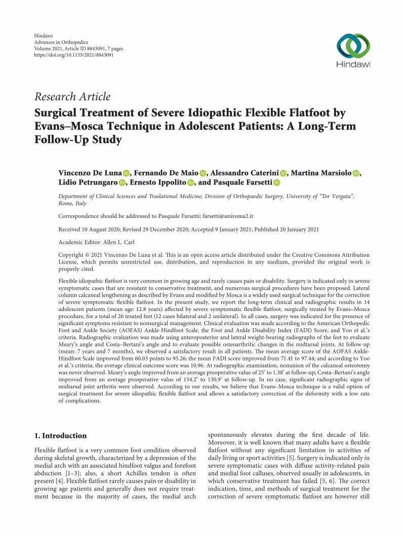
(a)
(b)
Figure 2: Same patient of Figure 1, three years later. *e patient was surgically treated by Evans–Mosca procedure bilaterally with aninterval of six months. *e radiographic examination of the left foot, performed in oblique view 3 months after surgery, showed goodlengthening of the calcaneus with the presence of the tricortical bone graft (a). At an intermediate follow-up, 7 years later, the graft wasperfectly remodeled in the calcaneal bone with the maintenance of calcaneal lengthening and without any sign of osteoarthritis of thecalcaneocudoid joint (b).
Advances in Orthopedics 3
feet (11.5%), while significant calcaneocuboid or talona-vicular joint osteoarthritis was never observed (Figure 3).
4. Discussion
Flexible idiopathic flatfoot is very common in children andrarely causes pain or disability. On the contrary, congenitalflatfoot caused by tarsal coalition, Marfan syndrome, orother congenital disorders, is usually painful, but uncom-mon in comparison to other congenital, pediatric, ortho-pedic diseases [15–18]. However, in some cases, especially inadolescents, flexible flatfoot may cause pain and disability,and surgery may be indicated when conservative treatmentfails [19].
In severe idiopathic symptomatic adolescent flatfoot,surgical indication and the type of surgical treatment toperform are still controversial. Arthroereisis of the subtalarjoint for the correction of severe flexible flatfoot in growingchildren is a widely used technique [20, 21]. *is surgicalprocedure restricts excessive subtalar joint eversion byplacing a synthetic implant. Different devices have beenproposed over time including bioabsorbable implants. Goodresults have been reported in the literature for the correctionof flexible flatfoot in growing children with arthroereisis ofthe subtalar joint [9, 20, 22], but pain at the level of the sinustarsi is a possible complication of these techniques, and asecond surgery for implant removal may be required[5, 23, 24]. For this reason, we preferred Evans–Moscasurgical procedures instead of arthroereisis in our series ofadolescent patients.
Evans [10] believed that the lateral column in flatfeet wasshorter than the medial column and first proposed a cal-caneal lengthening osteotomy for the correction of valgusdeformity, without an opening wedge osteotomy. *isconcept was elaborated by Mosca [5, 11] that published amodified technique utilizing a trapezoidal, tricortical iliaccrest wedge to perform both an opening wedge and a dis-tracting osteotomy and reported correction of all compo-nents of the deformity.
*e calcaneal lengthening osteotomy for the surgicaltreatment of severe symptomatic idiopathic flexible flatfootwas later used by other authors with satisfactory results.Dogan et al. [25], in a series of 13 patients (25 feet) treatedfor flexible pes planovalgus by calcaneal lengtheningosteotomy, reported that foot pain was eliminated in allpatients but one. *ey concluded that the advantages of thisprocedure are the preservation of subtalar joint motion and
the correction of the deformity in multiple plans. Moraledaet al. [26] also reported good clinical and radiographic re-sults in a series of 21 children (33 feet) with symptomaticflexible flatfoot surgical treatment with a calcaneal length-ening osteotomy. More recently, Kumar and Sonanis [27]reported a systematic review on lateral column lengtheningperformed in adolescent pes planovalgus deformity. *eseauthors identified seven studies with 103 patients involving156 feet and concluded that lateral column lengthening leadsto good clinical and radiological outcome with high patientsatisfaction and acceptable complication rate. *ey alsoconcluded that the literature is mostly retrospective, andthere is need for other studies.
*e indication of lateral calcaneal lengthening osteot-omy for idiopathic flatfoot in children has been extended toother forms of flatfoot with a low incidence of complica-tions [26]. Marengo et al. [28] reported the clinical andradiological outcome of calcaneal lengthening osteotomyfor flatfoot deformity of various etiologies in 27 skeletallyimmature patients (38 feet). Clinical outcome was satis-factory in 89% of cases, and all radiographic parametersimproved significantly. *ey concluded that calcaneallengthening osteotomy is not contraindicated in symp-tomatic flatfoot of different etiologies, except neuromus-cular disease-related flatfoot that can affect bone qualityand reduce foot flexibility. *ey also reported that calca-neocuboid joint subluxation is frequently observed but haslittle functional impact as it tends to remodel over time.Similar to other CT studies that analyzed the results oftreatment of the congenital clubfoot [29], Canavese et al.[30] published a study on postoperative CT-scan 3D re-construction of the calcaneus following lateral calcaneallengthening osteotomy performed in 14 children (20 feet)affected by symptomatic flatfoot with different etiologies.*is study showed that subtalar anatomy presented sig-nificant anatomical variations among these examined pa-tients; however, clinical evaluation at follow-up showedsatisfactory outcome in 80% of cases. Calcaneal length-ening for flatfoot deformity in patients with cerebral palsyhas also been reported with good results [31]. Andreacchioet al. [32] concluded that calcaneal lengthening is a suc-cessful treatment for flexible planovalgus foot deformity inambulatory children with spastic CP. Regarding otheretiologies, Mosca and Bevan [33] reported good results forcorrecting deformity and relieving pain in rigid flatfeet of 8patients (13 feet), affected by talocalcaneal tarsal coalition,treated by calcaneal lengthening osteotomy with
Table 1: Preoperative and final (at follow-up) clinical and radiographic results in 26 flatfeet surgically treated by Evans–Mosca technique.
AOFAS (preop) 69.03 (59–79)p value <0.01AOFAS (follow up) 95.26 (88–100)
FADI (preop) 71.41 (59.5–75.3)p value <0.01FADI (follow up) 97.44 (88.5–100)
Yoo et al. (follow up) 10.96 (8–12) —Meary’s angle (preop) 25° (22°–32°)
p value <0.01Meary’s angle (follow up) 1.4° (0°–4°)Costa–Bertani angle (preop) 154.2° (150°–160°)
p value <0.01Costa–Bertani angle (follow up) 130.9° (124°–138°)
4 Advances in Orthopedics

(a) (b)
(c) (d)
(e) (f )
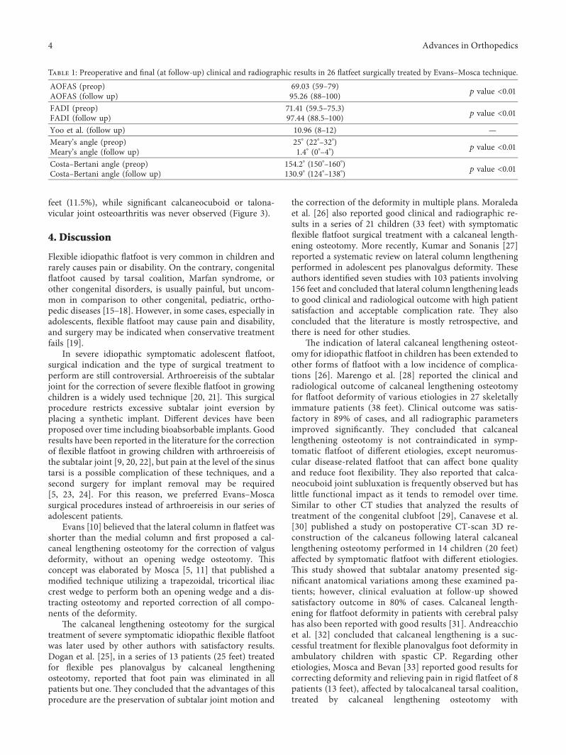
Figure 3: Same patient of Figure 1, at the final follow-up, 11 years after surgery.*e patient was pain free. Clinical and podoscopic aspects ofboth feet showed an excellent result ((a)–(d)). At radiographic examination, Meary’s angle was normal bilaterally (0°), in the absence ofmidtarsal joint osteoarthritis ((e) and (f)).
Advances in Orthopedics 5

gastrocnemius or Achilles tendon lengthening. Guha andPerera [34] reported that calcaneal lengthening osteotomymay be performed even in the correction of adult flexibleflatfoot, but they concluded that an essential prerequisitefor using this technique is the absence of arthritis of thesubtalar joint.
Our long-term results in a group of adolescents surgi-cally treated for severe symptomatic idiopathic flexibleflatfoot by the Evans–Mosca technique, in association with astrain of the tibialis posterior tendon and joint capsule,confirmed the good results reported by the aforementionedstudies. All our patients were satisfied with the final resultand referred a significant improvement of the preoperativesymptoms.
In our patients, we paid particular attention to per-forming the calcaneal osteotomy into the interval betweenthe anterior and middle facets of the subtalar joint assuggested by Ragab et al. [35], and at follow-up, we neverobserved significant radiographic changes of the midtarsaljoint as reported by some studies [36, 37]. We always used atricortical iliac crest autologous bone graft inserted into theosteotomy, as described by Mosca [11], to obtain length-ening of the lateral column of the calcaneus and neverobserved complications in terms of nonunion or loss ofcorrection. Also, we never observed any patient morbidityfrom graft harvesting at the level of the iliac crest; however,some authors reported satisfactory results using autogenousbone graft from a different donor site or allogenic bone graft[38–40]. We had no personal experiences with titaniumwedge insertion. We prefer to use bone autograft to obtain abetter and faster integration in the native bone and to bettermaintain the correction obtained. However, to avoid the riskof pain at the level of the donor site, the use of allograft mightbe considered.
5. Conclusion
According to our long-term results, we believe that Evan-s–Mosca surgical procedure is an effective solution for thetreatment of idiopathic symptomatic flexible flatfoot resis-tant to conservative treatment and allows themaintenance ofthe surgical correction without the development of osteo-arthritic changes.
Data Availability
*e data used to support the findings of this study areavailable from the corresponding author upon reasonablerequest.
Conflicts of Interest
*e authors declare that there are no conflicts of interest.
References
[1] L. T. Staheli, D. E. Chew, and M. Corbett, “*e longitudinalarch. a survey of eight hundred and eighty-two feet in normalchildren and adults,” "e Journal of Bone and Joint Surgery.American Volume, vol. 69, no. 3, pp. 426–428, 1987.
[2] R. Vanderwilde, L. T. Staheli, D. E. Chew, and V. Malagon,“Measurements on radiographs of the foot in normal infantsand children,” "e Journal of Bone & Joint Surgery, vol. 70,no. 3, pp. 407–415, 1988.
[3] E. J. Harris, “*e natural history and pathophysiology offlexible flatfoot,” Clinics in Podiatric Medicine and Surgery,vol. 27, no. 1, pp. 1–23, 2010.
[4] R. I. Harris and T. Beath, “Hypermobile flat-foot with shorttendo achillis,” "e Journal of Bone & Joint Surgery, vol. 30,no. 1, pp. 116–150, 1948.
[5] V. S. Mosca, “Flexible flatfoot in children and adolescents,”Journal of Children’s Orthopaedics, vol. 4, no. 2, pp. 107–121,2010.
[6] N. M. Blitz, R. J. Stabile, R. J. Giorgini, and L. A. DiDomenico,“Flexible pediatric and adolescent pes planovalgus: conser-vative and surgical treatment options,” Clinics in PodiatricMedicine and Surgery, vol. 27, no. 1, pp. 59–77, 2010.
[7] B. S. Jones, “Flatfoot. a preliminary report of an operation forsevere cases,” Journal of Bone and Joint Surgery British Vol-ume, vol. 57, no. 3, pp. 279–282, 1975.
[8] P. Angus and H. Cowell, “Triple arthrodesis. a critical long-term review,” "e Journal of Bone and Joint Surgery. BritishVolume, vol. 68-B, no. 2, pp. 260–265, 1986.
[9] S. Giannini, F. Ceccarelli, M. G. Benedetti, F. Catani, andC. Faldini, “Surgical treatment of flexible flatfoot in children,”"e Journal of Bone and Joint Surgery-American Volume,vol. 83, no. suppl. 2, pp. 73–79, 2001.
[10] D. Evans, “Calcaneo-valgus deformity,” "e Journal of Boneand Joint Surgery. British Volume, vol. 57-B, no. 3, pp. 270–278, 1975.
[11] V. S. Mosca, “Calcaneal lengthening for valgus deformity ofthe hindfoot. results in children who had severe, symptomaticflatfoot and skewfoot,” "e Journal of Bone & Joint Surgery,vol. 77, no. 4, pp. 500–512, 1995.
[12] H. B. Kitaoka, I. J. Alexander, R. S. Adelaar et al., “Clinicalrating systems for the ankle-hindfoot, midfoot, hallux, andlesser toes,” Foot & Ankle International, vol. 15, no. 7,pp. 349–353, 1994.
[13] R. L. Martin, R. G. Burdett, and J. J. Irrgang, “Development ofthe foot and ankle disability index (FADI),” Journal of Or-thopaedic & Sports Physical "erapy, vol. 29, pp. A32–A33,1999.
[14] W. J. Yoo, C. Y. Chung, I. H. Choi, T.-J. Cho, and D. H. Kim,“Calcaneal lengthening for the planovalgus foot deformity inchildren with cerebral palsy,” Journal of Pediatric Ortho-paedics, vol. 25, no. 6, pp. 781–785, 2005.
[15] S. J. Kumar, J. T. Guille, M. S. Lee, and J. C. Couto, “Osseousand non-osseous coalition of the middle facet of the talo-calcaneal joint,” "e Journal of Bone & Joint Surgery, vol. 74,no. 4, pp. 529–535, 1992.
[16] F. De Maio, A. Fichera, V. De Luna, F. Mancini, andR. Caterini, “Orthopaedic aspects of marfan syndrome: theexperience of a referral center for diagnosis of rare diseases,”Advances in Orthopedics, vol. 2016, Article ID 8275391, , 2016.
[17] P. Farsetti, M. Dragoni, and E. Ippolito, “Tibiofibular torsionin congenital clubfoot,” Journal of Pediatric Orthopaedics B,vol. 21, no. 1, pp. 47–51, 2012.
[18] P. Farsetti, R. Caterini, V. Potenza, and E. Ippolito, “Devel-opmental dislocation of the hip successfully treated by pre-operative traction and medial open reduction: a 22-year meanfollowup,” Clinical Orthopaedics and Related Research,vol. 473, no. 8, pp. 2658–2669, 2015.
6 Advances in Orthopedics

[19] J. V. Basmajian and G. Stecko, “*e role of muscles in archsupport of the foot,” "e Journal of Bone & Joint Surgery,vol. 45, no. 6, pp. 1184–1190, 1963.
[20] V. Pavone, A. Vescio, C. A. Di Silvestri, A. Andreacchio,G. Sessa, and G. Testa, “Outcomes of the calcaneo-stopprocedure for the treatment of juvenile flatfoot in youngathletes,” Journal of Children’s Orthopaedics, vol. 12, no. 6,pp. 582–589, 2018.
[21] C. Indino, J. H. Villafañe, R. D’Ambrosi et al., “Effectiveness ofsubtalar arthroereisis with endorthesis for pediatric flexibleflat foot: a retrospective cross-sectional study with final followup at skeletal maturity,” Foot and Ankle Surgery, vol. 26, no. 1,pp. 98–104, 2020.
[22] D. Y. Chong, B. A. Macwilliams, T. A. Hennessey, N. Teske,and P. M. Stevens, “Prospective comparison of subtalararthroereisis with lateral column lengthening for painfulflatfeet,” Journal of Pediatric Orthopaedics B, vol. 24, no. 4,pp. 345–353, 2015.
[23] C. Faldini, A. Mazzotti, A. Panciera, F. Perna, N. Stefanini,and S. Giannini, “Bioabsorbable implants for subtalararthroereisis in pediatric flatfoot,” Musculoskeletal Surgery,vol. 102, no. 1, pp. 11–19, 2018.
[24] A. Bernasconi, F. Lintz, and F. Sadile, “*e role ofarthroereisis of the subtalar joint for flatfoot in children andadults,” EFORT Open Reviews, vol. 2, no. 11, pp. 438–446,2017.
[25] A. Dogan, G. Zorer, E. I. Mumcuoglu, and E. Y. Akman, “Acomparison of two different techniques in the surgicaltreatment of flexible pes planovalgus: calcaneal lengtheningand extra-articular subtalar arthrodesis,” Journal of PediatricOrthopaedics B, vol. 18, no. 4, pp. 167–175, 2009.
[26] L. Moraleda, M. Salcedo, T. P. Bastrom, D. R. Wenger,J. Albiñana, and S. J. Mubarak, “Comparison of the calcaneo-cuboid-cuneiform osteotomies and the calcaneal lengtheningosteotomy in the surgical treatment of symptomatic flexibleflatfoot,” Journal of Pediatric Orthopaedics, vol. 32, no. 8,pp. 821–829, 2012.
[27] S. Kumar and S. V. Sonanis, “Lateral column lengthening foradolescent idiopathic pes planovalgus deformity-systematicreview,” Journal of Orthopaedics, vol. 14, no. 4, pp. 571–576,2017.
[28] L. Marengo, F. Canavese, M. Mansour, A. Dimeglio, andF. Bonnel, “Clinical and radiological outcome of calcaneallengthening osteotomy for flatfoot deformity in skeletallyimmature patients,” European Journal of Orthopaedic Surgery& Traumatology, vol. 27, no. 7, pp. 989–996, 2017.
[29] P. Farsetti, F. De Maio, L. Russolillo, and E. Ippolito, “CTstudy on the effect of different treatment protocols forclubfoot pathology,” Clinical Orthopaedics and Related Re-search, vol. 467, no. 5, pp. 1243–1249, 2009.
[30] F. Canavese, A. Dimeglio, and F. Bonnel, “Postoperative CT-scan 3D reconstruction of the calcaneus following lateralcalcaneal lengthening osteotomy for flatfoot deformity inchildren. is the surgical procedure potentially associated withsubtalar joint damage?” Foot and Ankle Surgery, vol. 24, no. 5,pp. 453–459, 2018.
[31] A. M. Aboelenein, M. L. Fahmy, H. M. Elbarbary,A. Z. Mohamed, and S. Galal, “Calcaneal lengthening for thepes planovalgus foot deformity in children with cerebralpalsy,” Journal of Clinical Orthopaedics and Trauma, vol. 11,no. 2, pp. 245–250, 2020.
[32] A. Andreacchio, C. A. Orellana, F. Miller, and T. R. Bowen,“Lateral column lengthening as treatment for planovalgus footdeformity in ambulatory children with spastic cerebral palsy,”
Journal of Pediatric Orthopaedics, vol. 20, no. 4, pp. 501–505,2000.
[33] V. S. Mosca and W. P. Bevan, “Talocalcaneal tarsal coalitionsand the calcaneal lengthening osteotomy: the role of defor-mity correction,” "e Journal of Bone and Joint Surgery-American Volume, vol. 94, no. 17, pp. 1584–1594, 2012.
[34] A. R. Guha and A. M. Perera, “Calcaneal osteotomy in thetreatment of adult acquired flatfoot deformity,” Foot andAnkle Clinics, vol. 17, no. 2, pp. 247–258, 2012.
[35] A. A. Ragab, S. L. Stewart, and D. R. Cooperman, “Impli-cations of subtalar joint anatomic variation in calcaneallengthening osteotomy,” Journal of Pediatric Orthopaedics,vol. 23, no. 1, pp. 79–83, 2003.
[36] P. S. Cooper, M. D. Nowak, and J. Shaer, “Calcaneocuboidjoint pressures with lateral column lengthening (Evans)procedure,” Foot & Ankle International, vol. 18, no. 4,pp. 199–205, 1997.
[37] K. H. Sung, S.-S. Kwon, C. Y. Chung, K. M. Lee, andM. S. Park, “Radiographic changes of the mid-tarsal joint aftercalcaneal lengthening for planovalgus foot deformity,” Footand Ankle Surgery, vol. 26, no. 1, pp. 110–115, 2020.
[38] D. Templin, K. Jones, and D. S. Weiner, “*e incorporation ofallogeneic and autogenous bone graft in healing of lateralcolumn lengthening of the calcaneus,”"e Journal of Foot andAnkle Surgery, vol. 47, no. 4, pp. 283–287, 2008.
[39] J. Rhodes, A. Mansour, A. Frickman et al., “Comparison ofallograft and bovine xenograft in calcaneal lengtheningosteotomy for flatfoot deformity in cerebral palsy,” Journal ofPediatric Orthopaedics, vol. 37, no. 3, pp. e202–e208, 2017.
[40] K. M. S. Mohamed, C. Fenelon, J. G. Galbraith, andL. G. D’Souza, “Obtaining local bone graft for evans calcanealosteotomy in pes planovalgus deformity correction,” Foot andAnkle Surgery, vol. 23, no. 3, pp. 208–210, 2017.
Advances in Orthopedics 7