Embed Size (px)
Citation preview

394 Giovani et al Surgical treatment and outcome of cerebral cavernomas
Surgical treatment and outcome of cerebral cavernomas –
a 10 years’ experience
A. Giovani, Aurelia Sandu, Angela Neacsu, R.M. Gorgan
Abstract: Cavernous malformations (cavernomas) are congenital low flow angiographic
occult vascular lesions with a high tencency to bleeding. The prevalence of cerebral
cavernous vascular malformations is estimated to be 0.4% to 0.9%.2,7 CMs in deep
locations, including the brainstem, thalamus, and basal ganglia, account for 9% to 35%
of all malformations in the brain. We performed a retrospective 10 years study on 130
operated cerebral cavernomas and discussed the clinical status at presentation, the choice
and timing of the surgical approach and the short and longterm follow up. The 130
operated cases were divided into a supratentorial 102 (78.46%) group and an
infratantorial group 28 (21.53%). The average age at presentation was 43,62 years old
(17-76) and there was no sex predominance, male/female = 1,44 (77/53). Only in 14 cases
(10.76%) we could find multiple cavernomas which had relatives with multiple
cavrnomas, but the familial inheritance was not studied, and only the symptomatic lesion
was resected. We divided the outcome results reporting for agroup with superficial
respectively profound lesions. The long term follow up for the patients in the profound
lesions group showed that 31/37 (83,78%) of patients had a mRS between 0 and 2, and
the rest had a poor long term outcome. After surgery there was no clinical deterioration
in the superficial lesions group and 22 patients from those who presented in mRS 2
showed neurological improvement on long term follow up, meaning that 82,79% of
patients had a 0 or 1 mRS. Microsurgery is the treatment of choice in symptomatic brain
cavernomas, total resection being the only curative treatment, capable to prevent further
bleeding and to offer an efficient control of seizures.
Key words: cavernous malformation, microneurosurgery, seizures.
Introduction
Cavernous malformations (cavernomas)
are congenital low flow vascular lesions, which
can be associated with other lesions from the
same spectrum like capillary telangiectasias
and developmental venous anomalies, they
account for 1% of intracranial vascular lesions
an d 15% of vascular malformations. (1, 6) The
prevalence of cerebral cavernous vascular
malformations is estimated to be 0.4% to 0.9%
(2, 7). Malformations in deep locations

Romanian Neurosurgery (2014) XXI 4: 394 – 404 395
including the brainstem, thalamus, and basal
ganglia are of particular interest because their
critical location renders them very challenging
for surgical resection (3, 7, 10). Cavernomas
are well circumscribed, benign, low-flow
vascular malformations, composed of
irregular sinusoidal vascular channels, lacking
smooth muscle and elastic fibers.
They lack feeding arteries or draining veins
and contain no neural tissue. By comparison
to other vascular malformations included in
McCormick classification (1966), angiography
is not a good diagnostic tool as there is no
blood flow inside these lesions, yet their
characteristic mulberry shape on MRI directs
the diagnostic. (8, 9, 10)
Though the mechanism of growth of these
lesions was not completely elucidated most
studies sugest that repeated subclinical
bleeding episodes inside the lesion and the
subsequent thrombus formation and
trombolisis create the “caverne” that give the
name and the typical mulberry appearance on
T2 MRI. (5)
After Rigamonti’s first description of this
aspect in 1986, Zabramsky developed a 4 grade
classification of cavernous malformations
based on MRI, in which grade 1are
hyperintense grade II are both hyper and
hypointense, grade III are hypointense and
grade IV are small “black spot lesions” visible
only on gradient echo these were considered
capillary telangiectasies. (21, 25)
CMin deep locations is not completely
understood, but most reports suggest that they
have a higher rate of hemorrhage than
superficial CMs as well as high rebleed rates. (2)
Treatment options include observation,
and surgery, and a few authors in the past
reported cases treated with radiosurgery but
now it gained acceptance that radiosurgery has
no positive long term impact in the treatment
of cavernomas. Especially superficial
supratentorial lesions with a low risk of
bleeding and producing neurologic deficit can
be followed by observation alone but this
approach is not safe for patients harboring
deep seated lesions prone to produce new
neurological deficits in case of rerupture. Most
of the studies report good outcomes with almost
100% complete removal, though this is not the
case in the posterior fossa where the post-
operative morbidity can be as high as 30%. (4)
Materials and methods
The objectives of the study were to analyze
the the factors that influence the short and
long term outcome, to establish the best
indications for surgery and to define the safe
corridors to approach deep seated lesions. We
retrospectively reviewed the clinical records
the surgical details and the acute and delayed
surgical complications for 130 consecutive
patients operated between January 2000 and
December 2009 in our department. The
average follow up was around 5 years (between
6 months and 10 years). All the patients were
investigated with CT scan and MRI as the
cavernomas are angiographically occult
lesions. The outcome was evaluated
comparing preoperative with postoperative
modified Rankin Scale (mRS).
The senior surgeon (R.M.G) did not feel
the need to use neuronavigation or of
Stereotaxy for locating the lesions nor for
those located immediately subcortical or for

396 Giovani et al Surgical treatment and outcome of cerebral cavernomas
those in deep locations. We did not use motor,
sensitive or cranial nerves evoked potentials.
Based on the information from the MRI the
surgical approach was directed on the shortest
way to the lesion. The indication for surgery
depends on the clinical state of the patient , for
the supratentorial location either repeated
seizures, motor or sensory deficit if the
internal capsule or the thalamus are injured or
visual disturbance if the optic radiations are
injured. (20, 22) As the natural course of the
brainstem cavernomas is more aggressive than
of those located supratentorial and the risk of
clinical significant hemorrhage is higher
surgical resection is indicated in all
symptomatic and accessible lesions. Even if
some studies report a 30% risk of new
neurological deficit after surgery these make
often a significant clinical recovery and there
is no risk of rebleeding. (6, 8, 14)
In almost all subcortical and deep seated
supratentorial lesions the approach was
transcortical with care not to injure eloquent
areas or important veins, but respecting the
rule of the shortest way to the lesion. The
approaches used for the infratentorial lesions
including those of the brainstem were
retrosigmoid, supracerebelar infratentorial,
suboccipital and suboccipital telovelar. All the
lesions were approached from the point where
they came closest to the surface (the 2 point
method). (26, 27)
The microsurgical technique included
sharp dissection and piecemeal resection or
one piece resection where possible in more
superficial lesions. Except for the deep seated
lesions the surrounding hemosiderin rich
gliotic ring was also removed, but the
associated venous anomalies where it was the
case were all left untouched.
In cases with multiple cavernomas surgery
is indicated for the lesions responsible for
neurological deficit, the other lesions are just
imagistically followed. (7)
Results
Between 2000 and 2009 130 consecutive
patients with cavernomas were operated in our
department. The mean age at presentation was
43,64 years range between 17 and 76.
The 130 operated cases were divided into a
supratentorial 102 (78.46%) group and an
infratantorial group 28 (21.53%). The average
age at presentation was 43,62 years old (17-76)
and there was no sex predominance,
male/female = 1,44 (77/53). Only in 14 cases
(10.76%) we could find multiple cavernomas
which had relatives with multiple cavrnomas,
but the familial inheritance was not studied,
but only the symptomatic lesion was resected.
The supratentorial cavernomas were
located in frontal lobes in 52 patients (40%), in
temporal lobes in 27 patients (20.76%) in
parietal lobes in 25 patients (19,23%) in
occipital lobes in 2 patients (1,53%) and deep
in 24 (18,46%)
The infratentorial cavernomas were
distributed in cerebellum 15 patients (53.57%),
and in brainstem and cerebellar peduncles in
13 patients (46,42%). Out of these 13 patients,
3 were in medulla, 2 in the pontomedullary
junction 4 in the pons and 4 in the
mesencephalus.
The clinical presentation was differentin
the two groups: in the supratentorial one,
seizures and headache while the dominant
Romanian Neurosurgery (2014) XXI 4: 394 – 404 397
symptoms but in the infratentorial group
where the cranial nerve deficits, hemiparesis,
numbness and cerebellar syndrome were
dominant (Table 1).
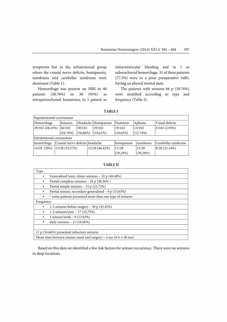
Hemorrhage was present on MRI in 40
patients (30.76%) in 38 (95%) as
intraparenchymal hematoma, in 1 patient as
intraventricular bleeding and in 1 as
subarachnoid hemorrhage. 31 of these patients
(77.5%) were in a poor preoperative mRS,
having an altered mental state.
The patients with seizures 66 p (50.76%)
were stratified according to type and
frequency (Table 2).
TABLE I
Supratentorial cavernomas
Hemorrhage Seizures Headache Hemiparesis Numness Aphasia Visual deficits
29/102 (28,43%) 66/102
(64.70%)
58/102
(56,86%)
19/102
(18,62%)
19/102
(18,62%)
13/102
(12.74%)
3/102 (2.94%)
Infratentorial cavernomas
hemorrhage Cranial nerve deficits headache hemiparesis numbness Cerebellar syndrome
14/28 (50%) 15/28 (53,57%) 13/28 (46,42%) 11/28
(39,28%)
11/28
(39,28%)
9/28 (32.14%)
TABLE II
Type
• Generalized tonic-clonic seizures – 32 p (48.48%)
• Partial complexe seizures – 24 p (36,36% )
• Partial simple seizures – 15 p (22.72%)
• Partial seizure, secondary generalized – 9 p (13.63%)
• * some patients presented more than one type of seizures
Frequency
• 1-2 seizures before surgery – 30 p (45,45%)
• 1-2 seizures/year – 17 (25,75%)
• 1 seizure/week – 9 (13.63%)
• daily seizures – 11 (16.66%)
11 p (16.66%) presented refractory seizures
Mean time between seizure onset and surgery = 4 mo (0.4 → 40 mo)
Based on this data we identified a few risk factors for seizure reccurence. There were no seizures
in deep locations.

398 Giovani et al Surgical treatment and outcome of cerebral cavernomas
TABLE III
Risk factors for seizures occurrence:
• Cortical location
• T location, especially temporomesial location
• Large lesions, including hemosiderinic ring
• Focal neurological deficits
• Left side cavernomas
• Acute hemorrhage
• Subacute hemorrhage
• Surrounding edema*
were not confirmed as risk factors for seizures occurrence
• Sex,
• age,
• diameter of hemosiderinic ring,
• diameter of surrounding edema,
• multiple lesions
Outcome
When we analyzed the outcome we studied
the group of corticalised or superficial
subcortical cavernomas and that of deep
seated lesions, in brainstem, thalamus and
basal ganglia.
Total resection was performed in all cases,
but the surrounding hemosiderin ring was not
removed in deep seated lesions where such a
maneuver could harm grey matter nuclei and
no mortality was registered on short or long
term follow up in this series of 130 patients.
Both Karnovsky and mRS improved after
surgery. In patients with deep seated lesions
(37 patients), 12 (32,43%) presented in a poor
preoperative state mRS > 2. 10 patients from
this group had worsened deficits immediately
after surgery, 4 of these in brainstem
cavernomas but all of them showed a good
recovery with follow up 5 of them remained in
mRS 2 and 3 with mRS>2. The complications
included worsened hemiparesis, worsened
ataxia, 7th nerve paresis and VI or IIIrd nerve
paresis.
The immediate postoperative status for this
group was excellent in 10/37 (27,02%) of
patients, good in 13/37 (35,13%) patients and
poor in 14 (37,83%) patients. The long term
follow up for these patients showed that 31/37
(83,78%) of patients had a mRS between 0 and
2, and the rest had a poor long term outcome.
The situation was different in the group of
superficial cavernomas where 55/93 (59.13%)
of patients presented with a mRS of 0 or 1 and
36/93 (38,7%) presented with mRS of 2 and 2
patients with an mRS>2.
After surgery there was no clinical
deterioration in this group and 22 patients
from those who presented in mRS 2 showed
neurological improvement on long term
follow up, meaning that 82,79% of patients had
a 0 or 1 mRS.

Romanian Neurosurgery (2014) XXI 4: 394 – 404 399
Case presentations
Case 1: A 70 years old male was admitted
for persistent headache and left homonymous
hemianopsia that worsened in the week
previous presentation.
Figure 1 - MRI – right occipital heterogenous lesion,
with salt and pepper appearance suggestive for
cavernoma
The tumor was completely removed with
the surrounding hemosiderin ring using a
right suboccipital occipital-supratentorial
approach. Using the gravity with no need for
mechanical retraction, the lesion was exposed
corticalised on the tentorial surface of the right
occipital lobe. The tumor was piecemeal
removed and then the hemosiderin ring was
resected. Hemostasis was achieved with
bipolar and intensive lavage with serum. After
the dural closure the scalp is closed on epidural
drain. The postoperative CT scan confirmed
complete removal.
Figure 2 - Same patient with postop CT images of the
occipital cavernoma – total removal

400 Giovani et al Surgical treatment and outcome of cerebral cavernomas
Case 2: A 45 years old female presented
with altered mental state (14 p) and a history
of headache, vomiting, swallowing disorders
(liquid and solid food), gait disturbances
(astasia-abasia), mild right faciobrachial
paresis with onset 7 days before admission.
The Neurologic exam showed mild right
faciobrachial paresis and IX, X, XI and XII
cranial nerves paresis.
Figure 3 - CT scan - spontaneous hyperdense lesion
within the right medulla, Ø = 13 mm
Figure 4 - MRI: right medulla lesion, H-iso in T1 and h-HT2,
Ø = 13 mm, suggestive for cavernoma, with subacute bleeding

Romanian Neurosurgery (2014) XXI 4: 394 – 404 401
The lesion was completely resected using a
telovelar approach. A midline suboccipital
craniectomy was performed, and after opening
the cisterna magna a telovellar approach is
used to access the medulla. As the lesion
appeared subpial it was entered directly and
then piecemeal resected completely. The
surrounding yellow gliotic plane was left
intact. Dura mater was reconstructed with
Kolagen and then with the drain left in place
the muscles plane and the skin were closed.
The outcome was favorable with partial
remission of the cranial nerves deficits and
improvement of swallowing disorders at 2
months follow up. (Figure 4)
Figure 5 - Postop. MRI with complete resection of the
cavernoma
Discussions
The frequency of asymptomatic
cavernomas can be as high as 40%
(Zabramsky), but the actual data is impossible
to quantify.
The most common presenting symptoms
for the supratentorial group were seizures,
headache, hemiparesis and numbnes. The
most common infratentorial presentation
were cranial nerve deficits followed by
hemiparesis and numbness and cerebellar
symptoms.
Compared to the infratentorial group
where the hemorrhagic presentation was
almost the rule (93%) only 39% of the
supratentorial group patients had this
presentation.
The division into supratentorial and
infratentorial groups is not enough and
further studies should focus on the differences
of the groups of patients with cortical or
subcortical cavernomas compared to those

402 Giovani et al Surgical treatment and outcome of cerebral cavernomas
with cavernomas seated in brainstem,
thalamus or basal ganglia. (1, 3)
The best method for preoperative and
postoperative investigating the patient is the
MRI. It can show the borders of the lesion the
intra or extralesional hemorrhage and the
associated edema and also is invaluable in
planning the aproach especially if
neuronavigation or stereotaxy are not used for
localization. Angiography can be useful in
defining associated lesions because
cavernomas are not visible on it but we did not
make a standard from using it and this may be
a reason why venous associated anomalies
escaped our view.
As cavernomas in deep locations have an
annual hemorrhage rate of 5,1% and a rebleed
rate of 31,5% per patient per year, there was no
time for wait and see in these cases.
Observation was reserved only to those cases
we considered inoperable. Complete removal
was achieved in all cases but with the cost of
new neurological deficits in 4 patients (3%), all
of these presenting deep seated lesions. The
postoperative mRS was good or excellent in
97% of patients compared to 64% preoperative
and was even more evident in patients with
deep seated lesions. The control of the seizures
was achieved in all patients who presented
with seizures, and almost 60% of them were
free of medication after surgery.
The dimensions of the lesions were not
statistically different in the two groups but in
the infratentorial group they correlated with
the hemorrhage and neurologic deficit. Some
studies report larger sizes of cavernomas in
children but our study included only adults.
We could find an association with venous
anomalies only in 3 patients which is in
contrast to the reports of other studies.
As prospective studies are lacking the
hemorrhage rates which are the key to the
surgical indication are calculated based on
retrospective analysis considering cavernomas
as congenital lesions, yet more and more
studies document the appearance of de novo
cavernomas in sporadic or familial cases. Even
the results of different studies differ it became
clear that once symptomatic the cavernomas
have a high rate of rebleeding causing new
neurological deficits. (14, 18, 19)
Because of their tendency to bleed
cavernomas are always in the surgical focus,
yet surgery should not be proposed for all
lesions, most of the supratentorial lesions can
be treated conservatively if asymptomatic.
Most of the time the hemorrhage is inside the
cavrnoma causing it’s enlargement in time, but
when seated in deep nuclei even this
enlargement can give neurological deficit,
acting as a mass effect. When located in
eloquent areas even small hemorrhage outside
the lesion can be echoed in severe deficits. (11,
12)
Our study aimed at identifying those
lesions with a higher tendency to produce
deficits for which surgery is the best and the
safest treatment but as is the case with many
studies on cavernomas it is limited by its
retrospective nature.
As The most frequent manifestations of
supratentorial lesions are repeated seizures,
which disturb the patient’s life balance we
identified a few risk factors for seizures like the
cortical and more frequently the
temporomesial location, the large cavernomas,
Romanian Neurosurgery (2014) XXI 4: 394 – 404 403
the location in the left hemisphere, the acute or
subacute hemorrhage and edema. In all
supratentorial lesions we removed the
hemosiderin ring as well and we believe this is
one of the main reasons for the complete
disappearance of seizures in most of the cases.
(23, 24)
We identified a few positive prognostic
factors including a high preoperative
Karnofsky score coincident with a mRS of 0
or 1, the small size and the superficial location
of the lesion.
We did not consider radiosurgery an
alternative to deep seated lesions because we
considered the high rates of rebleeding after
radiosurgery inacceptable and many studies
showed that cavernomas can occur after
radiation therapy. (12)
Conclusions
Symptomatic deep cavernous
malformations in the brainstem, basal ganglia,
and thalamus have a high bleed and rebleed
rate and an aggressive natural history. Early
surgery provides excellent clinical results and
protects against future hemorrhages.
Microsurgery is the treatment of choice in
symptomatic brain cavernomas, total
resection being the only curative treatment,
capable to prevent further bleeding and to
offer an efficient control of seizures.
Complete cavernoma resection and
resection of surrounding hemosiderin is
recommended except for cavernomas located
in the brainstem or in eloquent areas
ACKNOWLEDGEMENT: This paper was
co-financed from the European Social Fund,
through the Sectorial Operational Programme
Human Resources Development 2007-2013,
project number POSDRU/159/1.5/S/138907
"Excellence in scientific interdisciplinary
research, doctoral and postdoctoral, in the
economic, social and medical fields -
EXCELIS", coordinator The Bucharest
University of Economic Studies.
Correspondence
Giovani Andrei
References
1.Abdulrauf SI, Kaynar MY, Awad IA. A comparison of
the clinical profile of cavernous malformations with and
without associated venous malformations. Neurosurgery.
1999;44(1):41-46.
2.Abla AA, Lekovic GP, Garrett M, et al. Cavernous
malformations of the brainstem presenting in childhood:
surgical experience in 40 patients. Neurosurgery. 2010;67
(6):1589-1598; discussion 1598-1599.
3.Abla AA, Lekovic GP, Turner J, deOliveira JG, Porter R,
Spetzler RF. Advances in the treatment and outcome of
brainstem cavernous malformation surgery: a single-
center case series of 300 surgically treated patients.
Neurosurgery. 2011;68(2):403-414.
4.Casazza M, Broggi G, Franzini A, Avanzini G, Spreafico
R, Bracchi M, Valentini MC: Supratentorial cavernous
angiomas and epileptic seizures: Preoperative course and
postoperative outcome. Neurosurgery 39:26–34, 1996.
5.Clatterbuck RE, Elmaci I, Rigamonti D: The nature and
fate of punctate (type IV) cavernous malformations.
Neurosurgery 49:26–32, 2001.
6.Cohen DS, Zubay GP, Goodman RR: Seizure outcome
after lesionectomy for cavernous malformations. J
Neurosurg 83:237–242, 1995.
7.Dandy WE. Venous abnormalities and angiomas of the
brain. Arch Surg. 1928;17 (5):715-793.
8.Del Curling O Jr, Kelly DL Jr, Elster AD, Craven TE: An
analysis of the natural history of cavernous angiomas. J
Neurosurg 75:702–708, 1991.
9.Engel JJ: Outcome with respect to epileptic seizures, in
Engel JJ (ed): Surgical Treatment of the Epilepsies. New

404 Giovani et al Surgical treatment and outcome of cerebral cavernomas
York, Raven Press, 1987, pp 553–571.
10.Ferroli P, Casazza M, Marras C, Mendola C, Franzini
A, Broggi G: Cerebral cavernomas and seizures: A
retrospective study on 163 patients who underwent pure
lesionectomy. Neurol Sci 26:390–394, 2006.
11.Ferroli P, Sinisi M, Franzini A, Giombini S, Solero CL,
Broggi G. Brainstem cavernomas: Long-term results of
microsurgical resection in 52 patients. Neurosurgery.
2005;56(6):1203-1212.
12.Hasegawa T, McInerney J, Kondziolka D, Lee JY,
Flickinger JC, Lunsford LD. Long-term results after
stereotactic radiosurgery for patients with cavernous
malformations. Neurosurgery. 2002;50(6):1190-1197.
13.Kondziolka D, Lunsford LD, Kestle JR. The natural
history of cerebral cavernous malformations. J
Neurosurg. 1995;83(5):820-824.
14.Kupersmith MJ, Kalish H, Epstein F, et al. Natural
history of brainstem cavernous malformations.
Neurosurgery. 2001;48(1):47-53.
15.Mathiesen T, Edner G, Kihlstrom L. Deep and
brainstem cavernomas: a consecutive 8-year series. J
Neurosurg. 2003;99(1):31-37.
16.McCormick WF: Pathology of vascular malformations
of the brain, in Wilson CB, Steihn BM (eds): Intracranial
Arteriovenous Malformations. Baltimore,Williams &
Wilkins, 1984, pp 44–63.
17.Mizoi K, Yoshimoto T, Suzuki J. Clinical analysis of
ten cases with surgically treated brain stem cavernous
angiomas. Tohoku J Exp Med. 1992;166(2): 259-267.
18.Nimjee SM, Powers CJ, Bulsara KR: Review of the
literature on de novo formation of cavernous
malformations of the central nervous system after
radiation therapy. Neurosurg Focus 21:e4, 2006.
19.Pozzati E, Acciarri N, Tognetti F, Marliani F,
Giangaspero F: Growth, subsequent bleeding, and de
novo appearance of cerebral cavernous angiomas.
Neurosurgery 38:662–670, 1996.
20.Quinones-Hinojosa A, Lyon R, Du R, Lawton MT.
Intraoperative motor mapping of the cerebral peduncle
during resection of a midbrain cavernous malformation:
technical case report. Neurosurgery. 2005;56(2
Suppl):E439.
21.Rigamonti D, Hadley MN, Drayer BP, Johnson PC,
Hoenig-Rigamonti K, Knight JT, Spetzler RF: Cerebral
cavernous malformations. Incidence and familial
occurrence. N Engl J Med 319:343–347, 1988.
22.Robinson JR, Awad IA, Little JR: Natural history of the
cavernous angioma. J Neurosurg 75:709–714, 1991.
23.Samii M, Eghbal R, Carvalho GA, Matthies C. Surgical
management of brainstem cavernomas. J Neurosurg.
2001;95(5):825-832.
24.Vinas FC, Gordon V, Guthikonda M, Diaz FG.
Surgical management of cavernous malformations of the
brainstem. Neurol Res. 2002;24(1): 61-72.
25.Zabramski JM, Wascher TM, Spetzler RF, et al. The
natural history of familial cavernous malformations:
results of an ongoing study. J Neurosurg. 1994;80(3):422-
432.
26.Wang CC, Liu A, Zhang JT, Sun B, Zhao YL. Surgical
management of brainstem cavernous malformations:
report of 137 cases. Surg Neurol.
2003;59(6):444-454.
27.Ziyal IM, Sekhar LN, Salas E, Sen C. Surgical
management of cavernous malformations of the brain
stem. Br J Neurosurg. 1999;13(4):366-375.