Embed Size (px)
Citation preview

JewelACLTM
Surgical Technique Manual
The Tissue Graft Sparing Device for Anterior Cruciate Ligament Reconstruction
279703-2020

JewelACLTM
Introduction
The JewelACL is specifically designed for implantation with approaches that are familiar to most surgeons. Any necessary variations or adjustments to standard approaches when using the JewelACL are described later in the surgical technique. Thus bone tunnels may be prepared with appropriate standard instrumentation in current use. Also, the JewelACL (alone or in combination with a hamstring) may be secured to the bone with approved cortical suspension, cross pin, interference screw and staple fixation devices. Further details are included later in the surgical technique.
The JewelACL thus offers flexibility in ACL reconstruction by the way of approach, graft selection and graft anchoring to the bone.
The JewelACL is a tissue graft sparing device for the reconstruction of the anterior cruciate ligament (ACL).
It has been designed for use in partial or total tissue sparing ACL reconstruction with hamstring tendons. In a partial tissue graft sparing reconstruction procedure the JewelACL may be used in conjunction with either the semitendinosus or the gracilis tendon. In a total tissue graft sparing reconstruction the JewelACL may be implanted alone.
These approaches will either significantly reduce or totally eliminate the amount of autologous tissue used in the reconstruction. This will not only shorten the operative time but will also ameliorate or totally eliminate donor site morbidity and the consequent deficiency in the power of the hamstring muscle group caused by harvesting of the autologous tissue grafts.
When used in a partial or total tissue graft sparing procedure the JewelACL allows earlier weight-bearing and earlier aggressive rehabilitation than is permitted when autologous tissue grafts alone are used. This is because such grafts go through a debilitating necrotic stage after implantation whereby they lose much of their strength, which is slowly (but not fully) recovered through the processes of revascularization, re-colonization with collagen laying indigenous cells and remodelling of new tissue. During this period the autologous grafts are weak and vulnerable to stretching and so recipients are recommended to follow a slow and careful, non-aggressive rehabilitation regime to avoid compromising the reconstruction.

Flexibility
Surgical Approaches
Anteromedial ApproachFemoral tunnel is drilled through the anteromedial (AM) portal
or
Transtibial ApproachFemoral tunnel is drilled through the tibial tunnel
Graft Choices
Partial Tissue Graft SparingThe reconstruction is performed utilizing just one hamstring tendon alongside or inside the JewelACL
or
Total Tissue Graft SparingThe reconstruction is performed with the JewelACL alone, thus retaining all soft tissue structures intact
Fixation Options
Femoral FixationVarious fixation devices such as suspension devices and cross pins can be used. Approved devices include:• EndoButton® CL Ultra (Smith & Nephew)• TransFix® (Arthrex)
and
Tibial FixationSimilarly, a range of fixation devices such as interference screws or staples can be used. Approved devices include:• RCI Screw® (Smith & Nephew)• Interference Screw (Medgal)
Product Overview
IMPLANT
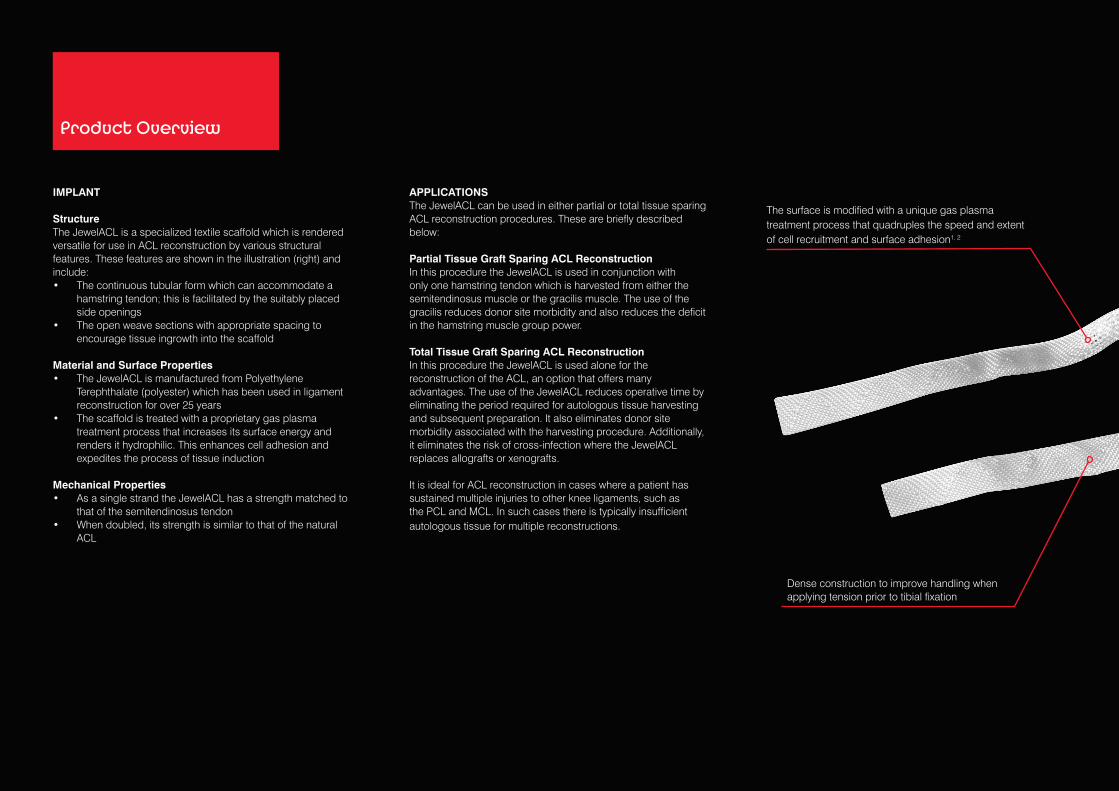
StructureThe JewelACL is a specialized textile scaffold which is rendered versatile for use in ACL reconstruction by various structural features. These features are shown in the illustration (right) and include:• The continuous tubular form which can accommodate a
hamstring tendon; this is facilitated by the suitably placed side openings
• The open weave sections with appropriate spacing to encourage tissue ingrowth into the scaffold
Material and Surface Properties• The JewelACL is manufactured from Polyethylene
Terephthalate (polyester) which has been used in ligament reconstruction for over 25 years
• The scaffold is treated with a proprietary gas plasma treatment process that increases its surface energy and renders it hydrophilic. This enhances cell adhesion and expedites the process of tissue induction
Mechanical Properties• As a single strand the JewelACL has a strength matched to
that of the semitendinosus tendon• When doubled, its strength is similar to that of the natural
ACL
APPLICATIONSThe JewelACL can be used in either partial or total tissue sparing ACL reconstruction procedures. These are briefly described below:
Partial Tissue Graft Sparing ACL ReconstructionIn this procedure the JewelACL is used in conjunction with only one hamstring tendon which is harvested from either the semitendinosus muscle or the gracilis muscle. The use of the gracilis reduces donor site morbidity and also reduces the deficit in the hamstring muscle group power.
Total Tissue Graft Sparing ACL ReconstructionIn this procedure the JewelACL is used alone for the reconstruction of the ACL, an option that offers many advantages. The use of the JewelACL reduces operative time by eliminating the period required for autologous tissue harvesting and subsequent preparation. It also eliminates donor site morbidity associated with the harvesting procedure. Additionally, it eliminates the risk of cross-infection where the JewelACL replaces allografts or xenografts.
It is ideal for ACL reconstruction in cases where a patient has sustained multiple injuries to other knee ligaments, such as the PCL and MCL. In such cases there is typically insufficient autologous tissue for multiple reconstructions.
The surface is modified with a unique gas plasma treatment process that quadruples the speed and extent of cell recruitment and surface adhesion1, 2
Dense construction to improve handling when applying tension prior to tibial fixation

INDICATIONThe JewelACL is indicated for ACL reconstruction.
Contraindications to surgery include:• Known hypersensitivity to implant materials. If the patient is
suspected of having any foreign body sensitivity, appropriate tests should be made prior to implantation.
• Infections, or any structural or pathological condition of the bone or soft tissue that would be expected to impair healing or secure fixation.
• Patients unable or unwilling to restrict activities to prescribed levels or follow a rehabilitation programme during the healing period.
• The JewelACL may not be suitable for skeletally immature patients as it will not elongate and so must not bridge, disturb, or disrupt the growth plate.
• Patients for whom it is not possible to bend the knee to at least 90º as it will not be possible to reach the correct position for drilling the bone tunnels.
WARNINGIf the patient is suspected of having any foreign body sensitivity, appropriate tests should be made prior to material selection or implantation.
INSTRUMENTATIONThe JewelACL can be implanted using modern ACL guidewire systems with a similar anteromedial or transtibial surgical technique to that utilized for hamstring grafts.
Openings allow harvested tissue to be placed inside the implant Dense structure indicates mid-length where the device is
looped around a femoral suspension fixation device
Tensile strength for a single strand matches that of the semitendinosus tendon which is typically 1200 N. This facilitates early mobilization and rehabilitation. There is no need to wait until the end of the typical tissue remodelling phases of autografts and allografts
The open weave tubular structure acts as a scaffold that encourages tissue ingrowth

FIXATION DEVICESThe JewelACL can be secured to the bone with the following approved fixation devices. When using these devices it is critical to follow the instructions for use supplied with them and to observe the conditions and necessary adjustments required in the technique described in the notes section in the following tables.
The strength figures quoted in the tables have been obtained from a rigorous in-house testing programme. Each test was repeated on six units of each of the devices listed in the tables. For more detailed information on the test conditions please see the white paper “Mechanical properties and fixation performance testing of the JewelACL” (WP 006).
Fixation Devices
Femoral Fixation
Fixation device
Procedure
Bone tunnel diameter
Approach to making the tunnels
Strength of femoral fixation
Notes
TransFix (Arthrex)
Partial tissue graft sparing only
TGS: Not applicablePGS: Sized to overall graft diameter
Transtibial only
TGS: Not applicablePGS: Since fixation strength was adequate when the JewelACL was tested alone (1977 N), the test was not repeated with a PGS graft which uses the hamstring graft inside the JewelACL.
Benefit: Strong fixation in weak bone.
Limitation: It can only be used when a tissue graft is incorporated with the JewelACL. The smallest femoral tunnel diameter which the TransFix surgical instruments can create is 7 mm, which is too large for use with the JewelACL alone as this requires a 4 mm tunnel. If a larger tunnel is used, this would result in a gap between the JewelACL and the bone tunnel wall which would not allow the required bone ingrowth and anchorage.
Suitable tibial fixation: RCI Screw or Interference Screw (Medgal).
Technique: Follow the technique supplied by the manufacturer, matching the tunnels to the graft diameter. If the graft has a very small diameter, make the smallest diameter bone tunnel which the instruments allow (e.g. 7 mm TransFix II Tunnel Hook).
EndoButton CL Ultra (Smith & Nephew)
Total tissue graft sparing (TGS) orPartial tissue graft sparing (PGS)
TGS: 4.5 mmPGS: Sized to overall graft diameter
Anteromedial orTranstibial
TGS: 1296 NPGS: Since fixation strength is adequate when the JewelACL is tested on its own, the test was not repeated with a PGS graft which uses the hamstring graft inside the JewelACL.
Benefit: Small diameter tunnel aids anatomic placement. Snug fit between the tunnel wall and the graft.
Suitable tibial fixation: RCI Screw, Interference Screw (Medgal).
Technique: When implanting the JewelACL alone use a zero or small offset (3-4 mm) femoral aimer since only a single diameter tunnel of 4.5 mm is required. There is no need to over-drill to create a stepped bone tunnel. Use the smallest loop to maximize the length of the device in the bone tunnel.
When incorporating a tissue graft with the JewelACL, follow the manufacturer’s instructions (Technique Guide for EndoButton CL, ref. 10600026, Smith & Nephew) to create a stepped bone tunnel with the appropriate offset aimer and cannulated drill matched to the total graft diameter.
It is advantageous to use a low accessory medial portal rather than the AM portal when creating the femoral tunnel for such cortical suspension devices to maximize the tunnel length (40-45 mm), as described by Brown3. The low accessory medial portal should be located as low as possible, but just above the medial joint line, while avoiding the anterior horn of the medial meniscus. The medial-lateral placement of this portal should be close to the medial edge of the patellar tendon.
Fixation device
Procedure
Bone tunnel diameter
Approach to making the tunnels
Strength of tibial fixation
Notes
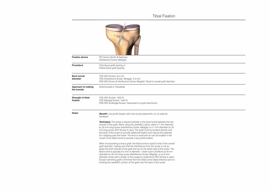
RCI Screw (Smith & Nephew)Interference Screw (Medgal)
Total tissue graft sparing or Partial tissue graft sparing
TGS (RCI Screw): 6.5 mmTGS (Interference Screw, Medgal): 5.5 mmPGS (RCI Screw & Interference Screw, Megdal): Sized to overall graft diameter
Anteromedial or Transtibial
TGS (RCI Screw): 1202 NTGS (Medgal Screw): 1544 NPGS (RCI & Medgal Screw): Dependent on graft size/choice
Benefit: Low profile fixation with intra-tunnel placement, so no external hardware.
Technique: The screw is placed centrally in the bone tunnel between the two strands of the graft. When using the JewelACL alone, either a 7 mm diameter by 30 mm long screw (Interference Screw, Medgal) or a 7 mm diameter by 25 mm long screw (RCI Screw) is used. The graft must be knotted directly over the back of the screw to provide additional fixation and reduce the potential for it slipping past the screw. The knot is small and so can be located in the mouth of the tibial tunnel to provide a low profile fixation.
When incorporating a tissue graft, the tibial tunnel is sized to that of the overall graft diameter, making sure that the interference from the screw is not so great that both strands of the graft end up on the same side of the screw. The tibial tunnel is typically 6.5 mm in diameter. Under such conditions an 8 mm diameter by 30 mm long screw (Interference Screw, Megdal), or an 8 mm diameter screw with a length of the surgeon’s preference (RCI Screw) is used. Excess hamstring graft is trimmed from the tibial tunnel distal entrance prior to knotting the JewelACL portion of the graft over the back of the screw.
Tibial Fixation
Mechanical Properties
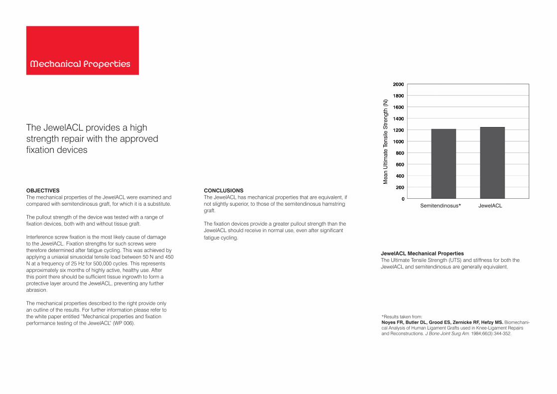
OBJECTIVESThe mechanical properties of the JewelACL were examined and compared with semitendinosus graft, for which it is a substitute.
The pullout strength of the device was tested with a range of fixation devices, both with and without tissue graft.
Interference screw fixation is the most likely cause of damage to the JewelACL. Fixation strengths for such screws were therefore determined after fatigue cycling. This was achieved by applying a uniaxial sinusoidal tensile load between 50 N and 450 N at a frequency of 25 Hz for 500,000 cycles. This represents approximately six months of highly active, healthy use. After this point there should be sufficient tissue ingrowth to form a protective layer around the JewelACL, preventing any further abrasion.
The mechanical properties described to the right provide only an outline of the results. For further information please refer to the white paper entitled “Mechanical properties and fixation performance testing of the JewelACL” (WP 006).
JewelACL Mechanical PropertiesThe Ultimate Tensile Strength (UTS) and stiffness for both the JewelACL and semitendinosus are generally equivalent.
Semitendinosus*
JewelACL
*Results taken from:Noyes FR, Butler DL, Grood ES, Zernicke RF, Hefzy MS. Biomechani-cal Analysis of Human Ligament Grafts used in Knee-Ligament Repairs and Reconstructions. J Bone Joint Surg Am. 1984;66(3):344-352.
The JewelACL provides a high strength repair with the approved fixation devices
CONCLUSIONSThe JewelACL has mechanical properties that are equivalent, if not slightly superior, to those of the semitendinosus hamstring graft.
The fixation devices provide a greater pullout strength than the JewelACL should receive in normal use, even after significant fatigue cycling.

Femoral FixationThe pullout strength for the TransFix and EndoButton CL Ultra when used with the JewelACL alone (i.e. TGS) are both above 1000 N, which is more than the fixation device should ever normally receive. These fixation devices are therefore recommended for use with the JewelACL.
Tibial Fixation after FatiguingThe pullout strengths of the RCI Screw and Interference Screw when used without (TGS) or with a tissue graft (PGS) remained well above that likely to be seen in normal use.
Tibial FixationThe pullout strength for the RCI Screw and Interference Screw when used with the JewelACL alone (i.e. TGS) are above 1200 N, which is more than the fixation device should ever normally receive. These fixation devices are therefore recommended for use with the JewelACL.
TransFix 15 mm ECLU RCI Screw Interference Screw
RC
I Scr
ew
Inte
rfere
nce
Scr
ew
RC
I Scr
ew+
Tis
sue
Inte
rfere
nce
Scr
ew +
Tis
sue
1
INTRODUCTIONThe JewelACL can be implanted using an anteromedial or transtibial surgical technique similar to that followed when reconstructing the ACL with hamstring grafts. The technique followed may be determined by the surgeon’s preference or may be dictated by the preferred fixation devices. The reconstruction can therefore be performed using commonly available modern ACL guidewire systems.
The instructions to follow describe an anteromedial approach with the JewelACL incorporating a single hamstring tendon to produce a partial tissue sparing graft which is fixed to the femur with a cortical suspension device (EndoButton CL Ultra from Smith & Nephew) and to the tibia with an interference screw (e.g. Megdal, which is available from Komak, Poland, or RCI Screw from Smith & Nephew).
Since the above procedure for ACL reconstruction is well known, the following text focuses on the important differences from a standard ACL technique.
PATIENT PREPARATIONThe procedure is performed with the patient in the supine position under general anaesthesia with a tourniquet inflated. Pre-operative antibiotics are administered.
The pre-operative preparation of the patient is carried out following standard procedures. When adopting an anteromedial approach the patient should be positioned so that the knee can be flexed beyond 90º.
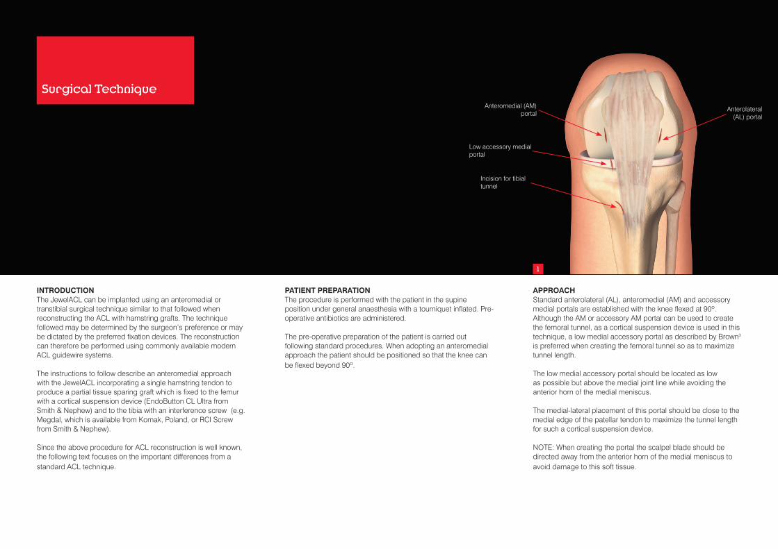
APPROACHStandard anterolateral (AL), anteromedial (AM) and accessory medial portals are established with the knee flexed at 90º. Although the AM or accessory AM portal can be used to create the femoral tunnel, as a cortical suspension device is used in this technique, a low medial accessory portal as described by Brown3 is preferred when creating the femoral tunnel so as to maximize tunnel length.
The low medial accessory portal should be located as low as possible but above the medial joint line while avoiding the anterior horn of the medial meniscus.
The medial-lateral placement of this portal should be close to the medial edge of the patellar tendon to maximize the tunnel length for such a cortical suspension device.
NOTE: When creating the portal the scalpel blade should be directed away from the anterior horn of the medial meniscus to avoid damage to this soft tissue.
Surgical Technique
Incision for tibial tunnel
Low accessory medial portal
Anteromedial (AM) portal
Anterolateral (AL) portal
2 3 4
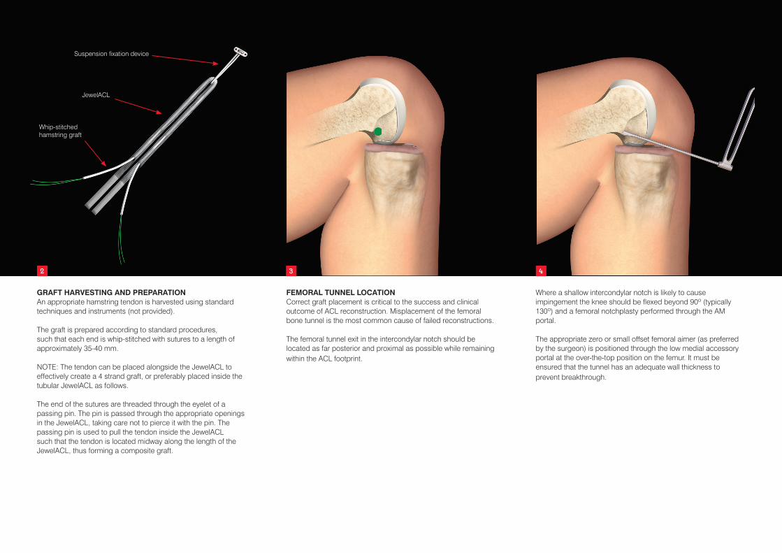
GRAFT HARVESTING AND PREPARATIONAn appropriate hamstring tendon is harvested using standard techniques and instruments (not provided).
The graft is prepared according to standard procedures, such that each end is whip-stitched with sutures to a length of approximately 35-40 mm.
NOTE: The tendon can be placed alongside the JewelACL to effectively create a 4 strand graft, or preferably placed inside the tubular JewelACL as follows.
The end of the sutures are threaded through the eyelet of a passing pin. The pin is passed through the appropriate openings in the JewelACL, taking care not to pierce it with the pin. The passing pin is used to pull the tendon inside the JewelACL such that the tendon is located midway along the length of the JewelACL, thus forming a composite graft.
FEMORAL TUNNEL LOCATIONCorrect graft placement is critical to the success and clinical outcome of ACL reconstruction. Misplacement of the femoral bone tunnel is the most common cause of failed reconstructions.
The femoral tunnel exit in the intercondylar notch should be located as far posterior and proximal as possible while remaining within the ACL footprint.
Where a shallow intercondylar notch is likely to cause impingement the knee should be flexed beyond 90º (typically 130º) and a femoral notchplasty performed through the AM portal.
The appropriate zero or small offset femoral aimer (as preferred by the surgeon) is positioned through the low medial accessory portal at the over-the-top position on the femur. It must be ensured that the tunnel has an adequate wall thickness to prevent breakthrough.
Whip-stitchedhamstring graft
JewelACL
Suspension fixation device

5 6 7
FEMORAL TUNNEL CREATIONA passing pin is drilled through the femoral aimer, into the femur and out through the anterolateral cortex.
NOTE: Care should be taken to avoid drilling into the peroneal nerve, or damaging the cartilage surface of the medial femoral condyle.
The aimer is removed, leaving the passing pin. The passing pin is over-drilled with a 4.5 mm cannulated bone tunnel drill until the cortex is breached. The length of the tunnel is measured and the appropriate size ECLU chosen and assembled with the graft following the technique described by the manufacturers to form a two-strand graft. A sizing block is used to determine the appropriate diameter of the cannulated drill bit used to create the bone tunnels.
The femoral socket is drilled to the appropriate depth with a drill matched to the graft size. The edges of the distal tunnel at its intercondylar exit are chamfered with an ACL tunnel rasp where possible.
NOTE: Care should be taken to ensure the drill does not breach the lateral femoral cortex, otherwise fixation with the EndoButton CL Ultra cannot be performed.
TIBIAL TUNNEL LOCATIONThe tibial footprint of the ACL is left intact for its proprioceptive and vascular contributions. Later it will be attached to the JewelACL to provide a cell source for tissue ingrowth and subsequent abrasion protection.
The intra-articular tibial attachment should be located slightly medial and slightly anterior to the centre of attachment of the natural ACL. It should not interfere with the anterior attachment of the medial meniscus and should also avoid damaging the articular cartilage. Placement too far anteriorly should be avoided as this can lead to impingement of the ligament on the roof of the notch at full extension.
TIBIAL TUNNEL CREATIONAn appropriate tibial guide is used to drill a passing pin into the tibia.
NOTE: Ensure an adequate tunnel length is produced to accommodate the interference screw.
The tibial guide is removed. The guidewire is over-drilled with an appropriately sized cannulated bone tunnel drill.
NOTE: A tunnel of an equal diameter to that of the overall graft is drilled, or a tunnel of a diameter that is 1 mm smaller than that of the graft is made and then expanded to the desired size using serial dilators.
Care should be taken to avoid damage to the articular cartilage.
The edges of intercondylar exit are chamfered with an ACL tunnel rasp.

9
TIBIAL FIXATIONTension is applied to the two strands of the graft which are separated to allow the screw to be introduced centrally between them in the bone tunnel. Care must be taken to ensure that the strands of the graft do not twist and end up on the same side of the screw. The excess hamstring graft is trimmed from the tibial tunnel entrance and the remaining strands of the JewelACL are tied using a reef knot over the back of the screw.
NOTE: The knot provides additional fixation and prevents the graft slipping past the screw. The knot is small and so can be located in the mouth of the tibial tunnel, thus providing a low profile fixation.
GRAFT INSERTION AND FEMORAL FIXATIONThe EndoButton CL Ultra and graft are pulled into the tunnels. The EndoButton CL Ultra is applied following the recommended technique described by the manufacturers.
NOTE: Ensure the button is securely seated on the femoral cortex.
8
TIBIAL ACL STUMP ATTACHMENT ANDTRIMMING TO LENGTHWhere possible attach the remnants of the ACL to the synthetic graft using appropriate sized sutures.
The knee is cycled through a full range of motion while examining the graft arthroscopically to ensure that it has been placed isometrically and allows a full range of motion with no graft impingement.
Ensure the knot is locked before trimming anyexcess strands of the JewelACL. Each cord is cut with scissors at right angles to its length, to minimize the generation of loose fibres.
IMPORTANT:• Any loose fibres created when trimming to length must be
carefully removed from the incision site.• It is vital to ensure that the knot is covered with, and remains
buried in, tissue.
10

POST-OPERATIVE MANAGEMENTThe rehabilitation programme (below) provides only an outline of the prescribed regime. For a full description refer to the document entitled “JewelACL Rehabilitation Programme: For Anterior Cruciate Ligament Reconstruction” (LAB 145).
The rehabilitation programme should be supervised by a specialist physiotherapist. All mobilization and exercises should be performed within the pain free range of movement.
The patient should be warned not to exceed the prescribed activity levels or to overload the repair before complete healing has occurred.
The rehabilitation regime was developed in conjunction with Ian Horsley MSc, MCSP, Clinical Lead Physiotherapist, English Institute of Sport (EIS) North West, of BackinAction Physiotherapy and Sports Injury Clinic, Wakefield, UK.
Day 1• The leg is placed in a Continuous Passive Motion (CPM)
machine and subjected to passive flexion of between 20-60º for as many hours as possible.
Days 2-7• CPM between 20-90º flexion is continued but the range
should be reduced to 20-60º if the patient complains of any pain.
Week 2• Active Range of Motion (ROM), allowing flexion and
extension, is initiated with the patient wearing a brace limiting the range of motion flexion/extension to 20-90º of knee flexion.
Week 3• Continue with week 2 activities.
• In addition, passive full extension is allowed twice per day as the patient tolerates.
• Partial weight-bearing is commenced with two-handed support, e.g. crutches.
Weeks 4-5• Active ROM exercise, with unblocked brace, is allowed to
the patient’s maximum flexion/extension capability (this is usually between 90-110º).
• Partial weight-bearing is continued with one-handed support, e.g. a cane.
Weeks 6-10• Limitation on both flexion and extension is no longer
imposed but wearing an unblocked brace is continued.
• Full weight-bearing may be commenced at this stage.
• Extension training for muscles is begun, aiming at full development of ROM.
Weeks 10-12• Light sports activities such as jogging or swimming may be
commenced.
Weeks 12-24• Gradual return to sporting activities is permitted.
• However, it must be noted that the speed of return to full pre-injury sporting activities should be governed by the state of the muscles of the injured leg.
• If they are inadequate it is recommended that the patient rehabilitates them to an adequate degree before engaging in any strenuous activities that might jeopardize the reconstructed ligament.
• It should also be noted that, on the occurrence of any significant discomfort, it may be necessary to extend the rehabilitation programme accordingly.
REFERENCES1. Rowland JR, Tsukazaki S, Kikuchi T, Fujikawa K,
Kearney J, Lomas R, Wood E, Seedhom BB. Radio frequency-generated glow discharge treatment: potential benefits for polyester ligaments. J Orthop Sci. 2003;8(2):198-206.
2. Tsukazaki S, Kikuchi T, Fujikawa K, Kobayashi T, Seedhom BB. Comparative study of the covered area of Leeds-Keio (LK) artificial ligament and radio frequency generated glow discharge treated Leeds-Keio (Bio-LK) ligament with synovial cells. J Long Term Eff Med Implants. 2003;13(4):355-62.
3. Brown CH Jr, Spalding T, Robb C. Medial portal technique for single-bundle anatomical Anterior Cruciate Ligament (ACL) reconstruction. International Orthopaedics. 2013;37(2):253-269. Doi: 10.1007/s00264-012-1772-9.

Ordering Information
Implant
102-6003 JewelACL 7 mm ID x 710 mm (supplied sterile)
Optional Tibial Fixation Devices:
7 mm x 30 mm Interference Screw (non-sterile)8 mm x 30 mm Interference Screw (non-sterile)Manufactured by Medgal and available from Komak, Poland([email protected]. tel./fax +48 061 662 44 01, tel. +48 664 027 656)
Please refer to the Instructions for Use leaflet packed with the JewelACL for essential information including Use, Sterility, Indications, Contraindications, Warnings and Precautions, Potential Adverse Effects and Storage.
Additional copies may be obtained from the Neoligaments™ Sales Department, or downloaded from http://www.neoligaments.com

Neoligaments™ A division of Xiros™ Springfield House Whitehouse Lane Leeds LS19 7UEUK
Tel. +44 (0) 113 238 7202 Fax. +44 (0) 113 238 7201 [email protected] www.neoligaments.com
Xiros Limited, registered in England No. 1664824.
All rights reserved. © Neoligaments™ 2020. Worldwide patents and patents pending. Neoligaments, JewelACL and Xiros are trademarks of Xiros.
TransFix is a registered trademark of Arthrex, Inc. EndoButton and RCI Screw are registered trademarks of Smith & Nephew, Inc.
Developed and manufactured by
LAB 138 8.00