Embed Size (px)
Citation preview

Surgical Considerations in theOpen Rhinoplasty Approach toClosure of Septal PerforationsDavid P. Arnstein, MD, Gerald S. Berke, MD
\s=b\Repair of nasoseptal perforations isa difficult problem for the otolaryngolo-gist. Recently, there has been anincreased incidence among patients, par-ticularly with the rise in cocaine abuseand trauma. The variety of proposedmethods of repair points to the lack of a
definitive solution for successful surgicaltreatment of nasoseptal perforations.Successful septal perforation repairsusing an open rhinoplasty approach withbipedicled mucoperichondrial flaps andtemporal fascia grafts were achieved ineight of nine patients in a series. Residentotolaryngologists in training were the pri-mary surgeons in all nine patients. The
Nasoseptal perforations are beingseen with increasing frequency
by otolaryngologists. This increase is,in large part, related to soaringcocaine abuse and trauma in largemetropolitan regions. Small or veryposterior perforations are usuallyasymptomatic.1 Larger or more ante¬rior perforations may present withcrusting, whistling on respiration,epistaxis, nasal obstruction, postnasaldrip, odor, or aberrations in smell.Patients with large perforations maylose so much support that a saddlenasal deformity with columellarretraction will result.2
The most common origin of septalperforations is reported to be trauma.3
Accepted for publication Oct 4,1988.From the Division of Head and Neck Surgery,
UCLA School of Medicine.Presented in part before the Western Section
Meeting of the American College of Surgeons,Newport Beach, Calif, Jan 23, 1988.
Reprint requests to Division of Head and NeckSurgery, 62-126 Center for Health Sciences,UCLA School of Medicine, Los Angeles, CA 90024(Dr Berke).
open rhinoplasty approach affords betterexposure to the septal perforation thandoes a closed technique, and it facilitatesthe elevation of mucoperichondrial flapson all sides of the perforation. This meth¬od also allows the surgeon access toperform a limited concurrent rhinoplastywhen indicated. The open rhinoplastyapproach is ideally suited for teaching thetechnique of large septal perforation clo¬sure In surgical training programs. Thesurgical considerations in using this meth¬od are discussed.
(Arch Otolaryngol Head Neck Surg1989;115:435-438)
Surgical trauma during septoplasty isthe usual cause. Other frequent causes
include nasal packing, cauterization,nasotracheal intubation, and nose
picking. Unfortunately, cocaine abuseas a factor in septal perforations is anall-too-familiar finding in today'ssociety. Some unusual causes includeinfection, autoimmune diseases, gran-ulomatous diseases, syphilis, and car¬cinoma. Workers exposed to industri¬al chemicals, such as those who han¬dle salt, lime, cement, chrome, arse¬
nic, and calcium nitrate, as well as
glass blowers, have also been found tohave a higher incidence of perfora¬tions.2
The mainstays of conservativetreatment have been frequent salineirrigation and moisturizing ointmentto relieve the nasal crusting andrecurrent epistaxis. Prosthetic obtu¬rators were first employed in the ear¬ly 1950s." The first prostheses weremade of nylon. Currently, they aremade of Silastic. However, many
patients find the prosthesis difficultto tolerate due to the foreign bodysensation in the nasal cavity. Largeperforations often make prostheticinsertion and maintenance in positionexacting for the physician, as well asfor the patient. A number of physi¬cians have therefore advocated thatnonoperative management be re¬served for patients who are asymp¬tomatic or who are considered pooroperative risks.5 High-risk patientsinclude those with long-term cocaineor substance abuse, active granuloma-tous disease or vasculitis, active infec¬tion (syphilis), or carcinoma. Evenwhen repair of a septal perforationfails, the remaining perforation istypically smaller and more posterior¬ly located and is therefore less symp¬tomatic.
That there is no widely acceptedsurgical technique for repair of septalperforations suggests that they arenot easily treated. The first surgicaltreatment involved enlargement ofthe perforations to make them lesssymptomatic. Hopefully, this methodis no longer in vogue. The first largeseries of repairs with good closurerates was reported by Fairbanks andChen.3 They combined Gollum's6 localmucosal flap method with the inser¬tion of a connective tissue graftbetween the sutured septal flaps.They reported a closure rate of 95% in20 patients with one to seven years offollow-up. However, the ability to ele¬vate intact mucosal flaps on all sidesof a perforation was crucial to graftrevascularization and viability.
Fundamental to the notion of usingfascia or pericranial autografts
Downloaded From: http://jamanetwork.com/ by a University of California - Los Angeles User on 08/02/2017

Fig 1.—Blood supply to nasal septum.
between flaps was Wright's7 observa¬tion that connective tissue grafts andthe tympanic membrane becameadherent within 24 hours and thatrevascularization followed shortlythereafter. This implied that mucosalflaps need not cover the connectivetissue graft on both sides, becausewhen the graft becomes vascularizedfrom the covered side, the mucosa onthe uncovered side will migrate across
and epithelialize the vascularized sur¬face. Also, epithelial migration proba¬bly occurs more readily on connectivetissue grafts than on bone or cartilageautografts.2
An important rule in repairing sep¬tal perforations is that mucosal flapsshould be designed to preserve maxi¬mal blood supply. Understanding theanatomy of the blood supply to thenasal septum is thus essential to anymethod of repair (Fig 1). The anteriorand posterior ethmoidal arteries andthe sphenopalatine artery arise fromthe posterosuperior area of the sep¬tum. There are anastomoses betweenthe palatine artery through the inci¬sor foramen, the descending palatineartery, and the septal branch of thesuperior labial artery. Thus, incisionsin the posterosuperior area of theseptum should be avoided. Bipedicledadvancement flaps based on the ante¬rior floor of the nose and on the
posterosuperior area of the septum asfirst described by Gollum6 maintainan intact blood supply.
Every surgeon who has tried toclose a large septal perforation knowsthat using tissue autografts anddesigning appropriate flap incisionsdoes not necessarily ensure a goodresult. The closure methods detailedby Fairbanks,8 while effective, are
technically not facile. The transnaresapproach to closure of large or poste¬rior perforations can be a frustratingexperience, even for a skilled surgeon.9This is especially true for residents intraining who may be in the earlystages of nasal surgical adeptness.Open rhinoplasty as suggested byGoodman and Strezlow5 providesexcellent exposure for these repairs.We report on a series of nine patientswho underwent attempted repair ofnasoseptal perforations using an openrhinoplasty approach with bipedicledmucoperichondrial flaps and fasciagrafts. Resident physicians in otolar-yngology-head and neck surgery werethe primary surgeons in all ninecases.
TECHNIQUEA general or long-acting local anesthesia
was preferred, since the procedure can belengthy (two to ZVi hours), depending onwhether concurrent rhinoplasty was
planned (Fig 2). The postauricular skin
was injected for harvesting the graft mate¬rial, and standard septorhinoplasty injec¬tions were performed.
The graft material was harvested ini¬tially so that it could be allowed to drywhile the nasal dissection was beingaccomplished. A dried and stiffened graftwas found to be easier to handle duringplacement. A superior postauricular inci¬sion was made, and the temporal fasciawas harvested. It is important to harvestenough for placement on both sides of theperforation, with a generous overlappingarea (approximately 20 cm2). If the tempo¬ral fascia was attenuated, harvesting ofpericranium also yielded usable graftmaterial.
The technique used standard externalrhinoplasty incisions.8 A columellar skinincision was made with an inverted V (Fig3). This was then connected to bilateralalar margin incisions. The columellar skinneeds to be elevated with great care
because it is very thin and easily torn. Thenasal skin was then reflected back toexpose the medial and lateral crura, theupper lateral cartilages, and the nasalbones (Fig 4). Following this, the medialcrura and domes were separated, withsharp dissection providing exposure to thecaudal end of the septum (Fig 5). Exposingthe caudal end of the septum and gettinginto the correct plane between the mucosa
and cartilage were frequently the mosttedious and arduous parts of the proce¬dure.
Mucoperichondrial flaps were thendeveloped on both sides of the septum (Fig6). The mucoperichondrium was leftattached to the edges of the perforationuntil the flap was elevated, to lessen thechance of an inadvertent tear. One impor¬tant detail is that the upper lateral carti¬lages often needed to be shaved from theseptum to facilitate development of themucoperichondrial flaps. However, ade¬quate exposure was obtained in this serieswithout having to resort to medial osteot¬omies. Because the surgical field was nar¬
row and deep, adequate retraction couldhave presented a problem. A medium-length nasal speculum placed between themucoperichondrial flaps provided a good,readily available retractor.
After elevation, a relaxing incision was
created in at least one of the flaps (usuallythe flap that had been most completelyelevated). An incision was made on thelateral nasal wall just beneath the inferiorturbinate (Fig 7). This flap was then mobi¬lized from the floor of the nose medially tothe maxillary crest and then up onto theseptum, in continuity with the mucosalflap. If there was insufficient tissue forprimary closure, a second incision was
Downloaded From: http://jamanetwork.com/ by a University of California - Los Angeles User on 08/02/2017

Fig 2.—A 2.5-cm septal perforation (arrow)caused by cocaine abuse in 16-year-old girl.
Fig 3.—Columellar incision. Fig 4.—
Exposed nasal skeleton showing low¬er (small arrow) and upper (large arrow)lateral cartilage.
Fig 5.—Caudal septum (large, curved arrow)and mucoperichondrial flaps. Smaller arrow
indicates right lower lateral cartilage andmucoperichondrial flap.
Fig 6.—Exposed septum (arrow). Fig 7.—Elevation of floor of nose. Arrowindicates inferior turbinate.
Fig 8.—
Suture closure of perforation in leftmucoperichondrial flap (arrow).
Fig 9.—Placement of connective tissue graft.Arrow indicates temporal fascia graft.
Fig 10.—Reapproximation of lower lateralcartilage.
Downloaded From: http://jamanetwork.com/ by a University of California - Los Angeles User on 08/02/2017
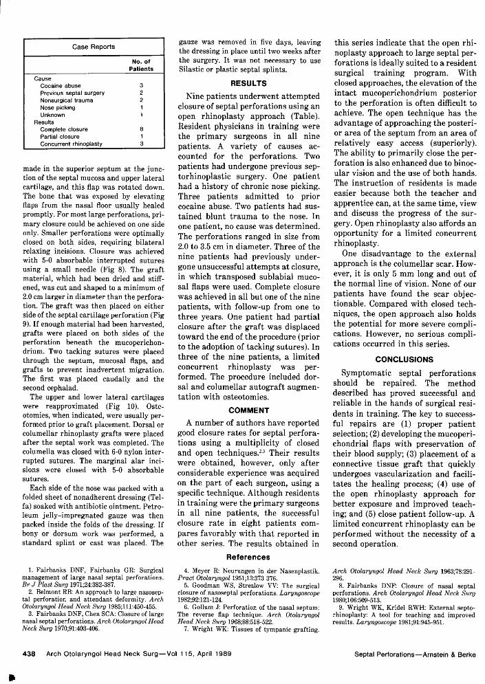
Case ReportsNo. of
PatientsCause
Cocaine abuse 3Previous septal surgery 2Nonsurgical trauma 2Nose picking 1Unknown 1
ResultsComplete closure 8Partial closure 1Concurrent rhinoplasty 3
made in the superior septum at the junc¬tion of the septal mucosa and upper lateralcartilage, and this flap was rotated down.The bone that was exposed by elevatingflaps from the nasal floor usually healedpromptly. For most large perforations, pri¬mary closure could be achieved on one sideonly. Smaller perforations were optimallyclosed on both sides, requiring bilateralrelaxing incisions. Closure was achievedwith 5-0 absorbable interrupted suturesusing a small needle (Fig 8). The graftmaterial, which had been dried and stiff¬ened, was cut and shaped to a minimum of2.0 cm larger in diameter than the perfora¬tion. The graft was then placed on eitherside of the septal cartilage perforation (Fig9). If enough material had been harvested,grafts were placed on both sides of theperforation beneath the mucoperichon-drium. Two tacking sutures were placedthrough the septum, mucosal flaps, andgrafts to prevent inadvertent migration.The first was placed caudally and thesecond cephalad.
The upper and lower lateral cartilageswere reapproximated (Fig 10). Oste¬otomies, when indicated, were usually per¬formed prior to graft placement. Dorsal orcolumellar rhinoplasty grafts were placedafter the septal work was completed. Thecolumella was closed with 6-0 nylon inter¬rupted sutures. The marginal alar inci¬sions were closed with 5-0 absorbablesutures.
Each side of the nose was packed with afolded sheet of nonadherent dressing (Tel-fa) soaked with antibiotic ointment. Petro¬leum jelly-impregnated gauze was thenpacked inside the folds of the dressing. Ifbony or dorsum work was performed, astandard splint or cast was placed. The
gauze was removed in five days, leavingthe dressing in place until two weeks afterthe surgery. It was not necessary to useSilastic or plastic septal splints.
RESULTSNine patients underwent attempted
closure of septal perforations using an
open rhinoplasty approach (Table).Resident physicians in training were
the primary surgeons in all ninepatients. A variety of causes ac¬counted for the perforations. Twopatients had undergone previous sep-torhinoplastic surgery. One patienthad a history of chronic nose picking.Three patients admitted to priorcocaine abuse. Two patients had sus¬tained blunt trauma to the nose. Inone patient, no cause was determined.The perforations ranged in size from2.0 to 3.5 cm in diameter. Three of thenine patients had previously under¬gone unsuccessful attempts at closure,in which transposed sublabial muco¬sal flaps were used. Complete closurewas achieved in all but one of the ninepatients, with follow-up from one tothree years. One patient had partialclosure after the graft was displacedtoward the end of the procedure (priorto the adoption of tacking sutures). Inthree of the nine patients, a limitedconcurrent rhinoplasty was per¬formed. The procedure included dor¬sal and columellar autografi augmen¬tation with osteotomies.
COMMENTA number of authors have reported
good closure rates for septal perfora¬tions using a multiplicity of closedand open techniques.2-3 Their resultswere obtained, however, only afterconsiderable experience was acquiredon the part of each surgeon, using a
specific technique. Although residentsin training were the primary surgeonsin all nine patients, the successfulclosure rate in eight patients com¬
pares favorably with that reported inother series. The results obtained in
this series indicate that the open rhi¬noplasty approach to large septal per¬forations is ideally suited to a residentsurgical training program. Withclosed approaches, the elevation of theintact mucoperichondrium posteriorto the perforation is often difficult toachieve. The open technique has theadvantage of approaching the posteri¬or area of the septum from an area ofrelatively easy access (superiorly).The ability to primarily close the per¬foration is also enhanced due to binoc¬ular vision and the use of both hands.The instruction of residents is madeeasier because both the teacher andapprentice can, at the same time, viewand discuss the progress of the sur¬
gery. Open rhinoplasty also affords an
opportunity for a limited concurrentrhinoplasty.
One disadvantage to the externalapproach is the columellar scar. How¬ever, it is only 5 mm long and out ofthe normal line of vision. None of our
patients have found the scar objec¬tionable. Compared with closed tech¬niques, the open approach also holdsthe potential for more severe compli¬cations. However, no serious compli¬cations occurred in this series.
CONCLUSIONS
Symptomatic septal perforationsshould be repaired. The methoddescribed has proved successful andreliable in the hands of surgical resi¬dents in training. The key to success¬ful repairs are (1) proper patientselection; (2) developing the mucoperi¬chondrial flaps with preservation oftheir blood supply; (3) placement of aconnective tissue graft that quicklyundergoes vascularization and facili¬tates the healing process; (4) use ofthe open rhinoplasty approach forbetter exposure and improved teach¬ing; and (5) close patient follow-up. Alimited concurrent rhinoplasty can beperformed without the necessity of asecond operation.
References1. Fairbanks DNF, Fairbanks GR: Surgical
management of large nasal septal perforations.Br J Plast Surg 1971;24:382-387.
2. Belmont RB: An approach to large nasosep-tal perforation and attendant deformity. ArchOtolaryngol Head Neck Surg 1985;111:450-455.
3. Fairbanks DNF, Chen SCA: Closure of largenasal septal perforations. Arch Otolaryngol HeadNeck Surg 1970;91:403-406.
4. Meyer R: Neurungen in der Nasenplastik.Pract Otolaryngol 1951;13:373-376.
5. Goodman WS, Strezlow VV: The surgicalclosure of nasoseptal perforations. Laryngoscope1982;92:121-124.
6. Gollum J: Perforation of the nasal septum:The reverse flap technique. Arch OtolaryngolHead Neck Surg 1968;88:518-522.
7. Wright WK: Tissues of tympanic grafting.
Arch Otolaryngol Head Neck Surg 1963;78:291\x=req-\296.
8. Fairbanks DNF: Closure of nasal septalperforations. Arch Otolaryngol Head Neck Surg1980;106:509-513.
9. Wright WK, Kridel RWH: External septo-rhinoplasty: A tool for teaching and improvedresults. Laryngoscope 1981;91:945-951.
Downloaded From: http://jamanetwork.com/ by a University of California - Los Angeles User on 08/02/2017


![Injec%c3%87%c3%83o Diesel Bosch Common Rail[1]](https://img.dokumen.tips/doc/110x75/5571f93249795991698f0775/injecc387c383o-diesel-bosch-common-rail1.jpg)


