Embed Size (px)
Citation preview
Surgical and Medical Management of Diseases of the
Thyroid and Parathyroid

Surgical and Medical Management of Diseases of the
Thyroid and Parathyroid
Ashok R. Shaha, MD, FACS
Cherie-Ann O. Nathan, MD, FACS
Jyotika K. Fernandes, MD, MBBS
Chris de Souza, MS, DORL, DNB, FACS, FRCS
Shashank R. Joshi, MD, DM, FRCP, FACE, FACP, FICP
5521 Ruffin RoadSan Diego, CA 92123
e-mail: [email protected]: http://www.pluralpublishing.com
Copyright © 2020 by Plural Publishing, Inc.
Typeset in 10.5/13 Garamond by Flanagan’s Publishing Services, Inc.Printed in the United States of America by Integrated Books International
All rights, including that of translation, reserved. No part of this publication may be reproduced, stored in a retrieval system, or transmitted in any form or by any means, electronic, mechanical, recording, or otherwise, including photocopying, recording, taping, Web distribution, or information storage and retrieval systems without the prior written consent of the publisher.
For permission to use material from this text, contact us byTelephone: (866) 758-7251Fax: (888) 758-7255e-mail: [email protected]
Every attempt has been made to contact the copyright holders for material originally printed in another source. If any have been inadvertently overlooked, the publishers will gladly make the necessary arrangements at the first opportunity.
NOTICE TO THE READERCare has been taken to confirm the accuracy of the indications, procedures, drug dosages, and diagnosis and remedia-tion protocols presented in this book and to ensure that they conform to the practices of the general medical and health services communities. However, the authors, editors, and publisher are not responsible for errors or omissions or for any consequences from application of the information in this book and make no warranty, expressed or implied, with respect to the currency, completeness, or accuracy of the contents of the publication. The diagnostic and remediation protocols and the medications described do not necessarily have specific approval by the Food and Drug administration for use in the disorders and/or diseases and dosages for which they are recommended. Application of this information in a particular situation remains the professional responsibility of the practitioner. Because standards of practice and usage change, it is the responsibility of the practitioner to keep abreast of revised recommendations, dosages, and procedures.
Library of Congress Cataloging-in-Publication Data
Names: Shaha, Ashok R., editor. | Nathan, Cherie-Ann O., editor. | Fernandes, Jyotika K., editor. | De Souza, Chris (Surgeon), editor. | Joshi, Shashank R., editor.Title: Surgical and medical management of diseases of the thyroid and parathyroid / [edited by] Ashok R. Shaha, Cherie-Ann O. Nathan, Jyotika K. Fernandes, Chris de Souza, Shashank R. Joshi.Description: San Diego, CA : Plural Publishing, [2020] | Includes bibliographical references and index.Identifiers: LCCN 2019001824| ISBN 9781597568548 (alk. paper) | ISBN 1597568546 (alk. paper)Subjects: | MESH: Thyroid Diseases — surgery | Thyroid Neoplasms — surgery | Parathyroid Diseases--surgery | Parathyroid Neoplasms--surgery | Thyroid Gland--pathology | Parathyroid Glands — pathologyClassification: LCC RD599.5.T46 | NLM WK 280 | DDC 617.5/39 — dc23LC record available at https://lccn.loc.gov/2019001824
v
Contents
Preface ixAbout the Editors xiContributors xvii
section I. Introduction 1
1 Anatomy and Development of Hypothalamic Pituitary Thyroid Axis 3Shashank R. Joshi, Jimit Vadgama, and Nikita Srinivasan
2 Anatomy and Pathology of Thyrotrophs 9Shashank R. Joshi and Nikita Srinivasan
3 Thyroid Hormone Synthesis and Transport 17Pramod Gandhi and Parimal Tayde
4 Thyroglobulin Structure, Function, and Biosynthesis 21Shashank R. Joshi and Nikita Srinivasan
5 Peripheral Thyroid Hormone Metabolism 33Emma Jakoi
6 Laboratory Assessment of Thyroid Function 43Shashank R. Joshi
7 Imaging of the Thyroid in Health and Disease 57Jeffrey Harris Henderson and Brittany Bohinc Henderson
8 Pathology of Thyroid Neoplasms 71Shubhada Kane and Neha Mittal
section II. Benign thyroid Disease 95
9 Thyroid Disorders in Pregnancy 97Jagdeesh Ullal and Joseph A. Aloi
10 Hyperthyroidism: Diagnosis, Evaluation, and Management 111David C. Lieb and Joseph A. Aloi
11 Hypothyroidism: Diagnosis, Evaluation, and Management 129Maria Papaleontiou and Nazanene H. Esfandiari
vi Surgical and Medical ManageMent of diSeaSeS of the thyroid and Parathyroid
12 Subacute, Postpartum, and Silent Thyroiditis 147Morgan Jones and Jyotika K. Fernandes
13 Thyroid Emergencies 153Rashi Agarwal and Katherine A. Lewis
14 Nontoxic Multinodular Goiter 165Gloria Ortiz and Sanjay Mediwala
15 Thyroid Nodules: Workup and Modern Concepts 173Jeffrey Harris Henderson and Brittany Bohinc Henderson
16 Surgical Anatomy of the Thyroid and Parathyroid Glands 191Madan Kapre and Neeti Kapre Gupta
17 Physical Examination of the Thyroid Gland 203Madan Kapre and Neeti Kapre Gupta
18 Thyroid Cancer Genetics 209Thomas E. Heineman, Travis L. Shiba, and Maie A. St. John
19 Proliferation, Clonality, and Autonomy of Thyroid Lesions 231Munita Menon
section III. Preoperative evaluation 243
20 FNAC for Thyroid and Parathyroid Disorders 245Uma P. Chaturvedi, Raji T. Naidu, Prachi R. Gaddam, and Preeti Dhingra
21 Ultrasound of the Thyroid and Parathyroid Glands 269Ashita Rastogi and Supreeta Arya
22 LASER and Radiofrequency Treatment of Thyroid Nodules and 295 Parathyroid AdenomaPankaj Chaturvedi and Abhishek Vaidya
section IV. thyroid neoplasia 307
23 Papillary Thyroid Carcinoma 309Robert W. A. Hone and Iain J. Nixon
24 Management of Papillary Thyroid Microcarcinoma 317Robert L. Witt
25 Postoperative Management of Differentiated Thyroid Cancer 329Fernanda Vaisman and R. Michael Tuttle
26 Anaplastic Thyroid Cancer and Thyroid Lymphoma 339Steven Anderson and James Paul O’Neill
contentS vii
27 Sporadic Medullary Thyroid Microcarcinoma 361Kendall J. Keck and James R. Howe
28 Syndromic Medullary Thyroid Carcinoma: MEN 2A and MEN 2B 371Mark Lee, Steven G. Waguespack, Paul H. Graham, Mimi I. Hu, and Mark E. Zafereo
29 Hurthle Cell Neoplasms 391Jennifer R. Cracchiolo and Ashok R. Shaha
30 Thyroid Cancer in Children: A Comprehensive Overview 401Siobhan T. Pittock and Geoffrey B. Thompson
31 Radiation Induced Thyroid Cancer 447Yogesh More, Manjiri Gupte, and Shaikh Irfan Basha
32 Surgery for Locally Advanced Infiltrating Thyroid Cancer Involving the 455 Larynx and TracheaKathryn M. Van Abel and Daniel L. Price
33 Central Compartment Dissection 469Ameya Asarkar, Cherie-Ann O. Nathan, Ashok R. Shaha, and Vikas Mehta
34 Lateral Neck Dissection and Technique 479Kelley Michele Malloy and Jayne Ryder Stevens
35 Medullary Thyroid Cancer 489A. I. Kaleva, Ashok R. Shaha, and Iain J. Nixon
36 Postoperative Radioiodine Ablation and Treatment of Differentiated 499 Thyroid CancerSomali Gavane, Bae P. Chu, and Ravinder K. Grewal
37 External Beam Radiation in Treatment of Thyroid Cancer 515 Amy J. Xu, Nancy Y. Lee, and C. Jillian Tsai
38 Revision Thyroid Surgery for Recurrent Differentiated Thyroid Cancer 525Nick Lilic, Iain J. Nixon, and Ashok R. Shaha
39 Medical Treatment of Metastatic Thyroid Cancer 533Kacey B. Wanland, Eiman Y. Ibrahim, and Naifa L. Busaidy
40 Positron Emission Tomography for Thyroid Disorders 543Sonia Mahajan and Ravinder K. Grewal
41 Thyroid Surgery 561Shorook Na’ara and Ziv Gil
42 New Innovations in Thyroid Surgery: Endoscopic and Robotic 573 Surgical TechniquesMichael C. Singer
viii Surgical and Medical ManageMent of diSeaSeS of the thyroid and Parathyroid
43 Long-Term Follow-Up of Thyroid Cancer Patients 581Fernanda Vaisman and R. Michael Tuttle
44 Thyroglossal Duct Cyst and Ectopic Thyroid 589Prathamesh Pai and Vidisha Tuljapurkar
45 Prognostic Factors for Thyroid Carcinoma 597Yasuhiro Ito and Akira Miyauchi
section V. the Parathyroids 607
46 Primary Hyperparathyroidism: Indications for Surgery and Preoperative 609 InvestigationsSteven N. Levine and Joshua D. Maier
47 Preoperative Localization of Parathyroid Adenoma 625Vibushitha Narendra and Surender K. Arora
48 Principles of Parathyroid Surgery 645Matthew D. Cox, Kurt L. Nelson, and Brendan C. Stack, Jr.
49 Intraoperative Parathyroid Hormone 667Vikas Mehta, Cherie-Ann O. Nathan, and Ashok R. Shaha
50 Surgical Management of Multigland Parathyroid Disease 675William S. Duke and David J. Terris
51 The Pre- and Postoperative Medical Management of Secondary 689 HyperparathyroidismAdrian P. Abreo and Kenneth Abreo
52 Revision Surgery for Sporadic Primary Hyperparathyroidism 699Madan Kapre
53 Cancer of the Parathyroid Glands 703Oluwafunmilola T. Okuyemi and Nitin A. Pagedar
54 Hiroshima, Nagasaki, Chernobyl, Fukushima, and Thyroid Cancer: 717 Lessons LearnedDaniel Yafit and Dan M. Fliss
55 Bilateral Parathyroidectomy for Multiglandular Disease 729Lourdes Quintanilla-Dieck and Maisie Shindo
Index 741
ix
PrefaCe
The thyroid gland is considered to be the mas-ter organ of the body. A provocative paper
even went so far as to suggest that iodination of the thyroid gland was instrumental in conferring intelligence and thus was responsible for the evo-lutionary separation of humans from hominids. The thyroid gland regulates 13 vital functions of the body, including that of the brain and heart. Understanding the role of the thyroid and para-thyroid glands and the consequences of their dysfunction has taken a long time to uncover. It was in 1908 that Theodor Kocher was awarded the Nobel prize for his work on the physiology, pathology, and surgery of the thyroid gland. The-odor Kocher is considered by many to be the father of thyroid surgery. The era before his work was clouded with catastrophes that surrounded thyroid surgery. Hemorrhage and sepsis were usu-ally the cause of mortality in those days. Since then the trajectory of thyroid gland surgery has evolved swiftly. The surgeons of today are lucky to be practicing in an era when thyroid surgery has its finesse and nuances; the mortality associated with thyroid surgery is almost negligible.
The new era of endocrine surgery has started as a subspecialty. Each discipline in medicine has experienced major developments and advances. All of these have greatly increased the safety of thyroid and parathyroid gland surgery. With the advent of robotics, greatly improved radiological imaging, molecular biology, and a vast array of treatment modalities, physicians can now better treat most problems associated with tumors and dysfunc-tions of the thyroid and parathyroid glands.
All the editors and authors of this book under-stand that its contents need to be updated at frequent intervals if it is to remain relevant. It is our hope that all those who read this book will find themselves better prepared to face the challenges associated with diseases of the thyroid and parathyroid glands.
Our primary intention is to pass on relevant, meaningful, and helpful information to our read-ers. This in turn should help them to transform this information into useful principles in everyday practice. When physicians and patients benefit from the information obtained from this book, all of us involved in its publishing will feel that its purpose is well served.
Ashok R. ShahaCherie-Ann O. NathanJyotika K. FernandesChris de SouzaShashank R. Joshi

xi
aBout the eDItors
Ashok R. Shaha, MD, FACS, is an attending sur-geon on the Head and Neck Service at Memo-rial Sloan-Kettering Cancer Center, Jatin P. Shah Chair in Head and Neck Surgery, and Professor of Surgery at Cornell University Medical College, New York. He completed his surgical training in Baroda, India and worked as a house surgeon at Tata Memorial Hospital, where he developed an interest in head and neck surgical oncology. After his arrival at Memorial Sloan-Kettering Cancer Center in 1975, Dr. Shaha did a surgical oncol-ogy fellowship completing his surgical training at Downstate Medical Center in Brooklyn, New York. Dr. Shaha returned to Memorial Hospital in 1981 as a Fellow in Head and Neck Surgery, and he joined the Department of Surgery at Downstate Medical Center in 1982 as a head and neck sur-geon, rising to the rank of Professor of Surgery in 1992. During this period, he was also Chief of
Head and Neck Surgery at King’s County, Brook-lyn VA Hospital and University Hospital. In August of 1993, Dr. Shaha moved to Sloan Kettering.
During his post-graduate training, Dr. Shaha was awarded several gold medals and was given the Golden Apple Teaching Award at Downstate Medical Center. Other awards include: Faculty Member of the AOA Honor Medical Society, the Outstanding Teacher Award at Memorial Sloan-Kettering Cancer Center in 1996, the Honor Award from the American Academy of Otolaryngology/Head and Neck Surgery, and being named Visit-ing Professor of the Society of Head and Neck Surgeons in 1997 and 1998. Dr. Shaha has been honored by visiting professorships at University of Santa Tomas, Manila, and Sun Yat-Sen Univer-sity, Guang Zhou, China. He has been actively involved in local and national head and neck societies, as well as having been President of the New York Head and Neck Society, the American Society for Surgeons of Indian Origin, and the Brooklyn Surgical Society, and was co-president of the American Head and Neck Society, 1998–1999, and president of the New York Cancer Society, 1999–2000 and the New York Surgical Society, 2004–2005. He is a member of many scientific organizations and serves on the editorial boards of the Journal of Surgical Oncology, Head and Neck, Annals of Surgical Oncology, Brazilian Journal of Surgery, and Journal of Clinical Oncology. He is an honorary member of the Brazilian College of Surgeons, the Cuban Surgical Society, Association of Surgeons of India, the Korean Head and Neck Society, Latin Head and Neck Society, Panamanian Society of Oncology, Chilean College of Surgeons, and Costa Rican Endocrine Society, and was the Program Chairman for the Fifth International
xii Surgical and Medical ManageMent of diSeaSeS of the thyroid and Parathyroid
Head and Neck Oncology Meeting in San Fran-cisco (2000), and Conference Chairman for the Sixth International Head and Neck Meeting in 2004. Recently Dr. Shaha was the recipient of the Distinguished Service Award by the Ameri-can Academy of Otolaryngology — Head and Neck Surgery and President of the American Associa-tion of Endocrine Surgeons, and was elected to the American Surgical Association. In July 2016, he delivered the Hayes Martin Lecture, and has served on the steering committee for World Con-gress in Thyroid Cancer.
Dr. Shaha has been academically active at national and international meetings, with approxi-mately 650 papers, 540 of which are peer-reviewed (Pubmed and Scopus). His curriculum vitae includes 150 published abstracts, 63 posters, and
45 scientific exhibits. He has delivered more than 2,000 presentations nationally and internationally. His research interests include tracheal reconstruc-tion and an experimental model of tracheomalacia and thyroid cancer. He has been actively involved in the training of head and neck fellows nationally and was chairman of the Advanced Training Council for head and neck fellowship in the United States.
Dr. Shaha has dedicated his professional career to the training of medical students and residents and has developed a preceptorship program at Cornell University Medical College in head and neck training for medical students. He was Chairman of the Advanced Training Council for Head and Neck Oncology Fellowships in the United States for ten years and recipient of the Distinguished Service Award by the Head and Neck Society twice.
Cherie-Ann O. Nathan, MD, FACS, is the Jack W. Pou Endowed Professor and Chairman of the Department of Otolaryngology/Head and Neck Surgery at LSU-Health in Shreveport, Louisiana. She is also Director of Head and Neck Oncologic Surgery and Research at the Feist-Weiller Cancer Center. She completed her Otolaryngology/Head and Neck Surgery residency and head and neck
fellowship in 1995 at University of California, San Diego. She was a post-doctoral fellow at Johns Hopkins where she started her research career. Following her fellowship, she began her academic career at LSU-Health Sciences Center, Shreveport.
Her passion to improve outcomes for patients with head and neck cancer was the reason she moved from Mumbai India, where she went to medical school. She is a Surgeon-Scientist who maintains a busy practice treating head and neck cancer, thyroid, parathyroid, and salivary gland tumors, and leads an active research team. The National Cancer Institute has funded her transla-tional research since 2000 with a focus on targeted therapy for head and neck patients. Dr. Nathan is recognized nationally and internationally for her seminal work on molecular analysis of surgi-cal margins. She has pioneered multi-institutional clinical trials using mTOR inhibitors in HNSCC patients with both Wyeth and Novartis. She has also received NIH funding for chemoprevention of cancer with curcumin and has a patent for a curcumin chewing gum. Her new RO1 on Target-ing the FGFR-2 pathway for cutaneous SCC holds potential for transplant patients with aggressive cSCC. She has published extensively. Dr. Nathan has over 180 publications in peer-reviewed jour-

about the editorS xiii
nals and has authored multiple textbooks and encyclopedia chapters.
Dr. Nathan is currently the President-Elect of the American Head and Neck Society. She serves on many national committees including the NCI Steering committee, the American Cancer Society-CDC HPV Steering Committee, executive board of directors for the Head and Neck Cancer Alli-ance, council member for the Society of Univer-sity Otolaryngology, and the Larynx Preservation Guideline Panel. She is currently co-president of the ASTRO-ASCO Multidisciplinary meeting and secretary treasurer of the Association of Academic Departments of Otolaryngology/Head and Neck Surgery. She has served on the nominating com-mittee for the American Academy of Otolaryn-gology-HNS Program and is the Head and Neck CORE grants research leader. She is also associate editor for “Laryngoscope Investigative Otolaryn-
gology.” At the local level she is active, having been on the board of directors for Shreveport Medical Society, Disaster Reform committee, and the Science Museum.
The Shreveport-Bossier Commerce Depart-ment awarded her the Athena Award for com-munity service and she received the Leonard Tow Humanism award from the Arnold Gold Foundation. The Board of Regents in Louisiana established the “Cherie-Ann O. Nathan Endowed Professorship in Otolaryngology/Head and Neck Surgery” initiated by grateful patients to honor her dedication and expertise.
Dr. Nathan is married to pulmonary and critical care physician Raghu Nathan and they have two boys Sean and Neil. Her favorite hobby is to perform with the “Nathan Family Trio” to raise money for the arts and cancer research in Shreveport.
Jyotika K. Fernandes, MD, MBBS is cur-rently Professor in Internal Medicine—Endo-crine Division at the Medical University of South Carolina, Charleston (MUSC) South Carolina. She is also Chief of the Endocrine Section at the Ralph H. Johnson Veterans Affairs Medical
Center, Charleston. Dr. Fernandes completed her early medical training in India — MBBS from Christian Medical College, Ludhiana, India and MD Medicine at PGIMER (Post Graduate Institute of Medical Education and Research), Chandigarh, India.
After her move to the United States, she did her internship at Mayo Clinic Rochester, and finished her residency at University of Texas, Houston. This was followed by a joint endocrine fellowship at Baylor College of Medicine and The MD Anderson Cancer Center, Houston, Texas. Her clinical interest is endocrine neoplasias and she leads a Multidisciplinary Endocrine Neoplasia Clinic at the Hollings Cancer Center at MUSC. The Multidisciplinary Endocrine Neoplasia Clinic com-prises specialists from Endocrinology, Endocrine Surgery, Head and Neck Surgery, Neurosurgery, Radiation, and Medical Oncology. Dr. Fernandes is actively involved in endocrine medical educa-tion and mentorship of the training fellows and residents at the Medical University and the VA hospital. She has authored several publications in peer-reviewed journals and is the lead investigator in several NIH and pharmaceutical trials

xiv Surgical and Medical ManageMent of diSeaSeS of the thyroid and Parathyroid
Chris de Souza, MS, DORL, DNB, FACS, FRCS, trained at the University of Minnesota with Dr. Michael Paparella, and completed his externship in otology and neurotology with Dr. Michael Glass-cock and Dr. C. Gary Jackson at Baptist Hospital in Nashville, Tennessee. He furthered his train-ing in India and was given the gold medal in the DORL exam from the College of Physicians and Surgeons in Mumbai, India. In 1995, Dr. de Souza
was the second awardee of the Orbit Silver Medal for his work on the nose, paranasal sinuses, and skull base. In 2018, Dr. de Souza was awarded an FRCS degree by the Royal College of Surgeons of England. He was visiting assistant professor of Otorhinolaryngology — Head and Neck Surgery at the State University of New York, Brooklyn and also at the Louisiana State University Health Sci-ence Center, Shreveport, where he has conducted temporal bone surgery workshops and has held these appointments for the past 22 years. Dr. de Souza has published extensively in internationally peer-reviewed journals and several of his publica-tions are considered landmark papers. He has pub-lished 35 postgraduate ENT medical textbooks in the United States, Germany, and India. Currently, he is editor-in-chief of the International Journal of Head and Neck Surgery. Dr. de Souza’s current clinical appointments include senior ENT consul-tant at Lilavati Hospital, Tata Memorial Hospital, Holy Family Hospital, and the Holy Spirit Hospital in Mumbai, India, and he is also the coordinator for implantable hearing devices in children and adults at Holy Family Hospital. He is the Direc-tor of the Hearing Disability Clinic, as well as the Director of the Cochlear Implant Foundation.
Shashank R. Joshi, MD, DM, FRCP, FACE, FACP, FICP is the President, API (Association of Physi-cians of India), President of Indian Academy of Diabetes, and Past President of RSSDI (Research
Society for Study of Diabetes in India). He is also an endocrinologist at Lilavati and Bhatia Hospi-tals. He is on the faculty at Grant Medical College and Sir JJ Group of Hospitals in Endocrinology. Dr. Joshi is a practicing endocrinologist and dia-betologist who has topped all years of MBBS, MD, and DM with Gold Medals. He is a Fellow of the American College of Endocrinology, Ameri-can College of Physicians and the Royal College of Physicians (Glasgow and Edinburgh). He has more than 600 research publications to his credit. He is the Hon. Emeritus Editor of JAPI (Journal of The Association of Physicians of India), and former editor of the Indian Journal of Obesity, Indian Journal of Endocrinology and Metabolism, and Indian Journal of Clinical Pharmacology and Therapeutics as well as several other leading med-ical journals. He is affiliated with several leading hospitals, including Lilavati and Bhatia Hospitals,

about the editorS xv
and he is the Past President of AIAARO (All India Association of Advancement for Research in Obe-sity, IASO Affiliate), Chapter Chair (India), and American Association of Clinical Endocrinology (AACE). He is visiting faculty to several Indian and international universities. Dr. Joshi is actively
involved with evidence-based work in endocrinol-ogy including diabetes, obesity, thyroid, osteopo-rosis, and growth. He was awarded “International Clinician of the Year 2012” by the American College of Endocrinology. He has been conferred “Padma Shri” in 2014 by the Government of India.


xvii
ContrIButors
Adrian P. Abreo, MDAssistant Professor of Medicine, Associate
Internal Medicine Program Director, Associate Internal Medicine Student Clerkship Director
Department of Medicine, Nephrology SectionLSU Health Shreveport School of MedicineShreveport, LouisianaChapter 51
Kenneth Abreo, MD, FASDINProfessor of Medicine, Chief, Nephrology
Section, Vice ChairmanDepartment of Medicine, Nephrology SectionLSU Health Shreveport School of MedicineShreveport, LouisianaChapter 51
Rashi Agarwal, MD FACPDivision of Endocrinology, Metabolism, and
Medical GeneticsMedical University of South CarolinaCharleston, South CarolinaChapter 13
Joseph A. Aloi, MD, FACE, FACSProfessor of Internal MedicineChief, Section on Endocrinology and MetabolismWake Forest School of MedicineWinston-Salem, North CarolinaChapters 9 and 10
Steven AndersonSpecialist RegistrarDepartment of SurgeryBeaumont HospitalRoyal College of Surgeons IrelandDublin, IrelandChapter 26
Surender K. Arora, MDSection Chief, EndocrinologyOverton Brooks VA Medical CenterAssistant Professor of EndocrinologyLouisiana State University Health Science
CenterShreveport, LouisianaChapter 47
Supreeta Arya, MD, DNB, DMRDProfessorDepartment of RadiodiagnosisTata Memorial CentreMumbai, IndiaChapter 21
Ameya Asarkar, MDStaff Physician (Otolaryngology)Overton Brooks VA Medical CenterAssistant Professor of OtolaryngologyLouisiana State University HealthShreveport, LouisianaChapter 33
Shaikh Irfan Basha, MD, MBBS, MS, FRCS, ORL-HNS-EdDivision Head OtolaryngologyDepartment of SurgerySheikh Khalifa Medical CityAbu Dhabi, United Arab EmiratesChapter 31
Naifa L. Busaidy, MD, FACE, FACPAssociate ProfessorDirector, Thyroid Nodule ClinicEndocrine Neoplasia and Hormonal DisordersThe University of Texas MD Anderson Cancer
Center
xviii Surgical and Medical ManageMent of diSeaSeS of the thyroid and Parathyroid
Houston, TexasChapter 39
Pankaj Chaturvedi, MS, FACSProfessorDepartment of Head and Neck SurgeryTata Memorial HospitalMumbai, Maharashtra, IndiaChapter 22
Uma P. Chaturvedi, MDPathologistBARC HospitalMumbai, IndiaChapter 20
Bae P. Chu, MPHLead Health PhysicistDepartment of Medical PhysicsMemorial Sloan Kettering Cancer CenterNew York, New YorkChapter 36
Matthew D. Cox, MDDepartment of Otolaryngology — Head and Neck
SurgeryUniversity of Arkansas for Medical SciencesLittle Rock, ArkansasChapter 48
Jennifer R. Cracchiolo, MDDepartment of Surgery, Head and Neck ServiceMemorial Sloan Kettering Cancer CenterNew York, New YorkChapter 29
Preeti Dhingra, MD, MBBS, MS (ENT)Junior ConsultantENT, Head and Neck SurgeryLilavati Hospital and Research Centre LocationMumbai, Maharashtra, IndiaChapter 20
William S. Duke, MD, FACSDepartment of OtolaryngologyMultiCare Health SystemTacoma, WashingtonChapter 50
Nazanene H. Esfandiari, MD, FACEAssociate Professor of MedicineDivision of Metabolism, Endocrinology, and DiabetesUniversity of MichiganAnn Arbor, MichiganChapter 11
Jyotika K. Fernandes, MD, MBBSProfessor of MedicineThe Medical University of South CarolinaCharleston, South CarolinaChapter 12
Dan M. Fliss, MDProfessor and ChairmanDepartment of OtolaryngologyHead and Neck Surgery and Maxillofacial SurgeryDirectorThe Interdisciplinary Center for Head and Neck
Surgery OncologyTel Aviv Medical CenterTel Aviv, IsraelChapter 54
Prachi R. Gaddam, MDPathologistPathology UnitBarl HospitalMumbai, IndiaChapter 20
Pramod Gandhi, MD, DMEndocrinologistGhandi Endocrinology CenterNagpur, IndiaChapter 3
Somali Gavane, MDAssistant Professor of RadiologyNuclear Medicine and Molecular ImagingIcahn School of Medicine at Mount SinaiNew York, New YorkChapter 36
Ziv Gil, MD, PhDTechnicianRambam Healthcare Campus, Head and Neck
Service

contributorS xix
Haifa, IsraelChapter 41
Paul H. Graham, MD, FACSAssistant ProfessorDepartment of Surgical OncologyThe University of Texas MD Anderson Cancer
CenterHouston, TexasChapter 28
Ravinder K. Grewal, MDAssociate Attending, Molecular Imaging and
Therapy ServiceDepartment of RadiologyMemorial Sloan Kettering Cancer CenterAssociate Member, Memorial HospitalNew York, New YorkChapters 36 and 40
Neeti Kapre Gupta, MS, DNB (ENT)Consultant Head and Neck SurgeonNeck Clinic, NagpurFellowship (Head and Neck Surgery, Tata
Memorial Hospital)Internation Fellow, IFHNOSChapters 16 and 17
Manjiri Gupte, MS, MRCS, DNBStaff PhysicianDepartment of SurgeryDivision OtolaryngologySheikh Khalifa Medical CityAbu Dhabi, United Arab EmiratesChapter 31
Thomas E. Heineman, MDDepartment of Otolaryngology — Head and Neck
SurgeryUniversity of California, Los AngelesLos Angeles, CaliforniaChapter 18
Brittany Bohinc Henderson, MD, ECNUAssistant Professor of MedicineDivision of Endocrinology, Diabetes and
MetabolismWake Forest Baptist Health
Winston-Salem, North CarolinaChapters 7 and 15
Jeffrey Harris Henderson, MD, CAQSMStaff PhysicianCone Health Medical GroupKernersville, North CarolinaChapters 7 and 15
Robert W. A. Hone, MBBS, MCh (Otolaryngology), FRCS(Eng)Head and Neck FellowQueen Victoria Hospital NHS Foundation TrustEast Grinstead, East Sussux, EnglandChapter 23
James R. Howe, MDDirectorSurgical Oncology and Endocrine SurgeryProfessor of SurgeryUniversity of Iowa College of MedicineIowa City, IowaChapter 27
Mimi I. Hu, MDProfessorDeputy Department Chair, Clinical AffairsDepartment of Endocrine Neoplasia and
Hormonal DisordersThe University of Texas MD Anderson Cancer
CenterHouston, TexasChapter 28
Eiman Y. IbrahimResearch InternDepartment of Endocrine Neoplasia and
Hormonal DisordersThe University of Texas MD Anderson Cancer
CenterHouston, TexasChapter 39
Yasuhiro Ito, MDDepartment of SurgeryKuma HospitalKobe, JapanChapter 45

xx Surgical and Medical ManageMent of diSeaSeS of the thyroid and Parathyroid
Emma Jakoi, PhDAssociate Research ProfessorCell Biology DepartmentDuke UniversityDurham, North CarolinaChapter 5
Morgan Jones, MDAssistant Professor of MedicineUniversity of North CarolinaChapel Hill, North CarolinaChapter 12
Shashank R. Joshi, MD, DM, FRCP, FACE, FACP, FICPProfessorEndocrinologistPresident, Association of Physicians of IndiaPresident, Indian Academy of DiabetesGrant Medical College and Sir Jamshedjee
Jeejeebhoy Group of HospitalsMumbai, Maharashtra, IndiaChapters 1, 2, 4, and 6
A. I. Kaleva, BMBCh, BA (Oxon), MRCS (ENT)ENT RegistrarColchester General HospitalColchester, United KingdomChapter 35
Shubhada Kane, MDProfessor & HeadDepartment of PathologyTata Memorial Hospital, HBNI UniversityMumbai, IndiaChapter 8
Madan Kapre, FRCS, DLOENT and Head Neck Surgery ConsultantDirector, Neck Clinic NagpurPresident Indian Society of Thyroid SurgeryPash President Foundation of Head and Neck
OncologyChapters 16, 17, and 52
Kendall J. Keck, MDGeneral Surgery ResidentDepartment of Surgery
University of Iowa, Carver College of MedicineChapter 27
Mark Lee, BS, BAMedical StudentWeill Cornell MedicineResearch AssociateDepartment of Head and Neck SurgeryThe University of Texas MD Anderson Cancer
CenterNew York, New YorkChapter 28
Nancy Y. Lee, MD, FASTROAttendingMemorial Sloan Kettering Cancer CenterVice Chair of Radiation OncologyExperimental TherapeuticsChief of Head and Neck Radiation OncologyNew York, New YorkChapter 37
Steven N. Levine, MDProfessor of MedicineDepartment of Medicine, Section of
Endocrinology and MetabolismLouisiana State University Health Sciences CenterShreveport, LouisianaChapter 46
Katherine A. Lewis, MD, MSCRAssociate ProfessorDivision of Endocrinology, Diabetes, and
Medical GeneticsMedical University of South CarolinaCharleston, South CarolinaChapter 13
David C. Lieb, MD, FACE, FACPAssociate Professor of MedicineDivision of Endocrinology and MetabolismEastern Virginia Medical SchoolNorfolk, VirginiaChapter 10
Nick Lilic, MBChB, MScNHS Lothian Department of ENT/Head and
Neck Surgery

contributorS xxi
University of EdinburghEdinburgh, ScotlandChapter 38
Sonia Mahajan, MDNuclear Medicine ResidentDepartment of Radiology, Molecular Imaging,
and Therapy ServiceMemorial Sloan Kettering Cancer CenterNew York, New YorkChapter 40
Joshua D. Maier, MD, CCDStaff EndocrinologistOverton Brooks VA Medical CenterAssistant Professor of MedicineLouisiana State University Health ShreveportShreveport, LouisianaChapter 46
Kelley Michele Malloy, MD, FACSAssociate ProfessorFellowship Director, Head and Neck Surgical
Oncology — Microvascular Reconstruction Fellowship
Associate Chief Clinical Officer of Surgical ServicesUniversity Hospital, Michigan MedicineDepartment of Otolaryngology — Head and Neck
SurgeryUniversity of MichiganAnn Arbor, MichiganChapter 34
Sanjay Mediwala, MDAssistant Professor of MedicineBaylor College of MedicineStaff EndocrinologistMichael E. DeBakey VA Medical CenterHouston, TexasChapter 14
Vikas Mehta, MD, MPH, FACSAssistant ProfessorOtorhino LaryngologyMontefiore Medical CenterAlbert Einstein College of MedicineBronx, New YorkChapters 33 and 49
Munita Menon, MDProfessorPathologyHomi Bhabha National InstituteMumbai, Maharashtra, IndiaChapter 19
Neha MittalAssistant ProfessorDepartment of PathologyTata Memorial CentreMumbai, IndiaChapter 8
Akira Miyauchi, MDPresident and COODepartment of SurgeryKuma HospitalKobe, JapanChapter 45
Yogesh More, MS, FACSStaff PhysicianDepartment of SurgeryDivision of OtolaryngologySheikh Khalifa Medical CityAbu Dhabi, United Arab EmiratesChapter 31
Shorook Na’ara, PhDStudentLaboratory for Applied Cancer Research at
RambamResidentDepartment of Otolaryngology Head and Neck
Surgery at RambamTechnion Integrated Cancer CenterRappaport Faculty of Medicine and Research
InstituteIsrael Institute of Technology, HaifaChapter 41
Raji T. NaiduPathologistBARC HospitalMumbai, IndiaChapter 20
xxii Surgical and Medical ManageMent of diSeaSeS of the thyroid and Parathyroid
Vibushitha Narendra, MDEndocrinologistInstitute of Diabetes and EndocrinologyAjax, Ontario, CanadaChapter 47
Cherie-Ann O. Nathan, MD, FACSJack W. Pou Endowed Professor and ChairmanDepartment of Otolaryngology/HNSLouisiana State University Health — ShreveportDirector of Head and Neck Surgical OncologyFeist-Weiller Cancer CenterShreveport, LouisianaChapters 33 and 49
Kurt L. Nelson, MDENT Physicians of North MississippiTupelo, MississippiChapter 48
Iain J. Nixon, PhD, MBChB, FRCSSenior LecturerNHS Lothian/Edinburgh UniversityEdinburgh, ScotlandChapters 23, 35, and 38
Oluwafunmilola T. Okuyemi, MD, MSCIFellow Associate — Head and Neck Oncology
and Microvascular SurgeryDepartment of Otolaryngology — Head and Neck
SurgeryUniversity of Iowa Hospitals and ClinicsIowa City, IowaChapter 53
James Paul O’Neill, FRCSI, MD, MMSc, MBA, ORL-HNSProfessor of Otolaryngology — Head and Neck
SurgeryThe Royal College of Surgeons in IrelandDublin, IrelandChapter 26
Gloria Ortiz, MDEndocrinology, Diabetes, and MetabolismKnapp Medical CenterChapter 14
Nitin A. Pagedar, MD, MPHAssociate ProfessorOtolaryngology — Head and Neck SurgeryUniversity of IowaIowa City, IowaChapter 53
Prathamesh Pai, MS (ENT), DNB, DORL, MNAMSProfessor and Consultant SurgeonDepartment of Head and Neck Surgical
OncologyTata Memorial CentreMumbai, IndiaChapter 44
Maria Papaleontiou, MDAssistant Professor of Internal MedicineDivision of Metabolism, Endocrinology, and
DiabetesUniversity of MichiganAnn Arbor, MichiganChapter 11
Siobhan T. Pittock, MBBChConsultant Pediatric Endocrinology, Assistant
Professor of PediatricsCollege of MedicineMayo ClinicRochester, MinnesotaChapter 30
Daniel L. Price, MDAssistant ProfessorDepartment of Otolaryngology, Head and Neck
SurgeryMayo ClinicRochester, MinnesotaChapter 32
Lourdes Quintanilla-Dieck, MD, FACSAssistant ProfessorOtolaryngology Head and Neck SurgeryOregon Health and Science UniversityPortland, OregonChapter 55
contributorS xxiii
Ashita Rastogi, DNB, Fellowship in Cancer ImagingAssistant ProfessorDepartment of RadiodiagnosisTata Memorial HospitalMumbai, IndiaChapter 21
Ashok R. Shaha, MD, FACSDepartment of Surgery, Head and Neck ServiceMemorial Sloan Kettering Cancer CenterNew York City, New YorkChapters 29, 33, 35, 38, and 49
Travis L. Shiba, MDAssistant Clinical ProfessorDepartment of Otolaryngology — Head and Neck
SurgeryUniversity of California, Los AngelesLos Angeles, CaliforniaChapter 18
Maisie Shindo, MD, FACSProfessor, Director of Head and Neck Endocrine
SurgeryOtolaryngologyOregon Health and Science UniversityPortland, OregonChapter 55
Michael C. Singer, MD, FACSDirector, Division of Thyroid and Parathyroid
SurgeryDepartment of Otolaryngology — Head and Neck
SurgeryHenry Ford Health SystemDetroit, MichiganChapter 42
Nikita SrinivasanResearch AssociateJoshi ClinicMumbai, IndiaChapters 1, 2, and 4
Brendan C. Stack, Jr., MD, FACS, FACEProfessorDepartment of Otolaryngology — Head and Neck
Surgery
UAMS Thyroid CenterUniversity of Arkansas for Medical SciencesLittle Rock, ArkansasChapter 48
Jayne Ryder Stevens, MDDepartment of Otolaryngology, Head and Neck
SurgeryTripler Army Medical CenterHonolulu, HawaiiChapter 34
Maie A. St. John, MD, PhDSamuel and Della Pearlman Chair in Head and
Neck SurgeryCo-Director, UCLA Head and Neck Cancer
ProgramJonsson Comprehensive Cancer CenterDavid Geffen School of Medicine at UCLALos Angeles, CaliforniaChapter 18
Parimal TaydeEndocrinologistEndocare ClinicNagpur, IndiaChapter 3
David J. Terris, MD, FACS, FACERegents Professor of Otolaryngology and
EndocrinologySurgical Director of Augusta Thyroid CenterAugusta UniversityAugusta, GeorgiaChapter 50
Geoffrey B. Thompson, MD, FACS, FACESection Head, Endocrine Surgery, Mayo ClinicProfessor of SurgeryCollege of Medicine, Mayo ClinicRochester, MinnesotaChapter 30
C. Jillian Tsai, MD, PhDDepartment of Radiation OncologyMemorial Sloan Kettering Cancer CenterCommack, New YorkChapter 37

xxiv Surgical and Medical ManageMent of diSeaSeS of the thyroid and Parathyroid
Vidisha Tuljapurkar, MCh (Head Neck), MS (ENT)Senior RegistrarDepartment of Head and Neck Surgical
OncologyTata Memorial CentreMumbai, IndiaChapter 44
R. Michael Tuttle, MDClinical Director, Endocrinology ServiceDepartment of MedicineMemorial Sloan Kettering Cancer CenterNew York, New YorkChapters 25 and 43
Jagdeesh Ullal, MD, MS, FACE, FACPCenter for Endocrine and Metabolic DisordersEastern Virginia Medical SchoolNorfolk, VirginiaChapter 9
Jimit Vadgama, MD, C.Diab.Director and Head, Swaminarayan DiabetesThyroid and Hormone ClinicConsultant, Mahavir Multispecialty HospitalBAPS Hospital and Green Leaf HospitalFormer Assistant Professor, Department of
MedicineGovernment Medical CollegeSurat, IndiaChapter 1
Abhishek Vaidya, MD, MS, DNB, Fellowship Head-Neck SurgeryConsultant, Head and Neck OncosurgeonNational Cancer InstituteNagpur, IndiaAssistant ProfessorENT and Head and NeckNKP Salve Institute of Medical SciencesNagpur, IndiaChapter 22
Fernanda Vaisman, MD, PhDEndocrinologistNational Institute of Cancer
Professor, Post Graduate ProgramFederal University of Rio de JaneirolRio de Janeiro, BrazilChapters 25 and 43
Kathryn M. Van Abel, MDInstructorDepartment of Otolaryngology, Head and Neck
SurgeryMayo ClinicRochester, MinnesotaChapter 32
Steven G. Waguespack, MD, FACEProfessorDepartment of Endocrine Neoplasia and
Hormonal DisordersThe University of Texas MD Anderson Cancer
CenterHouston, TexasChapter 28
Kacey B. WanlandEndocrine Neoplasia and Hormonal DisordersThe University of Texas MD Anderson Cancer
CenterHouston, TexasChapter 39
Robert L. Witt, MD, FACSProfessorDepartment of Otolaryngology — Head and Neck
SurgeryThomas Jefferson UniversityPhiladelphia, PennsylvaniaAffiliate ProfessorDepartment of Biological SciencesUniversity of DelawareNewark, DelawareDirectorHead and Neck Multidisciplinary ClinicHeten F. Graham Cancer Center, Christiano CareNewark, DelawareChapter 24
Amy J. Xu, MD PhDResident Physician

contributorS xxv
Radiation OncologyMemorial Sloan Kettering Cancer CenterNew York City, New YorkChapter 37
Daniel Yafit, MDStaff PhysicianDepartment of Otolaryngology, Head and Neck
and Maxillofacial SurgeryTel-Aviv Sourasky Medical CenterTel Aviv, IsraelChapter 54
Mark E. Zafereo, MD, FACSSection Chief, Head and Neck Endocrine
SurgeryAssociate Medical Director, Endocrine SurgeryAssociate Professor, Head and Neck SurgeryThe University of Texas MD Anderson Cancer
CenterDivision of Surgery, Department of Head and
Neck SurgeryHouston, TexasChapter 28


This book is dedicated to
Mr. Ratan N. Tata
Chairman Emeritus of the Tata Group and of the Tata Trusts for amazing and incredible philanthropic work that has benefited millions of people all over India. The Tata Memorial
Hospital named after its founder (commissioned by the Sir Dorabji Tata Trust on 28th February, 1941) and bears the Tata name, owes its existence to the Tata vision of free,
competent, and comprehensive care for cancer patients regardless of their status in society.
Dr. Rajendra A. Badwe (Padmashree Awardee)
Director of Tata Memorial Centre, whose vision for the future of the Tata Memorial Hospital has taken it to new heights of excellence.
To the sacred memory of Dr. Ketayun A. Dinshaw
Director of the Tata Memorial Hospital who introduced many revolutionary changes during her tenure as director.


Section IIntroduction

3
1
anatomy anD DeVeloPment of hyPothalamIC
PItuItary thyroID axIs
Shashank r. Joshi, Jimit Vadgama, and nikita Srinivasan
Introduction: Development and anatomy of the hPt axis
Thyroid hormones are our body’s most widely active hormones, critically required for neu-
ronal development, growth, energetic metabolism, and even thermogenesis. They also affect the hepatic metabolism of various nutrients, the car-diovascular system, and fluid balance. The hypo-thalamus-pituitary-thyroid (HPT) axis determines amount or the set point of hormone production by the thyroid gland.
The thyroid gland is responsible for synthe-sis and controlled release of triiodothyronine (T3) and thyroxine (T4) hormones. T4 is the highest secreted hormone of the thyroid gland, while T3, though of less amount, is a more highly active hormone than T4, and they both act via nuclear receptors to exert effects throughout the body. T3 is also important for negative feedback effects on thyroid stimulating hormone (TSH; thyrotropin) secretion from the pituitary and for thyrotropin-releasing hormone (TRH) synthesis and release at the hypothalamus, respectively. TSH, by the anterior pituitary, is the main regulator of thyroid gland development, growth, and hormone synthe-
sis and secretion. The secretion of TSH is in turn from positive input by TRH from the hypothala-mus and the negative feedback THS level in blood.
hypothalamic Part of the hPt axis: Development and anatomy
The hypothalamus is an evolutionarily ancient part of the brain which is formed by multiple small nuclei with diverse variety of functions. It is located above the midbrain and just below the thalamus. It forms the ventral diencephalon. The diencephalon is considered an embryologic area of the vertebrate neural tube region that gives rise to forebrain structures of the posterior region. In the early embryo, neuroectoderm of the fore-brain (prosencephalon) primary brain vesicle divides to form two secondary brain vesicles, tel-encephalon (endbrain, cortex) and diencephalon. The historical description is that the diencepha-lon ventrolateral wall, intermediate zone prolif-eration generates the primordial hypothalamus. But a recent “Prosomeric model”1,2 based on gene expression in the mouse, suggests an overall dif-ferent origin of the hypothalamus arising from the

4 Surgical and Medical ManageMent of diSeaSeS of the thyroid and Parathyroid
telencephalon. Hypothalamus development also occurs differentially in male and female embryos, described as part of neural sexual dimorphism.
The hypothalamus integrates diverse sensory and hormonal inputs and provides coordinated responses through motor outputs to key regula-tory sites.3 The various nuclei of the hypothala-mus act as a conduit between the central nervous system and the endocrine systems via the pitu-itary gland (hypophysis) by synthesizing and secreting neurohormones. This way it regulates multiple homeostatic functions of the body (eg, circadian rhythms, metabolic rate, hunger, thirst, body temperature).4
The hypothalamus is roughly diamond shaped in sagittal section and is composed of numerous fiber tracts and nuclei which are situated symmet-rically near and about the third ventricle of brain. At the caudal end, the hypothalamus extends up to the periaqueductal gray matter of the midbrain, and at the rostral end it extends from the anterior commissure, lamina terminalis, and optic chiasm.5
The nuclei of the hypothalamus are divided into three groups depending on the blood supply they receive4:
1. Anterior or chiasmatic region (anterior cere-bral and anterior communicating arteries)
2. Median or tuberal region (the posterior com-municating artery)
3. Posterior or mammillary region (the posterior communicating, posterior cerebral, and basi-lar arteries)
These groups can be further segregated into three functional areas:
1. lateral, 2. medial, and 3. periventricular
Anterior hypothalamic nuclei include the medial/lateral preoptic, periventricular, supraop-tic, suprachiasmatic, and anterior/lateral hypo-thalamic nuclei. The medial preoptic nucleus generates gonadotropin-releasing hormone (GnRH). The supraoptic nucleus and the para-ventricular nucleus are composed primarily of
neurosecretory cells, which produce oxytocin and vasopressin.4
The paraventricular nucleus also contains important endocrine-related neurons, composed of two major parts: a lateral part containing magnocellular neurons and a medial part with parvocellular neurons. The parvocellular neuro-secretory neurons project into the median emi-nence, where their axon terminals release very critical hormones directly into the hypothalamic-pituitary portal system. The hormones include TRH, GnRH, growth hormone–releasing hormone (GHRH), corticotropin-releasing hormone (CRH), and somatostatin. TRH neurons are in the medial and periventricular parvocellular subdivisions, which are the main hypophysiotropic neurons regulating the HPT axis.6 A few TRH-producing neurons are also located in the anterior subdivi-sion of the parvocellular neurons, but they are functionally distinct from the above-mentioned hypophysiotropic neurons and their secreted TRH exerts other effects on the central nervous system centered on food intake and thermoregulation.
The suprachiasmatic nucleus of the hypo-thalamus lies directly dorsal to the optic chiasm and optic tracts. This proximity permits afferents from the retina, as well as some fibers from visual pathways, which allows this nucleus to act as a dominant regulator of circadian rhythms and cir-cardian variations of TRH/TSH release.
Tuberal nuclei control satiety and the hypo-thalamic-pituitary-gonadal (HPG) axis.
The posterior/mammillary nuclei are a com-ponent of the limbic system and play a role in recognition of memory.4
median eminence and hypophyseal Portal system
The location of the median eminence places it in a central position to serve both as an afferent sensory organ and as a connecting link between the hypothalamus and the pituitary gland.8 The hypophyseal portal system is a special and sec-ond portal system of the body, which allows the hypothalamus to secrete neurohormones in it and
1. anatoMy and deVeloPMent of hyPothalaMic Pituitary thyroid axiS 5
further regulate anterior pituitary gland function. The hypothalamus nuclei, as mentioned above, secrete vesicles containing hypophysiotropic (“releasing”) hormones (eg, TRH, GnRH, CRH, GHRH, somatostatin, and dopamine) at the portal capillary plexus, and these hormones reach the anterior pituitary gland by passing through the small fenestrated capillary endothelium. Arterial blood supply to the pituitary is derived from two branches of the internal carotid artery itself, called the superior and inferior hypophyseal arteries.7
thyrotropin-releasing hormone
TRH, the short peptide hypophysiotropic hor-mone, is the tripeptide pyroGlu-His-Pro-NH2. The human TRH pre-prohormone gene encodes six copies of the TRH peptide sequence, which are then processed by two prohormone convertases, PC1 and PC2, carboxypeptidase E, and peptidylgly-cine α-amidating mono-oxygenase (PAM) enzymes before the final TRH molecule is formed.8
the Pituitary Part of the hPt axis: Development and anatomy
The pituitary gland, also called the master gland of body, is situated within the sella turcica (“Turk-ish saddle,” because of its shape). Along with the hypothalamus, the pituitary gland orchestrates the structural integrity and function of endocrine glands of whole body.8 The pituitary gland is con-tiguous with the hypothalamus via the pituitary stalk. The pituitary gland consists of the predomi-nant anterior lobe, the posterior lobe, and a vesti-gial intermediate lobe.
All vertebrates have a pituitary gland, and its basic structure has remained essentially the same. The posterior lobe, or neurohypophysis, develops from the diencephalic floor of the brain and remains connected to the brain. The adeno-hypophysis/anterior part of the gland comprises around two-thirds of the whole pituitary gland and is derived from the oral ectoderm (Rathke’s
pouch) (from the adjacent ectoderm of the head or mouth). The sella turcica forms the thin bony roof of the sphenoid sinus and is located at the base of the skull. The optic chiasm is directly above the diaphragma sella and is located anterior to the pituitary stalk. The posterior pituitary gland, in contrast to the anterior pituitary, is directly inner-vated by the supraopticohypophyseal and tuber-ohypophyseal nerve tracts of the posterior stalk. The anterior pituitary gland consists of five differ-entiated cell types that secrete six hormones. Thy-rotrophs (also called thyrotropes) are endocrine cells present in the anterior pituitary comprising less than 5% of the total adenohypophyseal cell population, and they produce TSH, in response to TRH from the hypothalamus, and regulate the function of the thyroid gland. Thyrotrophs are dis-cussed in detail in a separate chapter. Blood sup-ply of the pituitary is described above.
TSH is a 28-kD glycoprotein hormone secreted from thyrotrophs of the pituitary gland and is made up of two subunits, alpha and beta. The alpha subunit has a common structure among follicle-stimulating hormone, luteinizing hormone, and human chorionic gonadotropin. The beta sub-unit, on the other hand, is specific for TSH only.
thyroid Glandular Part of the hPt axis: Development and anatomy
Origin-wise the thyroid gland derives from a diverticulum of the pharynx. The gland originates at the base of the tongue and then during fur-ther development migrates downward. Its relation with the base of the tongue is evidenced by and related to the foramen cecum. This downward course is along the midline and reaches the final place nearby the trachea in the center of the neck. This downward course is finally indicated by a duct called the thyroglossal duct. Many a time, few remnants of this duct remain prominent (or fail to disappear) into adult life, where they pro-duce mucus-filled cysts called thyroglossal cysts.
The adult or developed thyroid has an appear-ance of a Greek shield as described in ancient lit-erature and is composed of two lobes which are
6 Surgical and Medical ManageMent of diSeaSeS of the thyroid and Parathyroid
pear shaped, surrounding the two sides of the trachea. They are joined together by an isthmus. The adult gland weighs around 15 g. In size and weight, it is the largest endocrine gland of the human body. In half of adults, a median lobe can be found arising from the isthmus, which should be noted during surgery and never to be missed in total thyroidectomy.9 Apart from this median lobe during surgery, the two other important things to note by a surgeon are the four parathyroid glands at each pole of the two thyroid lobes and the recurrent laryngeal nerve. The gland is encapsu-lated by connective tissue, invaginates the gland many times, and forms small lobules. One lob-ule is made up of around 30 follicles and each follicle represents a fully independent functional unit of the thyroid gland. The adult thyroid gland contains around 3 million follicles.10 Each follicle has a single line of epithelium, which are called thyroid follicular cells or thyrocytes. The thyroid gland also contains neuroendocrine C cells, also called parafollicular C cells, and they comprise only 0.1 % of the gland. C cells are derived from the ultimobranchial body, (from the fourth pha-ryngeal pouch) and later on migrates to the thy-roid gland.11 The apex of the thyroid follicular cells is pointed toward the lumen of the follicle, which contains colloid (containing a large amount of protein called thyroglobulin), while the basolat-eral surface is directed toward the interfollicular space12 and contains the TSH receptors.
Blood supply of the thyroid gland varies significantly, but generally it is supplied by the superior thyroid artery, inferior thyroid artery, and lowest accessory thyroid arteries.13 The circulation is markedly increased in Graves’ disease and can be felt over the gland or auscultated also. The
venous drainage of the thyroid gland drains via superior, lateral, and inferior thyroid veins. The oncologically significant lymphatic drain occurs into cervical lymph nodes.
functional Part of hPt axis
After increase in concentration of TRH in humans, serum TSH levels rise within a few minutes only14 and is followed by a rise in serum thyroid hormone levels. TRH action on the pituitary is blocked by previous treatment with thyroid hormone, which forms an important negative feedback control of pituitary TSH release. TRH is also a potent pro-lactin-releasing factor (PRF) and involved in the regulation of prolactin (PRL) secretion. Stimula-tory effects of TRH are initiated by binding of the peptide to its G protein coupled receptor on the cell membrane of the thyrotrophs.15 Thyroid hormone itself and somatostatin antagonize the effects of TRH and interfere with its binding also. Binding on these receptors starts a cascade of intracellular reactions and leads to secretion and glycosylation of TSH hormone.16
So, regulation of thyrotropin release is con-trolled by these two related elements: negative feedback by thyroid hormone and open-loop neu-ral control by hypothalamic hypophysiotropic fac-tors (Figure 1–1).
TSH secreted in blood, via circulation, then binds to a specific G protein coupled receptor on the basolateral membrane of thyroid follicular cells, which is followed by intracellular cascade of reactions, and regulates the hormone synthesis function of the thyroid gland.16

7
figure 1–1. regulation of the htP axis. Source: adapted from Melmed S, Polonsky K, larsen P, Kronenberg h. Williams Textbook of Endocrinology. 13th ed. elsevier; 2016:212–214.
8 Surgical and Medical ManageMent of diSeaSeS of the thyroid and Parathyroid
references
1. Rubenstein J, Martinez S, Shimamura K, Puelles L. The embryonic vertebrate forebrain: the proso-meric model. Science. 1994;266(5185):578–580.
2. Hughes A, Guilding C, Piggins H. Neuropeptide signaling differentially affects phase mainte-nance and rhythm generation in SCN and extra-SCN circadian oscillators. PLoS ONE. 2011;6(4): e18926.
3. Melmed S, Polonsky K, Larsen P, Kronenberg H. Williams Textbook of Endocrinology. 13th ed. Philadelphia, PA: Elsevier; 2016:113–118.
4. Carpenter MB. Core Text of Neuroanatomy. 4th ed. Baltimore, MD: Williams and Wilkins; 1991.
5. Lechan RM, Toni R. Functional anatomy of the hypothalamus and pituitary. Endotext.org. November 2008.
6. Segerson TP, Kauer J, Wolfe HC, et al. Thyroid hormone regulates TRH biosynthesis in the para-ventricular nucleus of the rat hypothalamus. Sci-ence 1987;238:78–80.
7. Netter FH, Craig JA, Perkins J, Hansen JT, Koeppen BM. Atlas of Neuroanatomy and Neurophysiol-ogy. Teterboro, NJ: Icon Custom Communications; 2002.
8. Melmed S, Polonsky K, Larsen P, Kronenberg
H. Williams Textbook of Endocrinology. 13th ed. Philadelphia, PA: Elsevier; 2016:212–214.
9. Braun EM, Windisch G, Wolf G, et al. The pyra-midal lobe: clinical anatomy and its importance in thyroid surgery. Surg Radiol Anat. 2007;29: 21–27.
10. Sugiyama S. Histological studies of the human thyroid gland observed from the viewpoint of its postnatal development. Ergeb Anat Entwick-lungsgesch. 1967;39:1–71.
11. Borda A, Berger N, Turcu M, et al. The C-cells: current concepts on normal histology and hyper-plasia. Rom J Morphol Embryol. 1999;45:53–61.
12. Nitsch L, Tramontano D, Ambesi-Impiombato FS, et al. Morphological and functional polarity of an epithelial thyroid cell line. Eur J Cell Biol. 1985; 38:57–66.
13. Toni R, Della Casa C, Mosca S, et al. Anthropo-logical variations in the anatomy of the human thyroid arteries. Thyroid. 2003;13:183–192.
14. Jackson IM. Thyrotropin-releasing hormone. N Engl J Med. 1982;306:145–155.
15. Yu R, Ashworth R, Hinkle PM. Receptors for thyrotropin-releasing hormone on rat lactotropes and thyrotropes. Thyroid. 1998;8:887–894.
16. Wondisford F, Radovick S. Clinical Management of Thyroid Disease. Philadelphia, PA: Saunders/Elsevier; 2009:3–6.

9
2
anatomy anD PatholoGy of thyrotroPhs
Shashank r. Joshi and nikita Srinivasan
Introduction
The pituitary gland, also called the master gland of the body is situated within the sella tur-
cica (“Turkish” saddle, because of its shape). The name “pituitary” was given from the Greek word ptuo and Latin word pituita, which mean phlegm, reflecting its nasopharyngeal origin. Along with the hypothalamus, the pituitary gland orchestrates the structural integrity and function of endocrine glands of whole body.1 The pituitary gland is con-tiguous with the hypothalamus via the pituitary stalk. The anterior pituitary gland consists of five differentiated cell types that secrete six hormones. Thyrotrophs (also called thyrotropes) are endo-crine cells present in the anterior pituitary which produce thyroid stimulating hormone (TSH), in response to thyrotropin releasing hormone (TRH) from the hypothalamus, and regulate the function of the thyroid gland. The hypothalamic-pituitary-thyroid (HPT) system plays a very important role in intrauterine development, growth, and cell level basal metabolism. This is done by regulat-ing thyroid hormone availability, and its action is controlled by many complex mechanisms at the cellular targets.
anatomy
The pituitary gland consists of the predominant anterior lobe, the posterior lobe, and a vestigial intermediate lobe. The anterior part of the gland comprises around two-thirds of the whole pitu-itary gland and is derived from the oral ectoderm (Rathke’s pouch), while the posterior pituitary gland is derived from the diencephalon. The sella turcica forms the thin bony roof of the sphenoid sinus and is located at the base of the skull. The optic chiasm is directly above the diaphragma sella and is located anterior to the pituitary stalk. The posterior pituitary gland, in contrast to the anterior pituitary, is directly innervated by the supraopticohypophyseal and tuberohypophyseal nerve tracts of the posterior stalk.
Five differentiated hormone-secreting endo-crine cell types are present in the anterior pitu-itary gland (Table 2–1)1:
1. Corticotroph cells secrete pro-opiomelano-cortin (POMC) peptides, which includes adre-nocorticotropic hormone (ACTH).
2. Somatotroph cells secrete growth hormone (GH).

10 Surgical and Medical ManageMent of diSeaSeS of the thyroid and Parathyroid
3. Thyrotroph cells secrete the common glyco-protein α-subunit and the specific TSH (thy-rotropin) β-subunit.
4. Gonadotroph cells secrete the α- and β-subunits for both follicle-stimulating hormone (FSH) and luteinizing hormone (LH).
5. Lactotroph cells secrete prolactin (PRL).
Each cell type is under highly specific signal con-trols from the hypothalamus and other hormonal, biochemical, or genetic signals.
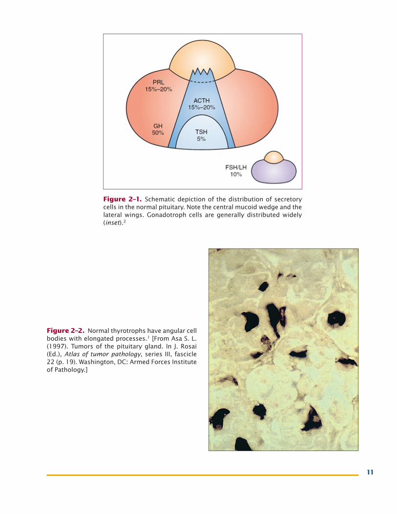
Arrangement of cells: Secretory cells of the anterior pituitary are generally zonal in distri-bution. This is most evident in horizontal sec-tions, in which the gland can be divided into a midline “mucoid wedge” and lateral “wings”2 (Figure 2–1).
thyrotroph Cells
The earliest light microscopic cell staining stud-ies differentiated all pituitary cell types by their reactions with different dyes. Trichrome stains differentiated three types of cells. The red cells were called acidophils, the blue cells were called basophils, and the colorless cells were called chro-mophobes. Thyrotrophs are basophils and stain with the PAS (periodic acid Schiff) reagent. Thy-rotrophs are one of the least common secretory cells of the pituitary gland and comprise approxi-mately 5% of the total secretory anterior pituitary cells1 (Figure 2–2). They are located mostly in the anteromedial areas of the pituitary gland. Thyro-trophs are identified by their content of very small
table 2–1. five differentiated hormone-Secreting endocrine cell types Present in the anterior Pituitary gland
Cell type (hormone)
Percent Cells
ChromosomalGene locus regulation
affected hormones effects
Somatotrophs (growth hormone) Gh
45–50 17q growth hormone– releasing hormone (ghrh)
insulin-like growth factor (igf-1) and somatostatin
linear and somatic growth. Metabolism (lipids and proteins carbohydrates)
lactotrophs (prolactin) Prl
15–25 6 thyrotropin releasing hormone (trh) and estrogen
dopamine lactation
gonadotrophs (luteinizing hormone and follicle stimulating hormone) fsh/lh
10 β-11p; β-19q gonadotropin releasing hormone (gnrh) estrogen late follicular phase of menstrual cycle
estrogen Progesterone testosterone (on fSh) inhibin
Sex steroids production. folliculogenesis and ovulation in female, spermatogenesis in male.
thyrotrophs (thyroid stimulating hormone) tsh
5 α-6q; β-1p trh t4, t3, Somatostatin
thyroid hormone production
corticotrophs (adrenocortico-tropin) aCth
15–20 2p corticotropin releasing hormone (crh)
cortisol glucocorticoid and dehydroepiandros-terone (dhea) production
11
figure 2–2. normal thyrotrophs have angular cell bodies with elongated processes.1 [from asa S. l. (1997). tumors of the pituitary gland. in J. rosai (ed.), Atlas of tumor pathology, series iii, fascicle 22 (p. 19). Washington, dc: armed forces institute of Pathology.]
figure 2–1. Schematic depiction of the distribution of secretory cells in the normal pituitary. note the central mucoid wedge and the lateral wings. gonadotroph cells are generally distributed widely (inset).2

12 Surgical and Medical ManageMent of diSeaSeS of the thyroid and Parathyroid
secretory granules (100–150 nm in diameter) and dilated profiles of rough endoplasmic reticulum. They are smaller than the other cell types and are irregularly shaped with flattened nuclei and relatively small secretory granules. They secrete TSH hormone.
TSH activity from the pituitary gland was first identified by Eduard Uhlenhuth in 1926. He showed that injecting bovine pituitary gland extract causes enlargement of thyroid gland fol-licular cells. TSH as a hormone was purified in the 1960s and found to have two subunits.3
TRH secreted by neurons in the paraventricu-lar nucleus of the hypothalamus stimulates TSH secretion from thyrotrophs.4 TSH is a glycopro-tein hormone comprising a 28-kDa heterodimer of two subunits, α and β.5 The α-subunit is com-mon to hormones like TSH, LH, FSH, and human chorionic gonadotropin (hCG), whereas the β-subunit is unique to TSH and it confers specific-ity of TSH action separately from other hormones. Production of the mature heterodimeric TSH mol-ecule requires complex co-translational glycosyl-ation and folding of nascent α- and β-subunits.6 Appropriate glycosylation is required for accurate molecular folding and subsequent combination of α- and β-subunits within the rough endoplasmic reticulum and Golgi apparatus. This TSH glycosyl-ation is regulated by both TRH and T3 in opposite directions for maintaining the negative and posi-tive feedbacks of the HPT axis. Increased TRH or low T3 level increases oligosaccharide addition to the TSH molecule.7
Disorders of thyrotrophs and tsh secretion
tsh Deficiency
TSH deficiency is most commonly seen as a part of pan-hypopituitarism. It can be defects in devel-opment of the whole pituitary gland or follow surgical removal of the gland or part of it. Pituitary damage may result in functional TSH deficiency, often without a clearly demonstrable reduction in serum TSH levels.
Congenital isolated TSH deficiency, a very rare condition, may arise from mutational defects of either the TSH hormone or TRH receptor genes. Cell differentiation in the developing pituitary gland is affected by genetic disorders of pituitary gland development. They give rise to TSH defi-ciency as a component of multiple pituitary hor-mone deficiencies. These genes with mutations include LHX3, PROP1, and POU1F1.1
Clinical features of tsh Deficiency
The consequences of TSH deficiency are similar to features of thyroid hormone deficiency, which in childhood can cause physical and mental growth retardation, while in adults it causes multiple clini-cal features of hypothyroidism, including low basal metabolic rate, weight gain, hypothermia, consti-pation, fluid retention, hair and skin changes, or frank myxedema. They will be discussed in detail in coming chapters.
assessment of the hPt axis and tsh Deficiency
Most thyrotroph disorders can be diagnosed by measuring basal TSH and thyroid hormone lev-els. TSH measurement is not helpful in diagnos-ing central hypothyroidism, which is identified by concurrent measurement of thyroid hormone levels. However, only around one-third of patients with secondary hypothyroidism have low TSH levels.8 The deficiency is usually associated with low T4 hormone levels along with low, normal, or (rarely) mildly elevated TSH levels (a similar profile may be seen in critically ill patients).
However, a TRH stimulation test may be required to effectively assess the integrity of the HPT axis.9 In this test, intravenous TRH (200–500 μg) is given and TSH levels are measured regularly at 15 minutes before administration and at 0, 15, 30, 60, and 120 minutes.
In normal persons, TSH levels rise and may peak up to 22-fold higher than basal levels after 30 minutes.10 Also after administration of T3 hor-
2. anatoMy and Pathology of thyrotroPhS 13
mone, the basal TSH levels fall, and TRH-stimu-lated TSH levels are also attenuated.
In subjects with secondary hypothyroidism due to any pituitary disease, TSH levels fail to increase in response to TRH administration.
thyrotropin-secreting Pituitary tumors11
The identification of thyrotropin-secreting pitu-itary adenomas (TSPAs) depends mainly on the presence of raised serum thyroid hormones along with increased or normal TSH levels, for which brain imaging helps sometimes. The combination of hyperthyroidism, a pituitary mass, and exces-sive TSH production demonstrated using a TSH bioassay was first described in 1960.12
They are usually very rare tumors and repre-sent only 0.6% of the adenomas found in postmor-tem cases,13 but around 0.9% to 1.5% of pituitary adenomas in surgical cases.14,15 TSH-secreting tumors, similar to the rest of the pituitary adeno-mas, are usually monoclonal in origin.16 Still, the exact mechanisms for pituitary cell transformation into adenoma remain unidentified.
A number of etiologic factors are proposed which either alone or with other of these factors interact and eventually transform and promote tumor cell proliferation. They include underex-pression of tumor suppressor genes, mostly those involved in cell cycle regulation, mutations in pitu-itary tumor-susceptibility genes, overactivation of cell-signaling pathways for proliferation, and dys-functioning of hormone-regulatory pathways.17
Pituitary thyrotroph hyperplasia in long-standing hypothyroidism
Hypothyroidism not only leads to loss of thyroid hormone feedback inhibition to TRH and TSH hypersecretion, but also leads to proliferation of TSH-secreting cells which can cause overt com-pensatory hyperplasia of thyrotrophs. Sometimes thyrotroph hyperplasia is associated with pro-
lactin cell hyperplasia and hyperprolactinemia, mostly due to sustained hypothalamic TRH stim-ulation.18 Histological staining shows the normal anterior pituitary acinar pattern, but each acini are larger, and contain many large pale cells called thyroidectomy cells. They have eccentric nuclei and abundant vacuolated cytoplasm in them; char-acteristically present in the pituitaries of patients with untreated protracted hypothyroidism and experimentally induced hypothyroid rats. These hyperplasic cells probably derive from division of pre-existing thyrotrophs as well as from differ-entiation of stem cells into mature TSH-secreting cells. Many times in addition to this, growth hor-mone and TSH bihormonal cells, which are also called thyrosomatotrophs, have been identified in similar patients or rats, supporting the hypoth-esis of transdifferentiation of somatotrophs to thyrotrophs, which leads to formation of thyroid cell hyperplasia.19 Rarely, thyrotroph hyperplasia to adenoma transformation can occur, and has been reported in a few cases with long-standing untreated hypothyroidism.20,21
Impaired thyroid hormone negative feedback
Negative feedback of T3, T4 thyroid hormones on TRH or TSH secretion can be defective and may be responsible for pathogenesis of TSH-secreting tumors. TSH levels do not suppress after admin-istration of thyroid hormones in most patients of TSPA. One possible explanation can be increase in expression or activity of deiodinase enzymes, which leads to reduced T3 hormone concentration in the adenoma.
Other causes include11:
n Hypothalamic signaling is altered by increased hypothalamic hormone stim- ulation or, alternatively, defective action of inhibitory hypothalamic hormones.
n TRH or its receptor mutations lead to abnormally increased activation of the TRH receptor or of its signal transduction pathway.

14 Surgical and Medical ManageMent of diSeaSeS of the thyroid and Parathyroid
n Dopamine (DA) receptor blockers like metoclopramide and domperidone can increase TSH concentration both in euthyroid and hypothyroid subjects.
n Loss of heterozygosity at the somatostatin SSTR5 locus has been described in one TSH secreting adenoma that was associated with unusual tumor aggressiveness and resistance to treatment with somatostatin analogues.21
n Multiple mechanisms via alterations in pituitary transcription factors like Pit-1; familial/genetic syndromes (MEN 1); multiple oncogenes, tumor suppressor genes, and growth factors; or abnormal cell signaling pathways like I3K/AKT/mTOR have been studied for their pathogenesis in TSPA.
Clinical features
TSPA generally presents with hyperthyroidism without suppression of TSH levels along with diffusely enlarged goiter. The pituitary mass can cause compression effects like those on optic chi-asma depending on size of adenoma. Co-secretion of GH or LH/FSH may be associated with TSPA and will manifest their other symptoms also.
management
With increased awareness of the disease and improvement in diagnostic techniques, these tumors are readily detected. Earlier diagnosis at initial stage of tumor growth improves the long-term prognosis. Pituitary microsurgery is the mainstay of TSPA management, providing a good chance of remission for early small size tumors, or improvement of symptoms by debulking larger tumors. Somatostatin analogues can also be used as second-line management after unsuccessful surgery or relapse, in view of their high effective-ness in controlling tumoral hypersecretion and tumor growth. Radiotherapy is usually reserved for somatostatin unresponsive cases.
references
1. Melmed S, Polonsky K, Larsen P, Kronenberg H. Williams Textbook of Endocrinology. 13th ed. Philadelphia, PA: Elsevier; 2016;212–214.
2. Scheithauer B, Kovacs K, Horvath E, Silva A, Lloyd R. Pathology of the pituitary and sellar region. In: Perry A, Brat DJ. Practical Surgical Neuropathology. Philadelphia, PA: Churchill Livingstone; 2010:371–416. doi:10.1016/b978-0- 443-06982-6.00018-3
3. Childs G. Pituitary gland (cell types, media-tors, development). In: Binder MD, Hirokawa N, Windhorst U, eds. Encyclopedia of Neurosci-ence. New York, NY: Springer; 2009:719–726. doi:10.1016/b978-008045046-9.01194-3
4. Chiamolera M, Wondisford F. Minireview: thyro-tropin releasing hormone and the thyroid hor-mone feedback mechanism. Endocrinology. 2009; 150:1091–1096.
5. Pierce JG, Parsons TF. Glycoprotein hormones: structure and function. Annu Rev Biochem. 1981; 50:465–495.
6. Grossmann M, Weintraub BD, Szkudlinski MW. Novel insights into the molecular mechanisms of human thyrotropin action: structural, physiologi-cal, and therapeutic implications for the glycopro-tein hormone family. Endocr Rev. 1997; 18:476–501.
7. Papandreou MJ, Persani L, Asteria C, et al. Vari-able carbohydrate structures of circulating thyrotropin as studied by lectin affinity chroma-tography in different clinical conditions. J Clin Endocrinol Metab. 1993;77:393–398.
8. Beck-Peccoz P, Persani L. TSH-producing adeno-mas. In: DeGroot LJ, Jameson JL, eds. Endo-crinology. 5th ed. Philadelphia, PA: Elsevier Saunders; 2006:475–484.
9. Faglia G. The clinical impact of the thyrotropin-releasing hormone test. Thyroid. 1998;8:903–908.
10. Spencer CA, Schwarzbein D, Guttler RB, et al. Thyrotropin (TSH)-releasing hormone stimula-tion test responses employing third and fourth generation TSH assays. J Clin Endocrinol Metab. 1993;76:494–498.
11. Rouach V, Greenman Y. Thyrotropin-secreting pituitary tumors. In: Melmed S, ed. The Pituitary. 3rd ed. London, UK: Academic Press; 2011:619–636. doi:10.1016/b978-0-12-380926-1.10017-3
12. Jailer J, Holub D. Remission of Graves’ disease following radiotherapy of a pituitary neoplasm. Am J Med. 1960;28:497–500.
2. anatoMy and Pathology of thyrotroPhS 15
13. Buurman H, Saeger W. Subclinical adenomas in postmortem pituitaries: classification and corre-lations to clinical data. Eur J Endocrinol. 2006; 154:753–758.
14. Davis J, Farrel W, Clayton R. Pituitary tumors. Reproduction 2001;121:363–371.
15. Saeger W, Ludecke D, Buchfelder M, Fahlbusch R, Quabbe H, Petersenn S. Pathohistological classification of pituitary tumors: 10 years of experience with the German Pituitary Tumor Registry. Eur J Endocrinol. 2007;156:203–216.
16. S. Mantovani S, Beck-Peccoz P, Saccomanno K, Spada A, Faglia G, Barbetti F. TSH-secreting pituitary adenomas are monoclonal in origin. Proceedings of the 77th Annual Meeting of the Endocrine Society. Washington DC, 1995, p. 412 (Abstract P2-485).
17. Dworakowska D, Grossman A. The pathophysi-
ology of pituitary adenomas. Best Prac Res Clin Endocrinol Metab. 2009;23:525–541.
18. Horvath E, Kovacs K, Scheithauer B. Pituitary hyperplasia. Pituitary. 1999;1(3-4):169-179.
19. Vidal S, Horvath E, Kovacs K, Cohen S, Lloyd R, Scheithauer B. Transdifferentiation of somato-trophs to thyrotrophs in the pituitary of patients with protracted primary hypothyroidism. Vir-chows Arch. 2000;436:43–51.
20. Ghannam N, Hammami M, Muttair Z, Bakheet S. Primary hypothyroidism-associated TSH-secret-ing pituitary adenoma/hyperplasia presenting as a bleeding nasal mass and extremely elevated TSH level. J Endocrinol Invest. 1999;22:419–423.
21. Ma W, Ikeda H, Watabe N, Kanno M, Yoshimoto T. A plurihormonal TSH-producing pituitary tumor of monoclonal origin in a patient with hypothy-roidism. Horm Res. 2003;59:257–261.