Embed Size (px)
Citation preview

Surgery of the Thyroid and Parathyroid Glands

Daniel Oertli · Robert Udelsman (Eds.)
Surgery of the Thyroid and Parathyroid Glands
With 235 Figures and 52 Tables
123
Editors
Daniel OertliDivision of General Surgery,University Hospital Basel,Vice-Dean of the Medical Faculty,University of Basel4031 BaselSwitzerland
Robert UdelsmanLampman Professor of Surgery and Oncology,Chairman, Department of Surgery,Surgeon-in-Chief –Yale-New Haven HospitalYale University School of MedicinePO Box 208062New Haven, CT 06520-8062USA
Library of Congress Control Number: 2005938803
ISBN-10 3-540-29165-2 Springer Berlin Heidelberg NewYork
ISBN-13 978-3-540-29165-7 Springer Berlin Heidelberg NewYork
This work is subject to copyright. All rights are reserved, whether the whole or part of the material is concerned, specifically the rights of translation, reprinting, reuse of illustrations, recitation, broadcasting, reproduction on microfilms or in any other way, and storage in data banks. Duplication of this publication or parts thereof is permitted only under the provisions of the German Copyright Law of September 9, 1965, in its current version, and permission for use must always be obtained from Springer-Verlag. Violations are liable for prosecution under the German Copyright Law.
Springer is a part of Springer Science + Business Mediaspringer.com© Springer-Verlag Berlin Heidelberg 2007
The use of general descriptive names, registered names, trademarks, etc. in this publication does not imply, even in the absence of a specific statement, that such names are exempt from the relevant protective laws and regulations and there-fore free for general use.
Editor: Gabriele M. Schröder, Heidelberg, GermanyDesk Editor: Stephanie Benko, Heidelberg, GermanyTypesetting and Production: LE-TeX Jelonek, Schmidt & Vöckler GbR, Leipzig, GermanyCover Design: estudio calamar, SpainPrinted on acid-free paper 24/3100/YL - 5 4 3 2 1 0
V
The Editors are deeply indebted to all authors and coauthors who have contributed to Surgery of the Thyroid and Parathyroid Glands. The Editors believe that this textbook is among the most comprehensive international references on surgical diseases of the thyroid and parathyroid glands. The diligent efforts of the contributors, who have provided insightful state-of-the art presentations, are gratefully acknowledged.
The Editors also wish to pay tribute to the diligent work of the Springer-Verlag staff members, who en-abled the realization of this first edition. Particularly appreciated were the efforts of Gabriele M. Schroeder, Executive Editor, and Stephanie Benko, Desk Editor, who provided strong encouragement and ongoing support during the creation of this textbook. Further-more, the Editors are most appreciative of the princi-pal artist, Mr. Jörg Kühn, who provided us with excel-lent drawings.
We also express our gratitude to the valuable coor-dinative work of our editorial assistants in Basel and New Haven: special thanks are addressed to Susan Demou, Madeleine Moser, and Dotty Franco.
Finally, our profound gratitude goes to all who were involved in the development of this text, in-cluding our immediate families, who expressed in-terest and encouragement in the completion of this textbook. We greatly appreciate their support, which stimulated us to pursue the ambitious goal of prepar-ing what we consider to be a concise, comprehensive textbook.
Daniel Oertli and Robert Udelsman
Acknowledgement
Preface
VII
Thyroid and parathyroid disorders are frequently en-countered by the endocrine surgeon in daily practice. The Editors therefore have designed this comprehen-sive textbook focusing on surgically relevant thyroid and parathyroid diseases. The Editors intend this book to become an important reference presenting the latest information regarding the management of both common and rare thyroid and parathyroid dis-orders. Internationally renowned physicians and sur-geons have provided detailed outlines and discussions on operative techniques and treatments accompanied by rationales for particular approaches advocated by the authors. The topics cover all surgically relevant thyroid and parathyroid pathologies, the preoperative surgical evaluation, decision making, and operative strategies including high quality step-by-step illustra-tions of the current surgical techniques. Furthermore, experts are presenting the molecular basis for thyroid neoplasia and the current understanding of the ge-netics of inherited thyroid and parathyroid diseases. Moreover, evolving modern operative techniques like the minimally invasive videoscopic approach to the thyroid and parathyroid glands are discussed in this textbook.
The current edition has been designed primarily to meet the requirements of young surgeons who wish to acquire profound knowledge of basic, clinical, and laboratory concepts as well as surgical techniques regarding the thyroid and parathyroid glands, thus complementing the surgeons’ training. These prin-ciples are presented together with advancements in technologic, molecular, cellular, and biologic sciences, thus meeting the criteria of the 21st century defini-tion of each subspecialty involving care of patients with thyroid and parathyroid disease. The prepara-tion of the text material has been a labor of love and represents an honest attempt to provide information that we believe is not only of clinical importance to surgeons, but also to endocrinologists, radiologists, and pathologists dealing with patients with thyroid and parathyroid disorders.
It is hoped that the reader will find the material in our textbook as helpful and exciting as we do.
Daniel Oertli and Robert Udelsman

Table of Contents
IX
1 History of Thyroid and Parathyroid Surgery . . . . . . . . . . . . . . . . . . . . . . . . . . . . . . . . . 1Hans-Dietrich Röher and Klaus-Martin Schulte
2 Embryology and Surgical Anatomy of the Thyroid and Parathyroid Glands . . . . 13William B. Stewart and Lawrence J. Rizzolo
3 Evaluation of Hyperthyroidism and Hyperthyroid Goiter . . . . . . . . . . . . . . . . 21Mirjam Christ-Crain, Nils G. Morgenthaler, and Beat Müller
4 Diagnostic Imaging of the Thyroid and Radioiodine Therapy . . . . . . . . . . . . . . . . 314.1 Walter Wiesner, Hermann Engel, and Wolfgang Steinbrich4.2 Egbert U. Nitzsche and Jan Mueller-Brand
5 Evaluation of Thyroid Nodules . . . . . . . . . . . 45Michel Procopiou and Christoph A. Meier
6 Fine-needle Aspiration Cytology of the Thyroid . . . . . . . . . . . . . . . . . . . . . . . . . . 61Anne E. Busseniers and Susan A. Silver
7 Technique of Thyroidectomy . . . . . . . . . . . . . 81Daniel Oertli
8 Surgery for the Solitary Thyroid Nodule . . 91Prabhat K. Bhama and Gerard M. Doherty
9 Modified Radical Neck Dissection . . . . . . . 101Robert Udelsman
10 Thyroid Pathology . . . . . . . . . . . . . . . . . . . . . 109Zubair W. Baloch and Virginia A. LiVolsi
11 Surgery for Medullary Thyroid Cancer . . . 147Oliver Gimm
12 Anaplastic Thyroid Carcinoma . . . . . . . . . . 161Christian Passler, Reza Asari, Christian Scheuba, and Bruno Niederle
13 Thyroid Lymphoma and Other Metastatic Lesions . . . . . . . . . . . . . . . . . . . . . . . . . . . . . . . . 171Rebecca S. Sippel and Herbert Chen
14 Multinodular and Retrosternal Goiter . . . 179Rachel Rosenthal and Daniel Oertli
15 Surgery for Hyperthyroidism . . . . . . . . . . . 191Peter E. Goretzki and Bernhard J. Lammers
16 Thyroiditis . . . . . . . . . . . . . . . . . . . . . . . . . . . . 207Michel Adamina and Daniel Oertli
17 Complications in Thyroid and Parathyroid Surgery . . . . . . . . . . . . . . . . . . . . . . . . . . . . . . . 217Andrea Frilling and Frank Weber
18 Outcomes Analysis in Thyroid Surgery: A Review of Patient and Provider Predictors . . . . . . . . . . . . . . . . . . . . . . . . . . . . . 225Kate V. Viola and Julie Ann Sosa
19 Physiology and Pathophysiology of the Parathyroid Glands and Preoperative Evaluation . . . . . . . . . . . . . . . . . . . . . . . . . . . . . 235Elizabeth H. Holt and Silvio E. Inzucchi
20 Parathyroid Imaging . . . . . . . . . . . . . . . . . . . 245David Cheng, Ludwig A. Jacob, and Leslie Scoutt
21 Conventional Surgical Management of Primary Hyperparathyroidism . . . . . . . . 261Heather Yeo, Paola Uranga, and Sanziana Roman
22 Minimally Invasive Parathyroidectomy . . 269Tobias Carling and Robert Udelsman

Table of ContentsX
23 Endoscopic Parathyroidectomy . . . . . . . . . 277Paolo Miccoli and Gabriele Materazzi
24 Multiglandular Parathyroid Disease and MEN Syndromes . . . . . . . . . . . . . . . . . . 285Peter Langer, Detlef K. Bartsch, and Matthias Rothmund
25 Pathophysiology and Treatment of Secondary and Tertiary Hyperparathyroidism . . . . . . 293Ulrich Güller and Michael Mayr
26 Parathyroid Carcinoma . . . . . . . . . . . . . . . . . 311Janice L. Pasieka and Moosa Khalil
27 Reoperative Parathyroid Surgery . . . . . . . . 327Cord Sturgeon, Nadine Caron, and Quan-Yang Duh
28 Outcomes Analysis in Parathyroid Surgery . . . . . . . . . . . . . . . . . . . . . . . . . . . . . . . 339Leon D. Boudourakis and Julie Ann Sosa
Subject Index . . . . . . . . . . . . . . . . . . . . . . . . . . . . . 349
List of Contributors
XI
Michel AdaminaDivision of General Surgery, University Hospital Basel, Spitalstrasse 21, 4031 Basel, Switzerland
Reza AsariDivision of Endocrine Surgery, Department of Surgery, Vienna University Hospital, Vienna, Austria
Zubair W. BalochDepartment of Pathology and Laboratory Medicine, University of Pennsylvania Medical Center, Philadelphia, PA, USA
Detlef K. BartschDepartment of Surgery, Philipps-University, Marburg, Germany
Prabhat K. BhamaDivision of Endocrine Surgery, University of Michigan Medical School, Ann Arbor, MI, USA
Leon D. BoudourakisYale University School of Medicine, New Haven, CT, USA
Anne E. BusseniersMetropolitan FNA Service, Washington, DC; Bethesda, MD, USAandDepartment of Pathology, Vrije Universiteit Brussels, Brussels,Belgium
Tobias CarlingDepartment of Surgery, Yale University School of Medicine, New Haven, CT,USA
Nadine CaronDepartment of Surgery, University of California San Francisco and UCSF Comprehensive Cancer Center at Mt Zion Medical Center, San Francisco, CA, USA
Herbert ChenDepartment of Surgery, University of Wisconsin, H4/750 Clinical Science Center, 600 Highland Avenue, Madison, WI, USA
David W. ChengDepartment of Diagnostic Radiology, Yale University School of Medicine, New Haven, CT, USA
List of ContributorsXII
Mirjam Christ-CrainDepartment of Endocrinology, John Vane Science Centre, Barts and the London Medical School,Charterhouse Square,London, UK
Gerard M. DohertyDivision of Endocrine Surgery, University of Michigan Medical School, Ann Arbor, MI; 2920B Taubman Health Care Center, Box 0331, 1500 E. Medical Center Drive, Ann Arbor, MI, USA
Quan-Yang DuhDepartment of Surgery, University of California San Francisco and Veterans Affairs Medical Center, San Francisco, CA, USA
Hermann EngelInstitute of Radiology and Nuclear Medicine, Hospital Waid, Zürich, Switzerland
Andrea FrillingDepartment of General Surgery, Visceral surgery and Transplantation, University Hospital Essen, Essen, Germany
Oliver GimmDepartment of General, Visceral and Vascular Surgery, University of Halle, Ernst-Grube-Strasse 40, 06097 Halle, Germany
Peter E. GoretzkiDepartment of Surgery, Lukaskrankenhaus of the Heinrich-Heine University Düsseldorf, Neuss, Germany
Ulrich GüllerDepartment of Surgery, Divisions of General Surgery and Surgical Research, University of Basel, Basel, Switzerland
Elizabeth H. HoltSection of Endocrinology, Yale University School of Medicine, New Haven, CT, USA
Silvio E. InzucchiSection of Endocrinology, Yale University School of Medicine, New Haven, CT, USA
Ludwig A. JacobSection of Interventional Radiology, Department of Radiology, University Hospital Basel, Basel,Switzerland
Mo sa KhalilUniversity of Calgary, Pathologist, Calgary Laboratory Services, Calgary, Alberta, Canada
Bernhard J. LammersDepartment of Surgery, Lukaskrankenhaus of the Heinrich-Heine University Düsseldorf, Neuss, Germany
Peter LangerDepartment of Surgery, Philipps-University Hospital, Baldingerstrasse, D-35043 Marburg, Germany
Virginia A. LiVolsiDepartment of Pathology and Laboratory Medicine, University of Pennsylvania Medical Center, Philadelphia, PA, USA
o
XIIIList of Contributors
Gabriele MaterazziDepartment of Surgery, Azienda Ospedale Università di Pisa, Pisa,Italy
Michael MayrDivision of Transplantation Immunology and Nephrology, Department of Internal Medicine, University of Basel, Basel, Switzerland
Christoph A. MeierEndocrine Unit, Division of Endocrinology, Diabetes and Nutrition, Department of Internal Medicine, University Hospital Geneva, CH-1211 Geneva 14, Switzerland
Paolo MiccoliDepartment of Surgery, Azienda Ospedale Università di Pisa, Pisa,Italy
Nils G. MorgenthalerInstitut für Experimentelle Endokrinologie,Charité Campus Mitte,Universitätsmedizin Berlin,Berlin,Germany
Jan Mueller-BrandUniversity of Basel, School of Medicine, Basel; Division of Nuclear Medicine and PET Center, University Hospital Basel,Basel, Switzerland
Beat MüllerDept. of Endocrinology,Diabetology and Clinical Nutrition,University Hospital,Petersgraben 4,Basel,Switzerland
Bruno NiederleDivision of Endocrine Surgery, Department of Surgery, Medical University of Vienna, Währingergürtel 18–20, A-1090 Vienna, Austria
Egbert U. NitzscheUniversity of Basel, School of Medicine, Basel; Division of Nuclear Medicine and PET Center, Aarau General (Cantonal) Hospital, Aarau, Switzerland
Daniel OertliDivision of General Surgery, University Hospital Basel, Vice-Dean of the Medical Faculty, University of Basel, Basel,Switzerland
Janice L. PasiekaDivision of General Surgery, Department of Surgery, Faculty of Medicine, University of Calgary, Calgary, Alberta, Canada
Christian PasslerDivision of Endocrine Surgery, Department of Surgery, Vienny University Hospital, Vienna, Austria
Michel ProcopiouDivision of Endocrinology, Diabetes and Nutrition, Department of Internal Medicine, University Hospital Geneva, CH-1211 Geneva 14, Switzerland
Lawrence J. RizzoloSection of Anatomy, Department of Surgery, Yale University School of Medicine, PO Box 208062, New Haven, CT 06520-8062, USA

List of ContributorsXIV
Hans-Dietrich RöherKlinik für Allgemein und Unfallchirurgie, Universitätsklinikum Duesseldorf, Moorenstrasse 5, 40225 Düsseldorf, Germany
Sanziana Alina RomanSection of Endocrine Surgery,Department of Surgery, Yale University School of Medicine, PO Box 208062, New Haven, CT 06520,USA
Rachel RosenthalDivision of General Surgery, Department of Surgery, University Hospital Basel, Spitalstrasse 21, CH-4031 Basel, Switzerland
Matthias RothmundDepartment of Surgery, Philipps-University, Marburg, Germany
Christian ScheubaDivision of Endocrine Surgery, Department of Surgery, Vienny University Hospital, Vienna, Austria
Klaus-Martin SchulteDepartment of Endocrine and General Surgery, King’s College Hospital, Denmark Hill, London SE5 9RS, UK
Leslie M. ScouttDepartment of Diagnostic Radiology, Yale University School of Medicine, New Haven, ConnecticutUSA
Susan A. SilverMetropolitan FNA Service, Bethesda, Washington, DC, USA
Rebecca S. SippelDepartment of Surgery, University of Wisconsin, H4/710 Clinical Science Center, 600 Highland Avenue, Madison, WI, USA
Julie Ann SosaSections of Oncologic, Endocrine, and General Surgery, Yale University School of Medicine, 330 Cedar Street, New Haven, CT 06520-8062, USA
Wolfgang SteinbrichInstitute of Diagnostic Radiology, University Hospital Basel, Petersgraben 4, 4031 Basel, Basel,Switzerland
William B. StewartDepartment of Surgery, Yale University School of Medicine, PO Box 208062, New Haven, CT 06520-8062, USA
Cord SturgeonDepartment of Surgery, Northwestern University Feinberg School of Medicine, Division of Gastrointestinal and Endocrine Surgery, Chicago, IL, USA
Robert UdelsmanDepartment of Surgery, Yale University School of Medicine, PO Box 208062, New Haven, CT 06520-8062, USA
Ana Paola UrangaDepartment of Surgery, Yale University School of Medicine, 330 Cedar Street FMB 102, New Haven, CT 06520, USA

XVList of Contributors
Kate V. ViolaSections of Oncologic, Endocrine, and General Surgery, Yale University School of Medicine, 330 Cedar Street, New Haven, CT 06520-8062, USA
Frank WeberDepartment of General Surgery, Visceral Surgery and Transplantation, University Hospital Essen, Essen,Germany
Walter WiesnerMedical Radiology Center, Klinik Stephanshorn, Brauerstrasse 95, 9016 St. Gallen, Switzerland
Heather YeoDepartment of Surgery, Yale University School of Medicine, 330 Cedar Street FMB 102, New Haven, CT 06520-8062, USA

1
1.1 Introduction
Endocrine surgery is the concept and practice of sur-gically applied human physiology. This concept has deep historical roots and has been nourished by many thoughts, ideas, and discoveries. Several individuals influenced this interesting field primarily provid-ing answers to human physiology and secondarily to questions on surgical technique. Indeed, the proper function of the thyroid as an endocrine gland was discovered by a surgeon. Moreover, surgeons pio-neered the recognition of parathyroid disease. Unlike in any other field, operative principles in endocrine surgery have been developed after recognition of the homeostatic regulations of different endocrine organ systems. Hence interest in molecular biology resulted in a profound understanding of the endocrine con-trol of cellular homeostasis. This resulted in the first entirely gene-directed surgical procedure in man, the prophylactic thyroidectomy in multiple endocrine syndrome 2A. Historically, endocrine surgery repre-
sents a major pathway for the import and export of ideas, a trafficking place, where anatomy, physiology, biological and analytical chemistry, imaging, and sur-gical technique have ideally met and influenced each other.
1.2 Recognition of Goitrous Disease
In the first and second centuries, Celsus and Galen described cervical masses such as cysts, tuberculous lymph nodes (scrofula), and goiters. In the eleventh century, the Arabic scientist Abulkasim, working in Cordoba, Spain, differentiated natural, endemic goiter from non-natural goiter [17]. Between the eleventh and the mid-thirteenth century, the renowned medi-cal school of Salerno near Naples, Italy, gave advice for conservative treatment of goiter disease using sea products such as burned sponge. On the other hand, the school recommended cauterization or seton im-plants in order to induce necrotizing inflammation and tissue destruction for selected cases [9]. In these times, nothing was really known about the organic source of goiter formation.
1.3 Anatomy of the Thyroid Gland
In 1543, the anatomist Andrea Vesalius (1514–1564; Fig. 1.1) originally described “glandulae laryngis” in his inauguration of modern anatomy De Humani Corporis Fabrica published in Basel, Switzerland. The first distinct image of the thyroid gland with the typi-cal horseshoe shape dates back to the work of Julius Casserius (1545–1616) [23]. Casserius was a servant of Hieronymus Fabricius of Aquapendente, Professor and Chairman of Anatomy and Surgery at the Uni-versity of Padua between 1609 and 1616. The first uti-lization of the term “glandula thyreoidea” was made by Thomas Wharton, London, UK (1656) and by Al-brecht von Haller, Göttingen, Germany. They attrib-uted endocrine secretory properties to this ductless gland.
1 History of Thyroid and Parathyroid Surgery
Hans-Dietrich Röher and Klaus-Martin Schulte
Contents
1.1 Introduction . . . 11.2 Recognition of Goitrous Disease . . . 11.3 Anatomy of the Thyroid Gland . . . 11.4 Early Attempts at Surgery . . . 21.5 Thyroid Physiology . . . 21.6 Morbidity of Thyroid Surgery During
the Nineteenth Century . . . 31.7 Relevance of Postoperative Loss
of Thyroid Function . . . 31.8 Surgery for Hyperthyroidism . . . 41.9 Thyroid Cancer Surgery . . . 51.10 Thyroid Surgery in Modern Times . . . 51.11 Discovery of the Parathyroid Glands . . . 61.12 Parathyroid Preservation . . . 71.13 Tetany and Hypoparathyroidism . . . 71.14 Hyperparathyroidism
and Parathyroidectomy . . . 81.15 Different Forms of Hyperparathyroidism . . . 10
References . . . 10

Hans-Dietrich Röher and Klaus-Martin Schulte2
1.4 Early Attempts at Surgery
Over several following centuries, written sources documented that attempts were undertaken for surgi-cal removal of goiters. The indication for surgery was primarily given by extensive cervical masses or severe tracheal obstruction resulting in dyspnea. However, the operative results were disastrous with extremely high mortality because of fatal bleeding or infection.
In 1742, Lorenz Heister (1683–1758; Fig. 1.2), who had founded scientific surgery in Germany, first de-scribed the surgical removal of a thyroid gland. While describing thyroid colloid substance in 1754, he was among the first who acknowledged the practical value of the understanding of endocrine substances. Heis-ter already knew that goiters can turn malignant, as he thought, after they had been treated with acid substances. He also recognized the erosion of neck vessels by malignant goiters and gave attention to thyroid cancer. In 1792, the French surgeon Pierre Jo-seph Desault (1744–1795) demonstrated that a partial thyroid resection may be feasible and thereby opened the proper way into thyroid surgery at a time when hormone replacement was not even theoretically known [10].
Following this, Johann August Wilhelm Hedenus (1760–1836) operated on six patients who had suf-fered from airway obstruction by giant goiters [24].
Thyroid surgery remained a life-threatening proce-dure during these times even in the hands of the most skilled surgeons. The mainly fatal outcome after goiter surgery motivated the leading German surgeon Jo-hann Dieffenbach from Berlin to make the statement in 1848 that “goiter surgery is one of the most thank-less, most perilous undertakings.” Also the French Academy of Medicine criticized any thyroid opera-tion in 1850, and Bernhard Rudolph Conrad Langen-beck (1810–1887) vehemently warned to avoid it.
1.5 Thyroid Physiology
Surgeons provided the first substantial evidence of endocrine secretion by the thyroid gland. Thomas Wilkinson King (1809–1847) from London wrote in his observations on the thyroid gland:
The most important novel fact concerning the thy-roid gland is doubtless this, that its absorbent vessels carry its peculiar secretion to the great veins of the body and the most simple and satisfactory method of demonstrating this fact is to expel the contents of the healthy gland by repeated and gentle compressions, into the lymphatics of the surface and then to coagulate the fluid on the surface… Whilst the nourishment of a part is indispensable to its existence, the influence which it exerts upon the circulating fluids may be more or less
Fig. 1.1 Andreas Vesalius (1514–1564) was a Belgian anato-mist and physician whose dissections of the human body and descriptions of his findings helped to correct misconceptions prevailing since ancient times. As Professor of Anatomy in Padua (Italy) Vesalius wrote the revolutionary texts De Humani Corporis Fabrica, seven illustrated volumes on the structure of the human body
Fig. 1.2 Lorenz Heister (1683–1758) served as an army sur-geon in several German campaigns before becoming Professor of Anatomy and Surgery at Altdorf. Distressed at the inferior state of surgery he published his Chirurgie (Nuremberg, 1718)
31 History of Thyroid and Parathyroid Surgery
needful for the healthful subsistence of the entire ani-mal [27].
In the same volume, Sir Astley Cooper (1768–1841) had included a report on his experimental thyroidectomies in dogs. In 1827 he observed how slowly the dogs recovered after a period of stupor and tiredness. In 1859 the German physiologist Moritz Schiff (1823–1896) published his experiences of total thyroidectomy and showed that all thyroidectomized dogs and guinea pigs eventually died postoperatively. Some simultaneously appearing treatises on the coin-cidence of goiter and cretinism should have rendered obvious the nature of an “endocrine organ,” but these observations remained without general practical con-sequences. Because this seemed so ridiculous to his contemporaries, Sir Felix Semon (1849–1921) was unable to publish his hypothesis in 1883 that myx-edema and cretinism were caused by the loss of thy-roid function.
1.6 Morbidity of Thyroid Surgery During the Nineteenth Century
Intra- and postoperative bleeding was the major prob-lem in thyroid surgery of those days and surgeons had just learned how to tackle it. William Blizzard (1743–1835) tried to cure thyroid overfunction by ligation of the upper pole arteries. Luigi Porta (1800–1875) contributed in two major respects. First he performed a targeted adenoma excision in 1849. Second, his at-tempt to cure thyroid hyperfunction by unilateral arterial ligation failed and therefore Porta concluded that bilateral ligations would be necessary. Edmund Rose (1836–1914) followed up this idea. After suc-ceeding Theodor Billroth as the Surgical Chairman in Zurich, Switzerland, Rose published his treaty on Der Kropftod und die Radikalkur der Kröpfe (death by goi-ter and radical cure of goiters). He stated that goitrous recurrence could only be prevented by a complete re-moval of the gland. For this he described meticulous ligation of every single vessel feeding the gland whilst respecting the recurrent laryngeal nerves, the vagal nerves, as well as the hypoglossal nerves.
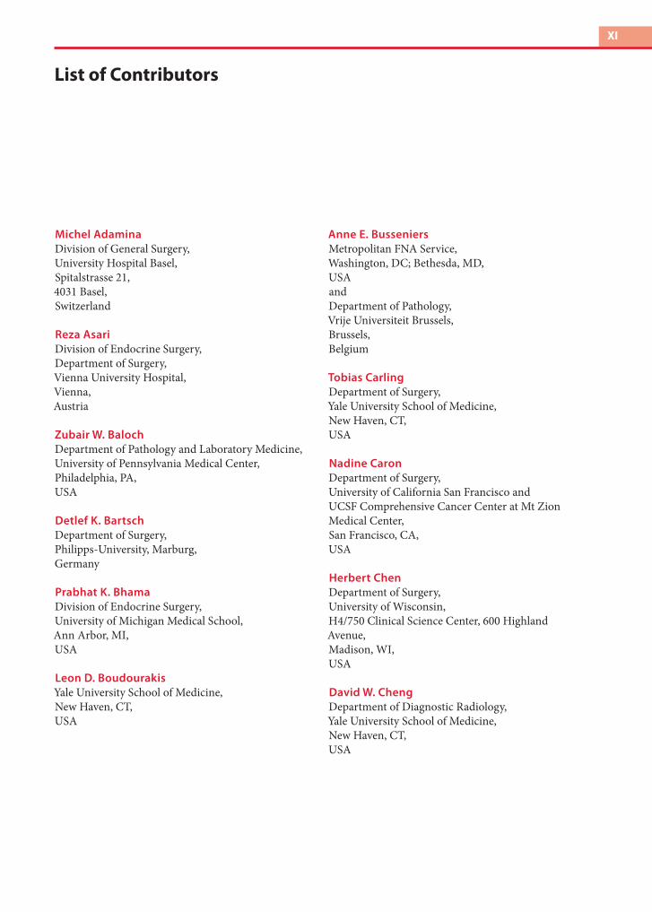
The main problems during the nineteenth century were bleeding and infection. Being among the leading surgical experts in Europe, Theodor Billroth (1829–1894; Fig. 1.3) reported an intraoperative mortality of 36% for thyroid surgery. During the second half of the nineteenth century, three key factors largely con-tributed to the development and progress of thyroid surgery. First, William Morton from Boston, USA, in-vented inhalation anesthesia in 1846. Second, in 1867, Lord Joseph Lister from Glasgow, UK, introduced the
principles of antisepsis [30]. Third, in 1874, Thomas Spencer Welles developed “hemostatic forceps” for surgery. These new developments enabled surgeons to substantially refine their operating technique.
This pioneering era in thyroid surgery was domi-nated by Theodor Billroth, who worked in Zurich, Switzerland, between 1860 and 1867 and later on in Vienna, Austria, and by his disciple Anton Wölfler (1850–1917). Their efforts targeted at complete re-moval of the thyroid gland in order to facilitate in-traoperative hemostasis after describing techniques using ligation of the arteries [57].
In 1879, Claude Bernard stated that “We do not know anything about the use of these organs (i.e. thyroid, thymus), we don’t even have an idea about their utility and importance...” Of note, this statement coincides with the first thyroidectomy for a goiter as-sociated with exophthalmus performed by Ludwig Rehn (1849–1930).
1.7 Relevance of Postoperative Loss of Thyroid Function
The discovery of the importance of human thyroid function dates back to 1882 and should be attributed to the surgeon Jacques-Louis Reverdin (1842–1929;
Fig. 1.3 Theodor Billroth (1829–1894) obtained his medical degree from the University in Berlin in 1852 and became as-sistant to Bernhard von Langenbeck in 1854. He was appointed Professor of Clinical Surgery in Zurich in 1860 and Professor of Surgery at the University of Vienna in 1867. Courtesy of Prof. U. Boschung, Institute of Medicine History, University of Berne, Switzerland

Hans-Dietrich Röher and Klaus-Martin Schulte4
Fig. 1.4), and to his cousin Auguste Reverdin (1848–1908). They called this postoperative state “myxo-edeme opératoire.” A letter of J.-L. Reverdin to The-odor Kocher in 1882 described a case of cretinism following thyroidectomy. In 1883, Reverdin pub-lished on the thyroprivic consequences of 22 thy-roidectomies. Thereafter, he vehemently advocated to avoid thyroidectomy and omitted this operation [47]. Theodor Kocher (1841–1917; Fig. 1.5), however, resolutely pursued the issue that had been raised by the Reverdins. This culminated in his historic manu-script Ueber Kropfexstirpation und ihre Folgen (on the removal of goiter and its consequences) [28]. For his work, Kocher was later awarded the Nobel prize for physiology or medicine in 1909.
Starting from the personal case of the eleven-year-old Maria Bichsel in 1874, Kocher developed the concept of “kachexia thyreopriva” that summarizes the various consequences for the entire body due to the lack of thyroid hormone function. After 1883, Kocher strongly promoted the use of the unilateral operation to avoid the thyroprivic state. This concept found worldwide acceptance and was transferred to the United States by Charles Mayo, the pioneer of en-docrine surgery in the new world [36–38].
In parallel one of Billroth’s disciples Johann v. Mi-kulicz-Radecki (1850–1905) recognized the problems of too extensive thyroid surgery. He replaced the complete thyroidectomy by a bilateral partial resec-tion in order to reduce the potential local harm to the parathyroids (whose function was only partially un-derstood) and to the laryngeal nerves. The remnant would take over the thyroid function. Whereas the
understanding of the consequence of complete loss of thyroid function can be attributed to the Reverdins and to Kocher, the recognition of postoperative tet-any was made by Billroth and his pupils Wölfler and von Eiselsberg. They recognized the association be-tween parathyroprivic symptoms and the loss of the parathyroid glands.
1.8 Surgery for Hyperthyroidism
Thyroid operations were initially performed to solve mechanical airway obstruction. Attention to hyper-thyroidism was paid much later in time. Caleb Hillier Parry first described an “exophthalmic goiter” [43]. In the English and the German literature, the clini-cal complex of autoimmune hyperthyroidism was de-scribed by Robert James Graves (Fig. 1.6) and by Carl Adolf von Basedow (Fig. 1.7), respectively [1,15]. In 1884, Ludwig Rehn (1849–1930) from Frankfurt, Germany, opened the way for surgical cure of Graves’ disease [46]. Mikulicz expanded the indication for surgery by “exophthalmic goitre.” Initially, many of these patients were treated with a unilateral approach, resulting in recurrences.
During the first two decades of the twentieth cen-tury, the unilateral operation for thyroid toxicosis was still the mainstay in the United States. In many in-stances a multistage procedure was performed. After
Fig. 1.4 Jacques-Louis Reverdin (1842–1929), Professor of Surgery, Geneva, Switzerland. Courtesy of Prof. U. Boschung, Institute of Medicine History, University of Berne, Switzerland Fig. 1.5 Theodor Kocher (1841–1917), Professor of Surgery. In
1872, he became Chairman at the University Hospital in Berne and remained in this post in spite of several invitations to for-eign universities. Courtesy of Prof. U. Boschung, Institute of Medicine History, University of Berne, Switzerland

51 History of Thyroid and Parathyroid Surgery
initial occlusion of the thyroid arteries, a lobectomy was done in a second operation. A third interven-tion was often indicated if the disease persisted or re-curred. F. Hartley (New York, USA, 1905) and Thomas Dunhill (Melbourne, Australia, 1907) changed this paradigm [11,22]. They showed that hyperthyroidism can be cured by unilateral lobectomy and contralat-eral subtotal resection. Dunhill also surmounted the contraindications for surgery in the presence of car-diac symptoms. He demonstrated that surgery may be successful even in patients with tachyarrhythmia and cardiac failure. The reports on his successes were first doubted and ignored, and Dunhill decided to travel from Australia to the United Kingdom and the United States. In the United States, his technique and his indications were well accepted, although without being quoted by the leading surgeons. This may be the reason why both Dunhill and Hartley were ignored until their preferred operation was elevated to the standard surgical technique for hyperthyroidism. An-other milestone was made by Charles Mayo, Henry Plummer, and Walter Boothby from the Mayo Clinic in the 1920s. They showed that the risks and severity of perioperative thyroid storm were greatly reduced by preoperative peroral administration of large doses of iodine using the Wolff-Chaikoff effect [44,45,55]. Perioperative mortality in 600 patients was reduced from 5% to less than 1%. The introduction of radioio-dine ablation therapy in 1942 and of thyrostatic drugs in 1943 dramatically changed the treatment patterns in hyperthyroidism and almost replaced surgery for a while.
During the 1950s the repertoire of diagnostic tools expanded. By detection of stimulating antibodies, the immunogenic nature of hyperthyroidism can now be proven and classified. The preoperative therapy using
high-dose iodine according to Plummer has been re-placed by thyrostatic drug therapy that allows for a fine hormonal tuning and for timing of surgery. Mod-ern surgical treatment of hyperthyroidism consists of a differentiated approach with either near-total or to-tal thyroidectomy for immunogenic hyperthyroidism and with lobectomy for toxic adenomas.
1.9 Thyroid Cancer Surgery
Histopathology has emerged as the preeminent tool for the classification of thyroid cancers. This led to the recognition of the biologically different behavior of different cancer subtypes and to the introduction of differentiated surgical strategies. Another major step is the, still incomplete, acceptance of prognostic scoring systems including tumor, node, metastases (TNM), European Organization for Research and Treatment of Cancer (EORTC), Age, Grade, Extent, Size (AGES), Age, Metastases, Extent, Size (AMES), and the Metastases, Age, Completeness of resection, Invasion, Size (MACIS). All these classifications al-low the comparison of treatment results in cohorts from different continents treated with a broad range of regimens.
1.10 Thyroid Surgery in Modern Times
Since 1980 molecular medicine has provided major in-sights into the impact of genetic mutations eventually
Fig. 1.7 Carl Adolf von Basedow (1799–1854)
Fig. 1.6 Robert James Graves (1796–1853)
Hans-Dietrich Röher and Klaus-Martin Schulte6
leading to thyroid tumors. This has not only yielded a further possibility of tumor classification accord-ing to their specific genetic changes, but it has also opened the path to the recognition of specific cancer predispositions in individual family members. Since mutations of the RET protooncogene can be detected in peripheral blood lymphocytes, prophylactic thy-roidectomy can be cancer preventive. Presymptom-atic thyroidectomy in individuals with the mutated RET protooncogene represents the first indication in the history of surgery which is fully and solely based on the genetic proof of a malignant trait, i.e., medul-lary thyroid cancer. Genetic research has also proven the unifying causative changes in familiar syndromes, such as multiple endocrine neoplasia type I and II [53,54] and diseases caused by changes in the succi-nate dehydrogenase complex. The lively interchange between basic molecular research and clinical prac-tice has revolutionized endocrine surgical practice.
The recent years represent an industrious period of relentless technical research and improvement in endocrine surgery. This has reduced the operative risks with regard to all forms of complications. Bleed-ing is now rare, and most thyroid surgery can safely be performed without the need of drainage. Infection has virtually disappeared. Due to vigorous protection of the anatomical structures and consequent identi-fication of the recurrent laryngeal nerve, permanent nerve palsies occur in less than 1% of cases. Hypo-parathyroidism has been reduced to frequencies be-low 1% due to meticulous preparation techniques and the generous use of parathyroid autotransplantation. Mortality is almost nil.
The most recent enrichments of the surgical reper-toire are the targeted, minimally invasive, and endo-scopic approaches. The evolutionary progress of tech-niques and indications is underway. A more secure estimation of the safety of these techniques needs fur-ther consolidation of data and experiences.
1.11 Discovery of the Parathyroid Glands
Small things often go unnoticed. It is no surprise that this holds true for the parathyroid glands that even today may sometimes be difficult to retrieve. Al-though Thomas Wharton gave a detailed report on the “glandulae thyroideae” in 1656, he did not men-tion the parathyroid glands.
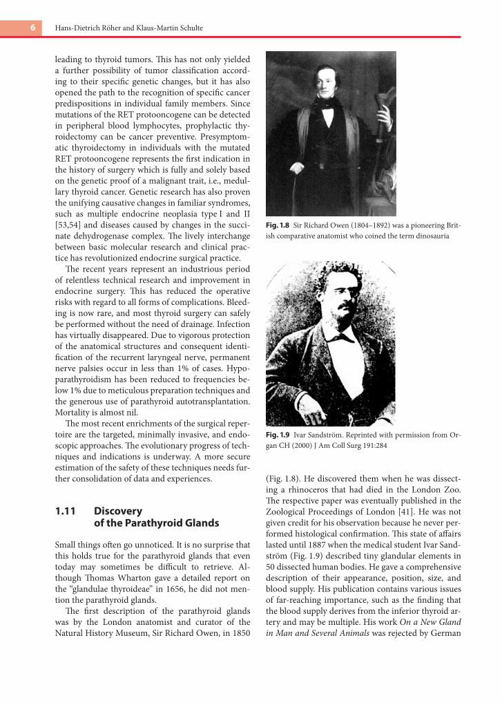
The first description of the parathyroid glands was by the London anatomist and curator of the Natural History Museum, Sir Richard Owen, in 1850
(Fig. 1.8). He discovered them when he was dissect-ing a rhinoceros that had died in the London Zoo. The respective paper was eventually published in the Zoological Proceedings of London [41]. He was not given credit for his observation because he never per-formed histological confirmation. This state of affairs lasted until 1887 when the medical student Ivar Sand-ström (Fig. 1.9) described tiny glandular elements in 50 dissected human bodies. He gave a comprehensive description of their appearance, position, size, and blood supply. His publication contains various issues of far-reaching importance, such as the finding that the blood supply derives from the inferior thyroid ar-tery and may be multiple. His work On a New Gland in Man and Several Animals was rejected by German
Fig. 1.8 Sir Richard Owen (1804–1892) was a pioneering Brit-ish comparative anatomist who coined the term dinosauria
Fig. 1.9 Ivar Sandström. Reprinted with permission from Or-gan CH (2000) J Am Coll Surg 191:284

71 History of Thyroid and Parathyroid Surgery
editors and eventually published in Swedish in the Uppsala Medical Journal [48]. This may have contrib-uted to the lack of recognition of this brilliant young man who later committed suicide.
1.12 Parathyroid Preservation
Sandström’s detailed dissection of the parathyroids and their blood supply were consolidated by the me-ticulous work of Herbert M. Evans, Johns Hopkins, Baltimore, who identified the variations of blood vessels and thereby heralded the protection of glan-dular function by maintenance of their blood supply. His mentor, William Halsted (Fig. 1.10) immediately derived the correct instinctive conclusion, that the thyroid artery should be ligated proximal to the thy-roid gland while sparing the parathyroid end arter-ies [20].
Kocher and Billroth were the two exponents of thyroid surgery at the end of the nineteenth century. Each had founded a surgical school—Kocher in Bern and Billroth in Vienna—and the respective postop-erative outcomes reflected the particular techniques utilized in each school. Kocher experienced the symp-toms associated with radical removal of the thyroid, leading to postoperative hypothyroidism “cachexia strumipriva.” Billroth’s patients experienced tetany. Halsted gives another example of his surgical instinct when he associated these differences to the characters of operating surgeons:
Kocher, neat and precise, operating in a relatively bloodless manner, scrupulously removed the entire thy-roid gland, doing little damage outside the capsule. Bill-roth, operating more rapidly, and as I recall his manner, with less regard for tissues and less concern for hemor-rhage, might easily have removed the parathyroids or at least interfered with their blood supply, and have left remnants of the thyroid [19].
This description still is of enormous value since it teaches us about some of the virtues needed for suc-cessful endocrine surgery.
Billroth’s pupils discovered the symptom complex of postoperative hypocalcaemia. Anton Wölfler gave a full and detailed account of tetany in the first patient who had undergone a total thyroidectomy by Theodor Billroth [56]. The patient recovered after having expe-rienced the full range of symptoms over a period of three weeks. Nathan Weiss collected more data from patients with postoperative tetany [51]. These experi-ences stimulated Mikulicz to develop his technique of protection of the posterior thyroid capsule. Surgical knowledge about the parathyroids emerged from sur-
gical complications and preceded the discovery of the parathyroid function.
1.13 Tetany and Hypoparathyroidism
In 1891 the French physiologist Eugene Gley clarified the relation between parathyroid gland function and tetany [14]. He described tetany in rats and rabbits as a consequence of the removal of the thyroid and para-thyroid glands. Moreover, he could show that removal of the parathyroids alone would have the same effect. The concept of parathyroid transplantation was born.
The first parathyroid autotransplantation was per-formed in 1892 by Anton von Eiselsberg (Fig. 1.11),Vienna, Austria. He transplanted thyroid and para-thyroid tissue into the preperitoneal space of cats and showed that tetany was absent and new vessels had formed at the transplants. In contrast, tetany occurred after these transplant were removed [12].
Also William J. MacCallum at Johns Hopkins, Bal-timore, described the use of parathyroid extracts to cure tetany in experimental animals [31,32]. He trans-ferred upcoming knowledge about the role of calcium in nerve conduction and muscle action and formed a hypothesis that the parathyroid glands may play a role in calcium metabolism. This ingenious conclu-sion was later proven in experiments by Carl Voegtlin and it was shown that tetany caused by parathyroid-
Fig. 1.10 William Stewart Halsted (1852–1922) was a true sur-gical innovator. Halsted revolutionized surgery by insisting on skill and technique rather than brute strength. Using an experi-mental approach, he developed new operations for intestinal and stomach surgery, gallstone removal, hernia repair, and dis-orders of the thyroid gland. He first practiced in New York and in 1886 became the first Professor of Surgery at Johns Hopkins
Hans-Dietrich Röher and Klaus-Martin Schulte8
ectomy could be corrected with parathyroid extract or by injections of calcium [33]. This was a major advance, although MacCallum remained uncertain about the value of his own discoveries for another decade. In 1907, William Halsted at Johns Hopkins used parathyroid extract and calcium chloride to treat postoperative tetany [18]. He reported on the cure of “hypoparathyrosis” by parathyroid transplantation. However, the problems with parathyroid extracts were the difficulty of their production, the lack of stability, and the variability of biological activity. Adolf Hansen developed a method for hormone extraction from bo-vine parathyroid glands. In animal experiments, these extracts were able to cure tetany and raise the serum calcium of parathyroprivic dogs. They also induced osteoporosis after administration over a prolonged period [21]. These findings were substantiated with detailed experiments conducted by James P. Collip [6,7]. An immunoassay for parathyroid hormone de-tection in peripheral blood was developed by Yalow and Berson [4,58]. In 1977 the DNA sequence of the gene for parathyroid hormone was identified [3] and the respective cDNA was cloned in 1981 [25]. Today, human recombinant parathyroid hormone is avail-able for treatment of hypoparathyroidism.
1.14 Hyperparathyroidism and Parathyroidectomy
After parathormone (PTH) deficiency was recog-nized and could be treated during the first decade
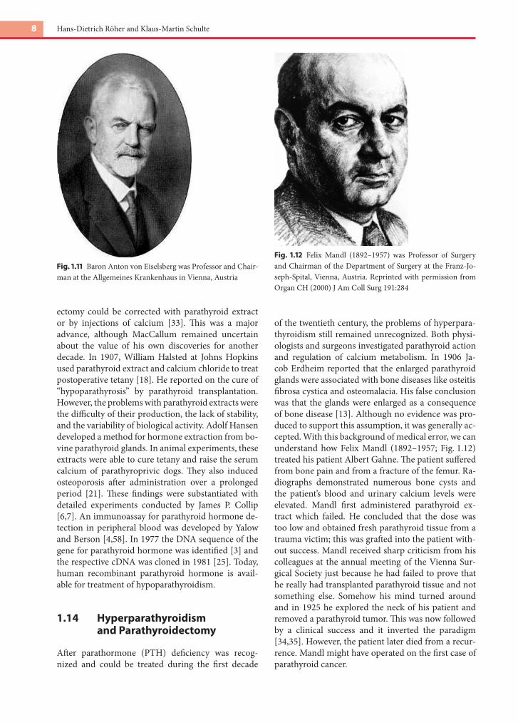
of the twentieth century, the problems of hyperpara-thyroidism still remained unrecognized. Both physi-ologists and surgeons investigated parathyroid action and regulation of calcium metabolism. In 1906 Ja-cob Erdheim reported that the enlarged parathyroid glands were associated with bone diseases like osteitis fibrosa cystica and osteomalacia. His false conclusion was that the glands were enlarged as a consequence of bone disease [13]. Although no evidence was pro-duced to support this assumption, it was generally ac-cepted. With this background of medical error, we can understand how Felix Mandl (1892–1957; Fig. 1.12)treated his patient Albert Gahne. The patient suffered from bone pain and from a fracture of the femur. Ra-diographs demonstrated numerous bone cysts and the patient’s blood and urinary calcium levels were elevated. Mandl first administered parathyroid ex-tract which failed. He concluded that the dose was too low and obtained fresh parathyroid tissue from a trauma victim; this was grafted into the patient with-out success. Mandl received sharp criticism from his colleagues at the annual meeting of the Vienna Sur-gical Society just because he had failed to prove that he really had transplanted parathyroid tissue and not something else. Somehow his mind turned around and in 1925 he explored the neck of his patient and removed a parathyroid tumor. This was now followed by a clinical success and it inverted the paradigm [34,35]. However, the patient later died from a recur-rence. Mandl might have operated on the first case of parathyroid cancer.
Fig. 1.11 Baron Anton von Eiselsberg was Professor and Chair-man at the Allgemeines Krankenhaus in Vienna, Austria
Fig. 1.12 Felix Mandl (1892–1957) was Professor of Surgery and Chairman of the Department of Surgery at the Franz-Jo-seph-Spital, Vienna, Austria. Reprinted with permission from Organ CH (2000) J Am Coll Surg 191:284

91 History of Thyroid and Parathyroid Surgery
In the United States E.J. Lewis at Cook County Hospital, Chicago, performed the first excision of a parathyroid tumor, again likely to be a carcinoma, in 1926 [16]. Unfortunately, the patient suffered from recurrences.
The case of Captain Charles Martell (Fig. 1.13)illustrates the problems of ectopic parathyroid ad-enoma [2]. The patient was a master mariner of the U.S. merchant marine with transport duties in the North Atlantic. In 1918, he was 22 years of age, about 1.85 m tall, and obviously in fine physical condition (Fig. 1.13a). A year later, Captain Martell’s disease became manifest with severe osteopathy and nephro-lithiasis. In 1926 when Martell entered the Massachu-setts General Hospital (MGH) for surgery, the patient had shrunk by about 18 cm (Fig. 1.13b). By this time he had experienced eight fractures and suffered from marked kyphosis and bone deformities. The two first cervical explorations done by Dr. E.P. Richardson were unsuccessful. A third operation was performed in 1932 by Dr. Russell Patterson in New York City, with no tumor being discovered, and Martell re-turned to the MGH. Dr. Oliver Cope (Fig. 1.14) and Dr. Edward D. Churchill (Fig. 1.15) performed three subsequent cervical reinterventions without finding an adenoma.
The captain, who was often found in his room reading anatomy texts, was now convinced that the
tumor was to be sought in the chest, and he urged a mediastinotomy. The seventh operation was per-formed by Churchill, with Cope’s assistance, and a mediastinal encapsulated brown tumor of 3 cm in diameter was found. The two surgeons excised only 90% of the adenoma, attaching the remnant with its vascular pedicle to the region of the sternal notch. Despite this, tetany developed three days after sur-gery. Six weeks postoperatively a kidney stone became impacted in the ureter and Captain Martell died from larnygospasm shortly after a surgical intervention to relieve his ureteral obstruction. After 1932, Cope and Churchill performed a number of successful parathy-roidectomies.
Fig. 1.15 Edward Churchill. Reprinted with permission from Organ CH (2000) J Am Coll Surg 191:284
Fig. 1.14 Oliver Cope. Reprinted with permission from Organ CH (2000) J Am Coll Surg 191:284
Fig. 1.13a,b Charles Martell, Captain of US merchant marine. Reprinted with permission from Bauer E, Federman DD (1962) Metabolism 11:22

Hans-Dietrich Röher and Klaus-Martin Schulte10
Isaac Y. Olch performed the first successful opera-tion of a parathyroid gland in the United States. In 1928, he removed a 3 × 3 cm adenoma from the left inferior thyroid pole from a patient at Barnes Hospi-tal of the Washington University School of Medicine in St. Louis, Missouri. The definitive breakthrough of parathyroid surgery occurred in the late 1920s and early 1930s and can be ascribed to the group around Fuller Albright (Fig. 1.16) who studied in detail the pathophysiology of parathyroid bone disease and recognized hyperparathyroidism as a distinct clinical syndrome.
1.15 Different Forms of Hyperparathyroidism
One of the major issues was the discovery that para-thyroid surgery may well be unsuccessful despite re-moval of one adenoma. Double adenomas and mul-tiglandular hyperplasia became recognized disease entities of primary hyperparathyroidism. This led Paloyan and many others to the recommendation of subtotal parathyroidectomy in all cases. The dominant adenoma and at least two further glands would have to be resected to prevent recurrence [42]. With the more widespread availability of calcium and PTH as-says the disease was considered to be due to hormone excess rather than adenoma formation. Early unsuc-cessful operations in cases of multiple diseased glands shifted the entire field of parathyroid surgery toward a principal bilateral exploration of all glands. In 1982, this paradigm was challenged when Tibblin advo-cated unilateral surgery for single adenomas [49].
In the late 1960s and 1970s surgery for secondary hyperparathyroidism due to chronic renal failure be-
came popular [26]. The general recommendation was that of total or subtotal parathyroidectomy with or without autotransplantation and cryopreservation of tissue [52].
For both situations, the difficult adenoma in pri-mary hyperparathyroidism and for retrieval of super-numerary glands in secondary hyperparathyroidism, attempts were made to improve preoperative local-ization by selective catheter angiography and venous sampling and computed tomography. Both did not offer satisfactory results. Rather they led to the quota-tion: “The most convincing localisation is to locate an experienced endocrine surgeon.”
Further technical innovations significantly influ-enced parathyroid surgery. In 1989, A.J. Coakely no-ticed that technetium sestamibi is rapidly taken up by the parathyroids [5]. This has provided surgery with a potent tool for preoperative imaging, useful both in primary and redo situations. A focused surgical access is possible thanks to preoperative scintigraphy and cervical ultrasonography. Quick methods for assess-ment of PTH emerged. In 1988, Nussbaum provided evidence that PTH measures can be produced during the operation and thereby identify success [39]. This has rendered intraoperative frozen section much less important. Various combinations of imaging tech-niques and intraoperative hormone assessments are actually under consideration with regard to success rates and cost efficiency.
Uniglandular disease may well be approached by minimally invasive techniques, such as focused mini-incisions or endoscopy.
Today, the diagnosis of hyperparathyroidism can readily be made. The association between elevated PTH and bone disease is well understood, whereas the effects of elevated PTH on the central nervous system needs further investigation. Parathyroid sur-gery for primary hyperparathyroidism has nowadays a success rate close to 99%, operative complications are below 1%, and mortality is virtually nil.
References
1. Basedow CA von (1840) Exopthalmos durch Hypertro-phie des Zellgewebes in der Augenhöhle. Wochenschr Gesamte Heilkunde 6:197
2. Bauer W, Federman DD (1962) Hyperparathyroidism epitomized: the case of Captain Charles E. Martell. Me-tabolism 11:21–29
3. Baxter JD, Seeburg PH, Shine J, Martial JA, Goodmann HM (1977) DNA sequence of a human coding for a poly-peptide hormone. Clin Res 25:514A
Fig. 1.16 Fuller Albright. Reprinted with permission from Or-gan CH (2000) J Am Coll Surg 191:284

111 History of Thyroid and Parathyroid Surgery
4. Berson SA, Yalow RS, Aurbach GD, Potts JT Jr (1963) Im-munoassay of bovine and human parathyroid hormone. Proc Natl Acad Sci USA 49:613–617
5. Coakley AJ, Kettle AG, Wells CP, et al (1989) 99mTc ses-tamibi a new agent for parathyroid imaging. Nucl Med Commun 10:791–794
6. Collip JP (1925a) A case of tetany treated with parathyrin. Can Med Assoc 15:59–60
7. Collip JP (1925b) Extraction of a parathyroid hormone which will prevent or control parathyroid tetany and which regulates the levels of blood calcium. J Biol Chem 63:395–438
8. Cope O (1966) The story of hyperparathyroidism at the Massachusetts General Hospital. N Engl J Med 274:1174–1182
9. Corner A (1931) Rise of medicine at Salerno in the twelfth century. Am Med Hist New Series 3:1–16
10. Desault PJ (1792) Giraud. J Chir (Paris) iii:311. Dunhill T (1909) Remarks on partial thyroidectomy, with
special reference to exophthalmic goitre, and observations on 113 operations under local anaesthesia. BMJ 1:1222
12. Eiselsberg A von (1892) Ueber erfolgreiche Einheilung der Katzenschilddrüse in die Bauchdecke und Auftreten von Tetanie nach deren Extirpation. Wien Klin Wochen-schr 5:81–85
13. Erdheim J (1906) Tetania parathyreopriva. Mitt Grenzgeb Med Chir 16:632–744
14. Gley ME (1891) Sur les functions du corps thyroide. CR Soc Biol 43:841–843
15. Graves RJ (1835) Clinical lectures (part II). London Med Surg J 7:516
16. Guy CC (1929) Tumors of the parathyroid glands. Surg Gynaecol Obstet 48:557–565
17. Haddad FS (1968) Albucasin. Abbotempo 3:2218. Halsted WS (1907) Hypoparathyreosis, status parathyreo-
privus, and transplantation of the parathyroid glands. Am J Med Sci 134:1–12
19. Halsted WS (1920) The operative story of goiter. The au-thor’s operation. John Hopkins Hosp Rep 19:71–257
20. Halsted WS, Evans HM (1907) The parathyroid glandules: their blood supply and their preservation in operations upon the thyroid gland. Ann Surg 46:489
21. Hanson AM (1923) An elementary chemical study of the parathyroid glands of cattle. Mil Surg 52:280–284
22. Hartley F (1905) Thyroidectomy for exophthalmic goiter. Ann Surg 42:33
23. Hast M (1970). The anatomy of the larynx: an aspect of renaissance anatomy by Julius Casserius. Proc Inst Med Chic 28:64
24. Heidel G, Wundrich B, Dehne A (1986) Our surgical heri-tage. The Dresden surgeon Johann August Wilhelm Hede-nus (1760–1836). Zentralbl Chir 111:1551–1558
25. Hendy GN, Kronenberg HM, Potts JT Jr, Rich A (1981) Nucleotide sequence of cloned cDNAs encoding human preproparathyroid hormone. Proc Natl Acad Sci U S A 78:7365–7369
26. Katz AI, Hampers CL, Wilson RE, Bernstein DS, Wachman A, Merrill JP (1968) The place of subtotal parathyroidec-tomy in the management of patients with chronic renal failure. Trans Am Soc Artif Intern Organs 14:376–384
27. King TW (1836) Guy’s Hospital Reports 1:429–44628. Kocher T (1883) Über Kropfextirpation und ihre Folgen.
Arch Klin Chir 29:25429. Kocher T (1910) Ueber Jodbasedow. Arch Klin Chir
92:1166–119330. Lister J (1909) The collected papers of Joseph Baron Lister.
Clarendon, London31. MacCallum WJ (1905) The physiology and the pathol-
ogy of the parathyroid glands. Bull Johns Hopkins Hosp 86:625–633
32. MacCallum WG (1912) The function of the parathyroid glands. JAMA 59:319
33. MacCallum WJ, Voegtlin C (1908) On the relation of the parathyroid to calcium metabolism and the nature of tet-any. Bull Johns Hopkins Hosp 19:91–92
34. Mandl F (1925) Therapeutischer Versuch bei Ostitis fi-brosa generalisata mittels Exstirpation eine Epithelkör-perchens. Wien Klin Wochenschr 38:1343–1344
35. Mandl F (1926) Attempt to treat generalized fibrous os-teitis by extirpation of parathyroid tumor. Zentralbl Chir 53:260–264
36. Mayo CH (1909) Ligation of the thyroid vessels in certain cases of hyperthyroidism. Ann Surg 50:1018–1024
37. Mayo CH (1910) Ligation and partial thyroidectomy for hyperthyroidism. In: Mellish MH (ed) Collected papers by the staff of St. Mary’s Hospital, Mayo Clinic. Mayo Clinic, Rochester, MN, p 476
38. Mayo CH (1913) Surgery of the thyroid. Observations on 5,000 operations. JAMA 61:10
39. Nussbaum SR, Thompson AR, Hutcheson KA, et al (1988) Intraoperative measurement of parathyroid hormone in the surgical management of hyperparathyroidism. Sur-gery 104:1121–1127
40. Organ CH (2000) The history of parathyroid surgery, 1850–1996: the Excelsior Surgical Society 1998 Edward D. Churchill lecture. J Am Coll Surg 191:284–299
41. Owen R (1862) On the anatomy of the Indian rhinoceros (Rh. Unicornis, L). Trans Zool Soc London 4:31–58
42. Paloyan E, Lawrence AM, Baker WH, Straus FH II (1969) Near-total parathyroidectomy. Surg Clin North Am 49:43–48
43. Parry CH (1825) Collections from the unpublished papers of the late Caleb Hilliel Parry, vol 2. Underwood, Fleet-street Press, London
44. Plummer HS (1923a) The value of iodine in exophthalmic goitre. Collect Pap Mayo Clin 15:565–576
45. Plummer HS (1923b) Results of administering iodine to patients having exopthalmic goiter. JAMA 80:1955
46. Rehn L (1884) Ueber die Exstirpation des Kropfs bei Mor-bus Basedowii. Berl Klin Wochenschr 163–166

Hans-Dietrich Röher and Klaus-Martin Schulte12
47. Reverdin J, Reverdin A (1883) Note sur vingt-deux opéra-tions de goitre, avec 3 pl. photographiques. Rev Med Su-isse Romande 3:169–198
48. Sandström I (1880) On a new gland in man and several mammals (in Swedish). Upsala Laekarefoeren Foerh 15:441–471
49. Tibblin SA, Bondeson AG, Ljungberg O (1982) Unilateral parathyroidectomy in hyperparathyroidism due to single adenoma. Ann Surg 195:245–252
50. Vassale G, Generali F (1896) Sugli effeti dell’estirpazione delle ghiandole paratiroide. Riv Patol Nerv Ment 1:95–99
51. Weiss N (1881) Ueber Tetanie. Sammlung Klinischer Vor-träge 189. Innere Medizin 63:1675–1704
52. Wells SA, Christiansen C (1974) The transplanted para-thyroid gland: evaluation of cryopreservation and other environmental factors which affect its function. Surgery 75:49–55
53. Wells SA, Chi D, Toshima K (1994) Predictive DNA test-ing and prophylactic thyroidectomy in patients at risk for multiple endocrine neoplasia type 2A. Ann Surg 220:237–250
54. Wermer P (1954) Genetic aspects of adenomatosis of en-docrine glands. Am J Med 16:363
55. Wolff J. Chaikoff I (1948) Plasma inorganic iodide as a homeostatic regulator of thyroid function. J Biol Chem 174:555–564
56. Wölfler A (1882) Die Kropfextirpationen an Hofr. Bill-roth’s Klinik von 1877 bis 1881. Wien Med Wochenschr 32:5
57. Wölfler A (1886) Die operative Behandlung des Kropfes durch Unterbindung der zuführenden Arterien. Wien Med Wochenschr 36:1013–1017
58. Yalow RS, Berson SA (1953) Assay of plasma insulin in hu-man subjects by immunologic methods. Nature 184:1648
13
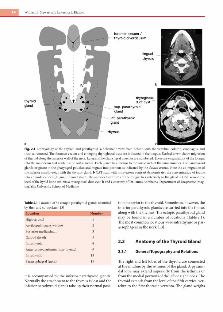
2.1 Embryology of the Thyroid
The primordial thyroid gland is first identifiable dur-ing the fourth week of gestation, beginning as an endodermal invagination of the tongue at the site of the foramen cecum (Fig. 2.1a). The foramen cecum lies where the midline intersects the sulcus termina-lis, which divides the tongue into anterior two thirds (oral part) and posterior one third (pharyngeal part). The thyroid diverticulum begins its descent through the tongue carrying with it the thyroglossal duct. The path of descent carries the developing gland anterior to the hyoid bone and the larynx. During the descent in the fifth week, the superior part of the duct degen-erates. By this time, the gland has achieved its rudi-mentary shape with two lobes connected by an isth-mus. It continues to descend until it reaches the level of the cricoid cartilage at about the seventh week. By the twelfth week of development, thyroid hormone is secreted. The distal part of the thyroglossal duct de-generates but may remain as a pyramidal lobe [8].
There is also a contribution to the thyroid from the fifth pharyngeal pouch (ultimobranchial body). These cells are believed to be neural crest in origin. They migrate into the thyroid and differentiate into the calcitonin-producing C cells (Fig. 2.1a) [4].
A number of developmental errors can affect thy-roid development. The thyroid may fail to descend.
In this case, a lingual thyroid is located at the junc-tion of the oral and pharyngeal parts of the tongue (Fig. 2.1b). Ectopic thyroid tissue may occur at any point along the pathway of the descent of the thy-roid. In rare conditions, the thyroid may descend into the thorax. There may also be remnants of the thyroglossal duct that hypertrophy and become cys-tic (Fig. 2.1c). Ectopic thyroid tissue may also be en-countered laterally in the neck [9]. Evaluation of the patient should consider whether the ectopic tissue is the sole active thyroid tissue. In very rare circum-stances thyroid tissue may be encountered inferior to the diaphragm in association with the gastrointestinal tract. This thyroid tissue, a struma ovarii, is derived from an ovarian germ cell tumor [5].
2.2 Embryology of the Parathyroid Glands
The parathyroid glands develop from the third and fourth pharyngeal (branchial) pouches (Fig. 2.1a).These pharyngeal pouches develop in association with the aortic arches that encircle the developing foregut. The pharyngeal arches have a mesodermal core, covered on their superficial surface by ectoderm and on their deep surface by endoderm. The pha-ryngeal pouches lie between successive pharyngeal arches and are endodermal evaginations of the fore-gut. The inferior parathyroid glands (parathyroid III)come from the third pharyngeal pouch and the supe-rior parathyroid glands (parathyroid IV) come from the fourth pharyngeal pouch. During the fifth week of development, the developing glands detach from the pouches and descend to join the thyroid gland during the seventh week. It should be noted that the inferior parathyroid glands actually arise from a more superior pharyngeal location (pouch III) than the su-perior thyroids (pouch IV). This relationship may be explained by the relationship of the developing infe-rior parathyroid gland with the thymus. The thymus arises from the caudal portion of the third pharyn-geal pouch. As the thymus descends into the thorax,
2 Embryology and Surgical Anatomy of the Thyroid and Parathyroid Glands
William B. Stewart and Lawrence J. Rizzolo
Contents
2.1 Embryology of the Thyroid . . . 132.2 Embryology of the Parathyroid Glands . . . 132.3 Anatomy of the Thyroid Gland . . . 142.3.1 General Topography and Relations . . . 142.3.2 Blood Supply . . . 152.4 Anatomy of the Parathyroid Glands . . . 172.5 Nearby Relations of the Thyroid and
Parathyroid at Risk During Surgery . . . 182.5.1 External Laryngeal Nerve . . . 182.5.2 Recurrent Laryngeal Nerve . . . 18
References . . . 19
William B. Stewart and Lawrence J. Rizzolo14
it is accompanied by the inferior parathyroid glands. Normally the attachment to the thymus is lost and the inferior parathyroid glands take up their normal posi-
tion posterior to the thyroid. Sometimes, however, the inferior parathyroid glands are carried into the thorax along with the thymus. The ectopic parathyroid gland may be found in a number of locations (Table 2.1).The most common locations were intrathymic or par-aesophageal in the neck [13].
2.3 Anatomy of the Thyroid Gland
2.3.1 General Topography and Relations
The right and left lobes of the thyroid are connected at the midline by the isthmus of the gland. A pyrami-dal lobe may extend superiorly from the isthmus or from the medial portions of the left or right lobes. The thyroid extends from the level of the fifth cervical ver-tebra to the first thoracic vertebra. The gland weighs
Fig. 2.1 Embryology of the thyroid and parathyroid. a Schematic view from behind with the vertebral column, esophagus, and trachea removed. The foramen cecum and emerging thyroglossal duct are indicated in the tongue. Dashed arrow shows migration of thyroid along the anterior wall of the neck. Laterally, the pharyngeal pouches are numbered. These are evaginations of the foregut into the mesoderm that contains the aortic arches. Each pouch lies inferior to the aortic arch of the same number. The parathyroid glands originate in the pharyngeal pouches and migrate into position as indicated by the dashed arrows. Note the co-migration of the inferior parathyroids with the thymus gland. b CAT scan with intravenous contrast demonstrates the concentration of iodine into an undescended (lingual) thyroid gland. The anterior two thirds of the tongue lies anteriorly to the gland. c CAT scan at the level of the hyoid bone exhibits a thyroglossal duct cyst. b and c courtesy of Dr. James Abrahams, Department of Diagnostic Imag-ing, Yale University School of Medicine
Table 2.1 Location of 54 ectopic parathyroid glands identified by Shen and co-workers [13]
Location Number
High cervical 1
Aorticopulmonary window 2
Posterior mediastinum 3
Carotid sheath 5
Intrathyroid 6
Anterior mediastinum (non-thymic) 9
Intrathymic 13
Paraesophageal (neck) 15
152 Embryology and Surgical Anatomy of the Thyroid and Parathyroid Glands
about 30 g, being somewhat heavier in females than in males [12]. The thyroid is surrounded by a sleeve of pretracheal fascia sometimes called the perithyroid sheath. Posteriorly, a thickening of this fascia attaches the gland to the cricoid cartilage. This fascia is the lat-eral ligament of the thyroid (ligament of Berry).
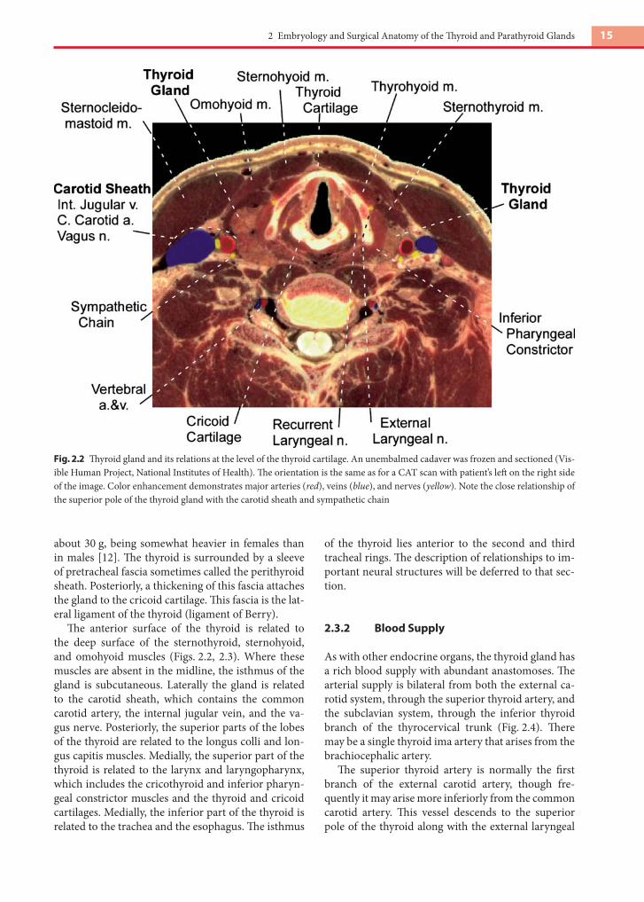
The anterior surface of the thyroid is related to the deep surface of the sternothyroid, sternohyoid, and omohyoid muscles (Figs. 2.2, 2.3). Where these muscles are absent in the midline, the isthmus of the gland is subcutaneous. Laterally the gland is related to the carotid sheath, which contains the common carotid artery, the internal jugular vein, and the va-gus nerve. Posteriorly, the superior parts of the lobes of the thyroid are related to the longus colli and lon-gus capitis muscles. Medially, the superior part of the thyroid is related to the larynx and laryngopharynx, which includes the cricothyroid and inferior pharyn-geal constrictor muscles and the thyroid and cricoid cartilages. Medially, the inferior part of the thyroid is related to the trachea and the esophagus. The isthmus
of the thyroid lies anterior to the second and third tracheal rings. The description of relationships to im-portant neural structures will be deferred to that sec-tion.
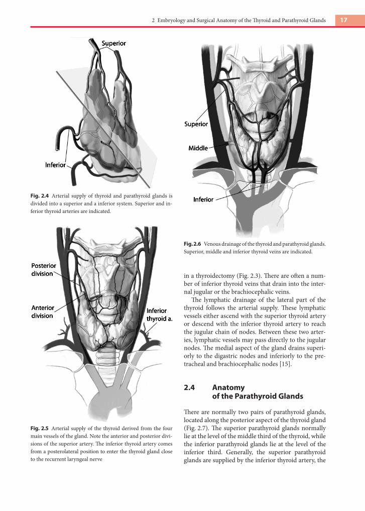
2.3.2 Blood Supply
As with other endocrine organs, the thyroid gland has a rich blood supply with abundant anastomoses. The arterial supply is bilateral from both the external ca-rotid system, through the superior thyroid artery, and the subclavian system, through the inferior thyroid branch of the thyrocervical trunk (Fig. 2.4). There may be a single thyroid ima artery that arises from the brachiocephalic artery.
The superior thyroid artery is normally the first branch of the external carotid artery, though fre-quently it may arise more inferiorly from the common carotid artery. This vessel descends to the superior pole of the thyroid along with the external laryngeal
Fig. 2.2 Thyroid gland and its relations at the level of the thyroid cartilage. An unembalmed cadaver was frozen and sectioned (Vis-ible Human Project, National Institutes of Health). The orientation is the same as for a CAT scan with patient’s left on the right side of the image. Color enhancement demonstrates major arteries (red), veins (blue), and nerves (yellow). Note the close relationship of the superior pole of the thyroid gland with the carotid sheath and sympathetic chain

William B. Stewart and Lawrence J. Rizzolo16
nerve. As it reaches the thyroid, the artery divides into anterior and posterior branches (Fig. 2.5). The anterior branch parallels the medial border of the lobe and anastomoses in the midline with the anterior branch of the other side. The posterior branch anasto-moses with branches of the inferior thyroid artery.
The inferior thyroid artery takes a looping course. It ascends along the anterior scalene muscle (Fig. 2.3).It turns medially to pass posteriorly to the carotid sheath and usually posteriorly to the sympathetic trunk as well. It descends along the longus colli to reach the inferior pole of the thyroid. There it passes to the thyroid either anteriorly or posteriorly to the recurrent laryngeal artery. At the thyroid, the artery branches into superior and inferior branches. The superior branch ascends on the posterior part of the
gland to anastomose with the posterior branch of the superior thyroid artery. The inferior branch supplies the inferior part of the gland as well as the inferior parathyroid glands. The inferior thyroid artery may be absent on either side. There is evidence that there are anthropologic differences in the incidence of thy-roid ima arteries, as well as in the symmetric origin of the superior thyroid arteries [17].
There are three main venous pathways from the thyroid: the superior, middle, and inferior thyroid veins (Fig. 2.6). The superior thyroid vein accompa-nies the superior thyroid artery and drains into the internal jugular vein. The middle thyroid vein is un-accompanied and drains directly into the internal jugular vein. Because of its posterior course, it is at risk when forward traction is applied to the gland, as
Fig. 2.3 Thyroid gland and its relations at the level of the third tracheal ring. Note the posteromedial relationships of the thyroid gland with the recurrent laryngeal nerve and middle thyroid veins. The thoracic duct (green) is atypically dilated close to where it joins the left internal jugular and subclavian veins. The inferior thyroid artery follows a looping course. In this image it is seen su-perior to its origin from the thyrocervical trunk of the subclavian artery. It will loop superiorly and medially before descending to join the thyroid gland near the recurrent laryngeal nerve. An inferior right parathyroid gland (orange) is evident near the recurrent laryngeal nerve and middle thyroid veins. Major nerves (yellow), arteries (red), and veins (blue) are indicated
172 Embryology and Surgical Anatomy of the Thyroid and Parathyroid Glands
in a thyroidectomy (Fig. 2.3). There are often a num-ber of inferior thyroid veins that drain into the inter-nal jugular or the brachiocephalic veins.
The lymphatic drainage of the lateral part of the thyroid follows the arterial supply. These lymphatic vessels either ascend with the superior thyroid artery or descend with the inferior thyroid artery to reach the jugular chain of nodes. Between these two arter-ies, lymphatic vessels may pass directly to the jugular nodes. The medial aspect of the gland drains superi-orly to the digastric nodes and inferiorly to the pre-tracheal and brachiocephalic nodes [15].
2.4 Anatomy of the Parathyroid Glands
There are normally two pairs of parathyroid glands, located along the posterior aspect of the thyroid gland (Fig. 2.7). The superior parathyroid glands normally lie at the level of the middle third of the thyroid, while the inferior parathyroid glands lie at the level of the inferior third. Generally, the superior parathyroid glands are supplied by the inferior thyroid artery, the
Fig. 2.4 Arterial supply of thyroid and parathyroid glands is divided into a superior and a inferior system. Superior and in-ferior thyroid arteries are indicated.
Fig. 2.5 Arterial supply of the thyroid derived from the four main vessels of the gland. Note the anterior and posterior divi-sions of the superior artery. The inferior thyroid artery comes from a posterolateral position to enter the thyroid gland close to the recurrent laryngeal nerve
Fig. 2.6 Venous drainage of the thyroid and parathyroid glands.Superior, middle and inferior thyroid veins are indicated.

William B. Stewart and Lawrence J. Rizzolo18
superior thyroid artery, or both. Anastomotic con-nections within the thyroid allow both vessels to con-tribute, especially to the superior parathyroid glands. A number of methods have been advocated for local-izing the glands. These include ultrasonography [6], intraoperative methylene blue [7], and technetium sestamibi scans [18].
2.5 Nearby Relations of the Thyroid and Parathyroid at Risk During Surgery
2.5.1 External Laryngeal Nerve
The external laryngeal is a division of the superior la-ryngeal nerve, a branch of the vagus. This nerve sup-plies the cricothyroid muscle. Since this muscle is in-volved in movements of the vocal apparatus, damage to the nerve will impair phonation. The nerve may run near the superior pole of the thyroid on the way
to its target. The external laryngeal nerve is frequently entrapped in the vascular pedicle that transmits the superior thyroid vessels. Consequently the nerve may be injured during the ligation of these vessels [2,3].
2.5.2 Recurrent Laryngeal Nerve
The recurrent laryngeal nerve, a branch of the vagus, supplies the remainder of the laryngeal musculature as well as sensation on and inferior to the vocal folds (Figs. 2.2, 2.3). On the right side, the nerve loops pos-teriorly to the subclavian artery to ascend obliquely until it reaches the tracheoesophageal groove near the inferior extent of the thyroid (Fig. 2.7). On the left side the nerve loops posteriorly to the arch of the aorta and ascends to the larynx in the tracheoesophageal groove. The nerve may divide into a number of branches that also supply the trachea and esophagus [10]. The nerve has a very close relationship with the inferior thyroid artery, where it might lie either an-teriorly or posteriorly to the vessel (Fig. 2.7). Because the left inferior thyroid artery may be absent in 6% of individuals, the identification of the recurrent la-ryngeal nerve may be more complicated [14]. The nerve may also be closely related to or within the ligament of Berry. Care must be taken in both re-traction and division of the ligament to ensure that the nerve is preserved. There are some cases where the nerve may run through the substance of the gland [11,16].
In a small number of individuals (approximately 1%) the right subclavian artery arises distally from the arch of the aorta [1]. As a consequence the right recurrent laryngeal nerve is not pulled into the tho-rax by its relationship with the subclavian artery. This non-recurrent right laryngeal nerve passes directly to the larynx posterior to the common carotid artery. It runs parallel to the inferior thyroid artery and can ascend for a short distance in the tracheoesophageal groove [15]. It is, therefore, at risk for injury during surgery.
The vagus nerve and sympathetic trunk are within or closely related to the carotid sheath (Figs. 2.2, 2.3, 2.8). The vagus nerve may receive some of its blood supply from the inferior thyroid artery [15]. Conse-quently, the artery should not be ligated too close to its origin. Lymph node dissection along the carotid artery and near the vertebral artery or any manipu-lation near the superior pole of the thyroid gland should also be performed with care to ensure that the cervical sympathetic chain ganglia are not damaged or removed (Figs. 2.2, 2.3).
Fig. 2.7 Schematic dorsal view shows the course of the inferior laryngeal nerve in relation to the inferior thyroid artery, the thyroid gland, and the parathyroid glands