Embed Size (px)
Citation preview
Surface tracking from the cortical mesh complements diffusion MRI fiber tracking near the cortex Etienne St-Onge1, Gabriel Girard1, Kevin Whittingstall2, and Maxime Descoteaux1
1Sherbrooke Connectivity Imaging Lab, Université de Sherbrooke, Sherbrooke, Québec, Canada, 2Department of Diagnostic Radiology, Faculty of Medicine and Health Science, Université de Sherbrooke, Sherbrooke, Québec, Canada
Target Audience: Researchers working on brain white matter connectivity and diffusion MRI tractography methods.
Purpose: Conventional diffusion MRI fiber tracking methods estimate fiber directions for each voxel from a diffusion MRI (dMRI) acquisition and then use a tracking algorithm to reconstruct white matter pathways[1]. However, because of the low spatial resolution of dMRI (approximately 2mm isotropic), fiber tracking techniques have difficulty reconstructing white matter fiber structures near gray matter, and in particular, have trouble penetrating into gyri and fully exploring the gyrus[2]. Hence, the main limitation of current tractography techniques is the partial volume effect due to the tracking mask discretization in gyri and poor spatial resolution of the data. This is why groups have recently pushed spatial resolution using high-resolution 7T acquisitions[3], high-resolution Human Connectome Project data[4,5] and post-mortem acquisitions[6]. These new datasets show new details of the gyri connectivity, with fiber orientations fanning into the gyri walls[3 fig.1,6c] and showing exquisite details of connections arriving orthogonal to the surface of the cortex. In this abstract, we take a completely different approach. We exploit the cortical gray matter (GW) - white matter (WM) mesh from T1 images as a complement to our standard clinical dMRI datasets to boost the resolution of the fiber tracking and connectivity when approaching the cortex and entering gyri. The goal of our new surface mesh tracking method is to improve tractography near the cortex without the need of high resolution acquisitions or sophisticated hardware.
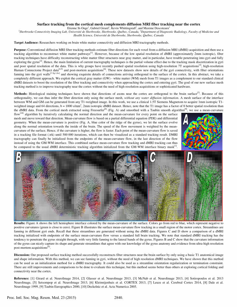
Methods: Histological staining techniques have shown that directions of axons near the cortex are orthogonal to the brain surface[7]. Because of this orthogonality, we can thus infer the fiber direction only using the surface mesh, without any water diffusion information. A mesh surface of the interface between WM and GM can be generated from any T1-weighted image. In this work, we use a clinical 1.5T Siemens Magnetom to acquire 1mm isotropic T1-weighted image and 64 directions, b = 1000 s/mm2, 2mm isotropic dMRI dataset. Hence, note that the T1 image has a factor of 8 better spatial resolution than the dMRI data. From the cortical mesh extracted using Freesurfer[8] (Fig. A) and smoothed with a Taubin smooth algorithm[9], we use a mean-curvature flow[10] algorithm by iteratively calculating the normal direction and the mean-curvature for every point on the surface mesh and move toward that direction. Mean-curvature flow is based on a partial differential equation (PDE) and differential geometry. When the mean-curvature is positive (Fig. A, blue color of the ridge of the gyrus), we let the surface evolve along the normal orientation towards the inside of the brain. The speed of the flow movement is weighted by the mean-curvature of the surface. Hence, if the curvature is higher, the flow is faster. Each point of the mean-curvature flow is saved in a tracking file format (.trk) until 500 000 iterations, which can then be visualized as a standard tracking result. DMRI tractography can finally be initialized from the endpoints of the mean-curvature flow, in the last direction of the flow instead of using the GM-WM interface. This combined surface mean-curvature flow tracking and dMRI tracking can thus be compared to the usual dMRI deterministic tracking algorithm initialized from the GM-WM interface binary mask[1].
Results: Figure A shows the left hemisphere interface colored by the mean-curvature of the surface. Colors go from red to blue, which represent negative to positive curvatures (green is close to zero). Figure B illustrates the surface mean-curvature flow tracking in a small region of the motor cortex. Streamlines are fanning in different gyri ends. Recall that these streamlines are generated without using the dMRI data. Figures C and D show a comparison of a dMRI tracking initialized with endpoints of the surface mean-curvature flow versus a standard full brain tracking. We note that standard dMRI tracking has the tendency to penetrate the gyrus straight through, with very little fanning to the lateral bands of the gyrus. Figures B and C show that the curvature information of the gyrus can nicely capture its shape and generate streamlines that agree with our knowledge of the gyrus anatomy and evidence from ultra-high resolution post-mortem acquisitions[6].
Discussion: Our proposed surface tracking method successfully reconstructs fiber structures near the brain surface by only using a basic T1 anatomical image and shape information. With this method, we can see fanning in gyri, without the need of high resolution dMRI techniques. We have shown that this method can be used as an initialization method for a dMRI tractography. It could also be used as a streamline orientation regularization or termination constraint. There are still improvements and comparisons to be done to evaluate this technique, but this method seems better than others at exploring cortical folding and connectivity near the cortex.
Reference: [1] Girard et al. NeuroImage 2014, [2] Glasser et al. NeuroImage 2013, [3] McNab et al. NeuroImage 2013, [4] Sotiropoulos et al. 2013 NeuroImage, [5] Setsompop et al. NeuroImage 2013, [6] Kleinnijenhuis et al. CORTEX 2013, [7] Leuze et al. Cerebral Cortex 2014, [8] Dale et al. NeuroImage 1999, [9] Taubin Eurographics 2000, [10] Deckelnic et al. Acta Numerica 2005.
Proc. Intl. Soc. Mag. Reson. Med. 23 (2015) 2840.