Embed Size (px)
Citation preview

Surface display for metabolic engineeringof industrially important acetic acidbacteria
Marshal Blank1 and Paul Schweiger2
1 Biology Department, Missouri State University, Springfield, MO, USA2 Department of Microbiology, University of Wisconsin-La Crosse, La Crosse, WI, USA
ABSTRACTAcetic acid bacteria have unique metabolic characteristics that suit them for a variety
of biotechnological applications. They possess an arsenal of membrane-bound
dehydrogenases in the periplasmic space that are capable of regiospecific and
enantioselective partial oxidations of sugars, alcohols, and polyols. The resulting
products are deposited directly into the medium where they are easily recovered for
use as pharmaceutical precursors, industrial chemicals, food additives, and
consumer products. Expression of extracytoplasmic enzymes to augment the
oxidative capabilities of acetic acid bacteria is desired but is challenging due to the
already crowded inner membrane. To this end, an original surface display systemwas
developed to express recombinant enzymes at the outer membrane of the model
acetic acid bacterium Gluconobacter oxydans. Outer membrane porin F (OprF)
was used to deliver alkaline phosphatase (PhoA) to the cell surface. Constitutive
high-strength p264 and moderate-strength p452 promoters were used to direct
expression of the surface display system. This system was demonstrated for
biocatalysis in whole-cell assays with the p264 promoter having a twofold increase in
PhoA activity compared to the p452 promoter. Proteolytic cleavage of PhoA from
the cell surface confirmed proper delivery to the outer membrane. Furthermore, a
linker library was constructed to optimize surface display. A rigid (EAAAK)1 linker
led to the greatest improvement, increasing PhoA activity by 69%. This surface
display system could be used both to extend the capabilities of acetic acid bacteria in
current biotechnological processes, and to broaden the potential of these microbes
in the production of value-added products.
Subjects Biotechnology, Microbiology, Molecular Biology
Keywords Surface display, Fusion linkers, Biocatalysis, Outer membrane proteins,
Gluconobacter oxydans
INTRODUCTIONGluconobacter oxydans is an industrially important microbe belonging to the family
Acetobacteriaceae, commonly referred to as the acetic acid bacteria. G. oxydans is an
incomplete oxidation specialist known for its ability to partially oxidize alcohols, polyols,
and monosaccharides to produce a diverse array of aldehydes, ketones, and organic acids.
These reactions occur in the periplasmic space, as G. oxydans possesses an arsenal of
dehydrogenases bound to the inner membrane. Therefore, industrially valuable
How to cite this article Blank and Schweiger (2018), Surface display for metabolic engineering of industrially important acetic acid
bacteria. PeerJ 6:e4626; DOI 10.7717/peerj.4626
Submitted 21 February 2018Accepted 26 March 2018Published 6 April 2018
Corresponding authorPaul Schweiger,
Academic editorRobert Winkler
Additional Information andDeclarations can be found onpage 16
DOI 10.7717/peerj.4626
Copyright2018 Blank and Schweiger
Distributed underCreative Commons CC-BY 4.0
compounds are excreted directly into the growth medium, where they are easily obtained
for use as food additives, pharmaceutical precursors, industrial chemicals, and consumer
products (Deppenmeier & Ehrenreich, 2009; Deppenmeier, Hoffmeister & Prust, 2002;
Prust et al., 2005). What is more, the membrane-bound dehydrogenases are both
regiospecific and enantioselective, allowing the microorganism to produce chiral
compounds from precursors containing multiple identical functional groups, such as
sugars and polyols. Additionally, the membrane-bound dehydrogenases contain
prosthetic groups that channel electrons to ubiquinone in the respiratory chain, which
facilitates rapid oxidation of substrates (Deppenmeier & Ehrenreich, 2009; Prust et al.,
2005). G. oxydans is also osmotolerant and acidophilic, which are desirable industrial
characteristics (Olijve & Kok, 1979).
Acetic acid bacteria serve many roles in biotechnology, but G. oxydans is particularly
important (Deppenmeier, Hoffmeister & Prust, 2002; Raspor & Goranovic, 2008).
Currently, G. oxydans is used to produce L-sorbose, the precursor to vitamin C
(Pappenberger & Hohmann, 2014; Yang & Xu, 2016) and 6-amino-6-desoxy-L-sorbose, the
precursor to the antidiabetic drug miglitol (Schedel, 2000). Additionally, this bacterium is
used to produce, dihydroxyacetone and erythrulose, which are primarily used as tanning
agents in cosmetics (De Muynck et al., 2007; Voss, Ehrenreich & Liebl, 2010), as well as
gluconate and gluconate derivatives, which serve as sequestering agents and drug
precursors (De Muynck et al., 2007; Deppenmeier, Hoffmeister & Prust, 2002). Despite their
widespread use in industry, progress toward metabolic engineering of acetic acid bacteria
has been partially limited by the lack of molecular tools available for genetic manipulation
of this group of microbes (Kallnik et al., 2010). Currently, constitutive expression vectors
are available for gene expression (Kallnik et al., 2010; Shi et al., 2014; Zhang et al., 2010),
and two markerless deletion systems have been developed (Kostner et al., 2013; Peters
et al., 2013). The ability to produce recombinant enzymes for the modification of
extracellular substrates has the potential to expand and compliment the natural
incomplete-oxidative metabolism of acetic acid bacteria. Signal peptides that allow for
periplasmic export are known (Kosciow et al., 2014), but the ability to produce additional
enzymes bound to the cytoplasmic membrane is limited because this space is already
crowded by the large number of native dehydrogenases (Guigas & Weiss, 2016).
Production of extracellular enzymes is also challenging because G. oxydans lacks the
machinery to secrete proteins across the outer membrane (Prust et al., 2005).
To overcome these limitations, a surface display system for expression of recombinant
enzymes at the cell surface of G. oxydans and other acetic acid bacteria was designed.
This molecular tool enables production of active enzymes with access to the extracellular
space, bypassing the crowded inner membrane. Surface display involves translational
fusion of a passenger protein with an anchor that innately localizes to the cell surface.
Surface display offers several advantages for biocatalysis. Most importantly, substrates do
not have to cross membrane barriers to interact with recombinant enzymes, and resulting
products are deposited directly into medium, permitting simplified extraction without the
need for cell lysis. Additionally, cells can be used for multiple rounds of biocatalysis, as
they can be removed from a spent reaction by centrifugation and then resuspended in a
Blank and Schweiger (2018), PeerJ, DOI 10.7717/peerj.4626 2/19

solution containing new substrate. Lastly, surface display eliminates the need to purify
enzymes (Schuurmann et al., 2014). To this end, a truncated version of outer membrane
porin F (OprF) from Pseudomonas aeruginosa was translationally fused to alkaline
phosphatase (PhoA), from Escherichia coli, and activity of the reporter enzyme was
quantified in a whole-cell assay. PhoA was proteolytically cleaved from the cell,
demonstrating that it was properly delivered to the outer leaflet of the outer membrane.
Finally, the surface display systemwas optimized by testing the effects of various linkers on
biocatalysis.
METHODSStrains and mediaPseudomonas aeruginosa PAO1 (DSMZ 22644) was grown in tryptic soy broth (Becton
Dickinson, Franklin Lakes, NJ, USA). E. coli 10b (New England Biolabs, Ipswich, MA,
USA), herein E. coli, was grown in lysogeny broth (1% tryptone, 0.5% yeast extract,
1% NaCl) with 100 mg/mL streptomycin added for strain maintenance. G. oxydans 621H,
hereafter G. oxydans, was grown in yeast mannitol (YM) broth composed of 2% mannitol
and 0.6% yeast extract, with 50 mg/mL cefoxitin added for strain maintenance. Plasmids
were maintained by addition of 50 mg/mL kanamycin. Electrocompetent G. oxydans was
prepared by growth in electroporation medium (Kallnik et al., 2010). After electroporation,
G. oxydans cells were plated on yeast glucose calcium carbonate (YGC) agar (2% glucose,
1.5% agar, 0.7% CaCO3, and 0.6% yeast extract) containing cefoxitin and kanamycin.
Materials and molecular techniquesStandard molecular techniques were done according to manufacturer’s protocols. Plasmid
DNA was extracted using a GeneJet Plasmid Miniprep kit (ThermoFisher Scientific,
Waltham, MA, USA) and genomic DNA was extracted using a GenElute Bacterial
Genomic DNA kit (Millipore-Sigma, St. Louis, MO, USA). Phusion DNA polymerase and
DreamTaq polymerase, FastDigest restriction enzymes, and T4 ligase were purchased from
ThermoFisher Scientific (Waltham, MA, USA). Factor Xa protease was purchased from
New England Biolabs (Ipswich, MA, USA). Primers were purchased from either Eurofins
Genomics (Louisville, KY, USA) or Integrated DNA Technologies (Coralville, IA, USA)
(Table 1). DNA sequencing was done by Eurofins Genomics (Louisville, KY, USA).
Construction of a surface display systemThe sequence encoding the native signal peptide plus the first 188 amino acids of OprF
(OprF188) was amplified from P. aeruginosa genomic DNA using primers oprF_F and
oprF_R, containing extended EcoRI and Eco105I restriction sites, respectively. The
resulting oprF188 amplicon was digested with these enzymes and ligated into similarly-cut
pBBR1p264-ST and pBBR1p452-ST to produce pBBR1p264-oprF-ST and pBBR1p452-
oprF-ST (Table 1). The gene encoding PhoA was amplified from E. coli genomic DNA
using primers phoA_F and phoA_R, containing extended Eco105I and HindIII sites,
respectively. Primer phoA_F was designed to exclude the native periplasmic signal
sequence of PhoA. The phoA amplicon was digested with Eco105I and HindIII and ligated
Blank and Schweiger (2018), PeerJ, DOI 10.7717/peerj.4626 3/19

in-frame into similarly-cut vectors to produce pBBR1p264-oprF-phoA and pBBR1p452-
oprF-phoA. Plasmids were transformed into E. coli and transformants were screened by
colony PCR and confirmed by sequencing. Plasmids were then transformed into G. oxydans
by electroporation (Kallnik et al., 2010; Kosciow et al., 2016; Kostner et al., 2013). Briefly,
100 mL of electroporation medium was inoculated with an overnight culture of
G. oxydans and grown to an OD600 nm of 0.8–1.0. The culture was placed on ice for 20 min,
Table 1 Plasmids and primers used for molecular cloning.
Plasmid or primer Description or sequencea Source or restriction site
Plasmids
pBBR1p264-ST pBBR1p264 derivative containing a Strep-tag (ST) sequence Zeiser et al. (2014)
pBBR1p452-ST pBBR1p452 derivative containing a ST sequence Kallnik et al. (2010)
pBBR1p264-oprF-ST pBBR1p264-ST derivative expressing oprF188 from P. aeruginosa This study
pBBR1p452-oprF-ST pBBR1p452-ST derivative expressing oprF188 from P. aeruginosa This study
pBBR1p264-oprF-phoA pBBR1p264-oprF-ST derivative with ST removed, expressing phoA from E. coli This study
pBBR1p452-oprF-phoA pBBR1p452-oprF-ST derivative with ST removed, expressing phoA from E. coli This study
pBBR1p452-oprF-CL-phoA pBBR1p452-oprF-phoA derivative with phoA removed, replaced by CL-phoA
encoding a Factor Xa cleavable linker
This study
pBBR1p264-oprF-FL1-phoA pBBR1p264-oprF-ST derivative with oprF-ST removed, replaced by oprF-FL1
and FL1-phoA encoding a (GGGGS)1 flexible linker
This study
pBBR1p264-oprF-FL2-phoA pBBR1p264-oprF-ST derivative with oprF-ST removed, replaced by oprF-FL2
and FL1-phoA encoding a (GGGGS)2 flexible linker
This study
pBBR1p264-oprF-FL3-phoA pBBR1p264-oprF-ST derivative with oprF-ST removed, replaced by oprF-FL2
and FL2-phoA encoding a (GGGGS)3 flexible linker
This study
pBBR1p264-oprF-RL1-phoA pBBR1p264-oprF-ST derivative with oprF-ST removed, replaced by oprF-RL1
and RL1-phoA encoding a (EAAAK)1 rigid linker
This study
pBBR1p264-oprF-RL2-phoA pBBR1p264-oprF-ST derivative with oprF-ST removed, replaced by oprF-RL2
and RL1-phoA encoding a (EAAAK)2 rigid linker
This study
pBBR1p264-oprF-RL3-phoA pBBR1p264-oprF-ST derivative with oprF-ST removed, replaced by oprF-RL2
and RL2-phoA encoding a (EAAAK)3 rigid linker
This study
Primers
oprF_F ATGGAATTCAGGAGGTAATATTTatgaaactgaagaacaccttaggc EcoRI
oprF_R ATCGTACGTAACTACCgacgttgtcgcaaacgccgtc Eco105I
phoA_F AATTTACGTAcctgttctggaaaaccggg Eco105I
phoA_R ATATAAGCTTtcatttcagccccagagcggc HindIII
CL-phoA_F ATATTACGTAATCGACGGCCGCGGCTCCcctgttctggaaaaccgg Eco105I
oprF-FL1_R AGAGGATCCGCCGCCGCCgacgttgtcgcaaacgcc BamHI
oprF-FL2_R TATGGATCCGCCGCCGCCCGAGCCGCCGCCGCCgacgttgtcgcaaacgcc BamHI
oprF-RL1_R CTTGGCGGCCGCTTCgacgttgtcgcaaacgcc NotI
oprF-RL2_R CTTGGCGGCCGCTTCCTTCGCCGCGGCTTCgacgttgtcgcaaacgcc NotI
FL1-phoA_F GGCGGCGGCGGATCCcctgttctggaaaaccgg BamHI
FL2-phoA_F GGCGGCGGCGGATCCGGCGGCGGCGGCTCGcctgttctggaaaaccgg BamHI
RL1-phoA_F GAAGCGGCCGCCAAGcctgttctggaaaaccgg NotI
RL2-phoA_F GAAGCGGCCGCCAAGGAAGCCGCGGCGAAGcctgttctggaaaaccgg NotI
Note:a The annealing portion of primers are shown in lowercase, synthetic additions uppercase, restriction sites 2 underlined, linker sequences italicized, and ribosomalbinding site bolded.
Blank and Schweiger (2018), PeerJ, DOI 10.7717/peerj.4626 4/19

and chilled cells were harvested by centrifugation at 2,000g for 10 min at 4 �C. Cellswere washed three times with 50–100% volume of 1 mM HEPES, with centrifugation at
4,000g for 10 min at 4 �C. Pellets were resuspended with HEPES and combined into a final
volume of 800 mL, to which 200 mL of glycerol was added. Cells were either used
immediately or aliquoted, flash-frozen, and stored at -80 �C. For electroporation, 40 mL
of electrocompetent cells were combined with 1 mL of plasmid DNA and pulsed with a
field strength of 22 kV/cm using a BioRad MicroPulser. After a 6–16 h outgrowth in
electroporation medium, transformed cells were plated on YGC agar containing kanamycin
and cefoxitin. To incorporate a cleavable linker (CL) into the OprF188-PhoA fusion protein,
a 5′-extended version of the PhoA gene, CL-phoA, was amplified from E. coli genomic DNA
using primers CL-phoA_F and phoA_R. Primer CL-phoA_F encoded the amino acid
sequence, Ile-Asp-Gly-Arg, recognized by Factor Xa protease (Nagai & Thogersen, 1987;
Terpe, 2003). The CL-phoA amplicon was cut with Eco105I and HindIII and ligated into
similarly-cut pBBR1p452-oprF-ST to produce pBBR1p452-oprF-CL-phoA (Table 1).
Construction of a linker library for surface displayA library of flexible (FL) and rigid (RL) linkers was assembled similar to Li et al. (2016).
The inserts, oprF-FL1, oprF-FL2, oprF-RL1, and oprF-RL2, were amplified from the
plasmid, pBBR1p264-oprF-ST, using the forward primer, oprF_F, and the respectively-
named reverse primers (oprF-FL1_R, oprF-FL2_R, oprF-RL1_R, and oprF-RL2_R)
containing either BamHI or NotI sites. The inserts, FL1-phoA, FL2-phoA, RL1-phoA, and
RL2-phoAwere amplified from the E. coli genome using the reverse primer, phoA_R, and
the respectively-named forward primers (FL1-phoA_F, FL2-phoA_F, RL1-phoA_F, and
RL2-phoA_F) containing either BamHI or NotI sites. The linker system inserts were cut
with their corresponding restriction enzymes—all oprF188 inserts were cut with EcoRI, all
phoA inserts were cut withHindIII, all FL inserts were cut with BamHI, and all rigid linker
(RL) inserts were cut with NotI. In parallel, vector pBBR1p264-oprF-ST was cut with
EcoRI and HindIII. The linker library inserts were ligated into a linearized vector
in a combinatorial fashion to produce six plasmids comprising the linker library:
pBBR1p264-oprF-FL1-phoA, pBBR1p264-oprF-FL2-phoA, pBBR1p264-oprF-FL3-phoA,
pBBR1p264-oprF-RL1-phoA, pBBR1p264-oprF-RL2-phoA, and pBBR1p264-oprF-
RL3-phoA (Table 1).
Phosphatase assaysAlkaline phosphatase assays were done using a modified method of Kosciow et al. (2014).
Experimental cultures were inoculated 1:100 from overnight cultures and grown to
mid-late exponential phase (OD600 of 0.9–1.1 for E. coli and 0.6–0.9 for G. oxydans). Forty
microliters of cells were mixed with 160 mL of substrate buffer (1M Tris base, 10 mM
MnSO4, 10 mM ZnSO4, 1.25 mM p-nitrophenylphosphate, pH 8.0) in a 96-well
microplate. The reactions were incubated with shaking for 60 min at 30 �C and the change
of absorbance was monitored at 405 nm in a BioTek EL808 plate reader. Phosphatase
activity was reported as absorbance change per hour, normalized by the optical density of
the bacterial culture, �A405/(h � OD600).
Blank and Schweiger (2018), PeerJ, DOI 10.7717/peerj.4626 5/19
PhoA localization assayLocalization of the passenger protein was conducted using a modified method Jiang &
Boder (2010). YM broth was inoculated 1:100 with an overnight culture of G. oxydans
harboring plasmid pBBR1p452-oprF-CL-phoA and grown to mid-late exponential phase
and 500 mL was centrifuged at 2,000g for 5 min. The pellet was resuspended in 200 mL of
Factor Xa buffer (100 mM NaCl, 20 mM Tris-base, 2 mM CaCl2, pH 8.0) and Factor Xa
protease was added to a final concentration of 20 ng/mL. Samples were incubated
overnight at 23 �C with shaking. After incubation, samples were pelleted at 2,000g for
5 min, and supernatant was transferred to a new microcentrifuge tube. The supernatant
was centrifuged at 16,100g for 2 min to remove intact cells and 40-mL aliquots were
used to quantify phosphatase activity as described above.
Growth behaviorYeast mannitol broth was inoculated to an OD600 of 0.05 from overnight cultures of
G. oxydans strains and 1 mL of inoculated broth was added to a 24-well microplate.
The plate was incubated at 30 �C with shaking at 150 rpm for up to 24 h, and
absorbance was monitored at 595 nm every 5 min in a Flurostar Optima plate reader
(BMG Labtech, GmbH, Ortenberg, Germany).
Statistical analysisR Studio was used to perform statistical analyses and to generate box-and-whisker plots,
strip charts, and growth curve graphics (R Core Team, 2017). Data were analyzed by
performing an analysis of variance and a post-hoc Tukey’s HSD test (q = 0.05) unless
otherwise noted. The R packages used in this study were dplyr, ggplot2, growthcurver,
plyr, multcomp, and reshape2 (Hothorn, Bretz & Westfall, 2008; Sprouffske & Wagner,
2016; Wickham, 2007, 2009, 2011; Wickham et al. 2017).
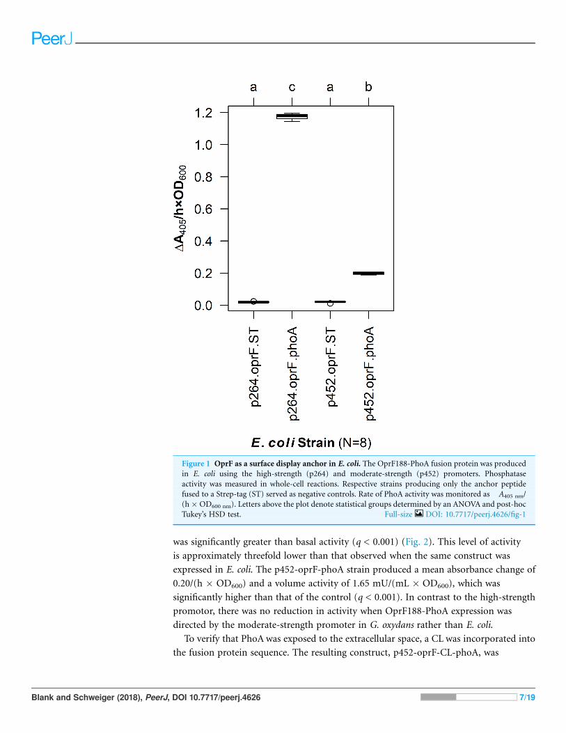
RESULTSSurface display in G. oxydansTo enable surface display in acetic acid bacteria, a truncated version of OprF (OprF188)
was tested for its ability to localize the PhoA reporter enzyme to the cell surface of
G. oxydans. PhoA was translationally fused to the C-terminal end of OprF188, and the
resulting OprF188-PhoA fusion protein was produced via two expression vectors, one
containing a high-strength promotor (p264) and the other containing a moderate-
strength promotor (p452) (Kallnik et al., 2010). As a preliminary test, these surface display
constructs were expressed in E. coli and phosphatase activity was quantified in a whole-cell
assay (Fig. 1). The OprF188-PhoA surface display systems produced statistically
significant absorbance changes compared to strains expressing the anchor protein alone
when using both the high- (q < 0.001) and moderate-strength (q < 0.001) promoters,
p264 and p452, respectively. Enzymatic rates were approximately sixfold higher in the
p264-oprF-phoA system compared to the p452-oprF-phoA system.
In G. oxydans, the p264-oprF-phoA strain produced a mean absorbance change of
0.39/(h � OD600), corresponding to a volume activity of 3.21 mU/(mL � OD600), which
Blank and Schweiger (2018), PeerJ, DOI 10.7717/peerj.4626 6/19
was significantly greater than basal activity (q < 0.001) (Fig. 2). This level of activity
is approximately threefold lower than that observed when the same construct was
expressed in E. coli. The p452-oprF-phoA strain produced a mean absorbance change of
0.20/(h � OD600) and a volume activity of 1.65 mU/(mL � OD600), which was
significantly higher than that of the control (q < 0.001). In contrast to the high-strength
promotor, there was no reduction in activity when OprF188-PhoA expression was
directed by the moderate-strength promoter in G. oxydans rather than E. coli.
To verify that PhoA was exposed to the extracellular space, a CL was incorporated into
the fusion protein sequence. The resulting construct, p452-oprF-CL-phoA, was
Figure 1 OprF as a surface display anchor in E. coli. The OprF188-PhoA fusion protein was produced
in E. coli using the high-strength (p264) and moderate-strength (p452) promoters. Phosphatase
activity was measured in whole-cell reactions. Respective strains producing only the anchor peptide
fused to a Strep-tag (ST) served as negative controls. Rate of PhoA activity was monitored as�A405 nm/
(h�OD600 nm). Letters above the plot denote statistical groups determined by an ANOVA and post-hoc
Tukey’s HSD test. Full-size DOI: 10.7717/peerj.4626/fig-1
Blank and Schweiger (2018), PeerJ, DOI 10.7717/peerj.4626 7/19

transformed into G. oxydans and an assay to localize PhoA was done (see Methods).
The mean level of phosphatase activity observed in the supernatant from cells treated
with Factor Xa protease was higher than that from untreated cells (two-sample t-test,
p < 0.001) (Fig. 3). These data suggest that PhoA was released into the supernatant after
cleavage, indicating that PhoA was attached to the outer membrane and oriented toward
the medium.
Linker system optimizationTo optimize surface display in acetic acid bacteria, a library of linkers was integrated into
the OprF188-PhoA surface display systems. First, overhang PCR was used to add linker
Figure 2 OprF as a surface display anchor in G. oxydans. The OprF188-PhoA fusion protein was
expressed in G. oxydans using the high-strength (p264) and moderate-strength (p452) promoters.
Phosphatase activity was measured in whole-cell reactions. Respective strains producing only the anchor
peptide fused to a Strep-tag (ST) served as negative controls. Rate of PhoA activity was monitored as
�A405 nm/(h�OD600 nm). Letters above the plot denote statistical groups determined by an ANOVA and
post-hoc Tukey’s HSD test. Full-size DOI: 10.7717/peerj.4626/fig-2
Blank and Schweiger (2018), PeerJ, DOI 10.7717/peerj.4626 8/19

building blocks to oprF188 and phoA. These building blocks encoded for either flexible or
RLs and contained either BamHI or NotI restriction sites, respectively. The modified
oprF188 inserts contained abridged linker sequences at their 3′ ends and the modified
phoA inserts contained abridged linker sequences at their 5′ ends. Therefore, complete
linker sequences resulted from ligation of compatible insert dyads. The building
blocks were assembled into the expression vector that showed the highest activity to
create six constructs encoding fusion proteins varying in linker composition and
linker length: Three containing FLs (FL1, FL2, and FL3) composed of a (GGGGS)1–3
motif, and three containing RLs (RL1, RL2, and RL3) composed of the (EAAAK)1–3
motif.
The linker library was first expressed in E. coli and PhoA activity was quantified (Fig. 4).
The FLs had slight negative effects on enzyme activity. While there was no difference
between the control lacking a linker and the fusion protein containing the small FL (FL1)
(q = 0.939), the medium (FL2), and large (FL3) FLs led to a slight but statistically
significant decrease in activity (q < 0.001 for each). In contrast, the small (RL1) and
medium (RL2) RLs did not have any effect on activity (q = 0.071 and 0.969, respectively).
Figure 3 Cleavable linker assay. To confirm proper localization of the passenger enzyme, a cleavable
linker motif was incorporated into the OprF188-PhoA surface display system. Whole G. oxydans cells
were treated with Factor Xa protease and the resulting supernatant (SN) was assayed for phosphatase
activity. Rate of PhoA activity was monitored as �A405 nm/(h � OD600 nm).
Full-size DOI: 10.7717/peerj.4626/fig-3
Blank and Schweiger (2018), PeerJ, DOI 10.7717/peerj.4626 9/19

The large RL (RL3) led to a slight but statistically significant increase in enzymatic activity
when compared to the control (q < 0.001). However, there was no difference between the
RL1 strain and the RL3 strain (q = 0.692), suggesting that the higher activity observed for
the RL3 strain was likely not biologically significant.
The linker systems had more pronounced effects on PhoA activity in G. oxydans
compared to the subtle effects of the flexible and RLs in E. coli (Fig. 5). While the small
FL (FL1) and the medium RL (RL2) had no effect on activity when compared to the
control lacking a linker (q = 0.967 and q = 0.765, respectively), the large RL (RL3) led to a
32% reduction in activity (q < 0.001). Interestingly, the small RL (RL1) drastically
improved PhoA activity (q < 0.001), having a 69% increase in biocatalysis that
corresponded to a volume activity of 5.42 mU/(mL � OD600). Attempts to transform
plasmids p264-oprF-FL2-phoA and p264-oprF-FL3-phoA into G. oxydans were
unsuccessful, suggesting that the medium (FL2) and large (FL3) FLs were toxic at
high expression.
Effect of surface display on growth of G. oxydansTo characterize the effects of protein production and surface engineering on the growth
behavior of G. oxydans cells, a method was developed to follow the growth of recombinant
G. oxydans strains using standard 24-well microplates, allowing automated monitoring of
cell growth in a plate reader. Strains containing OprF188 surface display systems were
compared to wildtype G. oxydans 621H growth (Fig. 6). When the high-strength p264
promoter was used, production of the OprF188 anchor protein without fusion to PhoA
Figure 4 The effects of linkers on biocatalysis in E. coli. To determine the effects of linkers on the
surface display system, a library of flexible (GGGGS)1–3 and rigid (EAAAK)1–3 linkers was integrated
into the OprF188-PhoA fusion protein and PhoA activity was measured. Rate of PhoA activity was
monitored as �A405 nm/(h � OD600 nm). Letters above the plot denote statistical groups determined by
an ANOVA and post-hoc Tukey’s HSD test. Full-size DOI: 10.7717/peerj.4626/fig-4
Blank and Schweiger (2018), PeerJ, DOI 10.7717/peerj.4626 10/19

led to a statistically significant increase in mean doubling time (97 min) compared to
wildtype cells (56 min) (q < 0.001) (Fig. 7). However, when the OprF188-PhoA fusion
was expressed, the doubling time (52 min) was not different than that of wildtype cells
(q = 0.603). The presence of the small FL (FL1) increased the doubling time (82 min)
compared to the strain expressing the OprF188-PhoA fusion without a linker (q < 0.001).
Conversely, the presence of the small RL (RL1) decreased doubling time (27 min)
compared to the strain lacking the linker (q < 0.001) and to wildtype cells (q < 0.001).
However, the lag time for this strain was approximately 8 h longer than that of wildtype
cells (Fig. 6). The medium RL (RL2) did not affect doubling time (58 min) compared to
the no-linker control strain (q = 0.166) or wildtype cells (q = 0.952) (Fig. 7). Lastly, the
large RL (RL3) increased doubling time to 121 min. Generally, the lag time was longer
for recombinant strains and the final optical density was lower than that of wildtype
G. oxydans, except for the p264-oprF-RL1-phoA containing strain (Fig. 6) that also
produced the highest PhoA activity. Interestingly, doubling time was inversely
proportional to PhoA activity (Figs. 5 and 7, respectively), suggesting that recombinant
PhoA positively contributed to the growth of G. oxydans.
Figure 5 The effects of linkers on biocatalysis in G. oxydans. A linker library consisting of flexible
(GGGGS)1 and rigid (EAAAK)1–3 linkers was integrated into the OprF188-PhoA fusion protein
and expressed in G. oxydans and the phosphatase activity was measured. Rate of PhoA activity was
monitored as �A405 nm/(h � OD600 nm). Letters above the plot denote statistical groups determined
by an ANOVA and post-hoc Tukey’s HSD test. Full-size DOI: 10.7717/peerj.4626/fig-5
Blank and Schweiger (2018), PeerJ, DOI 10.7717/peerj.4626 11/19

DISCUSSIONIn this study, the gene encoding PhoA was fused to OprF188 to test the ability of the
anchor peptide to transport recombinant enzymes to the cell surface of acetic acid
bacteria. The resulting constructs were expressed in the model acetic acid bacterium
G. oxydans and biocatalysis was quantified in whole-cell reactions. Based on enzymatic
activity, OprF188 localized active PhoA to the cell surface, regardless of expression level.
Nascent PhoA has no activity in the cytoplasm as it does not fold properly unless secreted
extracellularly where disulfide bonding takes place (De Geyter et al., 2016; Ehrmann,
Boyd & Beckwith, 1990; Hoffman & Wright, 1985; Manoil & Beckwith, 1985; Michaelis
et al., 1983). The native periplasmic signal sequence was removed from PhoA before it was
fused to OprF188, which contains an outer membrane signal peptide and is known to
localize to the outer membrane (Lee et al., 2005; Sugawara, Nagano & Nikaido, 2012).
The OprF188-PhoA fusion was produced using two expression vectors designed for
protein production in G. oxydans, one containing a high-strength p264 promoter, and
the other a moderate-strength p452 promoter. There was a twofold difference in
phosphatase activity produced byG. oxydans between the two promoters. This mirrors the
threefold difference in enzymatic activity reported previously (Kallnik et al., 2010).
Because PhoA is an innately periplasmic enzyme, its location needed to be verified
to show that OprF188 can correctly target recombinant enzymes to the outer leaflet of
the outer membrane in G. oxydans. To this end, a CL was integrated between OprF188
and PhoA as was done previously to validate another surface display systems (Jiang &
Boder, 2010). If PhoA was exposed at the cell surface via OprF188, then PhoA should be
Figure 6 Growth behavior of recombinant G. oxydans strains. Cells were cultured in 24-well
microplates and the growth of recombinant G. oxydans 621H strains was compared to that of wildtype.
Solid line, mean optical density; ribbon, 95% CI; n = 3. Full-size DOI: 10.7717/peerj.4626/fig-6
Blank and Schweiger (2018), PeerJ, DOI 10.7717/peerj.4626 12/19

removed from the outer membrane following treatment with Factor Xa protease,
increasing phosphatase activity in the medium. The CL assay demonstrated that the
level of phosphatase activity in the treated supernatant fractions was indeed higher than
that in the untreated supernatant fractions (p < 0.001), which suggests that PhoA was
present at the outer leaflet of the outer membrane.
In this study, we used a C-terminal truncated version of OprF, OprF188, containing
188 amino acids from the N-terminus of the original protein in its mature form. Valine
188 was previously determined to be the optimal fusion site for passenger proteins (Lee
et al., 2005). This anchor protein was previously used to localize a 49.9 kDa lipase, which is
comparable to the size of the 47 kDa PhoA monomer (Bradshaw et al., 1981), to the outer
membrane of E. coli (Lee et al., 2005) and Pseudomonas putida (Lee, Lee & Park, 2005).
The first 24 amino acids of nascent OprF188 encodes a Sec-dependent signal peptide.
OprF is an unusual outer membrane protein in that it is comprised of two domains: An
N-terminal domain that forms a small, eight-stranded b-barrel, and a C-terminal globular
domain that associates with the cell wall in the periplasm (Bodilis & Barray, 2006;
Sugawara, Nagano & Nikaido, 2012). Mature OprF188 consists of only the b-barreldomain with a C-terminal extracellular loop (Lee et al., 2005). OprF is also unusual
because the b-signal that is recognized by the b-barrel assembly complex to facilitate outer
membrane integration, is located internally at the end of the b-barrel domain, rather than
Figure 7 Doubling time of G. oxydans strains. Doubling time was calculated using the growthcurver R
package. Letters above the plot denote statistical groups determined by an ANOVA and post-hoc Tukey’s
HSD test. Full-size DOI: 10.7717/peerj.4626/fig-7
Blank and Schweiger (2018), PeerJ, DOI 10.7717/peerj.4626 13/19

C-terminally (Gessmann et al., 2014; Sugawara, Nagano & Nikaido, 2012). Thus, truncated
OprF188 maintains its b-signal, allowing proper membrane integration (Lee et al., 2005;
Sugawara, Nagano & Nikaido, 2012).
Interestingly, there have been many unsuccessful attempts to target PhoA to the cell
surface of E. coli using E. coli outer membrane proteins as anchors. PhoAwas fused to the
ferrichrome outer membrane transporter FhuA (Coulton, Reid & Campana, 1988). While
PhoAwas associated with the outer membrane, it was not necessarily exposed to the outer
surface. Similarly, the ferric enterobactin outer membrane transporter FepA localized
PhoA to the outer membrane of E. coli, but it was periplasmically oriented (Murphy &
Klebba, 1989). When a PhoA was fused to a lipoprotein-OmpA hybrid, again the fusion
protein was associated with the outer membrane but PhoA existed exclusively in the
periplasm (Stathopoulos, Georgiou & Earhart, 1996). Close inspection of these studies
reveals that the b-signal was removed from FhuA, FepA, and OmpA upon C-terminal
truncation. Therefore, deletion of the b-signal was likely sufficient to prevent proper outermembrane insertion by the b-barrel assembly complex in those studies. The use of
OprF188 as an anchor to successfully display proteins on the cell surface in the current
study and by others suggests that it is broadly functional as a surface display anchor
protein in gram-negative bacteria, likely due to the preservation of the b-signal (Lee et al.,2005; Lee, Lee & Park, 2005).
Despite their influence on fusion protein activity, the effect of linkers on bacterial
surface display has not previously been investigated. While linkers are sometimes included
in the design of surface display systems, their inclusion is rarely made explicit and even
fewer studies offer any explanation as to why a particular linker sequence was chosen. Yet
linker sequences can play a vital role in the design of fusion proteins, affecting protein
folding and production efficiency, and influencing enzyme activity (Chen, Zaro & Shen,
2013; Li et al., 2016). Here, a library of linkers varying in both composition and length
were integrated into a surface display system to explicitly test the effects of linkers on
biocatalysis at the cell surface of two bacterial species. Two types of linkers were
investigated in this study, FLs and RLs. FLs are composed of small, polar amino acids such
as glycine and serine and form random coils, such as the (GGGGS)1–3 linker used here.
RLs are often a-helical, such as the (EAAAK)1–3 linker used here (Chen, Zaro & Shen,
2013; Li et al., 2016). FLs provide passive separation and permit interaction between the
components of fusion proteins, while RLs maintain a set distance and can prevent
interaction between fusion protein domains (Li et al., 2016). The linker library was first
expressed in E. coli and, generally, the presence of linkers did not dramatically influence
product yield. In contrast, the linkers caused pronounced effects on biocatalysis in
G. oxydans. Fusion proteins containing the medium (FL2) and large (FL3) FLs—10 and
15 amino acids in length—were likely toxic to G. oxydans, as positive transformants were
not obtained after multiple attempts. The fusion proteins containing the small flexible
(FL1) and medium rigid (RL2) linkers had no influence on biocatalysis, whereas the large
rigid (RL3) linker was deleterious. Optimization of the surface display system was
achieved with the small RL (RL1), consisting of a single EAAAK pentapeptide, which
improved phosphatase activity by 69%.
Blank and Schweiger (2018), PeerJ, DOI 10.7717/peerj.4626 14/19

The disparate results of the linkers on E. coli and G. oxydans suggest that the effects of
linkers on biocatalysis at the cell surface may be species-specific. E. coli and G. oxydans are
phylogenetically distant, belonging to the Gammaproteobacteria and Alphaproteobacteria,
respectively. There are likely differences in the composition of their outer membranes and
thus the environment at the cell surface of these bacteria. It should be noted that the outer
membrane composition of G. oxydans and other acetic acid bacteria remains largely
uncharacterized, limiting direct comparison. Nevertheless, these differences may also be
why phosphatase activity was overall higher in E. coli than in G. oxydans. Differences in
promoter strength and/or efficiency of secretion (Choi & Lee, 2004) or outer membrane
protein assembly by the b-barrel assembly complex (Browning et al., 2015; Sikora et al.,
2017) may also play a role in the differences in the activities observed between E. coli and
G. oxydans. Additionally, the differences in linker effects could also be passenger protein-
specific. Overall, these results suggest that linker optimization is an important
consideration for each surface display system used and for the specific host organism.
To determine the effects of protein production and surface engineering on the growth
of G. oxydans, a novel method was developed for semi-high-throughput culturing of
G. oxydans. The strain containing plasmid p264-oprF-ST had significantly slower growth
compared to wildtypeG. oxydans 621H. It is possible that this growth defect was caused by
the metabolic burden of protein production. Alternatively, the production of the OprF188
peptide may have overwhelmed secretory machinery, preventing sufficient amounts of
required proteins from being processed. It is also possible that integration of the OprF188
anchor destabilizes outer membrane integrity. Interestingly, the p264-oprF-phoA strain
did not exhibit a growth defect, suggesting that PhoA may enable recovery of the OprF188
phenotype. It may be that high expression of PhoA allowed cells to scavenge more
phosphate from the medium, possibly allowing recovery from outer membrane lipid
defects. Indeed, the small RL (RL1) not only increased PhoA activity, but also improved
the growth rate with concomitant increased lag time. PhoA activity seemed to be inversely
proportional with doubling time, suggesting that inorganic phosphate availability may
contribute to the observed growth phenotypes.
CONCLUSIONSurface display is potentially a powerful tool to enable metabolic engineering of
G. oxydans and other industrially important acetic acid bacteria. G. oxydans currently has
a limited ability to grow on disaccharides and polysaccharides, and instead relies on
relatively expensive monomeric feedstocks (Kosciow et al., 2016). The ability to express
enzymes at the outer membrane could be used to improve current bioprocesses by
broadening the substrate range of this bacterium. Because this technique utilizes export
machinery found in all gram-negative bacteria, the surface display constructs could be
expressed in otherwise wildtype cells. Previously, a recombinant exoenzyme with a
periplasmic signal peptide was efficiently released from cells only when a knockout strain
of G. oxydans with a leaky outer membrane phenotype was used (Kosciow et al., 2016).
Surface display could also be used in conjunction with immobilized G. oxydans cells to
create stable bioreactors, such as those used in the production of dihydroxyacetone
Blank and Schweiger (2018), PeerJ, DOI 10.7717/peerj.4626 15/19

(Dikshit & Moholkar, 2016). In conclusion, a novel molecular tool for strain improvement
of acetic acid bacteria was produced, and the OprF188 surface display system described
herein is a significant first step toward outer membrane engineering of G. oxydans.
Such molecular tools will enable engineering of this unique bacterium to improve and
expand its ability to produce value-added products.
ACKNOWLEDGEMENTSWe thank Dr. Kyoungtae Kim and Dr. Laszlo Kovacs for lending their expertise and
providing feedback on this project.
ADDITIONAL INFORMATION AND DECLARATIONS
FundingThis work was supported by funds from the Missouri State University Biology
Department and the Graduate College. The funders had no role in study design,
data collection and analysis, decision to publish, or preparation of the manuscript.
Grant DisclosuresThe following grant information was disclosed by the authors:
Missouri State University Biology Department and the Graduate College.
Competing InterestsThe authors declare that they have no competing interests.
Author Contributions� Marshal Blank conceived and designed the experiments, performed the experiments,
analyzed the data, contributed reagents/materials/analysis tools, prepared figures and/or
tables, authored or reviewed drafts of the paper, approved the final draft.
� Paul Schweiger conceived and designed the experiments, analyzed the data, contributed
reagents/materials/analysis tools, prepared figures and/or tables, authored or reviewed
drafts of the paper, approved the final draft.
Data AvailabilityThe following information was supplied regarding data availability:
The raw data that was used to generate statistics/error bars and corresponding R code
for each figure are available as Supplemental Files.
Supplemental InformationSupplemental information for this article can be found online at http://dx.doi.org/
10.7717/peerj.4626#supplemental-information.
REFERENCESBodilis J, Barray S. 2006. Molecular evolution of the major outer-membrane protein gene (oprF)
of pseudomonas. Microbiology 152(4):1075–1088 DOI 10.1099/mic.0.28656-0.
Blank and Schweiger (2018), PeerJ, DOI 10.7717/peerj.4626 16/19

Bradshaw RA, Cancedda F, Ericsson LH, Neumann PA, Piccoli SP, Schlesinger MJ, Shriefer K,
Walsh KA. 1981. Amino acid sequence of Escherichia coli alkaline phosphatase. Proceedings of
the National Academy of Sciences of the United States of America 78(6):3473–3477
DOI 10.1073/pnas.78.6.3473.
Browning DF, Bavro VN, Mason JL, Sevastsyanovich YR, Rossiter AE, Jeeves M, Wells TJ,
Knowles TJ, Cunningham AF, Donald JW, Palmer T, Overduin M, Henderson IR. 2015.
Cross-species chimeras reveal BamA POTRA and beta-barrel domains must be fine-tuned for
efficient OMP insertion. Molecular Microbiology 97(4):646–659 DOI 10.1111/mmi.13052.
Chen X, Zaro JL, Shen WC. 2013. Fusion protein linkers: property, design and functionality.
Advanced Drug Delivery Reviews 65(10):1357–1369 DOI 10.1016/j.addr.2012.09.039.
Choi JH, Lee SY. 2004. Secretory and extracellular production of recombinant proteins
using Escherichia coli. Applied Microbiology and Biotechnology 64(5):625–635
DOI 10.1007/s00253-004-1559-9.
Coulton JW, Reid GK, Campana A. 1988. Export of hybrid proteins FhuA′-′LacZ and
FhuA′-′PhoA to the cell envelope of Escherichia coli K-12. Journal of Bacteriology
170(5):2267–2275 DOI 10.1128/jb.170.5.2267-2275.1988.
De Geyter J, Tsirigotaki A, Orfanoudaki G, Zorzini V, Economou A, Karamanou S. 2016.
Protein folding in the cell envelope of Escherichia coli. Nature Microbiology 1(8):16107
DOI 10.1038/nmicrobiol.2016.107.
De Muynck C, Pereira CS, Naessens M, Parmentier S, Soetaert W, Vandamme EJ. 2007. The
genus Gluconobacter oxydans: comprehensive overview of biochemistry and biotechnological
applications. Critical Reviews in Biotechnology 27(3):147–171
DOI 10.1080/07388550701503584.
Deppenmeier U, Ehrenreich A. 2009. Physiology of acetic acid bacteria in light of the genome
sequence of Gluconobacter oxydans. Journal of Molecular Microbiology and Biotechnology
16(1–2):69–80 DOI 10.1159/000142895.
Deppenmeier U, Hoffmeister M, Prust C. 2002. Biochemistry and biotechnological applications
of Gluconobacter strains. Applied Microbiology and Biotechnology 60(3):233–242
DOI 10.1007/s00253-002-1114-5.
Dikshit PK, Moholkar VS. 2016. Kinetic analysis of dihydroxyacetone production from crude
glycerol by immobilized cells of Gluconobacter oxydans MTCC 904. Bioresource Technology
216:948–957 DOI 10.1016/j.biortech.2016.06.042.
Ehrmann M, Boyd D, Beckwith J. 1990. Genetic analysis of membrane protein topology by a
sandwich gene fusion approach. Proceedings of the National Academy of Sciences of the United
States of America 87(19):7574–7578 DOI 10.1073/pnas.87.19.7574.
Gessmann D, Chung YH, Danoff EJ, Plummer AM, Sandlin CW, Zaccai NR, Fleming KG. 2014.
Outer membrane beta-barrel protein folding is physically controlled by periplasmic lipid head
groups and BamA. Proceedings of the National Academy of Sciences of the United States of
America 111(16):5878–5883 DOI 10.1073/pnas.1322473111.
Guigas G, Weiss M. 2016. Effects of protein crowding on membrane systems. Biochimica
et Biophysica Acta (BBA)–Biomembranes 1858(10):2441–2450
DOI 10.1016/j.bbamem.2015.12.021.
Hoffman CS, Wright A. 1985. Fusions of secreted proteins to alkaline phosphatase: an approach
for studying protein secretion. Proceedings of the National Academy of Sciences of the United
States of America 82(15):5107–5111 DOI 10.1073/pnas.82.15.5107.
Hothorn T, Bretz F, Westfall P. 2008. Simultaneous inference in general parametric models.
Biometrical Journal 50(3):346–363 DOI 10.1002/bimj.200810425.
Blank and Schweiger (2018), PeerJ, DOI 10.7717/peerj.4626 17/19

Jiang W, Boder ET. 2010. High-throughput engineering and analysis of peptide binding to class II
MHC. Proceedings of the National Academy of Sciences of the United States of America
107(30):13258–13263 DOI 10.1073/pnas.1006344107.
Kallnik V, Meyer M, Deppenmeier U, Schweiger P. 2010. Construction of expression vectors for
protein production in Gluconobacter oxydans. Journal of Biotechnology 150(4):460–465
DOI 10.1016/j.jbiotec.2010.10.069.
Kosciow K, Domin C, Schweiger P, Deppenmeier U. 2016. Extracellular targeting of an active
endoxylanase by a TolB negative mutant of Gluconobacter oxydans. Journal of Industrial
Microbiology & Biotechnology 43(7):989–999 DOI 10.1007/s10295-016-1770-6.
Kosciow K, Zahid N, Schweiger P, Deppenmeier U. 2014. Production of a periplasmic trehalase
in Gluconobacter oxydans and growth on trehalose. Journal of Biotechnology 189:27–35
DOI 10.1016/j.jbiotec.2014.08.029.
Kostner D, Peters B, Mientus M, Liebl W, Ehrenreich A. 2013. Importance of codB for new
codA-based markerless gene deletion in Gluconobacter strains. Applied Microbiology and
Biotechnology 97(18):8341–8349 DOI 10.1007/s00253-013-5164-7.
Lee SH, Choi JI, HanMJ, Choi JH, Lee SY. 2005.Display of lipase on the cell surface of Escherichia
coli using OprF as an anchor and its application to enantioselective resolution in organic
solvent. Biotechnology and Bioengineering 90(2):223–230 DOI 10.1002/bit.20399.
Lee SH, Lee SY, Park BC. 2005. Cell surface display of lipase in Pseudomonas putida KT2442 using
OprF as an anchoring motif and its biocatalytic applications. Applied and Environmental
Microbiology 71(12):8581–8586 DOI 10.1128/AEM.71.12.8581-8586.2005.
Li G, Huang Z, Zhang C, Dong BJ, Guo RH, Yue HW, Yan LT, Xing XH. 2016. Construction of a
linker library with widely controllable flexibility for fusion protein design. Applied Microbiology
and Biotechnology 100(1):215–225 DOI 10.1007/s00253-015-6985-3.
Manoil C, Beckwith J. 1985. TnphoA: a transposon probe for protein export signals. Proceedings of
the National Academy of Sciences of the United States of America 82(23):8129–8133
DOI 10.1073/pnas.82.23.8129.
Michaelis S, Inouye H, Oliver D, Beckwith J. 1983. Mutations that alter the signal sequence of
alkaline phosphatase in Escherichia coli. Journal of Bacteriology 154(1):366–374.
Murphy CK, Klebba PE. 1989. Export of FepA::PhoA fusion proteins to the outer
membrane of Escherichia coli K-12. Journal of Bacteriology 171(11):5894–5900
DOI 10.1128/jb.171.11.5894-5900.1989.
Nagai K, Thogersen HC. 1987. Synthesis and sequence-specific proteolysis of hybrid proteins
produced in Escherichia coli. Methods in Enzymology 153:461–481
DOI 10.1016/0076-6879(87)53072-5.
Olijve W, Kok JJ. 1979. An analysis of growth of Gluconobacter oxydans in chemostat cultures.
Archives of Microbiology 121(3):291–297 DOI 10.1007/bf00425070.
Pappenberger G, Hohmann HP. 2014. Industrial production of L-ascorbic Acid (vitamin C)
and D-isoascorbic acid. Advances in Biochemical Engineering/Biotechnology 143:143–188
DOI 10.1007/10_2013_243.
Peters B, Junker A, Brauer K, Muhlthaler B, Kostner D, Mientus M, Liebl W, Ehrenreich A.
2013. Deletion of pyruvate decarboxylase by a new method for efficient markerless gene
deletions in Gluconobacter oxydans. Applied Microbiology and Biotechnology 97(6):2521–2530
DOI 10.1007/s00253-012-4354-z.
Prust C, Hoffmeister M, Liesegang H, Wiezer A, Fricke WF, Ehrenreich A, Gottschalk G,
Deppenmeier U. 2005. Complete genome sequence of the acetic acid bacterium Gluconobacter
oxydans. Nature Biotechnology 23(2):195–200 DOI 10.1038/nbt1062.
Blank and Schweiger (2018), PeerJ, DOI 10.7717/peerj.4626 18/19
Raspor P, Goranovic D. 2008. Biotechnological applications of acetic acid bacteria. Critical
Reviews in Biotechnology 28(2):101–124 DOI 10.1080/07388550802046749.
R Core Team. 2017. R: A Language and Environment for Statistical Computing. Vienna:
R Foundation for Statistical Computing. Available at https://www.R-project.org/.
Schedel M. 2000. Oxidation of Aminosorbitol with Gox in Synthesis of Deoxynojirimycin. Weinheim:
Wiley-VHC.
Schuurmann J, Quehl P, Festel G, Jose J. 2014. Bacterial whole-cell biocatalysts by surface
display of enzymes: toward industrial application. Applied Microbiology and Biotechnology
98(19):8031–8046 DOI 10.1007/s00253-014-5897-y.
Shi L, Li K, Zhang H, Liu X, Lin J, Wei D. 2014. Identification of a novel promoter gHp0169
for gene expression in Gluconobacter oxydans. Journal of Biotechnology 175:69–74
DOI 10.1016/j.jbiotec.2014.01.035.
Sikora AE, Wierzbicki IH, Zielke RA, Ryner RF, Korotkov KV, Buchanan SK, Noinaj N. 2017.
Structural and functional insights into the role of BamD and BamE within the b-barrelassembly machinery in Neisseria gonorrhoeae. Journal of Biological Chemistry 293(4):1106–1119
DOI 10.1074/jbc.RA117.000437.
Sprouffske K, Wagner A. 2016. Growthcurver: an R package for obtaining interpretable metrics
frommicrobial growth curves. BMC Bioinformatics 17(1):172 DOI 10.1186/s12859-016-1016-7.
Stathopoulos C, Georgiou G, Earhart CF. 1996. Characterization of Escherichia coli expressing
an Lpp’OmpA(46-159)-PhoA fusion protein localized in the outer membrane. Applied
Microbiology and Biotechnology 45(1–2):112–119 DOI 10.1007/s002530050657.
Sugawara E, Nagano K, Nikaido H. 2012. Alternative folding pathways of the major porin OprF of
Pseudomonas aeruginosa. FEBS Journal 279(6):910–918 DOI 10.1111/j.1742-4658.2012.08481.x.
Terpe K. 2003. Overview of tag protein fusions: from molecular and biochemical fundamentals to
commercial systems. Applied Microbiology and Biotechnology 60(5):523–533
DOI 10.1007/s00253-002-1158-6.
Voss J, Ehrenreich A, Liebl W. 2010. Characterization and inactivation of the membrane-bound
polyol dehydrogenase in Gluconobacter oxydans DSM 7145 reveals a role in meso-erythritol
oxidation. Microbiology 156(6):1890–1899 DOI 10.1099/mic.0.037598-0.
Wickham H. 2007. Reshaping Data with the reshape Package. Journal of Statistical Software
21(12):1–20 DOI 10.18637/jss.v021.i12.
Wickham H. 2009. ggplot2: Elegant Graphics for Data Analysis. New York: Springer-Verlag.
Wickham H. 2011. The Split-Apply-Combine Strategy for Data Analysis. Journal of Statistical
Software 40(1):1–29 DOI 10.18637/jss.v040.i01.
Wickham H, Francois R, Henry L, Muller K. 2017. dplyr: A Grammar of Data Manipulation.
R package version 0.7.3. Available at https://CRAN.R-project.org/package=dplyr.
Yang W, Xu H. 2016. Industrial fermentation of vitamin C. In: Revuelta JL, Vandamme EJ, eds.
Industrial Biotechnology of Vitamins, Biopigments, and Antioxidants. Germany: Wiley-VCH
Verlag GmbH & Co. KGaA, 161–192.
Zeiser J, Muhlenbeck LH, Schweiger P, Deppenmeier U. 2014. Characterization of a periplasmic
quinoprotein from Sphingomonas wittichii that functions as aldehyde dehydrogenase.
Applied Microbiology and Biotechnology 98(5):2067–2079 DOI 10.1007/s00253-013-5016-5.
Zhang L, Lin J, Ma Y, Wei D, Sun M. 2010. Construction of a novel shuttle vector for
use in Gluconobacter oxydans. Molecular Biotechnology 46(3):227–233
DOI 10.1007/s12033-010-9293-2.
Blank and Schweiger (2018), PeerJ, DOI 10.7717/peerj.4626 19/19