Embed Size (px)
Citation preview

SUPPLEMENTARY ONLINE MATERIAL
The SUMO protease SENP7 is a critical component to ensure HP1 enrichment
at pericentric heterochromatin
Christèle Maison*, Kelly Romeo*, Delphine Bailly, Marion Dubarry, Jean-Pierre Quivy‡ and
Geneviève Almouzni‡
Institut Curie, Centre de recherche, 75248 Paris, France Centre National de la Recherche Scientifique (CNRS), Unité Mixte de Recherche UMR218, 75248 Paris, France * These authors contributed equally to this work ‡ Corresponding authors
This supplement contains:
Methods
6 Supplementary Figures
Nature Structural & Molecular Biology: doi:10.1038/nsmb.2244

METHODS
Cells, miRNA and siRNA treatment, extracts. We cultured NIH3T3 cells and MEFs
(provided by T. Jenuwein)1 in DMEM (Gibco BRL) containing 10% (v/v) FCS at 37ºC and 5%
CO2. We performed RNase treatment as described in2. We transfected NIH3T3 cells with
Lipofectamin 2000 (Invitrogen) and performed analyses 72 h after transfection. We prepared
total cell extract by resuspending cells in RIPA buffer (50 mM Tris-HCl pH 7.5, 150 mM NaCl,
5 mM EDTA, 15 mM MgCl2, 1% (v/v) Nonidet P-40, and 0.75% (w/v) sodium deoxycholate,
supplemented with protease and phosphatase inhibitors, and 20 mM NEM) and nuclear
extracts as described in3.
Plasmids. To generate GST-SENP7 and His6-SENP7, we cloned mouse SENP7 cDNA
obtained from I.M.A.G.E clone collection (isoform 1, clone ID IRAVp968H0599D6, RZPD)
into pGEX-4T1 and pET-30a vector (Novagen). We made GFP-SENP7 by inserting SENP7
cDNA in the pEGFP-C3 vector (Invitrogen). The catalytic-mutant GFP-SENP7C979S was
generated from GFP-SENP7 by site directed mutagenesis (Genescript). We used the
pEGFP-C3 plasmid to express GFP controls. HP1α-HA expressing plasmid was as
described in4. GFP-SUMO1, GFP-SUMO-2, GFP-SENP6 and GFP-SENP6C1030S5 were
kindly provided by R. Hay. We constructed the plasmid expressing the miRNA targeting
SENP7 (miSENP7) according to the Block-iT™ Pol II miR RNAi expression vector kit
(Invitrogen) with the sequence 5'-TGATGAAGAAAGTTGCTCTGA-3'. Control miRNA
(micont) with the sequence 5'-GAAATGTACTGCGCGTGGAGA-3' predicted not to target
known vertebrate genes was provided with the Block-iT™ Pol II miR RNAi system
(Invitrogen). The sequences of the siRNA were as follows: siSENP7, 5’-
ACAAGAAGCCUAAGAAAUA-3’; siSENP6, ON-TARGETplus SMARTpool J-062052-05, 06,
07 and 08 (Dharmacon); sicontrol, 5’-CGUACGCGGAAUACUUCGA-3’.
Immunofluorescence and image acquisition. We processed cells for immunostaining as
Nature Structural & Molecular Biology: doi:10.1038/nsmb.2244

described3,6. For simultaneous visualisation of GFP with SENP7 and HP1α or H3K9me3 and
HP1α, cells were not extracted prior to fixation. HP1α was detected with Alexa Fluor 647
secondary antibodies and SENP7 or H3K9me3 with Alexa Fluor 594 secondary antibodies.
We used a Zeiss Z1 epifluorescence microscope equipped with a x63 objective lens and a
chilled CCD camera (H2Q2, Ropper) for image acquisition.
Statistical analysis. The pvalue were calculated in R program. We used the Student paired-t
test to compare the % of cells with HP1 localization at pericentric domains between
miSENP7 and micont from 7 independent experiments. We counted more than 100 nuclei for
each case per experiment. Differences were considered significant when pvalue < 0.05.
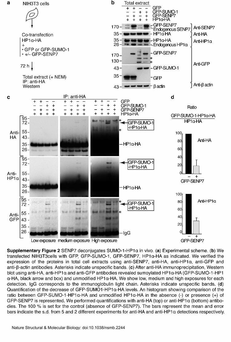
Antibodies and immunoprecipitation. We generated a rabbit polyclonal anti-mSENP7 by
injecting recombinant full-length His6-mSENP7 (Agro-Bio). We tested its specificity for
SENP7 by immunofluorescence and Western blot after depletion of SENP7 (Supplementary
Fig. 2a,b). Rabbit serum was used at 1:1,000 dilution for Western blot. For
immunofluorescence we affinity purified the antibodies by incubating 200μl of serum with 5
μg of purified His6-mSENP7 present on a nitrocellulose membrane according to7, and used
this purified antibody at 1:10 dilution. We used the following antibodies: for
immufluorescence, mouse monoclonal anti-HP1α (2HP-1H5-AS, Euromedex ; 1:1,000),
rabbit polyclonal anti-H3K9me3 (#07-442, Upstate; 1:1,000), goat polyclonal anti-SENP6
(#ab77619, Abcam; 1:50) and secondary antibodies Alexa Fluor 568, 594 or 647
(Invitrogen); for Western blot, rabbit polyclonal anti-HP1α (C7F11, Cell signaling; 1:1,000),
mouse monoclonal anti-HP1α (2HP-2G9-AS, Euromedex ; 1:1,000), mouse monoclonal anti-
HP1β (1MOD-1A9-AS, Euromedex; 1:1,000), mouse monoclonal anti-HP1γ (2MOD-1G6,
Euromedex; 1:1,000), goat polyclonal anti-SENP6 (#ab77619, Abcam; 1:250), mouse
monoclonal anti-GST (3G10/1B3, Abcam, 1/1,000), mouse monoclonal anti-β-actin (AC-15,
Sigma; 1:20,000), rat monoclonal anti-HA (#1867423, Roche; 1:2,000), mouse monoclonal
Nature Structural & Molecular Biology: doi:10.1038/nsmb.2244

anti-GFP (7.1 and 13.1 mix, Roche; 1:1,000), rabbit polyclonal anti-KAP1 (#A300-274A,
Bethyl; 1:1,000), rabbit polyclonal anti-SetDB1 (#07-378, Upstate ; 1:1,000) and secondary
antibodies coupled to horseradish peroxidase (Jackson Immuno Research Laboratories, Inc).
We performed Western blot immunodetection on the membrane with the Super Signal kit
(Pierce). We performed GFP and HP1α immunoprecipitation with anti-HA agarose-
conjugated beads (Roche) and GFP-trap_A beads (Chromotek). We performed HA
immunoprecipitations in RIPA buffer as in4. We performed GFP precipitations and washes in
10 mM Tris-HCl pH 7.5, 500 mM NaCl, 0.5 mM EDTA, 15 mM MgCl2 and 0.1% (v/v) Nonidet
P-40 supplemented with protease inhibitors. We quantified HP1α-HA and GFP-SUMO-1-
HP1α-HA from Western blots with a Chemi-doc XRS system and the QuantityOne software
(Biorad).
Cell cycle/DNA content analysis by flow cytometry. After washes in PBS supplemented
with 0.1% (w/v) BSA and 0.02% (w/v) sodium azide (PBA), we resuspended NIH3T3 cells
with continuous gentle vortexing in 70% (v/v) ethanol at -20ºC and incubated them on ice for
30 min. Then we stained cells with 20 ug.ml-1 propidium iodide plus 10ug.ml-1 RNase A in
PBA for 30 min. We performed analysis on a FACSCalibur (Becton Dickinson) or on a C6
(Accuri) cytometer and FlowJo software (Treestar). We selected cells for analysis on their
cycle profile by first gating on live cells with a forward scatter (FSC) vs side scatter (SSC)
gate, then excluding multiplets with a SSC-H vs SSC-W gate and finally selecting either GFP-
positive or GFP-negative cells.
Recombinant proteins and pull down. We bacterially expressed GST, GST-HP1α2, GST-
SENP7, and His-SENP7 from E. coli BL21 (DE3) strain and immobilized proteins on
glutathione (Amersham) or Ni-NTA beads (Novagen). We purified GST and GST-HP1α by
elution from the glutathione beads according to manufacturer’s instructions in buffer A (50
mM Tris-HCl pH 7.5, 250 mM NaCl, 0,05% (v/v) Nonidet P-40, supplemented with protease
Nature Structural & Molecular Biology: doi:10.1038/nsmb.2244
inhibitors). We performed GST pull-down by mixing 3T3 nuclear extract with GST or GST-
SENP7 beads and incubating for 2 hours at 4°C on a rotating wheel in buffer A. After five
washes in buffer A containing 500 mM NaCl, we recovered proteins by boiling in SDS-PAGE
loading buffer and analyzed by Western-blot. We performed direct interaction assay by
mixing GST or GST-HP1α with His-SENP7 beads and proceeded as above.
REFERENCES
1. Peters, A. et al. Loss of the Suv39h histone methyltransferases impairs mammalian heterochromatin and genome stability. Cell 107, 323-337 (2001).
2. Maison, C. et al. Higher-order structure in pericentric heterochromatin involves a distinct pattern of histone modification and an RNA component. Nat Genet 30, 329-34 (2002).
3. Martini, E., Roche, D.M., Marheineke, K., Verreault, A. & Almouzni, G. Recruitment of phosphorylated chromatin assembly factor 1 to chromatin after UV irradiation of human cells. J Cell Biol 143, 563-75 (1998).
4. Maison, C. et al. SUMOylation promotes de novo targeting of HP1alpha to pericentric heterochromatin. Nat Genet 43, 220-7 (2011).
5. Hattersley, N., Shen, L., Jaffray, E.G. & Hay, R.T. The SUMO protease SENP6 is a direct regulator of PML nuclear bodies. Mol Biol Cell 22, 78-90 (2011).
6. Quivy, J.P. et al. A CAF-1 dependent pool of HP1 during heterochromatin duplication. EMBO J 23, 3516-26 (2004).
7. Smith, D.E. & Fisher, P.A. Identification, developmental regulation, and response to heat shock of two antigenically related forms of a major nuclear envelope protein in Drosophila embryos: application of an improved method for affinity purification of antibodies using polypeptides immobilized on nitrocellulose blots. J. Cell Biol. 99, 20-28 (1984).
Nature Structural & Molecular Biology: doi:10.1038/nsmb.2244

Nature Structural & Molecular Biology: doi:10.1038/nsmb.2244
Nature Structural & Molecular Biology: doi:10.1038/nsmb.2244

Nature Structural & Molecular Biology: doi:10.1038/nsmb.2244

Nature Structural & Molecular Biology: doi:10.1038/nsmb.2244

Nature Structural & Molecular Biology: doi:10.1038/nsmb.2244
Nature Structural & Molecular Biology: doi:10.1038/nsmb.2244