Embed Size (px)
Citation preview
www.sciencemag.org/cgi/content/full/science.1227026/DC1
Supplementary Materials for
The Legionella Effector RavZ Inhibits Host Autophagy Through Irreversible Atg8 Deconjugation
Augustine Choy, Julia Dancourt, Brian Mugo, Tamara J. O’Connor, Ralph R. Isberg, Thomas J. Melia,* Craig R. Roy*
*To whom correspondences should be addressed. E-mail: [email protected] (T.J.M.); [email protected] (C.R.R.)
Published 26 October 2012 on Science Express
DOI: 10.1126/science.1227026
This PDF file includes:
Materials and Methods Figs. S1 to S11 Full Reference List
2
Materials and Methods:
Bacterial strains and plasmids
Legionella strains were grown on charcoal yeast extract plates as described previously
(16). L. pneumophila thyA (Lp02) (17), a thymidine auxotroph derived from the L.
pneumophila serogroup 1 strain Lp01, was used, along with isogenic strains Lp03 (dotA)
(17), ∆flaA (18) and pentuple and single chromosomal deletion strains (12). Bacteria
were cultured for 48 h on charcoal-yeast extract agar plates supplemented with
thymidine (100 µg/ml). The plasmid pJB1806 (19) was used to produce RavZ
(AAU27763.1), Ceg23 (AAU27701.1), Lem12, (AAU27705.1), Lpg1639 (AAU27719.1),
SidB (YP_095669.1), Lpg1654 (AAU27734.1), LegL3 (YP_095687.1), Lpg1663
(AAU27743.1), Ceg24 (AAU27746.1), Lpg1684 (AAU27764.1) and BlaM-RalF
(AAL23711.1) in Legionella. These genes were ligated into the vector using BamH1 and
Sal1 sites added during the PCR amplification. The plasmid pDEST-17 (Invitrogen) was
used for all His-tagged constructs and the plasmid pCDNA-DEST-53 (Invitrogen) was
used for all GFP fusion constructs. The plasmid pSR47S (20) was used to generate the
RavZ deletion mutant in Lp02.
Mice and bone marrow-derived macrophages
C57BL/6 mice were purchased from Jackson Laboratories. GFP-LC3 transgenic mice
were used with the permission of Dr. Mizushima (21), and provided by the laboratory of
Akiko Iwasaki (Yale University). Bone marrow was collected from the femurs and tibiae
of mice and cultured as described previously (22). Following differentiation,
3
macrophages were replated in RPMI 1640 from GIBCO (Carlsbad) containing 10% fetal
bovine serum (FBS) and 5% macrophage colony-stimulating factor (M-CSF).
Cell culture and transfection
HEK293 and HeLa cells were grown in Dulbecco’s modified Eagle medium (DMEM)
from Gibco (Carlsbad) containing 10% heat-inactivated fetal bovine serum (FBS; Gibco).
Cell lines were cultured at 37 °C in 5% CO2. For transfection, cells were added to 12-mm
coverslips in 24-well plates at a density of 2.5 × 10⁴ cells per well. Cells were transfected
with 0.8 µg of each plasmid using Lipofectamine 2000 (Invitrogen) and fixed 18 h after
transfection in 4% PFA, permeabilized with methanol and processed for
immunofluorescence microscopy as described previously (16).
Infection of HEK293 cells
HEK293 FcγRII (23) cells grown to near confluency in 24-well dishes were infected with
opsonized Legionella pneumophila strain Lp02 (wild type) or the isogenic mutants at an
estimated multiplicity of infection (MOI) of 10 bacteria to 1 host cell. For experiments
requiring LC3 immunoblots, infections were done in the presence of Bafilomycin A1
[160nM] (Sigma) as indicated.
Isolation of injected and uninjected macrophages
The Legionella Lp02-derived ∆flaA strain was used for infection of bone marrow-derived
macrophages to avoid flagellin-induced pyroptosis. Macrophages were plated on 10 cm
dishes and infected with Legionella expressing BlaM-RalF at an MOI of 50 in the
4
presence of Bafilomycin [160nM] for 2 hours. The media was then removed and cells
were loaded with the fluorescent substrate CCF4/AM, using the LiveBLAzer-FRET B/G
Loading Kit (Invitrogen) with 15 mM probenecid, in the dark for 2 hours at room
temperature. Cells were then washed and sorted based on the ratio of the signal of 460
nm vs. 535 nm when excited at 415 nm using an iCyt Reflection cell sorter. Immunoblot
analysis to determine LC3-II levels was conducted on the sorted cells and compared to
uninfected cells.
Generation of Atg5-deficient macrophages To generate myeloid lineage cell-specific Atg5-deficient mice using the Cre/loxP
recombination system, Atg5flox/flox mice (24), which contained two loxP sites flanking exon
3 of the Atg5 gene, were crossed with B6.129P2-Lyz2tm1(cre)Ifo/J mice (The Jackson
Laboratory) expressing Cre recombinase under the control of a Lyzs promoter. Bone
marrow was harvested from these mice and differentiated into macrophages. Non-
expression of Atg5 in these macrophages was confirmed by immunoblot analysis using
an Atg5-specific antibody (Sigma). All procedures relating to the care and treatment of
the animals were performed in accordance with the NIH guidelines.
Macrophage growth curves Following differentiation, macrophages were replated in RPMI 1640 from GIBCO
(Carlsbad) containing 10% fetal bovine serum (FBS) and 5% macrophage colony-
stimulating factor (M-CSF) at a density of 200,000 cells/well in a 24-well dish.
Macrophages were infected with Legionella at an MOI of 1 for 1 hour, washed 3 times
with PBS from GIBCO (Carlsbad) and incubated in fresh media. For collection of
Legionella from the wells, macrophages were lysed with sterile H2O at the indicated time
5
point. Serial dilutions of the lysates were plated to determine colony-forming units
(CFUs).
Immunoblotting
Total cell lysates were prepared by adding cells to Laemmli buffer (100mM Tris-HCl, pH
6.8, 2% SDS, 10% glycerol, 6% ß-mercaptoethanol, 0.0025% Bromophenol Blue).
Equal amount of proteins were resolved by SDS-PAGE, transferred to PVDF
membranes, and incubated with primary antibody for 1 hour. After 3 washes,
membranes were incubated with secondary antibodies conjugated with horseradish
peroxidase for 45 minutes. Signals were visualized with ECL on GE’s ImageQuant LAS
4000. LC3 antibody (Novus) was used at 1:3000, Actin antibody (Sigma) was used at
1:200, goat anti-mouse HRP antibody (Invitrogen) was used at 1:3000, and goat anti-
rabbit HRP (Invitrogen) antibody was used at 1:3000.
LC3 puncta assay, immunofluoresence and fluorescence microscopy
Cells infected or transfected as described above were incubated in DMEM (HEK293 and
HeLa) or RPMI (macrophages) with 10% FBS and 160nM Bafilomycin A1 for 2 hours.
Cells were then fixed in 4% PFA, permeabilized with methanol, blocked with goat serum,
incubated with primary antibody for 1 hour, incubated with secondary antibody for 45
minutes, and mounted using ProLong Gold (Invitrogen). LC3 antibody (Nanotools) was
used at 1:200, GFP antibody (Roche) was used at 1:1000. Rhodamine Red goat anti-
rabbit (Invitrogen) and Alexa Fluor 488 goat anti-mouse (Invitrogen) were used at
1:1000. Cells were scored as positive for LC3 puncta if they contained more than 5
puncta. For localization of GFP-LC3 and ubiquitin to the Legionella vacuole, GFP
antibody (Invitrogen) was used at 1:200 and ubiquitin antibody (Enzo) was used at
1:1000. For GFP-RavZ localization studies HeLa cells were stained using antibodies for
6
LC3 (Nanotools) and Atg16 (CosmoBio) in conjunction with DyLight-conjugated
secondary antibodies (Rockland). Digital images were acquired with a Nikon TE300
microscope using a ×100 1.4 N.A objective lens and a Hamamatsu ORCA-ER camera
controlled by IP Lab software.
Long-lived protein degradation assay
HeLa cells were pulse-labeled with 1 mCi/mL of 14C-Valine (Moravek Biochemicals Inc.)
for 4 hours in RPMI medium supplemented with 10% FCS. Cells were then incubated in
chase media (DMEM + 10% FCS + 10 mM L-Valine) for 15 hours before being washed
three times in HBSS and incubated either in full media (FM, same as chase media) or in
starvation media (Starv.: HBSS + 50 mM Hepes pH 7.4 + 10 mM L-Valine) for four
hours. Media and cells scraped in PBS + 1% Triton X-100 were then respectively
precipitated in 10% Trichloroacetic acid (TCA) by incubation on ice for 10 minutes. TCA-
precipitates were spun 10 minutes at 20 000g at 4°C. TCA-soluble and TCA-insoluble
fractions (resuspended in 0.2N NaOH) were separately submitted to scintillation
counting. Percent of long-lived protein degradation was obtained by dividing the counts
for the TCA-soluble fractions by the combined TCA-soluble and TCA-insoluble fractions
counts.
Protein expression and purification for in vitro biochemistry
To reconstitute the ubiquitin-like lipidation reaction of Atg8 homologues in vitro, the
mammalian homologues of Atg7, Atg3, and Atg8 proteins were expressed and purified
essentially as previously described for the yeast forms of these proteins (13). In brief, a
baculoviral expression plasmid carrying mouse Atg7 with an amino-terminal 6 histidine
tag was a kind gift from Xuejun Xiang (25). SF9 cells were infected with virus at 1x10⁶ to
1x10⁷ pfu/ml and collected after 72-96 hours. Cells were resuspended in Lysis Buffer
7
(20mM Tris, pH 7.6, 0.5M NaCl, 10% glycerol, 20 mM Imidizole, 1mM β-
mercaptoethanol (β-Me) (Sigma)) containing complete EDTA-free Protease Inhibitor
Cocktail (Roche) and lysed by sonication. The lysate was cleared by centrifugation and
incubated with Ni-NTA Agarose (Qiagen) beads for 2 hours at 4°C. Beads were washed
three times against a 10 bead-volume of Wash Buffer (20 mM Tris, pH 7.6, 10 mM NaCl,
20 mM Imidizole, 1 mM β-Me), and protein was eluted with Atg7 Elution Buffer (20mM
Tris, pH 7.6, 10mM NaCl, 500mM Imidizole, 1mM β-Me). Purified protein was
centrifuged at 900 rpm and 4°C for 5 minutes and the soluble material was stored at -
80°C in 20% glycerol.
Mouse Atg3, human Atg4B and each of the mammalian homologues of Atg8 tested were
cloned into pGEX-6p. The Atg8 homologues (human GABARAP, human GABARAP L1,
human GABARAP L2, human LC3A and rat LC3B) are ordinarily expressed in a pro-
form that is processed by Atg4 to cleave off the COOH-terminus and reveal a glycine
that is used in the lipidation reaction. To facilitate lipidation in vitro, each protein was
expressed as a COOH-terminal deletion such that the reactive glycine is the last residue.
Atg3, Atg4B, and each Atg8 homologue were expressed in Bl21-DE3 bacteria. Cells
were lysed in thrombin cleavage buffer (20 mM Tris, pH 7.6, 1 mM DTT, 100 mM NaCl,
5 mM MgCl2, 2 mM CaCl2) in the presence of protease inhibitor cocktails and the protein
was collected on glutathione beads. The beads were washed with 20 column volumes
of thrombin-cleavage buffer. Thrombin was added at 10 units/ml and cleavage was
performed for 4 hours at 4°C. Thrombin cleavage removes the protein from the GST tag
and results in a protein with wild type sequence (i.e. no additional amino acids). Purified
proteins were stored in 20% glycerol at -80°C.
8
For proteolysis experiments involving soluble protein, three additional proteins were
generated. Atg4-mediated processing of full-length GABARAP L1 to expose the COOH-
terminal glycine removes only a single COOH-terminal lysine. In order to detect this
processing event in vitro, we have generated a full-length GABARAP L1 fused to a
COOH terminal YFP (GR-YFP). Cloning to generate this construct included the
introduction of a SalI restriction site such that the amino acid sequence spanning the
fusion of the two proteins includes an insertion of the sequence “VD” between the
GABARAP L1 and YFP sequences to yield “…DESVYGKVDMVSKGEEL…”. We also
tested whether rat LC3B proteins carrying short COOH-extensions were substrates for
proteolysis. In this case, the plasmid coding for Rat MAP1LC3BG120 was mutated to
introduce a tail beyond the terminal glycine of sufficient length such that its cleavage
would be visible on a 12% Bis-Tris SDS-Page gel. Two tail sequences were introduced.
The tail on LC3-E1 is unrelated to the natural tail sequence in MAP1LC3B, with the
sequence EFIVTD being added after the glycine. The tail on LC3-E2 has the sequence
TALGFSDDLPRAFR, which includes the first three natural amino acids after the glycine
found in unprocessed MAP1LC3B. Each protein was expressed and purified as
described above.
Liposomes
Dioleyoyl-phosphatidylethanolamine (DOPE), palmitoyl-oleyl-phosphatidylcholine
(POPC), and dioleyoyl-phosphatidylserine (DOPS) were mixed in organic solvent at a
55:35:10 ratio and dried to a thin film. The lipids were resuspended in liposome buffer
(20 mM Tris, pH 7.6, 100 mM NaCl, 5 mM MgCl2), exposed to 7 rounds of snap freezing
in liquid nitrogen and thawing in a 37°C water bath and finally were sonicated to produce
small unilamellar liposomes. Liposomes were stored on ice for up to 7 days, and freshly
sonicated before each use.
9
Lipidation reactions
Coupling of GR or other homologues was performed essentially as described (13). In
brief, GR (1-20 µM), Atg3 (1-3 µM), Atg7 (1-3 µM), and liposomes (1-3 mM lipid) were
mixed in microcentrifuge tubes in reaction buffer (20 mM Tris, pH 7.6, 100 mM NaCl, 5
mM MgCl2, 1 mM DTT). Lipidation was initiated by the addition of 1 mM ATP and the
samples were moved to 37°C for the indicated time. In some experiments, additional
components were added during the reaction (including RavZ and Atg4). Completed
reactions were either stored on ice (before being applied to nycodenz gradients) or were
immediately mixed with SDS-loading buffer and boiled to stop further reaction.
Nycodenz flotation gradients
To isolate lipidated GR from unlipidated starting protein and other reaction components,
completed lipidation reactions were loaded onto a nycodenz gradient. In brief, reactions
were mixed 1:1 with 80% nycodenz. The resulting 40% nycodenz mixture (350 µl)
formed the basis of a step gradient that also included a 30% nycodenz (250 µl) and 0%
nycodenz step (50 µl; each nycodenz solution made up in reaction buffer). Gradients
were run for 4 hours at 48000 rpm in an SW-55 Beckman rotor. Proteo-liposomes were
recovered as a tight band at the 0%/30% interface.
Proteolysis of soluble Atg8 proteins
To test whether RavZ or Atg4B can process GR-YFP, 5uM of GR-YFP was incubated
with 100nM Atg4B or 700nM RavZ at 37°C in SN buffer (20mM Tris pH 7.6, 100mM
NaCl, 5mM MgCl2). Samples of each reaction were collected at 1, 2, 4, and 16 minutes
for Atg4 treated samples; and 10, 30, 60, and 120 minutes for RavZ treated samples.
These samples were immediately quenched by mixing with SDS-PAGE buffer and
10
boiling. To test whether RavZ or Atg4B can process rat LC3B proteins with COOH-
terminal extensions, 25 µM of each LC3 protein was incubated with either 1.9 µM Atg4B
or 875 nM RavZ in SN Buffer at 37°C for 30 minutes. The products were run out on
SDS-PAGE and imaged by Coomasie staining.
Protein labeling
GR was mutated to introduce an amino-terminal cysteine (forming cys-GR) to allow site-
specific labeling with a maleimide-associated fluorophore. Cys-GR was diluted into
conjugation buffer (20mM Tris pH 7.10, 100mM NaCl, 5mM MgCl2) to a final
concentration of 0.5 mg/ml. A hundred-fold excess of TCEP was added to this mixture
and the reaction vial incubated at room temperature for one hour. 5µL of Texas Red dye
(Invitrogen) dissolved in DMF to a concentration of 8mM was added per 500 µL of the
conjugation reaction. The reaction was covered with aluminum foil and incubated with
shaking at room temperature for one hour, and then moved to 4°C overnight. Following
incubation, the reaction was loaded onto an illustra NAP size exclusion column. Labeled
GABARAP-L1 protein (TxRed-GR) was separated from unconjugated dye by elution with
the conjugation buffer. The protein was passed twice through the column and purity of
the dye-labeled GABARAP-L1 was confirmed by SDS-PAGE and fluorescent gel
imaging.
Fluorescence anisotropy
TxRed-GR was coupled to liposomes in vitro by incubation of the reaction components
(10µM TxRed-GR, 2.5µM human Atg7, 2.5µM mouse Atg3, 1mM DTT, 1mM ATP, 3mM
55% DOPE sonicated liposomes) at 37°C for 90 minutes. Proteo-liposomes were
floated up and collected in SN buffer. For anisotropy measurements, proteoliposomes
equivalent to 67.5nM TxRed-GR were added to a quartz cuvette and constant traces of
11
anisotropy and polarization were recorded on a PC1 benchtop spectrofluorimeter (ISS;
Champaign, IL) at 575 nm excitation with a 625 nm longpass emission filter. Once a
baseline anisotropy was obtained, RavZ or Atg4 was added at different concentrations
and the delipidation reaction was followed in real-time for 10 minutes.
Mass spectrometry of in vitro decoupling reactions
The sequence exposed at the COOH terminus of GR following RavZ mediated
deconjugation was determined by mass spectrometry (LC-MS/MS). RavZ (350 nM) was
mixed with free GR (GR with a COOH-terminal glycine; 13 µM) or with GR-decorated
liposomes isolated from a nycodenz gradient (approximately 13 µM total GR protein).
Digestion continued for 60 minutes at 37°C. Samples were then mixed with SDS-PAGE
sample buffer, boiled, and run on a 12% Bis-Tris gel. Controls lacking RavZ were also
included. Each lane contained approximately 7 µg of GR. GR bands were cut out of the
gel with a methanol-rinsed, previously unused razor blade and stored in methanol-rinsed
microcentrifuge tubes at -20°C until processing. LC-MS/MS and subsequent analysis of
each sample was carried out at the Yale W.M. Keck Foundation Proteomics center.
12
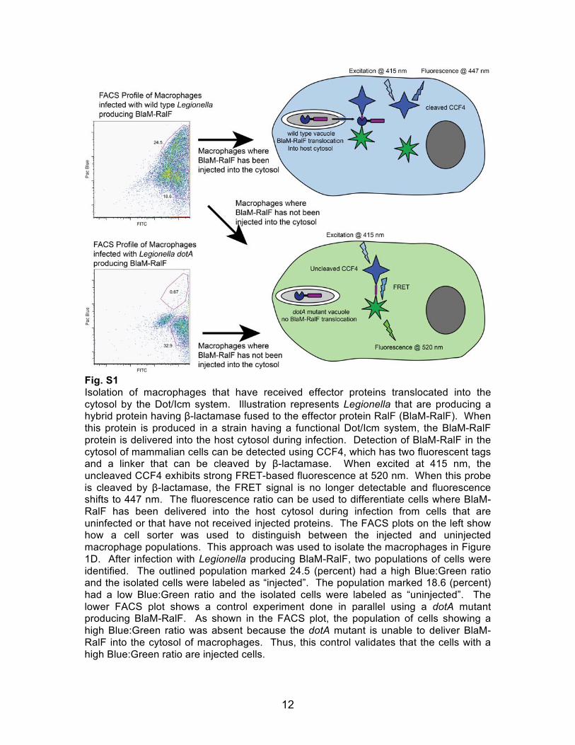
Fig. S1 Isolation of macrophages that have received effector proteins translocated into the cytosol by the Dot/Icm system. Illustration represents Legionella that are producing a hybrid protein having β-lactamase fused to the effector protein RalF (BlaM-RalF). When this protein is produced in a strain having a functional Dot/Icm system, the BlaM-RalF protein is delivered into the host cytosol during infection. Detection of BlaM-RalF in the cytosol of mammalian cells can be detected using CCF4, which has two fluorescent tags and a linker that can be cleaved by β-lactamase. When excited at 415 nm, the uncleaved CCF4 exhibits strong FRET-based fluorescence at 520 nm. When this probe is cleaved by β-lactamase, the FRET signal is no longer detectable and fluorescence shifts to 447 nm. The fluorescence ratio can be used to differentiate cells where BlaM-RalF has been delivered into the host cytosol during infection from cells that are uninfected or that have not received injected proteins. The FACS plots on the left show how a cell sorter was used to distinguish between the injected and uninjected macrophage populations. This approach was used to isolate the macrophages in Figure 1D. After infection with Legionella producing BlaM-RalF, two populations of cells were identified. The outlined population marked 24.5 (percent) had a high Blue:Green ratio and the isolated cells were labeled as “injected”. The population marked 18.6 (percent) had a low Blue:Green ratio and the isolated cells were labeled as “uninjected”. The lower FACS plot shows a control experiment done in parallel using a dotA mutant producing BlaM-RalF. As shown in the FACS plot, the population of cells showing a high Blue:Green ratio was absent because the dotA mutant is unable to deliver BlaM-RalF into the cytosol of macrophages. Thus, this control validates that the cells with a high Blue:Green ratio are injected cells.
13
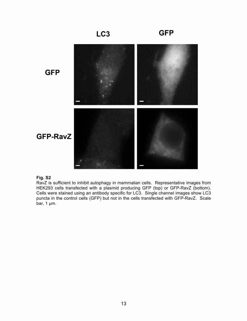
Fig. S2 RavZ is sufficient to inhibit autophagy in mammalian cells. Representative images from HEK293 cells transfected with a plasmid producing GFP (top) or GFP-RavZ (bottom). Cells were stained using an antibody specific for LC3. Single channel images show LC3 puncta in the control cells (GFP) but not in the cells transfected with GFP-RavZ. Scale bar, 1 µm.
14
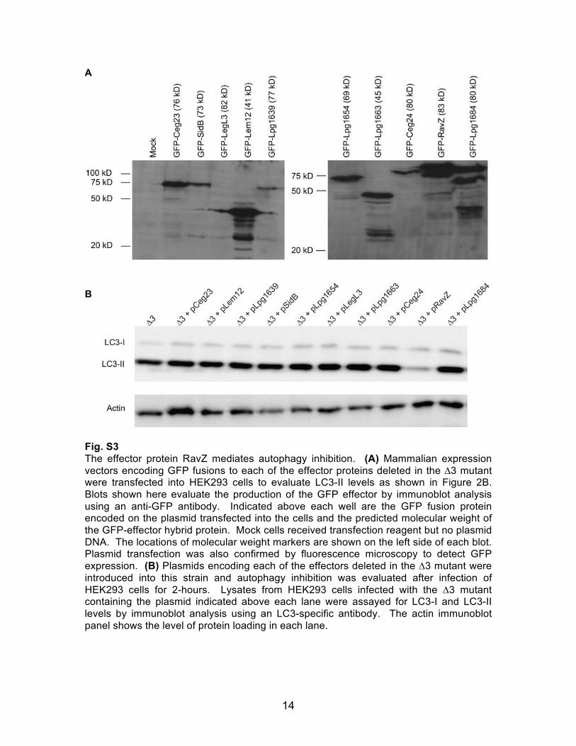
Fig. S3 The effector protein RavZ mediates autophagy inhibition. (A) Mammalian expression vectors encoding GFP fusions to each of the effector proteins deleted in the ∆3 mutant were transfected into HEK293 cells to evaluate LC3-II levels as shown in Figure 2B. Blots shown here evaluate the production of the GFP effector by immunoblot analysis using an anti-GFP antibody. Indicated above each well are the GFP fusion protein encoded on the plasmid transfected into the cells and the predicted molecular weight of the GFP-effector hybrid protein. Mock cells received transfection reagent but no plasmid DNA. The locations of molecular weight markers are shown on the left side of each blot. Plasmid transfection was also confirmed by fluorescence microscopy to detect GFP expression. (B) Plasmids encoding each of the effectors deleted in the ∆3 mutant were introduced into this strain and autophagy inhibition was evaluated after infection of HEK293 cells for 2-hours. Lysates from HEK293 cells infected with the ∆3 mutant containing the plasmid indicated above each lane were assayed for LC3-I and LC3-II levels by immunoblot analysis using an LC3-specific antibody. The actin immunoblot panel shows the level of protein loading in each lane.
15
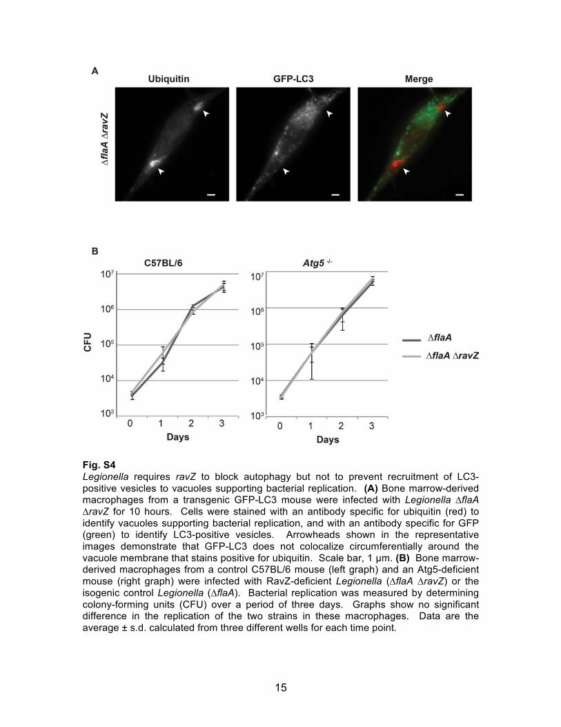
Fig. S4 Legionella requires ravZ to block autophagy but not to prevent recruitment of LC3-positive vesicles to vacuoles supporting bacterial replication. (A) Bone marrow-derived macrophages from a transgenic GFP-LC3 mouse were infected with Legionella ∆flaA ∆ravZ for 10 hours. Cells were stained with an antibody specific for ubiquitin (red) to identify vacuoles supporting bacterial replication, and with an antibody specific for GFP (green) to identify LC3-positive vesicles. Arrowheads shown in the representative images demonstrate that GFP-LC3 does not colocalize circumferentially around the vacuole membrane that stains positive for ubiquitin. Scale bar, 1 µm. (B) Bone marrow-derived macrophages from a control C57BL/6 mouse (left graph) and an Atg5-deficient mouse (right graph) were infected with RavZ-deficient Legionella (∆flaA ∆ravZ) or the isogenic control Legionella (∆flaA). Bacterial replication was measured by determining colony-forming units (CFU) over a period of three days. Graphs show no significant difference in the replication of the two strains in these macrophages. Data are the average ± s.d. calculated from three different wells for each time point.
16
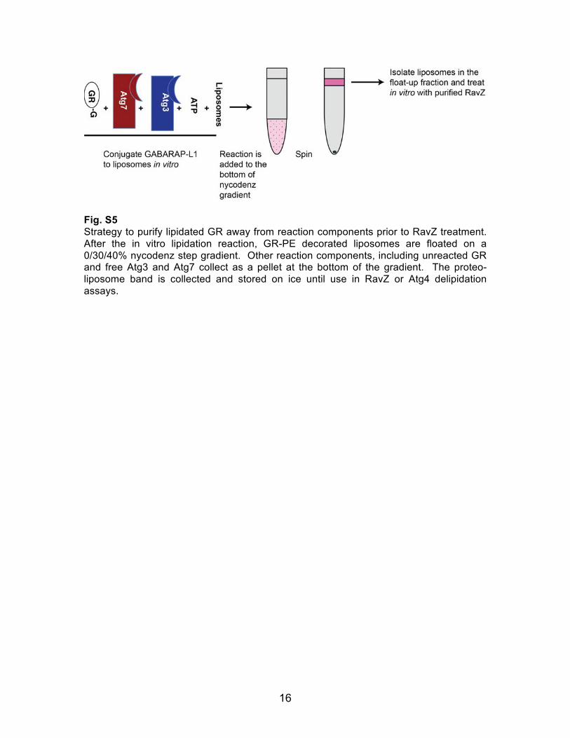
Fig. S5 Strategy to purify lipidated GR away from reaction components prior to RavZ treatment. After the in vitro lipidation reaction, GR-PE decorated liposomes are floated on a 0/30/40% nycodenz step gradient. Other reaction components, including unreacted GR and free Atg3 and Atg7 collect as a pellet at the bottom of the gradient. The proteo-liposome band is collected and stored on ice until use in RavZ or Atg4 delipidation assays.
17
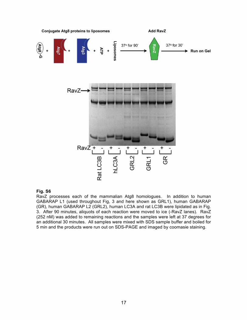
Fig. S6 RavZ processes each of the mammalian Atg8 homologues. In addition to human GABARAP L1 (used throughout Fig, 3 and here shown as GRL1), human GABARAP (GR), human GABARAP L2 (GRL2), human LC3A and rat LC3B were lipidated as in Fig. 3. After 90 minutes, aliquots of each reaction were moved to ice (-RavZ lanes). RavZ (252 nM) was added to remaining reactions and the samples were left at 37 degrees for an additional 30 minutes. All samples were mixed with SDS sample buffer and boiled for 5 min and the products were run out on SDS-PAGE and imaged by coomasie staining.
18
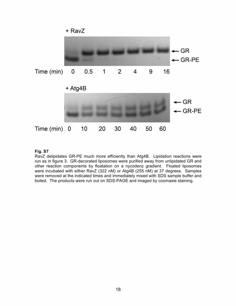
Fig. S7 RavZ delipidates GR-PE much more efficiently than Atg4B. Lipidation reactions were run as in figure 3. GR-decorated liposomes were purified away from unlipidated GR and other reaction components by floatation on a nycodenz gradient. Floated liposomes were incubated with either RavZ (322 nM) or Atg4B (255 nM) at 37 degrees. Samples were removed at the indicated times and immediately mixed with SDS sample buffer and boiled. The products were run out on SDS-PAGE and imaged by coomasie staining.
19
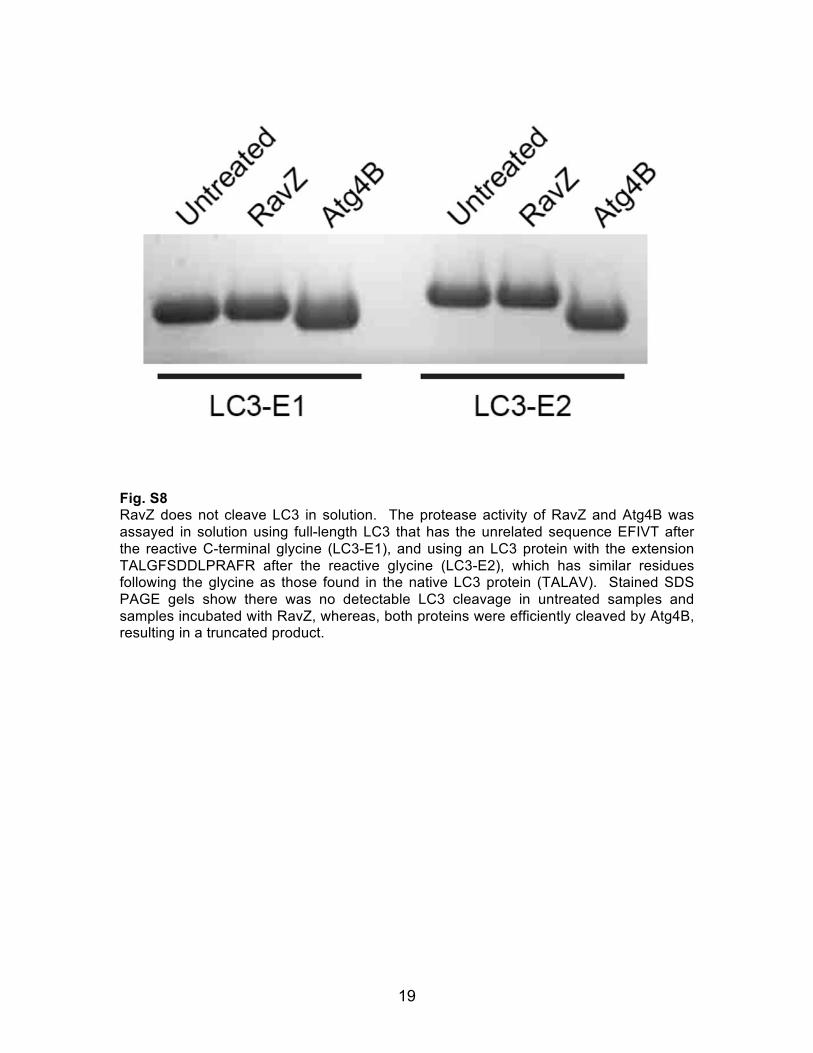
Fig. S8 RavZ does not cleave LC3 in solution. The protease activity of RavZ and Atg4B was assayed in solution using full-length LC3 that has the unrelated sequence EFIVT after the reactive C-terminal glycine (LC3-E1), and using an LC3 protein with the extension TALGFSDDLPRAFR after the reactive glycine (LC3-E2), which has similar residues following the glycine as those found in the native LC3 protein (TALAV). Stained SDS PAGE gels show there was no detectable LC3 cleavage in untreated samples and samples incubated with RavZ, whereas, both proteins were efficiently cleaved by Atg4B, resulting in a truncated product.
20
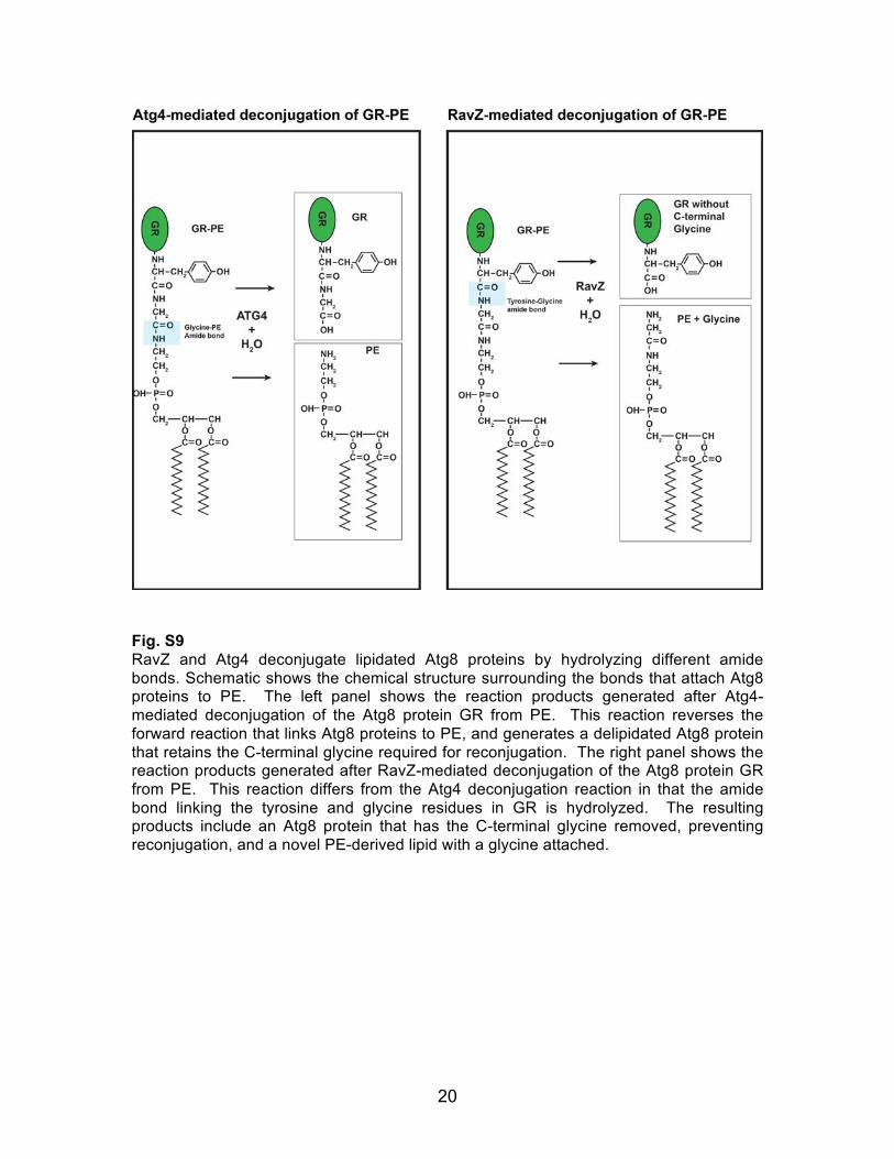
Fig. S9 RavZ and Atg4 deconjugate lipidated Atg8 proteins by hydrolyzing different amide bonds. Schematic shows the chemical structure surrounding the bonds that attach Atg8 proteins to PE. The left panel shows the reaction products generated after Atg4-mediated deconjugation of the Atg8 protein GR from PE. This reaction reverses the forward reaction that links Atg8 proteins to PE, and generates a delipidated Atg8 protein that retains the C-terminal glycine required for reconjugation. The right panel shows the reaction products generated after RavZ-mediated deconjugation of the Atg8 protein GR from PE. This reaction differs from the Atg4 deconjugation reaction in that the amide bond linking the tyrosine and glycine residues in GR is hydrolyzed. The resulting products include an Atg8 protein that has the C-terminal glycine removed, preventing reconjugation, and a novel PE-derived lipid with a glycine attached.
21
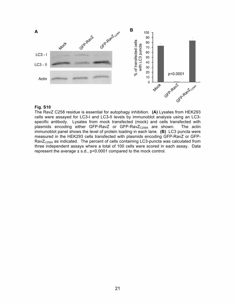
Fig. S10 The RavZ C258 residue is essential for autophagy inhibition. (A) Lysates from HEK293 cells were assayed for LC3-I and LC3-II levels by immunoblot analysis using an LC3-specific antibody. Lysates from mock transfected (mock) and cells transfected with plasmids encoding either GFP-RavZ or GFP-RavZC258A are shown. The actin immunoblot panel shows the level of protein loading in each lane. (B) LC3 puncta were measured in the HEK293 cells transfected with plasmids encoding GFP-RavZ or GFP-RavZC258A as indicated. The percent of cells containing LC3-puncta was calculated from three independent assays where a total of 100 cells were scored in each assay. Data represent the average ± s.d., p<0.0001 compared to the mock control.
22
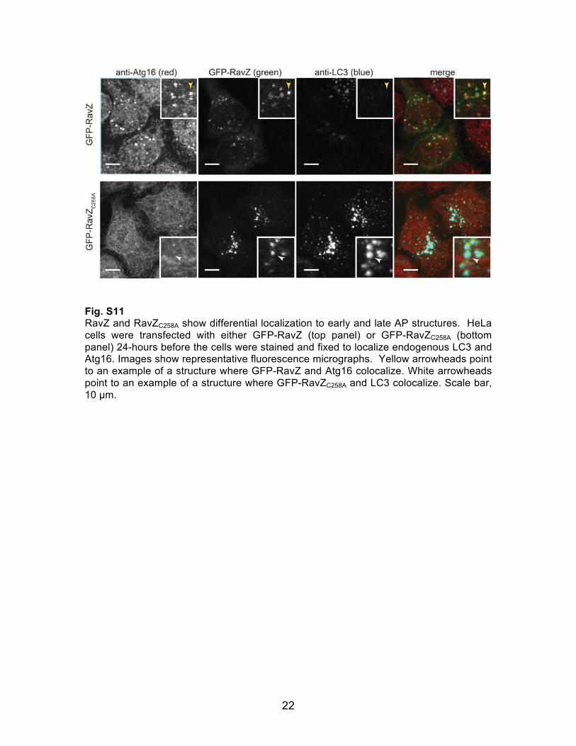
Fig. S11 RavZ and RavZC258A show differential localization to early and late AP structures. HeLa cells were transfected with either GFP-RavZ (top panel) or GFP-RavZC258A (bottom panel) 24-hours before the cells were stained and fixed to localize endogenous LC3 and Atg16. Images show representative fluorescence micrographs. Yellow arrowheads point to an example of a structure where GFP-RavZ and Atg16 colocalize. White arrowheads point to an example of a structure where GFP-RavZC258A and LC3 colocalize. Scale bar, 10 µm.
References and Notes 1. A. Hubber, C. R. Roy, Modulation of host cell function by Legionella pneumophila type IV
effectors. Annu. Rev. Cell Dev. Biol. 26, 261 (2010). doi:10.1146/annurev-cellbio-100109-104034 Medline
2. G. Segal, M. Purcell, H. A. Shuman, Host cell killing and bacterial conjugation require overlapping sets of genes within a 22-kb region of the Legionella pneumophila genome. Proc. Natl. Acad. Sci. U.S.A. 95, 1669 (1998). doi:10.1073/pnas.95.4.1669 Medline
3. J. P. Vogel, H. L. Andrews, S. K. Wong, R. R. Isberg, Conjugative transfer by the virulence system of Legionella pneumophila. Science 279, 873 (1998). doi:10.1126/science.279.5352.873 Medline
4. A. W. Ensminger, R. R. Isberg, Legionella pneumophila Dot/Icm translocated substrates: a sum of parts. Curr. Opin. Microbiol. 12, 67 (2009). doi:10.1016/j.mib.2008.12.004 Medline
5. Z. Xie, D. J. Klionsky, Autophagosome formation: core machinery and adaptations. Nat. Cell Biol. 9, 1102 (2007). doi:10.1038/ncb1007-1102 Medline
6. A. P. Hayward, S. P. Dinesh-Kumar, What can plant autophagy do for an innate immune response? Annu. Rev. Phytopathol. 49, 557 (2011). doi:10.1146/annurev-phyto-072910-095333 Medline
7. P. Kuballa, W. M. Nolte, A. B. Castoreno, R. J. Xavier, Autophagy and the immune system. Annu. Rev. Immunol. 30, 611 (2012). doi:10.1146/annurev-immunol-020711-074948 Medline
8. Y. Ichimura et al., A ubiquitin-like system mediates protein lipidation. Nature 408, 488 (2000). doi:10.1038/35044114 Medline
9. Y. Kabeya et al., LC3, a mammalian homologue of yeast Apg8p, is localized in autophagosome membranes after processing. EMBO J. 19, 5720 (2000). doi:10.1093/emboj/19.21.5720 Medline
10. D. J. Klionsky et al., Guidelines for the use and interpretation of assays for monitoring autophagy in higher eukaryotes. Autophagy 4, 151 (2008). Medline
11. H. Nagai, J. C. Kagan, X. Zhu, R. A. Kahn, C. R. Roy, A bacterial guanine nucleotide exchange factor activates ARF on Legionella phagosomes. Science 295, 679 (2002). doi:10.1126/science.1067025 Medline
12. T. J. O’Connor, Y. Adepoju, D. Boyd, R. R. Isberg, Minimization of the Legionella pneumophila genome reveals chromosomal regions involved in host range expansion. Proc. Natl. Acad. Sci. U.S.A. 108, 14733 (2011). doi:10.1073/pnas.1111678108 Medline
13. A. Jotwani, D. N. Richerson, I. Motta, O. Julca-Zevallos, T. J. Melia, Approaches to the study of Atg8-mediated membrane dynamics in vitro. Methods Cell Biol. 108, 93 (2012). doi:10.1016/B978-0-12-386487-1.00005-5 Medline
14. T. Kirisako et al., The reversible modification regulates the membrane-binding state of Apg8/Aut7 essential for autophagy and the cytoplasm to vacuole targeting pathway. J. Cell Biol. 151, 263 (2000). doi:10.1083/jcb.151.2.263 Medline
15. M. Y. Balakirev, S. O. Tcherniuk, M. Jaquinod, J. Chroboczek, Otubains: a new family of cysteine proteases in the ubiquitin pathway. EMBO Rep. 4, 517 (2003). doi:10.1038/sj.embor.embor824 Medline
16. X. Pan, A. Lührmann, A. Satoh, M. A. Laskowski-Arce, C. R. Roy, Ankyrin repeat proteins comprise a diverse family of bacterial type IV effectors. Science 320, 1651 (2008). doi:10.1126/science.1158160 Medline
17. K. H. Berger, R. R. Isberg, Two distinct defects in intracellular growth complemented by a single genetic locus in Legionella pneumophila. Mol. Microbiol. 7, 7 (1993). doi:10.1111/j.1365-2958.1993.tb01092.x Medline
18. T. Ren, D. S. Zamboni, C. R. Roy, W. F. Dietrich, R. E. Vance, Flagellin-deficient Legionella mutants evade caspase-1- and Naip5-mediated macrophage immunity. PLoS Pathog. 2, e18 (2006). doi:10.1371/journal.ppat.0020018 Medline
19. J. P. Bardill, J. L. Miller, J. P. Vogel, IcmS-dependent translocation of SdeA into macrophages by the Legionella pneumophila type IV secretion system. Mol. Microbiol. 56, 90 (2005). doi:10.1111/j.1365-2958.2005.04539.x Medline
20. J. J. Merriam, R. Mathur, R. Maxfield-Boumil, R. R. Isberg, Analysis of the Legionella pneumophila fliI gene: intracellular growth of a defined mutant defective for flagellum biosynthesis. Infect. Immun. 65, 2497 (1997). Medline
21. N. Mizushima, A. Yamamoto, M. Matsui, T. Yoshimori, Y. Ohsumi, In vivo analysis of autophagy in response to nutrient starvation using transgenic mice expressing a fluorescent autophagosome marker. Mol. Biol. Cell 15, 1101 (2004). doi:10.1091/mbc.E03-09-0704 Medline
22. C. L. Case, S. Shin, C. R. Roy, Asc and Ipaf Inflammasomes direct distinct pathways for caspase-1 activation in response to Legionella pneumophila. Infect. Immun. 77, 1981 (2009). doi:10.1128/IAI.01382-08 Medline
23. K. Arasaki, C. R. Roy, Legionella pneumophila promotes functional interactions between plasma membrane syntaxins and Sec22b. Traffic 11, 587 (2010). doi:10.1111/j.1600-0854.2010.01050.x Medline
24. T. Hara et al., Suppression of basal autophagy in neural cells causes neurodegenerative disease in mice. Nature 441, 885 (2006). doi:10.1038/nature04724 Medline
25. Y. Shao, Z. Gao, T. Feldman, X. Jiang, Stimulation of ATG12-ATG5 conjugation by ribonucleic acid. Autophagy 3, 10 (2007). Medline