Embed Size (px)
Citation preview

MicroVue CIC-Raji Cell Replacement EIA Page 1 of 14
SUMMARY
An enzyme immunoassay for the quantitation of circulating immune
complexes (CIC) containing C3 activation fragments in human serum
and plasma

MicroVue CIC-Raji Cell Replacement EIA Page 2 of 14
INTENDED USE The MicroVue CIC-Raji Cell Replacement Enzyme Immunoassay measures immune complexes containing C3 activation fragments in human serum and plasma.
SUMMARY AND EXPLANATION The importance of circulating immune complexes (CIC) and their relationship to various diseases has been the subject of investigation for a number of years. Formation of immune complexes is a protective, on-going, and usually benign process of a normally functioning immune system. CIC are removed from the circulation in the normal host by a number of complex biochemical, enzymatic, and cellular processes. The effective clearance of many CIC requires the activation of complement. Complement activation will result in C3 fragment deposition within the immune complex followed by enhanced elimination by phagocytic cells of the reticuloendothelial system. Under certain disease conditions, which are not fully understood, immune complexes may not be efficiently eliminated from the body. In these diseases, the immune complexes may accumulate and initiate complement-dependent injury in various organs and tissues. This activation of complement may begin a series of potentially destructive events in the host including anaphylatoxin production, cell lysis, leukocyte stimulation, and activation of macrophages and other cells.1 When immune complexes become fixed to vessel walls or cell membranes, destruction of normal tissue can occur, as in some cases of glomerulonephritis. Certain properties of CIC influence their potential pathogenicity. Of particular importance are: (1) nature, size and concentration of the antigen; (2) nature, size and concentration of the antibody; (3) rate of formation and clearance of the immune complexes.1,2 Circulating immune complexes have been measured in a variety of conditions: for example, infections, autoimmune disorders, trauma, and neoplastic proliferative diseases. Current studies suggest that CIC determinations can be important in the evaluation of certain diseases and, sometimes, in monitoring efficiency of therapy. This is especially true in systemic lupus erythematosus (SLE) and some forms of rheumatoid arthritis (RA).3,4 The first disease state linked to the formation of immune complexes was serum sickness, described in the early 1900’s by von Pirquet. Since that time elevated levels of CIC have been described in autoimmune diseases (SLE, SLE-related syndrome, RA), glomerulonephritis, neoplastic disease (Hodgkin’s, leukemia), bacterial infections (subacute bacterial endocarditis, leprosy), parasitic infections (malaria, schistosomiasis) and viral infections (hepatitis, mononucleosis). Over 40 assay techniques to detect or quantitate CIC have been described. Such tests as the Raji Cell assay, C1q deviation test, conglutinin test, fluid phase C1q binding procedures, rheumatoid factor assay, PEG precipitin test, and solid phase C1q assays have been described.1,5 Since the size and physiochemical properties of CIC vary markedly, none of these assays has been accepted as a standard. A collaborative study sponsored by the World Health Organization in 1978 determined that no single method was appropriate in all suspected disease states and recommended that at least two different assay techniques be performed to detect and measure CIC adequately.
PRINCIPLE OF THE PROCEDURE Fragments of the third complement component, C3, often become covalently bound to complement-activating immune complexes. Raji cells, which were derived from a continuous B lymphocyte culture cell line, bear CR2 complement receptors which bind the iC3b, C3d,g and C3d fragments of activated C3.6,7,8 The Raji Cell CIC assay is based upon the ability of the Raji cell CR2 receptors to bind immune complexes containing these C3 fragments.6 The MicroVue CIC-Raji Cell Replacement Enzyme Immunoassay also measures CIC-containing C3 fragments by using an immobilized monoclonal antibody that specifically binds the iC3b, C3d,g and C3d activation fragments of C3 in a manner which is analogous to the Raji cell CR2 binding reaction.

MicroVue CIC-Raji Cell Replacement EIA Page 3 of 14
In the first stage, standards and serum or plasma specimens diluted in Complement Specimen Diluent are added to microassay wells coated with monoclonal antibodies to human C3 fragments and incubated. During this incubation, immune complexes containing C3 activation fragments are captured by the solid phase antibody. After incubation, a wash cycle removes unbound serum or plasma proteins. In the second stage, horseradish peroxidase (HRP)-conjugated mouse anti-human IgG is added to each test well and incubated. During this incubation, the conjugate binds to the immune complexes which are now bound to the microassay wells. A wash cycle removes unbound conjugate. In the third stage, an enzyme substrate is added to each test well. The bound HRP-conjugated antibody reacts with the chromogenic substrate forming a green color. After incubation, a reagent is added to stop color development. The standard and test specimen absorbances (A405 values) are measured spectrophotometrically. The intensity of the green color that forms is proportional to the amount of CIC binding to the solid phase. A standard curve is generated by plotting the A405 values obtained with each standard versus its indicated concentration. The concentration of immune complexes present in the test specimen is determined by reference to the standard curve. Results are expressed as micrograms of serum-treated heat-aggregated human gamma globulin (HAGG) equivalents per mL (μg Eq/mL). To confirm a positive CIC result, the specimen may be assayed after dilution in CIC-Raji Confirmation Diluent which contains blocking antibodies with specificities similar to the antibody immobilized on the microassay well.
REAGENTS AND MATERIALS PROVIDED 96 Assays for CIC-Raji Cell Replacement Enzyme The CIC-Raji Cell Replacement EIA kit contains the following:
A B C D E
Standards Parts A9970-A9974 1 each, 2 mL Each contains a known quantity of serum-treated heat-aggregated human gamma globulin (HAGG) in PBS, 2.5% stabilizers, 0.01% thimerosal.
L Low Control Part A9919 3 each (Lyophilized) Contains low levels of serum-treated HAGG in human serum, 20 mM EDTA, 0.01% thimerosal
H High Control Part A9920 3 each (Lyophilized) Contains high levels of serum-treated HAGG in human serum, 20 mM EDTA, 0.01% thimerosal
❶ Microassay Plate Part A9512 12 each 96-well with retainer and holder consisting of 12 eight-well strips coated with mouse anti-human C3 fragments in a resealable foil pouch
❷ Stop Solution Part A3673 6 mL Contains 250 mM oxalic acid
❸ 20X Wash Solution Concentrate Part A9957 2 each, 50 mL Contains phosphate buffered saline (PBS), 1.0% Tween-20® and 0.035% Proclin® 300
❹ Complement Specimen Diluent Part A3670 50 mL Contains PBS, 2.5% stabilizers, 0.035% ProClin 300
❺ Substrate Diluent Part A3672 25 mL Contains 0.1M citrate buffer and 0.05% H2O2
❻ Substrate Concentrate Part A3671 1.5 mL Contains 0.7% 2,2’-Azino-bis(3-ethylbenzthiazoline-6-sulfonic acid), diammonium salt

MicroVue CIC-Raji Cell Replacement EIA Page 4 of 14
MATERIALS REQUIRED BUT NOT PROVIDED Timer (60 minute range) Calculator or other computational method to validate the assay Clean, unused microassay plates and/or test tubes and racks Container for wash buffer dilution Wash bottle or other immunoassay washing system Adjustable multichannel pipette (8 or 12 channels) or repeating micropipettes (optional) Clean pipettes, 1 mL, 5 mL, and 10 mL Micropipettes and pipette tips Plate reader capable of optical density A405 readings between 0.0 and 2.0 Deionized or distilled water
WARNINGS AND PRECAUTIONS For in vitro diagnostic use. Treat specimen samples as potentially biohazardous material. Follow Universal Precautions when handling contents of this kit and any patient samples. Use the supplied reagents as an integral unit prior to the expiration date indicated on the package label. Store assay reagents as indicated. Do not use Coated Strips if pouch is punctured. Thimerosal is used as a preservative. Incidental contact with or ingestion of buffers or reagents containing
thimerosal can lead to increased hypersensitivity reactions including irritation to the skin, eyes, or mouth. Seek medical attention if symptoms are experienced. Exposure to thimerosal may have potential mutagenic effects. Avoid contact with strong acids and bases.
ProClin 300 is used as a preservative. Incidental contact with or ingestion of buffers or reagents containing ProClin can cause irritation to the skin, eyes or mouth. Use good laboratory practices to reduce exposure. Seek medical attention if symptoms are experienced.
Use of multichannel pipettes or repeat pipettors is recommended to ensure timely delivery of reagents. For accurate measurement of samples, add samples and standards precisely. Pipette carefully using only
calibrated equipment. Proper collection and storage of test specimens are essential for accurate results (see SPECIMEN
COLLECTION AND STORAGE). Avoid microbial or cross-contamination of specimens, reagents, or materials. Incorrect results may be
obtained if contaminated. Test each sample in duplicate. Do not use a microassay well for more than one test. Using incubation times and temperatures other than those indicated in the Procedure section may give
erroneous results. The Substrate Concentrate is light sensitive. Avoid prolonged exposure to bright or direct light. Store
reagents in the dark when not in use. Do not allow microassay wells to dry once the assay has begun. When adding or aspirating liquids from the microassay wells, do not scrape or touch the bottom of the
wells. Heat-inactivated, hyperlipemic or contaminated specimens may give erroneous results.
❼ CIC-Raji Conjugate Part A9516 2 each, 3 mL Contains peroxidase-conjugated (mouse) anti-human IgG suspended in an HRP stabilizing buffer with preservative
❽ CIC-Raji Confirmation Diluent Part A9517 12 mL Contains PBS, 2.5% Stabilizers, anti-human C3 fragment antibody. 0.35% ProClin 300
Tween® 20 is a registered trademark of ICI Americas Inc. ProClin® is a registered trademark of Rohm and Haas Company.

MicroVue CIC-Raji Cell Replacement EIA Page 5 of 14
To avoid aerosol formation during washing, use an apparatus to aspirate the wash fluid into a bottle containing household bleach.
This assay may be performed with any validated washing method. For best results, do not use a multichannel pipette to wash the microassay plate.
Testing should be performed in an area with adequate ventilation. Dispose of containers and unused contents in accordance with Federal, State and Local regulatory
requirements. Wear suitable protective clothing, gloves, and eye/face protection when handling the contents of this kit. Wash hands thoroughly after handling. For additional information on hazard symbols, safety, handling and disposal of the components within this
kit, please refer to the Safety Data Sheet (SDS) located at quidel.com.
STORAGE Store unopened kit at 2°C to 8°C. After the kit is opened, the 20X Wash Solution Concentrate may be stored at 2°C to 30°C.
After selecting the reagents or materials to be used in the assay, return the unused reagents immediately to their appropriate storage temperatures. Bring reagents and materials to room temperature (15°C to 30°C) before use.
INDICATIONS OF INSTABILITY OR DETERIORATION OF REAGENTS The Substrate Concentrate may range in color from colorless to pale or dark green. This condition will not influence performance. However, the freshly prepared Substrate Solution should be colorless to pale green. A dark green color indicates that the prepared Substrate Solution has deteriorated, must be discarded, and new Substrate Solution prepared in clean glassware.
Cloudiness or discoloration of the diluted Wash Solution indicates a deterioration of this reagent. If this occurs, the solution should be discarded.
SPECIMEN COLLECTION AND STORAGE Handle and dispose of all specimens using Universal Precautions Serum or plasma specimens should be collected aseptically and prepared using standard techniques for clinical laboratory testing.9,10 Do not heat-inactivate the specimens. Any particulate matter should be cleared from the specimens by low speed centrifugation before testing.
The specimens may be stored on ice, approximately 0°C for up to 6 hours. If specimens are stored for longer periods, they should be frozen at –70°C or below (storage at –20°C may yield erroneous results).
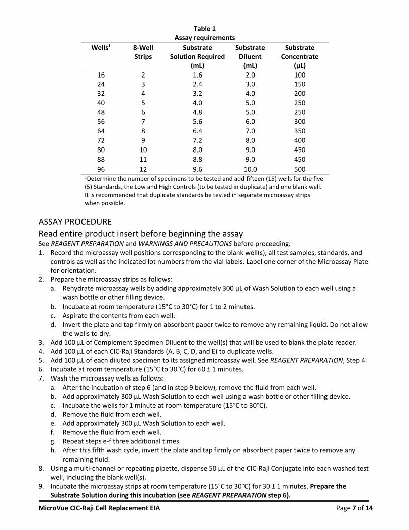
REAGENT PREPARATION Refer to Table 1 for the amounts of Substrate Solution and microassay strips required per number of tests. After removing the needed reagents and materials, return the unused items to their appropriate storage temperatures (see STORAGE). Bring all reagents and materials for the assay to room temperature (15 – 30°C) before use.
1. Wash Solution. Mix the 20X Wash Solution Concentrate by inverting the bottle several times. If the 20X Wash Solution Concentrate has been stored at 2°C to 8°C, crystals may have formed. To dissolve the crystals, warm the bottle in a 37°C to 50°C water bath until all crystals have dissolved and follow by mixing thoroughly. Prepare the Wash Solution by diluting the entire contents of one of the bottles of 20X Wash Solution Concentrate up to one liter with distilled or deionized water. Mix thoroughly. The Wash Solution is stable for 30 days when stored in a clean container at 2°C to 8°C. If discoloration or cloudiness occurs, discard the reagent.

MicroVue CIC-Raji Cell Replacement EIA Page 6 of 14
2. Selecting the Microassay Strips. Determine the number of microassay strips required for the assay by referring to Table 1. Remove the strip retainer from the assembled plate. Remove the unneeded strips and place them in the storage bag, reseal the bag and return it to storage at 2°C to 8°C. Secure the strips to be used in the assay.
3. Specimen Dilution Caution: Treat all specimens as if potentially infectious. Do not use heat-inactivated or contaminated specimens. Determine the number (N) of specimens to be tested. Label test tubes #1 through #N and record on the provided data sheet which specimen corresponds to each tube. Prepare a 1:50 dilution of each specimen using the Complement Specimen Diluent (e.g., 10 μL test specimen mixed with 490 μL Complement Specimen Diluent). Mix thoroughly, but avoid formation of foam and bubbles. Do not store or reuse diluted specimens.
4. Adding Diluted Specimens to the Microtiter Wells. Either of two methods can be used to add diluted specimens, standards, controls, and buffer, to the wells (see Step 5 of ASSAY PROCEDURE). For small assay runs where only a few specimens are being tested, the diluted specimens and other reagents can be added directly to their assigned wells with a micropipette (100 μL/well). For small or large runs, but especially larger runs, we recommend the use of a multichannel pipettor for adding specimens as follows. (A multi-channel pipettor may be used to conveniently add the Conjugate, Substrate and Stop Solution, as well.) In order to load the standards, controls and diluted specimens into the microassay wells as rapidly as possible, a “replica plating” procedure can be employed. Instead of adding 100 μL of each standard, control or diluted specimen to the antibody-coated wells individually, 120–130 μL of each solution can be added to individual wells in a blank plate (not provided) corresponding to the final EIA pattern desired. After all the solutions to be tested have been added to the microassay wells in the blank plate, rapidly transfer 100 μL from each blank well to the antibody-coated wells using a multichannel micropipettor. To avoid the possibility of cross-contamination, pipette tips must be changed each time there is a change in the composition of the samples to be transferred.
5. CIC-Raji Confirmation Dilution (optional). If confirmation of positive results is desired, determine the number (N) of specimens to be confirmed. Label test tubes #1c through #Nc and record. Prepare a 1:50 dilution using the CIC-Raji Confirmation Diluent. A specimen diluted in CIC-Raji Confirmation Diluent must be run concurrently with the same dilution of specimen in the Complement Specimen Diluent.
6. Preparation of Substrate Solution. Prepare just prior to use. Determine the required volume of Substrate Solution from Table 1, below. Prepare the Substrate Solution by adding 50 μL of Substrate Concentrate to each mL of Substrate Diluent. Mix thoroughly.
7. CIC Controls. Reconstitute each control with 1.0 ± 0.05 mL of the Complement Specimen Diluent provided. After reconstitution, mix each vial gently, but completely, to ensure complete rehydration. Allow the rehydrated solutions to incubate at room temperature (15°C to 30°C) for 10 to 15 minutes. Mix gently but completely again, and use. NO FURTHER DILUTION IS REQUIRED.
MicroVue CIC-Raji Cell Replacement EIA Page 7 of 14
Table 1 Assay requirements
Wells1 8-Well Strips
Substrate Solution Required
(mL)
Substrate Diluent
(mL)
Substrate Concentrate
(µL)
16 2 1.6 2.0 100 24 3 2.4 3.0 150
32 4 3.2 4.0 200
40 5 4.0 5.0 250
48 6 4.8 5.0 250
56 7 5.6 6.0 300
64 8 6.4 7.0 350
72 9 7.2 8.0 400
80 10 8.0 9.0 450
88 11 8.8 9.0 450
96 12 9.6 10.0 500 1Determine the number of specimens to be tested and add fifteen (15) wells for the five (5) Standards, the Low and High Controls (to be tested in duplicate) and one blank well. It is recommended that duplicate standards be tested in separate microassay strips when possible.
ASSAY PROCEDURE Read entire product insert before beginning the assay See REAGENT PREPARATION and WARNINGS AND PRECAUTIONS before proceeding. 1. Record the microassay well positions corresponding to the blank well(s), all test samples, standards, and
controls as well as the indicated lot numbers from the vial labels. Label one corner of the Microassay Plate for orientation.
2. Prepare the microassay strips as follows: a. Rehydrate microassay wells by adding approximately 300 μL of Wash Solution to each well using a
wash bottle or other filling device. b. Incubate at room temperature (15°C to 30°C) for 1 to 2 minutes. c. Aspirate the contents from each well. d. Invert the plate and tap firmly on absorbent paper twice to remove any remaining liquid. Do not allow
the wells to dry. 3. Add 100 μL of Complement Specimen Diluent to the well(s) that will be used to blank the plate reader. 4. Add 100 μL of each CIC-Raji Standards (A, B, C, D, and E) to duplicate wells. 5. Add 100 μL of each diluted specimen to its assigned microassay well. See REAGENT PREPARATION, Step 4. 6. Incubate at room temperature (15°C to 30°C) for 60 ± 1 minutes. 7. Wash the microassay wells as follows:
a. After the incubation of step 6 (and in step 9 below), remove the fluid from each well. b. Add approximately 300 μL Wash Solution to each well using a wash bottle or other filling device. c. Incubate the wells for 1 minute at room temperature (15°C to 30°C). d. Remove the fluid from each well. e. Add approximately 300 μL Wash Solution to each well. f. Remove the fluid from each well. g. Repeat steps e-f three additional times. h. After this fifth wash cycle, invert the plate and tap firmly on absorbent paper twice to remove any
remaining fluid. 8. Using a multi-channel or repeating pipette, dispense 50 μL of the CIC-Raji Conjugate into each washed test
well, including the blank well(s). 9. Incubate the microassay strips at room temperature (15°C to 30°C) for 30 ± 1 minutes. Prepare the
Substrate Solution during this incubation (see REAGENT PREPARATION step 6).

MicroVue CIC-Raji Cell Replacement EIA Page 8 of 14
10. Wash the microassay wells after the 30-minute incubation of Step 9, as described above, step 7. 11. Immediately following the wash procedure using a multichannel or repeating pipette, dispense 100 μL of
the freshly prepared Substrate Solution into each well, including the blank(s). 12. Incubate the microassay strips at room temperature (15°C to 30°C) for 30 ± 1 minutes. 13. Using a multi-channel or repeating pipette, add 50 μL of the Stop Solution to each well to stop the
enzymatic reaction. The Stop Solution should be added to the wells in the same order and at the same rate as was the Substrate Solution. Gently tap the plate to disperse the color development evenly.
14. Determine the absorbance reading at 405 nm (A405 value) for each test well within one hour after the addition of the Stop Solution (step 13), making a blank correction in accordance with the spectrophotometric system in use.
15. Keep the strip holder and strip retainer for future use. 16. Dispose of the remaining diluted specimens, substrate, and the used microassay strips in accordance with
Federal, State and Local regulations. Keep the strip holder and strip retainer for future use.
RECOMMENDED CONFIRMATION METHOD If independent confirmation of a positive result is required, or if a positive result is inconsistent with the clinical interpretation, the positive specimen may be reassayed using a confirmation test. A negative result cannot be confirmed. The confirmation method utilizes a specimen diluent (the CIC-Raji Confirmation Diluent) which contains anti-human C3 fragment antibodies. To confirm a positive result, an aliquot of the specimen must be diluted in the CIC-Raji Confirmation Diluent and a second aliquot diluted similarly in the Complement Specimen Diluent. Both samples are then assayed according to the usual MicroVue CIC-Raji Cell Replacement EIA procedure. See REAGENT PREPARATION and INTERPRETATION OF RESULTS sections for details.
QUALITY CONTROL If the positive and/or negative control fail to perform as specified, please contact Quidel Technical Support as soon as possible. In addition to controls, the MicroVue CIC-Raji Cell Replacement Assay also provides a RECOMMENDED CONFIRMATION METHOD and VALIDATION PARAMETERS. By utilizing the Controls, Standards with validation, and the confirmation method, you should achieve reproducible and accurate results.
INTERPRETATION OF RESULTS Calculation of Results Calculations: The standard curve is generated using the blank-subtracted A405 value of each CIC-Raji Standard on the y-axis versus the assigned micrograms of serum-treated, heat-aggregated gamma globulin equivalents/mL (μg Eq/mL) indicated on the vial label for each Standard on the x-axis. After linear regression, the generated standard curve must meet the validation requirements (see below). The sample concentrations are then calculated directly from the standard curve. Most computers and calculators are capable of performing these calculations. Alternatively, the data may be graphed manually and the values (μg Eq/mL) of the test samples read directly from the best-fit line of the standard curve. An example of a typical standard curve is shown in Figure 1.

MicroVue CIC-Raji Cell Replacement EIA Page 9 of 14
Figure 1 Example of Standard Curve
Confirmation Test Calculation: To confirm a positive result, the immune complex concentration [CIC] determined in the sample diluted in CIC-Raji Confirmation Diluent is divided by the immune complex concentration measured in the sample diluted in Complement Specimen Diluent to generate a ratio:
Ratio = [CIC] in CIC-Raji Confirmation Diluent
[CIC] in Complement Specimen Diluent
Sample (A405) µg Eq/mL
Standard A 0.06 0 Standard B 0.18 5 Standard C 0.45 17 Standard D 0.75 30 Standard E 1.16 48
Specimen 1 0.15 3.9 Specimen 2 0.50 19.1 Specimen 3 1.00 40.9
r = 1.00 m = 0.023 b = 0.06
Validation Determine the slope, intercept, and correlation coefficient of the derived best fit line. The values must be within the specified ranges to qualify the assay:
Correlation coefficient (r): > 0.95
Slope (m): 0.013 to 0.034
y-intercept (b): (–) 0.07 to (+) 0.10
Most normal subjects demonstrate measurable levels of CIC. Since there is no accepted abnormal level of CIC, the user should establish in-house normal levels. As a guideline, levels of CIC based on the results obtained from the normal populations described in EXPECTED VALUES are as follows: Normal Results: Values less than or equal to 15 μg Eq/mL are considered normal for levels of CIC. Abnormal Results: Values greater than or equal to 20 μg Eq/mL are considered abnormal for levels of CIC. Specimens which have measured concentrations of CIC greater than CIC-Raji Standard E should be reported as greater than the assigned CIC-Raji Standard E concentration indicated on the vial label.

MicroVue CIC-Raji Cell Replacement EIA Page 10 of 14
Equivocal Values: Values greater than 15 μg Eq/mL and less than 20 μg Eq/mL are equivocal. These specimens may be repeat-tested or a new sample may be drawn and tested, if indicated. If an equivocal specimen repeats as equivocal, the specimen is then considered significantly higher than normal and may be reported as abnormal. Confirmation Results: If the ratio is less than 0.5, the positive CIC result is confirmed. In other words, greater than 50% reduction in the apparent CIC concentration confirms a positive result. Occasionally, positive specimens may not confirm. Non-confirming specimens may be due to, among other reasons: (1) mishandled specimens (e.g. contaminated or heat-inactivated) or (2) specimens containing human IgG antibodies which bind to mouse IgG. Such specimens, however, are not necessarily negative for CIC. The material causing the apparent false positive result may mask concomitantly occurring CIC which, if they were present alone, would otherwise give a confirmable positive CIC result.
LIMITATIONS OF THE PROCEDURE This test measures immune complexes or aggregates of human IgG containing C3 activation fragments. Therefore conditions that promote aggregation of IgG or complement activation must be avoided during sample collection and processing.
EXPECTED VALUES Fifty (50) selected sera were obtained from a reference laboratory which receives specimens from throughout the United States. These sera were tested in the MicroVue CIC-Raji Cell Replacement EIA and in the reference laboratory’s Raji Cell assay. There was a 92% agreement between the two assays for the measurement of the presence of CIC. Sera obtained from sixty-two (62) SLE patients at two eastern U.S. clinics and from twenty-nine (29) RA patients at a southern U.S. rheumatology clinic were tested in the MicroVue CIC-Raji Cell Replacement EIA. In addition, sera from twenty-six (26) normal, healthy subjects at the two SLE clinic sites and from twenty-five (25) clinically non-autoimmune patients reporting to the rheumatology clinic were tested. The mean concentrations of CIC, the standard deviations, and the frequency distribution for each population are presented in Figures 2, 3, and 4.
Figure 2 CIC-Raji Cell Replacement EIA
Systemic Lupus Erythematosus Population

MicroVue CIC-Raji Cell Replacement EIA Page 11 of 14
Figure 3 CIC-Raji Cell Replacement EIA
Rheumatoid Arthritis Population
Figure 4 CIC-Raji Cell Replacement EIA
Control Population
Within one SLE site and the RA site, the degree of disease activity was assigned independently of any CIC laboratory data. This assessment was made by the attending physician(s). To assure consistent reporting, one physician at each site later reviewed the patient records and assigned the disease activity. One RA patient was described as late stage “burnout.” The CIC result for this patient was 1 μg Eq/mL. Since there were no other similar RA patients, this individual result is not included in Table 2. Table 2 shows the observed relationship between CIC measured by the MicroVue CIC-Raji Cell Replacement Enzyme Immunoassay and the disease activity of the patients.
Table 2 CIC-Raji Replacement Assay Results Compared to Disease Activity
% Abnormal
Low Activity Moderate Activity High Activity
SLE 8% (1/12) 36% (4/11) 79% (11/14) RA 0% (0/4) 19% (3/16) 50% (4/8)
1 Inside the parentheses (after each % abnormal) is indicated the number of patients testing abnormal divided by the number of patients classified with the particular disease activity.

MicroVue CIC-Raji Cell Replacement EIA Page 12 of 14
PERFORMANCE CHARACTERISTICS Accuracy A World Health Organization (WHO) Immune Complex Standard, i.e., tetanus toxoid-anti-tetanus toxoid complexes pre-incubated in fresh normal human serum, was used to standardize the assay. To test the accuracy of the assay, five dilutions of the WHO Standard were tested in triplicate in nine runs in the MicroVue kit. The assayed concentrations showed a correlation of 0.99 with the known values.
Reproducibility Patient specimens and Kit Standards were tested in nine assay runs in two different kit lots. Each was tested in triplicate within each assay run. The average variation between each run for the specimens and Kit Standards is shown in Table 3, as inter-assay variation. The average variation within each run for the specimens and Kit Standards is shown in Table 3 as intra-assay variation.
Table 3 Assay Reproducibility
Mean (µg Eq/mL)
Intra-assay S.D. (% CV)
Inter-assay S.D. (% CV)
Specimen 1 56 5 9
Specimen 2 13 9 23
Standard 1 4 8 30 Standard 2 11 7 15
Standard 3 23 5 9 Standard 4 32 4 6 Standard 5 40 5 6
Standard 6 48 4 5 Standard 7 59 3 4
Sensitivity The MicroVue CIC-Raji Cell Replacement EIA measures at least 4 μg Eq/mL or greater of CIC analyte based on comparison to the WHO Standard.
Specificity The fifty-one (51) control sera described in the EXAMPLE VALUES section were tested in the MicroVue CIC-Raji Cell Replacement assay. Only three were positive (repeatedly greater than 15 μg Eq/mL), yielding a 94% specificity.
ASSISTANCE To place an order or for technical support, please contact a Quidel representative at 800.874.1517 (in the U.S.) or 858.552.1100 (outside the U.S.), Monday through Friday, from 8:00 a.m. to 5:00 p.m., Eastern time. Orders may also be placed by fax at 740.592.9820. For e-mail support contact [email protected] or [email protected]. For services outside the U.S.A., please contact your local distributor. Additional information about Quidel, our products, and our distributors can be found on our website quidel.com.

MicroVue CIC-Raji Cell Replacement EIA Page 13 of 14
REFERENCES 1. McDougal, J.S, McDuffie, F.C., Immune Complexes in Man: Detection and Clinical Significance, Advances in
Clinical Chemistry, Vol. 24, p. 1, 1985. 2. Endo, L., Corman, L.C., Panush, R.S., Clinical Utility of Assays for Circulating Immune Complexes, Medical Clinics
of North America, Vol. 69, No. 4, p. 623, July 1985. 3. Abrass, C.K., Nies, K.M., Louie, J.S., Border, W.A., Glassock, R.J., Correlation and Predictive Accuracy of
Circulating Immune Complexes with Disease Activity in Patients with Systemic Lupus Erythematosus, Arthritis Rheum., Vol. 23, p. 273, 1980.
4. Duquesnoy, B., Circulating Immune Complexes and Complement in Rheumatoid Arthritis, Ann. Biol. Clin., Vol. 42, p. 71, 1984.
5. Theofilopoulos, A.N., The Raji, Conglutinin, and Anti-C3 Assays for the Detection of Complement-Fixing Immune Complexes, Methods in Enzymology, Vol. 74, p. 511, 1981.
6. Agnello, V., Winchester, R.J., Kunkel, H.G., Precipitin Reactions of the C1q Component of Complement with Aggregated Gamma-Globulin and Immune Complexes in Gel Diffusion, Immunology, Vol. 19, p. 909, 1970.
7. Hack, C.E., Huijbregts, C.C., Paardekooper, J., Influence of Ionic Strength, EDTA Concentration, Endogenous C1q and Polyanions on the 125I-C1q-Binding Test, J. Immunol. Meth., Vol. 72, p. 197, 1984.
8. Richardson, J.H., Barkley, W.E., Biosafety in Microbiological and Biomedical Laboratories, p. 11-13, 1984. 9. Davidson, I., et al., The Blood, p. 1 20. in Cl. Davidson and J.B. Henry (ed.), Todd-Sanford Clinical Diagnosis,
W.B. Saunders Co., Philadelphia, Pennsylvania, 1969. 10. Centers for Disease Control. Recommendations for prevention of HIV transmission in health-care settings.
MMWR 1987;36 (suppl no. 2S):001.
A002 – MicroVue CIC-Raji Cell Replacement EIA Kit
MDSS GmbH Schiffgraben 41 30175 Hannover, Germany Quidel Corporation 2005 East State Street, Suite 100 Athens, OH 45701 USA quidel.com PIA002001EN00 (04/17)

MicroVue CIC-Raji Cell Replacement EIA Page 14 of 14
GLOSSARY