1
8 WWW.CEN-ONLINE.ORG MAY 5, 2008 T HE FIRST TECHNIQUE for imaging carbohy- drates as they are produced on cell surfaces of live animals has been developed by glycobiology specialist Carolyn R. Bertozzi and coworkers at the University of California, Berkeley. Specific glycan (oli- gosaccharide) structures in cells are important markers and mediators of many physiological processes, but up to now changes in these glycans couldn’t be visualized in living organisms. The new technique, based on cell-friendly fluorescent-labeling chemistry devised by the same group, will help scientists more closely probe the functional roles of carbohydrates during development and in both health and disease. Chemistry professor M. G. Finn of Scripps Research Institute says the Bertozzi group’s technique “provides a ‘global positioning system’ for tracking carbohydrates through organisms. This is chemical biology at its fin- est: refined and reliable chemical tools illuminating— literally—molecular interactions in living biological systems.” In recent years, Bertozzi and cowork- ers have developed techniques for imag- ing glycans in live cells. First, they feed azide-derivatized sugars to live organ- isms, which incorporate the modified sugars into their glycans. After cells are removed from the organisms, the la- beled glycans can be imaged by using the Staudinger ligation, a reaction developed by Bertozzi’s group in 2000, to mark the glycans with fluorescent probes. But that reaction and similar ones are too slow or too toxic for use in live animals. Last year, Bertozzi’s group developed copper-free click chemistry with difluo- rinated cyclooctyne (DIFO) reagents (Proc. Natl. Acad. Sci. USA 2007, 104, 16793; C&EN, Oct. 22, 2007, page 15). This ligation reaction is faster than the Staudinger liga- tion and is nontoxic, unlike the copper-catalyzed click chemistry reactions on which it’s based. In the new study the researchers introduce azide-derivatized N-acetyl- galactosamine (GalNAc) metabolically into developing zebrafish embryos and treat the embryos with DIFO re- agent to fluorescently label GalNAc-containing glycans. They then use microscopy to image the labeled glycans in the developing animals (Science 2008, 320, 664). The technique makes it possible for Bertozzi and coworkers to detect increases in glycan biosynthesis in specific locations during zebrafish development and to visualize patterns of glycan expression that previously would have been undetectable. The researchers believe the technique can be extended to other sugars, such as sialic acid. The new study represents “a significant advance in visualizing the distribution of sugars on cell surfaces, where they are believed to mediate cell adhesion, mi- gration, and recognition processes,” says glycan-imag- ing specialist Mary L. Kraft of the University of Illinois, Urbana-Champaign. “Changes in cell-surface glycans also occur during embryo development and are char- acteristic of several diseases, such as cancer. However, how they mediate these processes is largely unknown” because no methods have existed to visualize them in live animals. The ability of the new approach “to deter- mine how glycans are distributed spatially within a tis- sue and in time can shed light on their functional roles and help identify cell-surface markers indicative of the physiological status of cells.” Chemistry Nobel Prize winner K. Barry Sharpless of Scripps, who helped develop the concept of click chem- istry, comments that Bertozzi’s “in vivo light-up of the zebrafish is a stunning first.”—STU BORMAN Cell-surface glycans shine in this microscopy image of the head of a three-day-old zebrafish embryo treated with the new technique. news of the week MAY 5, 2008 EDITED BY WILLIAM G. SCHULZ & KIMBERLY R. TWAMBLY COURTESY OF CAROLYN BERTOZZI AND COWORKERS SUGARS IMAGED IN LIVE ANIMALS CARBOHYDRATES: Technique is ‘global positioning system’ for tracking glycans in organisms AcO AcO OAc OAc HN O N 3 O O O F F NH N N N O O F F NH DIFO Azide-derivatized GalNAc Ac = acetyl Probe Probe N 3 SHINY REACTION Glycans in live zebrafish embryos light up when the embryos are fed azide-derivatized N-acetylgalactosamine (GalNAc) and then treated with difluorinated cyclooctyne (DIFO) reagents to attach fluorescent probes to GalNAc-containing glycans. © 2008 SCIENCE

SUGARS IMAGED IN LIVE ANIMALS

  • Upload
    stu

  • View
    212

  • Download
    0

Embed Size (px)

Citation preview

8WWW.CEN-ONLINE.ORG MAY 5, 2008

THE FIRST TECHNIQUE for imaging carbohy-drates as they are produced on cell surfaces of live animals has been developed by glycobiology

specialist Carolyn R. Bertozzi and coworkers at the University of California, Berkeley. Specific glycan (oli-gosaccharide) structures in cells are important markers and mediators of many physiological processes, but up to now changes in these glycans couldn’t be visualized in living organisms.

The new technique, based on cell-friendly fluorescent-labeling chemistry devised by the same group, will help scientists more closely probe the functional roles of carbohydrates during development and in both health and disease.

Chemistry professor M. G. Finn of Scripps Research Institute says the Bertozzi group’s technique “provides a ‘global positioning system’ for tracking carbohydrates through organisms. This is chemical biology at its fin-est: refined and reliable chemical tools illuminating—

literally—molecular interactions in living biological systems.”

In recent years, Bertozzi and cowork-ers have developed techniques for imag-ing glycans in live cells. First, they feed azide-derivatized sugars to live organ-isms, which incorporate the modified sugars into their glycans. After cells are removed from the organisms, the la-beled glycans can be imaged by using the Staudinger ligation, a reaction developed by Bertozzi’s group in 2000, to mark the glycans with fluorescent probes. But that reaction and similar ones are too slow or too toxic for use in live animals.

Last year, Bertozzi’s group developed copper-free click chemistry with difluo-

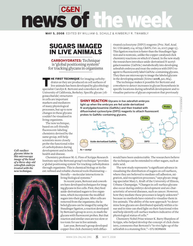
rinated cyclooctyne (DIFO) reagents (Proc. Natl. Acad. Sci. USA2007, 104, 16793; C&EN, Oct. 22, 2007, page 15). This ligation reaction is faster than the Staudinger liga-tion and is nontoxic, unlike the copper-catalyzed click chemistry reactions on which it’s based. In the new study the researchers introduce azide-derivatized N-acetyl-galactosamine (GalNAc) metabolically into developing zebrafish embryos and treat the embryos with DIFO re-agent to fluorescently label GalNAc-containing glycans. They then use microscopy to image the labeled glycans in the developing animals (Science2008, 320, 664).

The technique makes it possible for Bertozzi and coworkers to detect increases in glycan biosynthesis in specific locations during zebrafish development and to visualize patterns of glycan expression that previously

would have been undetectable. The researchers believe the technique can be extended to other sugars, such as sialic acid.

The new study represents “a significant advance in visualizing the distribution of sugars on cell surfaces, where they are believed to mediate cell adhesion, mi-gration, and recognition processes,” says glycan-imag-ing specialist Mary L. Kraft of the University of Illinois, Urbana-Champaign. “Changes in cell-surface glycans also occur during embryo development and are char-acteristic of several diseases, such as cancer. However, how they mediate these processes is largely unknown” because no methods have existed to visualize them in live animals. The ability of the new approach “to deter-mine how glycans are distributed spatially within a tis-sue and in time can shed light on their functional roles and help identify cell-surface markers indicative of the physiological status of cells.”

Chemistry Nobel Prize winner K. Barry Sharpless of Scripps, who helped develop the concept of click chem-istry, comments that Bertozzi’s “in vivo light-up of the zebrafish is a stunning first.”—STU BORMAN

Cell-surface glycans shine in this microscopy image of the head of a three-day-old zebrafish embryo treated with the new technique.

news of the weekMAY 5, 2008 EDITED BY WILLIAM G. SCHULZ & KIMBERLY R. TWAMBLY

CO

UR

TE

SY

OF

CA

RO

LY

N B

ER

TO

ZZ

I A

ND

CO

WO

RK

ER

S

SUGARS IMAGED IN LIVE ANIMALS

CARBOHYDRATES: Technique is ‘global positioning system’

for tracking glycans in organisms

AcO

AcO

OAc

OAc

HN

O

N3

O

O

OF

F

NH

N N

NO

OF

F

NH

DIFOAzide-derivatized GalNAc

Ac = acetyl

Probe

Probe

N3

SHINY REACTION Glycans in live zebrafish embryos

light up when the embryos are fed azide-derivatized

N-acetylgalactosamine (GalNAc) and then treated with

difluorinated cyclooctyne (DIFO) reagents to attach fluorescent

probes to GalNAc-containing glycans.

© 2

00

8 S

CIE

NC

E