Embed Size (px)
Citation preview

Subtractive single-chain antibody (scFv) phage-display: tailoring phage-display for high specificityagainst function-specific conformations of cellmembrane moleculesSteffen U Eisenhardt1, Meike Schwarz2, Nicole Bassler1 & Karlheinz Peter1
1Baker Heart Research Institute, Melbourne, Victoria 8008, Australia. 2Department of Cardiology, University of Freiburg, 79106 Freiburg, Germany. Correspondenceshould be addressed to S.U.E. ([email protected]).
Published online 21 November 2007; corrected online 5 June 2008 (details online); doi:10.1038/nprot.2007.455
Phage-display of antibody libraries is a powerful tool to select antibodies for specific epitopes. We describe a strategy for selecting
highly specific scFv-clones that discriminate between various conformational states of cell surface receptors. This approach adapts the
M13 pIII phage-display technology toward a cell suspension-based strategy, which allows panning against complex, multimeric, fully
functional cell membrane epitopes without alteration of structure due to purification or immobilization. As the functional properties
are preserved, phage can be specifically depleted or selected for neo-epitopes exposed after physiological alterations of the targeted
molecules. This subtractive strategy allows highly specific selection for single-chain antibodies directed against functionally regulated
epitopes on the cell surface molecules that can be tailored for diagnostic and therapeutic applications. Using this protocol,
activation-specific single-chain antibodies can be obtained within 4–6 weeks.
INTRODUCTIONConformation-specific antibodies are highly sought-after for bio-logical research, medical diagnosis as well as therapy. The classicalprocess of antibody generation does not allow targeting for con-formation-specific antibodies. Advanced protocols have beendeveloped to use artificial immobilization of target epitopes1,epitopes on intact mammalian cells2 or even in vivo epitopes3 forscreening purposes. We have developed and published a methodthat allows the selection of conformation-specific antibodiesagainst intact, functionally active, complex, multimeric epitopeson cell membrane molecules4–6.
Despite the introduction of novel display methods, such asbacterial surface display7, ribosomal display8, puromycin display9,or yeast surface display10, to date phage-display, based on the M13pIII peptide phage-display technology (Fig. 1)11 is the most widelyused method. Some of the aforementioned display systems maybe more suited for specific tasks than others and very often,the choice is dependent on personal preferences and expertise.However, a suitable selection method for a well-defined targetand highly specific selection are the main determinants for asuccessful panning strategy.
Phage-display is widely used for its advantages over the classicalhybridoma strategy, as the selection can be carried out on isolated,highly purified epitopes. In general, during the selection process,these antigens are presented to the phage library as immobilizedtargets on solid surfaces, such as 96-well polystyrole microtiter
plates12, polystyrole tubes13, nitrocellulose14 or magnetic beads15.This is clearly a limitation of the existing protocols as immobilizationcan induce conformational changes, and functionally importantepitopes may not be exposed or functionally regulated by stimuli.
p
uor
G g
n ih si l
bu
P eru ta
N 800 2©
nat
ure
pro
toco
ls/
moc.er
ut an.
ww
w//:ptt
h
Phagemid vector
pIII-protein
scFv
H-CDR 3
p3
His(6)-tag
Light chain variable region VL
Complementaritydetermining regions
Heavy chain variable region VHa
b
Figure 1 | Single-chain antibody display on pIII of the M13 phage.
(a) The phagemid encodes for single-chain antibodies with the antibody
heavy chain fused to the antibody light chain through a linker peptid. Each
scFv is His(6)-tagged for purification and detection. (b) The scFv is displayed
as a pIII-fusion protein on the surface of the M13 phage. The antibody
heavy chain is depicted in pink, and the light chain in yellow. Enlarged is
the 3D-structure of a typical scFv clone as described previously19. The model
was created with Rasmol-pdb viewer (University of Massachusetts).
NATURE PROTOCOLS | VOL.2 NO.12 | 2007 | 3063
PROTOCOL

One option to circumnavigate these limita-tions is to use in vivo phage-display16.However, these approaches have the samelimitations as hybridoma strategies andunspecific binding to the abundance ofantigens in the circulation is a disadvantageof this strategy.
Here, we describe a phage-display strat-egy performed with a single-chain antibody(scFv) phage library17,18 that allows theselection of highly specific scFv antibodyclones that are able to discriminate betweenvarious conformational states of cell surfacereceptors. Our method of subtractive, cellsuspension-based panning allows targetingof complex, multimeric, fully functionalcell membrane epitopes without alterationof structure otherwise due to purificationand immobilization. As the functionalproperties remain untouched, physiologi-cal changes can be induced and phage can be specifically depletedor selected for neo-epitopes that get exposed after stimulation orother physiological alterations.
This protocol provides detailed descriptions of the depletion andselection steps for phage-display based on the M13 pIII phage-display system. Our subtractive strategy, which is based on an initialdepletion step against unwanted epitopes followed by a selection stepfor the target epitope, allows a highly specific selection for activation-specific conformations (Fig. 2). Furthermore, this subtractivestrategy is highly effective in reducing the number of recoverednonspecific clones, in reducing the number of washes required in theselection steps, and ultimately the number of potential ligands thatare lost during the repeated washing steps. The latter is a typicaldrawback of classical panning strategies. The affinity maturationprocess can be easily screened by restriction digest and the binding ofselected clones can be screened by either ELISA or flow cytometry(fluorescence-activated cell sorting (FACS)).
This protocol has been used successfully for the selection of scFvsagainst adhesion receptors of the integrin family, which are knownto undergo a rapid shape change upon activation19,20, resulting in ahigh-affinity state for their ligands. We tested the protocol forleukocyte- and platelet receptors, and on recombinant cell linesexpressing activated or nonactivated conformations of cell surfacereceptors4,6,21. We found the combination of native and recombi-nant cell lines ideal to reduce binding to other neo-epitopes that areexposed on the membranes after cell activation. However, if arecombinant cell line is not available, all panning rounds can be
performed on native cells but a higher diversity of the resultingantibodies has to be expected. This multiple-step panning approachallows tailoring of specificity by changing the selection matrixbetween the panning rounds to obtain maximal specificity andtailoring of affinity by the variation of elution conditions and by thecompetitive elution steps (see Box 1). The latter is particularlyimportant when aiming for a blocking antibody or an antibody thatis competing with a known ligand of the receptor. We used thisapproach for the selection of an anti-GPIIb/IIIa activation-specificantibody to achieve fibrinogen-blocking characteristics for a poten-tial therapeutic use4. Other highly specialized selection strategiessuch as competitive deselection22, pathfinder23 or step-backselections24 have been designed and successfully used. Oursystem is unique in its specific design that allows targeting offunction-specific cell surface receptor epitopes rather thannonconformation-specific and/or soluble proteins. Furthermore,it is not dependent on pre-existing ‘guiding’-antibodies. Thus, thisprotocol is a valuable addition to the growing field of successfulphage-display strategies.
Our panning strategy is widely transferable and could be used foralternative approaches such as the BRASIL-protocol as described byGiordano et al.2 We recommend using phagemid libraries asdescribed in this protocol, as the low valency of phagemid-baseddisplay systems reduces background binding and increases theaffinity of the selected clones25. However, our protocol is transfer-able to other commercially available phage-display libraries26, andto other library display systems.
MATERIALSREAGENTS.0.1 M Gly (pH 2.2; Astral Scientific, cat. no. 0167).0.1 M PIPES (pH 6.6; Sigma, cat. no. P1851).0.4 M Sucrose (Merck, cat. no. 102747E).10� Cellfix (Becton Dickinson, cat. no. 340181).10� PBS without Ca2+/Mg2+ (Cambrex, cat. no. 17-515F).1 M Isopropyl-b-D-thiogalactopyranoside (IPTG; Invitrogen, cat. no. 15529-019).1� PBS with Ca2+/Mg2+ (Invitrogen, cat. no. 14040-133).2 M Tris–HCl pH 8 (Sigma, cat. no. T6666).Acetic acid (Sigma, cat. no. A6283).ADP (Moelab, cat. no. 0203011)
.Agarose (Sigma, cat. no. A9414) (see REAGENT SETUP)
.Alkaline phosphatase (Roche, cat. no. 713023)
.Ampicillin (Sigma, cat. no. A0166)
.Anti-His(6) antibody horseradish peroxidase (HRP; Roche, cat. no. 1965085)
.Bicinchoninic acid (BCA)protein assay kit (Pierce, cat. no. 23227)
.Blue/orange loading dye 6� (Promega, cat. no. G1881)
.BSA (Sigma, cat. no. A7906)
.BugBuster mastermix (Merck, cat. no. 71456.4)
.Calcium chloride (CaCl2; Sigma, cat. no. C3306)
.DNA ladder 1 kb (Promega, cat. no. G5711)
.Epifibatide (Essex Pharma)
p
uor
G g
n ih si l
bu
P eru ta
N 800 2©
nat
ure
pro
toco
ls/
moc.er
ut an.
ww
w//:ptt
h
Figure 2 | Flow chart of
the subtractive panning
(depletion/selection)
strategy for the
generation of
conformation-specific
single-chain antibodies.
Each panning round
starts with a depletion
step, in which phage are
incubated with
nonactivated cells.
Phage encoding for
scFv-antibodies that are binding to the nonactivated cells are centrifuged down with the cells and
discarded. The supernatant, containing the phage that are not binding to the nonactivated cells will
be transferred to the selection round. Phage that are transferred from the depletion step are added to
activated cells expressing receptors in their activated conformation. Cells are centrifuged down with
the binding phage. The supernatant, containing phage that bound neither to nonactivated cells in the
depletion step, nor to activated cells in the selection step, is discarded. Phage, that did not bind to
the nonactivated cells in the depletion step, but to the activated cells in the selection step are bound
to the pelleted cells and will be eluted and transferred to the next panning round.
Elute phage and transfer to next panning round
SelectionDepletion
Stimu-lation
3064 | VOL.2 NO.12 | 2007 | NATURE PROTOCOLS
PROTOCOL
.Ethanol (Biolab, cat. no. BSPEL975.2.5)
.Ethidium bromide (Astral Scientific, cat. no. AMX328) ! CAUTIONCarcinogenic. Wear appropriate protection and handle with care.
.Formyl-met-leu-phe (fMLP; Sigma, cat. no. F3506)
.Glucose (Riedel de Haen, cat. no. 16325)
.Glycerol (Sigma, cat. no. G5516)
.Helper-Phage M13K07 (GE Healthcare, cat. no. 27-1542-01)
.HEPES (Sigma, cat. no. H7523)
.Human scFv phage library (Affimed) or other commercially available scFvlibrary (e.g., Tomlinson I + J; MRC geneservice)
.Kanamycin (Invitrogen, cat. no. 55-0204)
.Potassium chloride (KCl; Merck, cat. no. 1.04936.0500)
.Luria Bertani (LB) agar (Sigma, cat. no. L3027)
.Liquid nitrogen ! CAUTION Causes burns. Wear appropriate protection andhandle with care.
.Lipopolysaccharide (LPS; Sigma, cat. no. L4391)
.Luria Bertani (LB) media (Sigma, cat. no. L3522)
.Manganese (Mn; Sigma, cat. no. 244589)
.Magnesium chloride (MgCl2; Sigma, cat. no. M2670)
.Mouse IgG1 isotype control (Beckman Coulter, cat. no. IM0639)
.Sodium bicarbonate (NaHCO3; Merck, cat. no. 1.0632905)
.Nickel-nitrilotriacetic acid (Ni-NTA)agarose (Qiagen, cat. no. 30210)
.PEG 6000 (Sigma, cat. no. 81260)
.Penta-His AlexaFluor 488 (Qiagen, cat. no. 35310)
.pexHAM vector (Affimed)27
.pHOG-21 Vector (Affimed)28
.Phorbol 12-myristate 13-acetate (PMA; Sigma, cat. no. P1585)
.QIAquick gel extraction kit (Qiagen, cat. no. 28704)
.QIAprep spin plasmid preparation kit (Qiagen, cat. no. 27106)
.Rapid DNA ligation kit (Roche, cat. no. 1635379)
.Restriction endonucleases: NcoI (cat. no. R0193S), NotI (cat. no. R0189S),BstNI (cat. no. R0168L), RsaI (cat. no. R0167S) (all from New EnglandBiolabs)
.SOC-Media (Invitrogen, cat. no. 15544-034)
.Tetracycline (Sigma, cat. no. T7660)
.Theophylline (Sigma, cat. no. T1633)
.Trizma base (Sigma, cat. no. T1503)
.Escherichia coli (E. coli) XL-1 blue (Stratagene, cat. no. 200249) m CRITICALGrow in LB media containing 20 mg ml–1 tetracycline.
.E. coli TG-1 (Stratagene, cat. no. 200123) m CRITICAL Grow in LB media.
.Tris-acetate-EDTA (TAE) buffer (see REAGENT SETUP)
.Hyperosmotic shock solution (see REAGENT SETUP)
.LBGAT agar plate (see REAGENT SETUP)
.LBGAT media (see REAGENT SETUP)
p
uor
G g
n ih si l
bu
P eru ta
N 800 2©
nat
ure
pro
toco
ls/
moc.er
ut an.
ww
w//:ptt
h
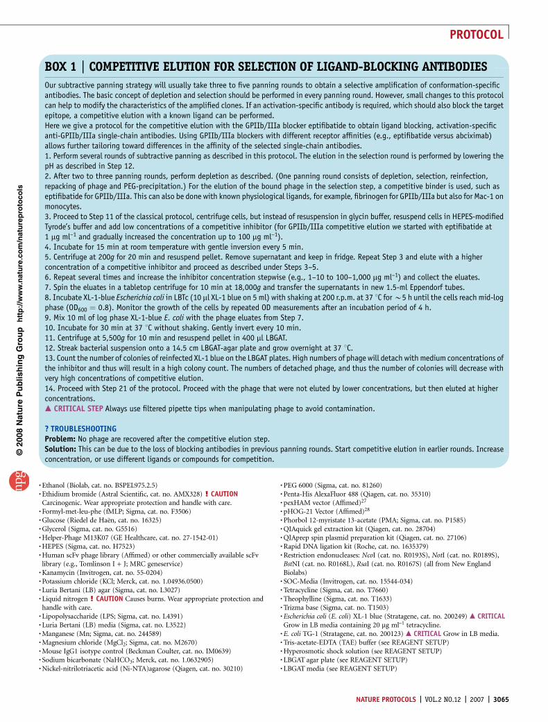
BOX 1 | COMPETITIVE ELUTION FOR SELECTION OF LIGAND-BLOCKING ANTIBODIES
Our subtractive panning strategy will usually take three to five panning rounds to obtain a selective amplification of conformation-specificantibodies. The basic concept of depletion and selection should be performed in every panning round. However, small changes to this protocolcan help to modify the characteristics of the amplified clones. If an activation-specific antibody is required, which should also block the targetepitope, a competitive elution with a known ligand can be performed.Here we give a protocol for the competitive elution with the GPIIb/IIIa blocker eptifibatide to obtain ligand blocking, activation-specificanti-GPIIb/IIIa single-chain antibodies. Using GPIIb/IIIa blockers with different receptor affinities (e.g., eptifibatide versus abciximab)allows further tailoring toward differences in the affinity of the selected single-chain antibodies.1. Perform several rounds of subtractive panning as described in this protocol. The elution in the selection round is performed by lowering thepH as described in Step 12.2. After two to three panning rounds, perform depletion as described. (One panning round consists of depletion, selection, reinfection,repacking of phage and PEG-precipitation.) For the elution of the bound phage in the selection step, a competitive binder is used, such aseptifibatide for GPIIb/IIIa. This can also be done with known physiological ligands, for example, fibrinogen for GPIIb/IIIa but also for Mac-1 onmonocytes.3. Proceed to Step 11 of the classical protocol, centrifuge cells, but instead of resuspension in glycin buffer, resuspend cells in HEPES-modifiedTyrode’s buffer and add low concentrations of a competitive inhibitor (for GPIIb/IIIa competitive elution we started with eptifibatide at1 mg ml–1 and gradually increased the concentration up to 100 mg ml–1).4. Incubate for 15 min at room temperature with gentle inversion every 5 min.5. Centrifuge at 200g for 20 min and resuspend pellet. Remove supernatant and keep in fridge. Repeat Step 3 and elute with a higherconcentration of a competitive inhibitor and proceed as described under Steps 3–5.6. Repeat several times and increase the inhibitor concentration stepwise (e.g., 1–10 to 100–1,000 mg ml–1) and collect the eluates.7. Spin the eluates in a tabletop centrifuge for 10 min at 18,000g and transfer the supernatants in new 1.5-ml Eppendorf tubes.8. Incubate XL-1-blue Escherichia coli in LBTc (10 ml XL-1 blue on 5 ml) with shaking at 200 r.p.m. at 37 1C for B5 h until the cells reach mid-logphase (OD600 ¼ 0.8). Monitor the growth of the cells by repeated OD measurements after an incubation period of 4 h.9. Mix 10 ml of log phase XL-1-blue E. coli with the phage eluates from Step 7.10. Incubate for 30 min at 37 1C without shaking. Gently invert every 10 min.11. Centrifuge at 5,500g for 10 min and resuspend pellet in 400 ml LBGAT.12. Streak bacterial suspension onto a 14.5 cm LBGAT-agar plate and grow overnight at 37 1C.13. Count the number of colonies of reinfected XL-1 blue on the LBGAT plates. High numbers of phage will detach with medium concentrations ofthe inhibitor and thus will result in a high colony count. The numbers of detached phage, and thus the number of colonies will decrease withvery high concentrations of competitive elution.14. Proceed with Step 21 of the protocol. Proceed with the phage that were not eluted by lower concentrations, but then eluted at higherconcentrations.m CRITICAL STEP Always use filtered pipette tips when manipulating phage to avoid contamination.
? TROUBLESHOOTINGProblem: No phage are recovered after the competitive elution step.Solution: This can be due to the loss of blocking antibodies in previous panning rounds. Start competitive elution in earlier rounds. Increaseconcentration, or use different ligands or compounds for competition.
NATURE PROTOCOLS | VOL.2 NO.12 | 2007 | 3065
PROTOCOL

.LBTc (see REAGENT SETUP)
.Lysis buffer (see REAGENT SETUP)
.HEPES-modified Tyrode’s buffer (see REAGENT SETUP)
.Modified Tyrode’s buffer (see REAGENT SETUP)
.Transformation buffer (see REAGENT SETUP)
.Wash buffer (see REAGENT SETUP)
.Saccharose
.Roche T4 Ligase kit (Roche)EQUIPMENT.Agar plates,15 cm (Sarstedt, cat. no. 82.1184) (see EQUIPMENT SETUP).Centrifuge bottles.ChemiDoc XRS system (Bio-Rad).Dry block heater.Filtered tips.Flow cytometer (FACS Calibur; Becton Dickinson).Gel electropheresis system (Bio-Rad).Slide-A-Lyzer cassettes (Pierce, cat. no. 66380).Spectra Por Dialysis Membrane (Spectrum Laboratories, cat. no. 132680).Tube rotator.Tubes (15, 50 and 1.5 ml).Image Pro PlusREAGENT SETUP3% (wt/vol) Agarose gel 300 ml 1� TAE buffer, 3 g agarose. Heat inthe microwave until the agarose is completely dissolved and then add30 ml EthBr. ! CAUTION EthBr is carcinogenic. Always handle withcare. We recommend using double layers of gloves and avoiding directcontact with the gels or using safer alternatives such as GelRed or SYBR Green.503 TAE buffer 2 M Tris, 5 mM EDTA pH 8, 5.5% acetic acid.
Elution buffer (pH 8.0) 50 mM Sodium phosphate, monobasic (NaH2PO4),300 mM sodium chloride (NaCl), 250 mM imidazole, adjust pH.HEPES-modified Tyrode’s buffer (pH 6.5) 10 mM HEPES, 12 nM NaHCO3,138 mM NaCl, 2.9 mM KCl, 2 mM MgCl2, 2 mM CaCl2, 1 g l–1 glucose, 1 g l–1
BSA, adjust pH.Hyperosmotic shock solution (pH 8) 20% sucrose, 1 mM EDTA, 50 mMTrizma base.LBGAT agar plate LB agar, 50 mM glucose, 100 mg ml–1 ampicillin, 20 mg ml–1
tetracycline. Autoclave LB agar, then add glucose, ampicillin, and tetracyclineand pour the plates.LBGAT media LB medium, 50 mM glucose, 100 mg ml–1 ampicillin, 20 mg ml–1
tetracycline, autoclave LB medium, then add glucose, ampicillin, and tetracyclineLBTc LB medium, 20 mg ml–1 tetracycline, autoclave LB medium, let mediumcool down, then add tetracycline.Lysis buffer (pH 8.0) 50 mM NaH2PO4, 300 mM NaCl, 10 mM imidazole,adjust pH with 1 M NaOH to raise pH, or with 1 M hydrochloric acid (HCl) tolower pH.Modified Tyrode’s buffer (pH 7.4) 150 mM NaCl, 12 mM NaHCO3, 2.5 mMKCl, 2 mM CaCl2, 2 mM MgCl2, 1 mg ml–1 BSA, 1 mg ml–1 glucose, adjust pH.Phage dilution buffer (PDB, pH 7.5) 10 mN Tris–HCl, 20 mM NaCl, 2 mMEDTA, adjust pH and autoclave.Transformation buffer 25 ml 1 M CaCl2, 150 ml 50% glycerol, 0.1 M PIPES(pH 6.6), 275 ml H2O and autoclave.Wash buffer (pH 8.0) Add 50 mM NaH2PO4, 300 mM NaCl, 20 mMimidazole and adjust pH.EQUIPMENT SETUPAgar plate 25 g LB agar, 50 mM glucose, 100 mg ml–1 ampicillin, 20 mg ml–1
tetracycline. Autoclave LB agar, then add glucose, ampicillin, and tetracyclineand pour the plates.
PROCEDUREDepletion of unspecific phage1| Thaw frozen aliquots of human scFv phage library (e.g., 1 ml phage in PDB) on ice. The number of phage should exceed thecomplexity of the used library by 1,000-fold.
2| Isolate cells expressing the target receptor and preserve them in a resting state. For cells that are easily activated bypreparative steps, such as platelets, activation can be suppressed by adding inhibitors (e.g., theophylline in the case ofplatelets) to the buffer. For leukocytes, preparative steps that include plastic adhesion should be avoided, as leukocyte adhesioncan cause cell activation. Magnetic separation methods are preferable. Alternatively, a recombinant cell line, expressing thetarget receptor can be used.m CRITICAL STEP The purity of the cell lines should be monitored using flow cytometry. For cells originating from whole bloodpreparation it should beZ90%. As this protocol does not use purified epitopes, the preparation and the purity of the cells are crucialfor the selection of specific antibodies. In particular this is the case for the selection step. In the depletion step contamination withother cells is either less crucial or might be even beneficial, as phage binding to the contaminating epitopes will be discarded.
3| Centrifuge the harvested or isolated cells for 10 min at 140g and resuspend the pellet to a concentration of 2 � 106 in1 ml modified Tyrode’s buffer.
4| Add phage library and incubate for 2 h at room temperature (20–23 1C) by gently rotating on a tube rotator. If a rotator isunavailable keep cells in suspension by periodic inversion.m CRITICAL STEP Phage internalization can occur during incubation of the phage library with the cell line at room temperature.This can be avoided by performing Step 4 at 4 1C. However, lowering the temperature to 4 1C can lead to receptor clustering,unspecific cell activation (in particular with platelets), and may also inhibit the expression of function (e.g., cell activation)-specific epitopes. Thus, for targeting of function-specific epitopes, we recommend performing the incubation step at roomtemperature. Decreasing the incubation period of the phage with target cells reduces phage internalization.
5| Pellet cells at 140g for 20 min and transfer supernatant into a fresh Falcon tube.m CRITICAL STEP Always use filtered pipette tips when manipulating phage to avoid contamination.
Selection of activation-specific binding phage6| Prepare selection cell line, that bears the target receptor in its activated conformation or that should be activated toinduce conformational changes of the target receptor. We used 20 mM ADP and 100 ng ml–1 PMA for platelet and monocytepanning, respectively. If you are working with recombinant cell lines expressing integrins, conformational changes can beachieved either by mutations22 or by incubation with Mn23.
p
uor
G g
n ih si l
bu
P eru ta
N 800 2©
nat
ure
pro
toco
ls/
moc.er
ut an.
ww
w//:ptt
h
3066 | VOL.2 NO.12 | 2007 | NATURE PROTOCOLS
PROTOCOL

7| Pellet 2 � 106 cells and resuspend in the supernatant from Step 5, which contains the unbound phage from the depletionstep. Mix by gentle inversion. The low pH of the selection buffer reduces the unspecific binding of insert-free phage24. The cellnumber in the selection rounds influences the result. Low numbers of cells increase the selection pressure, which results inhigh-affinity clones, whereas high cell numbers reduce the selection pressure.
8| Incubate for 30 min at room temperature with gentle rotation to allow binding of phage to recognition patterns that werenot available in the depletion step.
9| Pellet cells for 10 min at 140g and remove the supernatant that contains the unbound phage. The unbound phage can bediscarded.
10| Wash cells by resuspending pelleted cells in 50 ml HEPES buffer.
11| Centrifuge for 20 min at 200g and resuspend pellet in 1 ml HEPES buffer and transfer to a 1.5-ml Eppendorf tube. Repeatwashing (Steps 10 and 11) with lower volumes (e.g., 1 ml) two times.
12| Elute phage by resuspending the pellet in 1 ml Glycine elution buffer (pH 2.2) and incubating for 15 min at roomtemperature with gentle inversion every other minute.
13| Neutralize by adding 100 ml of 2 M Tris (pH 8), and transfer into a new 1.5-ml Eppendorf tube. Transfer to Step 14 quicklyas neutralized phage could rebind to the cells that are still present in the tube.
14| Spin in a tabletop centrifuge for 10 min at 18,000g and transfer supernatant into a new 1.5-ml Eppendorf tube.m CRITICAL STEP Choosing a suitable cell line for selection is a crucial step in this protocol. It is important that conformationalchanges in the target epitope can be induced by a known stimulant and that the cells can be isolated to a sufficient purity.Changing to another cell line expressing the same receptors can be useful to reduce background binding. We recommend startingwith the least specific cell line for the initial depletion and selection step. We started with native cells purified from wholeblood in the first round and then switched to the more controlled environment of recombinant Chinese hamster ovary (CHO)cells. This strategy not only reduces binding to nonactivated target receptors, but also reduces binding to nontargeted membraneepitopes that are exposed upon activation. By changing cell lines, binding to surface epitopes other than the target epitope is reduced.’ PAUSE POINT The phage eluate can be kept at 4 1C overnight if convenient.
Reinfection of XL-1 blue E. coli with the selected phage15| Incubate XL-1-blue E. coli in LBTc (10 ml XL-1 blue in 5 ml) with shaking at 200 r.p.m. at 37 1C for B5 h until the cellsreach mid-log phase (OD600 ¼ 0.8). Monitor the growth of the cells by taking multiple OD measurements after an incubationperiod of 4 h.
16| Mix 10 ml of log phase XL-1-blue E. coli with the phage eluate from Step 13.
17| Incubate for 30 min at 37 1C without shaking. Gently invert every 10 min.
18| Centrifuge at 5,500g for 10 min and resuspend pellet in 400 ml LBGAT media. The phage carry an ampicillin-resistancemarker. Only successfully infected bacteria will grow.m CRITICAL STEP The resistance marker can vary between different vector systems and phage libraries. Resistance markers haveto be carefully checked before adding suitable antibiotics.
19| Plate bacterial suspension onto a 14.5-cm LBGAT-agar plate and grow overnight at 37 1C.
20| Count the number of colonies of reinfected XL-1 blue on the LBGAT plates. This can be done by imaging and analysis withcontrast-based counting programs, such as Image Pro Plus. A total of 2,000–5,000 colonies can be expected after the firstround.m CRITICAL STEP The number of colonies is a valuable parameter to monitor the progress of panning. A low number of coloniesafter the first panning round suggests a successful depletion of unspecific binding phage. Over the next rounds the number willreach a plateau. After a few specifically binding clones have been amplified through the course of panning (after three to fourpanning rounds, see ANTICIPATED RESULTS), the number of colonies will be amplified twofold to fourfold compared with theprevious panning rounds.
Repacking of phage with M13K07 helper phage for the following panning rounds21| Pipette 5–10 ml LBGAT media on the plates and carefully dilute the bacterial colonies by mixing them with the media usinga spatula.
p
uor
G g
n ih si l
bu
P eru ta
N 800 2©
nat
ure
pro
toco
ls/
moc.er
ut an.
ww
w//:ptt
h
NATURE PROTOCOLS | VOL.2 NO.12 | 2007 | 3067
PROTOCOL

22| Remove the resuspended bacteria by pipetting and transfer to a 15-ml Falcon tube. Remove 10 ml and add to 990 ml ofLBGAT and measure the OD600 of this 1:100 dilution.
23| Inoculate 20 ml LBGAT media with the bacteria at an OD600 of 0.1. (e.g., if you measured an OD600 of 0.09 for your 1:100dilution, the OD600 of your bacterial suspension is 9 and to achieve an OD600 of 0.1 you need to prepare a 1:90 dilution, whichequals 222 ml on 20 ml).
24| The rest of the bacterial suspension can be kept as glycerol stocks. This allows going back to previous panning roundsif one panning round failed or did not result in the amplification of a clone with the sought-after characteristics. Add sterileglycerol to a final concentration of 15% to the bacteria from Step 22 and store at –80 1C.’ PAUSE POINT The panning can be stopped here, if convenient. Bacterial stocks from Step 22 can be kept for several yearsat –80 1C. When proceeding with the panning continue with Step 23.
25| Incubate the 20 ml LBGAT inoculated with the XL-1 blue carrying phage with shaking at 200 r.p.m. at 37 1C for 1 h.
26| Add M13K07 helper phage in a multiplicity of infection (MOI: phage/bacteria) of 10–20. You can estimate the number ofbacteria. An OD600 of 1 equals 1 � 109 bacteria per ml. The culture was started at an OD of 0.1, which corresponds to 1 � 108
bacteria per ml (see Step 23). In the 20-ml suspension, 4 � 109 bacteria can be expected as the number has doubled within 1 hof cultivation. 4 � 1010 phage should be added (MOI of 10). 40 ml of the phage suspension at a phage titre of 1012 phage perml should be added.
27| Mix carefully by gentle inversion and incubate for 30 min at 37 1C with gentle inversion every 10 min, but withoutshaking.
28| Incubate a further 30 min with shaking in a bacterial shaker at 200 r.p.m. at 37 1C.m CRITICAL STEP For successful phage infection, bacteria have to be grown at temperatures 434 1C as this is essential forpili formation.
29| Pellet bacteria by centrifugation for 5 min at 5,500g.
30| Discard supernatant and resuspend in 20 ml LB media containing ampicillin and kanamycin (50 mg ml–1) without glucoseand tetracycline. The M13K07 helper phage carries a kanamycin-resistance marker that allows the identification of successfullyco-infected bacterial clones.
31| Incubate overnight with shaking in a bacterial shaker at 200 r.p.m. and 30 1C.m CRITICAL STEP Always use filtered pipette tips when manipulating phage to avoid contamination.
PEG-precipitation of phage32| Centrifuge bacterial culture at 5,500g for 10 min.
33| Precipitate phage particles by transferring the supernatant to a new tube containing one-fifth the volume of PEG/NaCl.Mix carefully by inversion, do not vortex.
34| Incubate for 2 h on ice.
35| Centrifuge for 1 h at 5,500g at 4 1C and discard the supernatant.
36| Resuspend pellet in 1 ml PDB and transfer to a 1.5-ml Eppendorf tube.
37| Centrifuge for 5 min at 14,000g in a tabletop centrifuge, remove supernatant and store at 4 1C for the next panning round.’ PAUSE POINT The stock is stable at 4 1C for up to 6 months. For long-term storage sterile glycerol can be added to a finalconcentration of 15% and phage stored at –80 1C.
38| For the next panning round the number of phage should be determined. Inoculate 5 ml LBTc-media with 100 ml of thawedXL-1-blue stock. Incubate log-phase bacteria with the phage dilution and infect as described in Step 17. Make step-wisedilutions (e.g., 103–1010) and spread transduced bacteria on several LBGAT agar plates, or subdivide on 14.5 cm LBGAT agarplates in multiple sections and spread different dilutions in these sections. A LBGAT plate with kanamycin can serve as control.
39| Incubate overnight in a bacterial incubator and count colonies the next day. The number of colonies multiplied with thedilution factor will give an estimate of how many phage are in the dilution after the panning round. 1011–1013 colonies can beexpected.m CRITICAL STEP Always change pipette tips after each dilution step to avoid false-high results and use filtered pipette tips.
p
uor
G g
n ih si l
bu
P eru ta
N 800 2©
nat
ure
pro
toco
ls/
moc.er
ut an.
ww
w//:ptt
h
3068 | VOL.2 NO.12 | 2007 | NATURE PROTOCOLS
PROTOCOL

Random pattern restriction analysis40| When the number of colonies after reinfection (see Step 20) is amplified from twofold to fourfold (Fig. 3a) the panningshould be stopped and the colonies should be screened for diversity.
Pick 10 to 20 clones using sterile pipette tips from the LBGAT plates, drop the tips into 5-ml LBGAT media and grow themovernight at 37 1C with shaking at 220 r.p.m. The picked colonies should be marked and numbered using a permanent markerat the backside of the plate and the plates kept at 4 1C.
41| Isolate DNA of the picked clones using a commercial mini-prep kit (Qiagen) and follow the manufacturer’s instructions.Determine the concentration. Approximately 0.3 mg ml–1 can be expected. If preferred, all picked clones can be sequenced andthe diversity can be directly analyzed.
42| Digest 10–20 DNA preparations with BstN1 or RSA-1 for random pattern restriction analysis. Prepare reaction solution asdescribed in the table below.
BstN1 RSA-1
1 ml NEBuffer 2 1 ml NEBuffer 10.1 ml BSA 0.1 ml BSA4.9 ml ddH2O 4.9 ml ddH2O1 mg DNA 1 mg DNA1 ml BSTN I 1 ml RSA IDigest for 1 h at 60 1C Digest for 1 h at 37 1C
43| Add 2 ml loading dye per tube and run on a 3% agarose gel in TAE buffer. The time of the run depends on the gelelectrophoresis system used. We recommend loading a marker to assess the progress of the run; however, the marker is notnecessary for the analysis of the results.
44| Stain DNA-gels with EthBr and analyze restriction patterns (Fig. 3b). At this stage, glycerol stocks should be made of eachdifferent clone. The numbered clones can be picked from the agar plate (from Step 40), grown overnight, and glycerol stocksprepared as described in Step 24.
p
uor
G g
n ih si l
bu
P eru ta
N 800 2©
nat
ure
pro
toco
ls/
moc.er
ut an.
ww
w//:ptt
h
104103102101100104103102101100104103102101100
104103102101100 104103102101100 104103102101100
FL1-HFL1-HFL1-H+ADPNo additionanti-His FITC
0
100C
ount
s
0
100
Cou
nts
0
100
Cou
nts
0
100
Cou
nts
0
100
Cou
nts
0
100
Cou
nts
FL2-HFL2-HFL2-Hanti-His PE No addition +ADP
Panning round 4Panning round 3Panning round 2
Bst
NI
4th round3rd round2nd round1st round0
2,000
4,000
6,000
8,000
Num
ber
of c
lone
saf
ter
rein
fect
ion
a
b
c
Figure 3 | Monitoring for success of the panning process. (a) Numbers of
recovered phage after each panning round. After each round of panning, the
rescued phage are used for infection of log-phase XL-1 blue Escherichia coli
bacteria. The numbers of colonies, which are representing the number of
phage clones, are counted. Expect low yields with B1,000 colonies in the
first panning round, which reflects the loss of a large number of nonbinding
phage in the selection step as well as in the depletion step. Once a few,
activation-specific clones have been selected, the number of recovered phage
increases significantly, as these specific clones are not lost in depletion or
selection steps and thus amplified over the course of panning. This reflects
the final goal of the panning procedure and recovered phage should be
screened for diversity by random pattern restriction digest or sequencing.
(b) Fingerprinting of scFv clones by BstN1. The diversity of the scFv clones can
be evaluated by digestion with the restriction enzyme BstN1. Here, phagemid
DNA of ten randomly picked natural clones of panning rounds 2–4 was purified
and digested with the restriction enzyme and separated in agarose-gel-
electrophoresis and stained with ethidium bromide. The DNA markers Lambda
DNA/HindIII (lane 1) and PhiX174DNA/HaeIII (lane 2) are used as
comparison. The restriction pattern of the scFv clones differs widely after
panning rounds 2 and 3. In contrast, after panning round 4 all investigated
clones demonstrate an identical restriction pattern. These results demonstrate
the power of positive clone amplification using the developed depletion/
selection system over the course of consequent panning rounds. Shown
is the successful panning for an anti-Mac-1 integrin antibody as described
previously5. (c) Screening for targeted binding characteristics in flow
cytometry. scFv clones can be screened by flow cytometry. A secondary anti-
His(6) tag antibody, either fluorescein isothiocyanate (FITC)- or phycoerythrin
(PE)-labeled is used for detection of binding. The upper panel shows a typical
binding pattern of a nonactivation-specific scFv against a platelet receptor.
The shift indicating positive binding in flow cytometry is identical with or
without ADP stimulation. Given in the lower panel are the typical binding
characteristics of an activation-specific scFv, an increase in mean fluorescence
can only be detected upon stimulation with the platelet activator ADP.
NATURE PROTOCOLS | VOL.2 NO.12 | 2007 | 3069
PROTOCOL

! CAUTION EthBr is carcinogenic. Always handle with care. We recommend using double layers of gloves and avoid directcontact with the gels or using safer alternatives such as GelRed or SYBR Green.’ PAUSE POINT Bacterial stocks of the picked clones can be kept for several years at –80 1C.? TROUBLESHOOTING
Small-scale preparation45| All clones that differ in their restriction patterns should be screened for their binding characteristics by FACS (Fig. 3c).For testing, a small-scale preparation of the antibodies is sufficient. The scFvs are expressed as periplasmic proteins and forscreening purposes it is not necessary to purify them from other periplasmic proteins. Using this method 40–50 clones can bescreened at once. Thaw glycerol stocks or pick clones from the agar plate from Step 40. Inoculate 5 ml LBGA media and growovernight at 37 1C with shaking and 220 r.p.m.’ PAUSE POINT Bacteria grow overnight.
46| Inoculate 5 ml LBGAT media with 100 ml preculture and grow at 37 1C with shaking at 220 r.p.m. for B5 h until the cells reachmid-log phase (OD600 ¼ 0.8). Monitor the growth of the cells by taking multiple OD measurements after an incubation period of 4 h.
47| Centrifuge at 1,800g for 10 min at room temperature.
48| Discard supernatant and resuspend pellet in 5 ml LB-media with 0.4 M sucrose, 50 mg ml–1 ampicillin, and 0.1 mMIPTG, and grow overnight at 24 1C with shaking at 220 r.p.m.Note: The pexham vector contains a TAG amber-stop codon between the scFv sequence and the pIII sequence. XL-1 blue is asuppressor strain, and thus does not stop translation at the amber codon. The translation stops after the pIII protein and thescFv are expressed and purified as scFv-pIII fusion proteins for initial testing of binding.
49| Centrifuge bacteria at 5,000g for 10 min at 4 1C.
50| Resuspend the pellet in 1/20 of the starting volume in ice-cold hyperosmotic shock solution containing 50 mM Tris, 20%saccharose, 1 mM EDTA at pH 8 (e.g., if you inoculated 5 ml LB media in Step 46 with the bacteria, use 250 ml buffer). Transferto 1.5-ml Eppendorf tube and incubate for 1 h on ice.
51| Centrifuge for 30 min at 30,000g at 4 1C. (Place tabletop centrifuge in a cool room.)
52| Take the periplasma supernatant and transfer to a Slide-A-Lyzer MINI dialysis unit. Dialyse for up to 12 h against PBS.Change buffer several times. The dialysis is necessary to remove IPTG from the samples.
53| Analyze periplasmic product for the expression of scFv by SDS–polyacrylamide gel electrophoresis and western blottingand staining with anti-His(6)-Tag-HRP. Thereby, scFv expression level, correct size and purity of the scFv preparation can beevaluated. Figure 4 shows thesuccessful production and purificationof an scFv antibody as assessed bySDS–polyacrylamide gel electrophoresisand western blotting of an scFv clonedinto the pHOG-21 vector expressed inTG-1 E. coli (Steps 58–70).m CRITICAL STEP The expressionand purification conditions can varybetween different vector and expressionsystems. If you are using a commerciallyavailable library, refer to the datasheet on which culture, induction andpurification conditions are suggested.
Screening for binding characteristics54| Purify target cells (e.g., plateletsor monocytes; whole blood preparationsare a time-saving option as well) orharvest cell lines expressing the epitopeof interest and adjust to 6 � 106 perml. Prepare one nonactivated and oneactivated sample.
p
uor
G g
n ih si l
bu
P eru ta
N 800 2©
nat
ure
pro
toco
ls/
moc.er
ut an.
ww
w//:ptt
h
Figure 4 | Production and purification of selected
single-chain antibodies in TG-1 Escherichia coli.
(a) Silver staining of SDS–polyacrylamide gel
electrophoresis (SDS-PAGE). (b) Western blot
probed with an anti-His-tag horseradish
peroxidase(HRP)-coupled antibody. Gel and blot
show: lane I: bacterial culture; lane II: supernatant
of the first centrifugation step; lane III: lysate of
bacterial pellet; lane IV: flow through of the
nickel-nitrilotriacetic acid (Ni-NTA)-agarose-
column; lane V: first washout of the Ni-NTA-
agarose-column; lane VI: second wash out of the
Ni-NTA-agarose-column; lane VII: empty; lane VIII:
eluate of the Ni-NTA-agarose-column. The bacterial
suspension contains a low concentration of scFv
(lane I). After centrifugation, the scFv is not
detectable in the supernatant (lane II) but at a
high concentration in the lysate of the bacterial
pellet (lane III). The flow through of an Ni-NTA-
agarose-column (shown in lane IV) shows a large
amount of protein (silver staining), but no signal
in the anti-His-tag staining, indicating that scFvs
are bound to the Ni-NTA-agarose, whereas nonspecific proteins flow through. The following washing steps
(lane V and VI) demonstrate further washout of decreasing amounts of proteins that include only a small
portion of scFv. The comparison between the bacterial suspension (lane I) and the final eluate (lane VIII)
demonstrates the power of bacterial protein expression/purification and the Ni-His-tag purification
system. Shown is a purification of an antibody clone as performed previously4–6.
I. II. III. IV. V. VI. VII. VIII.
scFv
anti-His-tag staining
scFv
Silver staining
I. II. III. IV. V. VI. VII. VIII.
a
b
3070 | VOL.2 NO.12 | 2007 | NATURE PROTOCOLS
PROTOCOL

m CRITICAL STEP The same stimulant that was used in the panning rounds to induce conformational changes should be used for theinitial screening. However, once a successfully binding clone has been identified, other stimulants can be used to confirm theactivation-specificity (e.g., we used PMA for the initial panning on activated monocytes and confirmed activation-specificity withLPS, and fMLP).
55| Incubate both samples with 30 ml periplasma product for 15 min at room temperature.
56| Add 1 ml anti-His-tag-AlexaFluor 488 conjugate and incubate for 15 min. Also prepare adequate negative controls. Forscreening purposes, incubating the cells with 1 ml anti-His-tag-AlexaFluor 488 conjugate alone without primary antibody orisotype-matched control antibody is sufficient.
57| Add 200 ml Cellfix and proceed to flow cytometric analysis.m CRITICAL STEP Screening for binding can also be performed by ELISA. However, immobilization of the target epitope can lead toconformational changes and thus alter the results.? TROUBLESHOOTING
Cloning of successfully binding antibodies into the pHOG-21 expression vector and transformation into TG-1 E. coli58| Isolate DNA of the selected scFv using a commercially available mini-prep kit (Qiagen) following the manufacturer’sinstructions and prepare pHOG-21 DNA.
59| Digest vector and scFv DNA with NcoI and NotI following the manufacturer’s instructions. Separate DNA in agarose gelelectrophoresis and cut scFv insert and pHOG-21 vector DNA and extract using a gel-extraction kit (Qiagen).
60| Add the DNA of insert and vector in a 1:4 ratio and ligate using a commercially available ligation kit (Roche T4 Ligase kit).
61| For transformation into TG-1 E. coli, thaw competent bacteria on ice (for generation of competent TG-1 E. coli, please referto Box 2).
62| Add 1 ml DNA to 100 ml of bacterial stock in an Eppendorf tube.
63| Incubate for 30 min on ice.
64| Incubate for 60 s at 42 1C and put back on ice for 2 min.
65| Add 900 ml of SOC media, transfer to a 15-ml Falcon tube and grow at 37 1C with shaking at 220 r.p.m. for 1 h.
66| Spread 100 ml of transformed bacteria on LBGA plates and incubate at 37 1C overnight.m CRITICAL STEP The change to TG-1 cells is necessary to increase the yield of scFv expression and purification, as TG-1 is a moresuitable strain for protein expression. Only the scFv-encoding sequence is cloned into pHOG-21, but not the pIII sequence, whichfurther increases the yield, as the scFv are not expressed as scFv-pIII fusion proteins but as scFv only.? TROUBLESHOOTING
67| Pick clones and inoculate 5 ml LBGA media and grow for 6–8 h, prepare DNA and confirm the successful cloning byDNA-sequence analysis.
p
uor
G g
n ih si l
bu
P eru ta
N 800 2©
nat
ure
pro
toco
ls/
moc.er
ut an.
ww
w//:ptt
h
BOX 2 | COMPETENT CELLS PROTOCOL FOR TG-1 E. COLI
1. Thaw TG-1 Escherichia coli glycerol stocks on ice. Inoculate 5 ml LB media and grow overnight at 37 1C with shaking at 220 r.p.m.2. Inoculate 500 ml LB media with 5 ml of the preculture and grow at 37 1C with shaking at 220 r.p.m. for B5 h until the cells reach mid-logphase (OD600 ¼ 0.8). Monitor the growth of the cells by taking multiple OD measurements after an incubation period of 4 h.3. Put on ice for 10 min on a rocking shaker.4. Centrifuge at 1,800g for 15 min at 4 1C.5. Discard supernatant and resuspend pellet in 250 ml transformation buffer (prepare 25 ml 1 M CaCl2, 150 ml 50% glycerol, 50 ml 0.1 M PIPES(pH 6.6) in 275 ml aqua destillata).6. Put on ice for 20 min on a rocking shaker.7. Centrifuge at 1,800g for 15 min at 4 1C.8. Discard supernatant and resuspend pellet in 25 ml transformation buffer.9. Put on ice for 40 min on a rocking shaker.10. Aliquot into 500 ml stocks and throw into liquid nitrogen to shock freeze stocks.11. Store at –80 1C.! CAUTION Liquid nitrogen can cause severe cold burns. Always handle with care and wear protective glasses and eyewear.
NATURE PROTOCOLS | VOL.2 NO.12 | 2007 | 3071
PROTOCOL

68| If the cloning was successful, purify scFv clones as described above (starting from Step 45). For the isolation of the scFvsfrom the periplasmic product dialyse the supernatant from Step 51 against lysis buffer for 6 h at 4 1C. Load the supernatant onan Ni-NTA-agarose column that has been equilibrated with lysis buffer. Collect flow-through for troubleshooting if purification isunsuccessful.
69| Wash the column two times with washing buffer and elute with 500 ml elution buffer.
70| Proceed with dialysis against PBS (Step 51), determine protein concentration by BCA or spectrophotometry at 280 nm andconfirm activation-specific binding in flow cytometry (Steps 53–56). You now have an activation-specific scFv clone that can beproduced in large-scale purifications in TG-1 E. coli.’ PAUSE POINT Protease inhibitor and sodium azide can be added for long-term storage. scFv antibodies can be stored at 4 or–20 1C for long-term storage.
� TIMINGSteps 1–39, one panning round: depletion, selection, reinfection, repacking of phage, and PEG-precipitation: 5–10 d;Steps 40–44, random pattern restriction analysis: 4 h; Steps 45–53, small-scale preparation of scFv: 2–4 d; Steps 54–57,flow cytometry analysis of scFv binding to target epitope: 2–4 h; Steps 58–66, cloning and tranformation: 2–4 d.
? TROUBLESHOOTINGTroubleshooting advice can be found in Table 1.
p
uor
G g
n ih si l
bu
P eru ta
N 800 2©
nat
ure
pro
toco
ls/
moc.er
ut an.
ww
w//:ptt
h
TABLE 1 | Troubleshooting table.
Step Problem Solution
44 No identical clones in therestriction pattern analysis
No amplification of specific phage. More panning rounds are needed. Transfer the repacked phage to thenext panning round
Alternatively, modify panning conditions (e.g., decrease cell number, perform additional washing steps,use competitive elution)
Evaluate the panning conditions and the suitability of the used phage library carefully. Modern phagelibraries can reach a complexity of up to 1011 unique clones. A greater complexity increases theprobability of the selection of a highly specific clone. However, for the screening of high complexitylibraries, it is necessary that, at least in the first panning round, the large number of phage have to bematched by a large number of antigens. Thus, low copies of the target receptor on the surface of theselection and depletion cell line can reduce the success of depletion and selection. We recommend usinga recombinant cell line for depletion and selection, if available. However, reducing the antigenconcentration in the course of the panning can be a useful tool to enrich high-affinity binders amongthe phage selected in the previous rounds
All picked clones showidentical restriction patterns
Proceed with small-scale purification and test for positive binding. A very strong binder may have beenamplified. If the amplified clone does not show the sought-after binding characteristics, screen theclones of the previous panning round for diversity and binding
57 No binding clones in screeningof the periplasmic product
The screening of the periplasmic product is a crude method to obtain preliminary information on thebinding characteristic of individual clones. As the amount of purified scFv can be very low, whenexpressed in pexHAM, negative binding in this assay does not generally rule out a positive clone amongthe clones that are tested. However, it is a good screening method for high numbers of clones. In case oflow numbers (one to two different clones in ten picked clones), we recommend proceeding with thecloning of the DNA into the pHOG-21 expression vector and testing the selected scFvs
Other problems might be the loss of protein during dialysis or an insufficient dialysis with remainingEDTA in the buffer, which can inhibit activation and binding
In the case of low protein expression (see western blot) the volume of the periplasmic product needs tobe increased (e.g., 50 ml per sample)
66 No colonies observed followingbacterial transformation
Bacterial transformation efficiency depends on DNA and cell concentrations. If the transformationefficiency is low, increase the amount of plasmid DNA in the transformationIf the transformation generally failed, repeat the transformation with new competent cells
3072 | VOL.2 NO.12 | 2007 | NATURE PROTOCOLS
PROTOCOL

ANTICIPATED RESULTSThe number of colonies after reinfection is a parameter to screen the process of in vitro affinity maturation. The number ofcolonies on the agar plates after reinfection will drop after the first panning round, when the majority of nonspecificallybinding clones are lost in the first depletion step. Over the following rounds, the numbers will plateau. Once specific bindershave been selected and are transferred to the next panning round, they will not be lost in the depletion step and the number ofcolonies after reinfection will dramatically increase (threefold to fivefold). This is a good indicator of the amplification of a fewclones. The restriction digest of a few picked clones will reveal different patterns and thus identify identical clones. Two to fourdifferent patterns out of ten picked clones can be expected. These should be screened for binding. In our hands, we found asufficient number of clones after four panning rounds, which proved to be activation specific4–6. Depending on the receptor andthe selection-matrix cell line at least one to two among ten picked different clones should show binding and should exhibit thesought-after activation-specific characteristic.
Published online at http://www.natureprotocols.comReprints and permissions information is available online at http://npg.nature.com/reprintsandpermissions
1. Clackson, T., Hoogenboom, H.R., Griffiths, A.D. & Winter, G. Making antibodyfragments using phage display libraries. Nature 352, 624–628 (1991).
2. Giordano, R.J., Cardo-Vila, M., Lahdenranta, J., Pasqualini, R. & Arap, W.Biopanning and rapid analysis of selective interactive ligands. Nat. Med. 7,1249–1253 (2001).
3. Arap, W. et al. Steps toward mapping the human vasculature by phage display.Nat. Med. 8, 121–127 (2002).
4. Schwarz, M. et al. Single-chain antibodies for the conformation-specific blockadeof activated platelet integrin alphaIIbbeta3 designed by subtractive selectionfrom naive human phage libraries. FASEB J. 1704–1706 (2004).
5. Eisenhardt, S.U. et al. Generation of activation-specific human anti-alphaM/beta2 single-chain antibodies as potential diagnostic tools and therapeuticagents. Blood 109, 3521–3528 (2007).
6. Schwarz, M. et al. Conformation-specific blockade of the integrin GPIIb/IIIa:a novel antiplatelet strategy that selectively targets activated platelets.Circ. Res. 99, 25–33 (2006).
7. Fuchs, P., Breitling, F., Dubel, S., Seehaus, T. & Little, M. Targeting recombinantantibodies to the surface of Escherichia coli: fusion to a peptidoglycan associatedlipoprotein. Biotechnology 9, 1369–1372 (1991).
8. Zahnd, C., Amstutz, P. & Pluckthun, A. Ribosome display: selecting and evolvingproteins in vitro that specifically bind to a target. Nat. Methods 4, 269–279(2007).
9. Roberts, R.W. & Szostak, J.W. RNA-peptide fusions for the in vitro selection ofpeptides and proteins. Proc. Natl. Acad. Sci. USA 94, 12297–12302 (1997).
10. Boder, E.T. & Wittrup, K.D. Yeast surface display for screening combinatorialpolypeptide libraries. Nat. Biotechnol. 15, 553–557 (1997).
11. Smith, G.P. Filamentous fusion phage: novel expression vectors that displaycloned antigens on the virion surface. Science 228, 1315–1317 (1985).
12. Barbas, C.F. III, Kang, A.S., Lerner, R.A. & Benkovic, S.J. Assembly ofcombinatorial antibody libraries on phage surfaces: the gene III site. Proc. Natl.Acad. Sci. USA 88, 7978–7982 (1991).
13. Hust, M., Maiss, E., Jacobsen, H.J. & Reinard, T. The production of a genus-specific recombinant antibody (scFv) using a recombinant potyvirus protease.J. Virol. Methods 106, 225–233 (2002).
14. Hawlisch, H. et al. Site-specific anti-C3a receptor single-chain antibodies selectedby differential panning on cellulose sheets. Anal. Biochem. 293, 142–145 (2001).
15. Moghaddam, A. et al. Identification of scFv antibody fragments that specificallyrecognise the heroin metabolite 6-monoacetylmorphine but not morphine.J. Immunol. Methods 280, 139–155 (2003).
16. Robert, R. et al. Identification of human scFvs targeting atherosclerotic lesions:selection by single round in vivo phage display. J. Biol. Chem. 281, 40135–40143(2006).
17. Little, M. et al. Generation of a large complex antibody library from multipledonors. J. Immunol. Methods 231, 3–9 (1999).
18. Dorsam, H. et al. Antibodies to steroids from a small human naıve IgM library.FEBS Lett. 414, 7–13 (1997).
19. Takagi, J. & Springer, T.A. Integrin activation and structural rearrangement.Immunol. Rev. 186, 141–163 (2002).
20. Hynes, R.O. Integrins: versatility, modulation, and signaling in cell adhesion.Cell 69, 11–25 (1992).
21. Eisenhardt, S.U. et al. Generation of activation-specific human anti-alphaMbeta2single-chain antibodies as potential diagnostic tools and therapeutic agents.Blood 109, 3521–3528 (2007).
22. Parsons, H.L. et al. Directing phage selections towards specific epitopes. ProteinEng. 9, 1043–1049 (1996).
23. Osbourn, J.K., Derbyshire, E.J., Vaughan, T.J., Field, A.W. & Johnson, K.S.Pathfinder selection: in situ isolation of novel antibodies. Immunotechnology 3,293–302 (1998).
24. Osbourn, J.K. et al. Directed selection of MIP-1 alpha neutralizing CCR5antibodies from a phage display human antibody library. Nat. Biotechnol. 16,778–781 (1998).
25. Levitan, B. Stochastic modeling and optimization of phage display. J. Mol. Biol.277, 893–916 (1998).
26. de Wildt, R.M., Mundy, C.R., Gorick, B.D. & Tomlinson, I.M. Antibody arrays forhigh-throughput screening of antibody-antigen interactions. Nat. Biotechnol.18, 989–994 (2000).
27. Le Gall, F., Kipriyanov, S.M., Moldenhauer, G. & Little, M. Di-, tri- and tetramericsingle chain Fv antibody fragments against human CD19: effect of valency oncell binding. FEBS Lett. 453, 164–168 (1999).
28. Kipriyanov, S.M., Moldenhauer, G. & Little, M. High level production of solublesingle chain antibodies in small-scale Escherichia coli cultures. J. Immunol.Methods 200, 69–77 (1997).
p
uor
G g
n ih si l
bu
P eru ta
N 800 2©
nat
ure
pro
toco
ls/
moc.er
ut an.
ww
w//:ptt
h
NATURE PROTOCOLS | VOL.2 NO.12 | 2007 | 3073
PROTOCOL

CORRIGENDUM
Corrigendum: Subtractive single-chain antibody (scFv) phage-display: tailoring phage-display for high specificity against function-specific conformations of cell membrane moleculesSteffen U Eisenhardt, Meike Schwarz, Nicole Bassler & Karlheinz PeterNat. Protoc. 2, 3063–3073 (2007); doi:10.1038/nprot.2007.455; published online 21 November 2007; corrected online 5 June 2008.
In the version of this article initially published, some reagents were referred to incorrectly or unclearly. In Step 11, “1 ml buffer” should have been “1 ml HEPES buffer”. In Step 48, “saccharose” should have been “sucrose”. In Box 2, Step 1, “LBG media” should have been “LB media”.
Some units were listed incorrectly. In Box 1, Step 3, “1,000 g ml-1” should have been “100 µg ml-1”. In the table in Step 42, the four instances of “ml” should have been “µl”. In Box 2, Step 2, “Inoculate 5 ml LB media with 100 µl” should have been “Inoculate 500 ml LB media with 5 ml”.
The relationship between Procedure steps and Figure 4 was unclear, leaving the figure open to misinterpretation. The following sentence has been added to the end of Step 53, before the Critical Step: “Figure 4 shows the successful production and purification of an scFv antibody as assessed by SDS–polyacrylamide gel electrophoresis and western blotting of an scFv cloned into the pHOG-21 vector expressed in TG-1 E. coli (Steps 58–70).” The two labels on Figure 4 that read “36,000 Da” have been changed to “scFv”. In the description of panel b in the legend, “anti-HIS-tag” has been changed to “anti-His-tag”.
In Step 15, “XL-1 blue on 5 ml” should have been “XL-1 blue in 5 ml”.
In the legend to Figure 3, the citation to ref. 18 should have been to ref. 5. At the end of the legend to Figure 4 and in “Anticipated Results,” the citation “18,20” should have been “4–6”.
These errors have been corrected in the HTML and PDF versions of the article.


![Development of specific scFv antibodies to detect ...Phage clones displaying specific peptides to NC were obtained according to Ribeiro [12]. 2.3. scFv phage-display library Antibodies](https://img.dokumen.tips/doc/110x75/5eaa547bca83f15a83239fa6/development-of-specific-scfv-antibodies-to-detect-phage-clones-displaying-speciic.jpg)


![2005-[Sachdev S. Sidhu] Phage Display in Biotechnology](https://img.dokumen.tips/doc/110x75/552b81704a795963588b46ae/2005-sachdev-s-sidhu-phage-display-in-biotechnology.jpg)


