Embed Size (px)
Citation preview
MOL #101014
1
Substance P enhances keratocyte migration and neutrophil recruitment through
interleukin-8
Marta Słoniecka, Sandrine Le Roux, Qingjun Zhou, Patrik Danielson
Department of Integrative Medical Biology, Umeå University, Umeå, Sweden
(M.S.,S.LR.,Q.Z., P.D.); Department of Clinical Sciences, Ophthalmology, Umeå University,
Umeå, Sweden (M.S.); Shandong Provincial Key Laboratory of Ophthalmology, Shandong
Eye Institute, Qingdao, China (Q.Z.)
This article has not been copyedited and formatted. The final version may differ from this version.Molecular Pharmacology Fast Forward. Published on December 8, 2015 as DOI: 10.1124/mol.115.101014
at ASPE
T Journals on June 5, 2018
molpharm
.aspetjournals.orgD
ownloaded from
MOL #101014
2
Running title: Substance P enhances keratocyte migration
To whom correspondence should be addressed:
Professor Patrik Danielson, M.D., Ph.D.; Dept. of Integrative Medical Biology, Umeå
University, SE-901 87, Umeå, Sweden; Phone: +46 90 786 58 93; e-mail:
Number of pages: 43
Number of tables: 0
Number of figures: 7
Number of references: 58
Word count in Abstract: 240
Word count in Introduction: 728
Word count in Discussion: 1926
Abbreviations: α-SMA, alpha smooth muscle actin; CXCR1, Interleukin 8 receptor, alpha;
CXCR2, Interleukin 8 receptor, beta; DMEM/F12, Dulbecco’s Modified Eagle Medium:
Nutrient Mixture F-12; EIA, enzyme immunoassay; ELISA, enzyme-linked immunosorbent
assay; FBS, Fetal Bovine Serum; HBSS, Hanks’ Balanced Salt Solution; IL-8, interleukin-8;
HRP, horseradish peroxidase; L-733,060, (2S,3S)-3-[[3,5
bis(Trifluoromethyl)phenyl]methoxy]-2-phenylpiperidine hydrochloride; NK-1R, neurokinin
1 receptor; PMA, phorbol 12-myristate 13-acetate; PBS, phosphate buffered saline; PI3K,
Phosphoinositide 3-kinase; PKC, protein kinase C; PLC, phospholipase C; PVDF,
polyvinylidene difluoride; Rac1, Ras-related C3 botulinum toxin substrate 1; RhoA, Ras
homolog gene family, member A; RIPA, radioimmunoprecipitation buffer; RPMI, Roswell
This article has not been copyedited and formatted. The final version may differ from this version.Molecular Pharmacology Fast Forward. Published on December 8, 2015 as DOI: 10.1124/mol.115.101014
at ASPE
T Journals on June 5, 2018
molpharm
.aspetjournals.orgD
ownloaded from

MOL #101014
3
Park Memorial Institute medium; SP, substance P; TBS, TRIS-buffered saline; TBS-T, TRIS-
buffered saline containing 0.1% Tween-20; TRITC, tetramethylrhodamine.
This article has not been copyedited and formatted. The final version may differ from this version.Molecular Pharmacology Fast Forward. Published on December 8, 2015 as DOI: 10.1124/mol.115.101014
at ASPE
T Journals on June 5, 2018
molpharm
.aspetjournals.orgD
ownloaded from
MOL #101014
4
Abstract
Keratocytes are the resident cells of the corneal stroma responsible for maintaining the
turnover of this tissue by synthesizing extracellular matrix components. When the cornea is
injured, the keratocytes migrate to the wounded site and participate in the stromal wound
healing. The neuropeptide substance P (SP), which is also known to be produced by non-
neuronal cells, has previously been implicated in epithelial wound healing after corneal
injury. As corneal scarring, which occurs in the stroma when the process of wound healing
has malfunctioned, is one of the major causes of preventable blindness, this study aimed to
elucidate the potential role of SP in keratocyte migration and therefore in stromal wound
healing. We report that the expression and secretion of SP in human keratocytes were
increased in response to injury in vitro. Moreover, SP enhances the migration of keratocytes
by inducing the actin cytoskeleton reorganization and focal adhesion formation through the
activation of the phosphatidylinositide 3-kinase and Rac1/RhoA pathway. Furthermore, SP
stimulation leads to upregulated expression of the proinflammatory and chemotactic cytokine
interleukin-8 (IL-8), which also contributes significantly to SP-enhanced keratocyte
migration and is able to attract neutrophils. In addition, the preferred SP receptor, the
neurokinin-1 receptor, is necessary to induce keratocyte migration and IL-8 secretion. In
conclusion, we describe new mechanisms by which SP enhances migration of keratocytes
and recruits neutrophils, two necessary steps in the corneal wound healing process, which are
also likely to occur in other tissue injuries.
This article has not been copyedited and formatted. The final version may differ from this version.Molecular Pharmacology Fast Forward. Published on December 8, 2015 as DOI: 10.1124/mol.115.101014
at ASPE
T Journals on June 5, 2018
molpharm
.aspetjournals.orgD
ownloaded from

MOL #101014
5
Introduction
The cornea is the transparent front part of the eye and it is responsible for the majority of the
eye’s total optical power. It comprises three functionally distinct layers: An outer epithelial
layer, a middle stromal layer, and an inner endothelial layer. The stroma accounts for up to 90
percent of the corneal thickness and it consists of regularly packed collagen fibrils arranged
into lamellae (Benedek, 1971; Farrell et al., 1973; Hassell and Birk, 2010; Maurice, 1957;
Ruberti et al., 2011). Sparsely distributed between the lamellae, the quiescent cells of the
stroma, keratocytes, are found. They have a dendritic morphology and are responsible for
maintenance and repair of the stroma. Keratocytes produce lumican and keratocan, which
regulate corneal transparency and shape (Musselmann et al., 2005). The cornea is protected
from injury both by physical and molecular barriers. However, under some circumstances,
such as trauma, infection or surgery, the cornea may get injured. When the cornea gets
injured a healing response is activated, which ideally leads to a healed, healthy cornea (Lim
et al., 2003). The corneal wound healing process is a complex interplay between the
epithelium, the stroma, and the immune system, and it is mediated by various cytokines and
growth factors. Briefly, after an injury occurs, keratocytes adjacent to the wound undergo
apoptosis. As apoptosis results in an area devoid of keratocytes, the remaining cells start to
proliferate and migrate towards the wound. Part of the mitotic keratocytes generate
myofibroblasts, which are mainly responsible for producing new extracellular matrix
components (Wilson et al., 2001). At 12-24 hours after injury there is an influx of
inflammatory cells to the stroma (O'Brien et al., 1998). Cells such as macrophages,
monocytes, and neutrophils scavenge cellular components released from dying keratocytes.
In the final stage of corneal wound healing, restoration of the quiescent state of the
keratocytes and remodeling of any disordered collagen takes place (Wilson et al., 2001).
This article has not been copyedited and formatted. The final version may differ from this version.Molecular Pharmacology Fast Forward. Published on December 8, 2015 as DOI: 10.1124/mol.115.101014
at ASPE
T Journals on June 5, 2018
molpharm
.aspetjournals.orgD
ownloaded from

MOL #101014
6
However, when the process of the wound healing is incomplete or disturbed, corneal scarring
may occur, transforming the transparent cornea to an opaque tissue with consequent visual
impairment. Despite extensive research on corneal wound healing, corneal scarring remains a
major cause of preventable blindness (Stepp et al., 2014). More in-depth understanding of
this process is still needed.
The migration of keratocytes is an important step towards proper wound closure and healing.
The process of migration involves reassembly of the actin cytoskeleton, and in this step a
small G protein called Rac1 is one of the main regulators (Ridley, 2001). Moreover,
phosphoinositide 3-kinases (PI3Ks) are the key players in maintaining cell polarity and
defining the leading edge of the migrating cell (Cain and Ridley, 2009). Well-coordinated
cooperation of Rac1, RhoA and PI3Ks is needed for a successful cell migration and thus
wound closure.
Substance P (SP) is a neuropeptide well-known for its role in the sensory nervous system,
including that it has been detected in sensory nerve fibers of the cornea (Muller et al., 2003).
Nevertheless, it has been shown that SP can also be produced by non-neuronal cells, such as
corneal epithelial cells and keratocytes (Watanabe et al., 2002). SP acts through binding to G
protein-coupled receptors; the neurokinin 1 receptor (NK-1R) being the subtype with the
highest affinity for SP (Regoli et al., 1994). SP activates the phospholipase C (PLC)
(Nakajima et al., 1992), protein kinase C (PKC), and PI3K (Sun et al., 2009) signaling
pathways to initiate diverse physiological functions such as modulation of immune cell
activity (Maggi, 1997), vasodilation of blood vessels (Baluk et al., 1997), and contraction of
smooth muscles (Lundberg and Saria, 1982). Moreover, SP has been shown to sensitize
corneal epithelial cells to migrate (Yamada et al., 2005; Yang et al., 2014), and to stimulate
synthesis of the proinflammatory cytokine interleukin 8 (IL-8), which recruits and activates
This article has not been copyedited and formatted. The final version may differ from this version.Molecular Pharmacology Fast Forward. Published on December 8, 2015 as DOI: 10.1124/mol.115.101014
at ASPE
T Journals on June 5, 2018
molpharm
.aspetjournals.orgD
ownloaded from
MOL #101014
7
neutrophils during inflammation (Hammond et al., 1995; Tran et al., 2000). Importantly,
recent studies have demonstrated that increased SP levels accelerate corneal wound healing in
an alkali burn model in rabbits (Hong et al., 2009). Considering all the reports on SP and the
fact that most corneal wound healing research is done on corneal epithelial cells, we aimed to
explore the role of SP in keratocyte migration and therefore in stromal wound healing. The
results demonstrate that SP enhances keratocyte migration through activation of the
PI3K/Rac1/RhoA pathways and via IL-8, and therefore that it may facilitate corneal wound
healing.
This article has not been copyedited and formatted. The final version may differ from this version.Molecular Pharmacology Fast Forward. Published on December 8, 2015 as DOI: 10.1124/mol.115.101014
at ASPE
T Journals on June 5, 2018
molpharm
.aspetjournals.orgD
ownloaded from
MOL #101014
8
Materials and Methods
Collection of human corneas
Healthy human corneas from deceased individuals who had chosen, when alive, to donate
their corneas post-mortem for transplantation and research, according to Swedish law, were
kept in a corneal biobank at the University Hospital of Umeå, Sweden. If these healthy
donated corneas were not used for transplantation after their collection, they were delivered
to the laboratory for research purpose. If corneas were used for transplantation, some or all of
the transplantation graft leftovers were retrieved for study purpose: the donor healthy limbal
part or the donor healthy anterior or posterior central lamella. The study was vetted by the
Regional Ethical Review Board in Umeå (2010-373-31M) without objections. The study was
performed according to the principles of the Declaration of Helsinki.
Isolation and primary culture of human keratocytes
Healthy human corneas were obtained from donated transplantation grafts or graft leftovers,
as described in the previous section. Primary culture of human keratocytes was established.
Samples were scraped using a sterile scalpel to remove any remaining epithelial or
endothelial cells before being washed in sterile Hanks’ Balanced Salt Solution (HBSS;
Invitrogen, Carlsbad, CA, USA, # 14170). In cases of whole corneal samples, the central part
was separated from the peripheral part using a scalpel. Each part was then minced with a
scalpel and digested in 2mg/ml collagenase (Clostridopeptidase A, Sigma, St. Louis, MO,
USA, # C-1030) diluted in Dulbecco's Modified Eagle Medium: Nutrient Mixture F-12
(DMEM / F-12, Gibco, Carlsbad, CA, USA, # 31330-095) O/N at 37°C. The samples were
then transferred and cultured in DMEM / F-12 medium supplemented with 2% Fetal Bovine
Serum (FBS; Gibco, # 10082-147), 1% penicillin-streptomycin (Gibco, # 15410-122).
Medium was replaced every second to third day until the cells reached confluence. Confluent
This article has not been copyedited and formatted. The final version may differ from this version.Molecular Pharmacology Fast Forward. Published on December 8, 2015 as DOI: 10.1124/mol.115.101014
at ASPE
T Journals on June 5, 2018
molpharm
.aspetjournals.orgD
ownloaded from

MOL #101014
9
cells were detached with 0.05% Trypsin-EDTA (Gibco, # 15400-054) and split in a 1:2 ratio.
Cells from the central cornea in passages 4 to 5 were used for experiments. Corneas were
assessed individually.
Cell culture and treatments
In order to maintain keratocyte phenotype, central keratocytes were cultured in DMEM/F12
medium supplemented with 2% FBS (instead of 10% FBS, which transforms keratocytes into
fibroblasts and activates them) and 1% penicillin-streptomycin. Culturing keratocytes in
medium supplemented with 2% FBS maintains their proper phenotype: cells express
keratocan and lumican, markers of keratocytes, and only small amounts of α-SMA, a marker
of activated keratocytes (Sloniecka et al., 2015). For experiments cells were seeded in either
6 well plates (Sarstedt, Helsingborg, Sweden, # 83.1839): 0.25 x 10⁶ cells per well (western
blot and RT- qPCR), 0.4 x 10⁶ cells per well (scratch and wound healing assay), 1 x 10⁶ cells
per well (scratch assay for SP EIA) or in 96 well plates (Sarstedt, # 83.1835): 8000 cells per
well (IL-8 ELISA) in DMEM/F12 medium containing 0.1% FBS (unless stated otherwise)
and allowed to adhere overnight. Vehicle controls (DMSO for wortmannin [concentration
0.00495%], water for SP and L-733,060; referred to as controls, as no differences between
controls and vehicle controls were found) were included. In order to block NK-1 receptor,
cells were incubated with L-733,060 (Tocris, Bristol, UK, # 1145) for 30 minutes at 37ºC.
Afterwards cells were treated with two different concentrations of SP: 10-7M and 10-5M
(Sigma, # S6883). To confirm IL-8 induced migration, either IL-8 neutralizing antibody
(3μg/ml; Pepro Tech, # 500-P28) was added to the cells 8h after SP treatment, or cells were
treated with various concentrations of recombinant human IL-8 (Pepro Tech, #200-08).
Normal rabbit IgG antibody was used as an isotype control (R&D Systems, Minneapolis,
MN, USA, #AB-105-C) at a concentration of 3μg/ml. Wortmannin (Sigma, # 681675) was
This article has not been copyedited and formatted. The final version may differ from this version.Molecular Pharmacology Fast Forward. Published on December 8, 2015 as DOI: 10.1124/mol.115.101014
at ASPE
T Journals on June 5, 2018
molpharm
.aspetjournals.orgD
ownloaded from

MOL #101014
10
used to inhibit PI3K. Cells were pretreated with 100nM wortmannin for 1h at 37ºC and
afterwards they were treated with SP. Various concentrations of L-733,060, SP and
wortmannin were tested regarding effect on keratocyte viability, and concentrations which
were non-toxic to the cells were chosen for the subsequent experiments. CXCR1 and CXCR2
antibodies (R&D Systems, #MAB330 and #MAB331-100, respectively) were used to block
IL-8 receptors. Cells were incubated with 2μg/ml of CXCR1 and CXCR2 antibodies for 1h at
37ºC. Mouse IgG2A antibody was used as an isotype control (R&D Systems, #MAB003) at a
concentration of 2μg/ml. At specified time points supernatants were collected, cells were
either lysed in RLT buffer or freeze/thawed and lysed in RIPA buffer or fixed and
immunostained. Images of the cells (migration scratch assay and wound healing assay) were
taken at specified time points.
Isolation of human neutrophils
Blood was collected from three healthy donors and anticoagulated in glass aprotinine
K3EDTA tubes (Becton Dickinson, Franklin Lakes, NJ, USA, #BDAM361017).
Polymorphprep™ (Axis-Shield, Rodelokka, Norway, #1114683) was used to isolate
neutrophil polymorphonuclear granulocytes (PMNs). 5 ml of blood was layered over 5 ml of
separation media and centrifuged at 500 RCF for 35 minutes at RT. After separation into six
distinct bands, the top three bands were disposed and the layer of neutrophils and isolation
media beneath it was placed into a clean centrifuge tube, resuspended in HBSS without
Ca2+/Mg2+ (Invitrogen, #14175-053) and centrifuged at 350 RCF for 10 minutes. Next,
residual red blood cells were lysed with red blood cell lysis buffer. Afterwards, cells were
centrifuged at 250 RCF for 5 minutes and washed with HBSS without Ca2+/Mg2+ two times.
Neutrophils were counted, adjusted to desired concentration and used within 2 hours after
This article has not been copyedited and formatted. The final version may differ from this version.Molecular Pharmacology Fast Forward. Published on December 8, 2015 as DOI: 10.1124/mol.115.101014
at ASPE
T Journals on June 5, 2018
molpharm
.aspetjournals.orgD
ownloaded from
MOL #101014
11
isolation. Isolated neutrophils were primed with 100 ng/ml phorbol 12-myristate 13-acetate
(PMA; Sigma Aldrich, #P8139)
RT-qPCR Assay
IL-8, TAC1 (SP) and TACR1 (NK-1R) mRNA levels were assessed by RT-qPCR. Total
RNA was isolated from keratocytes by RNeasy Mini Kit (Qiagen, Venlo, The Netherlands,
#74104) according to the manufacturer’s protocol and reverse transcribed into cDNA with
High Capacity cDNA Reverse Transcription Kit (Life Technologies, Carlsbad, CA, USA, #
4368814). To determine IL-8, TAC1 and TACR1 gene expression, TaqMan® Gene
Expression Assay (Applied Biosystems, Carlsbad, USA, # Hs00174103_m1, #
Hs00243225_m1, # Hs00185530_m1, respectively) were used. cDNA transcribed from 40ng
of RNA was run in duplicates by ViiA™ 7 Real-Time PCR system (Applied Biosystems),
with 18S as an internal control (Life Technologies, #4333760F), and analyzed with ViiA™ 7
Software (Applied Biosystems).
Western blot analysis
Keratocytes were washed with PBS and freeze/thawed 3 times. Cells were further lysed in
RIPA buffer supplemented with 0.5% Proteinase inhibitor cocktail (Sigma, # P1860). Protein
concentration was assessed by Bradford assay (Bio-Rad, Hercules, CA, USA, # 500-0006).
Total proteins were separated by sodium dodecyl sulfate / polyacrylamide gel electrophoresis
and transferred to PVDF membranes (GE Healthcare, Little Chalfont, UK, #
GEHERPN303F). Membranes were blocked with 5% (w/v) BSA (Sigma, # A9647) or 5%
non-fat milk in TRIS-buffered saline (TBS) containing 0.1% Tween-20 (TBS-T) for one hour
at room temperature and incubated overnight at 4°C with primary antibodies: Anti-NK-1R
from R&D Systems, Minneapolis, MN, USA (#MAB6687), anti-RhoA, anti-SAPK/JNK,
anti-phospho-SAPK/JNK (Thr183/Tyr185), anti-NF-κB p65, anti-phospho-NF-κB p65
This article has not been copyedited and formatted. The final version may differ from this version.Molecular Pharmacology Fast Forward. Published on December 8, 2015 as DOI: 10.1124/mol.115.101014
at ASPE
T Journals on June 5, 2018
molpharm
.aspetjournals.orgD
ownloaded from

MOL #101014
12
(Ser536), and anti-β-actin from Cell Signaling, Leiden, The Netherlands (# 2117, # 9252, #
9251, # 8242, #3033, and # 4967, respectively), and anti-CXCR1 and anti-CXCR2 from
Abcam, Cambridge, UK (# ab60254 and # ab14935, respectively). After washing, HRP-
conjugated secondary antibody was added and incubated for 1h at room temperature. For
detection of Rac1, membranes were incubated with Rac1 antibody (Cytoskeleton, Denver,
CO, USA, # ARC03) for one hour at room temperature in TBS-T, washed and incubated with
HRP-conjugated secondary antibody for 30 minutes at room temperature in TBS-T. Images
were taken by Odyssey® Fc imaging system (LI-COR, Lincoln, NE, USA). Densitometry
was performed using Image J analysis software (NIH). Densitometry analysis of
phosphorylated NF-κB and SAPK/JNK was calculated as follows: Intensity of the
phosphorylated protein of interest was divided by the intensity of β-actin. Intensity of total
protein of interest was divided by the intensity of β-actin. Ratio of phosphorylated protein of
interest was divided by ratio of the total protein of interest.
ELISA/EIA
Supernatants collected from scratch assay were subjected to Substance P EIA kit (Phoenix
Pharmaceuticals, Burlingame, CA, USA, # EK-061-05) in order to measure SP secretion,
according to the manufacturer’s protocol. IL-8 secretion was measured with Human
CXCL8/IL-8 DuoSet (R&D Systems, # DY208) according to the manufacturer’s protocol.
Immunofluorescence staining
104 cells per well were seeded in an 8 well chamber slides (Corning, Corning, NY, USA #
354118). After treatments, medium was removed and cells were washed twice with PBS.
Keratocytes were fixed with 3.7% paraformaldehyde in 1xPBS for 20 minutes at room
temperature, permeabilized with 0.1% Triton X-100 in 1xPBS for 5 minutes, then blocked for
30 minutes with 1% BSA in PBS containing 0.1% Tween-20 (PBS-T). Cells were incubated
This article has not been copyedited and formatted. The final version may differ from this version.Molecular Pharmacology Fast Forward. Published on December 8, 2015 as DOI: 10.1124/mol.115.101014
at ASPE
T Journals on June 5, 2018
molpharm
.aspetjournals.orgD
ownloaded from

MOL #101014
13
with anti-vinculin antibody (Sigma, #V9131) for 1 hour. After washing, secondary antibody
labelled with TRITC (Dako, Glostrup, Denmark, #R0270) was added together with
BODIPY®FL phallacidin (Life Technologies, #B607) for 30 minutes. Finally, cells were
mounted in ProLong® Diamond Antifade Mountant with DAPI (Life Technologies, #
P36962). A control well was also prepared for secondary antibody by replacing the primary
antibody with PBS. A Zeiss Axioskop 2 plus microscope equipped with epifluorescence and
an Olympus DP70 digital camera were used for analysis.
Scratch Assay
106 keratocytes were seeded into 6 well plates in DMEM /F12 medium supplemented with
0.1% FBS and allowed to adhere overnight. Next morning, medium was replaced with fresh
one, and bottom of the well was scratched with 200µl pipette tip, leaving control wells not
scratched. Cells were washed with PBS and fresh medium was added. Supernatants were
collected (for SP EIA) at 8 and 24 hours, cells were lysed in RLT buffer (for TAC1, TACR-1
RT-qPCR) at 8h.
For migration scratch assay, 0.4x10⁶ keratocytes were seeded into 6 well plates in
DMEM/F12 medium without FBS and allowed to adhere overnight. Next morning, medium
was replaced with fresh one, and bottom of all wells was scratched with 200μl pipette tip,
creating a wound field. Cell were washed with PBS and treated as described in cell culture
and treatments section. Images of the wounds were taken at same spots, at times 0, 8h, 24h
and 48h using Motic AE31 Trinocular inverted microscope. Cell migration was quantified by
counting cells which migrated into the created wound field at 0h, 8h, 24h and 48h and
compared to 0h (the number of cells that had migrated into the wound field at a specified
time point was divided by the number of cells that were in the wound field at time 0)..
Counting was done with ImageJ software.
This article has not been copyedited and formatted. The final version may differ from this version.Molecular Pharmacology Fast Forward. Published on December 8, 2015 as DOI: 10.1124/mol.115.101014
at ASPE
T Journals on June 5, 2018
molpharm
.aspetjournals.orgD
ownloaded from
MOL #101014
14
Chemotaxis
CytoSelect™ 24-Well Cell Migration Assay (Cell Biolabs, #CBA-103 and # CBA-101)
(transwell assay) was used to assess SP induced chemotaxis, involvement of PI3K in SP
enhanced migration, and role of IL-8 in keratocyte migration. Isolated human neutrophils and
keratocytes were used as migratory cell populations. Two different membrane pore sizes
were used: 3µm for neutrophils and 8µm for keratocytes. The assay was used according to
manufacturer’s protocol. Briefly: 500µl of supernatants collected from cell treatments or
fresh medium supplemented with SP or different concentrations of IL-8, was added to lower
well of the migration plate. Suspension of either 1.5 x 10⁶ neutrophils or 0.4 x 10⁶
keratocytes in serum free medium was added to the inside of each insert. Plates were
incubated for 18h in a cell culture incubator. After incubation time, media from the inside of
the insert was aspirated and the insert was transferred to a clean well containing Cell
Detachment Solution and incubated for 30 minutes at 37°C. Cells were dislodged from the
underside of the membrane by tilting the insert several times in the detachment solution.
Medium from the lower well of the plate was transferred to the well containing Cell
Detachment Solution for the same migration assay sample (only neutrophils assay). 180µl of
the mixture was transferred to a 96 well plate and treated with lysis buffer and CyQuant® GR
dye solution for 20 minutes at room temperature. Mixture was transferred into 96 well plate
suitable for fluorescence measurement (Corning, Corning, NY, USA, # 734-4122) and
fluorescence was read with a Synergy HT plate reader at 480/520nm (BioTek, Winooski, VT,
USA).
Cell proliferation assay
3,000 keratocytes per well were seeded into 96 well plate in DMEM/F12 medium containing
0.1% FBS and allowed to adhere overnight. Cell were treated with two concentrations of SP
This article has not been copyedited and formatted. The final version may differ from this version.Molecular Pharmacology Fast Forward. Published on December 8, 2015 as DOI: 10.1124/mol.115.101014
at ASPE
T Journals on June 5, 2018
molpharm
.aspetjournals.orgD
ownloaded from

MOL #101014
15
(10-7M and 10-5M) or the following concentrations of IL-8: 0.25 ng/ml, 0.5 ng/ml, 0.75
ng/ml, 1 ng/ml, 10 ng/ml, 50 ng/ml and 100 ng/ml. Cell proliferation was measured with
CellTiter 96® Aqueous One Solution Cell Proliferation Assay (MTS) according to
manufacturer’s instructions. Proliferation was measured at 0h, 8h, 24h, 48h and 72h after
stimulation.
Statistical analysis
Statistical analyses were performed by using GraphPad Prism 5 (GraphPad Software, La
Jolla, CA, USA). Data were expressed as means ± standard deviation (SD). Statistical
analysis was performed with one-way or two-way ANOVA and Bonferroni post hoc test. A p
value of <0.05 was predetermined as a statistically significant difference. All experiments
were successfully performed at least 3 times. (i.e. at least three separate experiments were
performed with cells isolated from different patients). All samples were prepared in
triplicates.
This article has not been copyedited and formatted. The final version may differ from this version.Molecular Pharmacology Fast Forward. Published on December 8, 2015 as DOI: 10.1124/mol.115.101014
at ASPE
T Journals on June 5, 2018
molpharm
.aspetjournals.orgD
ownloaded from

MOL #101014
16
Results
Scratch injury increases SP production by keratocytes cells in vitro
To test whether corneal injury has an effect on SP production by keratocytes, primary human
keratocytes were subjected to a scratch assay. Cells were grown in 6 well plates and a scratch
injury was created by using a 200µl pipette tip. Cells were lysed 8h after induction of the
injury and analyzed for TAC1 (SP) and TACR1 (NK-1R) gene expression quantification.
Both TAC1 and TACR1 genes were upregulated after scratch injury (Fig. 1A). Moreover,
supernatant from injured cells was collected and cells were lysed at 8h and 24h after injury.
SP secretion and NK-1R protein expression were then measured by SP EIA and western blot,
respectively. The analysis revealed that the levels of secreted SP were significantly elevated
at 8h and 24h after injury (Fig. 1B). Expression of NK-1R was significantly increased at both
8h and 24h in cells subjected to scratch injury (Fig. 1C).
SP increases migration of keratocytes in vitro through NK-1R
To investigate the possible effect of SP on migration of keratocytes, cells were subjected to
scratch injury, and treated with two different concentrations of SP (10-7 and 10-5M). To
evaluate if SP acts through its preferred receptor, NK-1R, cells were pretreated with the NK-
1R antagonist L-733,060 (10-6M) after the scratch injury before treatment with SP. Migration
of keratocytes was observed under microscope and pictures were taken at 0h, 8h, 24h and
48h (Supplemental Figure 1). Cells were counted inside the created injury and compared to
time 0. Cells treated with 10-5M SP showed enhanced migration towards the center of the
scratch injury at 24h after injury, and at 48h both concentrations of SP significantly enhanced
keratocyte migration (Fig. 2A). SP was shown to act through NK-1R, as the SP-induced
increased migration of the cells was abolished at both 24h and 48h after L-733,060
pretreatment (Fig. 2A). To further quantify SP induced migration, we used CytoSelect™ cell
This article has not been copyedited and formatted. The final version may differ from this version.Molecular Pharmacology Fast Forward. Published on December 8, 2015 as DOI: 10.1124/mol.115.101014
at ASPE
T Journals on June 5, 2018
molpharm
.aspetjournals.orgD
ownloaded from

MOL #101014
17
transwell migration assay. SP in fresh medium served as a chemoattractant and keratocytes as
migratory cells. Cells were allowed to migrate for 18h. Both concentrations of SP (10-7 and
10-5M) had a significantly positive effect on cell migration and this effect was dose dependent
(Fig. 2B). Pretreatment of keratocytes with L-733, 060 (10-6M) abolished the effect of SP on
their migration (Fig. 2B). Additionally, SP had no proliferative effect on keratocytes (data not
shown).
SP induces actin cytoskeleton reorganization and formation of focal adhesion points in
keratocytes in vitro by activation of PI3K
Cytoskeletal changes and formation of focal adhesion points after cells were stimulated with
SP were studied. Cytoskeleton changes occurred in cells stimulated with SP (10-5M) and
could be observed 8h and 48h after stimulation. As seen in Fig. 3, in control (untreated) cells
actin filaments (F-actin) were well organized and distributed throughout the cell. The control
cells had no formation of focal adhesion points (vinculin) and their shape was elongated.
However, after stimulation with 10-5M SP (8h and 48h), spike-like filopodia were observed
and lamellipodium could be found at the leading edges of the migrating cells. Prominent
stress fibers appeared. The migrating cells also showed formation of numerous focal adhesion
points. To confirm that SP acts through NK-1R in this regard, cells were pretreated with L-
733,060 (10-6M) and subsequently treated with 10-5M SP. Actin reorganization could then not
be observed, the cells did not form a leading edge with lamellipodia, and no formation of
focal adhesion points could be seen. The cells remained in their resting state. The same effect
was observed for SP at a concentration of 10-7M (Supplemental Figure 2).
Furthermore, we studied PI3K involvement in actin reorganization and formation of focal
adhesion points (Fig. 3). The PI3K inhibitor wortmannin treatment reduced formation of actin
stress fibers. The cells did not have a leading edge, and no filo- or lamellipodia. A slight
This article has not been copyedited and formatted. The final version may differ from this version.Molecular Pharmacology Fast Forward. Published on December 8, 2015 as DOI: 10.1124/mol.115.101014
at ASPE
T Journals on June 5, 2018
molpharm
.aspetjournals.orgD
ownloaded from

MOL #101014
18
membrane ruffling could be observed. The cells had a spindle-like shape and did not form
focal adhesion points at either 8h or 48h.
SP upregulates Rac1 and RhoA through activation of PI3K
The expression of Rac1 and RhoA, the two proteins involved in regulation of cytoskeleton, in
keratocytes was evaluated by western blotting at 8h and 48h after SP stimulation (10-7 and 10-
5M) (Fig. 4A). The results showed that expression of both Rac1 and RhoA was upregulated in
SP treated cells at both 8h and 48h. When cells were pretreated with L-733,060 (10-6M)
before SP-treatment, Rac1 and RhoA expression were at the same levels as for the control
group (except for cells pretreated with L-733,060 together with 10-5M SP at 8h, in which both
Rac1 and RhoA were upregulated). To examine whether PI3K is involved in SP-enhanced
migration, we treated cells with 100nM wortmannin (a PI3K inhibitor) and SP, and found out
that SP was not able to induce upregulation of either Rac1 or RhoA (Fig. 4A). To confirm
PI3K importance in SP enhanced migration, keratocytes were pretreated with 100nM
wortmannin and then subjected to chemotaxis in which SP 10-7M or SP 10-5M served as
chemoattractant. Cells pretreated with wortmannin and subsequently treated with SP showed
significantly decreased migration as compared cells treated with SP alone (Fig. 4B).
Moreover, keratocytes pretreated with wortmannin in the scratch injury model, migrated
significantly less when compared to cells treated with only SP at 8h, 24h and 48h (Fig. 4C).
SP increases IL-8 gene expression and secretion
As it has been reported that SP is able to stimulate cells to produce IL-8, we wanted to see if
SP would have this effect on keratocytes. First of all, IL-8 gene expression was assessed by
RT qPCR after stimulation with two concentrations of SP (10-7 and 10-5M) (Fig. 5A). SP
stimulation increased IL-8 gene expression in a dose dependent manner; higher concentration
being more potent. The maximum expression occurred at 6h after stimulation, with a sharp
This article has not been copyedited and formatted. The final version may differ from this version.Molecular Pharmacology Fast Forward. Published on December 8, 2015 as DOI: 10.1124/mol.115.101014
at ASPE
T Journals on June 5, 2018
molpharm
.aspetjournals.orgD
ownloaded from

MOL #101014
19
decline at 12h. Next we aimed to explore the mechanism by which SP induced IL-8
expression. Two pathways which control IL-8 transcription (SAPK/JNK, NF-κB) and one
pathway which control post-transcriptional changes to IL-8 mRNA (p38 MAPK) were
analyzed. SP stimulation (10-7M and 10-5M) led to phosphorylation of SAPK/JNK (p46) after
1 and 5 minutes. SP stimulation also led to phosphorylation of NF-κB p65 with a peak at 15
and 30 minutes for both concentrations of SP. Additionally, a second peak of phosphorylated
NF-κB p65 could be observed at 180 minutes for both concentrations of SP (Fig. 5B and C).
SP stimulation did not result in p38 MAPK phosphorylation (data not shown). Next, to check
if SP stimulation leads to increased IL-8 secretion, we treated cells with two different
concentrations of SP (10-7 and 10-5M) and performed ELISA (Fig. 5D). IL-8 secretion was
seen to also be dose dependent. Higher concentration of SP had a significant effect on IL-8
secretion already at 8h after stimulation, whereas lower concentration only at 48h. There was
a significant difference between the two concentrations used. However, at 72h after
stimulation, this difference could not be observed anymore. To check NK-1R involvement in
SP induction of IL-8 secretion, cells were pretreated with two concentrations of L-733,060
(10-7 and 10-6M) and subsequently treated with SP (10-7 and 10-5M) for 24h. IL-8 secretion
was measured by ELISA (Fig. 5E). The results showed that IL-8 secretion was decreased
when pretreated with both concentrations of L-733,060 as compared to SP alone, indicating
that SP acts through NK-1R in order to stimulate IL-8 expression.
IL-8 secreted by keratocytes after SP stimulation increases their migration
It is known that IL-8 induces cell migration. Therefore, we wanted to determine whether IL-8
secreted after SP stimulation has an effect on keratocytes migration. First, expression of IL-8
receptors CXCR1 and CXCR2 in keratocytes was confirmed by western blot (Supplemental
Figure 3). To test whether IL-8 is involved in keratocyte migration, a scratch assay was
This article has not been copyedited and formatted. The final version may differ from this version.Molecular Pharmacology Fast Forward. Published on December 8, 2015 as DOI: 10.1124/mol.115.101014
at ASPE
T Journals on June 5, 2018
molpharm
.aspetjournals.orgD
ownloaded from

MOL #101014
20
performed. Cells were scratched with a 200µl pipette tip and treated with 10-7M and 10-5M
SP. 8h after stimulation IL-8 neutralizing antibody or normal rabbit IgG isotype control were
added (3µg/ml). Images were taken at 0h, 8h, 24h and 48h to observe cell migration towards
the center of the scratch (Supplemental Figure 4). Cells were counted inside the created
injury and compared to time 0. Keratocytes treated with IL-8 neutralizing antibody showed
decreased migration when compared to cells treated with either SP or isotype control and SP.
This effect could be observed at 24h and 48h after SP stimulation (Fig. 6A). To test if cell
migration was dependent on IL-8 concentration, CytoSelect™ cell transwell migration assay
was used. Keratocytes served as migratory cells. As a chemoattractant we used supernatants
collected from keratocytes untreated (control), treated with SP (10-7M and 10-5M) or
pretreated with L-733,060 (10-6M) before SP treatment. IL-8 concentrations were measured
in these supernatants (Supplemental Table 1). Cells were allowed to migrate for 18h.
Migration of keratocytes, for which supernatant collected from SP treated cells served as a
chemoattractant, was increased when compared to the control. Cell migration was, however,
not enhanced when supernatant from L-733,060 and SP treated cells was used (Fig. 6B). An
enhanced migration was corresponding to higher concentration of IL-8 (Supplemental Table
1). To further confirm IL-8 involvement in keratocyte migration, we used supernatants from
keratocytes that were untreated or treated with SP (10-5M) and/or IL-8 neutralizing antibody
and/or normal rabbit IgG control (3µg/ml) as chemoattractants. Again, IL-8 concentrations
were measured in these supernatants (Supplemental Table 2). Keratocytes served as
migratory cells. Migration of keratocytes was increased with supernatant collected form SP
and isotype control + SP treated cells, whereas cell migration was significantly decreased
when supernatant collected from SP + IL-8 neutralization treated cells was used as a
chemoattractant (Fig. 6C). Interestingly, supernatant collected from cells treated with IL-8
neutralization antibody alone, also decreased cell migration when compared to supernatant of
This article has not been copyedited and formatted. The final version may differ from this version.Molecular Pharmacology Fast Forward. Published on December 8, 2015 as DOI: 10.1124/mol.115.101014
at ASPE
T Journals on June 5, 2018
molpharm
.aspetjournals.orgD
ownloaded from

MOL #101014
21
untreated cells. Again, enhanced migration was corresponding to higher concentration of IL-
8. Finally, IL-8 receptors (CXCR1 and CXCR2) were blocked on keratocytes and these cells
were allowed to migrate towards supernatant collected from cells treated with SP (10-5M) (in
which IL-8 concentration was measured (Supplemental Table 3)). Blockage of IL-8 receptors
significantly decreased cell migration when compared to cells with IL-8 receptors not
blocked or cells treated with mouse IgG2A isotype control, indicating that IL-8 is partially
involved in SP induced migration (Fig. 6D). Moreover, to confirm that IL-8 is able to induce
keratocyte migration, we tested seven concentrations (0.25ng/ml, 0.5ng/ml, 0.75ng/ml,
1ng/ml, 10ng/ml, 50ng/ml and 100ng/ml) of recombinant human IL-8 and performed scratch
assay and chemotaxis assay. Cells were scratched with a 200µl pipette tip and treated with
various concentrations of IL-8. Images were taken at 0h, 8h, 24h and 48h to observe cell
migration towards the center of the scratch. Cells were counted inside the created injury and
compared to time 0. At 8h, all concentrations of IL-8 except 0.25 ng/ml and 100 ng/ml
significantly increased keratocyte migration. 24h after scratch injury all concentrations of IL-
8 had positive effect on cell migration. At 48h only 0.5 ng/ml, 0.75 ng/ml and 1 ng/ml of IL-8
significantly increased keratocyte migration (Fig. 6E). CytoSelect™ cell transwell migration
assay was used to test if various concentrations of IL-8 are able to attract keratocytes.
Keratocytes were allowed to migrate for 18h. 0.5ng/ml, 0.75ng/ml and 1ng/ml of IL-8
significantly enhanced keratocyte migration, whereas 0.25 ng/ml, 10ng/ml, 50ng/ml and
100ng/ml had a positive effect on the migration, although not statistically significant (Fig.
6F). Additionally, IL-8 had no effect on keratocyte proliferation (data not shown).
SP increases neutrophil migration indirectly through IL-8
To test whether IL-8 secreted by keratocytes cells after SP stimulation is able to attract
neutrophils, or if SP is able to attract neutrophils directly, CytoSelect™ cell transwell
This article has not been copyedited and formatted. The final version may differ from this version.Molecular Pharmacology Fast Forward. Published on December 8, 2015 as DOI: 10.1124/mol.115.101014
at ASPE
T Journals on June 5, 2018
molpharm
.aspetjournals.orgD
ownloaded from

MOL #101014
22
migration assay was used. Isolated human neutrophils were used as migratory cells. Cells
were allowed to migrate for 18h. First, supernatants collected from keratocytes untreated
(control), treated with either SP (10-7M and 10-5M) or L-733,060 (10-6M) and SP (10-7M and
10-5M), were used as chemoattractant. Concentrations of IL-8 were measured in these
supernatants (Supplemental Table 4). The results showed that isolated human neutrophils
migrated significantly more towards the supernatants collected from SP treated keratocytes
than towards control. Supernatants collected from L-733,060 and SP treated cells did not
enhance cell migration (Fig. 7A). Again, enhanced migration was corresponding to higher
concentration of IL-8. Next, we tested if SP is able to attract neutrophils directly. Fresh
medium supplemented with two different concentrations of SP (10-7 and 10-5M) was used as a
chemoattractant. Neither concentration of SP had a positive effect on isolated human
neutrophils migration. Moreover, when neutrophils were pretreated with L-733,060 (10-6M)
and subsequently treated with SP, no migratory effect was observed (Fig. 7B), indicating that
SP induced neutrophil migration through IL-8 but not directly.
This article has not been copyedited and formatted. The final version may differ from this version.Molecular Pharmacology Fast Forward. Published on December 8, 2015 as DOI: 10.1124/mol.115.101014
at ASPE
T Journals on June 5, 2018
molpharm
.aspetjournals.orgD
ownloaded from

MOL #101014
23
Discussion
The cornea is the major refractive unit of the eye. Although relatively well protected from
injury, when injured, a proper wound healing response in the cornea is needed in order to
avoid scarring and subsequent visual loss or even blindness. SP and its receptor NK-1R, have
been identified in rat and rabbit corneal epithelial cells in which they are believed to play a
role in maintaining corneal integrity and wound healing (Nakamura et al., 2003; Nakamura et
al., 1997; Yamada et al., 2005). It has also been shown that human stromal keratocytes
express SP and NK-1R (Watanabe et al., 2002), which has also been confirmed in our
laboratory for the keratocyte cell culture model used in the present study (Sloniecka et al.,
2015). However, little is known about the role of SP and its receptor in corneal stroma wound
healing. In this study, we show that injured cultured human keratocytes produce more SP
than resting cells and that they are able to respond to SP stimulation with increased migration
and with expression of the proinflammatory and chemotactic protein IL-8, which further
stimulates the cells to migrate and also attracts neutrophils. Two different concentrations of
SP were tested. The concentrations used were tested for toxicity and found not to be toxic to
keratocytes. As there is no available data on endogenous SP levels in vivo in the human
cornea, it is not evident what concentrations should be used in in vitro studies. In other
species, such as for example mouse and rabbit, the endogenous SP levels are 40.5-68.6
pg/cornea and 5.1pmols/g, respectively (Keen et al., 1982; Stjernschantz et al., 1982). There
is data available for SP levels in human tear fluid: 306 ± 96.5 pg/ml (Yamada et al., 2002).
When compared to the concentrations of SP we have used in our experiments, the SP
concentration in tear fluid is about 400 (for SP 10-7M) and 40,000 (for SP 10-5M) times less.
This is reasonable, as SP in the tear fluid would be much diluted as compared to SP in the
interstitial extracellular fluid around the keratocytes that produce and secrete SP. Moreover,
the source of SP in the cornea in vivo is far more than keratocytes themselves, which makes it
This article has not been copyedited and formatted. The final version may differ from this version.Molecular Pharmacology Fast Forward. Published on December 8, 2015 as DOI: 10.1124/mol.115.101014
at ASPE
T Journals on June 5, 2018
molpharm
.aspetjournals.orgD
ownloaded from
MOL #101014
24
likely that the endogenous levels around keratocytes in vivo are far greater than what is
produced in culture dishes in vitro. For instance, nerves, such as the trigeminal nerve, also
produce SP (Garcia-Hirschfeld et al., 1994; Ko et al., 2014; Kowtharapu et al., 2014), and
corneal epithelial cells also have been found to produce it (Watanabe et al., 2002). The choice
of the two concentrations which have been used in this study is based on other studies in
which wide range of SP concentrations have been used, including the two concentrations
used in this study (Backman and Danielson, 2013; Koon et al., 2004; Koon et al., 2007;
Villablanca et al., 1994; Yang et al., 2014).
Both concentrations enhanced keratocytes migration in a dose dependent manner. Cell
migration is initiated in response to various signals coming from cells or extracellular matrix
(Ridley et al., 2003). Actin cytoskeleton reorganization, which includes formation of
filopodia, lamellipodia, and stress fibers, are essential for successful cell motility (Lamalice
et al., 2007). We found that treatment of keratocytes with SP leads to formation of filo- and
lamellipodia on the leading edge of the cells and it increased actin stress fibers. Moreover,
expression of focal adhesion protein, vinculin, was observed in the migrating cells. The
cytoskeleton changes induced by SP lasted for a long time, suggesting that the SP effect on
cell migration is relatively persistent. Expression of Rac1 and RhoA (Ridley, 2001), two
proteins involved in regulation of cytoskeleton, was increased in cells stimulated with SP.
Their expression was diminished in cells for which the SP receptor was blocked, indicating
that SP induced cell migration through its preferred receptor (NK1-R). PI3K is a major
regulator of key cellular functions, such as cell growth, aging and cell motility (Cain and
Ridley, 2009), and it has been shown to activate Rac1 at the leading edge of the migrating
cells (Welch et al., 2003). It is also involved in a SP signaling pathway leading to
proinflammatory responses in mouse macrophages (Sun et al., 2009). In this study we
showed that PI3K is critical for SP induced cell migration by inhibiting PI3K with
This article has not been copyedited and formatted. The final version may differ from this version.Molecular Pharmacology Fast Forward. Published on December 8, 2015 as DOI: 10.1124/mol.115.101014
at ASPE
T Journals on June 5, 2018
molpharm
.aspetjournals.orgD
ownloaded from

MOL #101014
25
wortmannin. By using western blot analysis we found that SP was not able to increase Rac1
and RhoA expression when PI3K was inhibited. Moreover, the changes of cytoskeleton
induced by SP were no longer observed when cells were pretreated with wortmannin. Taken
together, our data suggests that SP enhances keratocyte migration through PI3K, which also
acts as an upstream activator of Rac1 and RhoA.
It has been reported that SP stimulates human colonic epithelial cells (Koon et al., 2005),
human astrocytoma cells (Palma and Manzini, 1998), and human corneal epithelial cells
(Tran et al., 2000) to produce the chemotactic protein IL-8. It has furthermore been shown
that SP regulates the synthesis of this protein post-transcriptionally, making IL-8 transcripts
more stable in human astrocytoma cell lines and human corneal epithelial cells (Palma and
Manzini, 1998; Tran et al., 2000). Our results showed that SP upregulates IL-8 gene
expression in keratocytes, with a peak at 6h and the mechanism behind this IL-8 gene
increase seems to involve phosphorylation of both SAPK/JNK and NF-κB, which have been
shown to control IL-8 gene expression at transcription level (Baud et al., 1999), rather than
phosphorylation of p38 MAPK, which controls the IL-8 expression at post-transcriptional
level (Holtmann et al., 1999; Winzen et al., 1999). Although the p38 pathway contributes to
IL-8 expression, it is not essential, whereas NF-κB is (Feoktistov et al., 1999). In order to
have significant increase in IL-8 expression, activation of NF-κB and at least one other
pathway is needed (Hoffmann et al., 2002). However, maximal amounts of IL-8 can only be
achieved if NF-κB pathway, SAPK/JNK pathway, and p38 MAPK are all activated
(Hoffmann et al., 2002). The lack of p38 MAPK kinase activation after SP stimulation of
keratocytes in the present study, might explain the significant but not robust increase in IL-8
gene secretion and expression. SP treated cells secreted more IL-8 protein than controls and
this effect was dose dependent. Additionally, NK-1R was necessary to convey SP effect on
IL-8 production. IL-8 is a chemoattractant but it has also been shown to stimulate cell
This article has not been copyedited and formatted. The final version may differ from this version.Molecular Pharmacology Fast Forward. Published on December 8, 2015 as DOI: 10.1124/mol.115.101014
at ASPE
T Journals on June 5, 2018
molpharm
.aspetjournals.orgD
ownloaded from
MOL #101014
26
migration in endothelial cells (Lai et al., 2011) and cancer cells (Yin et al., 2014). Our results
suggest that IL-8 secreted by keratocytes due to SP simulation is responsible for SP enhanced
keratocyte migration. This could be established by the fact that neutralization IL-8 abolished
the SP effect on keratocyte migration. Furthermore, using supernatants collected from
keratocytes treated with SP and with measured IL-8 concentrations, we could see that
increased keratocyte migration correlates to higher IL-8 concentrations in the supernatant.
Interestingly, IL-8 neutralization revealed that endogenous levels of IL-8 produced by
keratocytes make the cells more motile, since when control cells (not treated with SP) were
treated with IL-8 neutralizing antibody, their migration was lower than control cells without
IL-8 neutralization. Lastly, when IL-8 receptors CXCR1 and CXCR2 were blocked,
migration of keratocytes was decreased, confirming that IL-8 is important in the SP enhanced
migration. However, also other cytokines may be produced by keratocytes due to SP
stimulation and thereby contribute to the enhanced migration, as we did observe that
recombinant IL-8 alone was not as effective in enhancing migration as SP or supernatants
collected from SP treated cells. In fact, we have found that at least two other cytokines are
produced after SP treatment: MCP-1 (Monocyte Chemoattractant Protein-1) and RANTES
(Regulated on Activation, Normal T Cell Expressed and Secreted) (unpublished data).
The issue of the high basal IL-8 secretion in untreated keratocytes of this study should be
addressed. One possibility is that the cultured keratocytes are in an activated state, due to for
example presence of FBS in the culture. However, in a recent study of ours (Sloniecka et al.,
2015), we have examined the phenotype of the cells used in this study. As FBS is known to
alter the phenotype of keratocytes and activate them, we have compared keratocyte markers
in keratocytes grown with or without 2% FBS. Our results showed that the keratocytes grown
both with and without FBS express keratocyte markers (such as keratocan, lumican, CD34,
This article has not been copyedited and formatted. The final version may differ from this version.Molecular Pharmacology Fast Forward. Published on December 8, 2015 as DOI: 10.1124/mol.115.101014
at ASPE
T Journals on June 5, 2018
molpharm
.aspetjournals.orgD
ownloaded from
MOL #101014
27
and ALDH), and express only very low amounts of e.g. α-SMA (a marker of
myofribroblasts), and therefore they are not in an activated state (Sloniecka et al., 2015). The
reason for high basal IL-8 level remains unknown. However, we have observed that the basal
IL-8 level is not passage dependent. It seems to be a cumulative event, i.e. the longer the cells
are in culture (at a specified passage) the more IL-8 is secreted. It has been shown that
growing keratocytes in medium containing low glucose concentrations (1,000 mg/L) has been
beneficial for maintaining keratocyte phenotype. Therefore, we have tested if medium
containing low glucose concentration has an effect on IL-8 secretion by keratocytes
(unpublished data). Our results have shown that there is a slight, but not significant, decrease
in IL-8 secretion in keratocytes grown in the low glucose medium when compared to the IL-8
secretion from cells grown in high glucose medium (3,151 mg/L). We have also tested IL-8
secretion in keratocytes grown on collagen I (as the cornea is composed of collagen I).
Surprisingly, keratocytes grown on collagen I secrete almost twice as much IL-8 when
compared to keratocytes grown on uncoated surface (both for high and low glucose media;
unpublished data). There is no data indicating how much IL-8 is produced by keratocytes or
other cells in the healthy cornea in vivo. However, studies show that IL-8 in tear fluid in
healthy individuals oscillate between 148-414 pg/ml (Carreno et al., 2010).
Neutrophils play an important role in the cornea protection. They provide protection against
pathogens and foreign substances on the eye surface (Waring and Rodrigues, 1987).
However, as the cornea has no blood supply or lymphatic drainage, the nearest source of
neutrophils can be found in blood vessels of the corneal limbus (Van Buskirk, 1989), i.e. in
the periphery adjacent to the tunica conjunctiva. Therefore, a powerful chemokine is needed
to attract neutrophils to the cornea, such as IL-8 which is known to be able to attract
neutrophils into inflamed tissues (Baggiolini et al., 1994). On the other hand, neutrophil
This article has not been copyedited and formatted. The final version may differ from this version.Molecular Pharmacology Fast Forward. Published on December 8, 2015 as DOI: 10.1124/mol.115.101014
at ASPE
T Journals on June 5, 2018
molpharm
.aspetjournals.orgD
ownloaded from

MOL #101014
28
infiltration in an injured cornea needs to be tightly controlled, otherwise the release of various
proteases by neutrophils may result in stromal degradation and ulceration, which in turn
might lead to corneal opacity and neovascularization (Brown et al., 1970), causing decreased
vision or even blindness. SP has been reported to chemoattract rabbit neutrophils (Marasco et
al., 1981), activate human neutrophils (Serra et al., 1988), and increase neutrophil adhesion to
bovine bronchial epithelial cells (DeRose et al., 1994). However, our results show that SP is
not able to attract human neutrophils directly, but that it does so by stimulating keratocytes to
produce IL-8, which then attracts neutrophils.
In summary, we conclude that SP, binding to its preferred receptor NK-1R, through
activation of IP3 kinases and the Rac1/RhoA pathway is able to enhance keratocyte migration
after injury, thus facilitating wound healing. Moreover, SP stimulates keratocytes to produce
IL-8, which further enhances cell migration and, as this IL-8 was also shown to be a
chemoattractant of human neutrophils, it is possible that SP might help initiate acute
inflammation within the corneal stroma after injury due to trauma or infection.
This article has not been copyedited and formatted. The final version may differ from this version.Molecular Pharmacology Fast Forward. Published on December 8, 2015 as DOI: 10.1124/mol.115.101014
at ASPE
T Journals on June 5, 2018
molpharm
.aspetjournals.orgD
ownloaded from

MOL #101014
29
Acknowledgements
The authors thank Dr. Ludvig Backman, Dr. Jialin Chen, and Dr. Gabor Borbely for technical
and scientific advice. We also thank Dr. Maria Brohlin and Ms. Randi Elstad for help in
providing the donated corneas from the biobank, as well as to all the OR-staff at the
Ophthalmic Surgery Clinic, especially Dr. Berit Byström, of the University Hospital of Umeå
for deliverance of graft leftovers.
This article has not been copyedited and formatted. The final version may differ from this version.Molecular Pharmacology Fast Forward. Published on December 8, 2015 as DOI: 10.1124/mol.115.101014
at ASPE
T Journals on June 5, 2018
molpharm
.aspetjournals.orgD
ownloaded from

MOL #101014
30
Authorship Contributions
Participated in research design: Słoniecka and Danielson
Conducted experiments: Słoniecka
Performed data analysis: Słoniecka, Le Roux, and Danielson
Wrote or contributed to the writing of the manuscript: Słoniecka, Zhou, and Danielson
This article has not been copyedited and formatted. The final version may differ from this version.Molecular Pharmacology Fast Forward. Published on December 8, 2015 as DOI: 10.1124/mol.115.101014
at ASPE
T Journals on June 5, 2018
molpharm
.aspetjournals.orgD
ownloaded from

MOL #101014
31
References
Backman LJ and Danielson P (2013) Akt-mediated anti-apoptotic effects of substance P in
Anti-Fas-induced apoptosis of human tenocytes. Journal of cellular and molecular
medicine 17(6): 723-733.
Baggiolini M, Dewald B and Moser B (1994) Interleukin-8 and related chemotactic
cytokines--CXC and CC chemokines. Advances in immunology 55: 97-179.
Baluk P, Bowden JJ, Lefevre PM and McDonald DM (1997) Upregulation of substance P
receptors in angiogenesis associated with chronic airway inflammation in rats. The
American journal of physiology 273(3 Pt 1): L565-571.
Baud V, Liu ZG, Bennett B, Suzuki N, Xia Y and Karin M (1999) Signaling by
proinflammatory cytokines: oligomerization of TRAF2 and TRAF6 is sufficient for
JNK and IKK activation and target gene induction via an amino-terminal effector
domain. Genes & development 13(10): 1297-1308.
Benedek GB (1971) Theory of transparency of the eye. Applied optics 10(3): 459-473.
Brown SI, Weller CA and Akiya S (1970) Pathogenesis of ulcers of the alkali-burned cornea.
Archives of ophthalmology 83(2): 205-208.
Cain RJ and Ridley AJ (2009) Phosphoinositide 3-kinases in cell migration. Biology of the
cell / under the auspices of the European Cell Biology Organization 101(1): 13-29.
Carreno E, Enriquez-de-Salamanca A, Teson M, Garcia-Vazquez C, Stern ME, Whitcup SM
and Calonge M (2010) Cytokine and chemokine levels in tears from healthy subjects.
Acta ophthalmologica 88(7): e250-258.
DeRose V, Robbins RA, Snider RM, Spurzem JR, Thiele GM, Rennard SI and Rubinstein I
(1994) Substance P increases neutrophil adhesion to bronchial epithelial cells. Journal
of immunology 152(3): 1339-1346.
This article has not been copyedited and formatted. The final version may differ from this version.Molecular Pharmacology Fast Forward. Published on December 8, 2015 as DOI: 10.1124/mol.115.101014
at ASPE
T Journals on June 5, 2018
molpharm
.aspetjournals.orgD
ownloaded from

MOL #101014
32
Farrell RA, McCally RL and Tatham PE (1973) Wave-length dependencies of light scattering
in normal and cold swollen rabbit corneas and their structural implications. The
Journal of physiology 233(3): 589-612.
Feoktistov I, Goldstein AE and Biaggioni I (1999) Role of p38 mitogen-activated protein
kinase and extracellular signal-regulated protein kinase kinase in adenosine A2B
receptor-mediated interleukin-8 production in human mast cells. Molecular
pharmacology 55(4): 726-734.
Garcia-Hirschfeld J, Lopez-Briones LG and Belmonte C (1994) Neurotrophic influences on
corneal epithelial cells. Experimental eye research 59(5): 597-605.
Hammond ME, Lapointe GR, Feucht PH, Hilt S, Gallegos CA, Gordon CA, Giedlin MA,
Mullenbach G and Tekamp-Olson P (1995) IL-8 induces neutrophil chemotaxis
predominantly via type I IL-8 receptors. Journal of immunology 155(3): 1428-1433.
Hassell JR and Birk DE (2010) The molecular basis of corneal transparency. Experimental
eye research 91(3): 326-335.
Hoffmann E, Dittrich-Breiholz O, Holtmann H and Kracht M (2002) Multiple control of
interleukin-8 gene expression. Journal of leukocyte biology 72(5): 847-855.
Holtmann H, Winzen R, Holland P, Eickemeier S, Hoffmann E, Wallach D, Malinin NL,
Cooper JA, Resch K and Kracht M (1999) Induction of interleukin-8 synthesis
integrates effects on transcription and mRNA degradation from at least three different
cytokine- or stress-activated signal transduction pathways. Molecular and cellular
biology 19(10): 6742-6753.
Hong HS, Lee J, Lee E, Kwon YS, Lee E, Ahn W, Jiang MH, Kim JC and Son Y (2009) A
new role of substance P as an injury-inducible messenger for mobilization of
CD29(+) stromal-like cells. Nature medicine 15(4): 425-435.
This article has not been copyedited and formatted. The final version may differ from this version.Molecular Pharmacology Fast Forward. Published on December 8, 2015 as DOI: 10.1124/mol.115.101014
at ASPE
T Journals on June 5, 2018
molpharm
.aspetjournals.orgD
ownloaded from

MOL #101014
33
Keen P, Tullo AB, Blyth WA and Hill TJ (1982) Substance P in the mouse cornea: effects of
chemical and surgical denervation. Neuroscience letters 29(3): 231-235.
Ko JA, Mizuno Y, Ohki C, Chikama T, Sonoda KH and Kiuchi Y (2014) Neuropeptides
released from trigeminal neurons promote the stratification of human corneal
epithelial cells. Investigative ophthalmology & visual science 55(1): 125-133.
Koon HW, Zhao D, Na X, Moyer MP and Pothoulakis C (2004) Metalloproteinases and
transforming growth factor-alpha mediate substance P-induced mitogen-activated
protein kinase activation and proliferation in human colonocytes. The Journal of
biological chemistry 279(44): 45519-45527.
Koon HW, Zhao D, Zhan Y, Moyer MP and Pothoulakis C (2007) Substance P mediates
antiapoptotic responses in human colonocytes by Akt activation. Proceedings of the
National Academy of Sciences of the United States of America 104(6): 2013-2018.
Koon HW, Zhao D, Zhan Y, Simeonidis S, Moyer MP and Pothoulakis C (2005) Substance
P-stimulated interleukin-8 expression in human colonic epithelial cells involves
protein kinase Cdelta activation. The Journal of pharmacology and experimental
therapeutics 314(3): 1393-1400.
Kowtharapu BS, Stahnke T, Wree A, Guthoff RF and Stachs O (2014) Corneal epithelial and
neuronal interactions: role in wound healing. Experimental eye research 125: 53-61.
Lai Y, Shen Y, Liu XH, Zhang Y, Zeng Y and Liu YF (2011) Interleukin-8 induces the
endothelial cell migration through the activation of phosphoinositide 3-kinase-
Rac1/RhoA pathway. International journal of biological sciences 7(6): 782-791.
Lamalice L, Le Boeuf F and Huot J (2007) Endothelial cell migration during angiogenesis.
Circulation research 100(6): 782-794.
Lim M, Goldstein MH, Tuli S and Schultz GS (2003) Growth factor, cytokine and protease
interactions during corneal wound healing. The ocular surface 1(2): 53-65.
This article has not been copyedited and formatted. The final version may differ from this version.Molecular Pharmacology Fast Forward. Published on December 8, 2015 as DOI: 10.1124/mol.115.101014
at ASPE
T Journals on June 5, 2018
molpharm
.aspetjournals.orgD
ownloaded from

MOL #101014
34
Lundberg JM and Saria A (1982) Bronchial smooth muscle contraction induced by
stimulation of capsaicin-sensitive sensory neurons. Acta physiologica Scandinavica
116(4): 473-476.
Maggi CA (1997) The effects of tachykinins on inflammatory and immune cells. Regulatory
peptides 70(2-3): 75-90.
Marasco WA, Showell HJ and Becker EL (1981) Substance P binds to the formylpeptide
chemotaxis receptor on the rabbit neutrophil. Biochemical and biophysical research
communications 99(4): 1065-1072.
Maurice DM (1957) The structure and transparency of the cornea. The Journal of physiology
136(2): 263-286.
Muller LJ, Marfurt CF, Kruse F and Tervo TM (2003) Corneal nerves: structure, contents and
function. Experimental eye research 76(5): 521-542.
Musselmann K, Alexandrou B, Kane B and Hassell JR (2005) Maintenance of the keratocyte
phenotype during cell proliferation stimulated by insulin. The Journal of biological
chemistry 280(38): 32634-32639.
Nakajima Y, Tsuchida K, Negishi M, Ito S and Nakanishi S (1992) Direct linkage of three
tachykinin receptors to stimulation of both phosphatidylinositol hydrolysis and cyclic
AMP cascades in transfected Chinese hamster ovary cells. The Journal of biological
chemistry 267(4): 2437-2442.
Nakamura M, Kawahara M, Nakata K and Nishida T (2003) Restoration of corneal epithelial
barrier function and wound healing by substance P and IGF-1 in rats with capsaicin-
induced neurotrophic keratopathy. Investigative ophthalmology & visual science
44(7): 2937-2940.
Nakamura M, Ofuji K, Chikama T and Nishida T (1997) The NK1 receptor and its
participation in the synergistic enhancement of corneal epithelial migration by
This article has not been copyedited and formatted. The final version may differ from this version.Molecular Pharmacology Fast Forward. Published on December 8, 2015 as DOI: 10.1124/mol.115.101014
at ASPE
T Journals on June 5, 2018
molpharm
.aspetjournals.orgD
ownloaded from

MOL #101014
35
substance P and insulin-like growth factor-1. British journal of pharmacology 120(4):
547-552.
O'Brien TP, Li Q, Ashraf MF, Matteson DM, Stark WJ and Chan CC (1998) Inflammatory
response in the early stages of wound healing after excimer laser keratectomy.
Archives of ophthalmology 116(11): 1470-1474.
Palma C and Manzini S (1998) Substance P induces secretion of immunomodulatory
cytokines by human astrocytoma cells. Journal of neuroimmunology 81(1-2): 127-
137.
Regoli D, Boudon A and Fauchere JL (1994) Receptors and antagonists for substance P and
related peptides. Pharmacological reviews 46(4): 551-599.
Ridley AJ (2001) Rho GTPases and cell migration. Journal of cell science 114(Pt 15): 2713-
2722.
Ridley AJ, Schwartz MA, Burridge K, Firtel RA, Ginsberg MH, Borisy G, Parsons JT and
Horwitz AR (2003) Cell migration: integrating signals from front to back. Science
302(5651): 1704-1709.
Ruberti JW, Roy AS and Roberts CJ (2011) Corneal biomechanics and biomaterials. Annual
review of biomedical engineering 13: 269-295.
Serra MC, Bazzoni F, Della Bianca V, Greskowiak M and Rossi F (1988) Activation of
human neutrophils by substance P. Effect on oxidative metabolism, exocytosis,
cytosolic Ca2+ concentration and inositol phosphate formation. Journal of
immunology 141(6): 2118-2124.
Sloniecka M, Le Roux S, Boman P, Bystrom B, Zhou Q and Danielson P (2015) Expression
Profiles of Neuropeptides, Neurotransmitters, and Their Receptors in Human
Keratocytes In Vitro and In Situ. PloS one 10(7): e0134157.
This article has not been copyedited and formatted. The final version may differ from this version.Molecular Pharmacology Fast Forward. Published on December 8, 2015 as DOI: 10.1124/mol.115.101014
at ASPE
T Journals on June 5, 2018
molpharm
.aspetjournals.orgD
ownloaded from
MOL #101014
36
Stepp MA, Zieske JD, Trinkaus-Randall V, Kyne BM, Pal-Ghosh S, Tadvalkar G and
Pajoohesh-Ganji A (2014) Wounding the cornea to learn how it heals. Experimental
eye research 121: 178-193.
Stjernschantz J, Gregerson D, Bausher L and Sears M (1982) Enzyme-linked immunosorbent
assay of substance P: a study in the eye. Journal of neurochemistry 38(5): 1323-1328.
Sun J, Ramnath RD, Tamizhselvi R and Bhatia M (2009) Role of protein kinase C and
phosphoinositide 3-kinase-Akt in substance P-induced proinflammatory pathways in
mouse macrophages. FASEB journal : official publication of the Federation of
American Societies for Experimental Biology 23(4): 997-1010.
Tran MT, Lausch RN and Oakes JE (2000) Substance P differentially stimulates IL-8
synthesis in human corneal epithelial cells. Investigative ophthalmology & visual
science 41(12): 3871-3877.
Van Buskirk EM (1989) The anatomy of the limbus. Eye 3 ( Pt 2): 101-108.
Villablanca AC, Murphy CJ and Reid TW (1994) Growth-promoting effects of substance P
on endothelial cells in vitro. Synergism with calcitonin gene-related peptide, insulin,
and plasma factors. Circulation research 75(6): 1113-1120.
Waring GO, 3rd and Rodrigues MM (1987) Patterns of pathologic response in the cornea.
Survey of ophthalmology 31(4): 262-266.
Watanabe M, Nakayasu K, Iwatsu M and Kanai A (2002) Endogenous substance P in corneal
epithelial cells and keratocytes. Japanese journal of ophthalmology 46(6): 616-620.
Welch HC, Coadwell WJ, Stephens LR and Hawkins PT (2003) Phosphoinositide 3-kinase-
dependent activation of Rac. FEBS letters 546(1): 93-97.
Wilson SE, Mohan RR, Mohan RR, Ambrosio R, Jr., Hong J and Lee J (2001) The corneal
wound healing response: cytokine-mediated interaction of the epithelium, stroma, and
inflammatory cells. Progress in retinal and eye research 20(5): 625-637.
This article has not been copyedited and formatted. The final version may differ from this version.Molecular Pharmacology Fast Forward. Published on December 8, 2015 as DOI: 10.1124/mol.115.101014
at ASPE
T Journals on June 5, 2018
molpharm
.aspetjournals.orgD
ownloaded from

MOL #101014
37
Winzen R, Kracht M, Ritter B, Wilhelm A, Chen CY, Shyu AB, Muller M, Gaestel M, Resch
K and Holtmann H (1999) The p38 MAP kinase pathway signals for cytokine-induced
mRNA stabilization via MAP kinase-activated protein kinase 2 and an AU-rich
region-targeted mechanism. The EMBO journal 18(18): 4969-4980.
Yamada M, Ogata M, Kawai M, Mashima Y and Nishida T (2002) Substance P and its
metabolites in normal human tears. Investigative ophthalmology & visual science
43(8): 2622-2625.
Yamada N, Yanai R, Inui M and Nishida T (2005) Sensitizing effect of substance P on
corneal epithelial migration induced by IGF-1, fibronectin, or interleukin-6.
Investigative ophthalmology & visual science 46(3): 833-839.
Yang L, Di G, Qi X, Qu M, Wang Y, Duan H, Danielson P, Xie L and Zhou Q (2014)
Substance P promotes diabetic corneal epithelial wound healing through molecular
mechanisms mediated via the neurokinin-1 receptor. Diabetes 63(12): 4262-4274.
Yin J, Zeng F, Wu N, Kang K, Yang Z and Yang H (2014) Interleukin-8 promotes human
ovarian cancer cell migration by epithelial-mesenchymal transition induction in vitro.
Clinical & translational oncology : official publication of the Federation of Spanish
Oncology Societies and of the National Cancer Institute of Mexico.
This article has not been copyedited and formatted. The final version may differ from this version.Molecular Pharmacology Fast Forward. Published on December 8, 2015 as DOI: 10.1124/mol.115.101014
at ASPE
T Journals on June 5, 2018
molpharm
.aspetjournals.orgD
ownloaded from
MOL #101014
38
Footnotes
This work was supported by the national Swedish Research Council [Grant 521-2013-2612];
the J.C. Kempe and Seth M. Kempe Memorial Foundations [Grant JCK-1222]; the Swedish
Society of Medicine [Grants SLS-176511, SLS-248321, SLS-329341, SLS-410021]; the
Cronqvist foundation [Grants SLS-120651, SLS-249071, SLS-329561]; the foundation
Kronprinsessan Margaretas Arbetsnämnd för synskadade [Grants 2010/30, 2012/26,
2013/10]; the foundation Ögonfonden; Västerbotten County Council ‘Spjutspetsmedel’; and a
regional agreement (ALF) between Umeå University and Västerbotten County Council.
Preliminary data from this work were presented by Słoniecka et al. in poster sessions at the
following meetings: The ARVO Annual Meeting 2014 May 4-8, Orlando, FL, USA, and the
Wound Healing Society Annual Meeting, San Antonio, TX, USA, 2015 April 29 - May 3.
These studies are part of the PhD-thesis work of Marta Słoniecka, Umeå University,
December 2015.
This article has not been copyedited and formatted. The final version may differ from this version.Molecular Pharmacology Fast Forward. Published on December 8, 2015 as DOI: 10.1124/mol.115.101014
at ASPE
T Journals on June 5, 2018
molpharm
.aspetjournals.orgD
ownloaded from

MOL #101014
39
Figure Legends
Fig. 1. Scratch injury upregulates TAC1 and TACR1 gene expression, as well as substance P
(SP) secretion and NK-1R expression, in human keratocytes. (A) RT qPCR showed that
genes for both SP (TAC1) and its receptor, the neurokinin-1 receptor (TACR1), were
upregulated in human keratocytes 8h after a scratch injury was induced in vitro. (B) SP
secretion, as quantified by EIA, was significantly increased in human keratocytes at 8h and
24h after scratch injury. (C) NK-1R expression, analyzed by western blot and densitometry
was significantly increased at 8h and 24h after scratch injury. Values are means ± SD. *p <
0.05, **p < 0.01, ***p<0.001
Fig. 2. SP enhances migration of keratocytes through the neurokinin-1 receptor (NK-1R). (A)
Quantification of keratocyte migration after scratch injury. Scratch injury was introduced to
keratocytes in vitro. Cells were pretreated with NK-1 R antagonist L-733,060 (10-6M).
Afterwards two different concentrations of SP (10-7M and 10-5M) were tested. Cells were
counted inside the created injury and compared to time 0 at 8h, 24h and 48h after scratch. SP
10-5M strongly enhanced cell migration at 24h. At 48h after introduction of scratch injury
both concentrations of SP significantly enhanced keratocyte migration. Pretreating cells with
L-733,060 abolished the SP enhanced migration. (B) Quantification of a keratocyte transwell
migration assay, in which SP served as a chemoattractant. Two concentrations of SP were
used (10-7M and 10-5M). Both SP 10-7M and SP 10-5M had a positive effect on cell migration.
Cell pretreatment with L-733,060 abolished that effect. Values are means ± SD. n.s. (not
significant) p > 0.05, *p < 0.05, **p < 0.01, ***p<0.001.
Fig. 3. SP induces the actin cytoskeleton reorganization and formation of focal adhesions
through the NK-1R and phosphoinisitide 3-kinase (PI3K) activation. Cells were pretreated
with the NK-1R antagonist L-733,060 or wortmannin, a PI3K inhibitor, and then SP (10-5M)
This article has not been copyedited and formatted. The final version may differ from this version.Molecular Pharmacology Fast Forward. Published on December 8, 2015 as DOI: 10.1124/mol.115.101014
at ASPE
T Journals on June 5, 2018
molpharm
.aspetjournals.orgD
ownloaded from

MOL #101014
40
for 8h or 48h. Cells were fixed and immunostained with BODIPY FL phallacidin (F-actin;
green), vinculin (red) and DAPI to visualize nuclei (blue). Filopodia (arrows) and
lamellipodia (arrowheads) could be observed at the leading edge of migrating cells treated
with SP at 8h and 48h (green). Prominent stress fibers were induced, especially at 48h
(intracellularly; green). Cells treated with SP showed expression of focal adhesion protein
(arrows), vinculin, at 8h and 48h (red). Cells pretreated with L-733,060 showed no formation
of filopodia and lamellipodia, nor focal adhesion points or stress fibers. Cells pretreated with
wortmannin did not show actin reorganization (no formation of stress fibers, leading edge,
filopodia and lamellipodia) and did not express the focal adhesion protein (vinculin) either at
8h or at 48h.
Fig. 4. SP induces keratocyte migration through activation of phosphoinisitide 3-kinase
(PI3K) and the Rac1/RhoA pathway. (A) Cells were left untreated (ctrl) or treated with two
different concentrations of SP (10-5M and 10-7M). L-733,060 (10-6M) was used as a NK-1R
antagonist and wortmannin (100nM) was used to inhibit PI3K. Protein expression was
assessed by western blot at 8h and 48h. Both Rac1 (22 kDa) and RhoA (21 kDa) levels were
higher in SP treated cells. Pretreatment of cells with L-733,060 before SP prevented the
increased Rac1 and RhoA levels in 10-7M SP treated cells. However, no such inhibitory effect
of the NK-1R antagonist was present when cells were treated with 10-5M SP. Also
wortmannin treatment prevented SP from acting and thus both Rac1 and RhoA protein levels
were not increased. β-actin was used as a loading control (45 kDa). (B) Quantification of
transwell migration assay in which keratocytes were pretreated with 100nM wortmannin for
1h and SP (10-7M or 10-5M) was used as a chemoattractant. Pretreatment of keratocytes with
wortmannin significantly decreased migration. (C) Quantification of keratocyte migration
after scratch injury. Scratch injury was introduced to keratocytes. Cells were treated with
This article has not been copyedited and formatted. The final version may differ from this version.Molecular Pharmacology Fast Forward. Published on December 8, 2015 as DOI: 10.1124/mol.115.101014
at ASPE
T Journals on June 5, 2018
molpharm
.aspetjournals.orgD
ownloaded from
MOL #101014
41
100nM wortmannin for 1h or left untreated. Cells were subsequently treated with two
different concentrations of SP (10-7M or 10-5M). Cells were counted inside the created injury
and compared to time 0 at 8h, 24h and 48h after scratch. Keratocytes treated with
wortmannin showed decreased migration.
Fig. 5. SP upregulates IL-8 gene expression and protein secretion in keratocytes. (A)
Stimulation of keratocytes with SP upregulated IL-8 gene expression in a dose dependent
manner with a peak at 6h, as quantified by RT qPCR. (B and C) Western blot and
densitometry analysis of keratocytes stimulated with SP (10-7M and 10-5M). SP stimulation
resulted in phosphorylation of SAPK/JNK (46 kDa) with a peak at 1 and 5 minutes after
stimulation and phosphorylation of NF-κB (65 kDa) with a peak at 15, 30 and 180 minutes
after stimulation. SAPK/JNK (46, 54 kDa) and NF-κB (65 kDa) served as unphosphorylated
controls. β-actin was used as a loading control (45 kDa). (D) SP stimulation increased IL-8
secretion by keratocytes in a dose dependent manner, as assessed by ELISA. (E) Pretreatment
of cells with the NK-1R inhibitor L-733,060 significantly decreased the SP-enhanced IL-8
secretion, assessed 24h after treatment by ELISA. Values are means ± SD. n.s. (not
significant) p > 0.05, *p < 0.05, **p < 0.01, ***p<0.001.
Fig. 6. SP induced migration is mediated through IL-8. (A) Quantification of keratocyte
migration after scratch injury. Scratch injury was introduced to keratocytes. Cells were
treated with two different concentrations of SP (10-7M and 10-5M). After 8h, IL-8
neutralizing antibody or normal rabbit IgG, as isotype control, were added (3μg/ml). Cells
were counted inside the created injury and compared to time 0 at 8h, 24h and 48h after
scratch. Treatment of cells with IL-8 neutralizing antibody significantly reduced SP enhanced
migration after 24h and 48h. Isotype control alone had no effect. (B) Quantification of
transwell migration assay, in which a supernatant collected from keratocytes treated with
This article has not been copyedited and formatted. The final version may differ from this version.Molecular Pharmacology Fast Forward. Published on December 8, 2015 as DOI: 10.1124/mol.115.101014
at ASPE
T Journals on June 5, 2018
molpharm
.aspetjournals.orgD
ownloaded from

MOL #101014
42
either SP (10-7M and 10-5M) or L-733,060 (10-6M) and SP, was used as a chemoattractant.
Supernatant collected from cells treated with SP had a positive effect on cell migration,
whereas supernatant collected from cell treated with L-733,060 and SP had no effect on cell
migration. (C) Quantification of transwell migration assay, in which a supernatant collected
from keratocytes treated with SP (10-5M) and/or with rabbit IgG control and/or with IL-8
neutralizing antibody was used as a chemoattractant. Supernatants from cells treated with IL-
8 neutralizing antibody and SP significantly reduced cell migration as compared to
supernatant from cells treated with SP alone. Isotype control had no effect. (D) Quantification
of transwell migration assay, in which CXCR1 and CXCR2 receptors were blocked on
keratocytes. Supernatant collected from keratocytes treated with SP (10-5M) was used as a
chemoattractant. Mouse IgG2A served as isotype control. Blocking CXCR1 and CXCR2
significantly reduced cell migration. (E) Quantification of keratocyte migration after scratch
injury. Scratch injury was introduced to keratocytes. Cells were treated with various
concentrations of IL-8. Cells were counted inside the created injury and compared to time 0
at 8h, 24h and 48h after scratch. At 8h all concentrations of IL-8 except 0.25 ng/ml and 100
ng/ml significantly increased keratocyte migration. 24h after scratch injury all concentrations
of IL-8 had positive effect on cell migration. At 48h only 0.5 ng/ml, 0.75 ng/ml and 1 ng/ml
of IL-8 significantly increased keratocyte migration. (F) Quantification of transwell migration
assay, in which different concentrations of IL-8 were used as a chemoattractant. 0.5ng/ml,
0.75 ng/ml and 1 ng/ml of IL-8 significantly enhanced keratocyte migration. Values are
means ± SD. n.s. (not significant), p > 0.05, *p<0.05,**p<0.01,***p<0.001.
Fig. 7. SP has a chemotactic effect on neutrophils via IL-8. Quantification of transwell
migration assay of isolated human neutrophils. (A) Supernatant from keratocytes with known
concentrations of secreted IL-8 after either SP or L-733,060 and SP treatment was used as a
This article has not been copyedited and formatted. The final version may differ from this version.Molecular Pharmacology Fast Forward. Published on December 8, 2015 as DOI: 10.1124/mol.115.101014
at ASPE
T Journals on June 5, 2018
molpharm
.aspetjournals.orgD
ownloaded from
MOL #101014
43
chemoattractant. Neutrophils migrated significantly more when supernatant collected from
SP treated keratocytes was used. Supernatant collected from keratocytes treated with L-
733,060 and SP had no effect on neutrophils migration. (B) SP was used as a direct
chemoattractant in fresh medium. SP had no direct effect on neutrophils migration.
Pretreating cells with L-733,060 and SP had no effect on neutrophils migration either.
Values are means ± SD. n.s. (not significant) ***p<0.001.
This article has not been copyedited and formatted. The final version may differ from this version.Molecular Pharmacology Fast Forward. Published on December 8, 2015 as DOI: 10.1124/mol.115.101014
at ASPE
T Journals on June 5, 2018
molpharm
.aspetjournals.orgD
ownloaded from

This article has not been copyedited and formatted. The final version may differ from this version.Molecular Pharmacology Fast Forward. Published on December 8, 2015 as DOI: 10.1124/mol.115.101014
at ASPE
T Journals on June 5, 2018
molpharm
.aspetjournals.orgD
ownloaded from

This article has not been copyedited and formatted. The final version may differ from this version.Molecular Pharmacology Fast Forward. Published on December 8, 2015 as DOI: 10.1124/mol.115.101014
at ASPE
T Journals on D
ecember 8, 2015
molpharm
.aspetjournals.orgD
ownloaded from
This article has not been copyedited and formatted. The final version may differ from this version.Molecular Pharmacology Fast Forward. Published on December 8, 2015 as DOI: 10.1124/mol.115.101014
at ASPE
T Journals on June 5, 2018
molpharm
.aspetjournals.orgD
ownloaded from
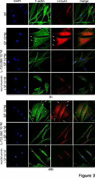
This article has not been copyedited and formatted. The final version may differ from this version.Molecular Pharmacology Fast Forward. Published on December 8, 2015 as DOI: 10.1124/mol.115.101014
at ASPE
T Journals on D
ecember 8, 2015
molpharm
.aspetjournals.orgD
ownloaded from
This article has not been copyedited and formatted. The final version may differ from this version.
Molecular Pharmacology Fast Forward. Published on December 8, 2015 as DOI: 10.1124/mol.115.101014
at ASPE
T Journals on June 5, 2018
molpharm
.aspetjournals.orgD
ownloaded from

This article has not been copyedited and formatted. The final version may differ from this version.Molecular Pharmacology Fast Forward. Published on December 8, 2015 as DOI: 10.1124/mol.115.101014
at ASPE
T Journals on June 5, 2018
molpharm
.aspetjournals.orgD
ownloaded from
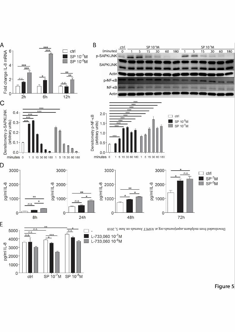
This article has not been copyedited and form
atted. The final version m
ay differ from this version.
Molecular Pharm
acology Fast Forward. Published on D
ecember 8, 2015 as D
OI: 10.1124/m
ol.115.101014
at ASPET Journals on June 5, 2018 molpharm.aspetjournals.org Downloaded from

This article has not been copyedited and formatted. The final version may differ from this version.Molecular Pharmacology Fast Forward. Published on December 8, 2015 as DOI: 10.1124/mol.115.101014
at ASPE
T Journals on D
ecember 8, 2015
molpharm
.aspetjournals.orgD
ownloaded from
This article has not been copyedited and formatted. The final version may differ from this version.Molecular Pharmacology Fast Forward. Published on December 8, 2015 as DOI: 10.1124/mol.115.101014
at ASPE
T Journals on June 5, 2018
molpharm
.aspetjournals.orgD
ownloaded from

This article has not been copyedited and formatted. The final version may differ from this version.Molecular Pharmacology Fast Forward. Published on December 8, 2015 as DOI: 10.1124/mol.115.101014
at ASPE
T Journals on D
ecember 8, 2015
molpharm
.aspetjournals.orgD
ownloaded from
This article has not been copyedited and formatted. The final version may differ from this version.Molecular Pharmacology Fast Forward. Published on December 8, 2015 as DOI: 10.1124/mol.115.101014
at ASPE
T Journals on June 5, 2018
molpharm
.aspetjournals.orgD
ownloaded from