Embed Size (px)
Citation preview
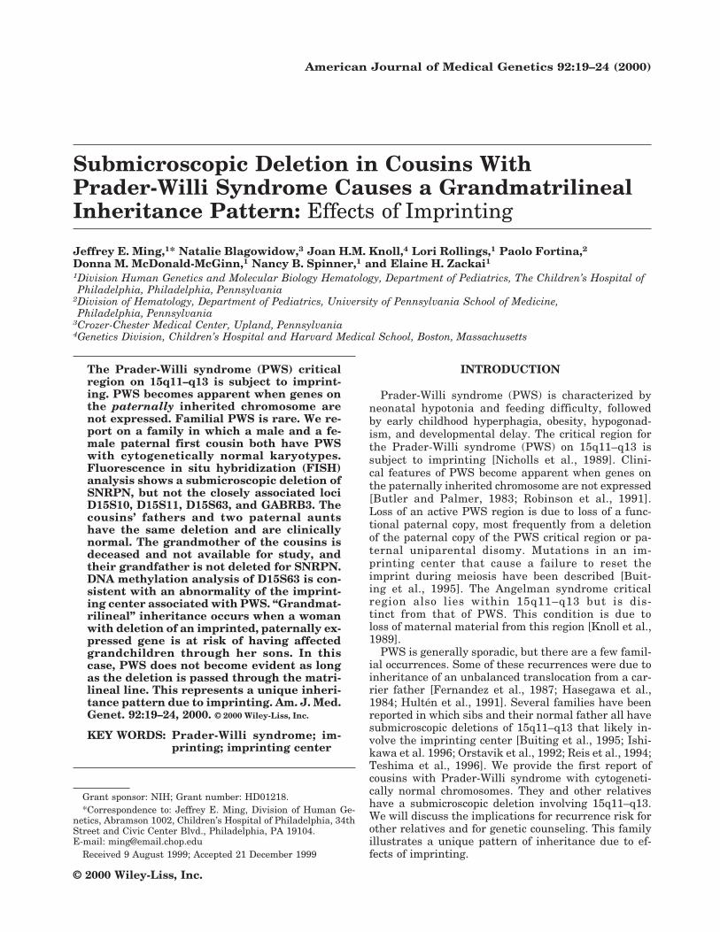
Submicroscopic Deletion in Cousins WithPrader-Willi Syndrome Causes a GrandmatrilinealInheritance Pattern: Effects of Imprinting
Jeffrey E. Ming,1* Natalie Blagowidow,3 Joan H.M. Knoll,4 Lori Rollings,1 Paolo Fortina,2Donna M. McDonald-McGinn,1 Nancy B. Spinner,1 and Elaine H. Zackai1
1Division Human Genetics and Molecular Biology Hematology, Department of Pediatrics, The Children’s Hospital ofPhiladelphia, Philadelphia, Pennsylvania
2Division of Hematology, Department of Pediatrics, University of Pennsylvania School of Medicine,Philadelphia, Pennsylvania
3Crozer-Chester Medical Center, Upland, Pennsylvania4Genetics Division, Children’s Hospital and Harvard Medical School, Boston, Massachusetts
The Prader-Willi syndrome (PWS) criticalregion on 15q11–q13 is subject to imprint-ing. PWS becomes apparent when genes onthe paternally inherited chromosome arenot expressed. Familial PWS is rare. We re-port on a family in which a male and a fe-male paternal first cousin both have PWSwith cytogenetically normal karyotypes.Fluorescence in situ hybridization (FISH)analysis shows a submicroscopic deletion ofSNRPN, but not the closely associated lociD15S10, D15S11, D15S63, and GABRB3. Thecousins’ fathers and two paternal auntshave the same deletion and are clinicallynormal. The grandmother of the cousins isdeceased and not available for study, andtheir grandfather is not deleted for SNRPN.DNA methylation analysis of D15S63 is con-sistent with an abnormality of the imprint-ing center associated with PWS. “Grandmat-rilineal” inheritance occurs when a womanwith deletion of an imprinted, paternally ex-pressed gene is at risk of having affectedgrandchildren through her sons. In thiscase, PWS does not become evident as longas the deletion is passed through the matri-lineal line. This represents a unique inheri-tance pattern due to imprinting. Am. J. Med.Genet. 92:19–24, 2000. © 2000 Wiley-Liss, Inc.
KEY WORDS: Prader-Willi syndrome; im-printing; imprinting center
INTRODUCTION
Prader-Willi syndrome (PWS) is characterized byneonatal hypotonia and feeding difficulty, followedby early childhood hyperphagia, obesity, hypogonad-ism, and developmental delay. The critical region forthe Prader-Willi syndrome (PWS) on 15q11–q13 issubject to imprinting [Nicholls et al., 1989]. Clini-cal features of PWS become apparent when genes onthe paternally inherited chromosome are not expressed[Butler and Palmer, 1983; Robinson et al., 1991].Loss of an active PWS region is due to loss of a func-tional paternal copy, most frequently from a deletionof the paternal copy of the PWS critical region or pa-ternal uniparental disomy. Mutations in an im-printing center that cause a failure to reset theimprint during meiosis have been described [Buit-ing et al., 1995]. The Angelman syndrome criticalregion also lies within 15q11–q13 but is dis-tinct from that of PWS. This condition is due toloss of maternal material from this region [Knoll et al.,1989].
PWS is generally sporadic, but there are a few famil-ial occurrences. Some of these recurrences were due toinheritance of an unbalanced translocation from a car-rier father [Fernandez et al., 1987; Hasegawa et al.,1984; Hulten et al., 1991]. Several families have beenreported in which sibs and their normal father all havesubmicroscopic deletions of 15q11–q13 that likely in-volve the imprinting center [Buiting et al., 1995; Ishi-kawa et al. 1996; Orstavik et al., 1992; Reis et al., 1994;Teshima et al., 1996]. We provide the first report ofcousins with Prader-Willi syndrome with cytogeneti-cally normal chromosomes. They and other relativeshave a submicroscopic deletion involving 15q11–q13.We will discuss the implications for recurrence risk forother relatives and for genetic counseling. This familyillustrates a unique pattern of inheritance due to ef-fects of imprinting.
Grant sponsor: NIH; Grant number: HD01218.*Correspondence to: Jeffrey E. Ming, Division of Human Ge-
netics, Abramson 1002, Children’s Hospital of Philadelphia, 34thStreet and Civic Center Blvd., Philadelphia, PA 19104.E-mail: [email protected]
Received 9 August 1999; Accepted 21 December 1999
American Journal of Medical Genetics 92:19–24 (2000)
© 2000 Wiley-Liss, Inc.

CLINICAL REPORTSPatient 1 (Propositus)
Patient 1 is a first cousin of patient 2; their fathersare brothers. He was born at 41 weeks of gestationweighing 3400 g. He had severe hypotonia in the neo-natal period and required nasogastric feeding for thefirst 10 days of life. He also has cryptorchidism. On ourexamination at age 6 months, he had bitemporal nar-rowness, unilateral esotropia, hypotonia, and the footlength was less than the 3rd centile bilaterally.
Patient 2
Patient 2 (Fig. 1) is a girl born at 33 weeks gestationsecondary to placental abruption weighing 1.5 kg (20thcentile). She had profound neonatal hypotonia and re-quired gavage feeding. She was also noted to have ahemivertebral body, fusion and hypoplasia of ribs, 13ribs on the left, and fusion of cervical vertebrae 6 and 7.She had a high arched palate, epicanthus, and strabis-mus noted neonatally. Electroencephalogram, nerveconduction studies, and electromyogram were normal.A muscle biopsy was unrevealing. In early childhood,
she developed hyperphagia and obesity. She hadBlount disease at the age of 12 years. She also hasdiabetes mellitus and hypoventilation. At the age of 12years 9 months, her height was less than the 5th cen-tile (50th centile for 10.5 years) and her foot length wasless than the 3rd centile. An evaluation at age 18 yearsshowed an IQ of 49 on the Slosson Intelligence Test.
Both patients fulfill proposed diagnostic criteria forPWS [Holm et al., 1993]. Patient 1 had neonatal hypo-tonia, neonatal feeding problems, hypogonadism, de-velopmental delay, molecular abnormality of PWS re-gion, and small feet. Patient 2 had neonatal hypotonia,neonatal feeding problems, rapid weight gain, globaldevelopmental delay, characteristic face, hyperphagia,molecular abnormality of PWS region, short stature,small feet, and strabismus.
Family History
Patient 1 has 3 sibs who are healthy and developingnormally (Fig. 2). Patient 2 had 1 sibling who washealthy with normal development until his death in adrowning accident. The patients’ fathers also have nosignificant medical problems. The patients have 3 ad-ditional paternal uncles and 5 paternal aunts. The pa-ternal grandmother died of emphysema. The paternalgrandfather is healthy.
MATERIALS AND METHODSCytogenetic Studies
High-resolution karyotypes with G-banding wereperformed on the two affected cousins as well as theirfathers.
Molecular Cytogenetic Analysis
Cosmid probes. Fluorescence in situ hybridizationstudies on metaphase spreads with probes for themarkers SNRPN and D15S10 (Oncor Inc., Gaithers-burg, MD) were performed on all relatives. A probe forthe locus PML on 15q22 was used to identify the chro-mosome 15 homologues. Cosmids for the markersD15S11 and GABRB3 (Oncor Inc., Gaithersburg, MD)were also used for FISH in the 2 clinically affectedcousins. FISH was carried out according to the manu-facturer’s instructions.
Fluorescence in situ hybridization probes.YAC clones were obtained from the Human GenomeProject Bank. The clones 307A12, 326F6, and 457B7were derived from the Centre d’Etude du Polymor-phism Humaine (CEPH) [Albertsen et al., 1990]. Totalyeast DNA as well as DNA from the 15 kb phage cloneJP3 (D15S63) [Knoll et al., 1993] were labeled by nicktranslation with biotin-16 dUTP. Hybridization wasperformed as previously described [Knoll and Lichter,1994].
DNA Methylation Analysis
PW71 methylation. Total genomic DNA was iso-lated from Patient 2 and her father (QIAGEN, Valen-cia, CA). The DNA was digested with HindIII andHpaII, run on an agarose gel, transferred to a nylon
Fig 1. One of the affected cousins with PWS at age 16 years (IndividualIV-2 in the pedigree in Fig. 2).
20 Ming et al.

membrane, and hybridized with the PW71 probe(D15S63) by standard techniques [Buiting et al., 1995].
SNRPN methylation. Total genomic DNA wasisolated from the father of patient 2 and two of herpaternal aunts (III.2, III.5). Using previously describedmethods [Kubota et al., 1997], methylation-specificPCR was performed after treatment of the genomicDNA with the CpGenome (Oncor) DNA modificationkit.
RESULTSMolecular Cytogenetic Studies
Both affected individuals and their fathers all hadcytogenetically normal karyotypes at the 550-bandlevel. Molecular cytogenetic (FISH) studies on both pa-tients revealed a deletion of SNRPN on one chromo-some 15 homologue (Fig. 3A). D15S10 (Fig. 3B),GABRB3, and D15S11 were all present on both homo-logues. An identical pattern was detected in both of thepatients’ fathers (Fig. 2, individuals III-4, III-10), andin 2 of the fathers’ 8 sibs (individuals III-5, III-6).SNRPN was not deleted in other relatives tested, in-cluding 3 paternal aunts, 2 paternal uncles, the pater-nal grandfather, and the paternal grandmother’s sis-ter. The paternal grandmother was deceased and notissue was available for study.
To further delineate the deletion, selected YACs en-compassing and flanking SNRPN [Mutirangura et al.,1993] were used as FISH probes (Fig. 4). These in-cluded YAC 307A12 (approximately 360 kb, containinglocus D15S13), YAC 326F6 (335 kb, containing locusD15S63), and YAC 457B7 (320 kb, containing locusD15S174). As expected, YACs 307A12 and 326F6 werepresent on each chromosome 15 homologue in the indexcases. YAC 457B4, that contains the SNRPN gene, wasalso present in two copies of strong intensity. In addi-
tion, a phage clone for D15S63 (JP3) was also used forFISH, and two copies were present in individuals withthe SNRPN deletion. These data suggest that the de-leted region is small compared to the 320 kb YAC.
DNA Methylation Studies
The PW71B probe detects DNA methylation at theD15S63 locus. When digested with HindIII and HpaII,normal individuals have a maternally derived band of6.0 kb, and a paternally derived band of 4.4 kb [Dittrichet al., 1992]. The affected Patient 2 showed only thematernal methylation pattern (Fig. 5). In combinationwith the FISH finding of two copies of D15S63 anddeletion of one copy of SNRPN, these data are sugges-tive of a deletion affecting the imprinting center. Herfather, who has the same FISH pattern as his daugh-ter, showed both paternal and maternal methylationpatterns.
To determine the origin of the deletion, we also per-formed methylation analysis of SNRPN on the father ofPatient 2 (III.4), and two paternal aunts of this patient,one of whom had a deletion of SNRPN by FISH (III.5),and one of whom had two copies of SNRPN (III.2). Boththe patient’s father and the aunt with one copy ofSNRPN showed only the presence of the paternally-imprinted 100 bp PCR product (data not shown). Theaunt with two copies of SNRPN showed both the ma-ternal (174 bp) and the paternal (100 bp) products.These results are consistent with the deletion beingpresent in the patient’s paternal grandmother.
DISCUSSION
We describe two paternal first cousins with PWS whohave a submicroscopic deletion of 15q11–q13. Bothchildren meet consensus diagnostic criteria for PWS[Holm et al., 1993]. This is the first report of cousins
Fig. 2. Pedigree. The propositi are paternal first cousins; their fathers are brothers. The affected patient’s fathers and two aunts carry the deletionbut are clinically normal. Individual IV-1 died in an accident and had normal somatic and intellectual development to that point. s, h: Deletion not tested,clinically normal. d, j: Prader-Willi syndrome. Ns, Nh: No deletion, clinically normal. (, ): Carries deletion 46,XX/Y.ish del(15)(q11.2q11.2)(SNRPN−D15S10+ GABRB3+ D15S11+), clinically normal.
Prader-Willi Syndrome in Cousins 21

affected with PWS who have cytogenetically normalchromosomes. In addition, the pedigree contains mul-tiple clinically normal carriers who also carry the samesubmicroscopic deletion, including each of the patient’sfathers and two paternal aunts (Fig. 3). The proposi-
tus’s paternal grandfather did not carry the deletion,and the grandmother was not available for testing. Wepostulate that she also carries the deletion. The sibs ofthe propositus were not tested for the deletion becauseall of them had normal growth and development andwere old enough so that clinical manifestations of PWSshould have been apparent. Approximately 60% of in-dividuals with PWS have a cytogenetically detectabledeletion of 15q11–q13 [Ledbetter et al., 1987]. Thesedeletions are almost always de novo and are generally4 megabases in size. Commonly deleted loci includeSNRPN, D15S10, GABRB3, and D15S11. Based on theFISH studies, the size of the deletion in the patients inthis report is much smaller than the common deletion,as only the clone for SNRPN is deleted.
There have been several previous reports of familialrecurrence of PWS. Some instances were due to unbal-anced translocations involving chromosome 15 in sibs[Fernandez et al., 1987; Hulten et al., 1991]. Cousinsinheriting an unbalanced translocation have also beenreported [Hasegawa et al., 1984]. Although both werediagnosed with PWS, one individual inherited the ab-normal chromosome 15 from his mother, and he hadseizures, severe developmental delay, and apparentprognathism. This individual might have actually hadAngelman syndrome. There are several reports of sibswith unequivocal clinical PWS with cytogeneticallynormal chromosomes. [Burke et al., 1987; Ishikawa etal., 1987; Lubinsky et al., 1987; Orstavik et al., 1992].Subsequent studies have demonstrated submicroscopicdeletions in some of these kindreds [Saitoh et al.,1997]. The submicroscopic deletion was also present inthe sibs’ father in two cases [Buiting et al., 1995;Teshima et al., 1996]. In another instance, the fatherwas not available for testing, but the deletion was pre-sent in the paternal grandmother [Reis et al., 1994]. Adeletion of the PWS critical region has not been notedon routine karyotype in any cases of familial recur-rence, unless a rearrangement was present.
These submicroscopic deletions are believed to dis-rupt an imprinting center (IC) [Buiting et al., 1995;Saitoh et al., 1997]. Mutations or deletions affectingthe IC will result in an inability to reset the maternaland paternal imprints during gametogenesis. In thecases described involving the IC, submicroscopic dele-tions of variable length around SNRPN have been de-tected [Buiting et al., 1995; Reis et al., 1994; Schuffen-hauer et al., 1996; Schulze et al., 1997; Sutcliffe et al.,1994] including, but not limited to the promoter andexon 1 [Saitoh et al., 1996; Ohta et al., 1999]. Individu-als with PWS due to imprinting mutations are not eas-ily distinguishable clinically from those with PWS dueto a deletion or UPD [Saitoh et al., 1997]. Hypopigmen-tation is not present, because this is thought to be dueto a deletion of the P gene locus.
The inheritance pattern seen in this family is uniqueto imprinting. The phenotype caused by the deletiononly becomes apparent when it has passed through thepaternal line. As long as the abnormality is passedthrough only the matrilineal line, no phenotypic effectwill occur. A woman with the deletion would not be atrisk for having affected children; however, her sons areat risk of having affected children. Her daughters
Fig. 3. FISH mapping of probes for 15q11–q13 on metaphases fromindividuals IV:2 or IV:7, who have PWS. (A) FISH of SNRPN with PMLcontrol. Only one copy of SNRPN is present. The PML marker indicates thetwo chromosome 15 homologues. (B) FISH of D15S10 with PML control.Both chromosome 15 homologues show hybridization to the probe forD15S10. (C) FISH of YAC 307A12 containing the marker D15S13. Bothhomologues show hybridization. Similar results were obtained with probescontaining the markers D15S63 and D15S174.
22 Ming et al.

would not be at risk, but the daughters’ sons wouldhave a risk of having children with PWS. Thus, awoman would not have affected children, but hergrandchildren through her sons are at risk of showingthe phenotype. This “grandmatrilineal” inheritancepattern presents new issues for genetic counseling be-cause only the grandchildren of a carrier woman havethe possibility of showing the PWS phenotype. In ad-dition, phenotypically normal males with the deletionhave a 50% recurrence risk for their children. In fami-lies where PWS is due to a deletion, the father shouldalso be evaluated by molecular methods for the dele-tion. This is especially important if the deletion is notapparent using high-resolution chromosome analysisand does not involve the Angelman syndrome criticalregion. The size of the deletion can be estimated byusing two probes in the commonly deleted region. Ifonly SNRPN is deleted, a familial imprinting mutationmust be suspected, and the potential implications forrecurrence risk should be discussed. A very small im-printing center deletion may not be detectable by a
cosmid probe, and in these cases, if FISH studies arenormal, a more detailed molecular investigation maybe required for accurate recurrence risk prediction.
ACKNOWLEDGMENTS
We would like to thank Wendy Hitchcock for assis-tance with the methylation studies. JEM was sup-ported in part by NIH Grant HD01218.
REFERENCES
Albertsen HM, Abderrahim H, Cann HM, Dausset J, LePaslier D, CohenD. 1990. Construction and characterization of a yeast artificial chro-mosome library containing seven haploid human genome equivalents.Proc Natl Acad Sci USA 87:4256–4260.
Buiting K, Saitoh S, Gross S, Dittrich B, Schwartz S, Nicholls RD, Horst-hemke B. 1995. Inherited microdeletions in the Angelman and Prader-Willi syndromes define an imprinting centre on human chromosome15. Nature Genet 9:395–400.
Burke CM, Kousseff BG, Gleeson M, O’Connell BM, Devlin JG. 1987. Fa-milial Prader-Willi syndrome—new developments. Arch Intern Med147:673–675.
Butler MG, Palmer CG. 1983. Parental origin of chromosome 15 deletion inPrader-Willi syndrome (letter). Lancet. 1:1285–1286.
Dittrich B, Robinson WP, Knoblauch H, Buiting K, Schmidt K, Gillessen-Kaesbach G, Horsthemke B. 1992. Molecular diagnosis of the Prader-Willi and Angelman syndromes by detection of parent-of-origin specificDNA methylation in 15q11–13. Hum Genet 90:313–315.
Fernandez F, Berry C, Mutton D. 1987. Prader-Willi syndrome in siblings,due to unbalanced translocation between chromosomes 15 and 22. ArchDis Child 62:841–843.
Hasegawa T, Hara M, Ando M, Osawa M, Fukuyama Y, Takahasi M,Yamada K. 1984. Cytogenetic studies of familial Prader-Willi syn-drome. Hum Genet 65:325–330.
Holm VA, Cassidy SB, Butler MG, Hanchett JM, Greenswag LR, WhitmanBY, Greenberg F. 1993. Prader-Willi syndrome: consensus diagnosticcriteria. Pediatrics 91:398–402.
Hulten M, Armstrong S, Challinor P, Gould C, Hardy G, Leedham P, LeeT, McKeown C. 1991. Genomic imprinting in an Angelman and Prader-Willi translocation family (letter). Lancet 338:638–639.
Ishikawa T, Kanayama M, Wada Y. 1987. Prader-Willi syndrome in twosiblings: one with normal karyotype, one with a terminal deletion ofdistal Xq. Clin Genet 32:295–299.
Ishikawa T, Kibe T, Wada Y. 1996. Deletion of small nuclear ribonucleo-protein polypeptide N (SNRPN) in Prader-Willi syndrome detected byfluorescence in situ hybridization: two siblings with the typical pheno-
Fig. 5. DNA Methylation Studies. Genomic DNA was digested withHindIII and HpaII and hybridized with the PW71B probe. The probe de-tects a 6.0 kb maternal fragment and a 4.4 kb paternal fragment. (Lane 1)Normal control with biparental band pattern; (lane 2) Patient 2, affectedwith PWS, showing only maternal band; (lane 3) Clinically normal fatherof Patient 2, showing biparental band pattern.
Fig. 4. Physical Map of 15q11 in the vicinity of the PWS critical region. The relevant YACS and their sizes are indicated. +, Not deleted by FISH; −,deleted by FISH.
Prader-Willi Syndrome in Cousins 23
type without a cytogenetic deletion in chromosome 15q. Am J MedGenet 62:350–352.
Knoll JHM, Lichter P. 1994. In situ hybridization to metaphase chromo-somes and interphase nuclei. In: Dracopoli NC, Haines JL, Korf BR,Moir DT, Morton CC, Seidman CE, Seidman JG, Smith DR, editors.Current Protocols in Human Genetics, Unit 4.3, Vol. 1. New York: JohnWiley & Sons, Inc.
Knoll JHM, Nicholls RD, Magenis RE, Graham Jr JM, Lalande M, Latt SA.1989. Angelman and Prader-Willi syndromes share a common chromo-some 15 deletion but differ in parental origin of the deletion. Am J MedGenet 32:285–290.
Knoll JHM, Sinnett D, Wagstaff J, Glatt K, Wilcox AS, Whiting PM, Win-grove P, Sikela JM, Lalande M. 1993. FISH ordering of reference mark-ers and of the gene for the alpha-5 subunit of the gamma-aminobutyricacid receptor (GABRA5) within the Angelman and Prader-Willi syn-drome chromosomal regions. Hum Mol Genet 2:183–189.
Kubota T, Das S, Christian SL, Baylin SB, Herman JG, Ledbetter DH.1997. Methylation-specific PCR simplifies imprinting analysis (letter).Nat Genet 16:16–17.
Ledbetter DH, Greenberg F, Holm VA, Cassidy SB. 1987.Conference re-port: second annual Prader-Willi scientific conference. Am J Med Genet28:779–790.
Lubinsky M, Zellweger H, Greenswag L, Larson G, Hansmann I, LedbetterD. 1987. Familial Prader-Willi syndrome with normal chromosomes.Am J Med Genet 28:37–43.
Multirangura A, Jayakumar A, Sutcliffe JS, Nakao M, McKinney MJ, Buit-ing K, Hosthemke B, Beaudet AL, Chinault AC, Ledbetter DH. 1993. Acomplete YAC contig of the Prader-Willi/Angelman chromosome region(15q11–q13) and refined localization of the SNRPN gene. Genomics18:546–552.
Nicholls RD, Knoll JHM, Butler MG, Karam S, Lalande M. 1989. Geneticimprinting suggested by maternal heterodisomy in nondeletion Prader-Willi syndrome. Nature 342:281–285.
Ohta T, Gray TA, Rogan PK, Buiting K, Gabriel JM, Saitoh S, MuralidharB, Bilienska B, Krajewska-Walasek M, Driscoll DJ, Horsthemke B,Butler MG, Nicholls RD. 1999. Imprinting-mutation mechanisms inPrader-Willi syndrome. Am J Hum Genet 64:397–413.
Orstavik KH, Tangsrud SE, Kiil R, Hansteen I-L, Steen-Johnsen J, Cas-
sidy SB, Martony A, Anvret M, Tommerup N, Brøndum-Nielsen K.1992. Prader-Willi syndrome in a brother and sister without cytoge-netic or detectable molecular genetic abnormality at chromosome15q11q13. Am J Med Genet 44:534–538.
Reis A, Dittrich B, Greger V, Buiting K, Lalande M, Gillessen-Kaesbach G,Anvret M, Horsthemke B. 1994. Imprinting mutations suggested byabnormal DNA methylation patterns in familial Angelman and Prader-Willi syndromes. Am J Hum Genet 54:741–747.
Robinson WP, Bottani A, Yagang X, Balakrishman J, Binkert F, MachlerM, Prader A, Schinzel A. 1991. Molecular, cytogenetic, and clinicalinvestigations of Prader-Willi syndrome patients. Am J Hum Genet49:1219–1234.
Saitoh S, Buiting K, Rogan PK, Buxton JL, Driscoll DJ, Arnemann J,Konig R, Malcolm S, Horsthemke B, Nicholls RD. 1996. Minimal defi-nition of the imprinting center and fixation of chromosome 15q11–q13epigenotype by imprinting mutations. Proc Natl Acad Sci USA 93:7811–7815.
Saitoh S, Buiting K, Cassidy SB, Conroy JM Driscoll DJ, Gabriel JM,Gillessen-Kaesbach G, Glenn CC, Greenswag LR, Horsthemke B, Ko-ndo I, Kuwajima K, Niikawa N, Rogan PK, Schwartz S, Seip J, Willi-ams CA, Nicholls RD. 1997. Clinical spectrum and molecular diagnosisof Angelman and Prader-Willi syndrome patients with an imprintingmutation. Am J Med Genet 68:195–206.
Schuffenhauer S, Buchholz T, Stengel-Rutkowski S, Buiting K, Schmidt H,Meitinger T. 1996. A familial deletion in the Prader-Willi syndromeregion including the imprinting control region. Hum Mutation 8:288–292.
Schulze A, Hansen C, Bækgaard P, Blichfeldt S, Petersen MB, TommerupN, Brøndum-Nielsen K. 1997. Clinical features and molecular geneticanalysis of a boy with Prader-Willi syndrome caused by an imprintdefect. Acta Paediatr 86:906–910.
Sutcliffe JS, Nakao M, Christian S, Orstavik KH, Tummerup N, LedbetterDH, Beaudet AL. 1994. Deletions of a differentially methylated CpGisland at the SNRPN gene define a putative imprinting control region.Nat Genet 8:52–58.
Teshima I, Chadwick D, Chitayat D, Kobayashi J, Ray P, Shuman C,Siegel-Bartelt J, Strasberg P, Weksberg R. 1996. FISH detection ofchromosome 15 deletions in Prader-Willi and Angelman syndromes.Am J Med Genet 62:216–223.
24 Ming et al.