Embed Size (px)
Citation preview

Subcutaneous tissue reaction and cytotoxicity of polyvinylidenefluoride and polyvinylidene fluoride-trifluoroethylene blendsassociated with natural polymers
Leonardo Marques,1 Leandro A. Holgado,1 Rebeca D. Sim~oes,2 Jo~ao D. A. S. Pereira,2
Juliana F. Floriano,3 L�ıgia S. L. S. Mota,4 Carlos F. O. Graeff,3 Carlos J. L. Constantino,2
Miguel. A. Rodriguez-Perez,5 Mariza Matsumoto,1 Angela Kinoshita1
1Universidade Sagrado Corac~ao – USC, Bauru, S~ao Paulo, Brazil2Faculdade de Ciencias e Tecnologia, UNESP Universidade Estadual Paulista, Presidente Prudente, S~ao Paulo, Brazil3Faculdade de Ciencias, UNESP Universidade Estadual Paulista, Bauru, S~ao Paulo, Brazil4Instituto de Biociencias, UNESP Universidade Estadual Paulista, Bauru, S~ao Paulo, Brazil5Condensed Matter Physics Department, CellMat Laboratory, Faculty of Science, University of Valladolid, Valladolid, Spain
Received 27 August 2012; revised 5 February 2013; accepted 6 March 2013
Published online 10 May 2013 in Wiley Online Library (wileyonlinelibrary.com). DOI: 10.1002/jbm.b.32941
Abstract: Cytotoxicity and subcutaneous tissue reaction of
innovative blends composed by polyvinylidene fluoride and
polyvinylidene fluoride-trifluoroethylene associated with nat-
ural polymers (natural rubber and native starch) forming
membranes were evaluated, aiming its applications associ-
ated with bone regeneration. Cytotoxicity was evaluated in
mouse fibroblasts culture cells (NIH3T3) using trypan blue
staining. Tissue response was in vivo evaluated by subcuta-
neous implantation of materials in rats, taking into account
the presence of necrosis and connective tissue capsule
around implanted materials after 7, 14, 21, 28, 35, 60, and
100 days of surgery. The pattern of inflammation was eval-
uated by histomorphometry of the inflammatory cells. Chem-
ical and morphological changes of implanted materials after
60 and 100 days were evaluated by Fourier transform infrared
(FTIR) absorption spectroscopy and scanning electron mi-
croscopy (SEM) images. Cytotoxicity tests indicated a good
tolerance of the cells to the biomaterial. The in vivo tissue
response of all studied materials showed normal inflamma-
tory pattern, characterized by a reduction of polymorphonu-
clear leukocytes and an increase in mononuclear leukocytes
over the time (p < 0.05 Kruskal–Wallis). On day 60, micro-
scopic analysis showed regression of the chronic inflamma-
tory process around all materials. FTIR showed no changes
in chemical composition of materials due to implantation,
whereas SEM demonstrated the delivery of starch in the me-
dium. Therefore, the results of the tests performed in vitro
and in vivo show that the innovative blends can further be
used as biomaterials. VC 2013 Wiley Periodicals, Inc. J Biomed
Mater Res Part B: Appl Biomater 101B: 1284–1293, 2013.
Key Words: biomaterial, piezoelectricity, tissue reaction, cyto-
toxicity, PVDF
How to cite this article: Marques L, Holgado LA, Sim~oes RD, Pereira JDAS, Floriano JF, Mota LSLS, Graeff CFO, Constantino CJL,Rodriguez-Perez MA, Matsumoto M, Kinoshita A. 2013. Subcutaneous tissue reaction and cytotoxicity of polyvinylidene fluorideand polyvinylidene fluoride-trifluoroethylene blends associated with natural polymers. J Biomed Mater Res Part B 2013:101:1284–1293.
INTRODUCTION
Currently the gold standard for the treatment of large bonedefects is the autologous bone that stimulates a series of cellularand molecular events that result in repair of the recipient area.Autologous bone is obtained from various donor sites; however,the morbidity of this procedure, the technical difficulty, and thehigh costs are factors that make necessary the search for newbiomaterials with properties that enhance bone healing.1,2
The most commonly used bioabsorbable polymers forbone tissue engineering are saturated aliphatic polymers
such as poly(lactic acid) (PLA), poly(glycolic acid),3 polycap-rolactone (PCL), and their copolymer, among others. Thesebased polymers and their derivatives have been successfullyused as internal fracture fixation devices (orthopedic bioab-sorbable osteofixation systems) for long bones in human,since the mid-1980.4 Another polymeric systems used arestarch-based blends. The starch is blended with thermoplas-tic polymers to increase their resistance against thermome-chanical degradation and make them less brittle and moreeasily processed. Blends of starch with PCL and PLA have
Correspondence to: A. Kinoshita; e-mail: [email protected] grant sponsors: FAPESP (Fundac~ao de Amparo �a Pesquisa do Estado de S~ao Paulo), CNPq (Conselho Nacional de Desenvolvimento
Cient�ıfico e Tecnol�ogico), CAPES (Coordenac~ao de Aperfeicoamento de Pessoal de N�ıvel Superior), FAP=USC (Fundac~ao de Amparo �a Pesquisa
da Universidade Sagrado Corac~ao)
1284 VC 2013 WILEY PERIODICALS, INC.

been proposed as potential alternative biodegradable materi-als for a wide range of biomedical applications, includingbone cements and bone substitutes.5 The association of bio-sorbable polymers with therapeutic factors was also investi-gated. For instance, Hench and Paschall6 studied theassociation of poly(D,L-lactic acid) (PLDLA) and bioactive glassto improve the osteoconductivity, guiding the bone growth asthe glass degrades. Carsten et al.7 developed composites withbiodegradable polymers and carbonated calcium phosphate(CaCO3). Macroporous were formed inside it due to fast deg-radation of PLDLA, promoting the bone formation, whereasthe slowly degradable poly-L-lactide outside of compositeensures the mechanical stability. In contrast with the strategyof inserting a therapeutic factor in polymeric matrix, in thepresent work no therapeutic factors of bone tissue regenera-tion were added since the polymers used already presents apropriety that may induce bone growth.
Polyvinylidene fluoride (PVDF) and its copolymer polyvi-nylidene fluoride-trifluoroethylene P(VDF-TrFE) applied herepresent scientific and technological interest due to theirferro, pyro, and piezoelectric properties.8,9 More recently,Hong et al.10 developed nanograss structures of P(VDF-TrFE) and observed an enhancement of piezoelectricity. Thisproperty can be associated with bone growth induction,since bones are piezoelectric.11–13 Callegari and Belangeroevaluated the interface formed between P(VDF-TrFE) andPVDF tubes (piezoelectric and nonpiezoelectric) in rat bonetissue. The results of conventional optical microscopy andbackscattered scanning electron microscopy (SEM) indicatethat the piezoelectric effect has an important role in thenew bone tissue formation inside the polymeric tubes.14
PVDF is formed by repeating units of –CH2–CF2–, corre-sponding to about 2000 monomeric units with an averagelength of about8 0.5 mm. Due to its electrical properties, lit-erature reports a wide range of applications for this poly-mer, including optoelectronic, electromechanical and morerecently, as a biomaterial.15,18 PVDF is a biocompatible ma-terial and some applications include vascular suture16,17;mesh materials for abdominal hernia repair18–20 as a sub-strate to enhance nerve fiber outgrowth21; controlled deliv-ery of drugs19; tissue engineering and cell biologyapplication including bone regeneration.11,13 These applica-tions are mainly due to the high piezoelectric activity ofPVDF, which depends on its polar crystalline phase.
The b phase of PVDF, characterized by a zigzag conforma-tion chain with planar orthorhombic crystal system and net-work parameters a 5 8.58 Å, b 5 4.91 Å, and c 5 2.56 Å,presents the highest piezoelectric response. Thus it is themost desirable form8; however, the most common crystallinephase is the a phase (nonpolar). On the other hand, randomcopolymers of vinylidene fluoride (VDF) with trifluoroethy-lene (TrFE) when associated in an appropriated molar ratioof VDF and TrFE, crystallize directly into a polar ferroelectricphase, in a transplanar chain similar to the PVDF b phase,presenting pyroelectricity and piezoelectricity comparable tob PVDF, after passing by a poling process.8
Membranes of PVDF and P(VDF-TrFE) can be producedwhen associated with natural rubber (NR) and=or cornstarch.
In a previous work, the fabrication procedure of these mem-branes was described.22 This process of fabrication discardsthe necessity of organic solvents to dissolve the synthetic co-polymer, contributing to biocompatibility. Besides, such proc-essing enables the fabrication of membranes with suitablemechanical properties. These blends were previously charac-terized by Fourier transform infrared (FTIR) spectroscopy,SEM, X-ray diffraction, density, melt flow index, hardness, andthermal conductivity. The results showed that the polymersdo not interact chemically with the additives leading to theformation of blends as physical mixtures where the additivesare well dispersed within the blends at micrometer level.However, it was observed that the adhesion of the starch isbetter in the case of blends with P(VDF-TrFE).23 Besides, thecrystalline structures of the a-PVDF and ferroelectric P(VDF-TrFE) are preserved in the blends. The density, hardness,melt flow index, and thermal conductivity values of theblends are the ones expected from physical mixtures.24
Tests for development of a new biomaterial include sev-eral stages from in vitro tests through cell cultures and invivo experiments using animal and human clinical trials.Such tests evaluate the biocompatibility, biological proper-ties, and the risks that the material can cause tohealth.12,24–27 Usually, the first stage to test a new materialfor biomedical use is the in vitro biocompatibility essays. Inthis work, the cytotoxicity of the polymeric blends was eval-uated in vitro using the cell line NIH3T3 of mouse fibro-blasts and in vivo by the study of tissue reaction aftersubcutaneous implant in rats. The results obtained withmembranes prepared with P(VDF-TrFE) were compared toPVDF=starch=NR latex, that is composed by well-known bio-compatible materials.16,28–30 As already mentioned, PVDFand P(VDF-TrFE) present piezoelectricity and can contributeto bone growth, indicating that the blends studied in thiswork can be useful for procedures associated with boneregeneration. Besides, the presence of interconnected po-rous, generated by the presence of the starch in the blends,is another factor that contributes to the bone tissue growth.
MATERIALS AND METHODS
The present study was approved by the Ethical Committeefrom the Universidade Sagrado Corac~ao – USC, Bauru, S~aoPaulo State, Brazil and was conducted according to recom-mendations of the National Institute of Health.31
MembranesThe innovative blends in form of membranes ofPVDF=starch=NR, P(VDF-TrFE)=starch and P(VDF-TrFE)=NRwere prepared by compressing=annealing (2 tons at 180�C)powders of the starting material leading to membranes of 1mm thick, as described in a previous work.24 They were cutin circular shape with 5 mm diameter and sterilized withgamma radiation (25 kGy) for in vivo and in vitro studies.The PVDF used was Florafon F4000 HD acquired from Ato-chem and P(VDF-TrFE) was 72=28 (in mass) acquired fromPiezotech (Piezotech S.A.S, H�esingue, France). The latex wascollected from different trees of Hevea brasiliensis, clones
ORIGINAL RESEARCH REPORT
JOURNAL OF BIOMEDICAL MATERIALS RESEARCH B: APPLIED BIOMATERIALS | OCT 2013 VOL 101B, ISSUE 7 1285

RRIM-600 (Rubber Research Institute of Malaysia), at theexperimental farm of EMBRAPA in Indiana, Sao Paulo, Brazil.The latex stabilization was made by using a commercial solu-tion of NH4OH (4.7 mL of NH4OH for 100 mL of latex). Theregular cornstarch with 28% amylose and particle size of 10lm (commercial name Amudex 3001) was supplied by CornProducts Brazil.
Cytotoxicity testThe biological properties of the materials were firstly eval-uated by in vitro cell tests.32 Cytotoxicity was evaluated byusing the cell line NIH3T3 of mouse fibroblasts. The cellswere placed in culture flasks (25 cm2) with 3.0 mL of culturemedium Dulbecco’s modified Eagle’s medium-HAM F12 (Invi-trogen) 1:1 ratio, supplemented with 20% fetal bovine serumand 1% penicillin=streptomycin and incubated at 38�C with95% humidity and 5% CO2. When the cells reached theirhighest degree of proliferation, they were isolated by usingtrypsin (Trypsin-Versen solution; Institute Adolfo Lutz) for 3min. The cell suspension obtained was transferred to a cen-trifuge tube for washing in culture media at 1000 rpm for 5min. This procedure was repeated twice. In the end cellswere suspended in 1 mL of culture medium.
The materials were placed in contact with the culturecells in concentration of 3.0 3 105 cells mL21, along withthe culture medium and incubated in the same conditionspreviously described for a period of 72 h. After this period,the material was removed. All samples were tested in tripli-cate. The parameter used to evaluate toxicity was cell viabil-ity using Trypan blue staining as marker (Sigma-AldrichCo.). Cell suspension of 40 mL was added to 40 mL of Trypanblue, homogeneized, and inoculated in a Neubauer chamber.Cell counting was performed in four quadrants of the exter-nal chamber. As negative control the culture plate itself wasused and as positive control phenol (0.02%), to validate thetest system. The stained blue cells were considered deadand the total viable cells was divided by the total number ofquadrants (four) and multiplied by 20,000 (10,000 3 dilu-tion factor 2). The nonparametric test Kruskal–Wallis wasused to compare the results and they were considered stat-istically different when p < 0.05.
The cell viability was observed only in the period of 72h, following the period described in ISO 10993-5 and ASTM19953, describing the periods of direct contact with thecells of the material by 72h.
Subcutaneous implant in vivoA total of 27 male Wistar rats, weighing an average of 300g, were used. The animals were kept in a plastic cage in anexperimental animal room and were fed with standard labo-ratory diet and water. Preoperatively, general anesthetic wasintramuscularly induced in animals with xylazine chlorhy-drate (5 mg kg21; Bayer, Brazil) and ketamine (35 mg kg21;Vetbrands, Brazil). The dorsal part was shaved and asepti-cally prepared for surgery. Incisions of 1cm long were madealong the back of the animal, symmetrical in relation to themidline, three on the right and three on the left side totaliz-ing six incisions according to the model of Minnen et al.33
The distance between implanted materials was approxi-mately 3 cm.
The skin was separated exposing the subcutaneous tissue,where the membranes were implanted. Two samples of eachblend were implanted into each animal in random position.The soft tissues and skin incisions were closed with 4-0 silkinterrupted sutures. After 7, 14, 21, 28, and 35 days, threeanimals were euthanized, and the pieces containing the mate-rial removed microscopic and macroscopic analysis. Six sam-ples (n 5 6) for each material implanted for a period ofobservation were obtained. After 60 and 100 days, six ani-mals were used: three for microscopic analysis and three forexperiments to characterize the implanted material.
For microscopic analysis, the excised tissues were pre-pared following routine histology procedures, parafinizedsections with 6 mm were stained with Masson’s trichromeand hematoxilin and eosin. The photomicroscope NikkonH550L was used to acquire the images. Six fields with 403
magnification around each implanted material were usedfor cell counting. Polymorphonuclear, mononuclear andgiant cells (GCs) were counted and the thickness of fibrousconnective tissue (CT) capsule was assessed using the soft-ware Image Pro-plus (Media-Cybernetics).
The average concentration of inflammatory cells in sixfields was classified according to the distribution adaptedfrom Yaltirik et al.34; for mononuclear and polymorphonu-clear cells, the classification was (1) <50 cells; (2) 50–70cells; (3) 70–100 cells, and (4) >100 cells and for GCs (1)<10 cells; (2) 10–20 cells, (3) 20–30 cells, and 4) >30 cells.This classification was adopted taking into account the dis-tribution of values found. The nonparametric test Kruskal–Wallis was used to compare the results and they were con-sidered statistically different when p < 0.05.
Characterization techniques of implanted membranesThe samples collected after 60 and 100 days postimplanta-tion were accessed by FTIR, to examine the chemicalchanges of the blends and by SEM, to study their morphol-ogy. FTIR measurements were carried out with a Brukerspectrometer model Tensor 27 in the ATR mode (not polar-ized), using 64 scans, and 4 cm21 spectral resolution. TheSEM images were recorded using a JEOL microscope modelJSM-820 (3 kV and 20 kV). The SEM were obtained fromthe transversal section after breaking the membranesthrough a fragile fracture (the membranes were frozen inliquid N2), the Au metallization (ca. 20 nm) was carried outusing a Balzers thermal evaporator model SCD 004.<
RESULTS
Macroscopic aspect postimplantationFigure 1 shows the membranes in the region of implanta-tion 60 days after surgery. The encapsulation of theimplanted material can be seen as well as the normal aspectof the tissues adjacent to the materials.
Microscopic analysisFigure 2 shows microscopic images of the implantationregion of the membranes, in the initial and final periods
1286 MARQUES ET AL. TISSUE REACTION AND CYTOTOXICITY OF PVDF AND P(VDF-TrFE)

after implantation. The region occupied by the membranesis marked (#). The images of 100 days after implantationsuggest that the membranes remained unchanged. At highermagnification, see Figure 3, CT and inflammatory cells maybe noted around the materials.
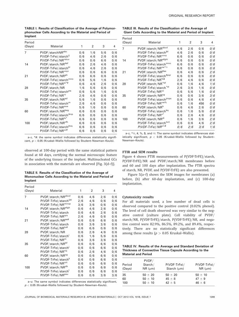
Tables I–III show the average counting of polymorpho-nuclear, mononuclear, and GCs after classification and arerepresented by the number of slices in the score divided bythe total number of slices observed. The differencesbetween the results were assessed by Kruskal–Wallis testand the same symbol inserted in superscript indicates p <
0.05. Table IV shows the average fibrous capsule thicknessfor each material as function of time. There are no statisti-cally significant differences between periods or materials[p > 0.05 analysis of variance (ANOVA), Tukey].
The microscopic images demonstrate that all testedmaterials presented similar responses (Figs. 2 and 3). Thisresult was statistically verified, since in general, there is nodifference between the responses of materials in the sameperiod. The statistically significant differences are found inthe cell counting performed after implant of the same mate-rial in different periods (Table III).
Seven days after implant, the materials are surroundedby granulation tissue [CT; Fig. 3(a–c)], with a moderate num-ber of mononuclear inflammatory cells infiltrate. Some paral-lel bundles of collagen fibers (*) can also be observed in theP(VDF-TrFE)=starch (b) and P(VDF-TrFE)=NR (c) images. At14 days, organized CT with collagen fibers arranged parallelto each other in contact with materials are observed with aslight diffuse mononuclear inflammatory infiltrate. At 21days, the microscopic pattern is similar to the previous pe-riod, with a decrease in the granulation tissue with persist-ence of mononuclear cell infiltration. In the PVDF=starch=NRresults, the increase in mononuclear cells is statistically sig-nificant (p < 0.05) in comparison to the 7-day period (TableII) and the number of foreign body GCs associated with theimplanted material is also in greater quantity (p < 0.05) incomparison to the 7-day period (Table III).
After 28 days, organization of collagen fibers with focalareas of mononuclear leukocytes is observed. In the P(VDF-TrFE)=starch images, vascular tissue close to the material isobserved. An increase in the number of mononuclear cellsand GCs in comparison to 7 days of implantation and a
FIGURE 1. The membranes of PVDF=starch=NR, P(VDF-TrFE)=starch
and P(VDF-TrFE)=NR 60 days after surgery. The normal aspect of the
tissues adjacent to the materials can be observed. [Color figure can
be viewed in the online issue, which is available at
wileyonlinelibrary.com.]
FIGURE 2. Microscopic images of the implantation region of the membranes, 7 and 100 days after surgery. The region occupied by the mem-
branes is indicated (#). (Masson Trichrome staining, original magnification 32). [Color figure can be viewed in the online issue, which is avail-
able at wileyonlinelibrary.com.]
ORIGINAL RESEARCH REPORT
JOURNAL OF BIOMEDICAL MATERIALS RESEARCH B: APPLIED BIOMATERIALS | OCT 2013 VOL 101B, ISSUE 7 1287

significant reduction in polymorphonuclear cells are found(p < 0.05, Tables I) in the P(VDF-TrFE)=starch and P(VDF-TrFE)=NR cell counting.
After 35 days, all tested implanted materials presentedsurrounded by mature fibrous CT showing collagen fibersparallel to each other, with a capsular pattern. There is areduction in the number of polymorphonuclear cells,
increase in mononuclear and GCs, in comparison with thebeginning period (p < 0.05; Tables I).
After 60 days of implantation, a reduction in the inflam-mation pattern is noticed for all tested materials, with areduction in polymorphonuclear cells and an increase in thenumber of mononuclear and GCs in comparison with theinitial periods (p < 0.05, Tables I). Similar results are
FIGURE 3. Microscopic images of the implantation region of the membranes, 7 (a–c) and 100 days (d–f) after surgery at higher magnification of
P(VDF-TrFE) (a and d), P(VDF-TrFE)=starch (b and e), and P(VDF-TrFE)=NR (c and f). At 7 days a moderate mononuclear inflammatory infiltrate in
the connective tissue (CT) is present. Parallel bundles of collagen fibers (*) surrounding the material are also observed. At 100 days,
multinucleated giant cells (GCs) in association with the membrane (hematoxylin and eosin staining, original magnification 340). [Color figure
can be viewed in the online issue, which is available at wileyonlinelibrary.com.]
1288 MARQUES ET AL. TISSUE REACTION AND CYTOTOXICITY OF PVDF AND P(VDF-TrFE)
observed at 100-day period with the same statistical patternfound at 60 days, certifying the normal microscopic featureof the underlying tissues of the implant. Multinucleated GCsin association with the materials are observed [Fig. 3(d–f)].
FTIR and SEM resultsFigure 4 shows FTIR measurements of P(VDF-TrFE)=starch,P(VDF-TrFE)=NR and PVDF=starch=NR membranes beforeand 60 and 100 days after implantation. The FTIR spectraof starch, NR, PVDF, and P(VDF-TrFE) are also presented.
Figure 5(a–f) shows the SEM images for membranes (a)before, (b) after 60-day implantation, and (c) 100-dayimplantation.
Cytotoxicity resultsFor all materials used, a low number of dead cells isobserved compared to the positive control (0.02% phenol).The level of cell death observed was very similar to the neg-ative control (culture plate). Cell viability of PVDF=starch=NR, P(VDF-TrFE)=starch, P(VDF-TrFE)=NR, and nega-tive control were 82.9%, 86.5%, 89.2%, and 89.4%, respec-tively. There are no statistically significant differencesamong these results (p > 0.05 Kruskal–Wallis).
TABLE I. Results of Classification of the Average of Polymor-
phonuclear Cells According to the Material and Period of
Implant
Period(Days) Material 1 2 3 4
7 PVDF=starchNRabc 0=6 1=6 5=6 0=6P(VDF-TrFe)=starchij 0=6 4=6 2=6 0=6P(VDF-TrFe)=NRrstuv 0=6 0=6 6=6 0=6
14 PVDF=starch=NRdef 0=6 2=6 4=6 0=6P(VDF-TrFe)=starchkl 0=6 4=6 2=6 0=6P(VDF-TrFe)=NRvxyz 0=6 0=6 6=6 0=6
21 PVDF=starch=NRgh 0=6 6=6 0=6 0=6P(VDF-TrFe)=starchmno 0=6 5=6 1=6 0=6P(VDF-TrFe)=NRw*# 0=6 4=6 2=6 0=6
28 PVDF=starch=NR 1=6 5=6 0=6 0=6P(VDF-TrFe)=starchpq 0=6 5=6 1=6 0=6P(VDF-TrFe)=NRrv 2=6 4=6 0=6 0=6
35 PVDF=starch=NRad 5=6 1=6 0=6 0=6P(VDF-TrFe)=starchm 2=6 4=6 0=6 0=6P(VDF-TrFe)=NRsxw 5=6 1=6 0=6 0=6
60 PVDF=starch=NRbeg 6=6 0=6 0=6 0=6P(VDF-TrFe)=starchiknp 6=6 0=6 0=6 0=6P(VDF-TrFe)=NRty* 6=6 0=6 0=6 0=6
100 PVDF=starch=NRcfh 6=6 0=6 0=6 0=6P(VDF-TrFe)=starchjloq 6=6 0=6 0=6 0=6P(VDF-TrFe)=NRuz# 6=6 0=6 0=6 0=6
a–z, *#: the same symbol indicates differences statistically signifi-
cant, p < 0.05 (Kruskal–Wallis followed by Student–Newman–Keuls).
TABLE II. Results of the Classification of the Average of
Mononuclear Cells According to the Material and Period of
Implant
Period(Days) Material 1 2 3 4
7 PVDF=starch=NRabcd 0=6 4=6 2=6 0=6P(VDF-TrFe)=starchijkl 2=6 4=6 0=6 0=6P(VDF-TrFe)=NRmnop 3=6 3=6 0=6 0=6
14 PVDF=starch=NRefgh 0=6 4=6 2=6 0=6P(VDF-TrFe)=starch 0=6 4=6 2=6 0=6P(VDF-TrFe)=NRqrs 2=6 4=6 0=6 0=6
21 PVDF=starch=NRae# 0=6 0=6 6=6 0=6P(VDF-TrFe)=starch 0=6 3=6 3=6 0=6P(VDF-TrFe)=NRtu# 0=6 6=6 0=6 0=6
28 PVDF=starch=NR 0=6 2=6 4=6 0=6P(VDF-TrFe)=starchi 0=6 1=6 5=6 0=6P(VDF-TrFe)=NRm 0=6 3=6 3=6 0=6
35 PVDF=starch=NRbf 0=6 0=6 6=6 0=6P(VDF-TrFe)=starchj 0=6 0=6 6=6 0=6P(VDF-TrFe)=NRnq 0=6 2=6 4=6 0=6
60 PVDF=starch=NRcg 0=6 0=6 6=6 0=6P(VDF-TrFe)=starchk 0=6 0=6 6=6 0=6P(VDF-TrFe)=NRort 0=6 0=6 6=6 0=6
100 PVDF=starch=NRdh 0=6 0=6 6=6 0=6P(VDF-TrFe)=starchl 0=6 0=6 6=6 0=6P(VDF-TrFe)=NRpsu 0=6 0=6 3=6 3=6
a–u: The same symbol indicates differences statistically significant,
p < 0.05 (Kruskal–Wallis followed by Student–Newman–Keuls).
TABLE III. Results of the Classification of the Average of
Giant Cells According to the Material and Period of Implant
Period(Days) Material 1 2 3 4
7 PVDF=starch=NRabcd 4=6 2=6 0=6 0=6P(VDF-TrFe)=starchjkl 4=6 2=6 0=6 0=6P(VDF-TrFe)=NRuvxy 6=6 0=6 0=6 0=6
14 PVDF=starch=NRefghi 6=6 0=6 0=6 0=6P(VDF-TrFe)=starchmno 6=6 0=6 0=6 0=6P(VDF-TrFe)=NRzw*@ 6=6 0=6 0=6 0=6
21 PVDF=starch=NR#ae 0=6 4=6 0=6 2=6P(VDF-TrFe)=starch#pqr 6=6 0=6 0=6 0=6P(VDF-TrFe)=NR%$ 2=6 4=6 0=6 0=6
28 PVDF=starch=NR*bf 0=6 1=6 4=6 1=6P(VDF-TrFe)=starch*st 2=6 3=6 1=6 0=6P(VDF-TrFe)=NRuz 0=6 5=6 1=6 0=6
35 PVDF=starch=NRcg 0=6 0=6 3=6 3=6P(VDF-TrFe)=starchjmps 0=6 0=6 4=6 4=6P(VDF-TrFe)=NRvw% 0=6 1=6 456 0=6
60 PVDF=starch=NRh 0=6 4=6 2=6 0=6P(VDF-TrFe)=starchkq 0=6 1=6 5=6 0=6P(VDF-TrFe)=NRx* 0=6 2=6 4=6 0=6
100 PVDF=starch=NRdi 0=6 1=6 3=6 2=6P(VDF-TrFe)=starchlort 0=6 0=6 4=6 2=6P(VDF-TrFe)=NRy@$ 0=6 2=6 3=6 1=6
a–z, *@, #, %, $, and @: The same symbol indicates differences stat-
istically significant, p < 0.05 (Kruskal–Wallis followed by Student–
Newman–Keuls).
TABLE IV. Results of the Average and Standard Deviation of
Thickness of Connective Tissue Capsule According to the
Material and Period
Period(Days)
PVDF=Starch=NR (mm)
P(VDF-TrFe)=Starch (mm)
P(VDF-TrFe)=NR (mm)
35 50 6 20 50 6 20 50 6 1060 50 6 10 45 6 8 47 6 9100 50 6 10 42 6 5 46 6 6
ORIGINAL RESEARCH REPORT
JOURNAL OF BIOMEDICAL MATERIALS RESEARCH B: APPLIED BIOMATERIALS | OCT 2013 VOL 101B, ISSUE 7 1289

DISCUSSION
Fukada and Yasuda35 demonstrated that bone is piezoelec-tric. Mechanical stress results in electric polarization and anapplied electric field causes strain, the converse effect. It isassumed that the surface of bone in remodeling is governed,at least in part, by the piezoelectric polarization producedwhen the bone is deformed.36 So, the association betweenpiezoelectric materials and natural polymers with biologicalactivity results in a promising material for bone repair. Mar-ino et al.11 implanted piezoelectric and nonpiezoelectricPVDF in rats and verified higher bone formation with piezo-electric PVDF, showing that materials with electrical polar-ization can alter bone cell function. In the same way,Gimenes et al.13 showed that the piezoelectricity producedby membranes made of P(VDF-TrFE) associated with bariumtitanate induce bone regeneration in rabbit tibiae, demon-strating the relationship between biological mechanismsand electrical phenomena in the osteogenesis. The in vitrobiocompatibility and the biological mechanisms associatedwith bone formation were also investigated using
osteoblastic cells from human alveolar bone37,38 and usinghuman periodontal ligament fibroblasts.39 NR is a biocom-patible material28 and some studies showed its potential inangiogenesis,40 thus the association with piezoelectric mate-rials might be interesting as a biomaterial for bone defecttreatment.
Literature reports that cytotoxicity experiments are suita-ble initial test recommended to evaluate new materials.41
The cell viability of all samples and negative control was thesame (p > 0.05, Kruskal–Wallis) and higher than 50%. Forthis reason, all materials can be considered biocompatible.
Since in vitro results are more limited in scope than invivo, in this work in vivo experiments were also used toevaluation of biological responses. For that purpose materi-als were implanted in the subcutaneous tissue of small ani-mals. This method is considered one of the mostappropriate for this application.42–44 In this work the animalmodel and periods of observation were adopted followingISO-10993.25,26 The macroscopic analysis of all pieces col-lected in postimplantation revealed that all the different
FIGURE 4. A: FTIR spectra of membranes of (a) P(VDF-TrFE) and (b) P(VDF-TrFE)=starch (before implantation). P(VDF-TrFE)=starch after implanta-
tion for (c) 60 days and (d) 100 days. (e) Starch powder. B: FTIR spectra of membranes of (f) P(VDF-TrFE) and (g) P(VDF-TrFE)=NR (before im-
plantation). P(VDF-TrFE)=NR after implantation for (h) 60 days and (i) 100 days; (j) NR film. (C) FTIR spectra for starch powder (k) and (l)
PVDF=starch=NR film (before implantation). PVDF=starch=NR film after implantation for (m) 60 days and (n) 100 days. o: NR film.
1290 MARQUES ET AL. TISSUE REACTION AND CYTOTOXICITY OF PVDF AND P(VDF-TrFE)
materials used remained intact without any signs of degra-dation. The tissues adjacent to the implanted material pre-sented normal characteristics, with no sign of rejection orsevere inflammatory response. The microscopic analysisrevealed in all cases that there is an inflammatory reactionin the initial periods, followed by chronic inflammatory fea-tures that evolve during tissue regeneration. The countingof inflammatory cells (Tables I) showed a normal inflamma-tory process, characterized by reduction of polimorphonu-clear cells and increase in mononuclear and GCs over time,for all studied materials (p < 0.05, Kruskal–Wallis). Thepresence of polymorphonuclear cells in larger quantitiesparticularly observed in earlier periods (7 and 14 days) inall tested materials is expected in the initial processes ofinflammation after an acute tissue aggression.45 In everystage analyzed, there are no statistically significant differen-ces in the number of inflammatory cells among the materi-als studied. Fibrous capsule is present around all materialsafter 35 days postimplantation (Fig. 2; Table IV), and thereare no differences in their thickness over time (p > 0.05ANOVA).
Figure 4 show FTIR measurements of (A) P(VDF-TrFE)=-starch, (B) P(VDF-TrFE)=NR, and (C) PVDF=starch=NR mem-branes before and 60 and 100 days after implantation. Themain assignments of the FTIR bands for native corn starchare based on literature.46,47 The FTIR spectra of the poly-mers (PVDF and its copolymer) in postimplanted mem-branes are similar to those in nonimplanted membranes,which indicate the polymer (matrix) does not present
chemical changes in their molecular structures for theimplanted period of 60 and 100 days. However, some differ-ences were found due to the action of the body on thestarch in the implanted membranes. For instance, forP(VDF-TrFE)=starch, the band at 1000 cm21 (C–O stretch-ing), which is dominant in the spectrum of neat starch [Fig.4(e)] and is present in the membrane before implantation[Fig. 4(b)], is hardly seen in the postimplanted membranes[Fig. 4(c,d)]. The same behavior was observed for the bandat 3300 cm21 (O–H stretching), which is strong in the spec-tra of neat starch [Fig. 4(e)] and in the membrane beforeimplantation [Fig. 4(b)], but has its relative intensitydecreased in comparison to the band at 2920 cm21 (C–Hstretching) for the postimplanted membranes [Fig. 4(c,d)].These results suggest the absorption of starch by theanimal.
On the other hand, the band attributed to water at 1650cm21 (O–H scissoring) has a different behavior. It appearsin neat starch [Fig. 4(e,k)], but its relative intensitydecreases in the nonimplanted membranes and increases inpostimplanted membranes for all samples (A–C). This resultis understood since the nonimplanted membranes are madeat 180�C; therefore, they have less moisture. The implantedmembranes remain in contact with body fluids and thuswater is incorporated, despite the fact that starch isabsorbed. In case of P(VDF-TrFE)=NR, particularly, the onlydifferences between the FTIR spectra of the membranesbefore and after implantation are in the bands related towater adsorption (1650 and 3300 cm21). In relation to NR,
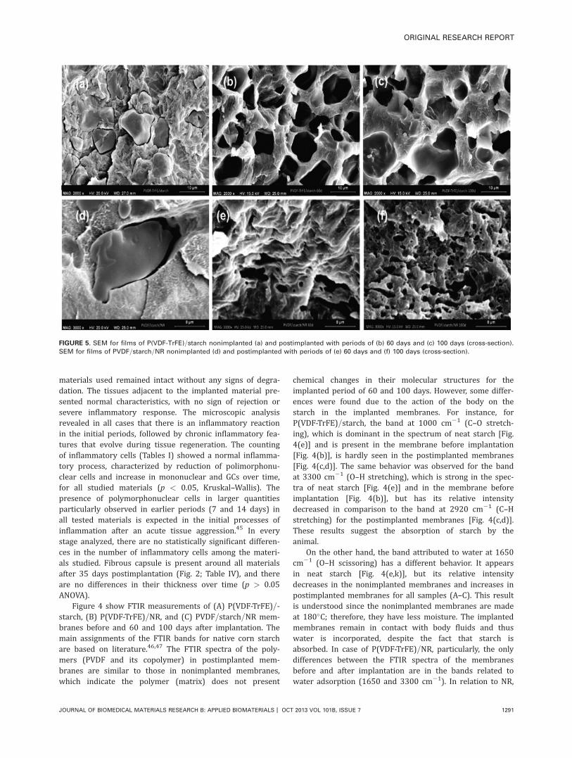
FIGURE 5. SEM for films of P(VDF-TrFE)=starch nonimplanted (a) and postimplanted with periods of (b) 60 days and (c) 100 days (cross-section).
SEM for films of PVDF=starch=NR nonimplanted (d) and postimplanted with periods of (e) 60 days and (f) 100 days (cross-section).
ORIGINAL RESEARCH REPORT
JOURNAL OF BIOMEDICAL MATERIALS RESEARCH B: APPLIED BIOMATERIALS | OCT 2013 VOL 101B, ISSUE 7 1291

no change was identified. It is worth mentioning that theband at 3300 cm21, assigned to O–H stretching, can berelated to water and starch content. Therefore, its relativeintensity tends to increase with water adsorption within thepostimplanted membranes and decrease with starch absorp-tion by the body.
SEM images were obtained from cross-section (cryogenicfracture) for the membranes of P(VDF-TrFE)=starch andPVDF=starch=NR. Figure 5(a–f) shows the SEM images formembranes (a, d) before, (b, e) after 60-day implantationand (c, f) 100-day implantation. Pores are clearly seen inthe implanted membranes, which is consistent with starchabsorption suggested by FTIR. For PVDF=starch=NR mem-branes, it seems to be an increase in the number of poreswith implantation time, which indicates that NR may hinderstarch absorption.
CONCLUSION
The blends tested in this work, PVDF and P(VDF-TrFE) asso-ciated with natural polymers (NR and native starch) formingmembranes do not present cytotoxicity from in vitro results.The tissue response after implantation presents reduction ininflammatory response with concomitant material encapsu-lation, a reduction in the number of polymorphonuclearcells and increase in mononuclear and GCs over time. Themembranes with starch in its composition lose starch withimplantation time (starch is absorbed by the body), leadingto formation of interconnected porous, which may also con-tribute to bone tissue growth. Besides, the polymeric matrix(PVDF and P(VDF-TrFE)) does not present chemical changeseven after 100 days after implantation. Therefore, ourresults attest favorable tissue reaction to these innovativeblends for bone regeneration.
ACKNOWLEDGMENTS
The authors are grateful to Prof. Dr. A.E. Job from UNESP=Bra-zil and Prof. Dr. A.J.F. Carvalho from UFSCar=Brazil for supply-ing the latex and the cornstarch, respectively.
REFERENCES
1. Banwart JC, Asher MA, Hassanein RS. Iliac crest bone graft har-
vest donor site morbidity: A statistical evaluation. Spine
1995;20(9):1055–1060.
2. Bloemers FW, Blokhuis TJ, Patka P, Bakker FC, Wippermann BW,
Haarman HJTM. Autologous bone versus calcium–phosphate
ceramics in treatment of experimental bone defects. J Biomed
Mater Res Part B: Appl Biomater 2003;66B(2):526–531.
3. Muhammad IS, Xiaoxue X, Li L. A review on biodegradable poly-
meric materials for bone tissue engineering applications. J Mater
Sci 2009;44(21):5713–5724.
4. Warren SM, Fong K, Nacamuli RP, Fang TD, Longaker MT. Bioma-
terials for skin and bone replacement and repair in plastic sur-
gery. Operat Tech Plast Reconstr Surg 2003;9(1):10–15.
5. Puppi D, Chiellini F, Piras AM, Chiellini E. Polymeric materials for
bone and cartilage repair. Prog Polym Sci 2010;35(4):403–440.
6. Hench LL, Paschall HA. Direct chemical bond of bioactive glass–
ceramic materials to bone and muscle. J Biomed Mater Res
1973;7(3): 35–42.
7. Carsten S, Christian R, Michael W, Felix B, Harald E, Matthias E,
Stephan W. Geometrically structured implants for cranial recon-
struction made of biodegradable polyesters and calcium phospha-
te=calcium carbonate. Biomaterials 2004;25(7):1239–1247.
8. Nalwa HS. Ferroelectric Polymers: Chemistry, Physics and Appli-
cations. New York: Marcel Dekker; 1995.
9. Fukada E, Furukawa T. Piezoelectricity and ferroelectricity in poly-
vinylidene fluoride. Ultrasonics 1981;19(1):31–39.
10. Hong C-C, Huang S-Y, Shieh J, Chen S-H. Enhanced piezoelectric-
ity of nanoimprinted sub-20 nm poly(vinylidene fluoride–trifluoro-
ethylene) copolymer nanograss. Macromolecules 2012;45(3):1580–
1586.
11. Marino AA, Rosson J, Gonzalez E, Jones L, Rogers S, Fukada E.
Quasi-static charge interactions in bone. J Electrostat 1988;21
(2–3):347–360.
12. Peppas NA, Langer R. New challenges in biomaterials. Science
1994;263:1715–1720.
13. Gimenes R, Zaghete MA, Bertolini MJ, Varela JA, Coelho LO,
Silva NF Jr. Composites PVDF-TrFE=BT used as bioactive mem-
branes for enhancing bone regeneration. Proc SPIE 2004;5385:
539–547.
14. Callegari B, Belangero WD. Analysis of the interface formed
among the poly(vinylidene) fluoride (piezoelectric and non-piezo-
electric) and the bone tissue of rats. Acta Ortop Bras 2004;12(3):
160–166.
15. Lang SB, Muensit S. Review of some lesser-known applications
of piezoelectric and pyroelectric polymers. Appl Phys A: Mater Sci
Process 2006;85(2):125–134.
16. Laroche G, Marois Y, Schwarz E, Guidoin R, King MW, Paris E,
Douville Y. Polyvinylidene fluoride monofilament sutures: Can
they be used safely for long-term anastomoses in the thoracic
aorta? Artif Organs 1995;19(11):1190–1199.
17. Bouaziz A, Richert A, Caprani A. Vascular endothelial cell
responses to different electrically charged poly(vinylidene fluo-
ride) supports under static and oscillating flow conditions. Bioma-
terials 1997;18(2):107–112.
18. Klinge U, Klosterhalfen B, €Ottinger AP, Junge K, Schumpelick V.
PVDF as a new polymer for the construction of surgical meshes.
Biomaterials 2002;23(16):3487–3493.
19. Junge K, Rosch R, Klinge U, Krones C, Klosterhalfen B, Mertens
PR, Lynen P, Kunz D, Preiß A, Peltroche-Llacsahuanga H, Schum-
pelick V. Gentamicin supplementation of polyvinyl fluoride mesh
materials for infection prophylaxis. Biomaterials 2005;26(7):787–
793.
20. Conze J, Junge K, Weiß C, Anurov M, Oettinger A, Klinge U,
Schumpelick V. New polymer for intra-abdominal meshes—PVDF
copolymer. J Biomed Mater Res Part B: Appl Biomater 2008;87B
(2):321–328.
21. Lee Y-S, Collins G, Livingston Arinzeh T. Neurite extension of pri-
mary neurons on electrospun piezoelectric scaffolds. Acta Bio-
mater 2011;7(11):3877–3886.
22. Simoes RD, Job AE, Chinaglia DL, Zucolotto V, Camargo-Filho JC,
Alves N, Giacometti JA, Oliveira ON, Constantino CJL. Structural
characterization of blends containing both PVDF and natural rub-
ber latex. J Raman Spectro 2005;36(12):1118–1124.
23. Simoes R, Rodriguez-Perez M, de Saja J, Constantino C. Thermo-
mechanical characterization of PVDF and P(VDF-TrFE) blends con-
taining cornstarch and natural rubber. J Therm Anal Calorim
2010;99(2):621–629.
24. Simoes RD, Rodriguez-Perez MA, De Saja JA, Constantino CJL.
Tailoring the structural properties of PVDF and P(VDF-TrFE) by
using natural polymers as additives. Polym Eng Sci 2009;49(11):
2150–2157.
25. ISO 10993-1:2009. Biological evaluation of medical devices. Part
1: Evaluation and testing in the risk management process.
26. ISO 10993-2:2006. Biological evaluation of medical devices. Part
2: Animal welfare requirements.
27. Wataha JC. Predicting clinical biological responses to dental
materials. Dent Mater 2012;28(1):23–40.
28. Balabanian CACA, Coutinho-Netto J, Lamano-Carvalho TL, Lac-
erda SA, Brentegani LG. Biocompatibility of natural latex
implanted into dental alveolus of rats. J Oral Sci 2006;48(4):201–
205.
29. Chunyan L, Pei-Ming W, Soohyun L, Gorton A, Schulz MJ, Ahn
CH. Flexible dome and bump shape piezoelectric tactile sensors
using PVDF-TrFE copolymer. J Microelectromech Syst 2008;
17(2):334–341.
1292 MARQUES ET AL. TISSUE REACTION AND CYTOTOXICITY OF PVDF AND P(VDF-TrFE)

30. Klink CD, Junge K, Binnebosel M, Alizai HP, Otto J, Neumann UP,
Klinge U. Comparison of long-term biocompatibility of PVDF and
PP meshes. J Investig Surg 2011;24(6):292–299.
31. Janet, CG, Barbee, RW, Bielitzki, JT, Clayton, LA, Donovan, JC,
Hendriksen, CFM, Kohn, DF, Lipman, NS, Locke, PA, Melcher, J,
Quimby, FW, Turner, PV, Wood, GA, W€urbel, H. Guide for the
Care and Use of Laboratory Animals, 8th ed, Washington, DC:
National Academies Press; 2011.
32. Strober W. Trypan blue exclusion test of cell viability. In: Coligan
JE, editor. Current Protocols in Immunology. New York: Wiley &
Sons; 2003.
33. Minnen B, Leeuwen M, Stegenga B, Zuidema J, Hissink C, Kooten
T, Bos R. Short-term in vitro and in vivo biocompatibility of a bio-
degradable polyurethane foam based on 1,4-butanediisocyanate.
J Mater Sci: Mater Med 2005;16(3):221–227.
34. Yaltirik M, Ozbas H, Bilgic B, Issever H. Reactions of connective
tissue to mineral trioxide aggregate and amalgam. J Endod
2004;30(2):95–99.
35. Fukada E, Yasuda I. On the piezoelectric effect of bone. J Phys
Soc Jpn 1957;12(10):1158–1162.
36. A G. Bone remodeling and piezoelectricity—I. J Biomech
1973;6(1):69–77.
37. Beloti MM, de Oliveira PT, Gimenes R, Zaghete MA, Bertolini MJ,
Rosa AL. In vitro biocompatibility of a novel membrane of the
composite poly(vinylidene-trifluoroethylene)=barium titanate.
J Biomed Mater Res Part A 2006;79A(2):282–288.
38. Teixeira L, Crippa G, Gimenes R, Zaghete M, de Oliveira P, Rosa
A, Beloti M. Response of human alveolar bone-derived cells to a
novel poly(vinylidene fluoride-trifluoroethylene)=barium titanate
membrane. J Mater Sci: Mater Med 2011;22(1):151–158.
39. Teixeira LN, Crippa GE, Trabuco AC, Gimenes R, Zaghete MA,
Palioto DB, de Oliveira PT, Rosa AL, Beloti MM. In vitro biocom-
patibility of poly(vinylidene fluoride–trifluoroethylene)=barium ti-
tanate composite using cultures of human periodontal ligament
fibroblasts and keratinocytes. Acta Biomater 2010;6(3):979–989.
40. Mendonca RJ, Maur�ıcio VB, de Bortolli Teixeira L, Lachat JJ, Cou-
tinho-Netto J. Increased vascular permeability, angiogenesis and
wound healing induced by the serum of natural latex of the rub-
ber tree Hevea brasiliensis. Phytother Res 2010;24(5):764–768.
41. Cao T, Saw TY, Heng BC, Liu H, Yap AUJ, Ng ML. Comparison of
different test models for the assessment of cytotoxicity of com-
posite resins. J Appl Toxicol 2005;25(2):101–108.
42. Olsson B, Sliwkowski A, Langeland K. Subcutaneous implantation
for the biological evaluation of endodontic materials. J Endod
1981;7(8):355–369.
43. Safavi KE, Pascon EA, Langeland K. Evaluation of tissue reaction
to endodontic materials. J Endod 1983;9(10):421–429.
44. Dawlee S and Jayabalan M. Iodinated glycidyl methacrylate co-
polymer as a radiopaque material for biomedical applications. J
Biomater Appl. DOI: 10.1177=0885328211434090.
45. Parirokh M, Mirsoltani B, Raoof M, Tabrizchi H, Haghdoost AA.
Comparative study of subcutaneous tissue responses to a novel
root-end filling material and white and grey mineral trioxide ag-
gregate. Int Endod J 2011;44(4):283–289.
46. Mano JF, Reis RL. Viscoelastic monitoring of starch-based bioma-
terials in simulated physiological conditions. Mater Sci Eng A
2004;370(1–2):321–325.
47. Park JW, Im SS, Kim SH, Kim YH. Biodegradable polymer blends
of poly(L-lactic acid) and gelatinized starch. Polym Eng Sci
2000;40(12):2539–2550.
ORIGINAL RESEARCH REPORT
JOURNAL OF BIOMEDICAL MATERIALS RESEARCH B: APPLIED BIOMATERIALS | OCT 2013 VOL 101B, ISSUE 7 1293