Embed Size (px)
Citation preview
lable at ScienceDirect
The Journal of Foot & Ankle Surgery 49 (2010) 298.e1–298.e4
Contents lists avai
The Journal of Foot & Ankle Surgery
journal homepage: www.j fas .org
Case Report
Subcutaneous Polypoid Lipoma of the First Intermetatarsal Space MimickingRudimentary Polydactyly
Joshua Decker, DPM 1, Zeeshan S. Husain, DPM, AACFAS 2, Charles G. Kissel, DPM, AACFAS 3,Doina David, MD 4, Benjamin Wehrli, DPM 5
1 Submitted during PGY-2 Detroit Medical Center PM&S-36, Detroit, MI2 Assistant Residency Director, Detroit Medical Center, Detroit, MI3 Residency Program Director, Detroit Medical Center, Podiatry Section Chief, Detroit, MI4 Associate Pathologist, Assistant Professor, Wayne State University–School of Medicine, Detroit, MI5 Submitted during PGY-2 Detroit Medical Center PM&S-36, Detroit, MI
a r t i c l e i n f o
Level of Clinical Evidence: 4Keywords:
benignmalignancysupernumerary digitTightRopetumorFinancial Disclosure: None reported.Conflicts of Interest: None reported.Address correspondence: Charles G. Kissel, DPM
48092.E-mail address: [email protected] (C.G. Kissel).
1067-2516/$ – see front matter � 2010 by the Ameridoi:10.1053/j.jfas.2010.01.003
a b s t r a c t
A case of a subcutaneous polypoid lipoma, presenting as a rudimentary polydactyly, is reported. The lesion waslocated between the first and second digits and extended into the first intermetatarsal space in a polypoidfashion. Radiographically, there were no osseous structures associated with the mass, and preliminary biopsyresults showed no evidence of malignancy. The patient was treated with marginal excision of the mass andreduction of the intermetatarsal space with a Mini TightRope (Arthrex, Inc., Naples, FL). The final pathologicaldiagnosis was polypoid lipomas, and the patient experienced a full and uneventful recovery.
� 2010 by the American College of Foot and Ankle Surgeons. All rights reserved.
Lipomas, the most common lipomatous tumor, account for nearly50% of all soft tissue tumors that occur in the extremities (1). Theyoccur more often in obese individuals and tend to be encapsulatedand well demarcated from surrounding tissues (2). Lipomas typicallydisplay morphologic features that enable them to be subclassified asconventional lipoma, fibrolipoma, angiolipoma, spindle cell lipoma,myelolipoma, and pleomorphic lipoma (2). The most common loca-tions for lipomas to occur are the upper back, neck, abdomen, and theproximal portions of the extremities (3), where an abundance ofadipose tissue is common; and occurrence in the hands and feet is farless common (4, 5). When a lipoma localizes to the foot, the patientmay complain of cosmetic concerns and/or irritation with shoe gear(3, 6, 7). To our knowledge, there has never been a case of a pedallipoma presenting in a fashion suggestive of rudimentary polydactyly.
Case Report
A 49-year-old man presented to the emergency department (ED)at Sinai-Grace Hospital (Detroit, MI) complaining of pain in a massbetween his hallux and second toe. Since high school, he had noticedthe slow but gradual development of a mass between his first and
, 29433 Ryan Rd, Warren, MI
can College of Foot and Ankle Surgeo
second toes, suggestive of a supernumerary toe. Three years beforepresentation to our ED, another physician had told the patient that themass represented an extra toe, even though the patient could onlyrecall the presence of the lesion since the time when he was in highschool. Furthermore, 2 weeks before the latest presentation to the ED,the patient had been evaluated in the same ED and was prescribed500 mg cephalexin 4 times daily and referred to the podiatry clinic. Atthe time of the latest visit, the patient related concern for the devel-opment of an ulcer at the tip of this mass. The patient also complainedof pain in the right foot, mainly due to the widened shape of the footand difficulty finding a suitable shoe. He described no history oftrauma, no past surgical intervention, and no family history ofa similar condition, and he had not yet presented to the podiatryclinic. He also denied the use of tobacco products, alcohol, or intra-venous drugs.
Physical examination of the patient’s right foot revealed a softtissue mass that protruded between the first and second toes ina fashion that was initially suggestive of the presence of 6 toes(Figure 1). Palpation of the lesion revealed fluctuance and tendernessdorsally and plantarly, there was no evidence of a toenail on the lesionbetween the first and second toes, and a round ulcer with a 1-cmdiameter was evident at the distal aspect of the lesion, withoutpurulence, drainage, tunneling, or a sinus tract. The base of the ulcerwas granular and fibrotic. He could not voluntarily move the lesionbetween the first and second toes, which suggested the absence ofa specific neuromuscular or tendinous supply to the lesion. Theoverall pedal neurovascular status was within normal limits, as were
ns. All rights reserved.

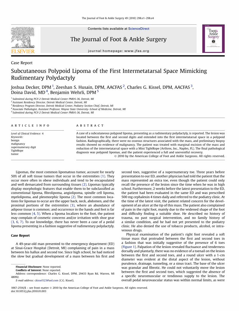
Fig. 1. Initial presentation in the emergency department after prior treatment thatinvolved a dorsal incision for drainage of suspected abscess (A); with persistence ofa wound (ulcer) at the tip of the mass situated between the first and second toes (B).
Fig. 2. The plain anteroposterior radiograph revealed increased soft tissue density andvolume suggestive of rudimentary polydactyly (supernumerary digit), without anyosseous structures, situated between the first and second toes. Although the first inter-metatarsal angle measured 12� , the soft tissue mass resulted in marked abduction of thesecond toe.
J. Decker et al. / The Journal of Foot & Ankle Surgery 49 (2010) 298.e1–298.e4 298.e2
the results of a complete blood count and biochemical profile. Stan-dard radiographs of the right foot revealed no evidence of ossification(specifically, no phalangeal elements), ectopic calcification, orsubcutaneous emphysema, although the soft tissue density andvolume suggested the presence of a supernumerary digit between thefirst and second toes, which led the radiologist to report polydactyly(Figure 2). Based on these findings, our differential diagnosis includedsoft tissue mass or rudimentary polydactyly, with distal ulcerationand concern for abscess. An incision and drainage were performed (atthe time of the second visit to the ED), and this failed to produce anypurulent drainage or abscess. The patient was referred, once again, to
the podiatry clinic, where he presented 1 week after the second visitto the ED. When he presented in follow-up the following week, a softtissue biopsy was performed with local anesthesia, by excision ofa small wedge of tissue from the subcutaneous layers centrally on thetoe. Based on the result of the biopsy, a provisional diagnosis ofa benign lipoma was made. A magnetic resonance image examinationwas also requested; however, the patient refused to undergo thisexamination and instead requested surgical excision of the lesion.After receiving the preliminary biopsy report and discussing thediagnosis, prognosis, and treatment options, the decision was made topursue complete excision via marginal (en bloc) resection of the mass,along with reduction of the first intermetatarsal space in an effort toeradicate the lesion and improve the shape of the foot so that it wouldbe possible to comfortably wear a normal shoe.
Afterward, the patient was taken to the operating room, where,with the use of intravenous sedation, local anesthesia, and an ankletourniquet, a diamond-shaped incision was made over the lesion.Dissection through the dermis revealed the presence of dense,encapsulated fat lobules throughout the subcutaneous fat andsuperficial fascia layers, distal to the first and second metatarsal headsbetween the first and second toes. The terminal branches of the deepperoneal nerve, as well as the terminal branches of the first inter-metatarsal artery, were observed to enter the proximal margin of themulti-lobular mass. Close inspection revealed these structures to be

Fig. 3. Marginal resection of the lesion (A) resulted in an excised mass that measured 8.0 � 7.0 � 3.0 cm (B) and displayed yellowish pink lobulated fat.
Fig. 4. Intraoperative anteroposterior radiographic inspection shows the distal placementof a Mini TightRope between the first and second metatarsals, which reduced the firstintermetatarsal space from 12� to 4� .
J. Decker et al. / The Journal of Foot & Ankle Surgery 49 (2010) 298.e1–298.e4298.e3
so incarcerated that the nerve and vascular elements had atrophied tothe extent that it was not possible to separate the mass from thesevital structures and there was no evidence of the nerve or vascularstructures distal to the mass. Based on these intraoperative findings,the decision was made to excise the lesion along its well-demarcatedmargins, resulting in an en bloc resection of the tumor (Figure 3A).Proximally, the common trunk of the deep peroneal nerve wastransected, the stump was buried in the first dorsal interosseousmuscle belly, and the artery was transected and tied with a 2-0 silkligature. Complete excision of the tumor showed that it involved theentire first intermetatarsal and interdigital spaces, with distalprotrusion between the first and second toes. It was completelysubcutaneous, except distally, where the lesion became contiguouswith the ulcerated skin. The tumor was well circumscribed andmultilobulated and did not display invasion of the surrounding softtissues or bone, except distally where the cutaneous ulcer waslocalized (Figure 3B).
After removal of the soft tissue mass, the first intermetatarsalspace displayed a large void and the first and second metatarsalsremained widely separated, creating a substantial space between thehallux and second toe. In an effort to reduce the separation betweenthe first and second metatarsals, and to eliminate much of the deadspace created by removal of the tumor, the decision was made toreduce the intermetatarsal angle with a Mini TightRope (Arthrex, Inc.,Naples, FL) (Figure 4). A closed suction drain was placed in the wound,after which the wound was closed in anatomic layers, a steriledressing was applied, and the foot protected with the use of a surgicalshoe for postoperative weight bearing. On gross examination, thesurgical pathology report described a multilobulated, yellowish pinkmass measuring 8.0 � 7.0 � 3.0 cm, with the distal margin of thelesion partially surfaced with wrinkled, eroded skin, revealing a 2.0 �2.7 cm ulcer. Microscopic evaluation showed skin with underlying

Fig. 5. Pathological inspection revealed many foam cells (single arrow, top portion of theimage), reactive mesenchymal cells (double arrow, top portion of the image), fat necrosis(single arrow, bottom portion of the image), and myxoid degeneration of the adiposetissue (double arrows, bottom portion of the image). (Top portion of the image, originalmagnification 400x; lower portion of the slide, original magnification 100x; hematoxylinand eosin stain.)
J. Decker et al. / The Journal of Foot & Ankle Surgery 49 (2010) 298.e1–298.e4 298.e4
polypoid adipose tissue displaying areas of fat necrosis, myxoiddegeneration, and reactive mesenchymal cells (Figure 5). The areasurrounding the ulceration displayed granulation tissue. There was noevidence of hyperchromatic atypical cells, such as those that aretypically associated with well-differentiated liposarcoma. The patientproceeded through an unremarkable postoperative course andresumed the use of a regular shoe by 6 weeks. Patient resumed workand all regular activities starting at eight weeks. He maintained thislevel until his final follow-up at six months. He was scheduled tofollow-up on a biannual basis in an effort to maintain routinesurveillance for the possibility of tumor recurrence or metatarsalstress fracture.
Discussion
Lipomas most commonly become apparent in the fifth and sixthdecades of life with a higher prevalence in women (8). Malignant
transformation of a lipoma is rare; however, a few examples havebeen described in literature (8). After thorough excision of a lipoma,recurrence is not very likely to occur (9). Although a typical lipomaseldom poses a diagnostic challenge to the pathologist, the distinctionfrom liposarcoma can be difficult in cases that present unusualfeatures, including angiolipoma, pleomorphic lipoma, and chondroidlipoma, or in cases where the lesion localizes to perineural or intra-muscular tissues (10).
Although the patient described in this report progressed wellpostoperatively, and the first intermetatarsal angle remained satis-factorily aligned, one concern associated with the long-term use ofthe Mini TightRope fixation system is the potential to create a meta-tarsal fracture due to stress riser formation caused by motion betweenthe first and second metatarsals (11–16). However, we consider thisform of fixation to tether the first and second metatarsals to bebeneficial because it allows motion between the first and secondmetatarsals, which is important relative to normal weight-bearingfunction of the metatarsus.
In conclusion, lipomas are benign soft tissue lesions that rarelybecome malignant, although they can produce cosmetic concerns aswell as irritation due to contact with shoe gear when the foot and/orankle is involved. The caring surgeon must maintain a high index ofsuspicion for potential malignancy whenever a soft tissue mass isconsidered. Moreover, the use of advanced imaging, such as magneticresonance image scans, computerized tomography, and ultrasound,can be useful diagnostic adjuncts; however, the key to an accuratediagnosis and appropriate treatment is biopsy. For symptomaticlesions in the foot, surgical excision is often curative, although long-term postexcision surveillance is important.
References
1. Cole DR, DeLauro TM. Soft-tissue neoplasia. In Neoplasms of the Foot and Leg, p 143,edited by CL Brown, Williams & Wilkins, Baltimore, 1990.
2. Rosenberg AE. Bone tumors and tumor like lesions. In Robbins and Cotran Patho-logic Basis of Disease, ed 7, pp 1292–1303, edited by V Kumar, N Fausto, A Abbas,Elsevier Saunders, Philadelphia, 2005.
3. Azam A, Rajagopalan S, Niezywinski W. A rapidly expanding massive lipoma of thetoe. J Foot Ankle Surg 46(6):499–501, 2007.
4. Booher RJ. Lipoblastic tumors of the hands and feet: review of the litera-ture and report of thirty-three cases. J Bone Joint Surg (Am) 47-A:727–740,1965.
5. Berlin SJ. A laboratory review of 67,000 foot tumors and lesions. J Am PodiatryAssoc 74(7):341–347, 1984.
6. Pontious J, Zielaskowski LA, King G. Extensive lipoma of the foot. J Am Podiatr MedAssoc 93(5):402–405, 2003.
7. Copeland CL, Kanat IO. A large lipoma involving the foot. J Foot Surg 30(6):571–573, 1991.
8. Enzinger FM. Benign lipomatous tumours. In Soft Tissue Tumours, pp 381–430,edited by FM Enzinger, SW Weiss, ed 3, Mosby, St. Louis, 1995.
9. Seale KS, Lange TA, Monson D, Hackbarth DA. Soft tissue tumors of the foot andankle. Foot Ankle 9:19–27, 1988.
10. Nilsen GP, Mandahl N. Adipocytic tumors. In WHO Classification of Tumours.Pathology and Genetics of Tumours of Soft Tissue and Bone, pp 20–35, edited byCDM Fletcher, KK Unni, F Mertens, IARC Press, Lyon, 2002.
11. Root ML, Orien WP, Weed JH. Clinical Biomechanics. Volume II: Normal andAbnormal Function of the Foot, Clinical Biomechanics Corporation, Los Angeles,1977.
12. Hicks JH. The mechanics of the foot I: the joints. J Anat 87(4):345–357, 1953.13. Kelikian H. Hallux Valgus, Allied Deformities of the Forefoot and Metatarsalgia,
Saunders, Philadelphia, 1965.14. Kelso SF, Richie DH, Cohen IR, Weed JH, Root ML. Direction and range of motion of
the first ray. J Am Podiatry Assoc 72(12):600–605, 1982.15. Kitaoka HB, Lundberg A, Luo ZP, An KN. Kinematics of the normal arch of
the foot and ankle under physiologic loading. Foot Ankle Int 16(8):492–499,1995.
16. Klaue K, Hansen ST, Masquelet AC. Clinical, quantitative assessment of first tar-sometatarsal mobility in the sagittal plane and its relation to hallux valgusdeformity. Foot Ankle Int 15(1):9–13, 1994.


