Embed Size (px)
Citation preview
POSTER PRESENTATION Open Access
Subclinical perfusion deficits in patients withType 2 diabetes detectable with CardiovascularMagnetic Resonance ImagingAnna Schmidt1*, David Lau2, Matthias G Friedrich3
From 2011 SCMR/Euro CMR Joint Scientific SessionsNice, France. 3-6 February 2011
BackgroundAn estimated 200 million people worldwide have type 2diabetes. Type 2 diabetes is recognized as an independentrisk factor for adverse cardiac events, such as myocardialinfarction. Cardiovascular disease is the leading cause ofdeath amongst diabetic patients. The subclinical pathophy-siology of diabetic heart disease predicts global microvas-cular disease, prior to the onset of overt ischemic heartdisease. Non-invasive screening methods are thereforeimportant for risk stratification of asymptomatic patients.Cardiovascular Magnetic Resonance Imaging (CMR) is avaluable tool for the assessment of subclinical microvascu-lar function in this high-risk population.
ObjectivesTo assess the degree of cardiac microvascular dysfunc-tion in individuals type 2 diabetes (T2D) without coron-ary disease or hypertension, compared to healthy, non-diabetic controls.
MethodsThis cross sectional pilot data included 2 groups of sub-jects: Patients diagnosed with type 2 diabetes (HbA1c
7.5-9.9%; mean age 59.3±7.17; n=6), and healthy,non-diabetic age-matched controls (mean age 51.9±10.3;n=10). Medical history and ECG were reviewed to ruleout ischemic heart disease. Qualified patients underwenta CMR-Adenosine stress perfusion,
ResultsSubendocardial perfusion delays were observed in 4 out of6 diabetic patients, and 0 of 10 healthy controls (p < 0.05).
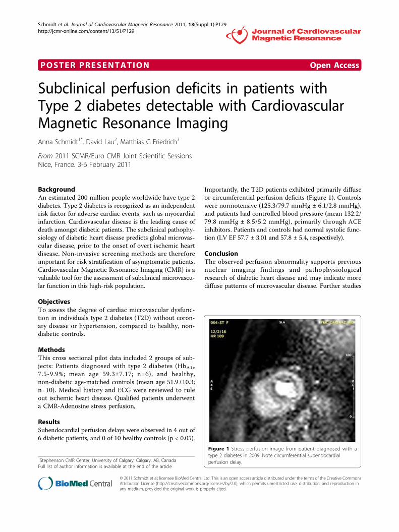
Importantly, the T2D patients exhibited primarily diffuseor circumferential perfusion deficits (Figure 1). Controlswere normotensive (125.3/79.7 mmHg ± 6.1/2.8 mmHg),and patients had controlled blood pressure (mean 132.2/79.8 mmHg ± 8.5/5.2 mmHg), primarily through ACEinhibitors. Patients and controls had normal systolic func-tion (LV EF 57.7 ± 3.01 and 57.8 ± 5.4, respectively).
ConclusionThe observed perfusion abnormality supports previousnuclear imaging findings and pathophysiologicalresearch of diabetic heart disease and may indicate morediffuse patterns of microvascular disease. Further studies
1Stephenson CMR Center, University of Calgary, Calgary, AB, CanadaFull list of author information is available at the end of the article
Figure 1 Stress perfusion image from patient diagnosed with atype 2 diabetes in 2009. Note circumferential subendocardialperfusion delay.
Schmidt et al. Journal of Cardiovascular Magnetic Resonance 2011, 13(Suppl 1):P129http://jcmr-online.com/content/13/S1/P129
© 2011 Schmidt et al; licensee BioMed Central Ltd. This is an open access article distributed under the terms of the Creative CommonsAttribution License (http://creativecommons.org/licenses/by/2.0), which permits unrestricted use, distribution, and reproduction inany medium, provided the original work is properly cited.

are required to assess the pathophysiologic context andprognostic impact of these findings.
Author details1Stephenson CMR Center, University of Calgary, Calgary, AB, Canada.2Stephenson CMR Centre, University of Calgray, Calgary, AB, Canada.3Stephenson CMR Centre, University of Calgary, Calgary, AB, Canada.
Published: 2 February 2011
doi:10.1186/1532-429X-13-S1-P129Cite this article as: Schmidt et al.: Subclinical perfusion deficits inpatients with Type 2 diabetes detectable with Cardiovascular MagneticResonance Imaging. Journal of Cardiovascular Magnetic Resonance 2011 13(Suppl 1):P129.
Submit your next manuscript to BioMed Centraland take full advantage of:
• Convenient online submission
• Thorough peer review
• No space constraints or color figure charges
• Immediate publication on acceptance
• Inclusion in PubMed, CAS, Scopus and Google Scholar
• Research which is freely available for redistribution
Submit your manuscript at www.biomedcentral.com/submit
Schmidt et al. Journal of Cardiovascular Magnetic Resonance 2011, 13(Suppl 1):P129http://jcmr-online.com/content/13/S1/P129
Page 2 of 2