Embed Size (px)
Citation preview

POLISH JOURNAL OF NATURAL SCIENCESAbbrev.: Pol. J. Natur. Sc., Vol 29(2): 189–195, Y. 2014
SUBCLINICAL ANGIOSTRONGYLUS VASORUMINFECTION IN A TERRIER DOG KENNEL
Angela Di Cesare1, Carlo Miotti2, Luigi Venco3,Fabrizio Pampurini4, Elena Centaro5, Donato Traversa1
1 Faculty of Veterinary MedicineUniversity of Teramo in Teramo, Italy
2 Veterinary Practice „Dr. Carlo Miotti, Dr. Marco Miotti” in Gallicano, Italy3 Veterinary Hospital „Citta di Pavia” in Pavia, Italy
4 Bayer S.p.A. in Milan, Italy5 Idexx Laboratories in Milan, Italy
K e y w o r d s: Angiostrongylus vasorum, dogs, diagnosis, imidacloprid, moxidectin, Italy.
A b s t r a c t
Angiostrongylus vasorum is a parasitic nematode causing severe clinical signs in infected dogs. Inthe past few years A. vasorum has been repeatedly described in both traditional endemic foci andpreviously free regions. Nonetheless, the infection is often neglected or unnoticed by vet practi-tioners, due to gaps of information on A. vasorum epidemiology, and to drawbacks inherent to theclinical and parasitological diagnosis. Indeed, subclinical infections may occur and, when present,clinical signs are difficult to differentiate from those of other canine cardio-pulmonary diseases.Additionally, the gold standard test for the aetiological diagnosis of the infection, i.e. the Baermann’smethod, is not commonly performed by veterinarians.
The present study describes cases of subclinical A. vasorum infection in a Jack Russell Terrierdog kennel in Italy and the ability of a newly marketed rapid kit (IDEXX Angio DetectTM Test) for thefield diagnosis of angiostrongylosis, pre- and post-treatment with a formulation licensed for thetreatment of A. vasorum.
SUBKLINICZNE ZARAŻENIE ANGIOSTRONGYLUS VASORUMW HODOWLI PSÓW TERIERÓW
Angela Di Cesare1, Carlo Miotti2, Luigi Venco3, Fabrizio Pampurini4,Elena Centaro5, Donato Traversa1
1 Wydział Medycyny Weterynaryjnej, Uniwersytet Teramo, Teramo, Włochy2 Praktyka Weterynaryna „Dr. Carlo Miotti, Dr. Marco Miotti”, Gallicano, Włochy
3 Klinika Weterynaryjna „Citta di Pavia”, Pavia, Włochy4 Bayer S.A., Milan, Włochy
5 Laboratoria Idexx, Milan, Włochy
S ł o w a k l u c z o w e: Angiostrongylus vasorum, psy, diagnostyka, Imidacloprid, Moxidectin, Włochy.
Address: Angela Di Cesare DVM, Ph.D, University of Teramo, Piazza A. Moro 45, 64100 Teramo,Italy, phone: +39 0 861 266 880, e-mail: [email protected].

A b s t r a k t
Angiostrongylus vasorum to pasożytniczy nicień powodujący ciężkie objawy kliniczneu zarażonych psów. Na przestrzeni ostatnich kilku lat wielokrotnie opisywano przypadki inwazjiA. vasorum zarówno w endemicznych ogniskach, jak również w regionach wcześniej wolnych od tegopasożyta. Często jednak zdarza się, że możliwość inwazji zostaje pominięta lub przypadek zarażeniapozostaje niedostrzeżony przez lekarza weterynarii z powodu niedostatecznej wiedzy na tematepidemiologii A. vasorum oraz niedociągnięć czy błędów podczas badania klinicznego i parazytologi-cznego. Podkliniczna postać inwazji może jednak występować – w takiej sytuacji objawy kliniczne sątrudne do odróżnienia od tych towarzyszących innym chorobom układu sercowo-naczyniowegoi chorobom płuc u psów. Co więcej, standard diagnostyki etiologicznej inwazji, czyli metodaBaermanna, nie jest powszechnie wykorzystywana przez lekarzy weterynarii.
W pracy opisano przypadki podklinicznej inwazji A. vasorum w hodowli Jack Russel terierów weWłoszech oraz podjęto zagadnienie możliwości zastosowania nowo wprowadzonego na rynek testu(IDEXX Angio DetectTM Test) w diagnostyce terenowej angiostrongylozy przed leczeniem preparatemdopuszczonym do stosowania w terapii inwazji A. Vasorum i po takiej kuracji.
Introduction
Canine angiostrongylosis is an emerging parasitic disease of dogs and wildanimals (e.g. foxes and wolves) caused by Angiostrongylus vasorum(Nematoda, Metastrongyloidea). The interest on this nematode is growing dueto the severe clinical outcome of the infection in dogs, the increase of reports inboth traditional endemic foci and previously free areas, and for the recentinsights achieved on the biology, treatment and the diagnosis of the infection(FERDUSHY and HASAN 2010, TRAVERSA et al. 2010, 2013, SCHNYDER et al.2014). Adult stages reside in the heart and pulmonary arteries, where adultfemales produce eggs that embryonate and hatch within alveolar ducts andalveoli. First stage larvae (L1) penetrate into the alveoli and migrate to thepharynx, are swallowed and released into the environment via the feces. Thelife cycle of A. vasorum is indirect, involving slugs and snails as intermediatehosts, in which L1 develop to the third infectious stage (L3). Dogs becomeinfected by ingesting mollusks harboring L3 (ANDERSON 2000, FERDUSHY andHASAN 2010, MORGAN and SHAW 2010). Angiostrongylus vasorum has beenconsidered for a long time to be present only in well-isolated endemic foci (i.e.areas of France, UK and Denmark). However, in the last decade, the nematodehas been recorded in dogs in previously free areas of northern and centralEurope, e.g. Sweden, Switzerland, Germany, of the Mediterranean Basin, e.g.Greece and Italy, and of Eastern Countries, e.g. Slovakia, Poland, Hungary(MAJOROS et al. 2010, TRAVERSA et al. 2010, 2013, DI CESARE et al. 2011,HURNIKOVA et al. 2013, GUARDONE et al. 2013, SCHNYDER et al. 2013).
Canine angiostrongylosis may be asymptomatic/sub-clinical or character-ized by iper-acute, acute or chronic signs, that may be life-threatening whena specific therapy is not administered (TRAVERSA and GUGLIELMINI 2008,
Angela Di Cesare et al.190

FERDUSHY and HASAN 2010, MORGAN et al. 2010, TRAVERSA et al. 2010).Cardiac, neurologic, gastrointestinal and hematological signs may be presentin infected animals, being coughing, dyspnea, and some non-specific signs,such as anorexia and weight-loss, lethargy, depression the most common.Cardiovascular signs (e.g. heart murmur, ascite, syncope) of congestive heartfailure (cor pulmonale) may be observed. Bleeding disorders and co-agulopathies may cause petechial or ecchymotic haemorrhages in the conjunc-tiva, episclera, gingiva and subcutis, as well as epistaxis, haemoptysis, post-surgical haematomas, gastrointestinal bleeding, haematuria and anaemia.Others common signs of angiostrongylosis include neurological (e.g vestibularsigns, convulsion and paralysis), ocular signs (e.g. uveitis) due to larvae or eggembolism or haemorrhages. Infected dogs may also show vomiting, diarrhoeaand anorexia (GOULD et al. 1999, CHAPMAN et al. 2004, OLIVEIRA-JUNIOR et al.2004, TRAVERSA and GUGLIELMINI 2008, TRAVERSA et al. 2008, 2010, 2013,KOCH et al. 2009). A recent multi-centric survey performed in dogs with clinicalpictures compatible with angiostrongylosis confirmed that non-specific gas-trointestinal, respiratory, hematological and neurological signs may occur andthat coughing is the most prevalent (TRAVERSA et al. 2013). Given the lack ofspecificity of signs, the clinical diagnosis of the infection is impossible. Thedetection of L1 in faeces of infected animals with the Baermann’s method isthe most reliable approach to achieve the aetiological diagnosis of angiostron-gylosis (TRAVERSA and GUGLIELMINI 2008). This technique is relatively easy toperform and cheap, although it is time-consuming (i.e. 24–36 h) and requireswell-trained microscopists. In fact, L1 should be recognized based on theirlength (i.e. 310–400 μm) and tail, having a typical sinus wave curve witha dorsal spine. These larvae need to be discriminated from those of otherfree-living or parasitic nematodes (i.e. Crenosoma vulpis, Oslerus osleri orFilaroides spp.) which can be present in canine faeces (TRAVERSA et al. 2010).
In additional, the Baermann’s method has major disadvantages like theinability to diagnose infections during the pre-patent period and when larvae arenot being shed, even in presence of severe clinical signs (CONBOY 2009,TRAVERSA et al. 2010). Innovative studies have been recently performed toovercome the constraints of copromicroscopic approaches (AL-SABI et al. 2010,SCHNYDER et al. 2011, 2013, 2014, SCHUCAN et al. 2012). After some of thesestudies, a newly marketed rapid kit (IDEXX Angio DetectTM Test) has beenrecently developed for the serological diagnosis of angiostrongylosis. The pres-ent study described cases of subclinical infections by A. vasorum in a JackRussell Terrier dog kennel in Italy and the efficiency of this new kit in the fielddiagnosis of angiostrongylosis. The diagnostic performance of both the Baer-mann’s test and the rapid kit has been evaluated before and after treatmentwith a parasiticide spot-on formulation licensed for the treatment of A. vasorum.
Subclinical Angiostrongylus Vasorum infection... 191

Materials and Methods
Study design and animals
The study was carried out in a private Jack Russell Terrier kennel locatedin Gallicano Municipality (Tuscany region, central Italy), selected for a previ-ous history of angiostrongylosis. At Day -15 all the fifteen dogs living in thekennel were clinically examined and then faeces and blood were collected to besubjected, respectively, to the Baermann’s method and to the detection of thecirculating antigen of A. vasorum with the Angio DetectTM Test. Dogs wereconsidered infected when positive at the Baermann’s and/or the Angio DetectTM
Test. At Day 0 positive dogs were clinically examined and treated witha spot-on formulation containing 10% imidacloprid and 2.5% moxidectin(Advocate®, Bayer). Two and four weeks after treatment (Days 14 and 28)these dogs were examined for clinical signs and samples were collected andexamined as above with the Baermann’s method and the Angio DetectTM Test.
Baermann methods and Angio DetectTM Test
The Baermann’s test was performed as on the follow: 3–5 grams of eachstool sample was put in the center of double layers of cheesecloth sheet.A pouch containing the fecal material was formed by holding the four cornersof the cheesecloth sheet together and molding the cloth around the fecalmaterial using a closing string. The pouch was placed in a funnel filled withwater and kept at room temperature. After 24 hours, 15 ml of fecal fluid wasdrawn off the bottom funnel into a tube and centrifuged at 2000 rpm for5 minutes. The sediment was transferred onto a slide and microscopicallyexamined using a light microscopy at 10X, 40X and 100X.
Angiostrongylus vasorum L1s retrieved at the copromicroscopic examin-ation were identified according to morphological and morphometrical keysfeatures (TRAVERSA et al. 2010). The Angio DetectTM Test was performed andinterpreted following manufacturer’s instructions using the plasma collectedfrom EDTA-blood samples.
Results and Discussion
At the clinical examinations performed before (Day-15) and after (Day14 and 28) treatment no dogs showed clinical signs suggestive of angiostron-gylosis.
Angela Di Cesare et al.192
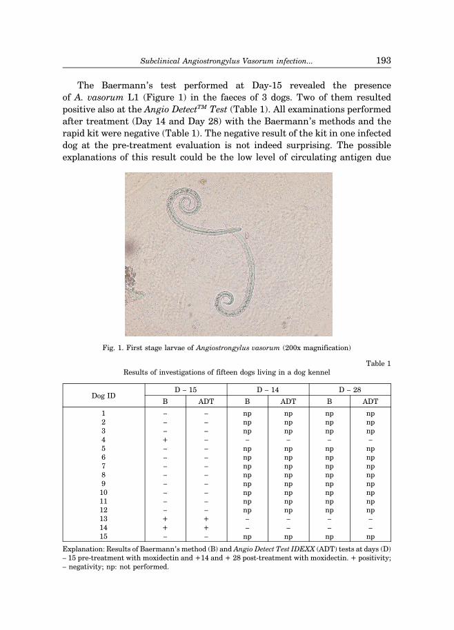
The Baermann’s test performed at Day-15 revealed the presenceof A. vasorum L1 (Figure 1) in the faeces of 3 dogs. Two of them resultedpositive also at the Angio DetectTM Test (Table 1). All examinations performedafter treatment (Day 14 and Day 28) with the Baermann’s methods and therapid kit were negative (Table 1). The negative result of the kit in one infecteddog at the pre-treatment evaluation is not indeed surprising. The possibleexplanations of this result could be the low level of circulating antigen due
Fig. 1. First stage larvae of Angiostrongylus vasorum (200x magnification)
Table 1Results of investigations of fifteen dogs living in a dog kennel
D – 15 D – 14 D – 28
B ADT B ADT B ADTDog ID
1 – – np np np np2 – – np np np np3 – – np np np np4 + – – – – –5 – – np np np np6 – – np np np np7 – – np np np np8 – – np np np np9 – – np np np np
10 – – np np np np11 – – np np np np12 – – np np np np13 + + – – – –14 + + – – – –15 – – np np np np
Explanation: Results of Baermann’s method (B) and Angio Detect Test IDEXX (ADT) tests at days (D)– 15 pre-treatment with moxidectin and +14 and + 28 post-treatment with moxidectin. + positivity;– negativity; np: not performed.
Subclinical Angiostrongylus Vasorum infection... 193

an early stage of the infection (as supported by the lack of clinical signs) and/orthe formation of antigen-antibody complexes, which may inhibit the detectionof A. vasorum antigens, as described also for Dirofilaria immitis (SCHNYDER etal. 2014). Overall, it has been recently shown that this kit has a specificity of~100% and a sensitivity of ~85%, which may lead to negative results in dogspositive at the BAERMANN’S test (SCHNYDER et al. 2014). Importantly, furtherstudies on a large scale are necessary to evaluate in field conditions theconcordance between the results of the Baermann’s test and the rapid kit, inboth clinically and subclinically infected dogs.
The results of the present study confirm the possible occurrence ofsubclinically infected dogs with no apparent signs compatible with the infec-tion. A diagnosis of angiostrongylosis should be always considered in thepresence of compatible clinical signs (TRAVERSA et al. 2013). However, dogsliving in endemic areas, especially young animals and those that usually eatmollusks, should be routinely screened for A. vasorum even in absence ofclinical signs. In fact, young dogs are more susceptible to the infection for theirage-related level of immunity (TRAVERSA and GUGLIELMINI 2008, FERDUSHY
and HASAN 2010). The Angio DetecTM Test is particularly suitable for thispurpose, as it can be directly used in veterinary practices and it representsa valid tool for a quick diagnosis for its high values of sensitivity and specificity(SCHNYDER et al. 2014).
Furthermore, the Angio DetectTM Test is a powerful tool in dogs withclinical signs and needing a prompt anthelmintic treatment. In fact, this testmay provide results in fifteen minutes, while the Baermann’s test requires atleast 24 hours. On the other hand, dogs presenting clinical signs compatiblewith angiostrongylosis but negative at the Angio DetectTM Test, should beexamined with the Baermann’s method before excluding the infection.
A reliable diagnosis of dog angiostrongylosis is of great importance undera practical standpoint. In fact, despite the severe pathogenic impact of A.vasorum, the available parasiticide options are straightforward and effective(TRAVERSA et al. 2010). Hence, it is noteworthy that the present studyconfirmed the high efficacy of moxidectin contained in Advocate® for thetherapy of canine angiostrongylosis (WILLESEN et al. 2007).
Accepted for print 28.07.2014
References
AL-SABI M.N., DEPLAZES P., WEBSTER P., WILLESEN J.L., DAVIDSON R.K., KAPEL C.M. 2010. PCRdetection of Angiostrongylus vasorum in faecal samples of dogs and foxes. Parasitol. Res., 107(1):135–140.
Angela Di Cesare et al.194

ANDERSON R.C. 2000. The superfamily metastrongyloidea. [In:] Nematode parasites of vertebrates.Their development and transmission (2nd edition), CABI Publishing, Guilford, UK, pp. 162–163.
CESARE A. DI., CASTAGNA G., MELONI S., MILILLO P., LATROFA M.S., OTRANTO D., TRAVERSA D. 2011.Canine and feline infections by cardiopulmonary nematodes in central and southern Italy.Parasitol. Res., 109(1): S87-96.
CHAPMAN P.S., BOAG A.K., GUITIAN J., BOSWOOD A. 2004. Angiostrongylus vasorum infection in 23 dogs(1999–2002). J. Small. Anim. Pract., 45(9): 435–440.
CONBOY G.A. 2009. Helminth parasites of the canine and feline respiratory tract. Vet Clin. North. Am.Small. Anim. Pract., 39: 1109–1126.
FERDUSHY T., HASAN M.T. 2010. Angiostrongylus vasorum: the „French Heartworm”. Parasitol. Res.,107(4): 765–771.
GUARDONE L., SCHNYDER M., MACCHIONI F., DEPLAZES P., MAGI M. 2013. Serological detection ofcirculating Angiostrongylus vasorum antigen and specific antibodies in dogs from central andnorthern Italy. Vet. Parasitol., 192(1–3): 192–198.
GOULD S.M., MCINNES E.L. 1999. Immune-mediated thrombocytopenia associated with Angiostron-gylus vasorum infection in a dog. J. Small. Anim. Pract., 40(5): 227–232.
HURNIKOVA Z., MITERPAKOVA M., MANDELIK R. 2013. First autochthonous case of canine Angiostron-gylus vasorum in Slovakia. Parasitol Res., 112: 3505–3508.
KOCH J., WILLESEN J.L. 2009. Canine pulmonary angiostrongylosis: an update. Vet. J., 179: 348–359.LEPRI E., VERONESI F., TRAVERSA D., CONTI M.B., MARCHESI M.C., MIGLIO A., MANDARA M.T. 2011.
Disseminated angiostrongylosis with massive cardiac and cerebral involvement in a dog fromItaly. Parasitol. Res., 109(2): 505–508.
MAJOROS G., FUKAR O., FARKAS R. 2010. Autochtonous infection of dogs and slugs with Angiostrongylusvasorum in Hungary. Vet Parasitol., 174(3–4): 351–354.
MORGAN E.R, SHAW S.E. 2010. Angiostrongylus vasorum infection in dogs: continuing spread anddevelopments in diagnosis and treatment. J. Small. Anim. Pract., 51(12): 616–621.
MORGAN E.R., JEFFERIES R., KRAJEWSKI M., WARD P., SHAW S.E. 2009. Canine pulmonary angiostron-gylosis: The influence of climate on parasite distribution. Parasitol. Int., 58: 406–410.
MORGAN E.R., JEFFERIES R., VAN OTTERDIJK L., MCENIRY R.B., ALLEN F., BAKEWELL M., SHAW S.E. 2010.Angiostrongylus vasorum infection in dogs: Presentation and risk factors. Vet. Parasitol.,173(3–4): 255–261.
OLIVEIRA-JUNIOR S.D., BARCANTE J.M., BARCANTE T.A., RIBEIRO V.M., LIMA W.S. 2004. Ectopic location ofadult worms and first-stage larvae of Angiostrongylus vasorum in an infected dog. Vet. Parasitol.,121(3–4): 293–296.
SCHNYDER M., TANNER I., WEBSTER P., BARUTZKI D., DEPLAZES P. 2011. An ELISA for sensitive andspecific detection of circulating antigen of Angiostrongylus vasorum in serum samples of naturallyand experimentally infected dogs. Vet. Parasitol., 179(1–3): 152–158.
SCHNYDER M., SCHAPER R., PANTCHEV N., KOWALSKA D., SZWEDKO A., DEPLAZES P. 2013. Serologicaldetection of circulating Angiostrongylus vasorum antigen- and parasite-specific antibodies in dogsfrom Poland. Parasitol. Res., 112, S1: 109–117.
SCHNYDER M., STEBLER K., NAUCKE T.J., LORENTZ S., DEPLAZES P. 2014. Evaluation of a rapid device forserological in-clinic diagnosis of canine angiostrongylosis. Parasit. Vectors., 7(1): 72.
SCHUCAN A., SCHNYDER M., TANNER I., BARUTZKI D., TRAVERSA D., DEPLAZES P. 2012. Detection of specificantibodies in dogs infected with Angiostrongylus vasorum. Vet. Parasitol., 185(2–4): 216–224.
TRAVERSA D., GUGLIELMINI C. 2008. Feline aelurostrongylosis and canine angiostrongylosis: a challeng-ing diagnosis for two emerging verminous pneumonia infections. Vet. Parasitol., 157: 163–174.
TRAVERSA D., TORBIDONE A., MALATESTA D., GUGLIELMINI C. 2008. Occurrence of fatal canine Angios-trongylus vasorum infection in Italy. Vet. Parasitol., 152(1–2): 162–166.
TRAVERSA D., DI CESARE A., CONBOY G. 2010. Canine and feline cardiopulmonary parasitic nematodesin Europe: emerging and underestimated. Parasit. Vectors., 3: 62.
TRAVERSA D., DI CESARE A., MELONI S., FRANGIPANE DI REGALBONO A., MILILLO P., PAMPURINI F.,VENCO L. 2013. Canine angiostrongylosis in Italy: occurrence of Angiostrongylus vasorum in dogswith compatible clinical pictures. Parasitol. Res., 112(7): 2473–2480.
WILLESEN J.L., KRISTENSEN A.T., JENSEN A.L., HEINE J., KOCH J. 2007. Efficacy and safety ofimidacloprid/moxidectin spot-on solution and fenbendazole in the treatment of dogs naturallyinfected with Angiostrongylus vasorum (Baillet, 1866). Vet. Parasitol., 147: 258–264.
Subclinical Angiostrongylus Vasorum infection... 195