Embed Size (px)
Citation preview

Subcellular Localization and Functional Domain Studies ofDEFECTIVE KERNEL1 in Maize and Arabidopsis Suggest aModel for Aleurone Cell Fate Specification InvolvingCRINKLY4 and SUPERNUMERARY ALEURONE LAYER1 W
Qing Tian,a,1,2 Lene Olsen,b,1 Beimeng Sun,a Stein Erik Lid,b Roy C. Brown,c Betty E. Lemmon,c Kjetil Fosnes,b
Darren (Fred) Gruis,a Hilde-Gunn Opsahl-Sorteberg,b Marisa S. Otegui,d and Odd-Arne Olsena,3,4
a Pioneer Hi-Bred International, A DuPont Business, Johnston, Iowa 50131b Norwegian University of Life Sciences, 1432 As, Norwayc University of Louisiana at Lafayette, Lafayette, Louisiana 70504-2451d Department of Botany, University of Wisconsin, Madison, Wisconsin 53706
DEFECTIVE KERNEL1 (DEK1), which consists of a membrane-spanning region (DEK1-MEM) and a calpain-like Cys proteinase
region (DEK1-CALP), is essential for aleurone cell formation at the surface of maize (Zea mays) endosperm. Immunolocal-
ization and FM4-64 dye incubation experiments showed that DEK1 and CRINKLY4 (CR4), a receptor kinase implicated in
aleurone cell fate specification, colocalized to plasma membrane and endosomes. SUPERNUMERARY ALEURONE LAYER1
(SAL1), a negative regulator of aleurone cell fate encoding a class E vacuolar sorting protein, colocalized with DEK1 and CR4
in endosomes. Immunogold localization, dual-axis electron tomography, and diffusion of fluorescent dye tracers showed that
young aleurone cells established symplastic subdomains through plasmodesmata of larger dimensions than those con-
necting starchy endosperm cells and that CR4 preferentially associated with plasmodesmata between aleurone cells. Genetic
complementation experiments showed that DEK1-CALP failed to restore wild-type phenotypes in maize and Arabidopsis
thaliana dek1 mutants, and DEK1-MEM also failed to restore wild-type phenotypes in Arabidopsis dek1-1 mutants. Instead,
ectopic expression of DEK1-MEM under the control of the cauliflower mosaic virus 35S promoter gave a dominant negative
phenotype. These data suggest a model for aleurone cell fate specification in which DEK1 perceives and/or transmits a
positional signal, CR4 promotes the lateral movement of aleurone signaling molecules between aleurone cells, and SAL1
maintains the proper plasma membrane concentration of DEK1 and CR4 proteins via endosome-mediated recycling/
degradation.
INTRODUCTION
The plant epidermis consists of specialized cell types that per-
form important functions such as mechanical protection, restric-
tion of transpiration, and water and gas exchange and absorption.
In addition, recent data have demonstrated that cell signaling from
the epidermis plays a role in directing the growth of the inner
cells in the shoot (Savaldi-Goldstein et al., 2007). In Arabidopsis
thaliana, epidermal cells are first specified in the embryo proper
as a single layer of protoderm cells on the surface of the globular
embryo (Laux et al., 2004). Marker genes that are specifically
expressed in the first protoderm cells, including ARABIDOPSIS
THALIANA MERISTEM LAYER1 (ATML1), are known, and ele-
ments in the ATML1 promoter that are essential for its transcrip-
tional activation have been identified (Takada and Jurgens,
2007). However, the factors driving this pattern of expression
remain unknown. Later in development, the embryo protoderm
gives rise to the meristematic L1 layer, and signaling by CLV3
from this cell layer to the underlying cell layers plays a vital role in
meristem function. The molecular mechanism for L1 cell layer
specification and maintenance is also unknown. Extensive evi-
dence supports the hypothesis that cell fate specification and
maintenance of a monolayer of epidermal cells in plants occurs
via positional signaling involving constant communication be-
tween cells (Stewart and Dermen, 1975; Kidner et al., 2000).
This conclusion was recently supported in a novel in vitro or-
gan culture system for maize (Zea mays) endosperm, in which
aleurone cells, the epidermal cells of the endosperm, form a
monolayer on all external endosperm surfaces (Gruis et al., 2006).
Defective Kernel1 (Dek1) is an essential gene for this position-
dependant aleurone cell formation (Becraft and Asuncion-Crabb,
2000; Gruis et al., 2006). DEK1 possesses several character-
istics that are compatible with a role in cell-to-cell communi-
cation in epidermal cell fate specification. First, a predicted
1 These authors contributed equally to this work.2 Current address: Monsanto Company, 800 North Lindbergh Boule-vard, Creve Coeur, MO 63167.3 Current address: Monsanto Company, 700 Chesterfield ParkwayWest, Chesterfield, MO 63017.4 Address correspondence to [email protected] authors responsible for distribution of materials integral to thefindings presented in this article in accordance with the policy describedin the Instructions for Authors (www.plantcell.org) are: Darren (Fred) Gruis([email protected]) for maize-related material and Hilde-GunnOpsahl-Sorteberg ([email protected]) for Arabidopsis-related material.W Online version contains Web-only data.www.plantcell.org/cgi/doi/10.1105/tpc.106.048868
The Plant Cell, Vol. 19: 3127–3145, October 2007, www.plantcell.org ª 2007 American Society of Plant Biologists

membrane-targeting signal suggests that DEK1 is located in the
plasma membrane, and second, its 21 predicted transmem-
brane segments with at least one potential extracytosolic loop
region (DEK1-MEM; Figure 1A) suggest a role in cell-to-cell
signaling (Lid et al., 2002). Although the three-dimensional struc-
ture of DEK1 is unknown, it has been speculated that the
predicted extracellular portion of the molecule interacts with
external ligands or membrane proteins representing positional
information. Furthermore, we have proposed that external sig-
nals perceived by DEK1-MEM are transmitted via the calpain-like
Cys protease (DEK1-CALP) predicted to be in the cytosol (Figure
1A) (Wang et al., 2003). Third, DEK1 is ubiquitously expressed in
the endosperm, fulfilling an important requirement for a protein
involved in sensing and/or transmitting cell surface positional
information (Lid et al., 2002). Mutations in At DEK1, the unique
Arabidopsis Dek1 homolog, lead to embryos that lack a proto-
derm and to a partial loss of the aleurone layer (Johnson et al.,
2005; Lid et al., 2005).
No direct observations have shown the subcellular localization
of the DEK1 protein. In animals, calpains consist of a highly con-
served family of cytosolic calcium-dependent Cys proteases
(Goll et al., 2003). Calpains have been linked to a wide variety of
functions, including early embryonic development in mice (Dutt
et al., 2006), muscle development (Goll et al., 2003), neuronal
growth and neurodegeneration (Saito et al., 1993), cell cycle pro-
gression (Xu and Mellgren, 2002), signal cascades triggered by
integrins and growth factors (Fox and Saido, 1999), membrane
protrusion (Franco et al., 2004), remodeling of the cytoskeleton
and cell migration (Dourdin et al., 2001; Glading et al., 2004), and
the regulation of cell death via both necrosis and apoptosis (Yu
et al., 2006). Animal calpains are activated in a multistep process
involving translocation to the plasma membrane, activation by
Ca2þ, and catalytic cleavage by an intramolecular process
(Zalewska et al., 2004).
In addition to DEK1, CRINKLY4 (CR4) is also implicated in
aleurone cell fate specification or maintenance and epidermis dif-
ferentiation. The effect of the cr4 mutation in maize endosperm
shows allele-dependent variation, ranging from relatively mild,
with homozygous mutant endosperms lacking small patches
of aleurone cells, to severe, with large areas of aleurone cells
missing (Becraft et al., 1996). Cr4 encodes a protein receptor–
like Ser/Thr kinase with a Cys-rich region with similarity to the
ligand binding domain of TNFR (for Tumor Necrosis Factor
Receptor) in its extracellular domain (Becraft et al., 1996). In
maize, Cr4 is also required for normal leaf epidermis develop-
ment (Becraft et al., 2001). Mutations in Arabidopsis CRINKLY4
(ACR4) demonstrate a role for this protein in the regulation of
cellular organization during the development of leaves, sepal
margins, ovule integuments, and the endothelium (Tanaka et al.,
2002; Watanabe et al., 2004; Cao et al., 2005). One study using
green fluorescent protein (GFP)–tagged ACR4 localized the
green fluorescent signal preferentially on the lateral and basal
plasma membrane domains in the epidermis of the leaf primordia
(Tanaka et al., 2002). A second study detected ACR4 in anticlinal
and inner periclinal plasma membranes of epidermal cells in
ovules (Gifford et al., 2003). A recent report concluded that ACR4
is internalized from the plasma membrane via a brefeldin
A–sensitive pathway, being detectable in two compartments in
root cells: protein export bodies and a population of internalized
vesicles (Gifford et al., 2005). The authors concluded that the
internalization and turnover of ACR4 are linked and depend on
functionality, suggesting that ACR4 signaling may be subjected
to endocytosis, endosomal trafficking, and degradation.
Although several observations appear to suggest that DEK1
and CR4 may act in the same pathway, including the enhanced
severity of leaf and embryo phenotypes when weak cr4 and dek1
Figure 1. Zm DEK1 Antibodies and Subcellular Localization.
(A) Zm DEK1 is predicted to consist of 21 membrane-spanning seg-
ments embedded in the plasma membrane and an external loop region
(DEK1-MEM). The first (unfilled) membrane-spanning segment repre-
sents a predicted signal peptide. The cytoplasmic portion harbors a
calpain-like Cys proteinase (DEK1-CALP). Polyclonal antibodies were
produced against peptides in the regions marked by Zm1, Zm3, Zm6,
Zm8, and Zm9.
(B) Immunoblot of protein extracts from wild-type and dek1/dek1 kernels
at 12 DAP probed with DEK1 Zm1 antibody. The arrowhead indicates the
band with the expected size (240 kD) of the DEK1 protein.
(C) Immunoblots of protein extracts from Zm Dek1-CALP:HA:FLAG:
AcGFP transgenic endosperm probed with anti-HA and DEK1 Zm1
antibodies. The arrowhead indicates a band with the expected size of the
fusion protein.
(D) Immunostaining of DEK1 using DEK1 Zm1 as the primary antibody
and a FITC-conjugated secondary antibody. Bar ¼ 10 mm.
3128 The Plant Cell

alleles are combined in both maize and Arabidopsis, a firm ex-
perimental basis for this conclusion is still lacking (Johnson et al.,
2005). The Cr4 transcript was detectable in dek1 mutant kernels,
indicating that Dek1 transcript or protein was not required for Cr4
transcript accumulation (Becraft et al., 2002). As stated above,
CR4 bears similarity to the ligand binding motif of TNFR. In
animals, two TNFRs bind the tumor necrosis factor (TNF) ligand,
a proinflammatory cytokine that plays an important role in di-
verse cellular events such as cell proliferation, differentiation,
and death (Locksley et al., 2001; MacEwan, 2002; Aggarwal,
2003). Both receptors belong to the TNF/nerve growth factor
receptor superfamily, all of which have multiple Cys-rich do-
mains of the type found in CR4. However, the mechanism by
which CR4 affects epidermal cell fate specification in plants is
unknown.
A third protein identified in maize affecting aleurone cell devel-
opment, SUPERNUMERARY ALEURONE LAYERS1 (SAL1) (Shen
et al., 2003), encodes a predicted 204–amino acid protein that is
a homolog of human CHMP1 (for CHARGED VESICULAR BODY
PROTEIN1/CHROMATIN-MODULATING PROTEIN1) (Howard
et al., 2001) and yeast DID2 (for DOA4-INDEPENDENT DEGRA-
DATION2) (Nickerson et al., 2006), members of a conserved gene
family encoding class E vacuolar sorting proteins. It is well
established that integral membrane proteins are sorted into
luminal vesicles of late multivesicular endosomes. These luminal
vesicles and their cargos are subsequently degraded in lyso-
somes upon fusion of the limiting endosomal membrane with the
lysosomal membrane (Babst, 2005). The sorting of transmem-
brane cargo proteins into the lumenal vesicles of endosomes
depends on the recruitment of endosomal sorting complexes
required for transport (ESCRTs) to the cytosolic face of endosomal
membranes. The subsequent dissociation of ESCRTs from endo-
somes requires VPS4P, and DID2P coordinates the dissociation
of ESCRT-III from endosomes (Nickerson et al., 2006). Mutations
in Sal1, sal1-1 and sal1-2, cause the differentiation of up to seven
layers of aleurone cells in maize endosperm (Shen et al., 2003).
The molecular mechanism creating this phenotype is unknown,
but one possibility is that the concentration of membrane mole-
cules regulating aleurone cell specification is controlled by
degradation through the endosomal pathway and that the sal1
mutation leads to an accumulation of such molecules at the mem-
brane, which in turn results in an increased number of aleurone
cell layers. In tobacco (Nicotiana tabacum), virus-induced si-
lencing of Sal1 resulted in only mild alteration of leaf structure
and color without significantly affecting plant architecture (Yang
et al., 2004).
Here, we present data to support the conclusion that DEK1-
CALP alone does not restore wild-type function in complemen-
tation experiments with the maize and Arabidopsis dek1
mutants. We also show that DEK1-MEM overexpression leads
to a dominant negative phenotype. Immunolocalization experi-
ments show that DEK1 and CR4 colocalize at the plasma mem-
brane and in endosomes and that SAL1 colocalizes with DEK1
and CR4 in endosomes. Finally, we establish that aleurone cells
of in vitro–grown endosperm form symplastic domains via en-
larged plasmodesmata in periclinal walls and that Cr4 preferen-
tially accumulates in plasma membranes associated with these
plasmodesmata.
RESULTS
DEK1 Localizes to the Plasma Membrane and to
Endosomal Compartments
DEK1 is predicted to have 21 membrane-spanning domains, a
loop region on the extracytosolic side, and a calpain Cys pro-
teinase domain in the cytosol (Figure 1A). Available sequence
data from angiosperms as well as gymnosperms demonstrate a
remarkable degree of sequence conservation for DEK1 (Lid et al.,
2002). For example, the maize and Arabidopsis homologs have
an overall identity of 70% over the 2129 amino acids of maize
DEK1, the identity being highest in the calpain domain, 86% (Lid
et al., 2002). For the DEK1 homolog of the gymnosperm loblolly
pine (Pinus taeda), the identity to maize DEK1 is 79% in the
calpain domain (Lid et al., 2002). Recently, the conserved nature
of DEK1 throughout plant evolution was confirmed by the DEK1
sequence of the moss Physcomitrella patens, separated from
angiosperms by ;500 million years of evolution (Quatrano et al.,
2007). Amino acid sequence comparison of DEK1 from moss
and maize reveals 59% identity and a similarity of 74%. In the
calpain domain, the identity is even higher, ;80%.
In order to determine the subcellular localization of DEK1,
polyclonal peptide antibodies were generated in rabbits against
five different regions of DEK1 (Figure 1A), all of which showed
high affinity to their corresponding peptides. To validate these
DEK1 antibodies, protein extracts from 12-DAP (for days after
pollination) dek1/dek1 and wild-type kernels were probed on
immunoblots with Zm1 antibody raised against a peptide in the
DEK1-CALP domain. The Zm1 antibody labeled a protein band
of the expected size for DEK1 (240 kD) only in wild-type, but not
in dek1/dek1, endosperm (Figure 1B). To validate the ability of
the Zm1 antibody to recognize DEK1, we expressed the fusion
protein ZmDEK1-CALP:HA:FLAG:AcGFP in maize under the
control of the 27-kD g-Zein promoter, expected to give high
levels of the transcript in starchy endosperm (Ueda and Messing,
1991; Russell and Fromm, 1997). Immunoblot analysis using
Zm1 antibody on extracts from 20-DAP GFP-positive endo-
sperm recognized a protein band at the expected size for the
fusion protein, the identity of which was confirmed using a com-
mercial hemagglutinin (HA) antibody (Figure 1C). Next, we per-
formed immunolabeling experiments to test the ability of Zm1
antibody to recognize DEK1 protein in sections from these
GFP-expressing endosperms using Zm1 antibody with rhodamine-
conjugated secondary antibody. The endosperm sections used
in this experiment as well as in other experiments throughout this
study were transverse sections of in vitro–grown endosperm in
which the outer cell layer consisted of aleurone cells and the
interior cells were starchy endosperm cells (Gruis et al., 2006). In
this experiment, the green GFP signal and the red signal from the
rhodamine-conjugated antibody overlapped, supporting the
conclusion that Zm1 antibody recognized DEK1 in situ (see
Supplemental Figures 1A to 1C online).
The Zm1 antibody was then used to label endogenous DEK1 in
sections of wild-type endosperms harvested at 6 DAP and grown
in vitro for 8 d. As shown in Figure 1D, the Zm1 antibody labeled
the plasma membrane and distinct ;500-nm compartments in
the cytoplasm. As a negative control for immunostaining, we
DEK1 in Maize and Arabidopsis 3129

omitted the primary antibody and used only fluorescein isothio-
cyanate (FITC)– or rhodamine-conjugated secondary antibodies.
Only faint diffused signals representing background were de-
tected in the red and green channels (see Supplemental Figures
1D and 1E online). The same localization pattern described in
Figure 1D was also observed using the antibodies against the
DEK1 regions Zm8, Zm3, Zm6, and Zm9 (data not shown). As a
final test of the specificity of the DEK1 antibodies in immunolo-
calization experiments, we used DEK1 peptide Zm8 (Figure 1A)
to block the binding of the Zm8 antibody to endogenous DEK1
(see Supplemental Figures 1F and 1G online). In these experi-
ments, staining associated with the plasma membrane as well as
the ;500-nm cytoplasmic compartments was lost when incu-
bated with the Zm8 peptide but not the control peptide, support-
ing the conclusion that the DEK1 antibody recognized the
endogenous DEK1 protein. Motivated by the need to carry out
double labeling experiments, we also developed a rat antibody
against the Zm1 peptide, named Zm1-2, that labeled the same
subcellular structures as the rabbit Zm1 antibody (see Supple-
mental Figures 1H to 1J online).
To investigate the possibility that the ;500-nm organelles
labeled by DEK1 antibodies were endosomes, in vitro–grown
endosperms were incubated with the endocytotic marker FM4-
64. For unknown reasons, the dye was not internalized by
endosperm cells. Since DEK1 showed the same localization pat-
tern in root cells as in endosperm cells (i.e., at the plasma mem-
brane and on the punctate cytoplasmic compartment) (Figure 2),
we used maize roots for our FM4-64 experiments. Fifteen min-
utes after incubation of intact young maize roots in fixable FM4-
64, the FM4-64 signal was observed in plasma membranes
and in punctate structures in the cytoplasm that we interpreted
to be endocytic compartments, likely endosomes. When fixed
FM4-64–stained samples were immunolabeled with DEK1 Zm1
(Figures 2A to 2C) and Zm8 (Figures 2D to 2F) antibodies,
both stained FM4-64–positive organelles, suggesting that DEK1
localizes to endosomal compartments.
DEK1-CALP Failed to Fully Complement the dek1 Mutant
Phenotypes in Maize and Arabidopsis
We previously proposed that DEK1 plays a role in maintaining
aleurone cell identity by acting as a calpain on an unknown
substrate(s) and that this activity is regulated by DEK1-MEM (Lid
et al., 2002). Here, we examined whether DEK1-CALP alone is
capable of functionally complementing the dek1 mutant pheno-
type of maize and Arabidopsis. First, we crossed maize plants
that expressed Zm DEK1-CALP under the control of the 27-kD
g-Zein promoter with heterozygous Dek1/dek1 plants. After one
round of self-pollination, we investigated the segregation ratio of
wild-type versus dek1/dek1 grains. In all crosses, we observed a
3:1 ratio of wild-type to typical dek1 endosperms lacking aleu-
rone cells. These experiments showed that Zm DEK1-CALP
alone is incapable of rescuing the dek1 mutant maize endosperm
phenotype when expressed under the control of the 27-kD
g-Zein promoter.
Next, we expressed At DEK1-CALP in heterozygous DEK1/
dek1-1 Arabidopsis plants under the control of the At DEK1
promoter that we had previously used to complement the dek1-1
mutant phenotype (Lid et al., 2005). Self-pollinated DEK1/dek1-1
plants are expected to carry a ratio of 3:1 wild-type to collapsed
Figure 2. Colocalization of DEK1 and FM4-64 in Endocytotic Compartments.
(A) to (C) Confocal microscopy images of maize root cells stained with FM4-64 (A) and with the DEK1 Zm1 antibody and a secondary antibody
conjugated with FITC (B). (C) shows a merged image of (A) and (B). Yellow indicates overlapping fluorescent signal (arrowheads).
(D) to (F) Confocal microscopy images of root cells stained with the endocytic marker FM4-64 (D) and DEK1 Zm8 antibody with a FITC-conjugated
secondary antibody (E). (F) shows a merged image of (D) and (E). Yellow indicates overlapping fluorescent signal (arrowheads).
Note the colocalization between DEK1 antibodies and FM4-64 in punctate cytoplasmic structures, likely endosomes (arrowheads) Bars ¼ 5 mm.
3130 The Plant Cell

dek1-1 seeds (Lid et al., 2005). Instead, plants that segregated
for DEK1/dek1-1 as well as the transgene carried only 15% of
typical collapsed dek1-1 mutant seeds (Figure 3). Upon closer
inspection, the 10% deficit of typically collapsed seeds could be
accounted for by a novel class of intermediate-type defective
seeds that failed to turn green but did not fully collapse when the
seeds matured (Figure 3A). Microscopy examination revealed
that, in contrast with typical dek1-1 seeds with underdeveloped
endosperm and embryo, the intermediate seeds developed a
larger endosperm that was retained longer than normal (Figures
3B to 3D). In wild-type seeds, the whole endosperm, with the
exception of the aleurone layer, became degraded as the em-
bryo reached maturity (Figures 3B and 3E). Similar to typical
dek1-1 endosperm, the intermediate seed type failed to differ-
entiate an aleurone layer (Figure 3F), which is clearly observable
in the late developmental stages of wild-type seeds (Figure 3E).
The embryos of the intermediate seeds were typically larger and
contained more cells (Figure 3G) than the homozygous dek1-1
embryos (Figure 3H). Similar to dek1-1 knockout embryos, em-
bryos of the intermediate seed type lacked proper organ forma-
tion. The same phenotypic ratio of seeds described above was
also obtained in complementation experiments in Arabidopsis
with Zm DEK1-CALP (overall 70% identity to At DEK1-CALP) (Lid
et al., 2002) (data not shown). These results showed that al-
though DEK1-CALP improved the growth of both endosperm and
embryos of homozygous dek1-1 seeds, a full complementation
Figure 3. The DEK1 Calpain Cys Proteinase Domain Alone Is Unable to Fully Complement the dek1 Mutant Phenotype in Arabidopsis.
(A) Arabidopsis seeds from a DEK1/dek1-1 plant transformed with the At DEK1-CALP expression cassette under the control of the At DEK1 promoter,
showing segregation of a wild-type seed (WT), an intermediate phenotype seed (dek1-int), and a collapsed dek1-1 seed (dek1).
(B) to (D) Longitudinal sections of Arabidopsis seed types shown in (A): wild-type seed (B); intermediate seed type (C); and typical collapsed dek1-1
seed (D).
(E) Detail from a wild-type seed with a peripheral aleurone layer (arrow).
(F) Detail from an intermediate seed type lacking aleurone differentiation in the peripheral position of the endosperm (arrow).
(G) An embryo from intermediate type seeds.
(H) A typical dek1-1 embryo.
E, embryo; EN, endosperm; SC, seed coat. Bars ¼ 50 mm.
DEK1 in Maize and Arabidopsis 3131
of the aleurone and embryo phenotypes to the wild type did not
occur.
Plants with Ectopic Expression of At DEK1-MEM in
Arabidopsis Have Similar Phenotypes as At DEK1 RNA
Interference Plants
DEK1-MEM has been proposed to regulate the DEK1 calpain
Cys proteinase activity (Lid et al., 2002). To further investigate the
role of the DEK1-MEM domain, we expressed At DEK1-MEM in
wild-type Arabidopsis plants under the control of the At DEK1
and cauliflower mosaic virus (CaMV) 35S promoters. In these ex-
periments, we expressed both untagged At DEK1-MEM protein
and an At DEK1-MEM-GFP fusion protein, with both constructs
giving identical phenotypes.
Whereas the expression of At DEK1-MEM and At DEK1-MEM-
GFP in wild-type plants under the control of the At Dek1 promoter
caused no deviating phenotype from the wild type, the overex-
pression of the At DEK1-MEM and At DEK1-MEM-GFP proteins
with the CaMV35S promoter resulted in a gradient of develop-
mental aberrations, ranging from plants with defective shoot
apical meristems unable to produce adult leaves (Figures 4A and
4B) to cotyledons with lack of or disorganized epidermal cells
(Figures 4C and 4D) to plants producing radialized rosette leaves
(Figure 4E). To demonstrate that the overexpressed At DEK1-
MEM proteins were targeted to plasma membranes, we performed
immunolabeling in roots overexpressing At DEK1-MEM-GFP with
an anti-GFP antibody. These experiments confirmed the presence
of the fusion protein in the plasma membrane of root cells (see
Supplemental Figure 2A online).
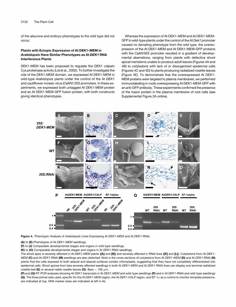
Figure 4. Phenotypic Analysis of Arabidopsis Lines Expressing At DEK1-MEM and At DEK1-RNAi.
(A) to (E) Phenotypes of At DEK1-MEM seedlings.
(F) to (J) Comparable developmental stages and organs in wild-type seedlings.
(K) to (O) Comparable developmental stages and organs in At DEK1-RNAi seedlings.
The shoot apex is severely affected in At DEK1-MEM plants ([A] and [B]) and severely affected in RNAi lines ([K] and [L]). Cotyledons from At DEK1-
MEM (C) and At DEK1-RNAi (M) seedlings are also distorted. Note in the cross sections of cotyledons from At DEK1-MEM (D) and At DEK1-RNAi (N)
plants that the cells exposed to both adaxial and abaxial surfaces contain chloroplasts, suggesting that they have not completely differentiated into
epidermal cells. Shoot apices from less severely affected seedlings in both At DEK1-MEM and At DEK1-RNAi lines can display one terminal radialized
rosette leaf (E) or several radial rosette leaves (O). Bars ¼ 100 mm.
(P) and (Q) RT-PCR analyses showing At DEK1 transcripts in At DEK1-MEM and wild-type seedlings (P) and in At DEK1-RNAi and wild-type seedlings
(Q). The three primer sets used, specific for the At DEK1-MEM region, the At DEK1-CALP region, and EF-1a as a control to monitor template presence,
are indicated at top. DNA marker sizes are indicated at left in kb.
3132 The Plant Cell

In order to provide a baseline for At DEK1-MEM overexpression
phenotypes, we also expressed an At DEK1 RNA interference
(RNAi) construct under the control of the CaMV35S promoter to
generate DEK1 knockout phenotypes. Phenotypes shown here
for the At DEK1-RNAi construct were comparable to those ob-
served previously (Johnson et al., 2005) in a similar experiment.
For a comparison with the At DEK1-MEM–overexpressing plants,
wild-type phenotypes are shown in Figures 4F to 4J.
Interestingly, the phenotypes of the At DEK1-MEM–over-
expressing plants very closely mimicked the range of pheno-
types observed in plants expressing the At DEK1-RNAi construct
(Figures 4K to 4O), suggesting that overexpression of the DEK1
membrane anchor domain induces a dominant negative effect
comparable to the loss or downregulation of DEK1 function.
Structural alterations were observed in ;50% of the obtained
independent T1 lines expressing the At DEK1-MEM and At
DEK1-RNAi constructs. The most severe phenotypes observed
when overexpressing At DEK1-MEM (Figure 4A) and At DEK1-
RNAi (Figure 4K) included seedlings grown on agar that did not
produce rosette leaves and died within 2 weeks after being trans-
ferred to soil. Microscopy analysis of the shoot apex showed that
in contrast with the wild type, consisting of densely cytoplasmic
cells organized into the L1 to L3 cell layers (Figure 4G), the
meristems of the At DEK1-MEM–expressing (Figure 4B) and At
DEK1-RNAi–expressing (Figure 4L) plants contained vacuolated
cells lacking a layered meristem organization. The At DEK1-MEM–
and At DEK1-RNAi–expressing plants (Figures 4C and 4M,
respectively) that did not produce rosette leaves also displayed
a disorganized cell organization on the cotyledon surfaces. On
these cotyledons, the epidermal cell identity appeared partially
lost, as shown by the appearance of chloroplast-containing cells
on the cotyledon surfaces (Figures 4D and 4N), in contrast with
the wild type, in which chloroplasts were only present in internal
mesophyll cells (Figure 4I).
The largest and less severely affected class of both At DEK1-
MEM– and At DEK1-RNAi–expressing plants displayed radial-
ized rosette leaves. This phenotype was manifested either as one
terminal organ with no further organ development from the shoot
apex, as exemplified in the At DEK1-MEM plants shown in Figure
4E, or, more frequently, as a series of two to five radialized leaves
emanating from the shoot apex, as exemplified in the At DEK1-
RNAi–expressing plants shown in Figure 4O. In the latter case,
the shoot apical meristems appeared normal and subsequent
rosette leaves beyond the first few radialized leaves developed
normally (data not shown). Although the radialized leaves initially
lacked trichomes, they invariably developed trichomes evenly
distributed around the circumference of the leaves as leaf growth
continued. Thus, as trichomes are normally restricted to the
adaxial leaf surface in wild-type plants, we infer that the radial-
ized leaves observed in the At DEK1-MEM– and At DEK1-RNAi–
expressing plants were adaxialized.
Additional experiments were performed to elucidate the nature
of the observed phenotypes. In order to rule out the possibility
that the DEK1-MEM phenotype represents a pleiotropic effect
caused by ectopic expression of a large membrane protein, we
expressed a version of At DEK1-MEM that lacked the loop region
(DEK1-MEM-DEL) (Figure 1A). In total, we studied 51 plants with
FLAG-tagged (at both the 59 and 39 ends) and His-tagged DEK1-
MEM-DEL, all of which were indistinguishable from wild-type
control plants. In addition, the presence of DEK1-MEM-DEL-
FLAG in the plasma membrane was demonstrated by immuno-
labeling (see Supplemental Figure 2B online). These results
support the conclusion that the phenotypes are caused specif-
ically by the overexpression of DEK1-MEM. RT-PCR analyses
showed that plants transformed with the CaM35S:At DEK1-MEM
construct had a higher level of At DEK1-MEM transcript than the
wild-type control. DEK1-RNAi plants showed lower At DEK1
transcript levels than wild-type control plants (Figures 4P and 4Q).
This indicates that the phenotypes observed were not caused by
silencing of the native At DEK1 gene.
CR4 Is Also Localized to the Plasma Membrane and
Endosomes in Maize
Antibodies were raised against the maize CR4 external and
cytosolic domains, GR2 and GR8, respectively (Figure 5A). Using
these antibodies in immunoblot analysis with extracts from cr4/
cr4 and wild-type plants, we detected a protein band with the
expected size of CR4 (;95 kD) in wild-type, but not in cr4/cr4,
Figure 5. CR4 Antibodies Recognize Zm CR4 in Wild-Type and Trans-
genic Maize Seeds.
(A) Domain structure and location of peptides used to generate Zm CR4
antibodies.
(B) Immunoblots of protein extracts from wild-type and cr4 mutant maize
roots probed with CR4 GR2 and GR8 antibodies. Arrowheads indicate
the band with the expected size of the CR4 protein (;95 kD).
(C) Immunoblots using protein extracts from CR4:HA:FLAG:AcGFP
transgenic endosperms (þ) or nontransgenic endosperm controls (�)
probed with GFP and CR4 antibodies. The arrowhead indicates the
fusion protein at 127 kD.
DEK1 in Maize and Arabidopsis 3133

samples (Figure 5B). To further characterize these antibodies, we
expressed the construct CR4:HA:FLAG:AcGFP under the con-
trol of the 27-kD g-Zein promoter in maize (Figure 5C). Trans-
genic 10-DAP endosperm expressed GFP in T2 kernels from nine
independent transformation events. The GFP signal intensified
up to 30 DAP, consistent with the known temporal activity of the
27-kD g-Zein promoter (Ueda and Messing, 1991; Russell and
Fromm, 1997). Immunoblot analysis of extracts from these trans-
genic endosperms detected the expected 127-kD CR4:HA:FLAG:
AcGFP fusion protein using antibodies against epitope tags as
well as the CR4 peptide antibodies GR2 and GR8 (Figure 5C).
We then studied the subcellular localization of CR4 in transgenic
in vitro–grown endosperms using GFP fluorescence from the
CR4:HA:FLAG:AcGFP fusion protein. In these endosperms, the
GFP signal was detectable in the plasma membrane and in small
spherical cytoplasmic compartments (Figure 6A). To corroborate
these observations, we also immunolabeled the endosperms
expressing GFP-tagged CR4 with GR2 antibody, observing a
high degree of colocalization of the two signals to the punctate
structure on the plasma membrane (see Supplemental Figures
3A to 3C online). We then labeled CR4-GFP–positive endo-
sperms with DEK1 antibody and showed that CR4 and DEK1
colocalized at the plasma membrane as well as in punctate
structures in the cytosol (Figures 6B and 6C). The subcellular
localization pattern for CR4 was similar in root tissues and in
endosperm cells (Figures 6A and 6D). Finally, root samples pre-
viously incubated in the endocytic dye FM4-64 were immunola-
beled with CR4 antibody and a FITC-conjugated secondary
antibody. This experiment showed colocalization between the
FM4-64 signal and CR4 staining (Figures 6D to 6F), suggesting
that the cytosolic CR4-positive organelles are endocytic com-
partments, likely endosomes. From these experiments, we con-
cluded that CR4 colocalizes with DEK1 at the plasma membrane
and in endosomes.
CR4Concentrated inPlasma MembraneDomains Associate
with Plasmodesmata That Connect Aleurone Cells
To further characterize the subcellular localization of CR4 in
maize in vitro–grown endosperm, we performed immunogold
labeling on high-pressure frozen/freeze-substituted material
(Figure 7). Using CR4 GR2 antibody, we confirmed that CR4
localized to plasma membranes of both aleurone and starchy
endosperm cells (Figures 7A to 7F). Interestingly, the highest
density of CR4 labeling was found at plasma membrane domains
of plasmodesmata located at the anticlinal cell walls between
aleurone cells (Figures 7B to 7D). Whereas 1.8 6 1.03 gold
particles per 500 nm of plasma membrane in cross section were
observed at the plasmodesmata between aleurone cells, only
0.3 6 0.23 gold particles per 500 nm of plasma membrane in
cross sections were recorded elsewhere in the aleurone cells.
This means that CR4 is approximately six times more abundant
at aleurone plasmodesmata than in any other plasma membrane
domain. By contrast, the CR4 labeling on plasmodesmata be-
tween starchy endosperm cells was low and did not drastically
differ from the labeling of the remaining plasma membrane
domains in the starchy endosperm cells (Figure 7F).
The observation that CR4 is associated with plasmodesmata
prompted us to look closer at the distribution and structure of
plasmodesmata in in planta– and in vitro–cultivated endosperms.
We obtained dual-axis electron tomograms of high-pressure
frozen/freeze-substituted aleurone and starchy endosperm cells,
and by imaging the reconstructed volume in different X, Y, and Z
settings, we were able to determine the precise size, shape, and
Figure 6. Colocalization of CR4 and DEK1 in Endosomes.
(A) to (C) Colocalization of CR4 (A) and DEK1 (B) in in vitro–grown CR4:HA:FLAG:AcGFP transgenic maize endosperm. (C) shows a merged image of (A)
and (B). The GFP signal and the DEK1 signal overlapped on plasma membrane and punctate structures in the cytoplasm (arrowheads). Bar ¼ 20 mm.
(D) to (F) Wild-type maize root sections immunostained with GR2 antibody and a secondary antibody conjugated with FITC (D). (E) shows staining with
FM-4-64. (F) shows the two signals colocalized on plasma membrane and in punctate structures in the cytoplasm, likely endosomes (arrowheads).
Bar ¼ 10 mm.
3134 The Plant Cell

distribution of plasmodesmata (Figures 7G and 7H). The aleu-
rone cells contained wide and branched plasmodesmata in
anticlinal cell walls (Figures 7A and 7G). On average, the inner
maximum diameter of plasmodesmata connecting aleurone cells
was 62 6 5 nm (n ¼ 9) and the distance between the plasma
membrane and the endoplasmic reticulum membrane was ;20
nm (Figure 7G). By contrast, plasmodesmata between starchy
endosperm cells were mostly unbranched and much narrower
(inner maximum diameter average was 41 6 4 nm, and plasma
membrane-to-endoplasmic reticulum membrane distance was
10.5 nm [n ¼ 14]) (Figure 7H) than those connecting aleurone
cells. These observations show variation in the distribution and
structure of plasmodesmata in endosperm, suggesting a greater
symplastic communication between aleurone cells than between
starchy endosperm cells. The plasmodesmata connecting aleu-
rone and starchy endosperm cells show intermediate features
between those connecting aleurone cells and those connecting
starchy endosperm cells.
Differentiating Aleurone Cells Establish a
Symplastic Subdomain
To test for the presence of more than one symplastic domain
established by plasmodesma connections in the developing
Figure 7. Electron Microscopy Analysis of CR4 Localization in Maize.
(A) General overview of branched plasmodesmata (PD) connecting aleurone cells in a high-pressure frozen/freeze-substituted in vitro–grown maize
endosperm. Al, aleurone; CW, cell wall.
(B) to (D) Immunogold labeling of CR4 (GR2 antibodies) on plasmodesmata (arrowheads) between aleurone cells.
(E) and (F) Immunogold labeling of CR4 on the plasma membrane (black arrowheads in [E]) of starchy endosperm cells. The plasmodesmata
connecting starchy endosperm cells (white arrowheads in [F]) are much narrower than those located between aleurone cells and showed very low or no
CR4 labeling.
(G) and (H) Tomographic slices of plasmodesmata in cell walls between aleurone cells (G) and between starchy endosperm cells (H). Line A to A9
indicates the distance between the plasma membrane and the endoplasmic reticulum membrane (ER), and line B to B9 indicates the widest total
diameter of the plasmodesmata.
Bars ¼ 100 nm in (A) to (F) and 50 nm (G) and (H).
DEK1 in Maize and Arabidopsis 3135

maize endosperm, we incubated in vitro–grown endosperm with
two fluorescent tracers, the small 524-D tracer 8-hydroxypyrene-
1,3,6-trisulfonic acid (HPTS) and 40-kD dextran conjugated to
FITC (40kD-F-dextran) (Kim et al., 2002). In endosperms grown in
vitro for 2 and 5 d, HPTS moved through all endosperm cells
(Figures 8A and 8B). However, the 40kD-F-dextran tracer moved
only through the aleurone cells, suggesting that the differentiating
aleurone cells form a symplastic subdomain within the endosperm
(Figures 8C to 8H). Interestingly, whereas the 40kD-F-dextran
tracer moved through up to 90% of the aleurone cells in 2-d in
vitro–grown endosperm, the same tracer was only able to move
through up to 40% of the aleurone cells in the 5-d in vitro–grown
endosperm. These results indicate that although symplastic con-
nectivity between aleurone cells remained, as shown by the
movement of the small HPTS tracer, the exclusion size of the
plasmodesmata located between aleurone cells was reduced
during endosperm development. Most of the aleurone cells
containing the 40kD-F-dextran tracer in the 5-d in vitro–grown
endosperm were located at young bulges or miniendosperms
(Figure 8F) that formed by localized high mitotic activity (Gruis
et al., 2006).
SAL1 Colocalizes with DEK1 and CR4 in Endosomes
The data presented above suggested that DEK1 and CR4
colocalize in endosomes. Therefore, we investigated whether
these endosomes associate with the SAL1 protein. Immunoblot
analysis of maize protein extracts from wild-type endosperm
showed that polyclonal rabbit antibodies against recombinant
SAL1 protein recognized a protein of ;27 kD, the expected size
for SAL1 (Figure 9, left panel). In extracts from sal1-2 mutant
endosperm, the band was weaker, suggesting that the sal1-2
allele was not a null allele. The maize SAL1 antibody also rec-
ognized a protein with the expected size for At SAL1 (;27.5 kD)
(Figure 9, right panel). Two weaker bands of higher molecular
mass were also recognized by the antibody in Arabidopsis ex-
tracts. Furthermore, the anti-SAL1 antibody labeled multivesicular
bodies (endosomes) in Arabidopsis embryo cells, confirming that
SAL1 localizes to endosomal compartments, as predicted based
on its high amino acid identity to class E vacuolar proteins such
as CHMP1 and DID2 (Figures 10A to 10C).
Next, we used the SAL1 antibody to immunolabel sections of
in vitro–grown maize endosperm. As shown in Supplemental
Figure 4A online, SAL1 is present in small structures distributed
throughout the cytoplasm in both aleurone and starchy endo-
sperm cells from wild-type plants. There was no difference in the
Figure 8. Cell-to-Cell Movement of Fluorescent Tracers in in Vitro–
Grown Maize Endosperms.
(A) and (B) Endosperms grown in vitro for 2 d (A) or 5 d (B) allow the
movement of HPTS through both differentiating aleurone (AL) and
starchy endosperm (ST) cells.
(C) to (H) In vitro–grown endosperm incubated in 40 kD of F-dextran. The
tracer is able to move only through the aleurone cells of endosperms
cultured for 2 d ([C], [E], and [G]) or 5 d ([D], [F], and [H]), but the
movement is highly reduced in the latter. Most of the aleurone cells that
contain the 40-kD F-dextran in the 5-d-in-culture endosperms are
located at growing bulges at the endosperm surface ([F], arrow). (G)
and (H) show paradermal views of the aleurone layer in endosperms
grown in vitro for 2 d (G) or 5 d (H). Note the reduction in the aleurone
cells containing the 40-kD F-dextran tracer in the older endosperms.
Bars ¼ 100 mm.
Figure 9. Characterization of SAL1 Antibodies.
Immunoblots of protein extracts from maize wild type and sal1-2 mutant
endosperms and roots as well as Arabidopsis leaf proteins (At) probed
with anti-SAL1 antibodies. Arrows show bands at the predicted size for
SAL1. The presence of a weak band in the mutant shows that the maize
sal1-2 mutant is not a null allele.
3136 The Plant Cell

distribution of SAL1 signal between aleurone and starchy endo-
sperm (data not shown), nor could we detect a difference in the
distribution of SAL1 between wild-type and sal1-2 mutant en-
dosperms (see Supplemental Figure 4B online). This observation
is not surprising in light of the fact that sal1-2 is not a null allele. As
reported above, we used maize roots for our FM4-64 labeling
experiments. In this system, double labeling with FM4-64 and the
maize SAL1 antibody confirmed the presence of SAL1 in endo-
somes (Figures 10D to 10F). To determine whether the sal1-2
mutation affected the pattern of localization of DEK1 and CR4,
we also immunostained sal1-2 in vitro–grown endosperm with
DEK1 Zm8 and CR4 GR2 antibodies and found no obvious
difference between wild-type and sal1-2 mutant endosperm (see
Supplemental Figures 4C and 4D online).
Finally, we investigated whether SAL1 colocalizes with CR4 and
DEK1. To test for DEK1 and SAL1 colocalization, we immuno-
labeled wild-type maize endosperms with the DEK1 Zm1-2 anti-
bodies (Figure 10G) followed by the SAL1 antibodies (Figure 10H).
We observed extensive colocalization on small cytoplasmic struc-
tures, likely endosomes (Figure 10I). Next, we localized CR4 by
GFP fluorescence from the fusion protein CR4:HA:FLAG:AcGFP
and then SAL1 by immunolabeling of the same section of in vitro–
grown endosperm (see Supplemental Figures 5A to 5C online).
The results supported the conclusion that SAL1 colocalized with
DEK1 and CR4 in endosomes of maize endosperm.
DISCUSSION
DEK1 Plays a Conserved Role in Epidermis
Cell Fate Specification
As demonstrated by the lack of aleurone cells in in vitro–grown
dek1 mutant endosperm (Gruis et al., 2006), DEK1 plays an es-
sential role either in the perception and/or in the transmission of
the positional cue initiating the epidermal cell fate. From the ob-
servation of similar epidermal dek1 mutant phenotypes in maize
and Arabidopsis, both in seeds and in vegetative organs, we infer
that the role of DEK1 in epidermal cell fate specification is
Figure 10. Localization of SAL1 in Arabidopsis and Maize Cells.
(A) to (C) Immunogold labeling of multivesicular endosomes in high-pressure frozen/freeze-substituted Arabidopsis embryo cells with anti-SAL1
antibodies. The antibodies labeled 60% of the multivesicular bodies analyzed (n ¼ 40).
(D) to (F) Colocalization of SAL1 and the endocytotic marker FM4-64 in punctate structures, likely endosomes (arrowheads in [F]), in wild-type maize
roots. (D) shows immunolabeling with anti-SAL1 antibodies and a FITC-conjugated secondary antibody, and (E) shows staining with fixable FM4-64. (F)
shows a merged image of (D) and (E).
(G) to (I) In vitro endosperm section immunostained with DEK1 rat antibody Zm1-2 (G) and SAL1 (H). Overlapping staining (I) is indicated by arrowheads
in the merged image of (G) and (H).
Bars ¼ 10 mm.
DEK1 in Maize and Arabidopsis 3137

conserved and that both systems provide valuable insight into
DEK1 function. The remarkable sequence conservation of DEK1
over the course of land plant evolution, as indicated by the high
degree of similarity between angiosperm and moss protein se-
quences, also suggests a conserved role for DEK1.
In maize in vitro cultures, interior starchy endosperm cells that
become positioned adjacent to internal voids that occasionally
develop convert to aleurone cells (Gruis et al., 2006). A prereq-
uisite for a protein postulated to respond to such positional infor-
mation is that it is ubiquitously distributed in all cells. This important
criterion is fulfilled for DEK1, as shown by in situ hybridization
experiments and high-resolution transcript profiling in maize and
Arabidopsis (Lid et al., 2005). The data showing ubiquitous dis-
tribution of DEK1 in peripheral and internal cell layers of in vitro
endosperm presented here also support this conclusion.
DEK1-CALP Fails to Fully Complement the dek1
Mutant Phenotype
We previously proposed that DEK1 is activated by external sig-
naling and that this activity is transmitted via the protease activity
of DEK1-CALP. This hypothesis was based on the highly con-
served active site of DEK1 compared with typical animal calpains
as well as on the fact that DEK1-CALP displays calpain Cys pro-
teinase activity in vitro (Wang et al., 2003). Here, we have shown
that maize DEK1-CALP failed to complement the loss-of-aleurone
cell phenotype in homozygous maize dek1 mutant endosperm
under the control of the 27-kD g-Zein promoter. Since DEK1-CALP
possesses proteinase activity in vitro (Wang et al., 2003), we infer
that DEK1 calpain activity is present in the peripheral cells of the
transgenic endosperm expressing DEK1-CALP. In spite of this,
the peripheral cell layer of the endosperm failed to assume an
aleurone cell fate. The expression of At DEK1-CALP also failed to
fully complement the Arabidopsis dek1 mutant phenotype. These
results support the conclusion that DEK1-MEM is necessary for
wild-type DEK1-CALP activity.
Recently, the membrane-bound transcription factor NTM1 (for
NAC WITH TRANSMEMBRANE MOTIF1) was identified as a
putative substrate for DEK1 in Arabidopsis (Kim et al., 2006). This
conclusion was supported by the similar phenotypes of Arabi-
dopsis plants that overexpress the Arabidopsis DEK1 gene and
ntm1-D mutant plants (Kim et al., 2006). In addition, the calpain
inhibitor N-acetyl-leucinyl-leucinyl-norleucinal inhibits NTM1 pro-
cessing, suggesting that NTM1 may be released from the mem-
brane by the DEK1 calpain protease. Future experiments should
reveal whether or not NTM1 is a substrate of DEK1.
Overexpression of At DEK1-MEM Gives a Dominant
Negative Phenotype
Ectopic expression of At DEK1-MEM under the control of
CaMV35S in Arabidopsis gives a phenotype that closely resem-
bles the mutant phenotype seen in At DEK1-RNAi suppression
lines. One explanation for the dominant negative phenotype
caused by DEK1-MEM overexpression is that DEK1-MEM inter-
acts with either a ligand or interacting membrane proteins that
normally mediate the activation of DEK1. The overexpressed
DEK1-MEM truncated protein competes with native DEK1 for the
DEK1 activators and/or interactors, and since it lacks the cyto-
plasmic calpain domain, it cannot trigger the downstream sig-
naling pathways, leading to a dominant negative phenotype. The
observation that expression of At DEK1-MEM under the weaker
At DEK1 promoter fails to give a detectable phenotype suggests
that a threshold level of truncated protein has to be surpassed to
interfere with the endogenous DEK1 protein function. The ob-
servation that plants overexpressing At DEK1-MEM-DEL, which
lack the loop region (Figure 1A), display a phenotype indistin-
guishable from the wild type strengthens the conclusion that the
observed dominant negative phenotype is specific to DEK1-
MEM. Furthermore, this observation supports the speculation
that the loop region is essential for either the perception and/or
the transmission of positional signals.
The nature of the signal for epidermal cell fate specification is
unknown. We previously speculated that that epidermal cell fate
specification is suppressed internally in the endosperm by lateral
inhibition from neighboring cells and that this inhibition is only
escaped in the outer membrane/cell wall of cells exposed to the
surface. Since the dominant negative phenotype caused by At
DEK1-MEM overexpression suggests that DEK1 is activated at
the plasma membrane, likely by an extracellular signal, DEK1
may function downstream of the primary signal for surface or
aleurone fate specification. A scenario in which DEK1 activation
occurs by autodimerization also represents a possibility that
cannot be ruled out. One way to identify putative DEK1 inter-
actors is by yeast two-hybrid experiments using the different pre-
dicted extracellular loop regions as baits. The design of these
studies, however, is hampered by the lack of insight into the
three-dimensional structure of DEK1, which may form a pore or a
channel-like structure in the membrane. Therefore, interaction by
extracellular molecules or other transmembrane proteins may be
with several DEK1 domains simultaneously, a situation that is
difficult to recreate in yeast two-hybrid screens.
DEK1 and CR4 Colocalize in SAL1-Positive Endosomes
In addition to the plasma membrane, DEK1 localized to mem-
branous structures that we interpret to be endosomes. Recently,
Johnson et al. (2005) presented data showing that GFP-tagged
ACR4 in Arabidopsis root cells was present in two distinct sub-
cellular compartments: protein export bodies or secretory ves-
icles and a population of internalized vesicles or endosomes. The
authors concluded that internalization and turnover for ACR4 are
linked and depend on functionality, suggesting that ACR4 sig-
naling may be subject to downregulation via internalization. In
their experiments with Arabidopsis root epidermal cells, identi-
fication of endocytic/endosomal compartments was based on
FM4-64 incubation experiments. Using maize roots, we show
here that FM4-64–positive compartments also stained positive
for SAL1. The observation that SAL1-positive compartments are
stained by FM4-64 after 15 min of incubation strongly supports
the conclusion that these structures represent endosomal com-
partments. In addition, the fact that the maize SAL1 antibodies
recognized epitopes in multivesicular bodies in Arabidopsis em-
bryos also supports this conclusion.
3138 The Plant Cell

In the same experiments, we also demonstrated that DEK1-
and CR4-positive organelles stained positive with FM4-64, lead-
ing us to speculate that DEK1 and CR4 are both internalized by
endocytosis and routed through endosomal compartments. The
functional significance of an internalization process has yet to be
determined, but a quantitative regulation of vital cellular recep-
tors by endosomal degradation is common in animal systems,
including animal growth hormone receptors (Mellman, 1996;
Katzmann et al., 2002). As demonstrated by the presence of
extra layers of aleurone cells in the maize sal1-2 mutant, SAL1 is
a negative regulator of aleurone cell fate. In animal systems,
the SAL1 homolog CHMP1 mediates the trafficking of cargo
proteins from multivesicular bodies to lysosomes for degradation
(Howard et al., 2001). We earlier proposed that the supernumer-
ary aleurone layer phenotype of sal1 mutants is caused by an
overabundance of positive regulators of aleurone cell fate due to
defective degradation via endosomes. The data presented here
support this proposal in two ways. First, by demonstrating that
SAL1 localizes to endosomes, and second, by demonstrating
that SAL1 colocalizes with the two known regulators of aleurone
cell fate, DEK1 and CR4. The immunolocalization experiments
performed here did not show differences between the distribu-
tion of DEK1 and CR4 in wild-type and sal1-2 mutant endo-
sperms. We ascribe this observation to the fact that the sal1-2
mutant is not a null mutant, the protein being present in mutant
endosperm at a reduced level compared with the wild type.
Nevertheless, sal1-2 mutant endosperms display multiple layers
of aleurone cells in in vitro organ cultures (Gruis et al., 2006).
Therefore, we speculate that the multiple aleurone layer pheno-
type of sal1 mutants is caused by an overabundance of aleurone
signaling membrane proteins (e.g., DEK1 and CR4).
Aleurone Cells of Young Endosperm Form a Symplastic
Subdomain through Wide Plasmodesmata Enriched in CR4
The localization pattern of CR4 in maize endosperm and roots
shown in this study is in agreement with data presented by
Johnson et al. (2005) on Arabidopsis root cells. These data
support the notion that CR4 is present in many different cell types
across monocot and dicot species and that targeting, function,
and regulation may be the same in all of these cells. In Arabidopsis
root cells, the ACR4 has been described as an L1 cell layer–
specific kinase (Johnson et al., 2005). This conclusion is not
supported by our data from endosperm, in which CR4 was shown
to be expressed in all cell layers. Interestingly, however, CR4 is
primarily associated with plasmodesmata only in the aleurone
layer and not in the starchy endosperm. Whether the preferential
presence of CR4 in periclinal epidermal cell walls of Arabidopsis
leaf primordia and ovules (Tanaka et al., 2002; Gifford et al., 2003)
also reflects an association with specialized plasmodesmata
remains to be determined. The CR4-positive plasmodesmata in
anticlinal walls of aleurone cells show a larger inner diameter and
also exhibit a larger plasmodesmata exclusion size, leading to the
development of symplastic subdomains of aleurone cells in
young endosperm, as demonstrated by the movement of fluo-
rescent tracers. The association of CR4 with these plasmodesmata
Figure 11. A Model for the Role of DEK1, CR4, and SAL1 in Aleurone Cell Specification.
DEK1 at the surface of the endosperm is activated by an unknown mechanism (a), its calpain domain in the cytosol cleaving a postulated substrate (b)
that leads to the specification of aleurone cell fate. DEK1 in all other positions is inactive (c). In cells with active DEK1 signaling, CR4 concentrates on
plasmodesmata between aleurone cells (pda) and increases the plasmodesma exclusion limit, allowing the activated DEK1 substrate to move laterally
between aleurone cells, thereby reinforcing the signal for aleurone cell fate specification (d). Plasmodesmata in cell walls between starchy endosperm
cells are narrow (pds), whereas plasmodesmata in cell walls between aleurone cells and starchy endosperm cells are intermediary in width (pdi). DEK1
and CR4 are internalized by endocytosis (e) and traffic through endosomes. Whereas some DEK1 and CR4 molecules may be recycled back to the
plasma membrane (f), others are sorted for degradation in the vacuole in a process that requires SAL1. Some endosomes are recycled back to the
plasma membrane (f).
DEK1 in Maize and Arabidopsis 3139

places CR4 downstream of DEK1 in aleurone signaling, a con-
clusion that is in agreement with previous observations in
Arabidopsis embryos (Johnson et al., 2005).
Although the establishment of symplastic domains during
plant development has been well documented in previous stud-
ies (Kim et al., 2002), one possibility that we raise here is that CR4
may be involved in increasing the exclusion limit for molecules
moving laterally between aleurone cells, in particular the acti-
vated substrate of DEK1 or a molecule downstream of this sub-
strate. If correct, the relative importance of this activity on aleurone
cell fate specification may be revealed by the phenotype of
homozygous cr4 mutant endosperms, in which ;10% mutant
kernels show mosaic aleurone layers (Becraft et al., 1996). One
interpretation is that sectors on the endosperm surfaces lacking
aleurone cells represent symplastic subdomain(s) in which lateral
transmission of the signal activating aleurone cell fate specifica-
tion failed due to a lack of CR4 activity. Interestingly, different cr4
mutants show distinct patterns of failure to develop aleurone
cells, ranging from the absence of aleurone cells in the abgermi-
nal region of the kernel to the absence in mainly germinal regions
(Becraft and Asuncion-Crabb, 2000). In addition, several other
mutants that disrupt aleurone differentiation also display distinct
mosaic aleurone phenotypes, including bareback and naked
(Becraft and Asuncion-Crabb, 2000). Based on the results shown
here, one possible explanation for these phenotypes is that they
are affected in functions needed to establish or maintain aleu-
rone symplastic subdomains. It is also interesting that Becraft
and coworkers (2001) concluded from their cr4 mutant sector
studies that although CR4 appears to function cell autono-
mously, the phenotype of several sectors with cr4 mutant cells
appeared defective in the production of lateral developmental
signals. Finally, a protein kinase that recognizes a specific subset
of viral as well as endogenous proteins associated with plasmo-
desmata has already been identified in plants (Lee et al., 2005).
Summary: A Model for Aleurone Cell Fate Specification
Aleurone cell fate specification in maize endosperm occurs ex-
clusively in response to surface position, a process that involves
the function of DEK1, CR4, and SAL1. The data presented here
show that DEK1 and CR4 are located at the plasma membrane
and in SAL1-positive endosomes in all cells investigated. Based
on these and earlier observations, we propose a model for
aleurone cell fate specification in which in DEK1 is active only in
the outer membrane of cells positioned on endosperm surfaces
(Figure 11). To be fully functional, the calpain activity must be
under the control of the membrane portion of DEK1, in which the
loop region plays an essential functional role. The DEK1 substrate
may be a cytoplasmic protein or, as suggested by Kim et al.
(2006), a membrane-bound transcription factor such as NTM1.
Once aleurone cell fate specification has been initiated, the pro-
cess is reinforced via lateral signaling in symplastic subdomains
mediated by CR4 in specialized plasmodesmata. Finally, we pre-
dict that the proper plasma membrane concentration of DEK1 and
CR4 is maintained by internalization and degradation at SAL1-
positive endosomes. We also hypothesize that this model is valid
for epidermal cell fate specification in all plants and that DEK1 was
recruited early during land plant evolution to serve in this role.
METHODS
Plant Material, Growth Conditions, and Generation of
Transgenic Plants
Maize (Zea mays) plants were grown under typical greenhouse conditions
(68 to 828F, 16 h of light/8 h of dark) using a commercial potting medium
(Metro-Mix 700; Scotts-Sierra) and fertilized as needed with a standard
fertilizer mixture (20:10:20 N:P:K). Two to 3 d after the first silks appeared,
pollination was performed to prepare material for in vitro endosperm
cultures. Maize in vitro endosperms were grown as detailed by Gruis et al.
(2006). For immunostaining of maize roots, maize seeds were sand-
wiched in four layers of wet paper towels and placed on a glass plate at
room temperature. Three days after germination, root tissues were har-
vested and processed for immunostaining. For immunoblot analyses,
12-DAP dek1/dek1 and Dek1 (wild-type) kernels from Dek1/dek1 ears
were frozen in liquid nitrogen for storage at �808C until use. Roots from
cr4/cr4 and wild-type plants were frozen at the V3 stage in liquid nitro-
gen for storage at �808C until use.
All Arabidopsis thaliana lines were produced in the Columbia ecotype.
Arabidopsis plants were grown in growth chambers at 228C and 70%
humidity under cool-white fluorescent light (85 mmol m�2 s�1) for 8 h of dark
and 16 h of light. Constructs were transformed into the Agrobacterium
tumefaciens strain C58C1 pGV2260 (Deblaere et al., 1985) by electropor-
ation. Arabidopsis plants were transformed by a modified version of the
floral dip method (Clough and Bent, 1998), in which the Agrobacterium cul-
ture was applied directly to flower buds using a pipette. Transformants were
selected on Murashige and Skoog (1962) medium containing 50 mg/mL
kanamycin.
Antibody Production and Purification
Polyclonal antibodies against DEK1, CR4, and SAL1 were raised in
rabbits or rats and affinity-purified according to the manufacturer’s man-
ual. For DEK1, we raised the antibodies against the following peptides: Zm1
and Zm1-2, 59-KGERNFTDQEFPPEDR-39; ZM3, 59-VREVDGHKL-39; ZM6,
59-PFVLSVFSKASIRLEAV-39; Zm8, 59-DKGLDPNFSYMLKDK-39; and ZM9,
59-LYKWKDDDWKISR-39. DEK1 rabbit antibodies were used at dilutions
of 1:3000 for immunoblot analyses and 1:300 for immunostaining. DEK1
rat antibody was used at dilutions of 1:3000 for immunoblot analyses and
1:200 for immunostaining. For CR4, antibodies were raised against
peptides in the external domain (GR2, 59-MTATHAVDEAVS-39) and in
the internal domain (GR8, 59-YRAPSWITFPSVTSSQRR-39). CR4 anti-
bodies were used at dilutions of 1:5000 for immunoblot analyses and
1:300 for immunostaining. SAL1 antibody was raised against the whole
protein and used at dilutions of 1:5000 for immunoblot analyses and 1:500
for immunostaining. Antibodies against HA (Sigma-Aldrich; catalog num-
ber H9658) and FLAG (Sigma-Aldrich; catalog number F3165) were used
at dilutions of 1:15,000 for immunoblot analyses and 1:1500 for immu-
nostaining. Antibody against GFP (BD Living Colors; catalog number
632380) was used at a dilution of 1:3000 for immunoblot analyses.
Plasmid Construction and Plant Transformation
To construct Zm DEK1-CALP:HA:FLAG:AcGFP, Dek1 cDNA from maize
(GenBank accession number AY061805; nucleotides 3683 to 6532) was
fused to cDNA for HA (amino acid sequence TSYPYDVPDYA), FLAG
(amino acid sequence DYKDDDDK), and the GFP gene (Clontech) from
Aequorea coerulescens downstream of the maize 27-kD g-Zein promoter
(1473 bp) isolated from the W64A maize line (Ueda and Messing, 1991;
Russell and Fromm, 1997). Several linker sequences were used that
generated the following amino acid sequences when translated: between
the g-Zein promoter and Zm DEK1-CALP, IGSEFELRRQ; between Zm
Dek1-CALP and HA, DV; between HA and FLAG, QL; between FLAG and
3140 The Plant Cell

AcGFP, A. For CR4:HA:FLAG:AcGFP expression, EST clsmc27 from
maize (GenBank accession number U67422; nucleotides 157 to 2859)
was fused to HA, FLAG, and AcGFP (as described above for Zm DEK1-
CALP:HA:FLAG:AcGFP expression) downstream of the maize 27-kD
g-Zein promoter (1473 bp) isolated from the W64A line. The linkers used
also generated extra amino acids in this construct: between CR4 and HA,
DV; between HA and FLAG, QL; between FLAG and AcGFP, A. These
constructs were transformed into maize embryos as described (Zhao and
Ranch, 2006). T1 kernels were analyzed for fluorescence protein expres-
sion or protein expression on immunoblots.
Constructs for the transformation of Arabidopsis were prepared in the
modified pCAMBIA2300 binary vectors pSEL1-ProCaMV35S -NOS or
pSEL1-NOS (Lid et al., 2005). All subclones derived by PCR amplification
were initially cloned in pCR4-TOPO (Invitrogen) and sequenced using the
BigDye Terminator version 3.1 cycle sequencing kit and the ABI PRISM
3100 genetic analyzer (Applied Biosystems) before cloning into their
respective binary vectors. The genomic region corresponding to At
DEK1-MEM was PCR-amplified from Arabidopsis genomic DNA of the
Columbia ecotype using primers 59-CCGGGCATGGAAGGGGATGAGC-
GAGGAGTCTT-39 and 59-ACTAGTCTAAATTATCAGACTAGCAACAAC-
ACCCCA-39 that introduced a unique XmaI site immediately upstream of
the ATG start site and a novel TAG stop codon at bp positions 5257 to
5259 relative to the ATG start as well as a SpeI site immediately 39 to this
stop codon. At DEK1-MEM was then cloned using XmaI and SpeI be-
tween CaMV35S and the NOS terminator in pSEL1- ProCaMV35-NOS.
In order to produce a C-terminal fusion of At Dek1-MEM and GFP, the
851-bp 39 part of At Dek1-MEM was PCR-amplified from pSEL1-
ProCaMV35-At Dek1-MEM-NOS using 59-CTCTCGAGGTGTATATGTT-
TTCTTTTCAAT-39 containing an integral XhoI site and 59-GGATCCAAT-
TATCAGACTAGCAACAACACCCC-39 introducing a BamHI site in place
of the stop codon and cloned into pCR4-TOPO (Invitrogen). The plasmid
pKEx4tr-smGFP (a gift from Tage Thorstensen) was digested with BamHI
and SpeI, and the resulting GFP fragment was cloned into the 39 end of At
Dek1-MEM. The in-frame C-terminal fusion of GFP and the 39 end of At
Dek1-MEM was then cloned into At Dek1-MEM-pCR4-TOPO using XhoI
and SpeI to give At Dek1-MEM-GFP-pCR4-TOPO. At Dek1-MEM-GFP
was finally cloned into pSEL1-ProCaMV35-At Dek1-MEM-NOS using PstI
and SpeI to give pSEL1-ProCaMV35-At Dek1-MEM-GFP-NOS.
For overexpression of the At DEK1-RNAi construct, a 590-bp At DEK1
cDNA fragment was first PCR-amplified from Arabidopsis Columbia
cDNA isolated from seedlings using oligo(dT) dynabeads (Invitrogen)
with the two primer sets 59-CCCGGGACAAGAGCATCCTGAACCAGTC-39/
59-GTCGACAGGAAACCCAGGATGGAGA-39and 59-ACTAGTACAGG-
AAACCCAGGATGGAGA-39/59-GGTACCGACAAGAGCATCCTGAAC-
CAGTC-39, introducing 59 XmaI and 39 SalI sites to one copy and 59 KpnI
and 39 SpeI sites to the other copy. The 190-bp Catalase intron from the
GUS gene in pCAMBIA2301 (AF234316) was PCR-amplified using
primers 59-GTCGACGGGTAAATTTCTAGTTTTTCTCC-39 and 59-ACT-
AGTGGTTCTGTAACTATCATCATCATC-39, introducing 59 SalI and 39 SpeI
sites. The XmaI/SalI and KpnI/SpeI cDNA copies of At DEK1 as well as the
SalI/SpeI clone of the Catalase intron were cloned between CaMV35S
and the NOS terminator in pSEL1-ProCaMV35-NOS using the added re-
striction sites, resulting in inverted repeats of the At DEK1 cDNA fragment
flanking the Catalase intron.
The 2.6-kb At DEK1 promoter was PCR-amplified from Arabidopsis
genomic DNA of the Columbia ecotype using primers 59-GGATCCAA-
GAGGGACACTGGTGGATGCAATTTG-39 and 59-CCCGGGCTTCTTCT-
CCCACCTACAACACCATGCA-39, introducing a 59 BamHI site and a
39 XmaI site immediately upstream of the ATG start codon. pSEL1-
ProCamV35S-At DEK1-CALP-NOS was digested with SalI and KpnI, and
the resulting DEK1-CALP fragment was cloned between the At DEK1
promoter and the NOS terminator in pSEL1-ProAtDEK1-NOS. At DEK1-CALP
was PCR-amplified from Arabidopsis Columbia cDNA isolated from
seedlings using oligo(dT) dynabeads (Invitrogen) with primers 59-CCCG-
GGCATGTGTGCCCATGCAAGAGTT-39 and 59-GGTACCTGACAATAC-
GGGCACTACAAAGCT-39, adding a 59 XmaI site and a 39 KpnI site, with
which At DEK1-CALP was cloned between the At DEK1 promoter and
the NOS terminator in pSEL1-ProAtDEK1-NOS. In order to produce a
C-terminal fusion of At DEK1-CALP and FLAG (amino acid sequence
DYKDDDDK), the 903-bp 39 part of At DEK1-CALP was PCR-amplified
from pSEL1-ProAtDEK1-At Dek1-CALP-NOS using 59-GGACTGGTTCAG-
GATGCTCTTGT-39, containing an integral SalI site, and 59-GGTAC-
CCTACTTGTCATCGTCGTCCTTGTAGTCCAAAGCTTCAAGAACAATGG-
ATGCT-39, introducing FLAG, a stop codon, and a new KpnI site in place
of the old stop codon, and cloned into pCR4-TOPO (Invitrogen). The in-
frame C-terminal fusion of the 39 end of At Dek1-CALP was finally cloned
into pSEL1-ProAtDEK1-At DEK1-CALP-NOS using SalI and KpnI to give
pSEL1-ProAt DEK1-At DEK1-CALP:FLAG-NOS.
In order to replace the loop domain of At Dek1-MEM with the trans-
membrane linker domain of At Dek1 exon 11 (amino acid sequence
RFSHSSA), the fragment was PCR-amplified from pSEL1-ProCaMV35-At
Dek1-MEM-NOS using two primer sets: 59-GGAGGAATCACATCAG-
CTGCAGT-39, containing an integral PstI site, and 59-CTCGAGTGGGA-
AAACCTTGGGTCTGTTATAGATAGATGCCGAG-39, introducing a XhoI
site; and 59-CTCGAGTGCTCCAGAGAGAGCATGGGGCCT-39, introduc-
ing a XhoI site, and 59-GTGGGCAACTGATCATCTCTAGATTTTA-39,
containing an integral XbaI site. The two fragments were ligated in pCR4-
TOPO, and the transmembrane linker was cloned into pSEL1-ProCaMV35-
At Dek1-MEM-NOS using PstI and XbaI, to give pSEL1-ProCaMV35-At
Dek1-MEM-DEL-NOS.
In order to produce a C-terminal fusion of At Dek1-MEM-DEL and FLAG
(amino acid sequence DYKDDDDK), the 1107-bp 39 part of At Dek1-MEM
was PCR-amplified from pSEL1-ProCaMV35-At Dek1-MEM-DEL-NOS us-
ing 59-CGTGGCATTTTGTGGAACTTCA-39, containing an integral XbaI
site, and 59-ACTAGTCTACTTGTCATCGTCGTCCTTGTAGTCAATTATC-
AGACTAGCAACAACACCCCA-39, introducing FLAG, a stop codon, and
SpeI. The in-frame C-terminal fusion of the At Dek1-MEM-DEL 39 end and
FLAG was finally cloned into pSEL1-ProCaMV35-At Dek1-MEM-DEL-NOS
using XbaI and SpeI, to give pSEL1-ProCaMV35-At Dek1-MEM-DEL:FLAG-
NOS. In order to produce a C-terminal fusion of At Dek1-MEM-DEL:FLAG
and 6xHIS, the 39 part of At Dek1-MEM-DEL:FLAG was PCR-amplified
using 59-CGTGGCATTTTGTGGAACTTCA-39, containing an integral XbaI
site, and 59-ACTAGTCTAGTGATGGTGATGGTGATGCTTGTCATCGT-
CGTCCTTGTAGTCA-39, introducing 6xHIS, a stop codon, and SpeI.
The in-frame C-terminal fusion of the At Dek1-MEM-DEL:FLAG 39 end
and the His tag was finally cloned into pSEL1-ProCaMV35-At Dek1-
MEM-DEL-NOS using XbaI and SpeI, to give pSEL1-ProCaMV35-At
Dek1-MEM-DEL:FLAG-HIS-NOS. In order to produce an N-terminal
fusion of At Dek1-MEM-DEL and FLAG, the 59 part of At Dek1-MEM
was PCR-amplified from pSEL1-ProCaMV35-At Dek1-MEM-DEL-NOS
using 59-CCCGGGATGGACTACAAGGACGACGATGACAAGATGGAAG-
GGGATGAGCGAGG-39, introducing XmaI, a start codon, and FLAG, and
59-CCAAGAACAATAACTGCAGCTGATG-39, containing an integral PstI
site. The in-frame N-terminal fusion of the At Dek1-MEM-DEL 59 end and
FLAG was then cloned into pSEL1-ProCaMV35-At Dek1-MEM-DEL-NOS us-
ingXmaI and PstI, to givepSEL1-ProCaMV35-FLAG:At Dek1-MEM-DEL-NOS.
RT-PCR Analyses
Isolation of poly(A)þ RNA from 35S-Dek1-MEM, 35S-Dek1-RNAi, and
wild-type control seedlings, as well as first-strand cDNA synthesis and
PCR conditions, were as described previously (Lid et al., 2005). To ensure
that amplification was in the logarithmic phase, the smallest number of
PCR cycles allowing detection on an ethidium bromide–stained agarose
gel was experimentally calculated and used. The number of cycles used
was 28 in Figure 4A and 29 in Figure 4B. All PCRs were performed at
578C annealing temperature. The primer pairs used were as follows: At
DEK1 in Maize and Arabidopsis 3141

Dek1-MEM, 59-TTTGGGCTGTTAATTGGCGG-39 and 59-CAACGTGAT-
TCCCTAGCTGTGAGA-39; At Dek1-CALP, 59-CAACGTGATTCCCTA-
GCTGTGAGA-39 and 59-ACTCCGCAAGTCAATCTCCTCA-39; EF-1a,
59-CACATCAACATTGTGGTCATTGG-39 and 59-GGTAGTGGCATCCA-
TCTTGTTACAA-39.
Immunoblot Analysis
Frozen wild-type and mutant materials were ground with a mortar and
pestle in liquid nitrogen. Equal fresh weights of the samples were ground
again with a plastic pestle in extraction buffer (50 mM Tris-HCl, pH 8.0, at
258C, 5 mM EDTA, and 2% SDS) with 13 protease inhibitor (Sigma-
Aldrich) at a 1:2 (w/v) ratio. Cellular debris was removed by centrifugation
at 140,000 rpm for 10 min. Protein extracts were diluted 1:2 (v/v) in sample
buffer and boiled for 5 min before separation through a 4 to 12% (w/v)
SDS polyacrylamide gel (Invitrogen) and then transferred to a cellulose
membrane in a submerged blotting system (Mini-Trans Blot; Invitrogen).
Membranes were blocked for 1 h with Tris-buffered saline containing 5%
(w/v) nonfat dry milk. Immunoblotting of DEK1 was performed using Zm1
and anti-rabbit secondary antibodies (Bio-Rad Laboratories) in Tris-
buffered saline containing 0.1% (v/v) Tween 20. Immunoblotting of CR4
was performed using GR2 and GR8 antibodies. Proteins that cross-
reacted with antibodies were detected with chemiluminescent substrates
(Pierce) and visualized on x-ray film.
Fluorescence Immunolabeling
Endosperms were fixed in 4% paraformaldehyde overnight at 48C,
sectioned at 60 mm using a vibratome, and placed on cover slips with a
thin layer of Meyer’s adhesive (50% egg white and 50% glycerol). After
dying, the sections were covered with a film of 0.75% low-melting-point
agarose and 0.75% gelatin in double-distilled water. After three washes
with PHEM/DMSO (50 mM PIPES, 5 mM EGTA, 1 mM MgSO4�7H2O,
pH 6.8, and 1% DMSO) buffer, the cover slips were incubated with 1%
b-glucuronidase (Sigma-Aldrich) and 1% Pectolyase (Kikkoman) for 20
min at room temperature followed by 1% Triton X-100 for 10 min at room
temperature to digest the cell wall. After three washes with PHEM/DMSO
buffer, they were incubated with block buffer (3% BSA and 5% goat
serum) for 1 h at room temperature followed by three washes with PHEM/
DMSO buffer. Primary antibodies were diluted in 0.5% BSA and hybrid-
ized at 378C for 1 h. Three washes were done with PHEM/DMSO buffer.
Secondary antibodies (Jackson Laboratories) conjugated with FITC
(fluorescein) or TRITC (rhodamine) were diluted in double-distilled water
and hybridized at 378C for 1 h followed by three washes with double-
distilled water. In double-labeling experiments, the antibodies were
concomitantly incubated. Primary antibodies were used at the following
concentrations: anti-DEK1, 1:300; anti-CR4, 1:300, anti-SAL1, 1:500.
Secondary antibodies were used at the following concentrations: anti-
rabbit FITC, 1:300; anti-rat TRITC, 1:300; anti-rabbit TRITC, 1:300. The
sections were then mounted in Anti-Fade reagent (Molecular Probes), air-
dried overnight, and examined by confocal microscopy. To exclude the
possibility that DEK1 immunosignals as round structures due to the Triton
X-100 treatment in the staining process, we skipped Triton X-100 treat-
ment in immunostaining and found similar DEK1 immunosignals as round
structures, indicating that Triton X-100 did not affect the signals (data not
shown). Therefore, we routinely treated the specimens with Triton X-100
to permeabilize plasma membranes.
Maize roots were harvested directly from agar plates and fixed in 4%
paraformaldehyde in 0.01 M PIPES buffer overnight at 48C and washed
three times for 5 min each in 0.05 M PIPES buffer. Root tips were then cut
off and placed on a cover slip with Meyer’s adhesive. The root tips were
gently crushed between two cover slips to separate the cells. The two
cover slips were then covered with a thin film (0.75% low-melting-point
agarose and 0.75% gelatin) and washed three times for 5 min each in
PHEM buffer. The roots were covered in a freshly prepared enzyme
mixture (0.1% cellulose and 0.1% b-glucuronidase) for 1 h to help digest
the plant cell wall. The cover slips were washed three times for 5 min each
in PHEM buffer before treatment with 1% Triton X-100 to help perme-
abilize the plasma membrane and then washed again. The cover slips
were incubated for 1 h at 378C in the dark with 1:500 anti-GFP rabbit IgG
fraction and Alexa Fluor 594 conjugate (Molecular Probes). The cover
slips were washed in the dark three times for 5 min each in PHEM buffer
and mounted in ProLong anti-fade mounting medium (Invitrogen). Slides
were stored in the dark at 48C until imaging. Confocal laser scanning
microscopy was performed with a Leica SP2 apparatus, and the Alexa
Fluor 594 was excited with a He/Ne 568-nm laser.
For immunostaining experiments described in Supplemental Figure 2
online, root tissues from At MEM-DEL-FLAG–expressing 2-week-old
Arabidopsis plants were harvested. After enzyme treatment with 0.1%
cellulose and 0.1% b-glucuronidase for 40 min at room temperature, the
cover slips were washed three times for 5 min each with PHEM buffer. The
cover slips with root tissue were incubated for 1 h at 378C in the dark with
1:200 monoclonal anti-FLAGM2-FITC conjugate antibody produced in
mouse (Sigma-Aldrich). The cover slips were washed in the dark three times
for 5 min each in PHEM buffer and mounted in ProLong anti-fade mounting
medium (Invitrogen). Slides were stored in the dark at 48C until imaging.
Confocal laser scanning microscopy was performed with a Leica SP2 ap-
paratus, and the FITC was excited with an argon ion 488-nm laser.
For the immunostaining experiments described in Supplemental Fig-
ures 1F and 1G online, in vitro–grown endosperm was harvested directly
from agar plates and fixed overnight in 4% paraformaldehyde in 0.1 M
PIPES buffer. The tissue was then washed three times for 5 min each in
0.05 M PIPES buffer, infiltrated with TissueTek, and frozen in the cryostat
chamber. The chamber was set at �208C, and the knife was at �218C.
Fourteen-micrometer-thick sections were dried down on precoated glass
slides. When the sections had thawed, the slides were washed three
times for 5 min each in 0.1 M PBS, then three times for 5 min each with
double-distilled water. Autofluorescence was quenched with 0.01%
NaBH4 for 2 min at room temperature. The slides were washed three
times for 5 min each in water and three times for 5 min each in 0.1 M PBS
and blocked with blocker (5% normal donkey serum [Sigma-Aldrich,
secondary host], 5% BSA, and 0.01% Triton X-100 in 0.1 M PBS) for 2 h at
room temperature. Primary Zm8 antibody was diluted in blocker solution
to a working concentration of 1:300 (13). Peptides were added in 103
excess with the primary antibody, giving three primary antibody solutions:
(1) Zm8 (DKGLDPNFSYMLKDK); (2) Zm8 þ negative peptide Zm8
(SPNDLKYDFKMDGKL); and (3) Zm8 þ original peptide Zm8. The pep-
tides were prediluted to 4 mg/mL in double-distilled water, and the purity
was >85% (Genescript).
The antibody–antigene mixture was added to the sections and incu-
bated overnight in a humid chamber at 48C. Slides were washed three
times for 5 min each in 0.1 M PBS. As secondary antibody, Rhodamine
Red-X–conjugated AffiniPure donkey anti-rabbit IgG (Jackson Immuno-
Research) was used at a 1:300 dilution with blocking buffer. Secondary
antibody was added to the sections and incubated for 30 min at 378C in
the dark. Slides were washed three times for 5 min each in 0.1 M PBS and
then three times for 5 min each in double-distilled water before mounting
in ProLong anti-fade mounting medium (Invitrogen). Slides were left to dry
for 6 h at room temperature and stored in the dark at 48C until imaging.
Confocal laser scanning microscopy was performed with a Leica SP2
confocal microscope. Rhodamine Red was excited with an Ar/Kr 514-nm
laser, and FITC was excited with an Ar ion 488-nm laser. Alternatively, we
used a Nikon C1 confocal microscope using the EZ C1 program. GFP and
FITC were excited by the 488-nm laser line in combination with the main
dichroic 545-nm filter and detected by a band-pass 500- to 530-nm filter.
TRITC was excited by the 543-nm laser line and detected by a band-pass
570- to 610-nm filter.
3142 The Plant Cell

Probe-Movement Assays on in Vitro–Grown Endosperms
HPTS (molecular mass of 524 D; Invitrogen) and FITC-conjugated 40-kD
dextran (Sigma-Aldrich) were prepared in endosperm liquid culture me-
dium at 5 mg/mL (Kim et al., 2002). Cell walls were stained with a 1 mg/mL
solution of Congo Red (Allied Chemical) in the endosperm culture
medium. All fluorescent probes were prepared just before the movement
assay. In vitro–grown endosperms harvested at 6 DAP and grown in
culture for 2 or 5 d were used. Thin slices of tissue were removed with a
razor blade at both ends of the cultured endosperms to allow penetration
of the fluorescent tracers. The endosperms were incubated in tracer
solutions for 10 min at room temperature. Fluorescent tracers were then
washed out by rinsing the endosperms three times with fresh endosperm
culture medium. Then, transverse slices of the cultured endosperm were
stained with the Congo Red solution and visualized using a Zeiss LSM510
Meta microscope using the 488-nm excitation line/505- to 530-nm band-
pass filter for FITC detection and the 543-nm excitation line/585- to 615-nm
band-pass filter for Congo Red detection.
Light and Scanning Electron Microscopy
For morphological examinations of Arabidopsis using light microscopy
and scanning electron microscopy, tissues were prepared as described
previously by Lid et al. (2004). For light microscopy, sections were cut at
0.5 to 1.0 mm, stained with Stevenels Blue (del Cerro et al., 1980), and
viewed using a Leitz Aristoplan microscope, and images were recorded
using a DC300F digital camera (Leica). For scanning electron micros-
copy, samples were critical-point-dried using a CPD 030 (Bal-Tec) drier,
coated with ;500 A Pt in a SC7640 sputter-coater, and analyzed using an
EVO-50 microscope (Zeiss).
Transmission Electron Microscopy and Immunogold Labeling
In vitro–grown maize endosperm and pieces of endosperm tissue from
developing maize kernels were high-pressure frozen/freeze-substituted
for electron microscopy analysis as described by Otegui et al. (2001). For
immunogold labeling, some high-pressure-frozen samples were sub-
stituted in 0.2% uranyl acetate (Electron Microscopy Sciences) plus 0.2%
glutaraldehyde (Electron Microscopy Sciences) in acetone at �808C for
72 h and then warmed to �508C for 24 h. After several acetone rinses,
these samples were infiltrated with Lowicryl HM20 (Electron Microscopy
Sciences) during 72 h and polymerized at �508C under UV light for 48 h.
Sections were mounted on Formvar-coated nickel grids and blocked for
20 min with a 5% (w/v) solution of nonfat milk in PBS containing 0.1%
Tween 20. The sections were incubated in the primary antibodies (1:10 in
PBS–Tween 20) for 1 h, rinsed in PBS containing 0.5% Tween 20, and then
transferred to the secondary antibody (anti-rabbit IgG, 1:50) conjugated to
15-nm gold particles for 1 h. Controls either omitted the primary anti-
bodies or used the preimmune serum.
Electron Tomography
Epon sections (250 nm thick) were mounted on Formvar-coated copper
slot grids and stained with 2% uranyl acetate in 70% methanol and
Reynold’s lead citrate (2.6% lead nitrate and 3.5% sodium citrate, pH 12).
Colloidal gold particles (15 nm) were used as fiducial markers for aligning
the series of tilted images. The sections were mounted in a tilt-rotate
specimen holder and observed in a Tecnai TF30 300-kV intermediate
voltage electron microscope (FEI Company) operated at 300 kV. The
images were taken at 15,0003 from þ608C to �608C at 1.08C intervals
about two orthogonal axes and collected with a Gatan digital camera at a
pixel size of 1.0 nm. The images were aligned as described (Ladinsky et al.,
1999). Tomograms were computed for each set of aligned tilts using the
R-weighted back-projection algorithm. Merging of the two single-axis
tomograms into a dual-axis tomogram involved a warping procedure.
Serial tomograms were obtained by combining dual-axis tomograms
(Ladinsky et al., 1999). Tomograms were displayed and analyzed with
3Dmod, the graphical component of the IMOD software package (Kremer
and Larson, 1983).
Accession Numbers
Sequences of the proteins mentioned in this article can be found in
GenBank: human CHMP1 (Q9HD42), Arabidopsis DEK1 (NM_104411),
loblolly pine DEK1 (AW043258), maize DEK1 (AY061804), yeast class E
protein of the vacuolar protein-sorting pathway (DID2) (NP_012961),
Arabidopsis CR4 (AB074762), maize CR4 (U67422), Arabidopsis SAL1
(Arabidopsis developmental protein; At1g73030), and maize SAL1
(AY243475). The DEK1 P. patens sequence (protein identifier 192068) can
be found at http://genome.jgi-psf.org//Phypa1_1/Phypa1_1.home.html.
Supplemental Data
The following materials are available in the online version of this article.
Supplemental Figure 1. Immunostaining of DEK1.
Supplemental Figure 2. Detection of At DEK1-MEM and At DEK1-
MEM-DEL in Arabidopsis.
Supplemental Figure 3. CR4 Peptide Antibodies Recognized the
CR4:HA:FLAG:AcGFP Fusion Protein in Sections of in Vitro Endo-
sperm.
Supplemental Figure 4. DEK1, SAL1, and CR4 Localization in sal1-2
Mutant Endosperms.
Supplemental Figure 5. Colocalization of CR4 and SAL1 Proteins.
ACKNOWLEDGMENTS
Guru Rao and Suman Kundu are gratefully acknowledged for providing
the CR4 antibodies, and Bo Shen and Changjiang Li are acknowledged for
providing the SAL1 antibodies. Kimberly Glassman and Shane Abbitt are
thankfully acknowledged for the maize vector constructs, and Jerry
Ranch is acknowledged for maize transformation work. Hena Guo, Karin
S. Olsen, and John Elwer are gratefully acknowledged for help with plant
materials, including dissections and maize endosperm in vitro cultures.
Paul Anderson is thankfully acknowledged for continued support. We also
thank members of the Boulder Laboratory for 3-Dimensional Electron
Microscopy of Cells for their support with the electron tomographic
analysis. Ragnhild Nestestog at the Norwegian Arabidopsis Research
Center is gratefully acknowledged for help with Arabidopsis transforma-
tion. The confocal imaging of Arabidopsis in Supplemental Figures 1F, 1G,
2A, and 2B online was performed at the Molecular Imaging Center
(Norwegian Research Council), University of Bergen. Ralph Quatrano is
acknowledged for making the P. patens DEK1 sequence available prior to
publication. This work was supported in part by Norwegian Research
Council Grant 159031/I20 and Grant MCB-0619736 from the U.S. Na-
tional Science Foundation to M.S.O.
Received November 12, 2006; revised September 18, 2007; accepted
September 25, 2007; published October 12, 2007.
REFERENCES
Aggarwal, B.B. (2003). Signalling pathways of the TNF superfamily:
A double-edged sword. Nat. Rev. Immunol. 3: 745–756.
Babst, M. (2005). A protein’s final ESCRT. Traffic 6: 2–9.
DEK1 in Maize and Arabidopsis 3143

Becraft, P.W., and Asuncion-Crabb, Y. (2000). Positional cues specify
and maintain aleurone cell fate in maize endosperm development.
Development 127: 4039–4048.
Becraft, P.W., Kang, S.-H., and Suh, S.-G. (2001). The maize CRIN-
KLY4 receptor kinase controls a cell-autonomous differentiation re-
sponse. Plant Physiol. 127: 486–496.
Becraft, P.W., Li, K., Dey, N., and Asuncion-Crabb, Y. (2002). The
maize Dek1 gene functions in embryonic pattern formation and cell
fate specification. Development 129: 5217–5225.
Becraft, P.W., Stinard, P.S., and McCarty, D. (1996). CRINKLY4: A
TNFR-like receptor kinase involved in maize epidermal differentiation.
Science 273: 1406–1409.
Cao, X., Li, K., Suh, S.G., Guo, T., and Becraft, P.W. (2005). Molecular
analysis of the Crinkly4 gene family in Arabidopsis thaliana. Planta
220: 645–657.
Clough, S.J., and Bent, A.F. (1998). Floral dip: A simplified method for
Agrobacterium-mediated transformation of Arabidopsis thaliana. Plant
J. 16: 735–743.
Deblaere, R., Bytebier, B., De Greve, H., Deboeck, F., Schell, J., Van
Montagu, M., and Leemans, J. (1985). Efficient octopine Ti plasmid-
derived vectors for Agrobacterium-mediated gene transfer to plants.
Nucleic Acids Res. 13: 4777–4788.
del Cerro, M., Cogen, J., and del Cerro, C. (1980). Stevenel’s Blue, an
excellent stain for optical microscopical study of plastic embedded
tissues. Microsc. Acta 83: 117–121.
Dourdin, N., Bhatt, A.K., Dutt, P., Greer, P.A., Arthur, J.S.C., Elce,
J.S., and Huttenlocher, A. (2001). Reduced cell migration and
disruption of the actin cytoskeleton in calpain-deficient embryonic
fibroblasts. J. Biol. Chem. 276: 48382–48388.
Dutt, P., Croall, D.E., Arthur, S.C., De Veyra, T., Williams, K., Elce,
J.S., and Greer, P.A. (2006). m-Calpain is required for preimplanta-
tion embryonic development in mice. BMC Dev. Biol. 6: 3.
Fox, J.E.B., and Saido, T.C. (1999). Calpain in signal transduction. In
Calpain: Pharmacology and Toxicology of Calcium-Dependent Pro-
tease, K. Wang, ed (Philadelphia: Taylor & Francis), pp. 103–126.
Franco, S., Perrin, B., and Huttenlocher, A. (2004). Isoform specific
function of calpain 2 in regulating membrane protrusion. Exp. Cell
Res. 299: 179–187.
Gifford, M.L., Dean, S., and Ingram, G.C. (2003). The Arabidopsis Acr4
gene plays a role in cell layer organisation during ovule integument
and sepal margin development. Development 130: 4249–4258.
Gifford, M.L., Robertson, F.C., Soares, D.C., and Ingram, G.C. (2005).
Arabidopsis CRINKLY4 function, internalization and turnover are depen-
dent on the extracellular crinkly repeat domain. Plant Cell 17: 1154–1166.
Glading, A., Bodnar, R.J., Reynolds, I.J., Shiraha, H., Satish, L.,
Potter, D.A., Blair, H.C., and Wells, A. (2004). Epidermal growth
factor activates m-calpain (calpain II), at least in part, by extracellular
signal-regulated kinase-mediated phosphorylation. Mol. Cell. Biol. 24:
2499–2512.
Goll, D.E., Thompson, V.F., Li, H.R., Wei, W., and Cong, J. (2003). The
calpain system. Physiol. Rev. 83: 731–801.
Gruis, D., Guo, H., Selinger, D.A., Tian, Q., and Olsen, O.-A. (2006).
Surface position, and not signaling from surrounding maternal tissues,
specify aleurone epidermal cell fate in maize endosperm organ
cultures. Plant Physiol. 141: 898–909.
Howard, T.L., Stauffer, D.R., Degnin, C.R., and Hollenberg, S.M.
(2001). CHMP1 functions as a member of a newly defined family of
vesicle trafficking proteins. J. Cell Sci. 114: 2395–2404.
Johnson, K.L., Degnan, K.A., Ross Walker, J., and Ingram, G.C.
(2005). AtDEK is essential for specification of embryonic epidermal
cell fate. Plant J. 44: 114–127.
Katzmann, D.J., Odorizzi, G., and Emr, S.D. (2002). Receptor down-
regulation and multivesicular-body sorting. Nat. Rev. Mol. Cell Biol.
12: 893–905.
Kidner, C., Sundaresan, V., Roberts, K., and Dolan, L. (2000). Clonal
analysis of the Arabidopsis root confirms that position, not lineage,
determines cell fate. Planta 221: 191–199.
Kim, I.G., Hempel, F.D., Sha, K., Pfluger, J., and Zambryski, P.C.
(2002). Identification of a developmental transition in plasmodesmatal
function during embryogenesis in Arabidopsis thaliana. Development
129: 1261–1272.
Kim, Y.-S., Kim, S.-G., Park, J.-E., Park, H.-Y., Lim, M.-H., Chua,
N.-H., and Park, C.-M. (2006). A membrane-bound NAC transcription
factor regulates cell division in Arabidopsis. Plant Cell 18: 3132–3144.
Kremer, A., and Larson, P.R. (1983). Genetic control of height growth
components in jack pine (Pinus banksiana) seedlings. Forest Sci. 29:
451–464.
Ladinsky, M.S., Mastronarde, D.N., McIntosh, J.R., Howell, K.E., and
Staehelin, L.A. (1999). Golgi structure in three dimensions: Functional
insights from the normal rat kidney cell. J. Cell Biol. 144: 1135–1149.
Laux, T., Wurschum, T., and Breuninger, H. (2004). Genetic regulation
of embryonic pattern formation. Plant Cell 16 (suppl.): S190–S202.
Lee, J.-Y., Taoka, K.-i., Yoo, B.-C., Ben-Nissan, G., Kim, D.-J., and
Lucas, W.J. (2005). Plasmodesmal-associated protein kinase in to-
bacco and Arabidopsis recognizes a subset of non-cell-autonomous
proteins. Plant Cell 17: 2817–2831.
Lid, S.E., Al, R.H., Krekling, T., Meeley, R.B., Ranch, J., Opsahl-
Ferstad, H.-G., and Olsen, O.-A. (2004). The maize disorganized
aleurone layer 1 and 2 (dil1, dil2) mutants lack control of the mitotic
division plane in the aleurone layer of developing endosperm. Planta
218: 370–378.
Lid, S.E., Gruis, D., Jung, R., Lorentzen, J.A., Ananiev, E., Chamberlin,
M., Niu, X., Meeley, R., Nichols, S.E., and Olsen, O.-A. (2002). The
defective kernel 1 (dek1) gene required for aleurone cell development in
the endospermof maize grains encodes a membrane protein of the calpain
gene superfamily. Proc. Natl. Acad. Sci. USA 99: 5460–5465.
Lid, S.E., Olsen, L., Nestestog, R., Aukerman, M., Brown, R.C.,
Lemmon, B.E., Mucha, M., Opsahl-Sorteberg, H.-G., and Olsen,
O.-A. (2005). Mutation in the Arabidopsis thaliana Dek1 calpain gene
perturbs endosperm and embryo development while over-expression
affects organ development globally. Planta 221: 339–351.
Locksley, R.M., Killeen, N., and Lenardo, M.J. (2001). The TNF and
TNF receptor superfamilies: Integrating mammalian biology. Cell 104:
487–501.
MacEwan, D.J. (2002). TNF receptor subtype signalling: Differences
and cellular consequences. Cell. Signal. 14: 477–492.
Mellman, I. (1996). Endocytosis and molecular sorting. Annu. Rev. Cell
Dev. Biol. 12: 575–625.
Murashige, T., and Skoog, T. (1962). A revised medium for rapid growth
and bioassays with tobacco tissue culture. Physiol. Plant. 15: 473–497.
Nickerson, D.P., West, M.A., and Odorizzi, G. (2006). Did2 coordi-
nates Vps4-mediated dissociation of ESCRT-III from endosomes.
J. Cell Biol. 175: 715–720.
Otegui, M.S., Mastronarde, D.N., Kang, B.-H., Bednarek, S.Y., and
Staehelin, L.A. (2001). Three-dimensional analysis of syncytial-type
cell plates during endosperm cellularization visualized by high reso-
lution electron tomography. Plant Cell 13: 2033–2051.
Quatrano, R.S., McDaniel, S.F., Khandelwal, A., Perroud, P.F., and
Cove, D.J. (2007). Physcomitrella patens: Mosses enter the genomic
age. Curr. Opin. Plant Biol. 10: 182–189.
Russell, D.A., and Fromm, M.E. (1997). Tissue-specific expression in
transgenic maize of four endosperm promoters from maize and rice.
Transgenic Res. 6: 157–168.
Saito, K., Elce, J.S., Hamos, J.E., and Nixon, R.A. (1993). Widespread
activation of calcium-activated neutral proteinase (calpain) in the brain
3144 The Plant Cell

in Alzheimer disease: A potential molecular basis for neuronal degen-
eration. Proc. Natl. Acad. Sci. USA 90: 2628–2632.
Savaldi-Goldstein, S., Peto, C., and Chory, A. (2007). The epidermis
both drives and restricts plant shoot growth. Nature 446: 199–202.
Shen, B., Li, C., Min, Z., Meeley, R.B., Tarczynski, M.C., and Olsen,
O.-A. (2003). sal1 determines the number of aleurone cell layers in
maize endosperm and encodes a class E vacuolar sorting protein.
Proc. Natl. Acad. Sci. USA 100: 6552–6557.
Stewart, R.N., and Dermen, H. (1975). Flexibility in ontogeny as shown
by the contribution of the shoot apical layers to the leaves of periclinal
chimaeras. Am. J. Bot. 62: 935–947.
Takada, S., and Jurgens, G. (2007). Transcriptional regulation of epider-
mal cell fate in the Arabidopsis embryo. Development 134: 1141–1150.
Tanaka, H., Watanabe, M., Watanabe, D., Tanaka, T., Machida, C.,
and Machida, Y. (2002). ACR4, a putative receptor kinase gene of
Arabidopsis thaliana, that is expressed in the outer cell layers of
embryos and plants, is involved in proper embryogenesis. Plant Cell
Physiol. 43: 419–428.
Ueda, T., and Messing, J. (1991). A homologous expression system for
cloned zein genes. Theor. Appl. Genet. 82: 93–100.
Wang, C., Barry, J.K., Min, Z., Tordsen, G., Rao, A.G., and Olsen, O.-A.
(2003). The calpain domain of the maize DEK1 protein contains the
conserved catalytic triad and functions as a cysteine proteinase. J. Biol.
Chem. 278: 34467–34474.
Watanabe, M., Tanaka, H., Watanabe, D., Machida, C., and Machida,
Y. (2004). The ACR4 receptor-like kinase is required for surface for-
mation of epidermis-related tissues in Arabidopsis thaliana. Plant J.
39: 298–308.
Xu, Y.H., and Mellgren, R.L. (2002). Calpain inhibition decreases the
growth rate of mammalian cell colonies. J. Biol. Chem. 277: 21474–
21479.
Yang, K.S., Jin, U.H., Kim, J., Song, K., Kim, S.J., Hwang, I., Lim,
Y.P., and Pai, H.S. (2004). Molecular characterization of NbCHMP1
encoding a homolog of human CHMP1 in Nicotiana benthamiana.
Mol. Cells 17: 255–261.
Yu, T., Wu, C., De Veyra, T., and Greer, P.A. (2006). Ubiquitous
calpains promote both apoptosis and survival signals in response to
different cell death stimuli. J. Biol. Chem. 281: 17689–17698.
Zalewska, T., Thompson, V.F., and Goll, D.E. (2004). Effect of phos-
phatidylinositol and inside-out erythrocyte vesicles on autolysis of
mu- and m-calpain from bovine skeletal muscle. Biochim. Biophys.
Acta 1693: 125–133.
Zhao, Z.Y., and Ranch, J. (2006). Transformation of maize via Agro-
bacterium tumefaciens using a binary co-integrate vector system.
Methods Mol. Biol. 318: 315–323.
DEK1 in Maize and Arabidopsis 3145

DOI 10.1105/tpc.106.048868; originally published online October 12, 2007; 2007;19;3127-3145Plant Cell
Darren (Fred) Gruis, Hilde-Gunn Opsahl-Sorteberg, Marisa S. Otegui and Odd-Arne OlsenQing Tian, Lene Olsen, Beimeng Sun, Stein Erik Lid, Roy C. Brown, Betty E. Lemmon, Kjetil Fosnes,
SUPERNUMERARY ALEURONE LAYER1 Suggest a Model for Aleurone Cell Fate Specification Involving CRINKLY4 andArabidopsis
Subcellular Localization and Functional Domain Studies of DEFECTIVE KERNEL1 in Maize and
This information is current as of August 8, 2018
Supplemental Data /content/suppl/2007/09/26/tpc.106.048868.DC1.html
References /content/19/10/3127.full.html#ref-list-1
This article cites 53 articles, 23 of which can be accessed free at:
Permissions https://www.copyright.com/ccc/openurl.do?sid=pd_hw1532298X&issn=1532298X&WT.mc_id=pd_hw1532298X
eTOCs http://www.plantcell.org/cgi/alerts/ctmain
Sign up for eTOCs at:
CiteTrack Alerts http://www.plantcell.org/cgi/alerts/ctmain
Sign up for CiteTrack Alerts at:
Subscription Information http://www.aspb.org/publications/subscriptions.cfm
is available at:Plant Physiology and The Plant CellSubscription Information for
ADVANCING THE SCIENCE OF PLANT BIOLOGY © American Society of Plant Biologists