Embed Size (px)
Citation preview
Stuttered and Fluent Speech Production:An ALE Meta-Analysis of Functional
Neuroimaging Studies
Steven Brown,1 Roger J. Ingham,2 Janis C. Ingham,2 Angela R. Laird,1
and Peter T. Fox1
1Research Imaging Center, University of Texas Health Science Center at San Antonio, Texas2Department of Speech and Hearing Sciences, University of California, Santa Barbara, California
� �
Abstract: This study reports an activation likelihood estimation (ALE) meta-analysis of imaging studiesof chronic developmental stuttering in adults. Two parallel meta-analyses were carried out: (1) stutteredproduction in the stutterers; (2) fluent production in the control subjects. The control subjects’ datareplicated previous analyses of single-word reading, identifying activation in primary motor cortex,premotor cortex, supplementary motor area, Rolandic operculum, lateral cerebellum, and auditory areas,among others. The stuttering subjects’ analysis showed that similar brain areas are involved in stutteredspeech as in fluent speech, but with some important differences. Motor areas were over-activated instuttering, including primary motor cortex, supplementary motor area, cingulate motor area, and cere-bellar vermis. Frontal operculum, Rolandic operculum, and anterior insula showed anomalous right-laterality in stutterers. Auditory activations, due to hearing one’s own speech, were essentially undetect-able in stutterers. The phenomenon of efference copy is proposed as a unifying account of the patternactivation revealed within this ALE meta-analysis. This provides the basis for a stuttering system modelthat is testable and should help to advance the understanding and treatment of this disorder. Hum BrainMapp 25:105–117, 2005. © 2005 Wiley-Liss, Inc.
Key words: ALE; developmental stuttering; brain imaging; efference copy
� �
INTRODUCTION
Speech is the most distinguishing and complex motoractivity that humans engage in, requiring smooth coordina-tion of processes related to respiration, phonation, and ar-ticulation. Syllable production, in particular, involves rapidand precisely controlled transitions between open andclosed configurations of the vocal tract. Speech requires finecontrol of physiological processes extending from the lungs
to the lips, made all the more complicated because compo-nents of the vocal system also serve critical functions unre-lated to speech (e.g., breathing, feeding, and facial expres-sion).
Like any complex motor activity, speech is subject todisruptions at many levels due to both congenital and ac-quired deficits, including those leading to syndromes likedysarthria, apraxia, dysphonia, and stuttering [Kent, 2000].Chronic developmental stuttering is a speech disorder char-acterized by involuntary syllable repetitions and prolonga-tions, especially during connected speech, thereby impairingnormally fluent speech. This disorder provides a fascinatingdisease model of speech production not only because of itshigh prevalence in the population (approximately 1%) butbecause of its marked gender ratio (3:1 ratio of men:women),probable genetic basis, and responsiveness to environmentalstimuli [Bloodstein, 1995]. There is a high rate of recovery in
*Correspondence to: Roger J. Ingham, Department of Speech andHearing Sciences, University of California Santa Barbara, SantaBarbara, CA 93106. E-mail: [email protected] for publication 7 February 2005; Accepted 8 February 2005DOI: 10.1002/hbm.20140Published online in Wiley InterScience (www.interscience.wiley.com).
� Human Brain Mapping 25:105–117(2005) �
© 2005 Wiley-Liss, Inc.

children, but stuttering that persists into adolescence oradulthood is much more resistant to recovery [Ingham,2001a]. Although the core pathology underlying develop-mental stuttering remains poorly understood, much re-search has effectively excluded the peripheral vocal systemas the cause of the disorder and has instead placed the focuson the central nervous system (CNS). One of the main piecesof evidence for this is that stuttering can be eliminatedalmost immediately although temporarily by simple manip-ulations that have no direct effect on the vocal system itselfbut that almost certainly affect a central planning mecha-nism. These manipulations, known as fluency-inducing con-ditions, include oral reading along with another speaker(so-called chorus reading), auditory masking, singing, read-ing to the accompaniment of a real or imagined rhythmicstimulus, among several others [Bloodstein, 1995]. Impor-tantly, the most effective fluency-inducing mechanisms in-volve either auditory stimulation or changes to the custom-ary speech pattern [Ingham, 1984]. The fact that simplemanipulations like hearing another speaker say the words tobe read are so effective in eliminating stuttering stronglysuggests that the pathology can be neither with the vocalorgan itself nor with the proximal motor mechanism butinstead at a locus closer to the level of vocal planning andinitiation. Finally, stuttering is distinct from other speech-motor disorders in being more or less specific for speech, incomparison to syndromes such as dysarthria that tend to bepart of generalized syndromes affecting motor controlthroughout much of the body [Kent, 2000]. The cause ofchronic developmental stuttering remains unknown, result-ing in a plethora of competing theories [Ingham, 2001a].
Neuroimaging studies have provided focus to the debateregarding the causation of stuttering by identifying func-tional and structural differences between the brains of stut-terers and nonstutterers. Three general classes of functionalneuroimaging findings have emerged: (1) overactivation ofcortical motor areas, such as the primary motor cortex andsupplementary motor area; (2) anomalous lateralization,such that speech-related brain areas that typically have left-hemisphere dominance in fluent speakers are active bilater-ally or with right-hemisphere dominance in stutterers; and(3) auditory suppression such that primary and secondaryauditory areas that are normally active during speech pro-duction are not activated [Fox, 2003; Ingham, 2001b]. Finally,anatomical imaging methods have pointed to structural ab-normalities in the left hemisphere of developmental stutter-ers occurring in regions such as the superior temporal gyrus[Foundas et al., 2001] and Rolandic operculum [Sommer etal., 2002], again supportive of suggestions that stutteringmay have a genetic basis. Stuttering can therefore provide aunique opportunity for understanding the neural basis ofspeech production by permitting the examination of corre-lations between speech production, brain activity, and brainanatomy [Fox, 2003].
Meta-analysis is an important means of examining theconcordance of results across a corpus of studies and ex-tracting the most significant and best-supported findings
from these studies. Imaging studies of stuttering have beenrelatively few in number and have been mainly restricted tothe oral reading of sentences or paragraphs rather than thetypes of spontaneous speech behaviors that prompt stutter-ing in everyday situations. Ingham [2001b] attempted to findregional commonalities among five positron emission to-mography (PET) studies using a traditional tabulation oflabel-reported regional activations and deactivations fromthese studies. This analysis found partially overlapping ab-normal activations in three of five studies in the supplemen-tary motor area (SMA) and anterior insula, as well as abnor-mal deactivations in auditory association areas. A secondmeta-analysis, that included performance-correlation analy-ses of PET studies and more restrictive comparison criteria[Ingham, 2004], found partial overlap in these regions butgreater agreement when task and image-analysis methodswere matched across studies. Both studies were limitedmethodologically being tabular, “label-based” meta-analy-ses. Tabular meta-analyses suffer from poor spatial precisionand high variability in labeling brain regions in differentpublications [Laird et al., 2005b]. Coordinate-based, voxel-wise meta-analysis [Chien et al., 2002; Turkeltaub et al.,2005; Wager et al., 2003] offers a powerful alternative tolabel-based meta-analyses by deriving statistical whole-brain images of convergence across a corpus of studies.These methods have been applied to normal speech produc-tion [Chien et al., 2002; Turkeltaub et al., 2002], but have notbeen applied previously to studies of abnormal subjects andmore specifically have not been applied in stuttering.
We apply the activation likelihood estimation (ALE)method to stuttered speech production and concurrentlyto fluent speech production, using data published onnormal control subjects in the stuttering literature. Noneof the normal-subject data had been utilized previously inmeta-analyses of speech production [Fiez and Petersen,1998; Indefrey and Levelt, 2000, 2004; Turkeltaub et al.,2002], offering a replication of these meta-analyses and awithin-study control for stuttering subjects. The objectiveof these parallel analyses is to understand the neurophys-iological basis of stuttering by reference to normal speech.An additional, more technical reason for carrying outvoxel-wise meta-analyses of stuttered and fluent speechproduction is to use the high spatial resolution of thesemethods (compared to label-based meta-analyses) to de-fine volumes of interest (VOIs) that can then be used toconstrain network models of these systems. By limitingthe data sets to data-driven VOIs, network-oriented ana-lytical techniques can be applied to raw data (e.g., usingstructural equation modeling) [McIntosh and Gonzalez-Lima, 1994] and to coordinate-based meta-data (e.g., us-ing replicator dynamics and related methods) [Neumannet al., this issue; Lancaster et al., this issue]. This hasspecial relevance for pathological conditions such as stut-tering [Fox, 2003] in which the breakdown of functionmost likely occurs at the level of functional systems ratherthan at the level of individual brain areas.
� Brown et al. �
� 106 �

MATERIALS AND METHODS
Inclusion Criteria for Articles
Two parallel meta-analyses of eight studies were carriedout using ALE analysis, one with the stutterer subjects andone with the control subjects (Table I). The same set of tasksand contrasts was used for both groups, making the twoanalyses overall comparable (but see caveats in followingparagraph). None of these studies had been included in thethree previous meta-analyses of speech production [Fiez andPetersen, 1998; Indefrey and Levelt, 2000, 2004; Turkeltaubet al., 2002]. Although the stuttering literature is quite small,several articles were excluded from the meta-analysis. Ourinclusion criteria were that: (1) the studies presented coor-dinate-based analyses of the data; (2) all or most of the brainwas imaged; and (3) overt speech was used as part of thetask. Using these criteria, the following stuttering articleshad to be excluded: Wu et al. [1995] and Van Borsel et al.[2003] because neither reported spatial coordinates for brainlocations; De Nil et al. [2001], because only a fraction of thebrain was imaged; and Ingham et al. [2000], because onlycovert speech was employed. As an aside, gender was not afactor in this meta-analysis. Most articles looked at malesubjects in both groups (see Table I), and so the meta-analysis has a disproportionate emphasis on male brains. Asstuttering mechanisms seem quite variable across the gen-ders [Ingham et al., 2004], it will be important that futurestudies address gender effects in greater detail.
In addition to including foci for brain activations, the meta-analyses include voxels showing positive correlations with ei-ther stuttering rate (stutterers) or syllable rate (controls) duringconnected speech. For Fox et al. [2000], comparable correlationdata was present for both groups. For Braun et al. [1997] andIngham et al. [2004], correlation data was presented only forthe stutterers. Because Braun et al. [1997] included activationdata (but not performance correlations) for the controls, it
contributed coordinates to the analysis of the controls. Thestudy of Ingham et al. [2004], based on correlations only, wasthe one article that contributed coordinates exclusively to stut-terers and not controls. Finally, no deactivations or negativecorrelations were examined in this study, mainly because thenumber of foci across the eight studies was insufficient to do areliable analysis.
ALE Analysis
Coordinates from conditional contrasts or performance cor-relations were taken from the original publications. MontrealNeurological Institute (MNI) coordinates were converted toTalairach coordinates using the Brett transform [Brett, 1999].ALE meta-analysis was carried out on this data as described byTurkeltaub et al. [2002], using a full-width at half-maximum(FWHM) of 10 mm as based on a modification of Laird et al.[2005b]. Statistical significance was determined using a permu-tation test of randomly distributed foci. Five thousand permu-tations were computed using the same FWHM value and thesame number of foci used in computing the ALE values. Thetest was corrected for multiple comparisons using the falsediscovery rate (FDR) method [Genovese et al., 2002]. All dataprocessing was carried out using an in-house Java version ofALE developed at the Research Imaging Center (available athttp://brainmap.org/ale). The ALE maps presented in Figure1 are shown overlaid onto an anatomical template generatedby spatially normalizing the International Consortium forBrain Mapping (ICBM) template to Talairach space [Kochunovet al., 2002].
Between-Group ALE Comparison
To create a comparison between the ALE maps for thestutterers and controls, their respective ALE maps weresubtracted from one another and a permutation test was runon the subtracted maps to obtain the appropriate threshold
TABLE I. Studies included in the meta-analysis
Reference Modality n Gender Vocal task Control Stutter
Fox et al., 1996 PET 10/10 M Paragraph reading Rest YesBraun et al., 1997 PET 18/20 M/F Spontaneous narrative � Sentence
constructionOrolaryngeal control Yes
Correlations w/dysfluency YesFox et al., 2000 PET 10/10 M Correlations w/stutter rate YesDe Nil et al., 2000 PET 10/10 M Word reading Silent reading NoDe Nil et al., 2003 PET 13/10 M Word reading Visual baseline NoNeumann et al., 2003 fMRI 16/16 M Sentence reading Visual baseline NoPreibisch et al., 2003 fMRI 16/16 M Sentence reading Visual baseline NoIngham et al., 2004 PET 10/10 F Correlations w/stutter rate Yes
Eight studies were included in the two ALE meta-analyses. For n, the first number represents the number of stutterer subjects, and thesecond number represents the number of fluent control subjects. All studies except that of Braun et al. [1997] had subjects of one gender.Three studies [Braun et al., 1997; Fox et al., 2000; Ingham et al., 2004] include performance correlations with stuttering/dysfluency rate. Onlyhalf of the studies elicited stuttering in the stuttering subjects. Those happened to be the ones that employed the more extensivereading/speaking tasks, such as paragraph reading or spontaneous narration. For Braun et al. [1997] and Ingham et al. [2004], thecorrelation data contributes exclusively to the stuttering meta-analysis. For Fox et al. [2000], positive correlations with syllable rate are usedfor the control subjects as well.
� Stuttered and Fluent Speech Neuroimaging �
� 107 �
for significance (P � 0.05), as described in Laird et al.[2005b].
Region-of-Interest Analysis of ALE Clusters
Once the two ALE meta-analyses for the studies werecomplete, the BrainMap database (www.brainmap.org) wassearched to determine the foci from the original datasets thatwere located within a region-of-interest (ROI) that was de-fined by the extent of various clusters from the two meta-analyses. Eleven clusters that showed interesting between-group differences were subjected to ROI analysis. Thebounding box of the ROIs was obtained from the ALE map(P � 0.05). Once the coordinates that fell within the bound-ing box were determined, they were inspected to verify theones that actually fell within the appropriate cluster border.
RESULTS
Two ALE meta-analyses were carried out using activationdata or performance correlations for the same tasks in bothgroups (but see caveats in the Methods section). In total, 154foci were analyzed for stutterers and 73 for controls. Thismarkedly larger number of foci for stutterers compared tothat for controls is in agreement with virtually all imagingstudies in the stuttering literature, showing more areas ofactivation and a wider distribution of these areas for stut-terers relative to controls when performing the same tasks.Such differences are seen even when behavioral perfor-mance is equated across groups, such as when fluency-inducing manipulations (e.g., chorus reading and treatmentprograms) are employed to eliminate stuttering [Fox et al.,1996; Ingham et al., 2003; Neumann et al., 2003].
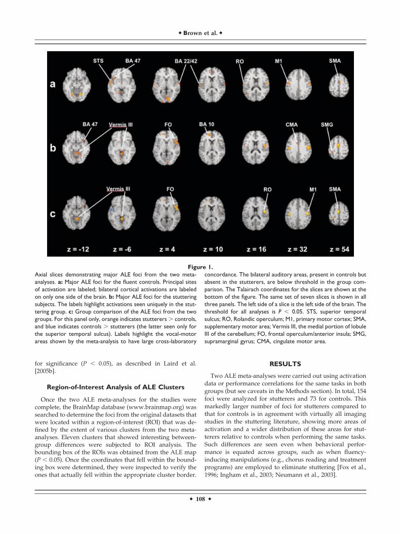
Figure 1.Axial slices demonstrating major ALE foci from the two meta-analyses. a: Major ALE foci for the fluent controls. Principal sitesof activation are labeled; bilateral cortical activations are labeledon only one side of the brain. b: Major ALE foci for the stutteringsubjects. The labels highlight activations seen uniquely in the stut-tering group. c: Group comparison of the ALE foci from the twogroups. For this panel only, orange indicates stutterers � controls,and blue indicates controls � stutterers (the latter seen only forthe superior temporal sulcus). Labels highlight the vocal-motorareas shown by the meta-analysis to have large cross-laboratory
concordance. The bilateral auditory areas, present in controls butabsent in the stutterers, are below threshold in the group com-parison. The Talairach coordinates for the slices are shown at thebottom of the figure. The same set of seven slices is shown in allthree panels. The left side of a slice is the left side of the brain. Thethreshold for all analyses is P � 0.05. STS, superior temporalsulcus; RO, Rolandic operculum; M1, primary motor cortex; SMA,supplementary motor area; Vermis III, the medial portion of lobuleIII of the cerebellum; FO, frontal operculum/anterior insula; SMG,supramarginal gyrus; CMA, cingulate motor area.
� Brown et al. �
� 108 �

Fluent Controls
ALE images for the fluent controls are presented in Figure1a, and the ALE scores and cluster sizes for these locationsare presented in Table II. The analysis shows that most coreareas of the vocal-control system highlighted in the previousmeta-analyses of single-word oral reading are present, eventhough the analyses have no overlap in the literature cov-ered. The main areas include the primary motor cortex,SMA, premotor cortex, Rolandic operculum (Brodmann area[BA] 4/43), left inferior frontal gyrus (BA47), cerebellarhemispheres (principally lobule VI), and bilateral auditoryassociation areas. Primary and secondary visual areas(BA17/18/19) were also seen, reflecting the use of writtentext as a stimulus. No activity was seen in the frontal oper-culum or anterior insula. Finally, the one part of the basalganglia showing a significant ALE score was the inferiorpart of the left globus pallidus (seen in Fig. 1a at slice levelz � �6). This focus is not listed in Table II because it did notmeet our cluster-volume criterion of 100 mm3.
Stutterers
The results with the stutterers performing the same tasksare shown in Figure 1b and Table III, and a color-codedcomparison between the stuttering group and control groupis shown in Figure 1c. From a qualitative standpoint, virtu-ally all areas seen in the ALE analysis with fluent controls
were present in the analysis with stutterers, including theprimary motor cortex, premotor cortex, SMA, Rolandicoperculum, cerebellar hemispheres, visual association cor-tex, and prefrontal cortex (BA10). At a basic level, the sameset of core areas involved in vocal production of read textwas therefore seen in parallel in both meta-analyses. Withinthat core most vocal-motor areas showed larger ALE scoresand cluster sizes in stutterers compared to that in controls,as highlighted in Figure 1c. Such was the case in the SMA,primary motor cortex (BA4/6), right Rolandic operculum,and cerebellar vermis (lobule VI). In fact, the strongest andlargest focus in the entire analysis was seen in the rightprimary motor cortex for the stutterers. It was both largerand stronger than was that for the controls, and larger andstronger than the left-hemisphere coordinates were for theprimary motor cortex for either group.
Next, a series of brain areas not seen in the fluentcontrols was found to have large concordance in the stut-terers, most notably the right frontal operculum/anteriorinsula, left cingulate motor area, cerebellar vermis of lob-ule III, supramarginal gyrus bilaterally, and frontal eyefields (BA8). The most striking of this group was the rightfrontal operculum bordering on the anterior insula(BA45/13), which achieved both a large ALE score and alarge cluster size in stutterers but had no counterpart inthe control subjects. In addition, the absence of activationin the auditory association cortex (BA22/42) bilaterally in
TABLE II. Major ALE foci for the fluent control subjects
Lobe Region x y z ALE (� 103) Size (mm3)
FrontalLeft Primary motor cortex (4/6) �49 �9 32 13.43 2,128
Inferior frontal gyrus (47) �36 19 �6 8.15 408Prefrontal cortex (10) �12 49 12 6.90 248
Right Primary/premotor cortex (4/6) 54 �10 34 11.90 2,312Rolandic operculum (4/43) 56 �8 20 11.80 SCSupplementary motor area (6) 5 �2 57 11.91 664
TemporalLeft Superior temporal sulcus (22/21) �51 �3 �5 9.19 888
Superior temporal gyrus (42) �58 �13 11 8.55 792Right Superior temporal gyrus (22) 62 �8 8 7.90 SC
OccipitalLeft Cuneus (17) �18 �94 1 7.27 488
Lingual gyrus (19) �10 �51 �3 8.13 432Lingual gyrus (18) �4 �75 4 7.19 336Lingual gyrus (19) �24 �57 �4 6.65 104
Right Lingual gyrus (17) 11 �84 5 8.24 752Cerebellum
Left Lobule VI �22 �63 �16 12.33 1,096Right Lobule VI 18 �62 �15 13.48 1,128
Vermis VI 4 �71 �15 7.05 344
The 17 principal ALE clusters derived from the analysis with the control subjects. After each anatomical name in the region column is theBrodmann area (BA) in parentheses. The columns labeled as x, y, and z are the Talairach coordinates for the weighted center of each cluster.The ALE score shown is the true value multiplied by 103. The right column shows the size (in mm3) of each cluster. The two right-hemisphere clusters labeled as SC in the size column (namely, 57, �9, 20 and 62, �8, 8) are derived from the right primary motor cortexcluster at 54, �10, 34, having a cluster size of 2,312 mm3. The Rolandic operculum is listed here in the frontal lobe, although it is listed forthe stuttering subjects in the parietal lobe due to a slight difference in the location of the weighted center of the cluster.
� Stuttered and Fluent Speech Neuroimaging �
� 109 �

the stutterers was notable. This absence can be seen incomparing the stutterers and controls in Figures 1b and1a, respectively; however, this difference did not achievesignificance in the group comparison, as shown in Figure1c. Finally, the weak left globus pallidus activation seenwith controls was not seen with stutterers, nor was anyother part of the basal ganglia seen to be active. Overall,the comparison of the two meta-analyses demonstratesthe presence of a common core of vocal-motor areas forboth groups but with the additional occurrence in stutter-ers of the following: (1) overactivations in these areas; (2)anomalous right-dominant lateralization in these areas;(3) additional areas of activation (motor and nonmotor)not seen in the controls (e.g., frontal operculum and ver-mis III); (4) an absence of auditory activations bilaterally;and (5) an absence of basal ganglia activations.
Region-of-Interest Analysis
Table IV presents an ROI analysis of the significant ALEfoci for stuttering subjects with regard to each of eight
studies included in the meta-analysis. Eleven major clustersshowing significant between-group differences were ana-lyzed. As can be seen at the top of the table, four majorlaboratories contributed to this literature, as represented bycity: San Antonio, Bethesda, Toronto, and Frankfurt. Thecritical finding from this analysis is that most of the corevocal areas, including the primary motor cortex, frontaloperculum, Rolandic operculum, cingulate motor area, andcerebellar vermis III, have contributions from three of fourlab groups. This is also the case for the right-lateralizedmotor activations in the primary motor cortex, frontal oper-culum, and Rolandic operculum. SMA activation was seenby only two groups; it was not scored for Braun et al. [1997]because their SMA activation sat 10 mm lateral to the peakcoordinate in our analysis.
In contrast to this picture, activation in several areas out-side of the core vocalization centers came principally fromone or two groups, perhaps reflecting specific features oftheir design or analysis. As shown in the lower part of TableIV, areas such as the frontal eye fields (BA8), supramarginal
TABLE III. Major ALE foci for the stuttering subjects
Lobe Region x y z ALE (� 103) Size (mm3)
FrontalLeft Supplementary motor area (6) �2 �5 54 16.40 1,664
Primary motor cortex (4) �45 �16 31 15.70 1,016Prefrontal cortex (10) �16 49 9 13.37 1,008Cingulate motor area (24) �6 8 35 9.16 256
Right Primary motor cortex (4) 48 �12 32 18.18 4,112Frontal operculum/insula (45/13) 47 13 2 14.14 1,904Inferior frontal gyrus (47) 35 16 �15 11.88 384Superior frontal gyrus (8) 17 24 47 10.37 616Premotor cortex (6) 35 13 46 8.62 432Anterior cingulate (32/24) 0 21 �8 8.21 184
ParietalLeft Supramarginal gyrus (40) �31 �40 55 9.21 216
Rolandic operculum (43/4) �54 �7 15 8.50 192Right Rolandic operculum (43/4) 44 �6 16 17.40 SC
Supramarginal gyrus (40) 36 �38 49 10.52 296Occipital
Left Lingual gyrus (18) �7 �75 �7 9.65 240Right Fusiform gyrus (19) 20 �55 �14 9.16 288
Lingual gyrus (17) 5 �86 5 8.18 200Temporal
Left Superior temporal gyrus (22) �56 �24 1 8.84 208Cerebellum
Left Lobule VI �37 �62 �25 12.54 912Lobule VI �24 �64 �17 11.98 712
Right Vermis III/IV 3 �46 �11 14.45 2,304Lobule VI 12 �68 �17 13.91 848Vermis VI 0 �70 �25 10.37 440
The 23 principal ALE clusters derived from the analysis with the stuttering subjects. After each anatomical name in the region column isthe Brodmann area in parentheses. Columns labeled as x, y, and z are the Talairach coordinates for the weighted center of each cluster. TheALE score shown is the true value multiplied by 103. The right column shows the size (in mm3) of each cluster. The right parietal clusterlabeled as SC in the size column (namely, 44, �6, 16) is derived from the right primary motor cortex cluster at 48, �12, 32, having a clustersize of 4,112 mm3. The Rolandic operculum is listed here in the parietal lobe, although it is listed for the control subjects in the frontal lobedue to a slight difference in the location of the weighted center of the cluster.
� Brown et al. �
� 110 �
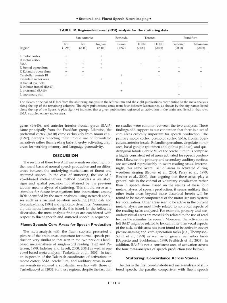
gyrus (BA40), and anterior inferior frontal gyrus (BA47)came principally from the Frankfurt group. Likewise, theprefrontal cortex (BA10) came exclusively from Braun et al.[1997], perhaps reflecting their unique use of formulatednarratives rather than reading tasks, thereby activating brainareas for working memory and language generativity.
DISCUSSION
The results of these two ALE meta-analyses shed light onthe neural basis of normal speech production and on differ-ences between the underlying mechanisms of fluent andstuttered speech. In the case of stuttering, the use of avoxel-based meta-analysis method provides a statisticalrigor and spatial precision not attained by the previoustabular meta-analyses of stuttering. This should serve as astimulus for future investigations into interactions amongROIs identified by the meta-analyses, using network analy-ses such as structural equation modeling [McIntosh andGonzalez-Lima, 1994] and replicator dynamics [Neumann etal., this issue; Lancaster et al., this issue]. In the followingdiscussion, the meta-analysis findings are considered withrespect to fluent speech and stuttered speech in sequence.
Fluent Speech: Core Areas for Speech Production
The meta-analysis with the fluent subjects presented apicture of the brain areas important for normal speech pro-duction very similar to that seen in the two previous label-based meta-analyses of single-word reading [Fiez and Pe-tersen, 1998; Indefrey and Levelt, 2000, 2004] as well as onevoxel-based meta-analysis [Turkeltaub et al., 2002]. In fact,an inspection of the Talairach coordinates of activations inmotor cortex, SMA, cerebellum, and auditory areas in ourmeta-analysis showed a substantial overlap with those ofTurkeltaub et al. [2002] for these regions, despite the fact that
no studies were common between the two analyses. Thesefindings add support to our contention that there is a set ofcore areas critically important for speech production. Theprimary motor cortex, premotor cortex, SMA, frontal oper-culum, anterior insula, Rolandic operculum, cingulate motorarea, basal ganglia (putamen and globus pallidus), and qua-drangular lobule (lobule VI) of the cerebellum thus comprisea highly consistent set of areas activated for speech produc-tion. Likewise, the primary and secondary auditory corticesare activated reproducibly in overt reading tasks. Interest-ingly, this same overall set of areas is activated duringwordless singing [Brown et al., 2004; Perry et al., 1999;Riecker et al., 2000], thus arguing that these areas play ageneral role in the control of voluntary vocalization ratherthan in speech alone. Based on the results of these fourmeta-analyses of speech production, it seems unlikely thatother brain areas beyond those described herein will befound to be major components of the motor-sensory systemfor vocalization. Other areas seen to be active in the currentmeta-analysis are most likely related to nonvocal aspects ofthe reading tasks analyzed. For example, primary and sec-ondary visual areas are most likely related to the use of readtext as the stimulus for speech. Moreover, the activation inleft BA47 might be related to lexical rather than vocal aspectsof the task, as this area has been found to be active in covertpicture-naming and verb-generation tasks [e.g., Thompson-Schill et al., 1999] as well as in general semantics tasks[Dapretto and Bookheimer, 1999; Preibisch et al., 2003]. Inaddition, BA47 is not a consistent area of activation acrossthe four meta-analyses of speech production (see below).
Stuttering: Concordance Across Studies
As this is the first coordinate-based meta-analysis of stut-tered speech, the parallel comparison with fluent speech
TABLE IV. Region-of-interest (ROI) analysis for the stuttering data
Region
San Antonio Bethesda Toronto Frankfurt
Fox(1996)
Fox(2000)
Ingham(2004)
Braun(1997)
De Nil(2000)
De Nil(2003)
Prebeisch(2003)
Neumann(2003)
L motor cortex � � �R motor cortex � � � � � �SMA � � � �R frontal operculum � � � � � �R Rolandic operculum � � � � �Cerebellar vermis III � � � �Cingulate motor area � �R frontal eye field � � �R inferior frontal (BA47) � �L prefrontal (BA10) �L supramarginal �
The eleven principal ALE foci from the stuttering analysis in the left column and the eight publications contributing to the meta-analysisalong the top of the remaining columns. The eight publications come from four different laboratories, as shown by the city names listedalong the top of the figure. A plus sign (�) indicates that a given publication registered an activation in the brain area listed in that row.SMA, supplementary motor area.
� Stuttered and Fluent Speech Neuroimaging �
� 111 �
provides an unprecedented opportunity to examine the pa-thology of stuttering. The stuttering neuroimaging literatureis relatively small, yet a high degree of concordance acrossstudies emerged when meta-analysis was carried out,thereby paving the way for establishing neural signatures ofstuttering. In addition, these findings show more consis-tency than the earlier label-based analyses do, while at thesame time validate the earlier observations of abnormalactivation in the right frontal operculum/anterior insula andcerebellum coupled with deactivation in right auditory as-sociation areas [Ingham, 2001b, 2004]. The following exam-ines a series of effects related to the neurobiology of stutter-ing. The most general effect is the overall increase in thenumber of activated brain areas in stutterers compared tothat in fluent subjects carrying out the same tasks, and themore widespread distribution of these activated areas in thebrain. Although such an observation does not lend itselfreadily to interpretation, it does show that the meta-analysisprovides an accurate picture of what virtually all the imag-ing studies have demonstrated individually.
The series of core areas comprising the vocal system in thefluent controls was also seen in the meta-analysis of stutter-ing, including the primary motor cortex, premotor cortex,SMA, Rolandic operculum, and cerebellum (hemispheresand vermis of lobule VI). Stuttered speech therefore de-pends, at least to a large degree, on the same series of coreareas important for speech in general. This is in contrast toa model in which stuttering might occur through somealternative vocalization route (e.g., cingulate vocalizationareas). But beyond this general level of commonality, threepoints of difference are observed. First, compared to theresults with the fluent controls, there is an increase in acti-vation in lateral vocal-motor areas (especially in the righthemisphere) and decrease in activation in auditory areas(bilaterally). Second, this is accompanied by a laterality shiftthat brings the balance of activity toward the right hemi-sphere, i.e., through a reduction of activity in left-hemi-sphere areas (primary motor cortex, auditory cortex, andRolandic operculum) and an increase in activity in right-hemisphere areas (frontal operculum and Rolandic opercu-lum). This general pattern of brain activity emerges as arightward shift in cerebral activation. This is consistent withthe findings of numerous preimaging studies of develop-mental stuttering [see Moore, 1993]. Finally, there is prom-inent overactivity in three medial motor structures: SMA,cingulate motor area, and cerebellar vermis (both lobules VIand III).
An important objective of a meta-analysis is not only toprovide a picture of concordance across a corpus of studiesbut a sense of which studies find activations in which areas,and to attempt to correlate them with task-specific effects[see Laird et al., 2005a]. The critical finding from the ROIanalysis was that most core vocal areas, including the pri-mary motor cortex, frontal operculum, Rolandic operculum,cingulate motor area, and cerebellar vermis, had contribu-tions from three of four lab groups. Importantly, this wasalso the case for the right-lateralized motor activations in the
primary motor cortex, frontal operculum, and Rolandicoperculum. SMA activation was seen by only two groups; itwas not scored for Braun et al. [1997] because their SMAactivation sat 10 mm lateral to the peak coordinate in ouranalysis. In contrast to this picture, activation in severalareas outside of the core vocalization centers came princi-pally from one or two groups, including the prefrontal cor-tex (BA10), the frontal eye fields (BA8), supramarginal gyrus(BA40), and anterior inferior frontal gyrus (BA47). Activityin right BA47, in particular, was argued by Preibisch et al.[2003] to be a negative correlate of stuttering severity. Com-parable activity in this area was seen by them in both avisual semantics task and a reading task, therefore arguingthat the role of right BA47 was tied more closely in withsemantics function than with vocalization (see above). Inany case, this area did not show strong concordance acrossthe studies in the meta-analysis.
The overall picture from the ROI analysis was a robustconcordance across laboratories in the core motor areas andlesser concordance outside of these areas. This is perhaps themost desirable outcome that the meta-analysis could haveprovided for the stuttering field. This result was not influ-enced by the presence or absence of stuttering in a particularstudy. The effect was seen in studies that both elicited stut-tering (the San Antonio and Bethesda studies) and those thatdid not (the Toronto and Frankfurt studies). The meta-anal-ysis was thus more successful at providing a general pictureof a stutterer phenotype than at pinpointing a profile ofactivity uniquely associated with stuttered speech. Clearly, amuch larger corpus of studies is needed to dissect sucheffects.
Neural Signatures of Stuttering
Whereas several of the stuttering effects described aboveinvolved relative changes in activity or laterality betweenstutterers and fluent controls, the meta-analyses highlightedthree neural signatures that seemed more or less specific tothe stuttering group: (1) overactivation in the right frontaloperculum/anterior insula; (2) absence of activation in au-ditory areas bilaterally; and (3) overactivation in the vermalregion of lobule III of the cerebellum. These three signatureswill now be described in more detail.
Activity in the right frontal operculum/anterior insula(BA45/13) stood out as being unique in our analysis in tworespects. First, unlike other lateral motor areas such as theprimary motor cortex and Rolandic operculum, activity wasfound exclusively in the right hemisphere. Second, againunlike the primary motor cortex and Rolandic operculum,activity was found uniquely in the stutterers. In the datasetof Fox et al. [1996], activation in the frontal operculum/insula was much higher during stutter-filled solo readingthan during stutter-free chorus reading. Moreover, in thetreatment study of Neumann et al. [2003], activation waspresent before treatment but was eliminated after treatment.Activity in this right-hemisphere region during readingtasks therefore may be a strong marker of stutterer status.
� Brown et al. �
� 112 �

Although the frontal operculum of the left hemisphere haswell-established functional linkages with speech [Acker-mann and Riecker, 2004] and language processes [Friedericiet al., 2000], and even with manual imitation [Iacoboni et al.,1999], the functional role of the right frontal operculum is farmore elusive. Results from several lines of research suggestthat one common link may be the processing of vocal fun-damental frequency during both production and perception.Activity in the right frontal operculum and anterior insulaare prominent during wordless singing tasks [Perry et al.,1999; Riecker et al., 2000], including vocal imitation of pitchsequences [Brown et al., 2004]. Regarding speech, activity inthe right frontal operculum and anterior insula is associatedmost closely with prosody tasks. For example, Hesling et al.[2004] found right-sided BA44 activity when they contrasted“expressive” presentation of a 30-s reading passage with a“flat” presentation in which the fundamental frequency con-tours were reduced greatly. Likewise, Meyer et al. [2004]demonstrated activation in the right frontal operculum(BA44) when subjects listened to degraded speech stimulithat preserved the intonational (melodic) properties but notsegmental properties of speech, as contrasted to normalspeech. Wildgruber et al. [2004] found bilateral activations inthe frontal operculum on discrimination tasks for both af-fective and linguistic prosody; their right-hemisphere acti-vations were located in BA45/46. Much neuropsychologicalevidence suggests that the right hemisphere may be domi-nant for production and perception of affective speech pros-ody [reviewed in Wymer et al., 2002]. Production and per-ception of vocal fundamental frequency therefore seemsmediated, at least in part, by the right frontal operculum/anterior insula. Abnormal activity in this region might con-tribute to aberrant phonological processing in stuttering.Another point relevant to aberrant phonology is the stronginhibition of auditory areas during oral reading (see below).In classical models of speech production, the frontal oper-culum is the recipient of projection fibers originating in theposterior part of the superior temporal gyrus that travelthrough the superior longitudinal (arcuate) fasciculus to thefrontal lobe [Catani et al., 2002]. Overactivation of the rightfrontal operculum coupled with inhibition of right (and left)auditory areas therefore might represent a disrupted func-tional connectivity between auditory and motor areas dur-ing speech planning in stutterers. Another reason for high-lighting the importance of frontal operculum/anteriorinsula to stuttering is the evidence that it is also abnormallyactive (bilaterally) in Tourette’s syndrome and that tic fre-quency may correlate with activity in this and other speech-related regions [Stern et al., 2000]. Tourette’s syndrome hasbeen related frequently to developmental stuttering becauseof its similar developmental pattern, responsiveness to re-lated stimuli, and comorbidity with stuttering [Comings andComings, 1994; Abwender et al., 1998].
The second important signature of stuttering is the reduc-tion in activity in auditory areas during vocalization tasks,especially because all previous meta-analyses of vocal pro-duction, as well as our own meta-analysis with the control
group, showed prominent and generally bilateral activationsduring overt speech. The inhibitory effect in stutterers wasdifficult to assess with the ALE meta-analysis, because it didnot include deactivations or negative correlations [Ingham,2001b]. It is therefore important to consider the literaturesuggestions of a fundamental abnormality in auditory areasduring overt reading tasks, as compared to fluent controls.Fox et al. [1996] was the first study to show that stutterershave marked reductions in superior temporal lobe activa-tions, and even deactivations, during reading tasks. Thiswas followed up by data showing negative correlationsbetween stuttering rate and auditory activations in male[Fox et al., 2000] and female [Ingham et al., 2004] cohorts.Braun et al. [1997] showed not only bilateral deactivations inauditory areas in stutterers during dysfluency-inducingtasks but strong negative correlations between dysfluencyand activation in right hemisphere auditory areas. In a studynot included in the meta-analysis because it did not reportcoordinates, Van Borsel et al. [2003] found an absence ofactivations bilaterally in auditory areas in stutterers on anovert speech task for which controls demonstrated strongactivations bilaterally. In De Nil et al. [2000], for the contrastof oral reading minus silent reading, nonstutterers showedonly left auditory activations (BA22) whereas stutterersshowed only right auditory activations, and the group com-parison of nonstutterers minus stutterers had significantsignal in left auditory association cortex. For the functionalmagnetic resonance imaging (fMRI) studies of Neumann etal. [2003] and Preibisch et al. [2003], only group comparisonswere reported and thus it is more difficult to assess task-dependent auditory effects within groups. Neumann et al.[2003] reported that bilateral BA22 was more active in peo-ple who stuttered less severely than it was in those withmore severe stuttering (based on clinical assessment), dem-onstrating that auditory activations seem to correlate nega-tively with stuttering severity. Stager et al. [2003] showedthat activity in auditory areas bilaterally was greater duringfluency-inducing than during dysfluency-inducing condi-tions, a result that parallels findings by Fox et al. [1996] onchorus reading. The only study to provide no indication ofan auditory effect in stutterers is the treatment study of DeNil et al. [2003]. The nonstuttering control subjects in thatstudy showed neither primary motor nor auditory activa-tions during overt reading, and so the data with the stutter-ers might be equally difficult to interpret. Looking at thegroup comparison in the meta-analysis (Fig. 1c), the areathat showed the largest inter-group difference (at z of �6) isa part of the superior temporal sulcus situated just anteriorto those areas found to have voice-selective auditory repre-sentations [Belin et al., 2000, 2002]. In sum, the publishedliterature supports a robust auditory inhibitory effect instutterers, which is consistent with the meta-analysis results.The inhibition of auditory activity seems amplified by theamount of stuttering during a reading task and by clinicalassessment of stuttering severity, and seems ameliorated byfluency-inducing manipulations and perhaps treatment.This might be one of the most distinctive markers of stut-
� Stuttered and Fluent Speech Neuroimaging �
� 113 �
tering in the neuroimaging literature [Ingham, 2001b]. Itremains an open question as to whether such auditory inhi-bitions occur only during self-produced speech or duringauditory perception in general.
Third, activity in the vermal part of lobule III stood out asa unique activation in the stuttering group. Cerebellar acti-vations during overt vocalization generally occur in lobuleVI (the quadrangular lobule) and the associated vermis[Brown et al., 2004; Perry et al., 1999; Turkeltaub et al., 2002],as was seen in our meta-analysis with the control subjects(see Table II). For Fiez and Petersen [1998], midline cerebel-lar activity was seen slightly more anteriorly, in lobule V. Incontrast to this, activity in lobule III was not generally seenwith vocalization tasks in normal subjects; however, itseemed to be found with chronic developmental stutterers.In the dataset of Fox et al. [1996], the principal midlinecerebellar activity for the stutterers was in vermis VI duringstutter-free chorus reading; activity in vermis III was onlyseen during stutter-filled solo reading. Likewise, Braun et al.[1997] observed activity in vermis III during their stutter-filled dysfluency tasks but not during their stutter-free flu-ency tasks; no activity was detected in their control subjects.De Nil et al. [2003] observed activity in vermis III in theirstutterer subjects before a treatment program but not at anypoint after treatment. Control subjects showed activity inlobule VI but not lobule III. These results overall suggest thatactivity in vermis III might not only be a marker for stuttererstatus but also one for stuttered speech as well. One pointthat raises doubts about the significance of vermis III foractual stuttering is that it did not show positive correlationswith stutter rate in either the male or female cohorts in theSan Antonio datasets [Fox et al., 2000; Ingham et al., 2004].Correlations with the cerebellar midline were only seen withvermis VI. Vermis VI and the hemispheric portion of lobuleVI have known somatotopic representations for the lips andtongue [Grodd et al., 2001]; therefore, their activation duringoral reading tasks is readily explainable in terms of a motormap of the cerebellum. In contrast, vermis III does not haveany functional properties attributed to it in somatotopystudies [Grodd et al., 2001]; therefore, its unique activationin stutterers during reading tasks is intriguing and in needof further investigation.
A comment about the basal ganglia is in order becausethis set of structures has been implicated in stuttering formany decades. Alm [2004], in reviewing a large literatureabout stuttering and the basal ganglia, proposed a model inwhich the core dysfunction of stuttering was suggested to bean “impaired ability of the basal ganglia to produce timingcues for the initiation” of speech motor activity (p. 325).Unfortunately, this proposal provided no predictions aboutwhether particular nuclei/circuits of the basal gangliawould be over- or underactivated during stuttering. Themeta-analysis data did not provide strong indications eitherfavoring or opposing this model. Essentially, a weak globuspallidus activation seen with the controls was eliminated inthe stutterers. This absence of basal ganglia effects is sur-prising given the established role of the left putamen in
vocalization, both for speech [Klein et al., 1994; Turkeltaub etal., 2002; Wildgruber et al., 2001] and for song [Brown et al.,2004]. Perhaps the most basal ganglia-specific effect seen inthe meta-analysis was the overactivation of the SMA (as wellas cingulate motor area) in stutterers. The SMA is associatedtraditionally with internal generation of motor activity andis often activated during mental imagery tasks, includingimagery of stuttered speech [Ingham et al., 2000]. An obser-vation of SMA overactivation must be seen in light of thereading tasks carried out in the studies for the meta-analysis,which were very much externally cued (i.e., by the text to beread). The spontaneous narrative task in Braun et al. [1997]was perhaps the closest thing to an internally-cued speechtask in the meta-analysis. They did in fact observe SMAactivations (10 mm lateral to the ALE focus for the SMA),although at a reduced level in stutterers compared to that incontrols. Although the basal ganglia may certainly be play-ing a contributing role in stuttering, this role is in need ofelucidation in future studies.
Efference Copy: A Unifying Hypothesis
As stated previously, the three most salient characteristicsof stuttering to emerge from this meta-analysis were over-activation in the right frontal operculum/anterior insula,absence of activation in auditory areas bilaterally, and over-activation in the vermal region of lobule III of the cerebel-lum. There are key questions to be addressed. Why are twoof the abnormalities hyperactivity, whereas the third is un-deractivity? Are the three phenomena independent orlinked? A tentative answer to both of these questions can beprovided by invoking the phenomenon of efference copy, asfollows.
Efference copy can be defined as a feed-forward projec-tion of a motor plan, at the movement of movement-planinitiation onto the sensory system(s), in which perceptualfeedback is anticipated to occur as a consequence of themovement. Efference copy was proposed initially in thecontext of perceptual constancy [von Holst and Mittelstaedt,1950], i.e., that the visual scene remains continuous duringeye movements. It has also been cited in explanation of thewell-known observation that we cannot tickle ourselves[Blakemore et al., 1999; Weiskrantz et al., 1971]. The signalprojected to the perceptual region receiving the efferencecopy is inhibitory, as the net effect is an attenuation of theperceptual response. For example, in the somatosensorysystem Leube et al. [2003] showed that “predictions gener-ated in motor areas attenuate sensory areas.” In the speechsystem, Houde et al. [2002], reported that, “during speechproduction, the auditory cortex (1) attenuates its sensitivityand (2) modulates its activity as a function of the expectedacoustic feedback” (p. 1125). Others have reported similareffects using other imaging modalities [Curio et al., 2000;Numminen and Curio, 1999]. Such findings have led to theconclusion that efference copy applies to motor control ingeneral [Haruno et al., 2001]. Max et al. [2004] have alsoconsidered its role in stuttering, albeit not as described be-low.
� Brown et al. �
� 114 �

In stuttering, the most characteristic performance abnor-mality is the failure to properly initiate the speech-motorplan. This is not likely a defect of motor programming perse, as developmental stutterers do not exhibit dysarthria,oral dyspraxia, or other signs of an incorrect mental modelof the desired movement, nor is this an abnormality of themotor execution system (motor cortex, basal ganglia, lowermotor neurons), as there is no oral weakness, slowness,spasticity, tremor, or hypophonia. The problem is limited tosuccessful initiation of the motor program. Importantly,stuttering is usually exhibited as a repetition of the initialsound of a word. In the context of efference copy, this wouldsuggest that the perceptual prediction (of speech sounds) isbeing delivered repeatedly to the auditory system as aninhibitory signal that will attenuate the effects of any suc-cessful utterances. Furthermore, if stuttering is sufficientlysevere, inhibition of auditory areas below baseline shouldoccur and has been reported [Fox et al., 1996]. Efference copythus can readily explain the noted lack of speech-relatedauditory activations in stutterers.
The motor-system overactivity observed in stuttering hastwo potential explanations. First, repeated initiation of thespeech-motor plan likely repeatedly activates some compo-nents of the speech motor system, resulting in overactiva-tion. Second, there is now considerable evidence that in-creased skill is associated with a concomitant decrease inactivation [Jansma et al., 2001; Just et al., 1996; Raichle et al.,1994]. The converse is also true. Disease conditions thatresult in less competence in task performance are associatedwith regional over-activation [Bookheimer et al. 2001; Habib,2000]. In stuttering, it is likely that both effects come intoplay. The right laterality of the motor region hyperactivityalso deserves comment. Studies from two labs have sug-gested that developmental stuttering might be associatedwith a structural lesion in the left hemisphere [Foundas etal., 2001, 2003; Sommer et al., 2002]. In the presence of aleft-hemisphere dysfunction, the right hemisphere assumesleft-hemisphere tasks at which it is intrinsically less compe-tent [Gandour et al., 2003, 2004], resulting in overactivation.
What, then, accounts for cerebellar overactivation? A fun-damental aspect of the efference copy concept is that despiteattenuation of the received sensory signal (in this instance,speech), there is self-monitoring that routinely compares theexpected and the actual. The cerebellum has been implicatedin the assessment of match between the predicted action andthe actual sensory consequences [Blakemore et al., 2001].The cerebellum has also been demonstrated to be involvedin auditory discrimination [Petacchi et al., 2005]. Conse-quently, the repeated observation of cerebellar overactiva-tion in stuttering may be associated not only with the motoroveractivity (as part of the motor system), but a response toan action–consequence mismatch.
The preceding stuttering system model linking motoroveractivity, auditory underactivity, and cerebellar overac-tivity lends itself to a network-based analysis. This could beaccomplished either by structural equation modeling ap-plied to raw data [Buchel et al., 1999; McIntosh and Gonza-
lez-Lima, 1994] or with one of the newly developed net-work-modeling strategies intended for meta-analysis[Neumann et al., this issue; Lancaster et al., 2005]. Both offerpromising strategies for extending the present meta-analy-sis. The efference copy mechanism would predict an inverserelationship between right anterior insula and left auditorycortex. It would also predict a direct relationship betweenthe cerebellar activity and the difference between right mo-tor and left auditory cortex (i.e., if the cerebellar activity isthe “discrepancy signal”). These effects should be presentboth on a study-by-study and on a trial-by-trial basis. Theseare both very testable predictions. In fact, the replicatordynamics [Neumann et al., 2005] and fractional similaritynetwork analysis [Lancaster et al., 2005] methods can beapplied to trial-by-trial data (i.e., to raw data) as well as tometa-analysis data.
REFERENCES
Abwender DA, Trinidad KS, Jones KR, Como PG, Hymes E, KurlanR (1998): Features resembling Tourette’s syndrome in develop-mental stutterers. Brain Lang 62:455–464.
Ackermann H, Riecker A (2004): The contribution of the insula tomotor aspects of speech production: a review and a hypothesis.Brain Lang 89:320–328.
Alm PA (2004): Stuttering and the basal ganglia circuits: a criticalreview of possible relations. J Commun Disord 37:325–369.
Belin P, Zatorre RJ, Lafaille P, Ahad P, Pike B (2000): Voice-selectiveareas in human auditory cortex. Nature 403:309–312.
Belin P, Zatorre RJ, Ahad P (2002): Human temporal-lobe responseto vocal sounds. Brain Res Cogn Brain Res 13:17–26.
Blakemore SJ, Frith CD, Wolpert DM (2001): The cerebellum isinvolved in predicting the sensory consequences of action. Neu-roreport 12:1879–1884.
Bloodstein O (1995): A handbook on stuttering. San Diego: SingularPublishing Group. 596 p.
Bookheimer SY, Sojwas MH, Cohen MS, Saunders AM, Pericak-Vance MA, Mazziotta JC, Small GW (2000): Patterns of brainactivation in people at risk for Alzheimer’s disease. N Engl J Med343:450–456.
Braun AR, Varga M, Stager S, Schulz G, Selbie S, Maisog JM, CarsonRE, Ludlow CL (1997): Altered patterns of cerebral activity dur-ing speech and language production in developmental stutter-ing: An H2
15O positron emission tomography study. Brain 120:761–784.
Brett M (1999): The MNI brain and the Talairach atlas, CambridgeImagers. Online at http://www.mrc-cbu.cam.ac.uk/Imaging/mnispace.html.
Brown S, Martinez MJ, Hodges DA, Fox PT, Parsons LM (2004): Thesong system of the human brain. Brain Res Cogn Brain Res20:363–375.
Buchel C, Coull JT, Friston KJ (1999): The predictive value ofchanges in effective connectivity for human learning. Science283:1538–1541.
Catani M, Howard RJ, Pajevic S, Jones DK (2002): Virtual in vivointeractive dissection of white matter fasciculi in the humanbrain. Neuroimage 17:77–94.
Chien JM, Fissell K, Jacobs S, Fiez JA (2002): Functional heteroge-neity within Broca’s area during verbal working memory.Physiol Behav 77:635–639.
� Stuttered and Fluent Speech Neuroimaging �
� 115 �
Comings DE, Comings BG (1994): TS, learning, and speech prob-lems. J Am Acad Child Adolesc Psychiatry 33:429–430.
Curio G, Neuloh G, Numminen J, Jousmaki V, Hari R (2000): Speak-ing modifies voice-evoked activity in the human auditory cortex.Hum Brain Mapp 9:183–191.
Dapretto M, Bookheimer SY (1999): Form and content: dissociatingsyntax and semantics in sentence comprehension. Neuron 24:427–432.
De Nil LF, Knoll RM, Kapur S, Houle S (2000): A positron emissiontomography study of silent and oral word reading in stutteringand nonstuttering adults. J Speech Lang Hear Res 43:1038–1053.
De Nil LF, Knoll RM, Houle S (2001): Functional neuroimaging ofcerebellar activation during single word reading and verb gen-eration in stuttering and nonstuttering adults. Neurosci Lett302:77–80.
De Nil LF, Knoll RM, Lafaille SJ, Houle S (2003): A positron emis-sion tomography study of short- and long-term treatment effectson functional brain activation in adults who stutter. J FluencyDisord 28:357–380.
Fiez JA, Petersen SE (1998): Neuroimaging studies of word reading.Proc Natl Acad Sci USA 95:914–921.
Foundas AL, Bollich AM, Corey DM, Hurley M, Heilman KM(2001): Anomalous anatomy of speech-language areas in adultswith persistent developmental stuttering. Neurology 57:207–215.
Foundas AL, Corey DM, Angeles V, Bollich AM, Crabtree-HartmanE, Heilman KM (2003): Atypical cerebral laterality in adults withpersistent developmental stuttering. Neurology 61:1378–1385.
Fox PT (2003): Brain imaging in stuttering: where next? J FluencyDisord 28:265–272.
Fox PT, Ingham RJ, Ingham JC, Hirsch TB, Downs JH, Martin C,Jerabek P, Glass T, Lancaster JL (1996): A PET study of the neuralsystems of stuttering. Nature 382:158–162.
Fox PT, Ingham RJ, Ingham JC, Zamarripa F, Xiong JH, Lancaster JL(2000): Brain correlates of stuttering and syllable production: aPET performance-correlation analysis. Brain 123:1985–2004.
Friederici AD, Meyer M, von Cramon DY (2000): Auditory languagecomprehension: an event-related fMRI study on the processingof syntactic and lexical information. Brain Lang 74:289–300.
Gandour J, Dzemidzic M, Wong D, Lowe M, Tong Y, Hsieh L,Satthamnuwong N, Lurito J (2003): Temporal integration ofspeech prosody is shaped by language experience: an fMRIstudy. Brain Lang 84:318–336.
Gandour J, Tong Y, Wong D, Talavage T, Dzemidzic M, Xu Y, Li X,Lowe M (2004): Hemispheric roles in the perception of speechprosody. Neuroimage 23:344–357.
Genovese CR, Lazar NA, Nichols TE (2002): Thresholding of statis-tical maps in functional neuroimaging using the false discoveryrate. Neuroimage 15:870–878.
Grodd W, Hulsmann E, Lotze M, Wildgruber D, Erb M (2001):Sensorimotor mapping of the human cerebellum: fMRI evidenceof somatotopic organization. Hum Brain Mapp 13:55–73.
Habib M (2000): The neurological basis of developmental dyslexia:an overview and working hypothesis. Brain 123:2373–2399.
Haruno M, Wolpert DM, Kawato M (2001): Mosaic model for sen-sorimotor learning and control. Neural Comput 13:2201–2220.
Hesling I, Clement S, Bordessoules M, Allard M (2004): Cerebralmechanisms of prosodic integration: evidence from connectedspeech. Neuroimage 24:937–947.
Houde JF, Nagarajan SS, Sekihara K, Merzenich MM (2002): Mod-ulation of the auditory cortex during speech: an MEG study. J.Cog Neurosci 14:1125–1138.
Iacoboni M, Woods RP, Brass M, Bekkering H, Mazziotta JC, Riz-zolatti G (1999): Cortical mechanisms of human imitation. Sci-ence 286:2526–2528.
Indefrey P, Levelt WJ (2000): The neural correlates of languageproduction. In: Gazzaniga MS, editor. The cognitive neuro-sciences (2nd ed.). Cambridge, MA: MIT Press. p 845–865.
Indefrey P, Levelt WJ (2004): The spatial and temporal signatures ofword production components. Cognition 92:101–144.
Ingham RJ (1984): Stuttering and behavior therapy: current statusand empirical foundations. San Diego: College-Hill Press. 486 p.
Ingham RJ (2001a): Stuttering. In: Blakemore C, Jennett S, editors.The Oxford companion to the body. Oxford: Oxford UniversityPress. p 659–660.
Ingham RJ (2001b): Brain imaging studies of developmental stutter-ing. J Commun Disord 34:493–516.
Ingham RJ (2004): Emerging controversies, findings, and directionsin neuroimaging and developmental stuttering: on avoiding pe-tard hoisting in Athens, Georgia. In: Bothe AK, editor. Evidence-based treatment of stuttering: empirical issues and clinical im-plications. Mahwah, NJ: Lawrence Erlbaum Associates. p 27–63.
Ingham RJ, Fox PT, Ingham JC, Xiong J, Zamarripa F, Hardies LJ,Lancaster JL (2004): Brain correlates of stuttering and syllableproduction: gender comparison and replication. J Speech LangHear Res 47:321–341.
Ingham RJ, Fox PT, Ingham JC, Zamarripa F (2000): Is overt stut-tered speech a prerequisite for the neural activations associatedwith chronic developmental stuttering? Brain Lang 75:163–174.
Ingham RJ, Ingham JC, Finn P, Fox PT (2003): Towards a functionalneural systems model of developmental stuttering. J FluencyDisord 28:297–317.
Jansma JM, Ramsey NF, Slagter HA, Kahn RS (2001): Functionalanatomical correlates of controlled and automatic processing. JCog Neurosci 13:730–743.
Just MA, Carpenter PA, Keller TA, Eddy WF, Thulborn KR (1996):Brain activation modulated by sentence comprehension. Science274:114–116.
Kent RD (2000): Research on speech motor control and its disorders:a review and prospective. J Commun Disord 33:391–428.
Klein D, Zatorre RJ, Milner B, Meyer E, Evans AC (1994): Leftputaminal activation when speaking a second language: evi-dence from PET. Neuroreport 21:2295–2297.
Kochunov P, Lancaster JL, Thompson P, Toga AW, Brewer P, Har-dies J, Fox PT (2002): An optimized individual target brain in theTalairach coordinate system. Neuroimage 17:922–927.
Laird AR, McMillan KM, Lancaster JL, Kochunov P, Turkeltaub PE,Pardo JV, Fox PT (2005a): A comparison of label-based meta-analysis and activation likelihood estimation in the Stroop task.Human Brain Mapp 25:6–21.
Laird AR, Fox PM, Price CJ, Glahn DC, Uecker AM, Lancaster JL,Turkeltaub PE, Kochunov P, Fox PT (2005b): ALE meta-analysis:controlling the false discovery rate and performing statisticalcontrasts. Hum Brain Mapp 25:155–164.
Lancaster JL, Laird AR, Fox PM, Glahn DE, Fox PT (2005): Auto-mated analysis of meta-analysis networks. Hum Brain Mapp25:xxx–yyy.
Leube DT, Knoblich G, Erb M, Grodd W, Bartels, Kircher TT (2003):The neural correlates of perceiving one’s own movement. Neu-roimage 20:2084–2090.
Max L, Guenther FH, Gracco VL, Ghash SS, Wallace ME (2004):Unstable or insufficiently activated internal models and feed-back biased motor control as sources of dysfluency: A theoreticalmodel of stuttering. CICSD 31:105–122.
� Brown et al. �
� 116 �

Meyer M, Steinhauer K, Alter K, Friederici AD, von Cramon DY(2004): Brain activity varies with modulation of dynamic pitchvariance in sentence melody. Brain Lang 89:277–289.
McIntosh AR, Gonzalez-Lima F (1994): Structural equation model-ing and its application to network analysis in functional brainimaging. Hum Brain Mapp 2:2–22.
Moore WH (1993): Hemispheric processing research. Past, present,and future. In: Boberg E, editor. Neuropsychology of stuttering.Edmonton: The University of Alberta Press. p 39–72.
Neumann J, Lohmann G, Derrfuss J, von Cramon DY (2005): Themeta-analysis of functional imaging data using replicator dy-namics. Hum Brain Mapp 25:165–173.
Neumann K, Euler HA, von Gudenberg AW, Giraud AL, Lanfer-mann H, Gall V, Preibisch C (2003): The nature and treatment ofstuttering as revealed by fMRI: a within- and between-groupcomparison. J Fluency Disord 28:381–410.
Numminen J, Curio G (1999): Differential effects of overt, covert andreplayed speech on vowel-evoked responses of the human au-ditory cortex. Neurosci Lett 272:29–32.
Petacchi A, Laird AR, Bower J (2005): The cerebellum and auditoryfunction: An ALE meta-analysis of functional neuroimagingstudies. Hum Brain Mapp 25:118–128.
Perry DW, Zatorre RJ, Petrides M, Alivisatos B, Meyer E, Evans AC(1999): Localization of cerebral activity during simple singing.Neuroreport 10:3979–3984.
Preibisch C, Neumann K, Raab P, Euler HA, von Gudenberg AW, Lan-fermann H, Giraud AL (2003): Evidence for compensation for stutter-ing by the right frontal operculum. Neuroimage 20:1356–1364.
Raichle ME, Fiez JA, Videen TO, MacLeod AM, Pardo JV, Fox PT,Petersen SE (1994): Practice-related changes in human brain func-tional anatomy during nonmotor learning. Cereb Cortex 4:8–26.
Riecker A, Ackermann H, Wildgruber D, Dogil G, Grodd W (2000):Opposite hemispheric lateralization effects during speaking andsinging at motor cortex, insula and cerebellum. Neuroreport11:1997–2000.
Sommer M, Koch MA, Paulus W, Weiller C, Buchel C (2002): Dis-connection of speech-relevant brain areas in persistent develop-mental stuttering. Lancet 360:380–383.
Stager SV, Jeffries KJ, Braun AR (2003): Common features of fluen-cy-evoking conditions in stuttering subjects and controls: anH2
15O PET study. J Fluency Disord 28:319–336.Stern E, Silbersweig DA, Chee KY, Holmes A, Robertson MM,
Trimble M, Frith CD, Frackowiak RS, Dolan RJ (2000): A func-tional neuroanatomy of tics in Tourette syndrome. Arch GenPsychiatry 57:741–748.
Thompson-Schill SL, D’Esposito M, Kan IP (1999): Effects of repe-tition and competition on activity in left prefrontal cortex duringword generation. Neuron 23:513–522.
Turkelbaub PE, Eden GF, Jones KM, Zeffiro TA (2002): Meta-anal-ysis of the functional neuroanatomy of single-word reading:method and validation. Neuroimage 16:765–780.
Van Borsel J, Achten E, Santens P, Lahorte P, Voet T (2003): fMRI ofdevelopmental stuttering: a pilot study. Brain Lang 85:369–376.
von Holst E, Mittelstaedt H (1950): Das reafferenzprinzip (wechsel-wirkungen zwischen zentralnervensystem und Peripherie).Naturwis-senschaften 37:464–476.
Wager TD, Jonides J, Reading S (2004): Neuroimaging studies ofshifting attention: a meta-analysis. Neuroimage 22:1679–1693.
Weiskrantz L, Elliott J, Darlington C (1971): Preliminary observa-tions on tickling oneself. Nature 230:598–599.
Wildgruber D, Ackermann H, Grodd W (2001): Differential contri-butions of motor cortex, basal ganglia, and cerebellum to speechmotor control: effects of syllable repetition rate evaluated byfMRI. Neuroimage 13:101–109.
Wildgruber D, Hertrich I, Riecker A, Erb M, Anders S, Grodd W,Ackermann H (2004): Distinct frontal regions subserve evalua-tion of linguistic and emotional aspect of speech intonation.Cereb Cortex 14:1384–1389.
Wu JC, Maguire G, Riley G, Fallon J, LaCasse L, Chin S, Klein E,Tang C, Cadwell S, Lottenberg S (1995): A positron emissiontomography [18F]deoxyglucose study of developmental stutter-ing. Neuroreport 6:501–505.
Wymer JH, Lindman LS, Booksh RL (2002): A neuropsychologicalperspective of aprosody: features, function, assessment, andtreatment. Appl Neuropsychol 9:37–47.
� Stuttered and Fluent Speech Neuroimaging �
� 117 �