Embed Size (px)
Citation preview
HAL Id: tel-02284561https://tel.archives-ouvertes.fr/tel-02284561
Submitted on 12 Sep 2019
HAL is a multi-disciplinary open accessarchive for the deposit and dissemination of sci-entific research documents, whether they are pub-lished or not. The documents may come fromteaching and research institutions in France orabroad, or from public or private research centers.
L’archive ouverte pluridisciplinaire HAL, estdestinée au dépôt et à la diffusion de documentsscientifiques de niveau recherche, publiés ou non,émanant des établissements d’enseignement et derecherche français ou étrangers, des laboratoirespublics ou privés.
Study of amphotericin B molecular aggregation intodifferent carrier system
Andre Silva
To cite this version:Andre Silva. Study of amphotericin B molecular aggregation into different carrier system. Pharma-ceutical sciences. Université Paris Saclay (COmUE); Universidade federal do Rio Grande do Norte(Natal, Brésil), 2017. English. �NNT : 2017SACLS326�. �tel-02284561�
Étude de l’état d’agrégation de l’amphotéricine B dans différents
systèmes d’administration
Thèse de doctorat de l'Université Paris-Saclay préparée à l’Université Paris-Sud
École doctorale n°569 Innovation thérapeutique : du fondamental à l’appliqué (ITFA)
Spécialité de doctorat: Pharmacotechnie e Biopharmacie
Thèse présentée et soutenue à Châtenay-Malabry, le 19 octobre 2017, par
M. André Leandro SILVA Composition du Jury : Christine VAUTHIER Directeur de Recherche, Université Paris-Sud (UMR 8612) Présidente
Philippe LEGRAND Professeur, Université de Montpellier (UMR 5253) Rapporteur
Frédéric FRÉZARD Professeur, Universidade Federal de Minas Gerais Rapporteur
Franceline REYNAUD Professeur, Universidade Federal do Rio de Janeiro Examinatrice
Gilles PONCHEL Professeur, Université Paris-Sud (UMR 8612) Directeur de thèse
Eryvaldo Sócrates T. Do EGITO Professeur, Universidade Federal do Rio Grande do Norte Co-Directeur de thèse
NN
T : 2
01
7S
AC
LS
326
Université Paris-Saclay
Université Paris-Sud / Faculté de Pharmacie
5, Rue J-B Clément / 92290 Châtenay-Malabry, France
MINISTÉRIO DA EDUCAÇÃO
UNIVERSIDADE FEDERAL DO RIO GRANDE DO NORTE
PROGRAMA DE PÓS-GRADUAÇÃO EM BIOTECNOLOGIA (RENORBIO)
ANDRÉ LEANDRO SILVA
Study of amphotericin B molecular aggregation into different
carrier systems
ORIENTADOR: Prof. Dr. ERYVALDO SÓCRATES TABOSA DO EGITO
CO-ORIENTADOR: Prof. Dr. GILLES PONCHEL
CHÂTENAY-MALABRY, FRANCE
2017
UNIVERSITÉ PARIS-SACLAY
ÉCOLE DOCTORALE 569 – INNOVATION THÉRAPEUTIQUE: DU
FONDAMENTAL À L’APPLIQUÉ
UMR CNRS 8612 – INSTITUT GALIEN PARIS SUD (ÉQUIPE 6)
UNIVERSIDADE FEDERAL DO RIO GRANDE DO NORTE
PROGRAMA DE PÓS-GRADUAÇÃO EM BIOTECNOLOGIA (RENORBIO)
LABORATÓRIO DE SISTEMAS DISPERSOS – LASID
ANDRÉ LEANDRO SILVA
STUDY OF AMPHOTERICIN B MOLECULAR AGGREGATION INTO
DIFFERENT CARRIER SYSTEMS
Tese apresentada ao Programa de Pós-Graduação em
Biotecnologia e à École Doctorale 569 como requisito
para a obtenção do grau de Doutor em Biotecnologia e
em Innovation Thérapeutique, em cotutela celebrada
entre a Universidade Federal do Rio Grande do Norte e
a Université Paris-Saclay.
ORIENTADOR: Prof. Dr. ERYVALDO SÓCRATES TABOSA DO EGITO
CO-ORIENTADOR: Prof. Dr. GILLES PONCHEL (Université Paris Sud)
CHÂTENAY-MALABRY
2017
AKNOWLEDGEMENTS
The conclusion of this PhD thesis closes a very important cycle in my life, the one
of scholar formation. Looking for knowledge, I have quite my cozy home too early, when
I was only 14 years old. When I was 17, I was living thousand kilometers away of my
family because I would like to become a pharmacist, and then to get my master and PhD
degrees. All along this journey I had the luck and pleasure of meeting people to whom I
would like to address my thanks and recognition.
The first person I would like to say a big “obrigado” (thank you in Portuguese) is
my mom, Mrs. Teresinha Lins. Back in 2003, it was her the one who encouraged me to
seek for a better education. I can’t guess how much questions passed through out her mind
and how much distress her heart felt seeing his son, that was only 14 years old, going to
live alone, in other federation state. Then, during the college, in spite of financial
difficulties, she encouraged me and payed for my interchange in Santiago do Chile. She
never let anything lack to me, in spite of the distance we live since 15 years ago, the thing
we have the much is love. - Mom, your efforts were not in vain! Your son is becoming
doctor, and he owes a lot to you! “Muito obrigado!!!”
I would like to thank to my family: my siglings (Adriana, Adriele, Júlio and
Maria), my cousins, uncles and aunts for all the support and love you’ve dedicated to me,
each one in its own way.
Thank you, my friends! You are an important part of this history. Thank you for
the good moments we have spent together. I’d like to mention Angélica and Renato, Iury,
Claudinha, Karol, Simone, Hennedy, Vanessa, Hendo, Duda, Diego, Ed, George and
Andrew. Sharing my live with you guys made this process much more “soft”.
This thesis was produced in a cooperation accord celebrated between the Federal
University of Rio Grande do Norte (Brazil) and the University of Paris-Sud (France). I
would like to thank to Capes and CNRS for the financial support and to the Capes-
COFECUB (Project 742/11) for the fellowship provided.
Many were the people contributing on my formation and construction of this
thesis. In Natal, the first person I would like to thank is my advisor, Professor Sócrates
Egito. Ten years ago, I was a little boy entering (maidenly) in your lab, the Laboratory of
Dispersed Systems (LaSiD). In this journey, I am very proud of the history we built
together, I am talking about science, respect, partnership and friendship. Thank you so
much for believing in me in so many opportunities. I hope the future reserves us much
more partnership, from now as colleagues. “Muito obrigado, meu amigo!”
I would like to thank to my friends from the LaSiD. Thank you so much for your
kindness and friendship, Julieta Genre, Francine Johansson, Franceline Reynaud,
Lourena Veríssimo, Henrique Marcelino, Belle Holanda, Bartô Santos, Katia Lira,
Alexandrino Junior, Andreza Morais, Junior Xavier, Éverton Nascimento, Sarah
Rafaelly, Renato Ribeiro, Marjorie Freire. I really appreciate all the scientific discussion
we have had and all the million liters of coffee we have shared along these 10 years.
A very important part of this work was prepared at the “Institut Galien Paris-
Sud”, in Châtenay-Malabry (France). Then, I would like to thank to Professor Elias Fattal
and all the UMR 8612 staff (Mme. Zemmour, Mme. Livet, Mme. Martin) for welcoming
me in your prestigious research unity.
I would also thank to Mme Lucie Landry, the secretary of the ED 569 for all the
attention and comprehension during this period.
I may address a special thanks to my French advisor, Professor Gilles Ponchel, for
welcoming me as part of his team and for being open to study one molecule that wasn’t
part of his arsenal, at that time. Thank you very much for all you have taught me and for
all the scientific discussion we have had. I cannot forget to thank you for the rides up to
the “Cité U” and for helping me to improve my spoken French (I hope someday I’ll be
able to pronounce “Je, J’ai, J’ait” properly). “Merci beaucoup, mon cher!”
As a member of “Équipe 6”, I had the opportunity of sharing the bench and the
office with friends that I hope to bring with me for good, coming from Italy, Mexico,
France, USA, Colombia and Brazil. “Muchas gracias” Any Taylor and Herman Palacio,
“Obrigado” Sarah Palácio, Henrique Marcelino, Mariane Lira, Élquio Eleamen and
Junior-Xavier, “Grazie” Martina Bombardi and Valeria Candioli, “Merci” Pierre-Louis
Destruel, Tiphany Grisin, Aurélia Nemo, Fanny and Jean Baptiste, and finally, thank you
Nick Frazier. Still in the team 6, I wish to thank to Dr. Christine Vauthier and Dr. Kawthar
Bouchemal, for the scientific discussions.
Thank you, the colleagues from other teams, partners of “cafeteria” and “on va
manger !?”. Especially the ones which became great friends, Thais Leite and Eloisa
Berbel.
To finalize the acknowledgments at the University of Paris-Sud, I would thank to
Magali Noiray for the training on ITC, to Hélène Chacun, for the training on
radiolabeling, to Gillian Barrat, for the training on DLS facility and especially to
Monique Chéron, for performing the circular dichroism assays.
The life in Paris was much more pleasurable because of the friends I have met on
my way: Alex de Carvalho and Thiago Queiroz, at Maison du Brésil Kellen Tjioe,
Barbara Fonseca and Georges, Cauê Souza, Monize Moura, Juliane Tamara. Muito
obrigado, meus amigos!
The Maison du Liban welcomed me during the 18 months I lived in Paris. Thanks
to Mme Atié and all the personal of MDL. Living at this place I’ve learned a little more
about the Lebanese culture, had the pleasure of tasting their food and dancing their
dances. I want to mention the ones which really made the difference during my stay in
the house: Rim Lebar, Ramy Bechara, Christelle El Haddad, Zahi Daoui, Ziad Salem,
Marwan Hu, Saly Barakat, Naila Bou Haidar, Rami El Zein and Mhamad El Houssaini.
جزيل شكرا
Last, but not least, I would like to thank to the jury for your time and
considerations, Prof. Dr. Phillippe Legrand, Prof. Dr. Frédéric Frezard, Prof. Dr.
Franceline Reynaud, Dr. Christine Vauthier. Other professors that already evaluated this
work in many other opportunities in Brazil, Prof. Dr. Maria Aparecida Maciel, Prof. Dr.
Arnóbio Silva-Junior, Prof. Dr. Hugo Rocha, Prof. Dr. Bolívar Damasceno and Prof. Dr.
Matheus Pedrosa.
« Muito obrigado a todos! Merci à tous ! »
« Celui que ne connaît pas les tourments de l’inconnu doit ignorer les joies de la
découverte qui sont certainement les plus vives que l’esprit de l’homme puisse jamais
ressentir »
“Aquele que não conhece as tormentas do desconhecido deve ignorar as alegrias da
descoberta, que são certamente as mais vivas que a mente do homem pode
experimentar”
Claude Bernard
ABSTRACT
The amphotericin B (AmB) is a drug of peculiar physicochemical features: being
amphiphilic and amphoteric. These characteristics turn difficult the drug load into
therapeutic systems. AmB is currently available in the market as micelles, liposomes and
lipid complex for injection. The literature show that there is an intimate correlation
between the AmB bound to the carrier and its biological response. However, there is a
deficiency concerning the physicochemical characterization of the available AmB-
containing products. Therefore, the aim of this work was to characterize AmB-containing
carriers seeking a prediction to its biological response. The AmB-containing micellar
system was the first product available for clinical use. The patent of this product has
already expired some years ago. In this work we have characterized the original system
and two other similar micellar products. In addition, we studied the stability increase of
heated systems, by the formation of AmB “super-aggregates”. AmBisome®, an AmB-
containing liposomal system, was also characterized and, for the first time, tested for the
possibility of super-aggregates formation. The AmB incorporation into nano and
microemulsion systems was presented and the physicochemical characteristics evaluated,
focusing mainly on applications for the treatment of fungal ocular diseases and also for
visceral leishmaniasis. The main techniques used for characterization were electronic
spectroscopy, circular dichroism and dynamic light scattering. The isothermal titration
calorimetry (ITC) was used as an attempt to measuring the super-aggregates energy
formation. Besides, an AmB soluble derivative was developed and characterized by
atomic mass spectroscopy, infra-red, UV-Vis and circular dichroism. Then, this AmB-
derivative was loaded into a microemulsion as a vehiculation strategy. The overall results
show that the AmB-containing systems presented different molecular aggregation states
that depends on the carrier, the way the drug is incorporated and also on the diluent.
According to the literature, the aggregation state is associated with both, drug efficiency
and toxicity. In nanoemulsion systems, the drug is found aggregated and multi-
aggregated. In microemulsions, AmB is loaded as monomers. The heated micellar
systems form AmB super-aggregates while the liposomal system is unable to form such
molecular structure. Moreover, the AmB soluble derivative presented distinct features
when compared to the original molecule. However, once incorporated into the
microemulsion, the aggregation state is similar to that of the original AmB molecule, as

supported by UV-Vis and circular dichroism results. It can be concluded that the AmB
aggregation state varies according to the kind of carrier, the drug concentration and also
the way of drug incorporation, even into one same carrier. Finally, the soluble derivative
opens the possibility for drug carrying into aqueous vehicles for the treatment of many
diseases by different administration routes.
RESUMO
A anfotericina B (AmB) é um fármaco de características físico-químicas bastante
peculiares: de caráter anfifílico e anfotérico. Essas características tornam difícil sua
veiculação em sistemas terapêuticos. Atualmente, a AmB é veiculada por via intravenosa
nas formas de micelas, lipossomas e complexos lipídicos. A literatura demonstra que
existe uma íntima relação entre a forma como a AmB está complexada ao sistema
carreador e sua resposta biológica. Entretanto, há uma deficiência nos dados de
caracterização fisico-química dos produtos disponíveis contendo AmB. Portanto, o
objetivo deste trabalho foi realizar a caracterização físico-química de sistemas
carreadores de AmB visando uma predição de sua resposta biológica. Os sistemas
micelares de AmB foram os primeiros produtos disponíveis à prática clínica, de forma
que sua patente expirou há alguns anos. Neste trabalho, o sistema original e dois de seus
similares foram caracterizados e o aumento da estabilidade destes sistemas após
aquecimento, pela formação dos super-agregados de AmB foi estudado. O sistema
liposomal AmBisome® também foi caracterizado e, pela primeira vez, foi estudada a
possibilidade de super-agregação, a exemplo dos sistemas micelares, a partir de
lipossomas. A incorporação de AmB em sistemas nano e microemulsionados foi
apresentada e as características físico-químicas destes sistemas foram estudados, com
demonstração de suas aplicações no tratamento ocular de infecções fúngicas e também
para o tratamento de leishmaniose visceral. As principais técnicas de caracterização
aplicadas foram espectroscopia UV-Vis, dicroísmo circular e espalhamento dinâmico de
luz. A técnica de calorimetria de titulação isotérmica (ITC) foi utilizada numa tentativa
de medir a energia de formação dos superagregados. Adicionalmente, um derivado
solúvel de AmB foi desenvolvido e caracterizado por espectroscopia de massa atômica,
infra-vermelho, UV-Vis e dicroísmo circular, bem como incorporado em sistema
microemulsionado como estratégia de veiculação deste derivado solúvel. Os resultados
revelam que os sistemas contendo AmB apresentam diferentes formas de agregação
molecular dependendo do carreador, da forma de incorporação do fármaco e do diluente
empregado para redispersar o sistema. Segundo a literatura, o estado de agregação está
intimamente ligado à eficácia e à toxicidade da molécula. Nos sistemas
nanoemulsionados a AmB apresenta-se na forma agregada e multi-agregada. Na
microemulsão, está incorporada na forma monomérica. Os sistemas micelares aquecidos
dão lugar à formação de super-agregados de AmB enquanto os sistemas lipossomais são
incapazes de se modificar em super-agregados. O derivado solúvel de AmB apresentou
características que diferem da AmB original. Contudo, quando incorporado na
microemulsão, o estado de agregação é similar ao da molécula original tanto nas análises
de UV-Vis quanto de dicroísmo circular. Pode-se concluir que a forma de agregação de
AmB varia não somente de acordo com o tipo de carreador, mas também com sua
concentração no meio e com o tipo de incorporação, ainda que num mesmo tipo de
carreador. Finalmente, o derivado solúvel abre a possibilidade de veiculação do fármaco
em carreadores de caráter aquoso para o tratamento de diversas enfermidades e por várias
vias de administração.
RÉSUMÉ
L'amphotéricine B (AmB) est une molécule utilisée en thérapeutique pour ses
propriétés antifongiques remarquables. Cependant, ses caractéristiques physico-
chimiques très particulières, puisqu’elle est à la fois amphiphile et amphotère, rendent
difficile la conception et la fabrication de systèmes thérapeutiques chargés en AmB qui
soient simultanément efficaces et peu toxiques. Au plan physico-chimique, les
médicaments contenant de l’AmB et disponibles sur le marché, sont constitués de
micelles, de liposomes ou de complexes lipidiques auxquels l’AmB est physiquement
associée. Ces médicaments sont tous administrés par la voie parentérale. La littérature
montre qu'il existe une relation intime entre la façon dont l’AmB est associée au système
transporteur et les effets pharmacologiques et toxicologiques qui sont observés. Malgré
de très nombreuses études, l’état d’association des molécules d’AmB dans les différentes
formulations commercialisées contenant de l'AmB n’est toujours pas connus avec
suffisamment de précision. Pour cette raison, le but de ce travail expérimental est de
caractériser différents systèmes contenant de l'AmB, dans l’objectif de prédire les effets
biologiques induits par l’état d’association de cette molécule à ces systèmes
supramoléculaires. Les systèmes micellaires contenant de l'AmB ont été les premiers
produits mis sur le marché dans les années 1960 et les brevets protégeant ces produits ont
expiré depuis quelques années. Dans ce travail, nous avons caractérisé un système
micellaire original ainsi que deux autres produits similaires tout en les comparant. De
plus, nous avons étudié les mécanismes par lesquels se forment des super-agrégats
d’AmB par l'augmentation de la stabilité des systèmes chauffés. Dans un second temps
et pour la première fois, la capacité de l’AmBisome®, un système liposomique
commercialement disponible d’AmB, à former des super-aggrégats a également été
caractérisée et testée. Enfin, l'incorporation de l'AmB dans des systèmes de type nano- et
micro-émulsion a été étudiée, ainsi que ses caractéristiques physico-chimiques lorsqu’elle
est associée à ces systèmes, avant d’être appliquée au traitement des maladies oculaires
fongiques et de la leishmaniose viscérale. Les principales techniques utilisées pour la
caractérisation physico-chimiques de l’état d’agrégation ont été : (i) la spectroscopie
électronique (UV-Vis), (ii) le dichroïsme circulaire (DC) et (iii) la diffusion dynamique
de la lumière (DLS). La calorimétrie à titrage isotherme (ITC) a été utilisée afin de tenter
de mesurer l’énergie de formation des super-agrégats. De plus, un dérivé soluble de
l’AmB a été développé et caractérisé par spectroscopie de masse atomique, infrarouge,
UV-Vis et dichroïsme circulaire (DC). Afin de disposer d’un système d’administration
adéquat, ce dérivé soluble a été ensuite incorporé dans une micro-émulsion. Au total,
l’ensemble des travaux expérimentaux conduits, montrent que l’état d'agrégation
moléculaire de l’AmB dépend très largement du système d’administration utilisé, ainsi
que des procédés par lesquels l’AmB est associées à ces systè-mes. Ces résultats ont une
réelle importance pratique puisque la littérature montre sans ambiguïté que l'efficacité du
médicament ainsi qu’à sa toxicité dépendent étroitement de l'état d'agrégation de l’AmB.
Ainsi, dans la nanoémulsion, l’AmB se trouve dans des états agrégés et multi-agrégés.
Au contraire, dans la micro-émulsion, l’AmB se présente plutôt sous forme
« monomère ». Une fois chauffés, les systèmes micellaires forment des super-agrégats
d'AmB, tandis que les liposomes étudiés sont incapables de donner naissance à cette
structure supramoléculaire. Enfin, le dérivé soluble d'AmB que nous avons préparé
présente des caractéristiques distinctes par rapport à la molécule d'origine. Cependant,
une fois associé à une microémulsion, son état d'agrégation est modifié et redevient
similaire à celui de l'AmB originale, comme l’indique les études en UV-Vis et en
dichroïsme circulaire. On peut donc conclure de ce travail que l'état d'agrégation d'AmB
varie considérablement en fonction du type de système d’administration utilisé, de la
concentration de l’AmB ainsi que du mode d'incorporation de la molécule, y compris
pour un même système. Enfin, ce travail a permis la mise au point d’un dérivé soluble
original de l’AmB qui offre la possibilité d’utiliser des formulations aqueuses adaptées à
différentes voies d’administration et pourrait renouveler l’intérêt de cette molécule
ancienne dans le traitement de différentes pathologies fongiques pour lesquelles il
n’existe pas de formulations réellement adapatées.
LIST OF FIGURES
General Introduction
Figure 1. Timeline of antibiotic discovery, modified from Silver (2011). 21
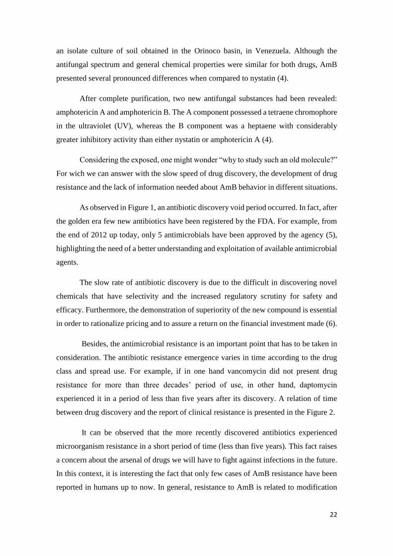
Figure 2. Time from antibiotic approval or introduction to detection of
resistance in clinical samples, extracted from Marston et al.
(2016).
23
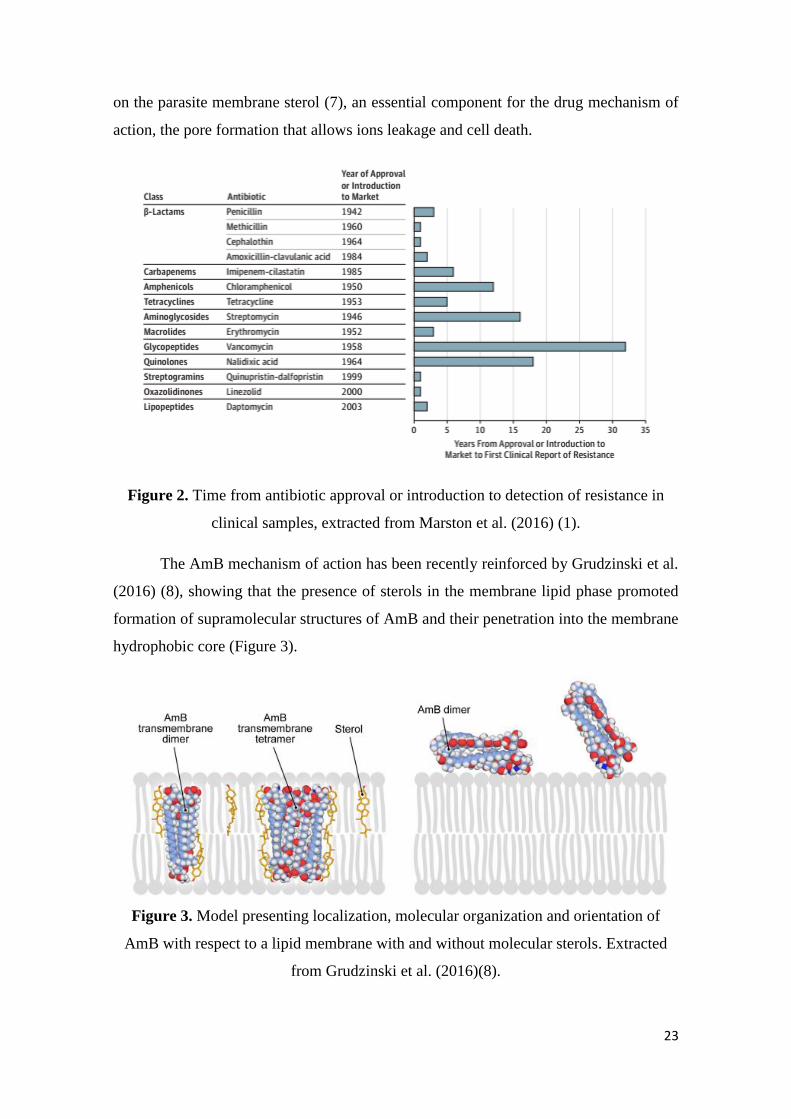
Figure 3. Model presenting localization, molecular organization and
orientation of AmB with respect to a lipid membrane with and
without molecular sterols. Extracted from Grudzinski et al.
(2016).
23
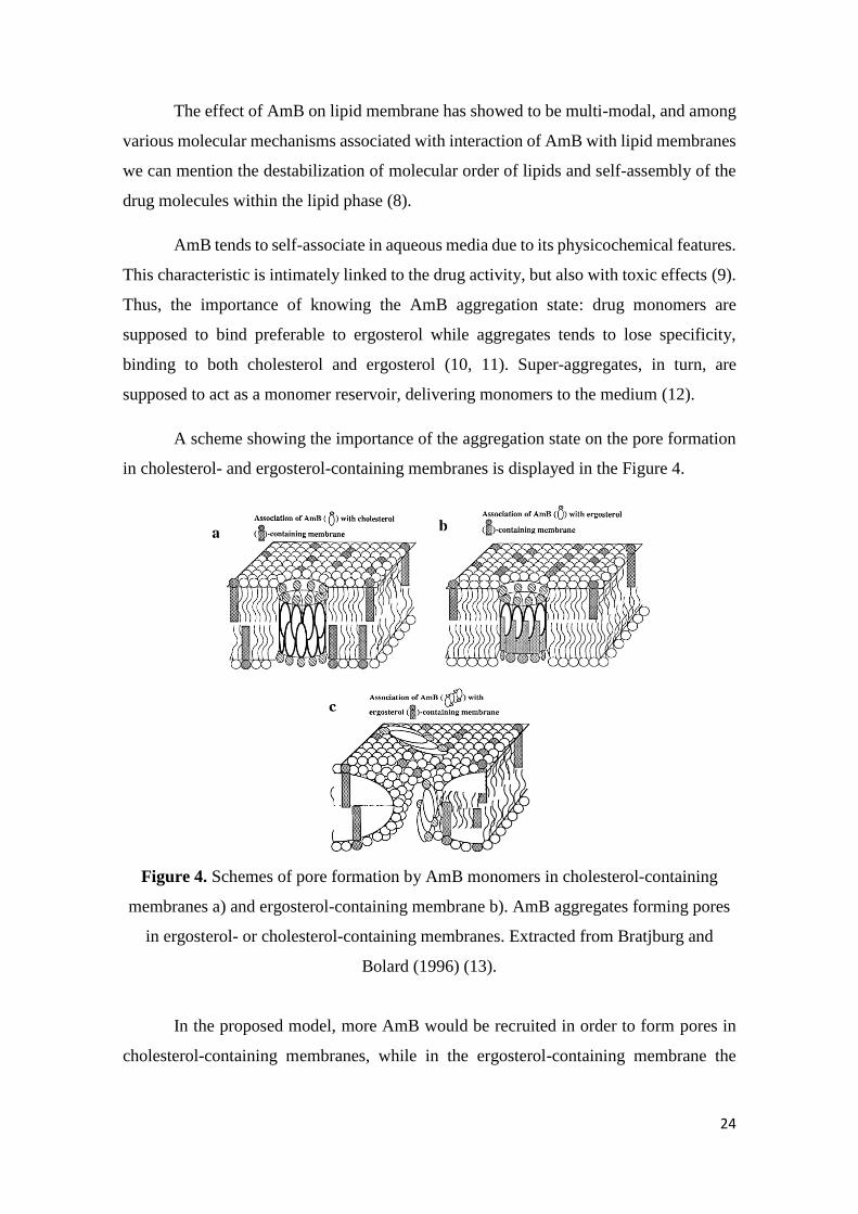
Figure 4. Schemes of pore formation by AmB monomers in cholesterol-
containing membranes a) and ergosterol-containing membrane
b). AmB aggregates forming pores in ergosterol- or cholesterol-
containing membranes. Extracted from Bratjburg and Bolard
(1996).
24
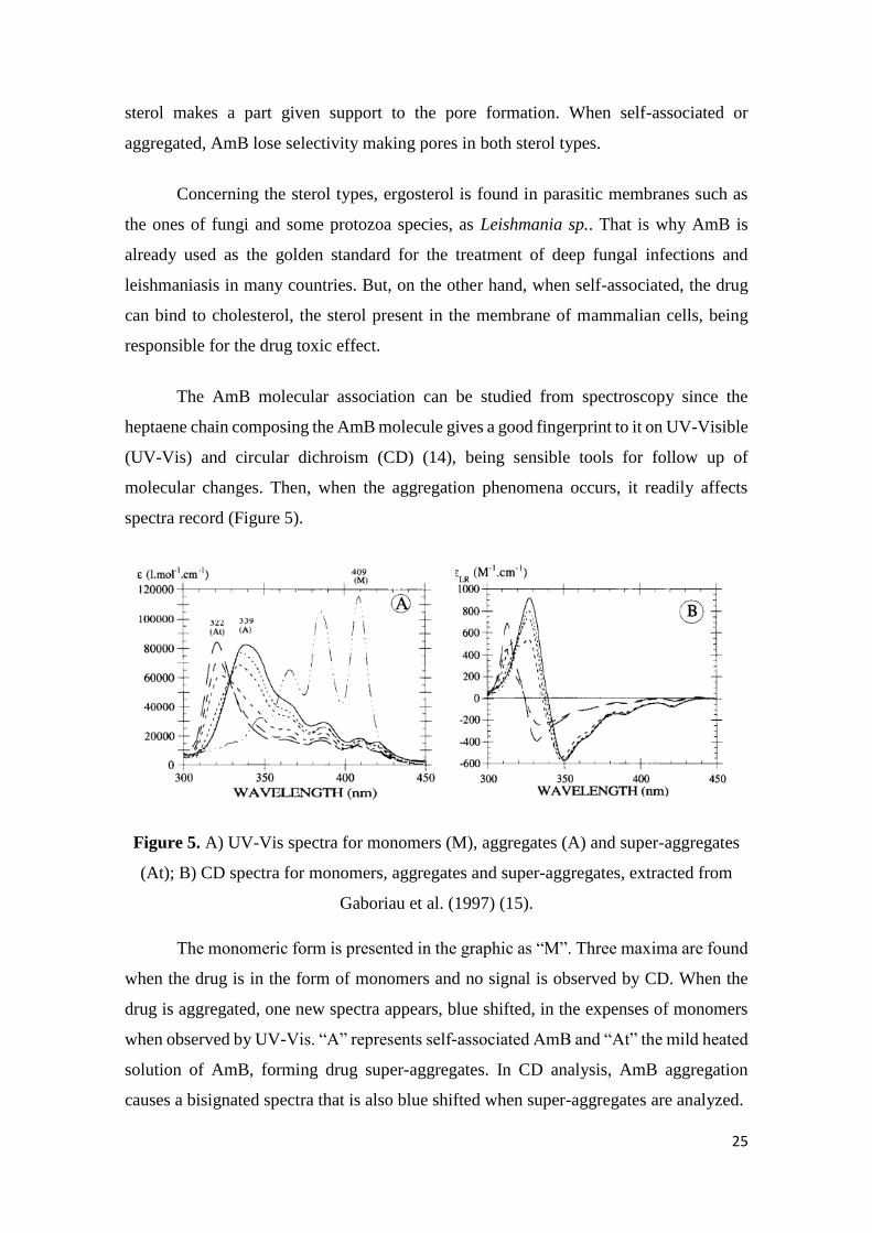
Figure 5. UV-Vis spectra for monomers (M), aggregates (A) and super-
aggregates (At); B) CD spectra for monomers, aggregates and
super-aggregates, extracted from Gaboriau et al. (1997)
25
Section I
Chapter I
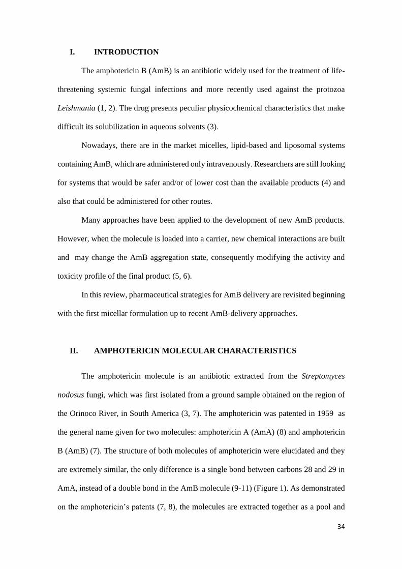
Figure 1. The molecular structure of amphotericin: a) amphotericin B b)
amphotericin A. 35
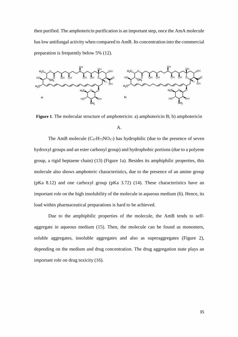
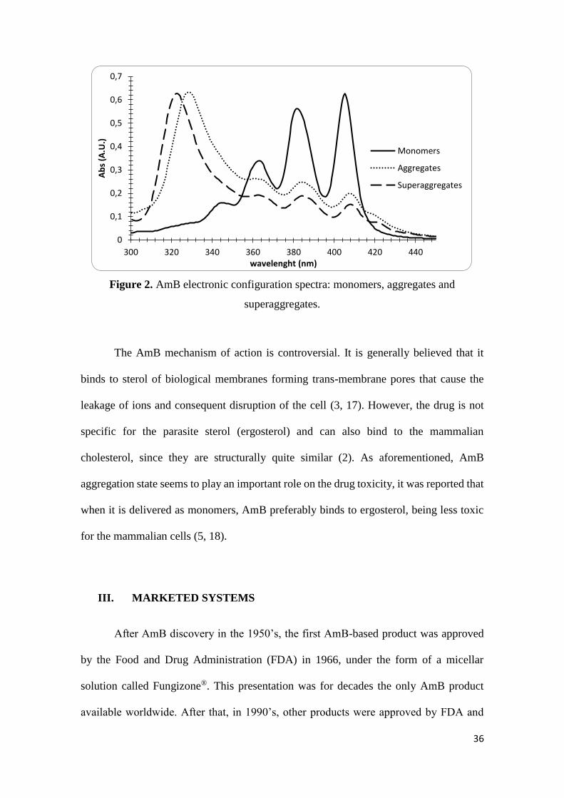
Figure 2. AmB electronic configuration spectra: monomers, aggregates
and superaggregates. 36
Figure 3. Structural schemes of AmB-based formulations already in the
market: a) micellar systems (Fungizone® and others) b) disk-like
lipid complex (Amphotec®) c) ribbon-like colloidal dispersion
(Abelcet®) d) liposomal systems (AmBisome® and others).
39
Section II
Chapter II
Figure 1. Amphotericin B molecular structure. 68
Figure 2. UV-Vis electronic spectrum of pure AmB in methanol. 72
Figure 3. AmB calibration curve in methanol. 73
Figure 4. AmB calibration curve in the range of Beer-Lambert law. 73
Figure 5. Electronic absorption spectra of samples A, B and C diluted in
water. Labeled concentration 5x10⁻⁶ M AmB. 74
Figure 6. Relation between AmB species: aggregates and monomers,
calculated from UV-Vis absorbance at 325 and 408 nm
respectively.
75
Figure 7. CD spectra of AmB-containing micellar systems at 5.10-5 M
AmB in water. Values were corrected in b) according to the
quantified AmB.
75
Figure 8. CD spectra of product A in different concentrations: from 5.10-
5 to 5.10-8 M AmB 76
Figure 9. Size distribution for products A, B and C: a) size distribution for
number b) size distribution for intensity. 77
Chapter III
Figure 1. UV-Vis spectra in water for a) Anforicin® B and b) AmBisome®
before (upper panel) and after (bottom panel) heating process,
recorded along 28 days from the initial concentration of 5x10⁻⁶
M AmB.
93
Figure 2. AmB CD spectra in water for a) liposomal and b) micellar
preparation. Before (upper panel) and after (bottom panel)
heating process, in three concentrations.
94
Figure 3. UV-Vis quantification (%) of AmB-containing micellar and
liposomal products. Heated (discontinuous lines) and unheated
products were analyzed.
95
Figure 4. Thermograms of the Anforicin B (5e-3 M) record in different
temperatures: a) 40 ºC; b) 50 ºC; c) 60 ºC and d) 70 ºC. The 96
injections define the beginning and the end of the counted time
(20 minutes).
Figure 5. Comparison of the size of an AmB micellar solution after
heating process at four different temperatures by naked eye
(upper panel) and DLS (bottom panel).
97
Figure 6. Spectra of Anforicin® B solution (5x10⁻³ M AmB) heated at
different temperatures in an ITC equipment. 97
Figure 7. Thermogram for the dilution of an AmB-SD solution, from
5x10⁻⁵ to 5x10⁻⁸ M AmB. 98
Figure 8. CD spectra of AmB-SD solution as oligomer aggregates (5x10-
5 M) and AmB monomers (5x10⁻⁸ M). 99
Figure 9. Scheme of super-aggregate formation: AmB coexists as
monomers and oligomers in solution after mild heating super-
aggregates are formed in the expenses of the previous AmB
forms (Based on Gaboriau et al. (1997).
101
Section III
Chapter IV
Figure 1. AmB solubilization in function of NaOH 1N addition and its
influence on the drug aggregation state. 117
Figure 2. AmB solubility profile in nano- and micro-emulsion. The
percentile of drug solubility (5mg/mL max) is showed in the
upper panel and the aggregation pattern in function of the added
NaOH 1N, by UV-Vis, is presented in the bottom panel.
118
Figure 3. AmB aggregation pattern according to the drug concentration
loaded into the nano-emulsion. 119
Figure 4. Change of drug aggregation pattern into nano-emulsion
according to the pH. 120
Figure 5. Characterization of nano-emulsion before and after AmB
loading. A) zeta potential B) droplet size. 120
Figure 6. AmB aggregation pattern into NanAmB according to the pH 121
Figure 7. CD spectra of blank nano-emulsion (Nano) and after AmB
loading by two incorporation methods. 122
Figure 8. DLS analysis of blank nano-emulsion and AmB-loaded systems
during (NanAmB) and after (Nano + AmB) nano-emulsion
preparation.
122
Chapter V
Figure 1. Pseudo-ternary phase diagrams of the system containing
Miglyol® 812N (oil phase- O), isotonic phosphate buffer pH 7.4
(aqueous phase- W), Lipoid® S100, and Tween® 80 (surfactants-
S), showing the area of existence of microemulsions (MEs) and
the point chosen to produce the blank ME formulation for future
studies.
129
Figure 2. Rheological behavior of the blank microemulsion (ME-2a) and
Amphotericin B-loaded microemulsion (AmB-ME-2b). 130
Figure 3. TEM microphotography of the microemulsion. 130
Figure 4. UV-Vis absorption spectra of the standard Amphotericin B
(AmB) in methanol and the Amphotericin B-loaded
microemulsion (AmB-ME) in microemulsion (ME) (4a). UV-
Vis absorption spectra of the micelle dosage form of
amphotericin B (AmB-M) and the Amphotericin B-loaded
microemulsion (AmB-ME) in phosphate buffer (pH 7.4) (4b)
131
Figure 5. Profile of toxicity of AmB-M and AmB-ME on RBC. (5a) Hb
leakage, and (5b) K+ leakage. 132
Section IV
Chapter VI
Figure 1. Infra-Red spectra of AmB (blue line) and its soluble derivative
(red line). The lost of a signal band is evidenced. 141
Figure 2. UV-Vis spectrum of AmB-derivative-containing microemulsion
diluted in water. 143
Figure 3. Scheme of AmB derivative aggregation in water and in
microemulsion medium. 144
Figure 4. CD spectra of AmB-derivative encapsulated in microemulsion.
Full line represents Day 0 and dotted line, Day 7. 145
LIST OF TABLES
Section I
Chapter I
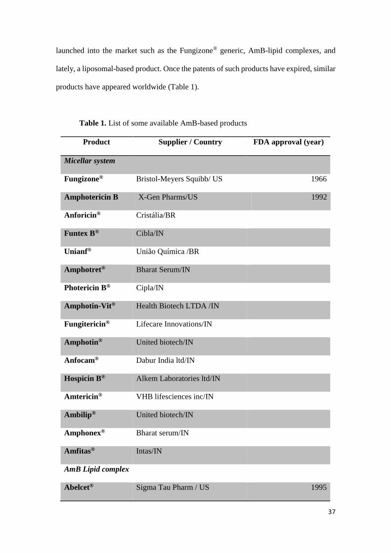
Table 1. List of some available AmB-based products 37
Section II
Chapter II
Table 1. List of some AmB-based micellar systems available in the
market. 69
Table 2. Quantification of the AmB content in AmB-based micellar
systems. 74
Section III
Chapter IV
Table 1. List of ingredients for micro-emulsion prepare. 115
Table 2. List of ingredients for nano-emulsion prepare. 115
Table 3. Relation between alkaline agent volume and solution pH. 116
Table 4. Droplet size and polydispersity index (PdI) of nano-emulsion
loaded with different amounts of AmB. 119
Chapter V
Table 1. Physicochemical parameters of blank microemulsion (ME) and
amphotericin B-loaded microemulsion (AmB-ME) (Mean ±
SD).
130
Table 2. Antifungal susceptibility of Amphotericin B-loaded
microemulsion (AmB-ME) against Candida and Fusarium
strains.
132
TABLE OF CONTENTS
General Introduction……………………………………………………….... 20
SECTION I PHARMACEUTICAL STRATEGIES FOR
AMPHOTERICIN B DELIVERY………………………... 29
CHAPTER I Pharmaceutical strategies for amphotericin B
delivery………
31
SECTION II AmB-CONTAINING MICELLAR SYSTEMS:
CHARACTERIZATION OF SIMILARS AND SUPER-
AGGREGATES FORMATION………………….………... 63
CHAPTER II Amphotericin B-containing sodium deoxycholate micellar
systems: a comparison of physicochemical characteristics
of marketed products …………………………………………… 65
CHAPTER III Amphotericin B super-aggregates: formation and influence
on the drug stability………………………………………….….. 86
SECTION III INCORPORATION OF AMPHOTERICIN B INTO
EMULSION SYSTEMS…………………………………... 111
CHAPTER IV AmB loading into micro- and nano-emulsion ………………... 113
CHAPTER V Development and characterization of a microemulsion
system containing amphotericin B with potential ocular
applications………………………………………………………. 125
SECTION IV VEHICULATION OF AN AmB-SOLUBLE
DERIVATIVE INTO A MICRO-EMULSION SYSTEM... 138
CHAPTER VI Microemulsion encapsulation of an AmB soluble
derivative………………………………………………………… 140
General Discussion ........................................................................................... 147
Conclusions ...................................................................................................... 160
Synthèse de la Thèse ........................................................................................ 163
Enclosures 176
List of abstracts presented in Conferences 177
Stearylamine-containing cationic nanoemulsions as a
promising carrier for gene delivery 179
Short Curriculum Vitae 181
20
General Introduction
21
GENERAL INTRODUCTION
The emergence of the antibiotics has revolutionized the practice of medicine by
enabling breakthroughs across the spectrum of clinical medicine (1). In this context, the
nature has been a source of medicinal products for millennia, with many useful active
substances obtained from plant sources and then, in the 20th century, the discovery of
penicillin was the start pointing for drug discovery from microbial sources.
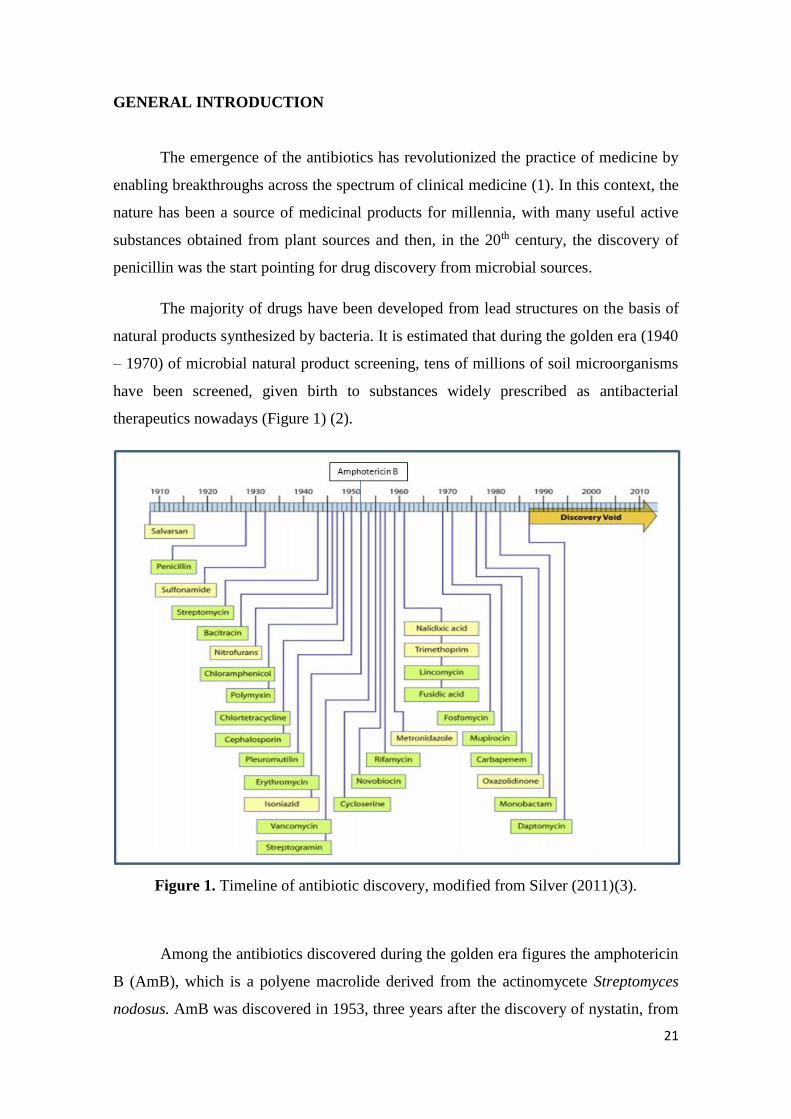
The majority of drugs have been developed from lead structures on the basis of
natural products synthesized by bacteria. It is estimated that during the golden era (1940
– 1970) of microbial natural product screening, tens of millions of soil microorganisms
have been screened, given birth to substances widely prescribed as antibacterial
therapeutics nowadays (Figure 1) (2).
Figure 1. Timeline of antibiotic discovery, modified from Silver (2011)(3).
Among the antibiotics discovered during the golden era figures the amphotericin
B (AmB), which is a polyene macrolide derived from the actinomycete Streptomyces
nodosus. AmB was discovered in 1953, three years after the discovery of nystatin, from
22
an isolate culture of soil obtained in the Orinoco basin, in Venezuela. Although the
antifungal spectrum and general chemical properties were similar for both drugs, AmB
presented several pronounced differences when compared to nystatin (4).
After complete purification, two new antifungal substances had been revealed:
amphotericin A and amphotericin B. The A component possessed a tetraene chromophore
in the ultraviolet (UV), whereas the B component was a heptaene with considerably
greater inhibitory activity than either nystatin or amphotericin A (4).
Considering the exposed, one might wonder “why to study such an old molecule?”
For wich we can answer with the slow speed of drug discovery, the development of drug
resistance and the lack of information needed about AmB behavior in different situations.
As observed in Figure 1, an antibiotic discovery void period occurred. In fact, after
the golden era few new antibiotics have been registered by the FDA. For example, from
the end of 2012 up today, only 5 antimicrobials have been approved by the agency (5),
highlighting the need of a better understanding and exploitation of available antimicrobial
agents.
The slow rate of antibiotic discovery is due to the difficult in discovering novel
chemicals that have selectivity and the increased regulatory scrutiny for safety and
efficacy. Furthermore, the demonstration of superiority of the new compound is essential
in order to rationalize pricing and to assure a return on the financial investment made (6).
Besides, the antimicrobial resistance is an important point that has to be taken in
consideration. The antibiotic resistance emergence varies in time according to the drug
class and spread use. For example, if in one hand vancomycin did not present drug
resistance for more than three decades’ period of use, in other hand, daptomycin
experienced it in a period of less than five years after its discovery. A relation of time
between drug discovery and the report of clinical resistance is presented in the Figure 2.
It can be observed that the more recently discovered antibiotics experienced
microorganism resistance in a short period of time (less than five years). This fact raises
a concern about the arsenal of drugs we will have to fight against infections in the future.
In this context, it is interesting the fact that only few cases of AmB resistance have been
reported in humans up to now. In general, resistance to AmB is related to modification
23
on the parasite membrane sterol (7), an essential component for the drug mechanism of
action, the pore formation that allows ions leakage and cell death.
Figure 2. Time from antibiotic approval or introduction to detection of resistance in
clinical samples, extracted from Marston et al. (2016) (1).
The AmB mechanism of action has been recently reinforced by Grudzinski et al.
(2016) (8), showing that the presence of sterols in the membrane lipid phase promoted
formation of supramolecular structures of AmB and their penetration into the membrane
hydrophobic core (Figure 3).
Figure 3. Model presenting localization, molecular organization and orientation of
AmB with respect to a lipid membrane with and without molecular sterols. Extracted
from Grudzinski et al. (2016)(8).
24
The effect of AmB on lipid membrane has showed to be multi-modal, and among
various molecular mechanisms associated with interaction of AmB with lipid membranes
we can mention the destabilization of molecular order of lipids and self-assembly of the
drug molecules within the lipid phase (8).
AmB tends to self-associate in aqueous media due to its physicochemical features.
This characteristic is intimately linked to the drug activity, but also with toxic effects (9).
Thus, the importance of knowing the AmB aggregation state: drug monomers are
supposed to bind preferable to ergosterol while aggregates tends to lose specificity,
binding to both cholesterol and ergosterol (10, 11). Super-aggregates, in turn, are
supposed to act as a monomer reservoir, delivering monomers to the medium (12).
A scheme showing the importance of the aggregation state on the pore formation
in cholesterol- and ergosterol-containing membranes is displayed in the Figure 4.
Figure 4. Schemes of pore formation by AmB monomers in cholesterol-containing
membranes a) and ergosterol-containing membrane b). AmB aggregates forming pores
in ergosterol- or cholesterol-containing membranes. Extracted from Bratjburg and
Bolard (1996) (13).
In the proposed model, more AmB would be recruited in order to form pores in
cholesterol-containing membranes, while in the ergosterol-containing membrane the
25
sterol makes a part given support to the pore formation. When self-associated or
aggregated, AmB lose selectivity making pores in both sterol types.
Concerning the sterol types, ergosterol is found in parasitic membranes such as
the ones of fungi and some protozoa species, as Leishmania sp.. That is why AmB is
already used as the golden standard for the treatment of deep fungal infections and
leishmaniasis in many countries. But, on the other hand, when self-associated, the drug
can bind to cholesterol, the sterol present in the membrane of mammalian cells, being
responsible for the drug toxic effect.
The AmB molecular association can be studied from spectroscopy since the
heptaene chain composing the AmB molecule gives a good fingerprint to it on UV-Visible
(UV-Vis) and circular dichroism (CD) (14), being sensible tools for follow up of
molecular changes. Then, when the aggregation phenomena occurs, it readily affects
spectra record (Figure 5).
Figure 5. A) UV-Vis spectra for monomers (M), aggregates (A) and super-aggregates
(At); B) CD spectra for monomers, aggregates and super-aggregates, extracted from
Gaboriau et al. (1997) (15).
The monomeric form is presented in the graphic as “M”. Three maxima are found
when the drug is in the form of monomers and no signal is observed by CD. When the
drug is aggregated, one new spectra appears, blue shifted, in the expenses of monomers
when observed by UV-Vis. “A” represents self-associated AmB and “At” the mild heated
solution of AmB, forming drug super-aggregates. In CD analysis, AmB aggregation
causes a bisignated spectra that is also blue shifted when super-aggregates are analyzed.
26
Due to the AmB low water solubility, the drug has been loaded in specially
designed pharmaceutical preparations. The first AmB-based product was a micellar
system, launched in the 1960’s. Almost thirty years later, in the middle of the 1990’s,
other products such as lipid complexes, colloidal dispersion and liposomes were approved
and put on the market for clinical use (11). However, the interaction among the drug and
its carrier influenced on the drug aggregation state, and consequently, on its biological
activity (13). Then, this work was dedicated to the study of AmB aggregation state into
different carrier systems, in which marketed and new systems were analyzed.
This thesis memoire is divided in four sections. In the first section we have one
review paper about pharmaceutical strategies for AmB delivery: this article presents the
already marketed products as well as new approaches aiming the improvement of AmB
safety. The second section is devoted to the micellar systems of AmB: they were the first
product launched and still useful in therapeutics. We have analyzed similar products from
three different countries and also the possibility of forming super-aggregates, comparing
micellar and liposomal systems. In the third section we report the AmB incorporation into
emulsion systems, reinforcing the potential of such preparations for ocular application
and antileishmanial treatment. The fourth and last section, presents a patent of the
production process of an AmB-soluble derivative and a report of the derivative
incorporation into a microemulsion system.
Concluding remarks closes this thesis, dedicated to the AmB. In addition, as an
appendix, there are an article and one patent dedicated to emulsion systems intended to
gene delivery, a contextualization is given as appendix introduction.
REFERENCES
1. Marston HD, Dixon DM, Knisely JM, Palmore TN, Fauci AS. Antimicrobial
Resistance. JAMA-J Am Med Assoc. 2016;316(11):1193-204.
2. Wohlleben W, Mast Y, Stegmann E, Ziemert N. Antibiotic drug discovery.
Microb Biotechnol. 2016;9(5):541-8.
3. Silver LL. Challenges of Antibacterial Discovery. Clin Microbiol Rev.
27
2011;24(1):71-109.
4. Dutcher JD. Discovery and development of amphotericin B. Dis Chest. 1968;S
54:296-8.
5. New Drugs at FDA [Internet]. 2017 [cited 08/03/2017]. Available from:
https://www.fda.gov/Drugs/DevelopmentApprovalProcess/DrugInnovation/ucm537040.
htm.
6. Fernandes P. The global challenge of new classes of antibacterial agents: an
industry perspective. Curr Opin Pharmacol. 2015;24:7-11.
7. Kumar A, Das S, Purkait B, Sardar AH, Ghosh AK, Dikhit MR, et al. Ascorbate
Peroxidase, a Key Molecule Regulating Amphotericin B Resistance in Clinical Isolates
of Leishmania donovani. Antimicrob Agents Chemother. 2014;58(10):6172-84.
8. Grudzinski W, Sagan J, Welc R, Luchowski R, Gruszecki WI. Molecular
organization, localization and orientation of antifungal antibiotic amphotericin B in a
single lipid bilayer. Sci Rep. 2016;6:1-11.
9. Espada R, Valdespina S, Alfonso C, Rivas G, Ballesteros MP, Torrado JJ. Effect
of aggregation state on the toxicity of different amphotericin B preparations. Int J Pharm.
2008;361(1-2):64-9.
10. Huang WM, Zhang ZL, Hang XJ, Tang JL, Wang JG, Dong SJ, et al. Ion channel
behavior of amphotericin B in sterol-free and cholesterol- or ergosterol-containing
supported phosphatidylcholine bilayer model membranes investigated by
electrochemistry and spectroscopy. Biophys J. 2002;83(6):3245-55.
11. Golenser J, Domb A. New formulations and derivatives of amphotericin B for
treatment of leishmaniasis. Mini-Rev Med Chem. 2006;6(2):153-62.
12. Cheron M, Petit C, Bolard J, Gaboriau F. Heat-induced reformulation of
amphotericin B-deoxycholate favours drug uptake by the macrophage-like cell line J774.
J Antimicrob Chemother. 2003;52(6):904-10.
13. Brajtburg J, Bolard J. Carrier effects on biological activity of amphotericin B. Clin
Microbiol Rev. 1996;9(4):512-31.
14. Espuelas MS, Legrand P, Cheron M, Barratt G, Puisieux F, Devissaguet JP, et al.
Interaction of amphotericin B with polymeric colloids: A spectroscopic study. Colloids
28
Surf B Biointerfaces. 1998;11(3):141-51.
15. Gaboriau F, Cheron M, Leroy L, Bolard J. Physico-chemical properties of the
heat-induced 'superaggregates' of amphotericin B. Biophys Chem. 1997;66(1):1-12.
29
SECTION I
PHARMACEUTICAL STRATEGIES FOR AMPHOTERICIN B
DELIVERY
____________________________________________________________
30
BRIEFING
Amphotericin B (AmB) is a quite old molecule, discovered in the 1950’s.
However, the studies dealing with this molecule are still in evidence. Much has been done
in the last 60 years: the antibiotic structure was fully described and the drug features
clarified. The AmB mechanism of action is still in discussion, despite the evidences of
mechanistic pore formation and membrane oxidation.
The drug is available in the market in a few pharmaceutical presentations and this
fact is mainly due to the drug physicochemical features. Then, the first section of this
thesis is composed of one chapter bringing a review manuscript discussing important
aspects of the AmB molecule, such as its physicochemical characteristics, aggregation
patterns, systems marketed worldwide and mainly the new pharmaceutical strategies for
AmB delivery.
31
CHAPTER I
Pharmaceutical strategies for amphotericin B delivery
32
Pharmaceutical strategies for amphotericin B delivery
André Leandro Silva1,2; Gilles Ponchel2; Eryvaldo Sócrates Tabosa do Egito1*
1 Programa de Pós-Graduação em Biotecnologia (Renorbio), Universidade Federal do Rio
Grande do Norte, Natal – Brazil;
2 Institut Galien Paris-Sud, UMR CNRS 8612, Université Paris-Sud, Châtenay-Malabry
– France.
*Corresponding author:
Eryvaldo Sócrates Tabosa do Egito
Rua Praia de Areia Branca, 8448. Ponta Negra, Natal – Brazil (59094-450).
Phone: +55 84 9 9431-8816 or +55 84 3342-9817
E-mail address: [email protected] or [email protected]
33
Abstract
The amphotericin B (AmB) is an antibiotic used for the treatment of systemic fungal
infections and leishmaniasis. The AmB molecule has amphiphilic and amphipathic
properties, which make difficult its load into pharmaceutical carriers. Since its discovery
in the 1950’s, just a few kind of pharmaceutical products were developed and are
available in the market worldwide. However, these products show problems regarding
toxicity (micelles) and high cost, besides the fact of been available only by intravenous
administration. Then, the development of new pharmaceutical systems is mandatory. In
this context we reviewed the strategies used for the development of new
nanotechnological devices that could improve AmB solubility and safety. In conclusion,
AmB is an old molecule that has not yet achieved its maximum on pharmaceutical use,
due to the lack of specific drug carriers, even though much has been done in the scenery
of lipid-based AmB-loaded systems, evidencing challenges and opportunities.
Key-words: amphotericin b; micelles; colloidal systems; liposomes; microemulsion;
nanoemulsions; cochleates; nanoparticles;
34
I. INTRODUCTION
The amphotericin B (AmB) is an antibiotic widely used for the treatment of life-
threatening systemic fungal infections and more recently used against the protozoa
Leishmania (1, 2). The drug presents peculiar physicochemical characteristics that make
difficult its solubilization in aqueous solvents (3).
Nowadays, there are in the market micelles, lipid-based and liposomal systems
containing AmB, which are administered only intravenously. Researchers are still looking
for systems that would be safer and/or of lower cost than the available products (4) and
also that could be administered for other routes.
Many approaches have been applied to the development of new AmB products.
However, when the molecule is loaded into a carrier, new chemical interactions are built
and may change the AmB aggregation state, consequently modifying the activity and
toxicity profile of the final product (5, 6).
In this review, pharmaceutical strategies for AmB delivery are revisited beginning
with the first micellar formulation up to recent AmB-delivery approaches.
II. AMPHOTERICIN MOLECULAR CHARACTERISTICS
The amphotericin molecule is an antibiotic extracted from the Streptomyces
nodosus fungi, which was first isolated from a ground sample obtained on the region of
the Orinoco River, in South America (3, 7). The amphotericin was patented in 1959 as
the general name given for two molecules: amphotericin A (AmA) (8) and amphotericin
B (AmB) (7). The structure of both molecules of amphotericin were elucidated and they
are extremely similar, the only difference is a single bond between carbons 28 and 29 in
AmA, instead of a double bond in the AmB molecule (9-11) (Figure 1). As demonstrated
on the amphotericin’s patents (7, 8), the molecules are extracted together as a pool and
35
then purified. The amphotericin purification is an important step, once the AmA molecule
has low antifungal activity when compared to AmB. Its concentration into the commercial
preparation is frequently below 5% (12).
Figure 1. The molecular structure of amphotericin: a) amphotericin B; b) amphotericin
A.
The AmB molecule (C47H73NO17) has hydrophilic (due to the presence of seven
hydroxyl groups and an ester carbonyl group) and hydrophobic portions (due to a polyene
group, a rigid heptaene chain) (13) (Figure 1a). Besides its amphiphilic properties, this
molecule also shows amphoteric characteristics, due to the presence of an amine group
(pKa 8.12) and one carboxyl group (pKa 3.72) (14). These characteristics have an
important role on the high insolubility of the molecule in aqueous medium (6). Hence, its
load within pharmaceutical preparations is hard to be achieved.
Due to the amphiphilic properties of the molecule, the AmB tends to self-
aggregate in aqueous medium (15). Then, the molecule can be found as monomers,
soluble aggregates, insoluble aggregates and also as superaggregates (Figure 2),
depending on the medium and drug concentration. The drug aggregation state plays an
important role on drug toxicity (16).
36
Figure 2. AmB electronic configuration spectra: monomers, aggregates and
superaggregates.
The AmB mechanism of action is controversial. It is generally believed that it
binds to sterol of biological membranes forming trans-membrane pores that cause the
leakage of ions and consequent disruption of the cell (3, 17). However, the drug is not
specific for the parasite sterol (ergosterol) and can also bind to the mammalian
cholesterol, since they are structurally quite similar (2). As aforementioned, AmB
aggregation state seems to play an important role on the drug toxicity, it was reported that
when it is delivered as monomers, AmB preferably binds to ergosterol, being less toxic
for the mammalian cells (5, 18).
III. MARKETED SYSTEMS
After AmB discovery in the 1950’s, the first AmB-based product was approved
by the Food and Drug Administration (FDA) in 1966, under the form of a micellar
solution called Fungizone®. This presentation was for decades the only AmB product
available worldwide. After that, in 1990’s, other products were approved by FDA and
0
0,1
0,2
0,3
0,4
0,5
0,6
0,7
300 320 340 360 380 400 420 440
Ab
s (A
.U.)
wavelenght (nm)
Monomers
Aggregates
Superaggregates
37
launched into the market such as the Fungizone® generic, AmB-lipid complexes, and
lately, a liposomal-based product. Once the patents of such products have expired, similar
products have appeared worldwide (Table 1).
Table 1. List of some available AmB-based products
Product Supplier / Country FDA approval (year)
Micellar system
Fungizone® Bristol-Meyers Squibb/ US 1966
Amphotericin B X-Gen Pharms/US 1992
Anforicin® Cristália/BR
Funtex B® Cibla/IN
Unianf® União Química /BR
Amphotret® Bharat Serum/IN
Photericin B® Cipla/IN
Amphotin-Vit® Health Biotech LTDA /IN
Fungitericin® Lifecare Innovations/IN
Amphotin® United biotech/IN
Anfocam® Dabur India ltd/IN
Hospicin B® Alkem Laboratories ltd/IN
Amtericin® VHB lifesciences inc/IN
Ambilip® United biotech/IN
Amphonex® Bharat serum/IN
Amfitas® Intas/IN
AmB Lipid complex
Abelcet® Sigma Tau Pharm / US 1995
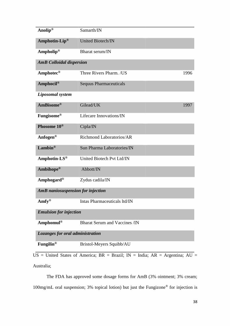
38
Anolip® Samarth/IN
Amphotin-Lip® United Biotech/IN
Ampholip® Bharat serum/IN
AmB Colloidal dispersion
Amphotec® Three Rivers Pharm. /US 1996
Amphocil® Sequus Pharmaceuticals
Liposomal system
AmBisome® Gilead/UK 1997
Fungisome® Lifecare Innovations/IN
Phosome 10® Cipla/IN
Anfogen® Richmond Laboratorios/AR
Lambin® Sun Pharma Laboratories/IN
Amphotin-LS® United Biotech Pvt Ltd/IN
Ambihope® Abbott/IN
Amphogard® Zydus cadila/IN
AmB naniosuspension for injection
Amfy® Intas Pharmaceuticals ltd/IN
Emulsion for injection
Amphomul® Bharat Serum and Vaccines /IN
Lozanges for oral administration
Fungilin® Bristol-Meyers Squibb/AU
US = United States of America; BR = Brazil; IN = India; AR = Argentina; AU =
Australia;
The FDA has approved some dosage forms for AmB (3% ointment; 3% cream;
100mg/mL oral suspension; 3% topical lotion) but just the Fungizone® for injection is
39
currently available in the USA. In 1992, X-Gen Pharm approved the first generic for
Fungizone®, marketed as “Amphotericin B® for injection”. After that, in 1995, the AmB
lipid complex Abelcet® was approved, being followed for Amphotec® colloidal
dispersion (1996) and the liposomal dosage form AmBisome® (1997) (19). The approval
of these three dosage forms, together with Doxil® (an anticancer liposome), were the first
FDA approvals to nanoengineered medicines (20). The approval of new biotechnological
drugs has received much more attention due to their increased specificity and lower toxic
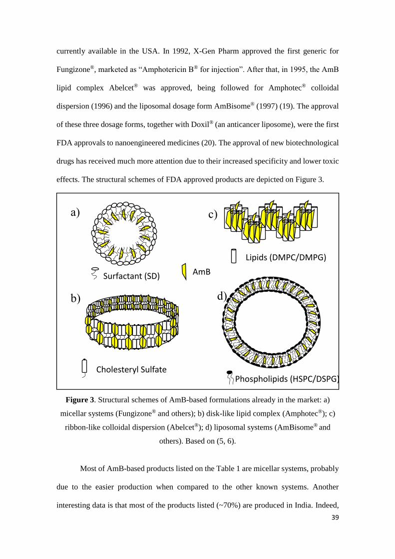
effects. The structural schemes of FDA approved products are depicted on Figure 3.
Figure 3. Structural schemes of AmB-based formulations already in the market: a)
micellar systems (Fungizone® and others); b) disk-like lipid complex (Amphotec®); c)
ribbon-like colloidal dispersion (Abelcet®); d) liposomal systems (AmBisome® and
others). Based on (5, 6).
Most of AmB-based products listed on the Table 1 are micellar systems, probably
due to the easier production when compared to the other known systems. Another
interesting data is that most of the products listed (~70%) are produced in India. Indeed,
a)
b)
c)
d)
Surfactant (SD)
Phospholipids (HSPC/DSPG)
Lipids (DMPC/DMPG)
Cholesteryl Sulfate
AmB
40
this fact is understandable once this country presents endemic leishmania infection areas
where patients are refractory to the treatment with antimonials (first line treatment) (21).
Some efforts have been made in India since 2005 in order to fight against leishmaniasis
and a great number of clinical trials using AmB probably gave birth to new AmB-
containing products (22).
All the systems listed in the Table 1 are intended for intravenous administration,
except Fungilin® lozanges, In fact the advances in the engineer of drug carriers allowed
the development of new AmB-based products, however their ability to cross the intestinal
barrier has not yet been achieved. Fungilin® lozanges are indicated for oral delivery but
with a topic pharmacological effect on the treatment of oral and intestinal candidiais.
In the following lines it will be presented the AmB-based products already
approved by the FDA that are/were available in the market.
Fungizone®
The Fungizone® was launched in the 1960’s consisting of a mix of 50 mg of AmB
and 41 mg of sodium deoxycholate (SD) (4, 5, 17) constituting for a long period of time
the only AmB-based product available in the market. Side effects such as headache, fever,
chills, nausea and mainly nephrotoxicity have been related to its infusion (2, 17, 23-26).
However, due to its low cost and high antifungal efficacy, it is broadly used nowadays.
To avoid the Fungizone’s drawbacks and to increase its therapeutic index some
strategies have been applied. The AmB aggregation state has an important role on the
host toxicity (18), when the AmB-SD is dissolved in water, the drug molecules are mainly
in the aggregated form (16, 27, 28) that is supposed to bind the cholesterol of host cells,
causing toxic effects. One strategy for increasing AmB-SD therapeutic index could be the
delivery of AmB monomers. In fact, it was demonstrated that AmB-SD has its deleterious
effects balanced by the presence of serum albumin, which influences the AmB
41
aggregation state towards monomers (29). In this way, other strategies have been
published such as the use of other micellar (30) or lipid systems (4, 31, 32) to dilute
Fungizone® as well as the mild heating of the product, in order to form AmB
superaggregates (25, 26, 28, 33).
Abelcet®
The product is an AmB lipid complex (AmBLC) consisting of a 1:1 molar ratio
of drug to lipids. The lipids herein used are dimyristoyl phosphatidylcholine (DMPC) and
dimyristoyl phosphatidylglycerol (DMPG) (7:3 molar ratio) (5, 6, 34-38).
The formation of the system is possible dependent of an AmB concentration
higher than 3 mol% (34, 38). Into this system, the drug is complexed with a ribbon-like
non-liposomal structure forming lipid-stabilized AmB aggregates. It is suggested that the
new structure is responsible for the drug toxicity attenuation (36). In fact, a low
concentration of monomers on the medium is enough to kill the fungal cell but insufficient
to cause toxicity to mammalian (34). Indeed, phospholipases are required to hydrolizate
the lipids in order to allow the drug release (35).
Due to Abelcet® low toxicity, it is possible to increase the quantity of drug
administered to the patient. In this way, better fungicidal level is achieved. Besides, even
in higher concentration, this formulation is better tolerated than AmB-SD (39). Moreover,
it is effective against fungal infections on bone marrow transplanted patients who had
failed to respond to previous antifungal therapy (even treated with AmB) (40).
Amphotec®
The Amphotec®, an AmB colloidal dispersion (ABCD) was the third FDA
approved AmB based product (34), it is presented as uniform disk shaped particles of
42
around 115 nm where the drug is complexed with cholesteryl sulphate in a 1:1 proportion
(41-43). ABCDs were marketed as Amphotec® (USA) and Amphocil® (Europe) (6, 17).
The disc-resemble particles of this system have similar antifungal activity and
decreased toxicity when compared to Fungizone®. Such an improvement in safety is due
to the strong interaction of the drug with cholesteryl sulfate, reducing the amount of free
AmB available in the blood (6, 42). However, ABCD showed up regulation of
inflammatory genes and consequently, similar or even more infusion-related reaction
compared to AmB-SD. It is probably why its production was discontinued in 2011 (17).
AmBisome®
The AmBisome® is a single bilayer liposome that was introduced in the European
market in 1989 and approved by the FDA for the treatment of leishmaniasis in 1997 (6).
The product consists of AmB, hydrogenated soya phosphatidylcholine (HSPC),
cholesterol, distearoyl phosphatidylglycerol (DSPG) and sucrose, where the AmB
molecule is intercalated within the liposomal bilayer (5, 17).
The low toxicity of liposomal systems is due to their small size which makes
possible their prolonged circulation in the blood, as well as due to their composition: once
cholesterol is a component of the system, AmB binds to it and remains attached until it
binds to an ergosterol-containing membrane. There is also a negative charged component
in the system (DSPG) that complex the positive AmB amine group (6, 44, 45).
Consequently, less free AmB is found in the medium interacting with mammalian cells
and causing side effects (17, 46).
The AmB encapsulation into liposomes could protect the drug against degradation
processes such as chemical inactivation, enzymatic degradation and immunological
neutralization (47). Indeed, it was demonstrated that liposomal-based eye drops were
stable for up to six months after reconstitution (48).
43
AmBisome® has its high cost (when compared to other marketed systems)
justified by the advantages in terms of high bioavailabitity and few side-effects (6). There
are already some AmBisome® similars on the market. However, studies showed that some
of these products do not have equivalence with AmBisome® regarding safety or efficacy
(44, 49). Indeed, even products having the same composition presented different results
concerning antifungal activity and toxicity both in vitro and in vivo. It is suggested that
the manufacture process could play an important role on the differences between products
(47, 50).
So far, many studies have been performed comparing the effectiveness of AmB-
loaded marketed products. As a common consensus, AmB-based micelle is the most toxic
system despite its high efficiency. The new lipid-based formulations are safer and well
tolerated even for immunocompromised and HIV co-infected patients, but they are cost-
limiting mainly for people in developing countries (17, 51, 52). That is the reason why
pharmaceutical strategies for AmB delivery are still under development.
IV. NEW STRATEGIES FOR AMB DELIVERY (NOT MARKETED)
The strategies described above are not completely new. In fact, the majority of
researchers deal with the nanotechnology in order to produce nanodevices well known
such as micelles, nanoparticles, microemulsions, nanosuspension among other systems.
However, the use of some components or even the methodology applied in the production
process may turn the drug more soluble and the product more effective or less toxic.
IV.A. Micellar Systems
The most used strategy is the one applied to the first AmB-based product,
micelization. It is well established that micelles are able to improve the solubility of many
drugs and that the location of the drug depends on its hydrophobicity (53). Besides, the
44
micelle dynamics (surfactant unimers exchanges) could be responsible for the release of
the AmB from the system (54).
The problem behind the use of AmB-loaded micellar systems is the aggregation
state of the drug. The AmB de-aggregation in Fungizone® was achieved by using a new
micellar system composed of poly (ethylene glycol) distearoyl phosphatidylethanolamine
(PEG-DSPE), a mixture that makes possible the delivery of the drug as monomers. The
antifungal efficacy of this new system did not change and the hemolytic activity was
lower than that of the original product (30). In the same way, but using pure AmB instead
of AmB-SD, self-assembled PEG-block-poly(ε-caprolactone-co-trimethylenecarbonate)
(PEG-p(CL-co-TMC)) system was able to solubilize AmB and to de-aggregate it. The
AmB solubility in a 20% PEG-p(CL-co-TMC) micellar solution was increased more than
100 times when compared to the drug solubility in water. AmB was found as monomers
in the micelles and this fact was reflected on the reduction of the hemolytic drug induced
effect. However, the antifungal effectiveness of the system was reduced (55). A similar
behavior was observed when a gemini surfactant derived from cysteine was used for AmB
solubilization (56).
Another strategy developed to increase the therapeutic index of AmB-SD was
introduced with the concept of the AmB super-aggregates. These super-aggregates
consist of the same AmB-SD mixture of components, therefore this dispersion passes
through a heat treatment (70 ºC for 20 minutes) that changes the supramolecular
organization of the micellar system (27). The new structure has been reported as less toxic
maintaining the drug activity, when compared to the un-heated AmB-SD (25, 26, 28, 33,
57). Recently, our group have demonstrated how micelles can be re-arranged to obtain
the super-aggregated structure after a heat process (26). Additionally, this re-builted
45
system can be freeze-dried and maintain its physicochemical and biological properties
(33).
The surfactant used in the Fungizone® preparation, SD, is a bile-acid detergent.
Other micellar systems were already prepared with SD-like components sodium cholate,
sodium deoxycholate sulfate (SDS) and potassium deoxycholate. These micelles were
tested for membrane permeation and they were able to increase this parameter two to five
folds when compared to pure AmB. Among these systems, the AmB-SDS was the less
toxic and the one who showed best permeability (58). The use of AmB-SDS showed to
be an interesting approach for the AmB pulmonary delivery, as an aerosol dosage form,
effective against C. albicans and C. neoformans (24).
The use of polymers to built micelles has been well studied for AmB delivery.
Modified alginate micelles were able to improve AmB water solubility up to 160 times,
and to control the druamg release (90% AmB release within 14 hours) (59). Using poly-
(dimethylmalic acid), AmB water solubility was increased 1000 times (60).
For copolymers-based systems, the composition (ratio) of the blocks influences
both the self-assemble and the drug incorporation. Due to it, for a system based on poly
(isoprene-b-ethyleneoxide) copolymers, the final product did not present good
polydispersity compromising AmB loading efficiency, droplet size and drug release (61).
This highlights the importance of controlling the molecular weight of copolymers. For
PDMAEMA-b-PCL-bPDMAEMA triblock copolymers, the micelles built from low
molecular weight polymers encapsulated more AmB than those produced with polymers
of high molecular weight. The consequence of high drug load was molecular aggregation.
Nanocontainers prepared with high molecular weight copolymers incorporated less drug,
presented as monomers into the system that had small droplet size and low hemotoxicity,
without loss of effectiveness (54). A study conducted with δ-decalacton homopolymers
46
and copolymers revealed slow release and long degradation time of the drug when loaded
in its micelles (62). It gives rise to the possibility of the use of polymeric micelles as a
kind of depot AmB-based system.
Cyclodextrin (CD) could be used to improve AmB solubilization and further
micelle preparation. The use of CD for the solubilization of AmB in poloxamer micelles
was described (63) as well as the use of hydroxypropyl-γ-CD for the treatment of
pulmonary aspergilosis (64). Indeed, when the drug is included in a CD, only the polyene
macrolide ring fits the CD cavity (65). Then, the arrangement of the cyclic
polysaccharides as monomer, dimer or hybrid cyclodextrins, influences the AmB binding
power (66).
As it could be seem, the use of micelles are very often intended for AmB
solubilization. New approaches such as the use of polymers on micelles formation have
raised the possibilities of the use of this kind of systems not only for AmB solubilization
but also for drug controlled release, consequently decreasing its toxicity.
IV.B. Emulsion systems
Emulsion systems are composed by two immiscible liquids dispersed within each
other and stabilized for a surfactant film (67). These kind of systems are of easy
preparation and can be classified as thermodynamically stable (microemulsions) or
unstable (true emulsions and nanoemulsions) systems (68). Both of them were already
used as AmB carriers, and the drug is supposed to be located on the droplet surface or
liquid interfaces (14).
Some techniques such as spontaneous emulsification, microfluidization, high
pressure homogenization and sonication were already employed for the development of
AmB-containing emulsion systems (69-72). Even the use of the already prepared

47
emulsion Lipofundin® have been related in the literature as an strategy for AmB
therapeutic index improvement (4, 31, 32).
Some studies have showed high AmB loading efficiency (> 90%) into emulsion
systems (73-75). Indeed, AmB had its solubility improved 1000 times in a microemulsion
system (71) and different load charges have been related, depending on the components
used. When Captex® 200 was used as oil core 6.8mg/mL (76) and 5.0 mg/mL (23) AmB
were loaded. For Miglyol® core, 3.0 mg/mL (72) and for cholesterol core, 2.5 mg/mL
(77). However, there is evidence that the AmB solubilization is due to the formulation
microstructure and not to the components themselves (78).
When loaded into the system, the AmB molecules are mainly in the monomeric
form (23, 70-73) specially in microemulsion formulations. It was also found in the
literature a case in which the drug was complexed within the emulsion droplet, but
released as monomers (79) in a safe and efficient manner (80).
In fact, the aggregation state of the drug was closely related to the formulation
safety profile. Emulsions in which the drug was mainly in the monomeric state presented
less side effects in vivo (73) and was 10-fold less toxic than AmB-SD (23). The presence
of self-aggregates in the formulation could be responsible for its time-dependent
cytotoxicity (71).
These systems were already intended for different administration routes such as
topical (75, 81), ocular (72), oral (71, 73, 74) and parenteral (4, 31, 32). For the treatment
of different diseases, mainly fungal-caused diseases (4, 31, 32, 72, 81), but also cutaneous
(75) and visceral leishmaniasis (73, 74).
Special attention should be paid for formulations intended for oral and topical
delivery. In the first case, the system must be stable in gastric and intestinal simulated
media, promote the absorption of the drug and be well distributed to the target organ (74).
48
In the second case, it could be interesting the use of a permeability enhancer such as the
transcutol-P (81). A formulation lacking such component failed to bypass the skin (75).
IV.C. Liposomal systems
Liposomes are vesicles made out of the same material as a cell membrane. It could
be multilamellar or unilamellar and the drug may be entrapped into the aqueous and/or in
the lipid phase (82). The launch of AmBisome®, in 90’s, improved the safety use of the
AmB. Researchers are now using liposomes seeking other administration routes such as
topical (83, 84), vaginal (85), oral (86) and pulmonary (87).
Comparing liposome components, the most common ingredient is cholesterol (83,
85, 86). Other components, like phospholipids, change in type and concentration among
formulations. Indeed, the use of cholesterol is a strategy to maintain the drug loaded into
the liposome.
Even the use of Fungizone® for liposome preparation is related in the literature
(83). In this case, liposomes were prepared with HSPD/cholesterol/stearylamine (7:2:1
molar ratio) intended for the topical application of AmB. The liposomes were of high
stability and deep penetration into the skin. The use of charged liposomes was also a
strategy for the production of a vaginal intended delivery product (85). For this purpose,
1,2 dioleoyl-sn-glycero-3-phosphoethanolamine, 1,2-dioleoyl-3 trimethylammonium-
propane, and cholesterol (4:5:1 molar ratio) were used for liposome prepare and then
incorporated in a thermosensitive gel of Poloxamer® 407 and Poloxamer® 188, gelling at
37 ºC.
The use of a plant sterol (stigmasterol) was the strategy of Iman et al. (2011) (88)
to produce liposomes, mixing it with the new lipid 1,2-Distigmasterylhemisuccinoyl-sn-
glycero-3-phosphocholine. The system was characterized and compared with
AmBisome®, showing similar activity against fungal and leishmania infections. The use
49
of vegetal ceramides was described by Skiba-Lahiani et al. (2015) (86), who developed
liposomes intended for AmB oral delivery. Ceramides were important to give liposome
membrane stability in the digestive medium. However, cholesterol and phospholipids
were required to prevent drug leakage and to provide liposomal lamellar structure.
A modification of the liposome preparation method was already suggested. In this
case, proliposomes were employed (87, 89). The concentrated proliposomes were used to
prepare unilamellar liposomes in situ, resulting in high entrapment efficiency (> 95%)
and AmBisome® comparable release and efficacy (89). Also a chitosan-coated
formulation intended for nebulisation was prepared by this method, showing to be active
against C. albicans and C. tropicalis, comparable to Fungizone® (87).
The most different liposome produced used ethanol in its composition (30-45%)
and was called ethosome (84). It was intended for the treatment of cutaneous fungal
infection, once the etholic nature of this carrier is supposed to enhance skin permeation.
Besides, the AmB-loaded ethosome was mixed with Carbopol® gel, in order to prepare a
final nanoethosome gel that could be retained for more time in the skin. The size of the
carrier was in the range of 180 – 300 nm, showing up to 89% AmB loading efficiency.
The drug was controlled released from the system (87% in 24 hours), although 95% of
the free drug was released to the medium within 2 hours. When compared with marketed
AmB by topical delivery, the new system was able to enhance drug permeation as well
as drug deposition and improve activity against C. albicans.
IV.D. Nanoparticles
Nanoparticles are very versatile systems due to the possibility of easy modulation
of important characteristics such as size, shape and surface charge, and are of particular
interest for the encapsulation of insoluble drugs such as AmB. It is suggested that this

50
sort of carrier could improve drug solubility and increase its distribution in biological
tissues (90).
Many polymers and copolymers such as N-Palmitoyl-N-monomethyl-N,N-
dimethyl-N,N,N-trimethyl-6-O-glycol chitosan (GCPQ)(91), poly(ε-caprolactone)
/poly(N,Ndimethylamino-2-ethyl methacrylate)(92), poly(lactide)-poly(ethylene glycol)
(PLA-PEG) (93), poly(D,L-lactide-co-glycolide) (PLGA) - D-α-tocopheryl polyethylene
glycol 1000 succinate(94), PLGA-PEG (90, 95), PLGA-b-poly(L-histidine)-b-PEG (96),
bovine serum albumin (BSA) (97) and hydroxypropylmethylcellulose (98) have already
been used for the production of AmB-containing nanoparticles. Besides, AmB-containing
magnetic nanoparticles were also developed for the treatment of pulmonary infections
(99, 100).
In general, the drug is loaded into the nanoparticle matrix (92) and, as
aforementioned for polymeric micelles (54), according to the polymer molecular weight
more or less AmB can be loaded into the nanoparticle matrix. As a consequence of the
high AmB incorporation amount, the drug is self-aggregated into the system (92, 93).
However, the drug could also be loaded on the nanoparticle surface (99, 100) and be
found as monomers (97, 101) depending on the nanoparticle type and polymer used. The
nanoparticle drug load varies, the herein reviewed articles loaded > 68% AmB (90, 92-
98). However, strategies using cluster dextrin (101) and solid lipid nanoparticles built
with theobroma oil/bees wax (102) did not present high drug load.
The nanoparticle structure permits the decoration of its surface. Tang et al. (2015)
(96) did it with anti-Candida albicans antibody. Besides the particle decoration, the
polymer used by them was pH responsive, which is a very interesting approach when it
is considered that during a fungal infection the local pH is diminished, lowering the AmB
activity. A modification of a chitosan-based nanoparticle with a ligand 4-sulfated N-
51
acetyl galactosamine (4-SO4GalNAc) is also related in the literature for the treatment of
leishmania infected macrophages (103). These strategies lie on the drug targeting. If
achieved, the dose may be diminished causing side effects decrease while activity is
maintained.
In fact, most of the nanoparticle systems were able to increase efficacy and
decrease the toxicity when compared to free-AmB (92, 94, 96, 99) and AmB-SD (93), or,
at least, maintain the drug efficacy while decreasing drug toxicity (96, 97, 101). Such
results could be explained by the sustained release of the drug from the system (93, 96,
98) that could be intended for alternative administration routes such as pulmonary (98-
100) and oral delivery (91, 95, 102).
IV.E. Other colloidal systems
Other colloidal systems have been studied in order to improve AmB solubilization
and target delivery. Nanodisks produced from phospholipids and amphipathic
apolipoproteins (104) and calcium sulfate and hydroxyapatite (105) have been described,
opening the possibility for the treatment of diseases such as fungal osteolyelitis (105),
that is not covered by using the marketed systems.
Carbon nanotubes could also be used for AmB delivery (106). Pruthi et al. (2012)
(107) loaded carbon nanotubes with AmB (which was physically adsorbed on the external
sidewalls of the nanostructure) called AmBitubes. This system was able to load up to 75%
AmB and sustain the drug deliver in a pH dependent manner. The system was 3 times
less hemotoxic than the free AmB solution and had improved drug uptake by
macrophages, resulting in an interesting strategy for AmB delivery in leishmaniasis.
Some natural products such as bixin have demonstrated ability to disperse AmB
in aqueous medium (108). This compound is antibacterial itself, and more effective than
AmB-SD against C. albicans (109).
52
The use of new colloidal systems as nanogels (110) and cochleates (111) opens
the possibility of AmB delivery by two important routes: topical and oral, respectively.
The nanogel developed by Qasim et al. (2014) (110) was based on poly(N-
isopropylacrylamide), achieving globule sizes of around 430 nm from where the drug was
well solubilized, controlled released and efficient against C. albicans.
The cochleates are nano or sub-micron sized multi-lamellar cylindrical structures
spontaneously generated by the fusion of negatively charged liposomes with metal
cations (6, 111-115) that where already used for the oral treatment of systemic candidiasis
(111). Results showed the system as efficient as AmB-SD by intraperitoneal injection.
When AmB-containing cochleates were tested for the treatment of leishmania, they were
also effective as AmB-SD with the advantage of being non-toxic for macrophages (114).
V. CONCLUDING REMARKS
Despite all drawbacks concerning AmB physicochemical properties and adverse
effects, scientists have been successful in creating dosage forms for this drug. Nowadays,
there are available in the market systems based on micelles, lipids and liposomes. The
studies comparing these products usually show the micellar system as the most effective
one. However, using the other systems it is possible to safely increase the drug posology
(116).
The main concern for the AmB lipid-based systems is the cost. Then, the majority
of the researchers try to develop systems with properties that overcome AmB-SD
concerning safety.
From the last 5 years, researches have been focused mainly on the development
of polymeric micelles and nanoparticles. In the first case, researchers try to have the drug
loaded as monomers once this presentation is supposed to be less toxic than AmB
53
aggregates, binding preferably to ergosterol-containing membranes. In the second case,
as the drug is mainly entrapped into the particle matrix, the drug could be aggregated and
the strategy lies on the AmB controlled release. Again, the aim is to release AmB
monomers, that are less toxic and more effective against parasites and fungi.
Besides the improvement on the AmB therapeutic index, led by the development
of new AmB delivery systems, many attempts are still being made on the development of
carriers to turn possible the administration of AmB by other routes, such as oral and
topical. So far, good results have been demonstrated by the use of cochleates and
emulsion systems.
The research for the “ideal carrier” continues. The society still waits for the AmB-
containing system able to treat infections without damaging kidneys, that would treat
diseases not only by intravenous, but by other routes and mainly, that would be affordable
by the most needed patients.
Acknowledgments
Authors are grateful to Dr. H. R. Marcelino and Dr. J. Genre for the paper critical read
before its publication and to CAPES-COFECUB (Project 742/11) for the scholarship
provided for A. L. Silva.
VI. REFERENCES
1. Sundar S, Chakravarty J. Liposomal Amphotericin B and Leishmaniasis: Dose
and Response. J Glob Infect Dis. 2010;2(2):159-66.
2. Hoeprich PD. Clinical use of amphotericin B and derivatives - Lore, Mystique,
and Fact. Clin Infect Dis. 1992;14:S114-S9.
3. Halperin A, Shadkchan Y, Pisarevsky E, Szpilman AM, Sandovsky H, Osherov
N, et al. Novel water-soluble amphotericin B-PEG conjugates with low toxicity and
potent in vivo efficacy. J Med Chem. 2016;59(3):1197-206.
4. Araújo IB, Brito CRN, Urbano IA, Dominici VA, Silva Filho MA, Silveira WLL,
et al. Similarity between the in vitro activity and toxicity of two different
fungizone™ / lipofundin™ admixtures. Acta Bras Cir. 2005;20:129-33.
54
5. Brajtburg J, Bolard J. Carrier effects on biological activity of amphotericin B. Clin
Microbiol Rev. 1996;9(4):512-31.
6. Torrado JJ, Espada R, Ballesteros MP, Torrado-Santiago S. Amphotericin B
formulations and drug targeting. J Pharm Sci. 2008;97(7):2405-25.
7. Dutcher JD, William G, Pagano JF, John V. Amphotericin b, its production, and
its salts. Google Patents; 1959.
8. Dutcher JD, William G, Pagano JF, John V. Amphotericin a and its salts. Google
Patents; 1959.
9. Sowinski P, Kolodziejczyk P, Borowski E. The structure of amphotericin-A.1.
Mass-spectrometric analysis of dodecahydroamphotericin-A. J Antibiotic.
1985;38(2):169-74.
10. Sowinski P, Pawlak JK, Borowski E, Iwashita T. The structure of amphotericin-
A. 2. The complete structure of the antibiotic. J Antibiotic. 1985;38(2):175-80.
11. Aszalos A, Bax A, Burlinson N, Roller P, McNeal C. Physico-chemical and
microbiological comparison of nystatin, amphotericin A and amphotericin B, and
structure of amphotericin A. J Antibiotic. 1985;38(12):1699-713.
12. Lemke A, Kiderlen AF, Kayser O. Amphotericin B. Appl Microbiol Biotechnol.
2005;68(2):151-62.
13. Asher IM, Schwartzman G, USARG. Amphotericin B. In: Florey K, editor.
Analytical Profile of Drug Substances. 6. New York: Academic Press INC; 1977. p. 1-
42.
14. Kupetz E, Bunjes H. Lipid nanoparticles: Drug localization is substance-specific
and achievable load depends on the size and physical state of the particles. J Control
Release. 2014;189:54-64.
15. Ernst C, Grange J, Rinnert H, Dupont G, Lematre J. Structure of amphotericin B
aggregates as revealed by UV and CD spectroscopies. Biopolymers. 1981;20:1575-88.
16. Espada R, Valdespina S, Alfonso C, Rivas G, Ballesteros MP, Torrado JJ. Effect
of aggregation state on the toxicity of different amphotericin B preparations. Int J Pharm.
2008;361(1–2):64-9.
17. Hamill RJ. Amphotericin B Formulations: A Comparative Review of Efficacy and
Toxicity. Drugs. 2013;73(9):919-34.
18. Barwicz J, Christian S, Gruda I. Effects of the aggregation state of amphotericin
B on its toxicity to mice. Antimicrob Agents Chemother. 1992;36(10):2310-5.
19. Administration F-USFaD. Drugs@FDA - FDA Approved Drug Products [cited
2016 17/03/2016]. Available from:
http://www.accessdata.fda.gov/scripts/cder/drugsatfda/index.cfm.
55
20. Weissig V, Pettinger TK, Murdock N. Nanopharmaceuticals (part 1): products on
the market. Int J Nanomed. 2014;9:4357-73.
21. Sundar S. Drug resistance in Indian visceral leishmaniasis. Trop Med Int Health.
2001;6(11):849-54.
22. Singh OP, Singh B, Chakravarty J, Sundar S. Current challenges in treatment
options for visceral leishmaniasis in India: a public health perspective. Infect Dis Poverty.
2016;5(1):19.
23. Damasceno BPGL, Dominici VA, Urbano IA, Silva JA, Araujo IB, Santos-
Magalhaes NS, et al. Amphotericin B microemulsion reduces toxicity and maintains the
efficacy as an antifungal product. J Biomed Nanotechnol. 2012;8(2):290-300.
24. Gangadhar KN, Adhikari K, Srichana T. Synthesis and evaluation of sodium
deoxycholate sulfate as a lipid drug carrier to enhance the solubility, stability and safety
of an amphotericin B inhalation formulation. Int J Pharm. 2014;471(1-2):430-8.
25. Zia Q, Azhar A, Kamal MA, Aliev G, Owais M, Ashraf GM. Super aggregated
form of amphotericin B: A novel way to increase its therapeutic index. Curr Pharm
Design. 2016;22(7):792-803.
26. Silva-Filho MA, Siqueira SD, Freire LB, Araujo IB, Holanda e Silva KG,
Medeiros Ada C, et al. How can micelle systems be rebuilt by a heating process? Int J
Nanomed. 2012;7:141-50.
27. Gaboriau F, Chéron M, Leroy L, Bolard J. Physico-chemical properties of the
heat-induced ‘superaggregates’ of amphotericin B. Biophys Chem. 1997;66(1):1-12.
28. Gaboriau F, Chéron M, Petit C, Bolard J. Heat-induced superaggregation of
amphotericin B reduces its in vitro toxicity: a new way to improve its therapeutic index.
Antimicrob Agents Chemother. 1997;41(11):2345-51.
29. Aramwit P, Yu BG, Lavasanifar A, Samuel J, Kwon GS. The effect of serum
albumin on the aggregation state and toxicity of amphotericin B. J Pharm Sci.
2000;89(12):1589-93.
30. Alvarez C, Shin DH, Kwon GS. Reformulation of Fungizone by PEG-DSPE
micelles: deaggregation and detoxification of amphotericin B. Pharm Res.
2016;33(9):2098-106.
31. Araujo IB, Damasceno BP, de Medeiros TM, Soares LA, do Egito ES. Decrease
in Fungizone toxicity induced by the use of Lipofundin as a dilutent: an in vitro study.
Curr Drug Deliv. 2005;2(2):199-205.
32. Egito EST, Araujo IB, Damasceno B, Price JC. Amphotericin B/emulsion
admixture interactions: An approach concerning the reduction of amphotericin B toxicity.
J Pharm Sci. 2002;91(11):2354-66.
33. Siqueira SD, Silva-Filho MA, Silva CA, Araujo IB, Silva AE, Fernandes-Pedrosa
56
MF, et al. Influence of the freeze-drying process on the physicochemical and biological
properties of pre-heated amphotericin B micellar systems. AAPS PharmSciTech.
2014;15(3):612-9.
34. Rapp RP, Gubbins PO, Evans ME. Amphotericin B lipid complex. Ann
Pharmacother. 1997;31(10):1174-86.
35. Swenson CE, Perkins WR, Roberts P, Ahmad I, Stevens R, Stevens DA, et al. In
vitro and in vivo antifungal activity of amphotericin B lipid complex: Are phospholipases
important? Antimicrob Agents Chemother. 1998;42(4):767-71.
36. Janoff AS, Boni LT, Popescu MC, Minchey SR, Cullis PR, Madden TD, et al.
Unusual lipid structures selectively reduce the toxicity of amphotericin B. Proc Natl Acad
Sci U S A. 1988;85(16):6122-6.
37. Swenson CE, Janoff AS. Preclinical studies with amphotericin B lipid complex.
Drugs Today. 1996;32(5):397-402.
38. Perkins WR, Minchey SR, Boni LT, Swenson CE, Popescu MC, Pasternack RF,
et al. Amphotericin B-phospholipid interactions responsible for reduced mammalian cell
toxicity. Biochim Biophys Acta. 1992;1107(2):271-82.
39. Fromtling RA. Experimental and clinical pharmacokinetics of amphotericin B
lipid complex. Drugs Today. 1996;32(5):403-9.
40. Wingard JR. Efficacy of amphotericin B lipid complex injection (ABLC) in bone
marrow transplant recipients with life-threatening systemic mycoses. Bone Marrow
Transplantat. 1997;19(4):343-7.
41. Patel R. Amphotericin B colloidal dispersion. Expert Opin Pharmacother.
2000;1(3):475-88.
42. Guo LSS, Fielding RM, Lasic DD, Hamilton RL, Mufson D. Novel antifungal
drug delivery: stable amphotericin B-cholesteryl sulfate discs. Int J Pharm.
1991;75(1):45-54.
43. Stevens DA. Overview of amphotericin B colloidal dispersion (Amphocil). J
Infect. 1994;28:45-9.
44. Adler-Moore JP, Gangneux JP, Pappas PG. Comparison between liposomal
formulations of amphotericin B. Med Mycol. 2016;54(3):223-31.
45. Adler-Moore J, Proffitt RT. AmBisome: liposomal formulation, structure,
mechanism of action and pre-clinical experience. J Antimicrob Chemother. 2002;49
Suppl 1:21-30.
46. Perea JRA, Barberan J. Liposomal amphotericin B: a unique pharmacokinetic
profile. An unfinished story. Rev Esp Quim. 2012;25(1):17-24.
47. Azanza JR, Sadaba B, Reis J. Liposomal formulations of amphotericin B:
57
differences according to the scientific evidence. Rev Esp Quim. 2015;28(6):275-81.
48. Morand K, Bartoletti AC, Bochot A, Barratt G, Brandely ML, Chast F. Liposomal
amphotericin B eye drops to treat fungal keratitis: Physico-chemical and formulation
stability. Int J Pharm. 2007;344(1–2):150-3.
49. Olson JA, Adler-Moore JP, Jensen GM, Schwartz J, Dignani MC, Proffitt RT.
Comparison of the physicochemical, antifungal, and toxic properties of two liposomal
amphotericin B products. Antimicrob Agents Chemother. 2008;52(1):259-68.
50. Olson JA, Schwartz JA, Hahka D, Nguyen N, Bunch T, Jensen GM, et al. Toxicity
and efficacy differences between liposomal amphotericin B formulations in uninfected
and Aspergillus fumigatus infected mice. Med Mycol. 2015;53(2):107-18.
51. Clemons KV, Stevens DA. Comparative efficacies of four amphotericin B
formulations - Fungizone, amphotec (Amphocil), AmBisome, and Abelcet - against
systemic murine aspergillosis. Antimicrob Agents Chemother. 2004;48(3):1047-50.
52. Goldsmith DR, Perry CM. Amphotericin B Lipid Complex. Drugs.
2004;64(17):1905-11.
53. Rangel-Yagui CO, Pessoa A, Jr., Tavares LC. Micellar solubilization of drugs. J
Pharm Pharm Sci. 2005;8(2):147-65.
54. Diaz IL, Parra C, Linarez M, Perez LD. Design of micelle nanocontainers based
on PDMAEMA-b-PCL-b-PDMAEMA triblock copolymers for the encapsulation of
amphotericin B. AAPS PharmSciTech. 2015;16(5):1069-78.
55. Vandermeulen G, Rouxhet L, Arien A, Brewster ME, Préat V. Encapsulation of
amphotericin B in poly(ethylene glycol)-block-poly(ɛ-caprolactone-co-
trimethylenecarbonate) polymeric micelles. Int J Pharm. 2006;309(1–2):234-40.
56. Faustino C, Serafim C, Ferreira I, Pinheiro L, Calado A. Solubilization power of
an amino acid-based gemini surfactant towards the hydrophobic drug amphotericin B.
Colloids Surf A Physicochem Eng Asp. 2015;480:426-32.
57. Baas B, Kindt K, Scott A, Scott J, Mikulecky P, Hartsel SC. Activity and kinetics
of dissociation and transfer of amphotericin B from a novel delivery form. AAPS
PharmSciTech. 1999;1(3):E10.
58. Adhikari K, Buatong W, Thawithong E, Suwandecha T, Srichana T. Factors
affecting enhanced permeation of amphotericin B across cell membranes and safety of
formulation. AAPS PharmSciTech. 2016;17(4):820-8.
59. Meng Y, Wu CY, Zhang JX, Cao QM, Liu QB, Yu YQ. Amphiphilic alginate as
a drug release vehicle for water-insoluble drugs. Colloid J. 2015;77(6):754-60.
60. Schott MA, Domurado M, Leclercq L, Barbaud C, Domurado D. Solubilization
of Water-Insoluble Drugs Due to Random Amphiphilic and Degradable
Poly(dimethylmalic acid) Derivatives. Biomacromolecules. 2013;14(6):1936-44.
58
61. Pippa N, Mariaki M, Pispas S, Demetzos C. Preparation, development and in vitro
release evaluation of amphotericin B-loaded amphiphilic block copolymer vectors. Int J
Pharm. 2014;473(1-2):80-6.
62. Bansal KK, Kakde D, Purdie L, Irvine DJ, Howdle SM, Mantovani G, et al. New
biomaterials from renewable resources - amphiphilic block copolymers from delta-
decalactone. Polym Chem. 2015;6(40):7196-210.
63. Jansook P, Pichayakorn W, Muankaew C, Loftsson T. Cyclodextrin-poloxamer
aggregates as nanocarriers in eye drop formulations: dexamethasone and amphotericin B.
Drug Dev Ind Pharm. 2016;42(9):1446-54.
64. Alsaadi M, Italia JL, Mullen AB, Kumar M, Candlish AA, Williams RAM, et al.
The efficacy of aerosol treatment with non-ionic surfactant vesicles containing
amphotericin B in rodent models of leishmaniasis and pulmonary aspergillosis infection.
J Control Release. 2012;160(3):685-91.
65. He J, Chipot C, Shao XG, Cai WS. Cyclodextrin-mediated recruitment and
delivery of amphotericin B. J Phys Chem C. 2013;117(22):11750-6.
66. He J, Chipot C, Shao XG, Cai WS. Cooperative recruitment of amphotericin B
mediated by a cyclodextrin dimer. J Phys Chem C. 2014;118(41):24173-80.
67. Khan BA, Akhtar N, Khan HMS, Waseem K, Mahmood T, Rasul A, et al. Basics
of pharmaceutical emulsions: A review. Afr J Pharm Pharmacol. 2011;5(25):2715-25.
68. Anton N, Vandamme TF. Nano-emulsions and micro-emulsions: clarifications of
the critical differences. Pharm Res. 2011;28(5):978-85.
69. Egito EST, Fessi H, Appel M, Puisieux F, Bolard J, Devissaguet JP. New
techniques for preparing submicron emulsions - Application to amphotericin B. STP
Pharm Sci. 1994;4(2):155-62.
70. Junghanns JU, Buttle I, Müller RH, Araújo IB, Silva AKA, Egito EST, et al.
SolEmuls® technology: a way to overcome the drawback of parenteral administration of
insoluble drugs. Pharm Dev Technol. 2007;12(5):437-45.
71. Silva AE, Barratt G, Cheron M, Egito EST. Development of oil-in-water
microemulsions for the oral delivery of amphotericin B. Int J Pharm. 2013;454(2):641-8.
72. Silveira WLLd, Damasceno BPGL, Ferreira LF, Ribeiro ILS, Silva KS, Silva AL,
et al. Development and characterization of a microemulsion system containing
amphotericin B with potential ocular applications. Curr Drug Deliv. 2016;13(6):982-93.
73. Caldeira LR, Fernandes FR, Costa DF, Frézard F, Afonso LCC, Ferreira LAM.
Nanoemulsions loaded with amphotericin B: A new approach for the treatment of
leishmaniasis. Eur J Pharm Sci. 2015;70:125-31.
74. Gupta PK, Jaiswal AK, Asthana S, Teja BV, Shukla P, Shukla M, et al. Synergistic
enhancement of parasiticidal activity of amphotericin B using copaiba oil in
59
nanoemulsified carrier for oral delivery: an approach for non-toxic chemotherapy. Br J
Pharmacol. 2015;172(14):3596-610.
75. Santos CM, de Oliveira RB, Arantes VT, Caldeira LR, de Oliveira MC, Egito
EST, et al. Amphotericin B-loaded nanocarriers for topical treatment of cutaneous
leishmaniasis: development, characterization, and in vitro skin permeation studies. J
Biomed Nanotechnol. 2012;8(2):322-9.
76. Pestana KC, Formariz TP, Franzini CM, Sarmento VHV, Chiavacci LA, Scarpa
MV, et al. Oil-in-water lecithin-based microemulsions as a potential delivery system for
amphotericin B. Colloid Surf B Biointerfaces. 2008;66(2):253-9.
77. Franzini CM, Pestana KC, Molina EF, Scarpa MV, do Egito EST, de Oliveira AG.
Structural properties induced by the composition of biocompatible phospholipid-based
microemulsion and amphotericin B association. J Biomed Nanotechnol. 2012;8(2):350-
9.
78. Salerno C, Perez S, Monteagudo E, Carlucci A, Bregni C. Solubility of
amphotericin B in water-lecithin-dispersions and lecithin-based submicron emulsions.
Pak J Pharm Sci. 2013;26(1):189-93.
79. Egito EST, Fessi H, Appel M, Barratt G, Legrand P, Bolard J, et al. A
morphological study of an amphotericin B emulsion-based delivery system. Int J Pharm.
1996;145(1):17-27.
80. Egito EST, Appel M, Fessi H, Barratt G, Puisieux F, Devissaguet JP. In-vitro and
in-vivo evaluation of a new amphotericin B emulsion-based delivery system. J
Antimicrob Chemother. 1996;38(3):485-97.
81. Hussain A, Samad A, Singh SK, Ahsan MN, Haque MW, Faruk A, et al.
Nanoemulsion gel-based topical delivery of an antifungal drug: in vitro activity and in
vivo evaluation. Drug Deliv. 2016;23(2):652-67.
82. Akbarzadeh A, Rezaei-Sadabady R, Davaran S, Joo SW, Zarghami N,
Hanifehpour Y, et al. Liposome: classification, preparation, and applications. Nanoscale
Res Lett. 2013;8(1):102-.
83. Manosroi A, Kongkaneramit L, Manosroi J. Stability and transdermal absorption
of topical amphotericin B liposome formulations. Int J Pharm. 2004;270(1–2):279-86.
84. Kaur L, Jain SK, Manhas RK, Sharma D. Nanoethosomal formulation for skin
targeting of amphotericin B: an in vitro and in vivo assessment. J Liposome Res.
2015;25(4):294-307.
85. Kang JW, Davaa E, Kim YT, Park JS. A new vaginal delivery system of
amphotericin B: a dispersion of cationic liposomes in a thermosensitive gel. J Drug
Target. 2010;18(8):637-44.
86. Skiba-Lahiani M, Hallouard F, Mehenni L, Fessi H, Skiba M. Development and
characterization of oral liposomes of vegetal ceramide based amphotericin B having
60
enhanced dry solubility and solubility. Mat Sci Eng C. 2015;48:145-9.
87. Albasarah YY, Somavarapu S, Stapleton P, Taylor KMG. Chitosan-coated
antifungal formulations for nebulisation. J Pharm Pharmacol. 2010;62(7):821-8.
88. Iman M, Huang ZH, Szoka FC, Jaafari MR. Characterization of the colloidal
properties, in vitro antifungal activity, antileishmanial activity and toxicity in mice of a
distigmasterylhemisuccinoyl-glycero-phosphocholine liposome-intercalated
amphotericin B. Int J Pharm. 2011;408(1-2):163-72.
89. Singodia D, Verma A, Khare P, Dube A, Mitra K, Mishra PR. Investigations on
feasibility of in situ development of amphotericin B liposomes for industrial applications.
J Liposome Res. 2012;22(1):8-17.
90. Carraro T, Khalil NM, Mainardes RM. Amphotericin B-loaded polymeric
nanoparticles: formulation optimization by factorial design. Pharm Dev Technol.
2016;21(2):140-6.
91. Serrano DR, Lalatsa A, Dea-Ayuela MA, Bilbao-Ramos PE, Garrett NL, Moger
J, et al. Oral particle uptake and organ targeting drives the activity of amphotericin B
nanoparticles. Mol Pharm. 2015;12(2):420-31.
92. Shim YH, Kim YC, Lee HJ, Bougard F, Dubois P, Choi KC, et al. Amphotericin
B aggregation inhibition with novel nanoparticles prepared with poly(epsilon-
caprolactone)/poly(N,N-dimethylamino-2-ethyl methacrylate) diblock copolymer. J
Microbiol Biotechnol. 2011;21(1):28-36.
93. Rodrigues CD, Casa DM, Dalmolin LF, de Camargo LEA, Khalil NM, Mainardes
RM. Amphotericin B-loaded poly(lactide)-poly(ethylene glycol)-blend nanoparticles:
Characterization and in vitro efficacy and toxicity. Curr Nanosci. 2013;9(5):594-8.
94. Tang XL, Zhu H, Sun LD, Hou W, Cai SY, Zhang RB, et al. Enhanced antifungal
effects of amphotericin B-TPGS-b-(PCL-ran-PGA) nanoparticles in vitro and in vivo. Int
J Nanomed. 2014;9:5403-13.
95. Al-Quadeib BT, Radwan MA, Siller L, Horrocks B, Wright MC. Stealth
amphotericin B nanoparticles for oral drug delivery: In vitro optimization. Saudi Pharm
J. 2015;23(3):290-302.
96. Tang XL, Dai JJ, Xie J, Zhu YQ, Zhu M, Wang Z, et al. Enhanced antifungal
activity by ab-modified amphotericin B-loaded nanoparticles using a pH-responsive
block copolymer. Nanoscale Res Lett. 2015;10:1-11.
97. Casa DM, Karam TK, Alves ADS, Zgoda AA, Khalil NM, Mainardes RM. Bovine
serum albumin nanoparticles containing amphotericin B: characterization, cytotoxicity
and in vitro antifungal evaluation. J Nanosci Nanotechnol. 2015;15(12):10183-8.
98. Mathpal D, Garg T, Rath G, Goyal AK. Development and characterization of
spray dried microparticles for pulmonary delivery of antifungal drug. Curr Drug Deliv.
2015;12(4):464-71.
61
99. Saldanha CA, Garcia MP, Iocca DC, Rebelo LG, Souza ACO, Bocca AL, et al.
Antifungal activity of amphotericin B conjugated to nanosized magnetite in the treatment
of paracoccidioidomycosis. Plos Neglect Trop Dis. 2016;10(6):18.
100. Santos CMB, da Silva SW, Saldanha CA, Santos M, Garcia MP, Azevedo RB, et
al. SERS as a valuable tool for detection and treatment follow-up of fungal infection in
mice lungs: use of Amphotericin B and its nanoencapsulation onto magnetic
nanoparticles. J Raman Spectrosc. 2013;44(5):695-702.
101. Kaneo Y, Taguchi K, Tanaka T, Yamamoto S. Nanoparticles of hydrophobized
cluster dextrin as biodegradable drug carriers: solubilization and encapsulation of
amphotericin B. J Drug Deliv Sci Technol. 2014;24(4):344-51.
102. Tan CSW, Billa N, Roberts CJ, Scurr DJ. Properties of an oral nanoformulation
of a molecularly dispersed amphotericin B comprising a composite matrix of theobroma
oil and bee'S wax. Nanomaterials. 2014;4(4):905-16.
103. Tripathi P, Dwivedi P, Khatik R, Jaiswal AK, Dube A, Shukla P, et al.
Development of 4-sulfated N-acetyl galactosamine anchored chitosan nanoparticles: A
dual strategy for effective management of Leishmaniasis. Colloid Surf B Biointerfaces.
2015;136:150-9.
104. Hargreaves PL, Nguyen T-S, Ryan RO. Spectroscopic studies of amphotericin B
solubilized in nanoscale bilayer membranes. Biochim Biophys Acta. 2006;1758(1):38-
44.
105. Karr JC, Lauretta J. In Vitro Activity of Calcium Sulfate and Hydroxyapatite
Antifungal Disks Loaded with Amphotericin B or Voriconazole in Consideration for
Adjunctive Osteomyelitis Management. J Am Podiatr Med Assoc. 2015;105(2):104-10.
106. Karimi M, Solati N, Ghasemi A, Estiar MA, Hashemkhani M, Kiani P, et al.
Carbon nanotubes part II: a remarkable carrier for drug and gene delivery. Expert Opin
Drug Deliv. 2015;12(7):1089-105.
107. Pruthi J, Mehra NK, Jain NK. Macrophages targeting of amphotericin B through
mannosylated multiwalled carbon nanotubes. J Drug Target. 2012;20(7):593-604.
108. Kikuchi IS, Takamoto RTO, Matuo MCS, Pinto TJA. Antimicrobial activity of
novel anionic nanodispersion prepared from bixin. Lat Am J Pharm. 2015;34(4):740-7.
109. Kikuchi IS, Takamoto RTO, Yshico PK, Matuo MCS, Pinto TJA. Effects of
amphotericin B prepared by using novel nanodispersion of bixin and commercial
formulations on Candida albicans in vitro assays. Lat Am J Pharm. 2016;35(4):705-11.
110. Qasim M, Baipaywad P, Udomluck N, Na D, Park H. Enhanced therapeutic
efficacy of lipophilic amphotericin B against Candida albicans with amphiphilic poly(N-
isopropylacrylamide) nanogels. Macromol Res. 2014;22(10):1125-31.
111. Santangelo R, Paderu P, Delmas G, Chen ZW, Mannino R, Zarif L, et al. Efficacy
of oral cochleate-amphotericin B in a mouse model of systemic candidiasis. Antimicrob
62
Agents Chemother. 2000;44(9):2356-60.
112. Papahadjopoulos D, Vail WJ, Jacobson K, Poste G. Cochleate lipid cylinders:
formation by fusion of unilamellar lipid vesicles. Biochim Biophys Acta.
1975;394(3):483-91.
113. Pawar A, Bothiraja C, Shaikh K, Mali A. An insight into cochleates, a potential
drug delivery system. RSC Adv. 2015;5(99):81188-202.
114. Sesana AM, Monti-Rocha R, Vinhas SA, Morais CG, Dietze R, Lemos EM. In
vitro activity of amphotericin B cochleates against Leishmania chagasi. Mem Inst
Oswaldo Cruz. 2011;106(2):251-3.
115. Rao RV, Squillante E, Kim KH. Lipid-based cochleates: A promising formulation
platform for oral and parenteral delivery of therapeutic agents. Crit Rev Ther Drug Carr
Syst. 2007;24(1):41-61.
116. González GM, Tijerina R, Najvar LK, Bocanegra R, Rinaldi MG, Graybill JR.
Efficacies of amphotericin B (AMB) lipid complex, AMB colloidal dispersion, liposomal
AMB, and conventional AMB in treatment of murine coccidioidomycosis. Antimicrob
Agents Chemother. 2004;48(6):2140-3.
63
SECTION II
AmB-CONTAINING MICELLAR SYSTEMS: CHARACTERIZATION
OF SIMILARS AND SUPER-AGGREGATES FORMATION
____________________________________________________________
64
BRIEFING
Micelles are known for their ability of solubilizing many water insoluble
molecules. Not surprisingly, this was the first successful strategy for amphotericin B
(AmB) solubilization.
In the 1960’s a mix of sodium deoxycholate and AmB was launched in the market
and after 50 years, in spite of the side-effects of this medicine, it is still used in the
therapeutics.
The second section of this thesis is dedicated to the AmB micellar systems,
bringing 2 chapters in which are presented and discussed issues such as the characteristics
of similar products and the formation of AmB super-aggregates.
65
CHAPTER II
Amphotericin B-containing sodium deoxycholate micellar systems: a
comparison of physicochemical characteristics of marketed products
66
Amphotericin B-containing sodium deoxycholate micellar systems: a comparison of
physicochemical characteristics of marketed products
André Leandro Silva1,2; Monique Chéron3; Gilles Ponchel2; Eryvaldo Sócrates Tabosa
do Egito*1;
1 Programa de Pós-Graduação em Biotecnologia (Renorbio), Universidade Federal do Rio
Grande do Norte, Natal – Brazil;
2 Institut Galien Paris-Sud, UMR CNRS 8612, Université Paris-Sud, Châtenay-Malabry
– France ;
3 UMR CNRS 7033, Université Pierre et Marie Curie, Paris – France ;
*Corresponding author:
Eryvaldo Sócrates Tabosa do Egito
Rua Praia de Areia Branca, 8448. Ponta Negra, Natal – Brazil (59094-450).
Phone: +55 84 9 9431-8816 or +55 84 3342-9817
E-mail address: [email protected] or [email protected]
67
Abstract
The amphotericin B (AmB) is an antibiotic used to treat deep life-threatening fungal
infections and also some protozoa-caused diseases such as leishmaniasis. The AmB
physico-chemical characteristics turn difficult the drug loading into pharmaceutical
carriers. Despite this fact, since the 1960’s, the drug has been commercialized as a mix
of AmB and Sodium Deoxycholate (SD). Many side-effects are attributed to this system.
However, after Fungizone’s patent expiration, similar products emerged on the market
worldwide. The aim of this work was to compare the characteristics of three AmB-
containing SD micellar systems. Drug label, carrier size and AmB aggregation state were
evaluated. The products presented size of around 30 nm. However, the drug label differed
between them and according to it, systems have different aggregation state. Besides of
evidencing the impossibility of product interchange, the findings of drug aggregation
pattern may suggest product efficacy and safety.
Key-words: Amphotericin B; UV-Vis; Circular Dicrhoism; Aggregation State;
Introduction
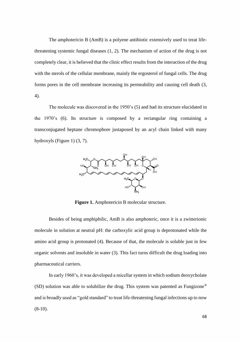
68
The amphotericin B (AmB) is a polyene antibiotic extensively used to treat life-
threatening systemic fungal diseases (1, 2). The mechanism of action of the drug is not
completely clear, it is believed that the clinic effect results from the interaction of the drug
with the sterols of the cellular membrane, mainly the ergosterol of fungal cells. The drug
forms pores in the cell membrane increasing its permeability and causing cell death (3,
4).
The molecule was discovered in the 1950’s (5) and had its structure elucidated in
the 1970’s (6). Its structure is composed by a rectangular ring containing a
transconjugated heptane chromophore justaposed by an acyl chain linked with many
hydroxyls (Figure 1) (3, 7).
Figure 1. Amphotericin B molecular structure.
Besides of being amphiphilic, AmB is also amphoteric, once it is a zwitterionic
molecule in solution at neutral pH: the carboxylic acid group is deprotonated while the
amino acid group is protonated (4). Because of that, the molecule is soluble just in few
organic solvents and insoluble in water (3). This fact turns difficult the drug loading into
pharmaceutical carriers.
In early 1960’s, it was developed a micellar system in which sodium deoxycholate
(SD) solution was able to solubilize the drug. This system was patented as Fungizone®
and is broadly used as “gold standard” to treat life-threatening fungal infections up to now
(8-10).
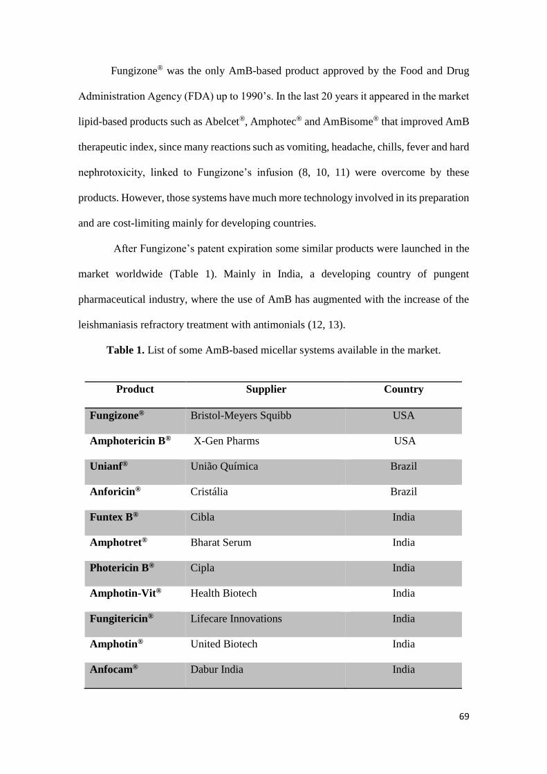
69
Fungizone® was the only AmB-based product approved by the Food and Drug
Administration Agency (FDA) up to 1990’s. In the last 20 years it appeared in the market
lipid-based products such as Abelcet®, Amphotec® and AmBisome® that improved AmB
therapeutic index, since many reactions such as vomiting, headache, chills, fever and hard
nephrotoxicity, linked to Fungizone’s infusion (8, 10, 11) were overcome by these
products. However, those systems have much more technology involved in its preparation
and are cost-limiting mainly for developing countries.
After Fungizone’s patent expiration some similar products were launched in the
market worldwide (Table 1). Mainly in India, a developing country of pungent
pharmaceutical industry, where the use of AmB has augmented with the increase of the
leishmaniasis refractory treatment with antimonials (12, 13).
Table 1. List of some AmB-based micellar systems available in the market.
Product Supplier Country
Fungizone® Bristol-Meyers Squibb USA
Amphotericin B® X-Gen Pharms USA
Unianf® União Química Brazil
Anforicin® Cristália Brazil
Funtex B® Cibla India
Amphotret® Bharat Serum India
Photericin B® Cipla India
Amphotin-Vit® Health Biotech India
Fungitericin® Lifecare Innovations India
Amphotin® United Biotech India
Anfocam® Dabur India India
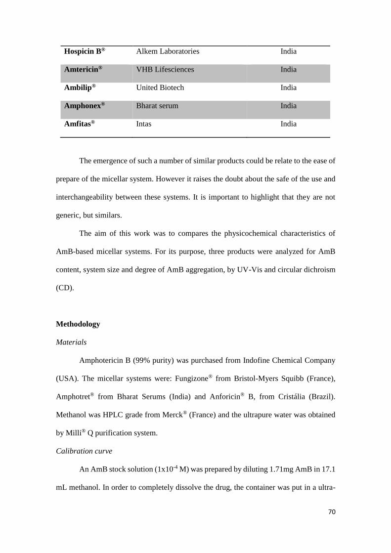
70
Hospicin B® Alkem Laboratories India
Amtericin® VHB Lifesciences India
Ambilip® United Biotech India
Amphonex® Bharat serum India
Amfitas® Intas India
The emergence of such a number of similar products could be relate to the ease of
prepare of the micellar system. However it raises the doubt about the safe of the use and
interchangeability between these systems. It is important to highlight that they are not
generic, but similars.
The aim of this work was to compares the physicochemical characteristics of
AmB-based micellar systems. For its purpose, three products were analyzed for AmB
content, system size and degree of AmB aggregation, by UV-Vis and circular dichroism
(CD).
Methodology
Materials
Amphotericin B (99% purity) was purchased from Indofine Chemical Company
(USA). The micellar systems were: Fungizone® from Bristol-Myers Squibb (France),
Amphotret® from Bharat Serums (India) and Anforicin® B, from Cristália (Brazil).
Methanol was HPLC grade from Merck® (France) and the ultrapure water was obtained
by Milli® Q purification system.
Calibration curve
An AmB stock solution (1x10⁻⁴ M) was prepared by diluting 1.71mg AmB in 17.1
mL methanol. In order to completely dissolve the drug, the container was put in a ultra-

71
sound bath during 5 minutes. Then, the stock solution was diluted in methanol for nine
points of AmB concentration (1.6x10⁻⁵; 8.0x10⁻⁶; 6.0x10⁻⁶; 5.0x10⁻⁶; 4.0x10⁻⁶; 2.5x10⁻⁶;
2.0x10⁻⁶; 1.0x10⁻⁶ and 5.0x10⁻⁷ M) that were read in a Perkin Elmer (France)
spectrophotometer with a 1cm length quartz cell. A screening was performed from 300
to 450 nm wavelength and λmax= 405 nm were used for AmB quantification.
AmB-SD solution preparation
All the products: Fungizone®, Amphotret® and Anforicin® B are presented as
lyophilized yellow powder containing 50 mg AmB and 41 mg SD. They were diluted in
10 mL water for injection, as recommended for the suppliers and reserved for further use.
The resulting solution was 5x10⁻³ M AmB concentrated. The formulations were identified
in this study as A; B and C, randomly, to avoid commercial appeal.
AmB quantification
The calibration curve was used to quantify the AmB-content into the products.
After water suspension (5x10⁻³ M), the products were diluted 1000-fold in methanol, in
order to result a 5x10⁻⁶ M AmB concentration, well fitting the calibration curve and
assuring accurate drug quantification at λmax= 405 nm.
Evaluation of AmB aggregation state
The systems were diluted in ultra-pure water in order to have its aggregation state
evaluated. The drug conformation was analyzed for both electronic and circular dichroic
(CD) spectroscopy.
UV-Vis spectra were recorded in a PerkinElmer (France) spectrophotometer after
1000x dilution of the re-suspended product, using a 1cm length quartz cell. The wavescan
was from λ = 300 to 450 nm (14).
For circular dicrhoism (CD), transitions of AmB were recorded from dilutions of
5x10⁻⁸ up to 5x10⁻⁵ M on a Jasco J-810 dichrograph (France), by using 10, 5, 1 and 0.1
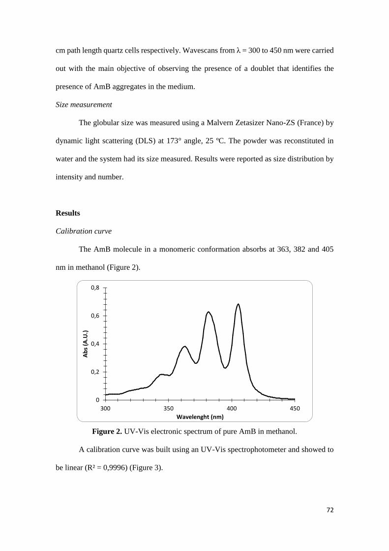
72
cm path length quartz cells respectively. Wavescans from λ = 300 to 450 nm were carried
out with the main objective of observing the presence of a doublet that identifies the
presence of AmB aggregates in the medium.
Size measurement
The globular size was measured using a Malvern Zetasizer Nano-ZS (France) by
dynamic light scattering (DLS) at 173° angle, 25 ºC. The powder was reconstituted in
water and the system had its size measured. Results were reported as size distribution by
intensity and number.
Results
Calibration curve
The AmB molecule in a monomeric conformation absorbs at 363, 382 and 405
nm in methanol (Figure 2).
Figure 2. UV-Vis electronic spectrum of pure AmB in methanol.
A calibration curve was built using an UV-Vis spectrophotometer and showed to
be linear (R² = 0,9996) (Figure 3).
0
0,2
0,4
0,6
0,8
300 350 400 450
Ab
s (A
.U.)
Wavelenght (nm)
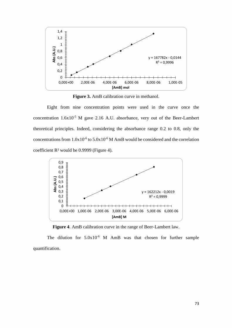
73
Figure 3. AmB calibration curve in methanol.
Eight from nine concentration points were used in the curve once the
concentration 1.6x10⁻⁵ M gave 2.16 A.U. absorbance, very out of the Beer-Lambert
theoretical principles. Indeed, considering the absorbance range 0.2 to 0.8, only the
concentrations from 1.0x10⁻⁶ to 5.0x10⁻⁶ M AmB would be considered and the correlation
coefficient R² would be 0.9999 (Figure 4).
Figure 4. AmB calibration curve in the range of Beer-Lambert law.
The dilution for 5.0x10⁻⁶ M AmB was that chosen for further sample
quantification.
y = 167782x - 0,0144R² = 0,9996
0
0,2
0,4
0,6
0,8
1
1,2
1,4
0,00E+00 2,00E-06 4,00E-06 6,00E-06 8,00E-06 1,00E-05
Ab
s (A
.U.)
[AmB] mol
y = 162212x - 0,0019R² = 0,9999
00,10,20,30,40,50,60,70,80,9
0,00E+00 1,00E-06 2,00E-06 3,00E-06 4,00E-06 5,00E-06 6,00E-06
Ab
s (A
.U.)
[AmB] M
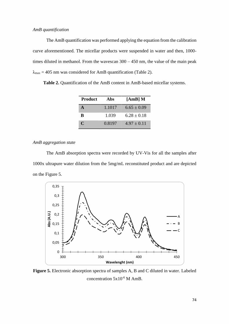
74
AmB quantification
The AmB quantification was performed applying the equation from the calibration
curve aforementioned. The micellar products were suspended in water and then, 1000-
times diluted in methanol. From the wavescan 300 – 450 nm, the value of the main peak
λmax = 405 nm was considered for AmB quantification (Table 2).
Table 2. Quantification of the AmB content in AmB-based micellar systems.
Product Abs [AmB] M
A 1.1017 6.65 ± 0.09
B 1.039 6.28 ± 0.18
C 0.8197 4.97 ± 0.11
AmB aggregation state
The AmB absorption spectra were recorded by UV-Vis for all the samples after
1000x ultrapure water dilution from the 5mg/mL reconstituted product and are depicted
on the Figure 5.
Figure 5. Electronic absorption spectra of samples A, B and C diluted in water. Labeled
concentration 5x10⁻⁶ M AmB.
0
0,05
0,1
0,15
0,2
0,25
0,3
0,35
300 350 400 450
Ab
s (A
.U.)
Wavelenght (nm)
A
B
C

75
The spectra were characterized for the presence of both monomers and aggregates.
The aggregation level in the samples was then calculated as the ratio between the
absorption of aggregates (λ = 325nm) by the absorption value of the main peak of
monomers (λ = 408nm) (Figure 6).
Figure 6. Relation between AmB species: aggregates and monomers, calculated from
UV-Vis absorbances at 325 and 408 nm respectively.
The aggregation state of the samples was also evaluated by CD. All of them
presented an exciton couplet characteristic of aggregated AmB forms (Figure 7).
Figure 7. CD spectra of AmB-containing micellar systems at 5.10-5 M AmB in water.
Values were corrected in b) according to the quantified AmB.
0 0,5 1 1,5 2
A
B
C
Ratio (325/408 nm)
Pro
du
ct
a) b)
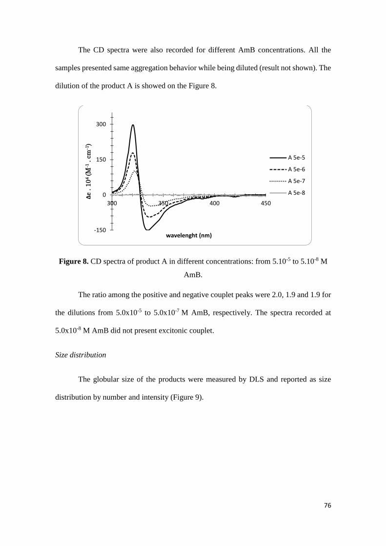
76
The CD spectra were also recorded for different AmB concentrations. All the
samples presented same aggregation behavior while being diluted (result not shown). The
dilution of the product A is showed on the Figure 8.
Figure 8. CD spectra of product A in different concentrations: from 5.10-5 to 5.10-8 M
AmB.
The ratio among the positive and negative couplet peaks were 2.0, 1.9 and 1.9 for
the dilutions from 5.0x10⁻⁵ to 5.0x10⁻⁷ M AmB, respectively. The spectra recorded at
5.0x10⁻⁸ M AmB did not present excitonic couplet.
Size distribution
The globular size of the products were measured by DLS and reported as size
distribution by number and intensity (Figure 9).
-150
0
150
300
300 350 400 450
∆ε
. 1
04
(M
-1. cm
-1)
wavelenght (nm)
A 5e-5
A 5e-6
A 5e-7
A 5e-8
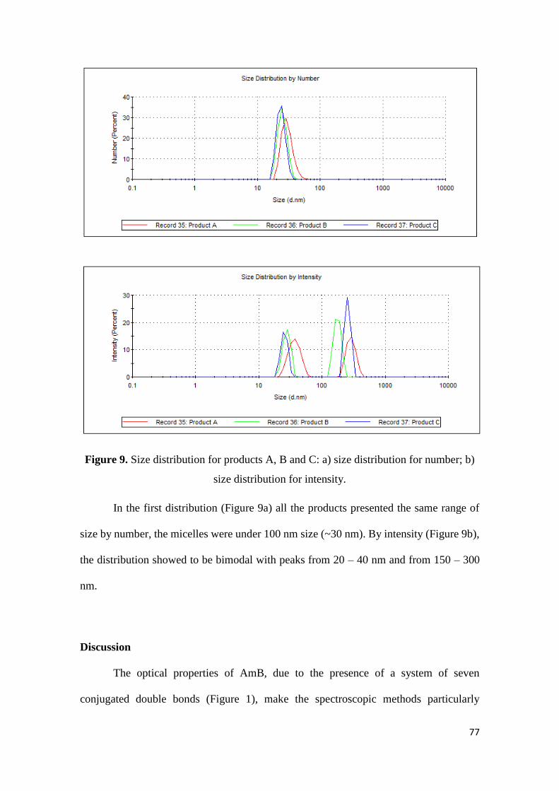
77
Figure 9. Size distribution for products A, B and C: a) size distribution for number; b)
size distribution for intensity.
In the first distribution (Figure 9a) all the products presented the same range of
size by number, the micelles were under 100 nm size (~30 nm). By intensity (Figure 9b),
the distribution showed to be bimodal with peaks from 20 – 40 nm and from 150 – 300
nm.
Discussion
The optical properties of AmB, due to the presence of a system of seven
conjugated double bonds (Figure 1), make the spectroscopic methods particularly
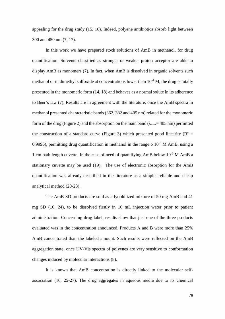
78
appealing for the drug study (15, 16). Indeed, polyene antibiotics absorb light between
300 and 450 nm (7, 17).
In this work we have prepared stock solutions of AmB in methanol, for drug
quantification. Solvents classified as stronger or weaker proton acceptor are able to
display AmB as monomers (7). In fact, when AmB is dissolved in organic solvents such
methanol or in dimethyl sulfoxide at concentrations lower than 10-4 M, the drug is totally
presented in the monomeric form (14, 18) and behaves as a normal solute in its adherence
to Beer’s law (7). Results are in agreement with the literature, once the AmB spectra in
methanol presented characteristic bands (362, 382 and 405 nm) related for the monomeric
form of the drug (Figure 2) and the absorption on the main band (λmax= 405 nm) permitted
the construction of a standard curve (Figure 3) which presented good linearity (R² =
0,9996), permitting drug quantification in methanol in the range o 10-6 M AmB, using a
1 cm path length cuvette. In the case of need of quantifying AmB below 10-6 M AmB a
stationary cuvette may be used (19). The use of electronic absorption for the AmB
quantification was already described in the literature as a simple, reliable and cheap
analytical method (20-23).
The AmB-SD products are sold as a lyophilized mixture of 50 mg AmB and 41
mg SD (10, 24), to be dissolved firstly in 10 mL injection water prior to patient
administration. Concerning drug label, results show that just one of the three products
evaluated was in the concentration announced. Products A and B were more than 25%
AmB concentrated than the labeled amount. Such results were reflected on the AmB
aggregation state, once UV-Vis spectra of polyenes are very sensitive to conformation
changes induced by molecular interactions (8).
It is known that AmB concentration is directly linked to the molecular self-
association (16, 25-27). The drug aggregates in aqueous media due to its chemical
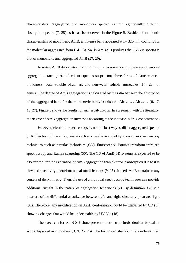
79
characteristics. Aggregated and monomers species exhibit significantly different
absorption spectra (7, 28) as it can be observed in the Figure 5. Besides of the bands
characteristics of monomeric AmB, an intense band appeared at λ= 325 nm, counting for
the molecular aggregated form (14, 18). So, in AmB-SD products the UV-Vis spectra is
that of monomeric and aggregated AmB (27, 29).
In water, AmB dissociates from SD forming monomers and oligomers of various
aggregation states (10). Indeed, in aqueous suspension, three forms of AmB coexist:
monomers, water-soluble oligomers and non-water soluble aggregates (14, 25). In
general, the degree of AmB aggregation is calculated by the ratio between the absorption
of the aggregated band for the monomeric band, in this case Abs325 nm/ Abs408 nm (8, 17,
18, 27). Figure 6 shows the results for such a calculation. In agreement with the literature,
the degree of AmB aggregation increased according to the increase in drug concentration.
However, electronic spectroscopy is not the best way to differ aggregated species
(18). Spectra of different organization forms can be recorded by many other spectroscopy
techniques such as circular dichroisim (CD), fluorescence, Fourier transform infra red
spectroscopy and Raman scattering (30). The CD of AmB-SD systems is expected to be
a better tool for the evaluation of AmB aggregation than electronic absorption due to it is
elevated sensitivity to environmental modifications (9, 15). Indeed, AmB contains many
centers of dissymmetry. Then, the use of chiroptical spectroscopy techniques can provide
additional insight in the nature of aggregation tendencies (7). By definition, CD is a
measure of the differential absorbance between left- and right-circularly polarized light
(31). Therefore, any modification on AmB conformation could be identified by CD (9),
showing changes that would be undetectable by UV-Vis (18).
The spectrum for AmB-SD alone presents a strong dichroic doublet typical of
AmB dispersed as oligomers (3, 9, 25, 26). The bisignated shape of the spectrum is an
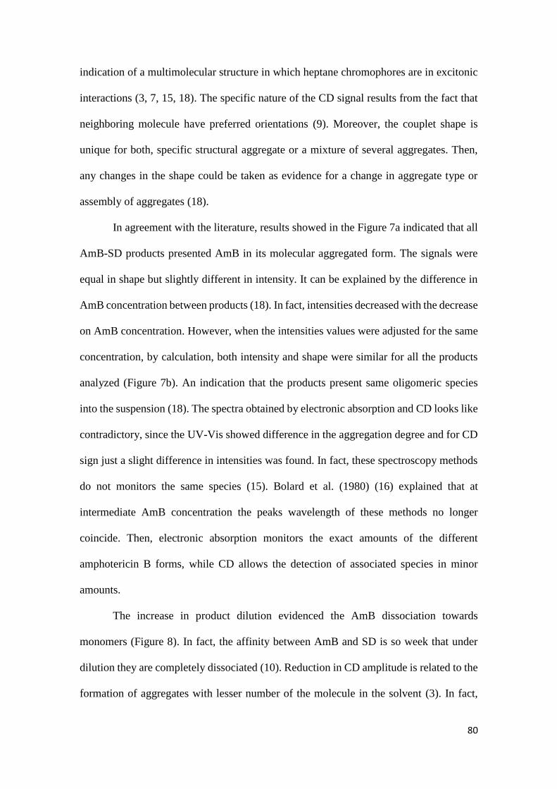
80
indication of a multimolecular structure in which heptane chromophores are in excitonic
interactions (3, 7, 15, 18). The specific nature of the CD signal results from the fact that
neighboring molecule have preferred orientations (9). Moreover, the couplet shape is
unique for both, specific structural aggregate or a mixture of several aggregates. Then,
any changes in the shape could be taken as evidence for a change in aggregate type or
assembly of aggregates (18).
In agreement with the literature, results showed in the Figure 7a indicated that all
AmB-SD products presented AmB in its molecular aggregated form. The signals were
equal in shape but slightly different in intensity. It can be explained by the difference in
AmB concentration between products (18). In fact, intensities decreased with the decrease
on AmB concentration. However, when the intensities values were adjusted for the same
concentration, by calculation, both intensity and shape were similar for all the products
analyzed (Figure 7b). An indication that the products present same oligomeric species
into the suspension (18). The spectra obtained by electronic absorption and CD looks like
contradictory, since the UV-Vis showed difference in the aggregation degree and for CD
sign just a slight difference in intensities was found. In fact, these spectroscopy methods
do not monitors the same species (15). Bolard et al. (1980) (16) explained that at
intermediate AmB concentration the peaks wavelength of these methods no longer
coincide. Then, electronic absorption monitors the exact amounts of the different
amphotericin B forms, while CD allows the detection of associated species in minor
amounts.
The increase in product dilution evidenced the AmB dissociation towards
monomers (Figure 8). In fact, the affinity between AmB and SD is so week that under
dilution they are completely dissociated (10). Reduction in CD amplitude is related to the
formation of aggregates with lesser number of the molecule in the solvent (3). In fact,
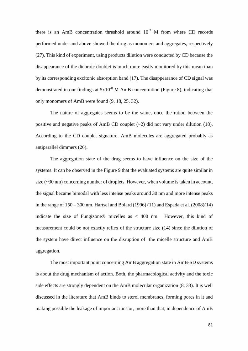
81
there is an AmB concentration threshold around 10-7 M from where CD records
performed under and above showed the drug as monomers and aggregates, respectively
(27). This kind of experiment, using products dilution were conducted by CD because the
disappearance of the dichroic doublet is much more easily monitored by this mean than
by its corresponding excitonic absorption band (17). The disappearance of CD signal was
demonstrated in our findings at 5x10⁻⁸ M AmB concentration (Figure 8), indicating that
only monomers of AmB were found (9, 18, 25, 32).
The nature of aggregates seems to be the same, once the ration between the
positive and negative peaks of AmB CD couplet (~2) did not vary under dilution (18).
According to the CD couplet signature, AmB molecules are aggregated probably as
antiparallel dimmers (26).
The aggregation state of the drug seems to have influence on the size of the
systems. It can be observed in the Figure 9 that the evaluated systems are quite similar in
size (~30 nm) concerning number of droplets. However, when volume is taken in account,
the signal became bimodal with less intense peaks around 30 nm and more intense peaks
in the range of 150 – 300 nm. Hartsel and Bolard (1996) (11) and Espada et al. (2008)(14)
indicate the size of Fungizone® micelles as < 400 nm. However, this kind of
measurement could be not exactly reflex of the structure size (14) since the dilution of
the system have direct influence on the disruption of the micelle structure and AmB
aggregation.
The most important point concerning AmB aggregation state in AmB-SD systems
is about the drug mechanism of action. Both, the pharmacological activity and the toxic
side effects are strongly dependent on the AmB molecular organization (8, 33). It is well
discussed in the literature that AmB binds to sterol membranes, forming pores in it and
making possible the leakage of important ions or, more than that, in dependence of AmB
82
concentration, causing the disruption of the cell, with consequently cell death (15, 16). It
has been proved that AmB-SD of high aggregation state is more toxic than the same
mixture at lower aggregation state (4, 8, 14, 25).
The drug bind to ergosterol and/or cholesterol is suggested to be the key for
understanding the appearance of AmB side effects (4, 26). It is admitted that the
interactions between AmB and sterols occur through hydrophobic interactions, where the
seven conjugated AmB double bonds interacts with ergosterol or cholesterol (4). AmB is
not specific for the binding to ergosterol, the sterol of fungi and protozoa cells. However,
it has been demonstrated that when the drug is in its monomeric form, it is not able to
cause toxicity for cholesterol-containing cells (4, 14). Then, the difference in AmB
aggregation in products supposed to be equal could result in differences in the therapeutic
response, incurring in patient’s hard side effects and consequently augmentation in the
time of hospital housing.
Conclusion
To our knowledge, this is the first time that a comparison between AmB-SD
micellar systems is made. The overall results showed that the marketed products analyzed
have similar mean diameter size, despite the unmatched labeled content. Only the product
C was in the designated concentration of 5 mg/mL. Products A and B were more than
25% up concentrated. All of the products were presented as a mix of AmB monomers and
oligomers as expected, and the level of AmB aggregation was in accordance to the drug
concentration. CD studies revealed that the same species of oligomers are presented into
the three formulations. This study suggests, in the light of AmB aggregation that the
AmB-SD systems in here evaluated cannot be interchangeable.
83
Acknowledgments
Authors are grateful to CAPES-COFECUB (project 742/11) for the scholarship provided
to A. L. Silva.
References
1. Hoeprich PD. Clinical use of amphotericin B and derivatives - Lore, Mystique,
and Fact. Clin Infect Dis. 1992;14:S114-S9.
2. Sundar S, Chakravarty J. Liposomal amphotericin B and leishmaniasis: dose and
response. J Glob Infect Dis. 2010;2(2):159-66.
3. Balakrishnan AR, Easwaran KRK. CD and NMR-studies on the aggregation of
amphotericin-B in solution. Biochim Biophys Acta. 1993;1148(2):269-77.
4. Barwicz J, Tancrède P. The effect of aggregation state of amphotericin-B on its
interactions with cholesterol- or ergosterol-containing phosphatidylcholine monolayers.
Chem Phys Lipids. 1997;85(2):145-55.
5. Dutcher JD, William G, Pagano JF, John V. Amphotericin b, its production, and
its salts. Google Patents; 1959.
6. Aszalos A, Bax A, Burlinson N, Roller P, McNeal C. Physico-chemical and
microbiological comparison of nystatin, amphotericin A and amphotericin B, and
structure of amphotericin A. J Antibiot (Tokyo). 1985;38(12):1699-713.
7. Brittain HG. Circular dichroism studies of the self-association of amphotericin B.
Chirality. 1994;6(8):665-9.
8. Barwicz J, Christian S, Gruda I. Effects of the aggregation state of amphotericin
B on its toxicity to mice. Antimicrob Agents Chemother. 1992;36(10):2310-5.
9. Barwicz J, Beauregard M, Tancrede P. Circular dichroism study of interactions of
Fungizone or AmBisome forms of amphotericin B with human low density lipoproteins.
Biopolymers. 2002;67(1):49-55.
10. Brajtburg J, Bolard J. Carrier effects on biological activity of amphotericin B. Clin
Microbiol Rev. 1996;9(4):512-31.
11. Hartsel S, Bolard J. Amphotericin B: new life for an old drug. Trends Pharmacol
Sci. 1996;17(12):445-9.
84
12. Singh OP, Singh B, Chakravarty J, Sundar S. Current challenges in treatment
options for visceral leishmaniasis in India: a public health perspective. Infect Dis Poverty.
2016;5(1):19.
13. Sundar S. Drug resistance in Indian visceral leishmaniasis. Trop Med Int Health.
2001;6(11):849-54.
14. Espada R, Valdespina S, Alfonso C, Rivas G, Ballesteros MP, Torrado JJ. Effect
of aggregation state on the toxicity of different amphotericin B preparations. Int J Pharm.
2008;361(1–2):64-9.
15. Boudet G, Bolard J. Interaction of the polyene antibiotic amphotericin B with
phospholipid bilayer membranes: A circular dichroism study. Biochem Biophys Res
Commun. 1979;88(3):998-1002.
16. Bolard J, Seigneuret M, Boudet G. Interaction between phospholipid bilayer
membranes and the polyene antibiotic amphotericin B. Biochim Biophys Acta
Biomembranes. 1980;599(1):280-93.
17. Bolard J. How do the polyene macrolide antibiotics affect the cellular membrane
properties? Biochim Biophys Acta Rev Biomembranes. 1986;864(3):257-304.
18. Jameson LP, Dzyuba SV. Circular dichroism studies on intermolecular
interactions of amphotericin B in ionic liquid-rich environments. Chirality.
2013;25(7):427-32.
19. Silva KGH, Xavier FH, Farias IEG, Silva AKA, Neto JAC, Souza LCA, et al.
Stationary cuvette: a new approach to obtaining analytical curves by UV-VIS
spectrophotometry. Phytochem Anal. 2009;20(4):265-71.
20. Botsoglou NA, Fletouris DJ, Papageorgiou GE, FlorouPaneri P, Mantis AJ. Rapid
determination of amphotericin B in serum and urine by third-order derivative
spectrophotometry. J Pharm Sci. 1996;85(4):402-6.
21. Casaccia P, Ladogana A, Xi YG, Ingrosso L, Pocchiari M, Silvestrini MC, et al.
Measurement of the Concentration of Amphotericin-B in Brain Tissue of Scrapie-
Infected Hamsters with a Simple and Sensitive Method. Antimicrob Agents Chemother.
1991;35(7):1486-8.
22. Millership JS, McCaffrey F, Tierney D. A novel application of ratio spectra
derivative spectroscopy for the determination of amphotericin in highly icteric plasma. J
Pharm Biomed Anal. 2008;48(2):408-13.
23. Damasceno BPGL, Dominici VA, Urbano IA, Silva JA, Araujo IB, Santos-
Magalhaes NS, et al. Amphotericin B microemulsion reduces toxicity and maintains the
efficacy as an antifungal product. J Biomed Nanotechnol. 2012;8(2):290-300.
24. Araújo IB, Brito CRN, Urbano IA, Dominici VA, Silva Filho MA, Silveira WLL,
et al. Similarity between the in vitro activity and toxicity of two different fungizone /
lipofundin admixtures. Acta Bras Cir. 2005;20:129-33.
85
25. Legrand P, Romero EA, Cohen BE, Bolard J. Effects of aggregation and solvent
on the toxicity of amphotericin B to human erythrocytes. Antimicrob Agents Chemother.
1992;36(11):2518-22.
26. Zielińska J, Wieczór M, Bączek T, Gruszecki M, Czub J. Thermodynamics and
kinetics of amphotericin B self-association in aqueous solution characterized in molecular
detail. Sci Rep. 2016;6:19109.
27. Tancréde P, Barwicz J, Jutras S, Gruda I. The effect of surfactants on the
aggregation state of amphotericin B. Biochim Biophys Acta Biomembranes.
1990;1030(2):289-95.
28. Jullien S, Vertut-Croquin A, Brajtburg J, Bolard J. Circular dichroism for the
determination of amphotericin B binding to liposomes. Anal Biochem. 1988;172(1):197-
202.
29. Siqueira SD, Silva-Filho MA, Silva CA, Araujo IB, Silva AE, Fernandes-Pedrosa
MF, et al. Influence of the freeze-drying process on the physicochemical and biological
properties of pre-heated amphotericin B micellar systems. AAPS PharmSciTech.
2014;15(3):612-9.
30. Starzyk J, Gruszecki M, Tutaj K, Luchowski R, Szlazak R, Wasko P, et al. Self-
association of amphotericin B: spontaneous formation of molecular structures responsible
for the toxic side effects of the antibiotic. J Phys Chem B. 2014;118(48):13821-32.
31. Circular Dicrhoism and the Conformational Analysis of Biomolecules. Fasman
GD, editor. New York: Springer US; 1996. 738 p.
32. Silva AE, Barratt G, Cheron M, Egito EST. Development of oil-in-water
microemulsions for the oral delivery of amphotericin B. Int J Pharm. 2013;454(2):641-8.
33. Gagoś M, Czernel G, Kamiński DM, Kostro K. Spectroscopic studies of
amphotericin B–Cu2+ complexes. BioMetals. 2011;24(5):915.
86
CHAPTER III
Amphotericin B super-aggregates: formation and influence on the drug
stability
87
Amphotericin B Super-aggregates: formation and influence on the drug stability
André Leandro Silva 1,2; An Young Sarahi Taylor de Castillo 2; Monique Chéron 3; Gilles
Ponchel 2; Eryvaldo Sócrates Tabosa do Egito *1.
1 Programa de Pós-Graduação em Biotecnologia (Renorbio), Universidade Federal do Rio
Grande do Norte, Natal – RN, Brazil;
2 UMR CNRS 8612, Institut Galien Paris-Sud, Université Paris-Saclay, Châtenay-
Malabry, France.
3 UMR CNRS 7033, Université Pierre et Marie Curie, Paris, France.
*Corresponding author:
Eryvaldo Sócrates Tabosa do Egito
Rua Praia de Areia Branca, 8448. Ponta Negra, Natal – Brazil (59094-450).
Phone: +55 84 9 9431-8816 or +55 84 3342-9817
E-mail address: [email protected] or [email protected]
88
Abstract
The amphotericin B (AmB) is a polyene antibiotic used to treat life-threatening systemic
fungal infections. The drug is currently available as AmB-micelles and -lipid-based
products. The micelles are a mix of AmB and sodium deoxycholate (SD), a biliar salt that
acts as a surfactant. This is a quite simple delivery system, and its injection is responsible
for important side-effects of the drug. The mild heating has been proved to improve the
safety of AmB-SD micelles by the formation of super-aggregates. In this work we tested
the possibility of super-aggregates formation from a liposomal system and tried to
measure the energy involved in the super-aggregation process. In addition, the heated
system was evaluated along the time, to evaluate the influence of the process on the drug
stability. It was demonstrated that only AmB-SD micelles can form super-aggregates that
improved the drug stability along the time. The process was accompanied of spectral
changes by UV-Vis and circular dichroism (CD). These changes, caused by the mild
heating seem to be irreversible. Isothermal titration calorimetry (ITC) was not able to
clarify about the energy spent/generated on super-aggregation process, but suggested that
some energy is generated during the re-organization of oligomers towards monomers of
AmB.
Key-words: amphotericin B; micellar system; liposomal system; super-aggregates;
circular dichroism; isothermal titration calorimetry;
89
Introduction
The amphotericin B (AmB) molecule is an antibiotic discovered in the 1950’s and
broadly used in the treatment of life-threatening systemic fungal infections up to now (1).
More recently, the drug has been used to fight against the leishmanial protozoa, in
refractory treatment with antimonials (2).
Drug physico-chemical characteristics are marked by hydrophobicity, due to a
polyene chain and hydrophilicity, due to a polyhydroxylated region. Besides, AmB has
amphoteric character. All together are responsible for the poor solubility of the drug (3).
Indeed, it is the main drawback to the development of AmB-containing pharmaceutical
formulations (4).
AmB was firstly marketed in the form of AmB-containing micelles, a mix of the
drug with sodium deoxycholate (SD). In spite of the problems such as headache, nausea,
vomiting, phlebitis and nephrotoxicity related to the AmB-SD micelles infusion, the
product is commonly used, mainly in developing countries (5, 6). In the last decades,
AmB-containing lipid-based products have been introduced in the market (7-9),
increasing AmB safety, mainly by nanomodification (10-13). Indeed, the lipid systems
developed are less toxic and have the possibility of improvement of doses, that was not
possible for AmB-SD (8, 14). However, due to their cost, this sort of systems are not
affordable for most of poor countries (4, 9).
Strategies those are still in the field of research deal with the use of micelles
themselves being diluted for other systems like nanoemulsions and microemulsions (6,
15), as an attempt of de-aggregate the drug, delivering monomers of AmB instead of
oligomers, proved to be more toxic AmB species (16).
The mild heat of AmB-SD has been related to the formation of a different
supramolecular organization of the drug. Through this process a new spectroscopic AmB
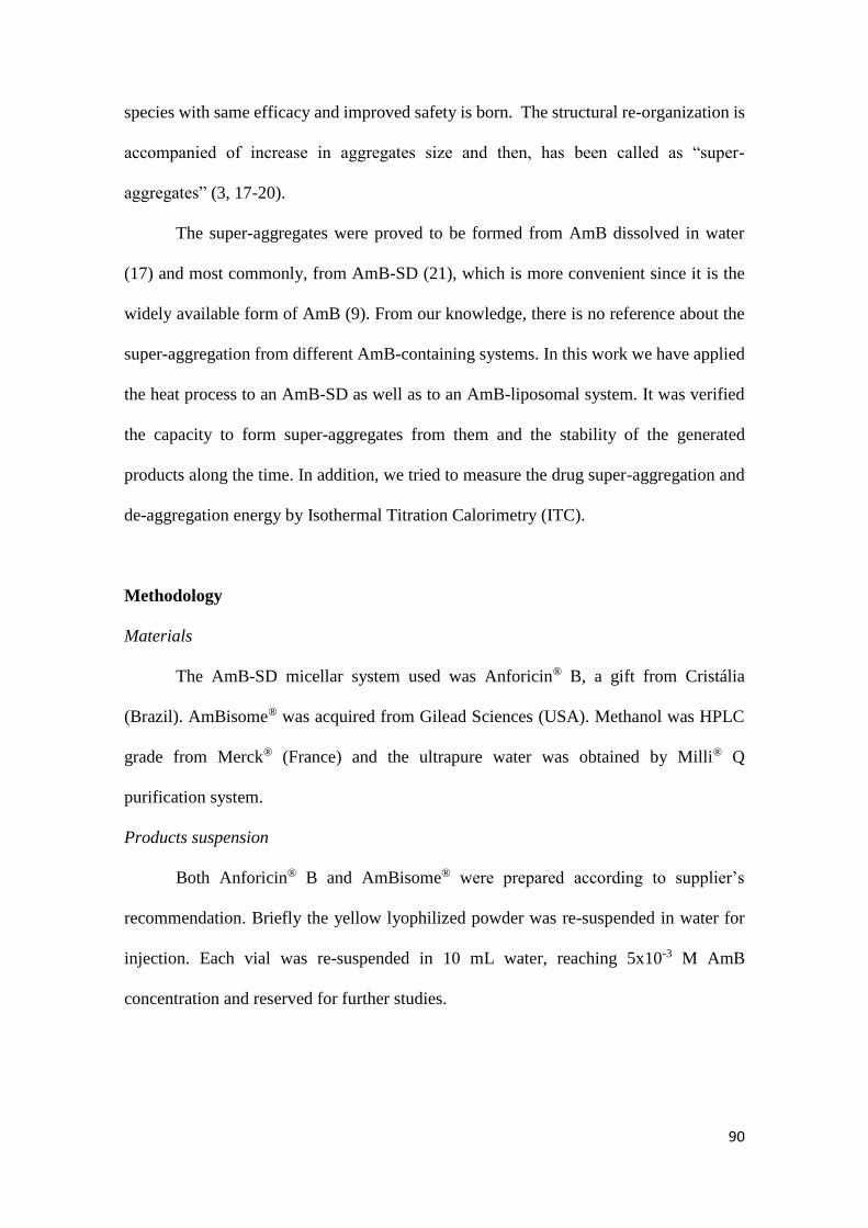
90
species with same efficacy and improved safety is born. The structural re-organization is
accompanied of increase in aggregates size and then, has been called as “super-
aggregates” (3, 17-20).
The super-aggregates were proved to be formed from AmB dissolved in water
(17) and most commonly, from AmB-SD (21), which is more convenient since it is the
widely available form of AmB (9). From our knowledge, there is no reference about the
super-aggregation from different AmB-containing systems. In this work we have applied
the heat process to an AmB-SD as well as to an AmB-liposomal system. It was verified
the capacity to form super-aggregates from them and the stability of the generated
products along the time. In addition, we tried to measure the drug super-aggregation and
de-aggregation energy by Isothermal Titration Calorimetry (ITC).
Methodology
Materials
The AmB-SD micellar system used was Anforicin® B, a gift from Cristália
(Brazil). AmBisome® was acquired from Gilead Sciences (USA). Methanol was HPLC
grade from Merck® (France) and the ultrapure water was obtained by Milli® Q
purification system.
Products suspension
Both Anforicin® B and AmBisome® were prepared according to supplier’s
recommendation. Briefly the yellow lyophilized powder was re-suspended in water for
injection. Each vial was re-suspended in 10 mL water, reaching 5x10⁻³ M AmB
concentration and reserved for further studies.
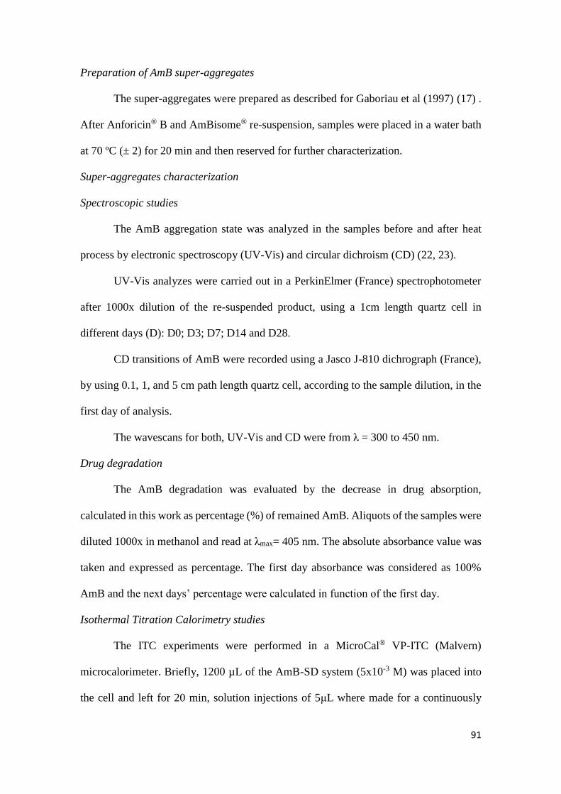
91
Preparation of AmB super-aggregates
The super-aggregates were prepared as described for Gaboriau et al (1997) (17) .
After Anforicin® B and AmBisome® re-suspension, samples were placed in a water bath
at 70 ºC (± 2) for 20 min and then reserved for further characterization.
Super-aggregates characterization
Spectroscopic studies
The AmB aggregation state was analyzed in the samples before and after heat
process by electronic spectroscopy (UV-Vis) and circular dichroism (CD) (22, 23).
UV-Vis analyzes were carried out in a PerkinElmer (France) spectrophotometer
after 1000x dilution of the re-suspended product, using a 1cm length quartz cell in
different days (D): D0; D3; D7; D14 and D28.
CD transitions of AmB were recorded using a Jasco J-810 dichrograph (France),
by using 0.1, 1, and 5 cm path length quartz cell, according to the sample dilution, in the
first day of analysis.
The wavescans for both, UV-Vis and CD were from λ = 300 to 450 nm.
Drug degradation
The AmB degradation was evaluated by the decrease in drug absorption,
calculated in this work as percentage (%) of remained AmB. Aliquots of the samples were
diluted 1000x in methanol and read at λmax= 405 nm. The absolute absorbance value was
taken and expressed as percentage. The first day absorbance was considered as 100%
AmB and the next days’ percentage were calculated in function of the first day.
Isothermal Titration Calorimetry studies
The ITC experiments were performed in a MicroCal® VP-ITC (Malvern)
microcalorimeter. Briefly, 1200 µL of the AmB-SD system (5x10⁻³ M) was placed into
the cell and left for 20 min, solution injections of 5μL where made for a continuously
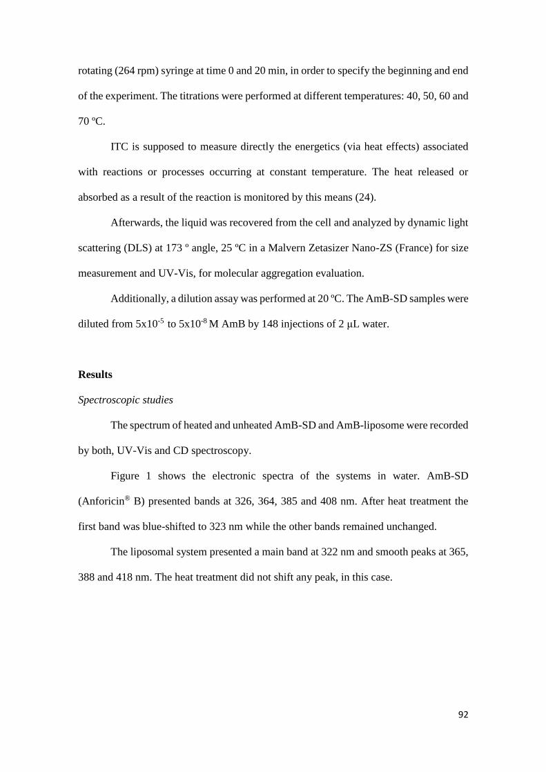
92
rotating (264 rpm) syringe at time 0 and 20 min, in order to specify the beginning and end
of the experiment. The titrations were performed at different temperatures: 40, 50, 60 and
70 ºC.
ITC is supposed to measure directly the energetics (via heat effects) associated
with reactions or processes occurring at constant temperature. The heat released or
absorbed as a result of the reaction is monitored by this means (24).
Afterwards, the liquid was recovered from the cell and analyzed by dynamic light
scattering (DLS) at 173 º angle, 25 ºC in a Malvern Zetasizer Nano-ZS (France) for size
measurement and UV-Vis, for molecular aggregation evaluation.
Additionally, a dilution assay was performed at 20 ºC. The AmB-SD samples were
diluted from 5x10⁻⁵ to 5x10⁻⁸ M AmB by 148 injections of 2 μL water.
Results
Spectroscopic studies
The spectrum of heated and unheated AmB-SD and AmB-liposome were recorded
by both, UV-Vis and CD spectroscopy.
Figure 1 shows the electronic spectra of the systems in water. AmB-SD
(Anforicin® B) presented bands at 326, 364, 385 and 408 nm. After heat treatment the
first band was blue-shifted to 323 nm while the other bands remained unchanged.
The liposomal system presented a main band at 322 nm and smooth peaks at 365,
388 and 418 nm. The heat treatment did not shift any peak, in this case.
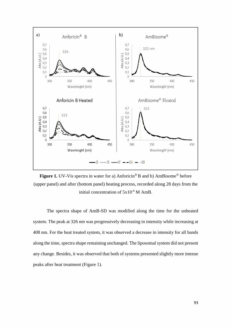
93
Figure 1. UV-Vis spectra in water for a) Anforicin® B and b) AmBisome® before
(upper panel) and after (bottom panel) heating process, recorded along 28 days from the
initial concentration of 5x10⁻⁶ M AmB.
The spectra shape of AmB-SD was modified along the time for the unheated
system. The peak at 326 nm was progressively decreasing in intensity while increasing at
408 nm. For the heat treated system, it was observed a decrease in intensity for all bands
along the time, spectra shape remaining unchanged. The liposomal system did not present
any change. Besides, it was observed that both of systems presented slightly more intense
peaks after heat treatment (Figure 1).
322 nm
322 nm 326 nm
323 nm
a) b)
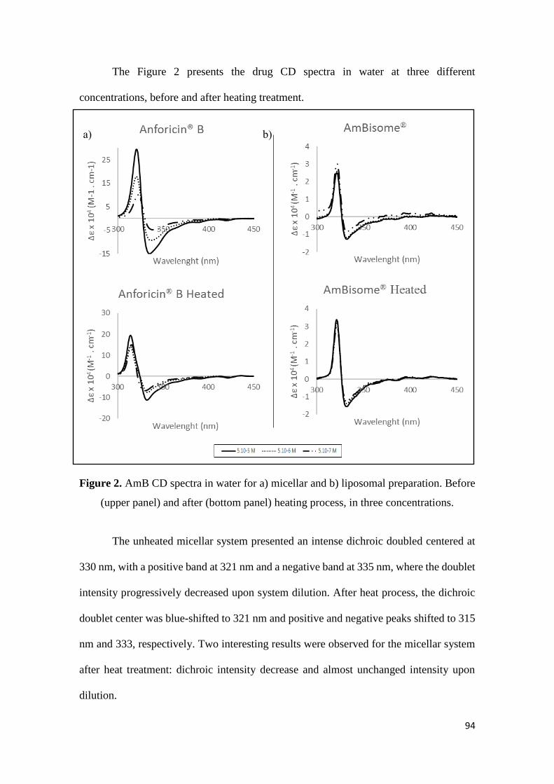
94
The Figure 2 presents the drug CD spectra in water at three different
concentrations, before and after heating treatment.
Figure 2. AmB CD spectra in water for a) micellar and b) liposomal preparation. Before
(upper panel) and after (bottom panel) heating process, in three concentrations.
The unheated micellar system presented an intense dichroic doubled centered at
330 nm, with a positive band at 321 nm and a negative band at 335 nm, where the doublet
intensity progressively decreased upon system dilution. After heat process, the dichroic
doublet center was blue-shifted to 321 nm and positive and negative peaks shifted to 315
nm and 333, respectively. Two interesting results were observed for the micellar system
after heat treatment: dichroic intensity decrease and almost unchanged intensity upon
dilution.
a) b)
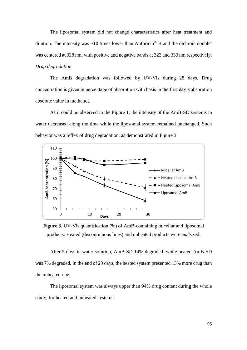
95
The liposomal system did not change characteristics after heat treatment and
dilution. The intensity was ~10 times lower than Anforicin® B and the dichroic doublet
was centered at 328 nm, with positive and negative bands at 322 and 333 nm respectively.
Drug degradation
The AmB degradation was followed by UV-Vis during 28 days. Drug
concentration is given in percentage of absorption with basis in the first day’s absorption
absolute value in methanol.
As it could be observed in the Figure 1, the intensity of the AmB-SD systems in
water decreased along the time while the liposomal system remained unchanged. Such
behavior was a reflex of drug degradation, as demonstrated in Figure 3.
Figure 3. UV-Vis quantification (%) of AmB-containing micellar and liposomal
products. Heated (discontinuous lines) and unheated products were analyzed.
After 5 days in water solution, AmB-SD 14% degraded, while heated AmB-SD
was 7% degraded. In the end of 29 days, the heated system presented 13% more drug than
the unheated one.
The liposomal system was always upper than 94% drug content during the whole
study, for heated and unheated systems.
50
60
70
80
90
100
110
0 10 20 30
Am
B c
on
cen
trat
ion
(%
)
Days
Micellar AmB
Heated micellar AmB
Heated Liposomal AmB
Liposomal AmB
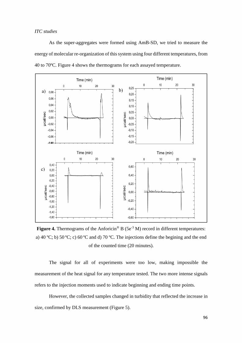
96
ITC studies
As the super-aggregates were formed using AmB-SD, we tried to measure the
energy of molecular re-organization of this system using four different temperatures, from
40 to 70ºC. Figure 4 shows the thermograms for each assayed temperature.
Figure 4. Thermograms of the Anforicin® B (5e-3 M) record in different temperatures:
a) 40 ºC; b) 50 ºC; c) 60 ºC and d) 70 ºC. The injections define the begining and the end
of the counted time (20 minutes).
The signal for all of experiments were too low, making impossible the
measurement of the heat signal for any temperature tested. The two more intense signals
refers to the injection moments used to indicate beginning and ending time points.
However, the collected samples changed in turbidity that reflected the increase in
size, confirmed by DLS measurement (Figure 5).
a)
c)
b)
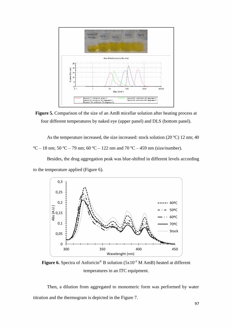
97
Figure 5. Comparison of the size of an AmB micellar solution after heating process at
four different temperatures by naked eye (upper panel) and DLS (bottom panel).
As the temperature increased, the size increased: stock solution (20 ºC) 12 nm; 40
ºC – 18 nm; 50 ºC – 79 nm; 60 ºC – 122 nm and 70 ºC – 459 nm (size/number).
Besides, the drug aggregation peak was blue-shifted in different levels according
to the temperature applied (Figure 6).
Figure 6. Spectra of Anforicin® B solution (5x10⁻³ M AmB) heated at different
temperatures in an ITC equipment.
Then, a dilution from aggregated to monomeric form was performed by water
titration and the thermogram is depicted in the Figure 7.
0
0,05
0,1
0,15
0,2
0,25
0,3
300 350 400 450
Ab
s (A
.U.)
Wavelenght (nm)
40ºC
50ºC
60ºC
70ºC
Stock
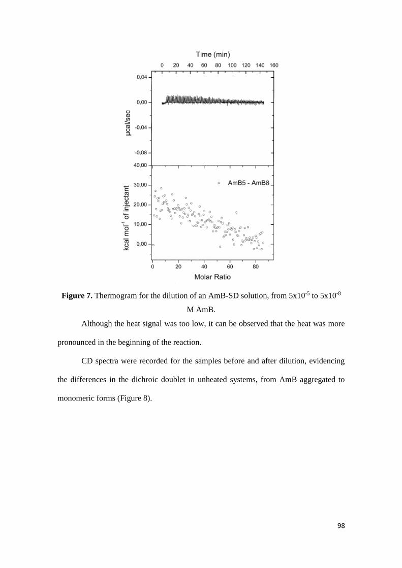
98
Figure 7. Thermogram for the dilution of an AmB-SD solution, from 5x10⁻⁵ to 5x10⁻⁸
M AmB.
Although the heat signal was too low, it can be observed that the heat was more
pronounced in the beginning of the reaction.
CD spectra were recorded for the samples before and after dilution, evidencing
the differences in the dichroic doublet in unheated systems, from AmB aggregated to
monomeric forms (Figure 8).
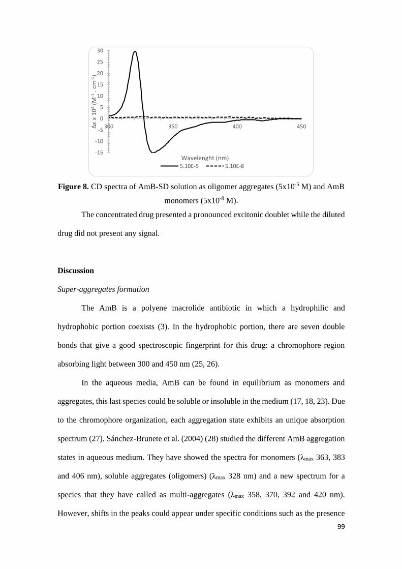
99
Figure 8. CD spectra of AmB-SD solution as oligomer aggregates (5x10⁻⁵ M) and AmB
monomers (5x10⁻⁸ M).
The concentrated drug presented a pronounced excitonic doublet while the diluted
drug did not present any signal.
Discussion
Super-aggregates formation
The AmB is a polyene macrolide antibiotic in which a hydrophilic and
hydrophobic portion coexists (3). In the hydrophobic portion, there are seven double
bonds that give a good spectroscopic fingerprint for this drug: a chromophore region
absorbing light between 300 and 450 nm (25, 26).
In the aqueous media, AmB can be found in equilibrium as monomers and
aggregates, this last species could be soluble or insoluble in the medium (17, 18, 23). Due
to the chromophore organization, each aggregation state exhibits an unique absorption
spectrum (27). Sánchez-Brunete et al. (2004) (28) studied the different AmB aggregation
states in aqueous medium. They have showed the spectra for monomers (λmax 363, 383
and 406 nm), soluble aggregates (oligomers) (λmax 328 nm) and a new spectrum for a
species that they have called as multi-aggregates (λmax 358, 370, 392 and 420 nm).
However, shifts in the peaks could appear under specific conditions such as the presence
-15
-10
-5
0
5
10
15
20
25
30
300 350 400 450∆ε
x 1
04
(M-1
. cm
-1)
Wavelenght (nm)5.10E-5 5.10E-8
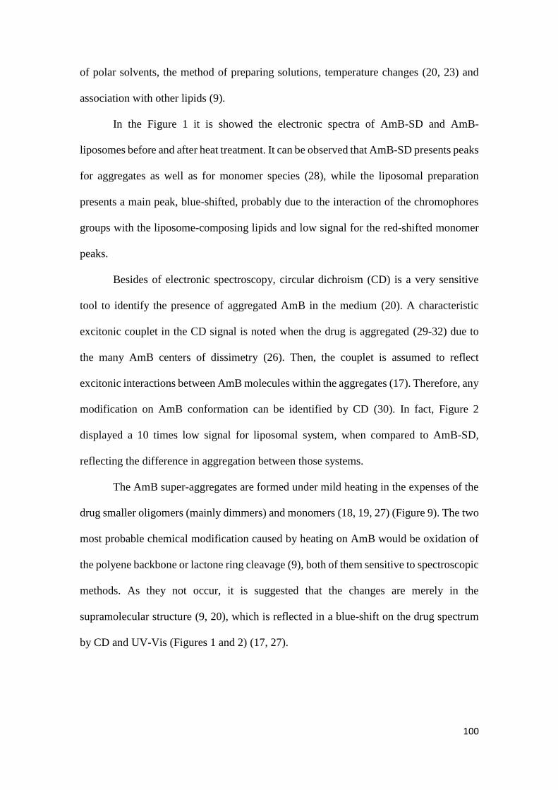
100
of polar solvents, the method of preparing solutions, temperature changes (20, 23) and
association with other lipids (9).
In the Figure 1 it is showed the electronic spectra of AmB-SD and AmB-
liposomes before and after heat treatment. It can be observed that AmB-SD presents peaks
for aggregates as well as for monomer species (28), while the liposomal preparation
presents a main peak, blue-shifted, probably due to the interaction of the chromophores
groups with the liposome-composing lipids and low signal for the red-shifted monomer
peaks.
Besides of electronic spectroscopy, circular dichroism (CD) is a very sensitive
tool to identify the presence of aggregated AmB in the medium (20). A characteristic
excitonic couplet in the CD signal is noted when the drug is aggregated (29-32) due to
the many AmB centers of dissimetry (26). Then, the couplet is assumed to reflect
excitonic interactions between AmB molecules within the aggregates (17). Therefore, any
modification on AmB conformation can be identified by CD (30). In fact, Figure 2
displayed a 10 times low signal for liposomal system, when compared to AmB-SD,
reflecting the difference in aggregation between those systems.
The AmB super-aggregates are formed under mild heating in the expenses of the
drug smaller oligomers (mainly dimmers) and monomers (18, 19, 27) (Figure 9). The two
most probable chemical modification caused by heating on AmB would be oxidation of
the polyene backbone or lactone ring cleavage (9), both of them sensitive to spectroscopic
methods. As they not occur, it is suggested that the changes are merely in the
supramolecular structure (9, 20), which is reflected in a blue-shift on the drug spectrum
by CD and UV-Vis (Figures 1 and 2) (17, 27).
101
Figure 9. Scheme of super-aggregate formation: AmB coexists as monomers and
oligomers in solution; after mild heating super-aggregates are formed in the expenses of
the previous AmB forms (Based on Gaboriau et al. (1997) (18)).
The formation of super aggregates has been reported from AmB aqueous solution
and mainly from micellar solutions (drug mixed with sodium deoxycholate) (3, 17-19,
21, 22, 33). There is no report of AmB super-aggregates formation from liposomal
systems. Maybe, because the super-aggregation is a strategy to lower the toxicity of the
micellar systems and that lipid-based systems already present improved safety. But, on
the other hand, it have to be taken in mind that in the liposomal system, AmB is strongly
associated with cholesterol (14, 23, 34), making difficult AmB release upon dilution. This
is probably why the CD signal is ~10 times lower than the AmB-SD signal (Figure 2): as
the drug remains bound to the lipid, less oligomers are free in the medium to form an
intense CD doublet signal. Consequently, the essential species above mentioned (AmB
aggregates and monomers) for the super-aggregate formation are not available in the
required concentrations.
Moreover, there is no shift in both UV-Vis (Figure 1) and in the couplet former
peaks (Figure 2), reinforcing the assumption of non-formation of AmB super-aggregates
from the liposomal system.
AmB oligomers AmB monomers
70 ºC
20 min
102
After a mild heating, super-aggregates were formed from AmB-SD systems as
evidenced by the blue-shift on the electronic band from 326 nm to 323 nm (Figure 1) and
also in the excitonic doublet observed by CD (Figure 2) (9, 17, 23). Indeed, increases in
temperature enhance the hydrophobic strength while decrease ionic and polar
stabilization. Then, the blue shift in the maximum band indicates a new aggregation form
in which the chromophore groups are highly organized in a nonpolar environment, where
the frequency of hydrophobic interactions is assumed to increase (17, 20, 27).
Although the Bee-Lambert law does not hold in the case of CD for aggregated
AmB (20), it is important to mention that the CD signal decreased after heating process.
Such a behavior was also found by super-aggregates characterized by other researchers
(17, 23). A change in CD bands amplitude is likely to reflect a change in the number of
molecules in an aggregated form, and variations of the other spectral characteristics are
related to conformational changes of aggregates (20). Decrease in CD doublet has also
been explained by the increased size of super-aggregates, that makes a blue-shift on the
electronic transition and could also be accompanied of weak CD doublets at lower
wavelength (17).
Stability after heating process
The super-aggregates appear to be more stable than the previous system. In our
study this assumption is supported for both, the maintenance of the electronic spectrum
shape (Figure 1) and the low change in intensity of CD signal upon dilution (Figure 2)
that happened only for heated AmB-SD and that is going to be discussed in the following
lines.
In the first case, it is seemed that the heat was able to change the AmB
conformation in a stable way. Along the study, for the un-heated micellar system, the
peak of aggregates decreased while the peak of monomers increased (Figure 2),
103
suggesting a modification on the aggregation state. The ratio between the peak of
aggregates and that of monomers is an interesting tool to evaluate the degree of drug
aggregation into the system (35, 36). In fact, when the concentration of the drug
decreases, the balance between monomers and aggregates moves towards monomers (19,
32). However, for the heated system, both peaks decreased in intensity and the spectra
remained similar in shape during the complete study.
Diluting an aliquot of the sample in methanol and analyzing it by UV-Vis, it was
confirmed that the heated system had its stability increased when compared with the un-
heated counterpart (Figure 3). The literature had already showed that heating AmB
solutions increase its chemical stability for at least 2h (17). However, it is the first time
that it is presented a study for such a long period as one month.
Such a behavior can be explained by the mechanism of drug degradation summed
with the dissociation of AmB species from the super-aggregates. The decrease in AmB
concentration is a process dependent of oxygen, corresponding to the drug auto-oxidation,
mainly related to the various oligomers types present in AmB-SD (9). Then, the increase
in stability might be explained by the drug re-structuration itself. The super-aggregates
would be a more cohesive particle, hiding the chromophore group and consequently
lowering its exposure to the aqueous solvent and also acting as a reservoir, being slowly
dissociated as monomers, those are not involved in auto-oxidation (17).
Besides of electronic spectroscopy, it was observed that after heat treatment CD
signal was quite constant even upon dilution (Figure 2), indicating that the drug remained
associated in its new structure (37). This behavior is explained by Silva-Filho et al. (2012)
(19) for the stronger bound of AmB to the surfactant when compared to un-heated system,
while Brajtburg and Bolard (1996) indicate that the signal is constant due to

104
uniformization of the type of oligomers in solution (11). All together suggests super-
aggregates as more stable organization than the previous micellar system.
The super-aggregates showed to be less degraded than the un-heated system.
However, as the drug is in solution, drug degradation does not stop occurring. Recent
studies have shown the possibility of freeze-drying AmB super-aggregates, maintaining
its physical and biological features (3).
The liposomal system was stable along the study, once no differences in
concentration were found among days and the heating process did not affect the system
aggregation behavior.
ITC experiments
Super-aggregates formation
Once observed that the super-aggregates only are formed from micellar systems
of AmB, it was conducted a study seeking to measure the energy dispensed or generated
during the AmB molecular re-organization.
ITC technique is highly used for evaluation of molecular interactions or
conformational changes based in the knowledge that any interaction can release or absorb
heat (24). The titrations performed in this work were set at 40, 50, 60 and 70 ºC,
considering that super-aggregation can also occurs at low temperature, spontaneously
(17).
Despite the sensibility of ITC equipment, the amount of heat generated/dispensed
in the process was not enough for track any molecular re-organization. In the titrations
obtained at different temperatures (Figure 4), it was possible to measure the two water
injections performed in the beginning and end of the titration. However, the tracked heat
during the 20 min does not show any significant heat peak.
105
The ITC recovered liquid evidenced physical changes in size at naked eye that
was further confirmed by DLS size measurement. Siqueira et al. (2014) (3) have also
reported a prominent increase in turbidity upon increased temperature, suggesting
molecular structural re-organization.
The size increased as the temperature increases: by naked eye, the cloudier the
sample, the larger it was found to be. The mean size of the stock solution was around 12
nm, and increased sequentially up to around 450 nm (volume/number), when 70 ºC was
used in the heating process (Figure 5). Such an increase in size justify the name give for
the new structure: super-aggregates (19).
It is assumed that the size of AmB-SD increases along time, probably due to AmB
release from the micelle and consequent aggregation. The heat process may accelerates
the dissociation process from the SD micelles, being followed by AmB re-association (9).
Such a greater size is responsible for an increase on the uptake of the drug by the cells
(22).
Previous studies have showed that thermal treatment does not induces molecular
dispersion, but seems to increase the size of aggregates until flocculation occurs (17).
Increasing the temperature upper than 75 ºC have been reported in the literature with loss
of drug solubility (27), probably due to precipitation caused by increased particle size.
Because of that, and also because that at 70 ºC practically all the self-associated form is
converted in the super-aggregated form (17), this temperature was set as the best one to
form super-aggregates.
The same samples, recovered from the ITC experiments conducted at different
temperatures were then analyzed by UV-Vis. It reinforced the role of the temperature in
the formation of the super-aggregates.

106
Different level of blue-shift and drug aggregation were found in accordance to the
temperature employed (Figure 6) (27). Gaboriau et al. (1997) (17) showed that solution
absorption spectrum stop to change for temperature higher than 70 ºC, reinforcing the
choose of such a temperature to super-aggregates formation.
De-aggregation
A final attempt to measure the energy of the aggregation process was tried by the
inverse of the process: diluting an aggregated AmB-SD solution (5x10⁻⁵ M AmB) to the
monomeric form of the drug, at 5x10⁻⁸ M AmB (Figure 7). Since the biding isotherms are
defined in terms of reaction heats, allowing a direct estimation of enthalpy changes related
to association/dissociation constant (24).
Unfortunately, the heat measurement done by ITC was quite low and did not
permit many inferences: the signal was more intense in the beginning of the dilution,
suggesting that the molecular re-organization generates heat. Then, in the end of the
dilution the signal was even weaker, probably due to the reduction in shocks of different
organized oligomeric structures.
Luengo-Alonso et al. (2015) (4) have performed studies in which AmB (1.1 x 10-
5 M) was de-aggregated with PEG5kDa-cholane (0.6 x 10⁻³ M). The isothermal results
highlighted the complexity of AmB interactions of AmB, which takes place with different
AmB soluble species.
Despite the failure of measuring the energy involved in the de-aggregation
process, CD spectra demonstrated that, in fact, the drug changed its conformation upon
dilution, from aggregated to monomeric form (Figure 8). The monomeric units do not
presents specific optical activity (17) (parallel transition moments do not lead to excitonic
CD), while the intense CD couplet is explained by interactions between the small units
(20).
107
Conclusion
The overall results showed that the formation of super-aggregates is possible by
heating AmB-micellar, but not liposomal systems. The process is responsible for the
increase in size of the nanostructures. Such an increase was dependent on the temperature
applied in the process. The super-aggregates seem to play an interesting role improving
the stability of the drug along the time, since the new structure was maintained for at least
28 days, although drug degradation occurred also for super-aggregates. Unfortunately,
ITC experiments were not able to accurately measure the heat involved in the process of
super-aggregates formation. However, it seems that the de-aggregation generates heat,
that becomes weaker when the drug is presented as monomers. Finally, it is assumed that
super-aggregates are less toxic than AmB aggregates, once its reorganization do not like
to have the propensity of reconverting to the toxic soluble oligomer species. The effect of
drug reservoir reported to super-aggregates means the release of a limited amount of
monomeric AmB to the medium, that would increase both drug safety and efficacy,
reinforcing the study of super-aggregates as an alternative to toxic AmB-SD products.
Acknowledgment
Authors would like to thanks to Mme. Magali Noiray for the training on the ITC facility
and to CAPES-COFECUB (Project 742/11) for the fellowship provided to the first author,
A. L. Silva.
References
1. Adhikari K, Buatong W, Thawithong E, Suwandecha T, Srichana T. Factors
affecting enhanced permeation of amphotericin B across cell membranes and safety of
formulation. AAPS PharmSciTech. 2016;17(4):820-8.
108
2. Sundar S, Chakravarty J. Liposomal amphotericin B and leishmaniasis: dose and
response. J Glob Infect Dis. 2010;2(2):159-66.
3. Siqueira SD, Silva-Filho MA, Silva CA, Araujo IB, Silva AE, Fernandes-Pedrosa
MF, et al. Influence of the freeze-drying process on the physicochemical and biological
properties of pre-heated amphotericin B micellar systems. AAPS PharmSciTech.
2014;15(3):612-9.
4. Luengo-Alonso C, Torrado JJ, Ballesteros MP, Malfanti A, Bersani S, Salmaso S,
et al. A novel performing PEG-cholane nanoformulation for Amphotericin B delivery. Int
J Pharm. 2015;495(1):41-51.
5. Hamill RJ. Amphotericin B formulations: a comparative review of efficacy and
toxicity. Drugs. 2013;73(9):919-34.
6. Araujo IB, Damasceno BP, de Medeiros TM, Soares LA, do Egito ES. Decrease
in Fungizone toxicity induced by the use of Lipofundin as a dilutent: an in vitro study.
Curr Drug Deliv. 2005;2(2):199-205.
7. Torrado JJ, Espada R, Ballesteros MP, Torrado-Santiago S. Amphotericin B
formulations and drug targeting. J Pharm Sci. 2008;97(7):2405-25.
8. Clemons KV, Stevens DA. Comparative efficacies of four amphotericin B
formulations - Fungizone, amphotec (Amphocil), AmBisome, and Abelcet - against
systemic murine aspergillosis. Antimicrob Agents Chemother. 2004;48(3):1047-50.
9. Baas B, Kindt K, Scott A, Scott J, Mikulecky P, Hartsel SC. Activity and kinetics
of dissociation and transfer of amphotericin B from a novel delivery form. AAPS
PharmSciTech. 1999;1(3):E10.
10. Barratt G, Bretagne S. Optimizing efficacy of amphotericin B through
nanomodification. Int J Nanomed. 2007;2(3):301-13.
11. Brajtburg J, Bolard J. Carrier effects on biological activity of amphotericin B. Clin
Microb Rev. 1996;9(4):512-31.
12. Damasceno BPGL, Dominici VA, Urbano IA, Silva JA, Araujo IB, Santos-
Magalhaes NS, et al. Amphotericin B Microemulsion Reduces Toxicity and Maintains
the Efficacy as an Antifungal Product. J Biomed Nanotechnol. 2012;8(2):290-300.
13. Halperin A, Shadkchan Y, Pisarevsky E, Szpilman AM, Sandovsky H, Osherov
N, et al. Novel water-soluble amphotericin B-PEG conjugates with low toxicity and
potent in vivo efficacy. J Med Chem. 2016;59(3):1197-206.
14. Adler-Moore JP, Gangneux JP, Pappas PG. Comparison between liposomal
formulations of amphotericin B. Med Mycol. 2016;54(3):223-31.
15. Araújo IB, Brito CRN, Urbano IA, Dominici VA, Silva Filho MA, Silveira WLL,
et al. Similarity between the in vitro activity and toxicity of two different Fungizone /
lipofundin admixtures. Acta Bras Cir. 2005;20:129-33.
109
16. Alvarez C, Shin DH, Kwon GS. Reformulation of Fungizone by PEG-DSPE
micelles: deaggregation and detoxification of amphotericin B. Pharm Res.
2016;33(9):2098-106.
17. Gaboriau F, Chéron M, Leroy L, Bolard J. Physico-chemical properties of the
heat-induced ‘superaggregates’ of amphotericin B. Biophys Chem. 1997;66(1):1-12.
18. Gaboriau F, Chéron M, Petit C, Bolard J. Heat-induced superaggregation of
amphotericin B reduces its in vitro toxicity: a new way to improve its therapeutic index.
Antimicrob Agents Chemother. 1997;41(11):2345-51.
19. Silva-Filho MA, Siqueira SD, Freire LB, Araujo IB, Holanda e Silva KG,
Medeiros Ada C, et al. How can micelle systems be rebuilt by a heating process? Int J
Nanomed. 2012;7:141-50.
20. Ernst C, Grange J, Rinnert H, Dupont G. Structure of amphotericin B aggregates
as revealed by UV and CD spectroscopies. Biopolymers. 1981;20(8):1575-88.
21. Zia Q, Azhar A, Kamal MA, Aliev G, Owais M, Ashraf GM. Super aggregated
form of amphotericin B: a novel way to increase its therapeutic index. Curr Pharm Design.
2016;22(7):792-803.
22. Cheron M, Petit C, Bolard J, Gaboriau F. Heat-induced reformulation of
amphotericin B-deoxycholate favours drug uptake by the macrophage-like cell line J774.
J Antimicrob Chemother. 2003;52(6):904-10.
23. Espada R, Valdespina S, Alfonso C, Rivas G, Ballesteros MP, Torrado JJ. Effect
of aggregation state on the toxicity of different amphotericin B preparations. Int J Pharm.
2008;361(1–2):64-9.
24. Freire E, Mayorga OL, Straume M. Isothermal Titration Calorimetry. Anal Chem.
1990;62(18):A950-A9.
25. Bolard J. How do the polyene macrolide antibiotics affect the cellular membrane
properties? Biochim Biophys Acta. 1986;864(3):257-304.
26. Brittain HG. Circular dichroism studies of the self-association of amphotericin B.
Chirality. 1994;6(8):665-9.
27. Burgess BL, He Y, Baker MM, Luo B, Carroll SF, Forte TM, et al. NanoDisk
containing super aggregated amphotericin B: a high therapeutic index antifungal
formulation with enhanced potency. Int J Nanomed. 2013;8:4733-43.
28. Sanchez-Brunete JA, Dea MA, Rama S, Bolas F, Alunda JM, Torrado-Santiago
S, et al. Amphotericin B molecular organization as an essential factor to improve
activity/toxicity ratio in the treatment of visceral leishmaniasis. J Drug Target.
2004;12(7):453-60.
29. Balakrishnan AR, Easwaran KRK. CD and NMR-Studies on the aggregation of
amphotericin-B in solution. Biochim Biophys Acta. 1993;1148(2):269-77.
110
30. Barwicz J, Beauregard M, Tancrede P. Circular dichroism study of interactions of
Fungizone or AmBisome forms of amphotericin B with human low density lipoproteins.
Biopolymers. 2002;67(1):49-55.
31. Legrand P, Romero EA, Cohen BE, Bolard J. Effects of aggregation and solvent
on the toxicity of amphotericin B to human erythrocytes. Antimicrob Agents Chemother.
1992;36(11):2518-22.
32. Zielińska J, Wieczór M, Bączek T, Gruszecki M, Czub J. Thermodynamics and
kinetics of amphotericin B self-association in aqueous solution characterized in molecular
detail. Sci Rep. 2016;6:19109.
33. Petit C, Yardley V, Gaboriau F, Bolard J, Croft SL. Activity of a heat-induced
reformulation of amphotericin B deoxycholate (Fungizone) against Leishmania
donovani. Antimicrob Agents Chemother. 1999;43(2):390-2.
34. Adler-Moore J, Proffitt RT. AmBisome: liposomal formulation, structure,
mechanism of action and pre-clinical experience. J Antimicrob Chemother. 2002;49
Suppl 1:21-30.
35. Barwicz J, Christian S, Gruda I. Effects of the aggregation state of amphotericin
B on its toxicity to mice. Antimicrob Agents Chemother. 1992;36(10):2310-5.
36. Jameson LP, Dzyuba SV. Circular dichroism studies on intermolecular
interactions of amphotericin B in ionic liquid-rich environments. Chirality.
2013;25(7):427-32.
37. Larabi M, Gulik A, Dedieu J-P, Legrand P, Barratt G, Cheron M. New lipid
formulation of amphotericin B: spectral and microscopic analysis. Biochim Biophys
Acta. 2004;1664(2):172-81.
111
SECTION III
INCORPORATION OF AMPHOTERICIN B INTO EMULSION
SYSTEMS
____________________________________________________________
112
BRIEFING
Emulsion systems are able to load hydrophilic and hydrophobic molecules, once
they are systems composed of oil and water. Micro-emulsion is one type of emulsion
characterized for presenting high amounts of surfactants and this fact helps this system to
better penetrate into different barriers. Nano-emulsion, in turn, are mainly aqueous system
composed of low amounts of surfactants.
These systems are eligible for the encapsulation of the amphotericin B (AmB),
due to its physicochemical characteristics, the drug is supposed to be placed in the
interface oil/water.
This section is dedicated to the AmB incorporation into emulsion systems. The
first chapter is a scientific report about the details of the drug loading into nano- and
micro-emulsion. The drug aggregation state into these systems is presented in the first
chapter for the nano-emulsion and in the second chapter for the micro-emulsion. The
second chapter was conducted in collaboration with other Ph.D student from the LaSiD
(Mr. Walteça Silveira) and present applications for AmB-loading micro-emulsions
treating fungal infections.
113
CHAPTER IV
AmB loading into micro- and nano-emulsion systems
114
AmB loading into micro- and nano-emulsion systems
(Scientific Report)
Introduction
The “Laboratório de Sistemas Dispersos” (LaSiD) is dedicated to the
development of emulsion systems, aiming to carry many types of molecules, mainly those
fragile (1-3) and insoluble (4-7).
Emulsions are biphasic systems, composed of oil and water, stabilized by a film
of surfactant molecules. These systems can be thermodynamically stable (e.g. micro-
emulsions) or unstable (e.g. nano-emulsions, real emulsions). Nano-emulsion systems, in
spite of being thermodynamically unstable, are considered to be kinetically stable, due to
their small droplet size. The main instability phenomena playing in this case is Ostwald
ripening (8).
Once emulsion systems present both aqueous and lipid phases, the incorporation
of different kind of substances of hydrophilic and hydrophobic character is believed to be
possible (9). In this scenery, emulsions are suggested to be promising carriers for
amphotericin B (AmB), a molecule of 924 Da, counting with a hydrophobic chain and
polyhydroxylated region (10).
Indeed some studies have been dealing with emulsions as carriers for AmB
delivery by different administration routes (4-6, 11-13)
In this work, the incorporation of AmB into nano- and micro-emulsion is reported,
evidencing the limit of drug loading according to the system and the correspondent drug
aggregation state.
Methodology
Micro-emulsion preparation
The micro-emulsion system was prepared based on the work of Silveira et al.
(2016) (4) (complete work in the next chapter), with some modifications. Briefly, Lipoid®
S100 and water were weighed in a flask and left under magnetic stirring up to the
formation of a milky suspension. Then, Tween® 80 was added to the suspension and left
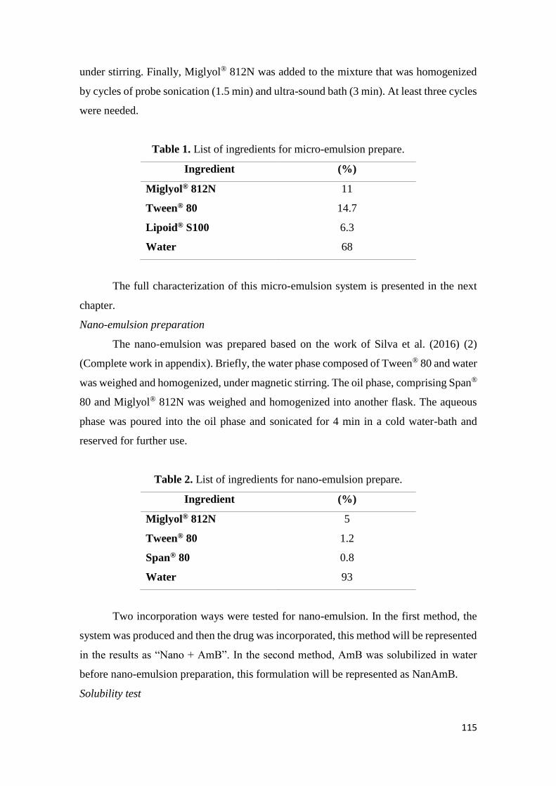
115
under stirring. Finally, Miglyol® 812N was added to the mixture that was homogenized
by cycles of probe sonication (1.5 min) and ultra-sound bath (3 min). At least three cycles
were needed.
Table 1. List of ingredients for micro-emulsion prepare.
Ingredient (%)
Miglyol® 812N
Tween® 80
Lipoid® S100
Water
11
14.7
6.3
68
The full characterization of this micro-emulsion system is presented in the next
chapter.
Nano-emulsion preparation
The nano-emulsion was prepared based on the work of Silva et al. (2016) (2)
(Complete work in appendix). Briefly, the water phase composed of Tween® 80 and water
was weighed and homogenized, under magnetic stirring. The oil phase, comprising Span®
80 and Miglyol® 812N was weighed and homogenized into another flask. The aqueous
phase was poured into the oil phase and sonicated for 4 min in a cold water-bath and
reserved for further use.
Table 2. List of ingredients for nano-emulsion prepare.
Ingredient (%)
Miglyol® 812N
Tween® 80
Span® 80
Water
5
1.2
0.8
93
Two incorporation ways were tested for nano-emulsion. In the first method, the
system was produced and then the drug was incorporated, this method will be represented
in the results as “Nano + AmB”. In the second method, AmB was solubilized in water
before nano-emulsion preparation, this formulation will be represented as NanAmB.
Solubility test
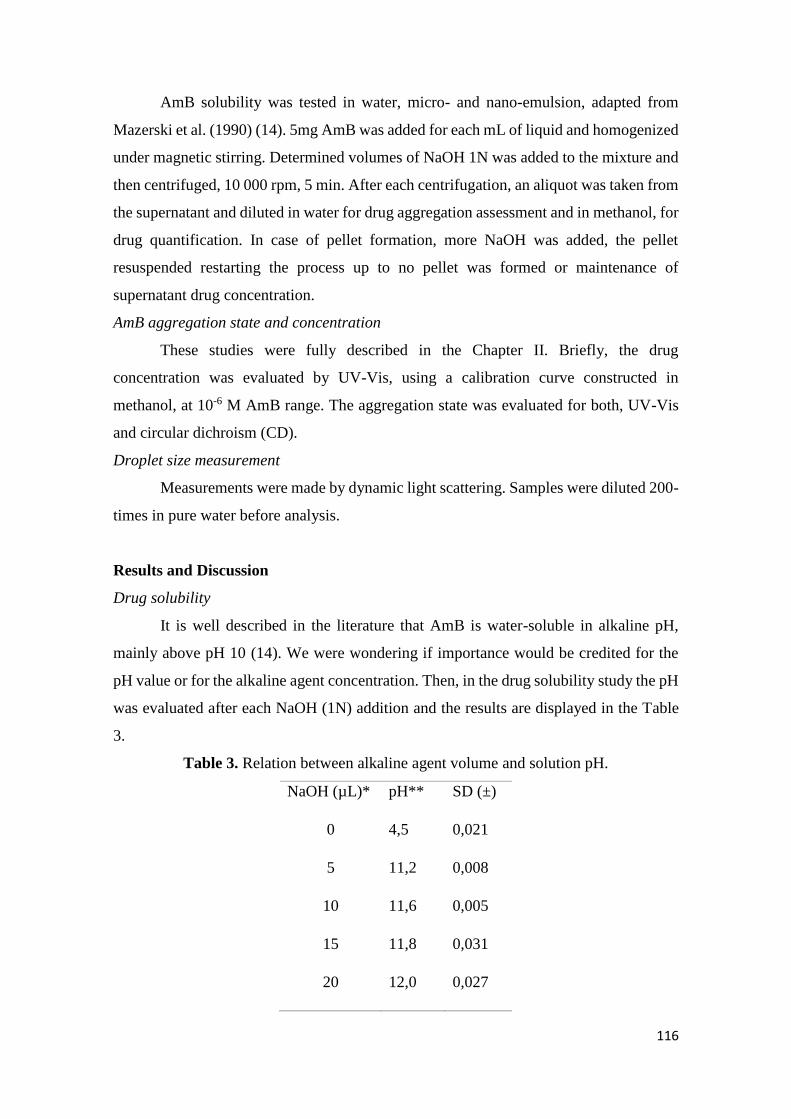
116
AmB solubility was tested in water, micro- and nano-emulsion, adapted from
Mazerski et al. (1990) (14). 5mg AmB was added for each mL of liquid and homogenized
under magnetic stirring. Determined volumes of NaOH 1N was added to the mixture and
then centrifuged, 10 000 rpm, 5 min. After each centrifugation, an aliquot was taken from
the supernatant and diluted in water for drug aggregation assessment and in methanol, for
drug quantification. In case of pellet formation, more NaOH was added, the pellet
resuspended restarting the process up to no pellet was formed or maintenance of
supernatant drug concentration.
AmB aggregation state and concentration
These studies were fully described in the Chapter II. Briefly, the drug
concentration was evaluated by UV-Vis, using a calibration curve constructed in
methanol, at 10-6 M AmB range. The aggregation state was evaluated for both, UV-Vis
and circular dichroism (CD).
Droplet size measurement
Measurements were made by dynamic light scattering. Samples were diluted 200-
times in pure water before analysis.
Results and Discussion
Drug solubility
It is well described in the literature that AmB is water-soluble in alkaline pH,
mainly above pH 10 (14). We were wondering if importance would be credited for the
pH value or for the alkaline agent concentration. Then, in the drug solubility study the pH
was evaluated after each NaOH (1N) addition and the results are displayed in the Table
3.
Table 3. Relation between alkaline agent volume and solution pH.
NaOH (µL)* pH** SD (±)
0 4,5 0,021
5 11,2 0,008
10 11,6 0,005
15 11,8 0,031
20 12,0 0,027
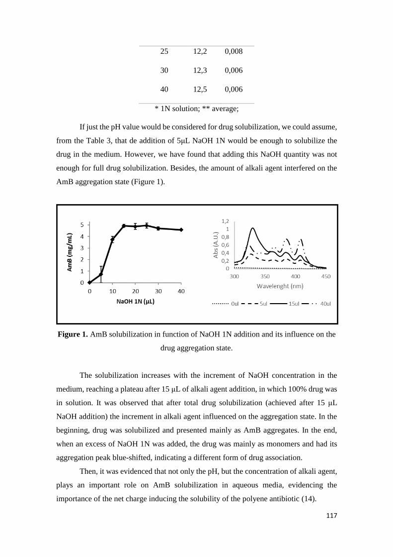
117
25 12,2 0,008
30 12,3 0,006
40 12,5 0,006
* 1N solution; ** average;
If just the pH value would be considered for drug solubilization, we could assume,
from the Table 3, that de addition of 5μL NaOH 1N would be enough to solubilize the
drug in the medium. However, we have found that adding this NaOH quantity was not
enough for full drug solubilization. Besides, the amount of alkali agent interfered on the
AmB aggregation state (Figure 1).
Figure 1. AmB solubilization in function of NaOH 1N addition and its influence on the
drug aggregation state.
The solubilization increases with the increment of NaOH concentration in the
medium, reaching a plateau after 15 μL of alkali agent addition, in which 100% drug was
in solution. It was observed that after total drug solubilization (achieved after 15 μL
NaOH addition) the increment in alkali agent influenced on the aggregation state. In the
beginning, drug was solubilized and presented mainly as AmB aggregates. In the end,
when an excess of NaOH 1N was added, the drug was mainly as monomers and had its
aggregation peak blue-shifted, indicating a different form of drug association.
Then, it was evidenced that not only the pH, but the concentration of alkali agent,
plays an important role on AmB solubilization in aqueous media, evidencing the
importance of the net charge inducing the solubility of the polyene antibiotic (14).

118
Results for the solubilization of the drug into the emulsion systems are displayed
in the Figure 2. Nano-emulsion presented results quite similar to the water solubility,
being able to solubilize up to 5mg AmB. In spite of solubilizing less amount of drug (up
to 3mg/mL), AmB presented similar aggregation patterns into the micro-emulsion
system, with bands of aggregates and monomers (Figure 2).
Figure 2. AmB solubility profile in nano- and micro-emulsion. The percentile of drug
solubility (5mg/mL max) is showed in the upper panel and the aggregation pattern in
function of the added NaOH 1N, by UV-Vis, is presented in the bottom panel.
The fact that same concentration of alkali agent was needed to solubilize the drug
in water and in nano-emulsion could be due to the high water content (93%) in this
system. However, the excess of NaOH did not cause changes in the aggregation pattern
of the drug. Probably because AmB was associated with the lipids into the nano-emulsion
system.
Micro-emulsion
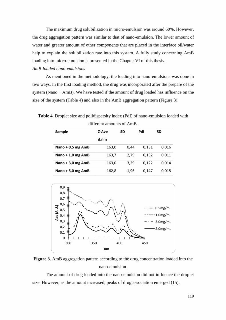
119
The maximum drug solubilization in micro-emulsion was around 60%. However,
the drug aggregation pattern was similar to that of nano-emulsion. The lower amount of
water and greater amount of other components that are placed in the interface oil/water
help to explain the solubilization rate into this system. A fully study concerning AmB
loading into micro-emulsion is presented in the Chapter VI of this thesis.
AmB-loaded nano-emulsions
As mentioned in the methodology, the loading into nano-emulsions was done in
two ways. In the first loading method, the drug was incorporated after the prepare of the
system (Nano + AmB). We have tested if the amount of drug loaded has influence on the
size of the system (Table 4) and also in the AmB aggregation pattern (Figure 3).
Table 4. Droplet size and polidispersity index (PdI) of nano-emulsion loaded with
different amounts of AmB.
Sample Z-Ave
d.nm
SD PdI SD
Nano + 0,5 mg AmB 163,0 0,44 0,131 0,016
Nano + 1,0 mg AmB 163,7 2,79 0,132 0,011
Nano + 3,0 mg AmB 163,0 3,29 0,122 0,014
Nano + 5,0 mg AmB 162,8 1,96 0,147 0,015
Figure 3. AmB aggregation pattern according to the drug concentration loaded into the
nano-emulsion.
The amount of drug loaded into the nano-emulsion did not influence the droplet
size. However, as the amount increased, peaks of drug association emerged (15).
0
0,1
0,2
0,3
0,4
0,5
0,6
0,7
0,8
0,9
300 350 400 450
Ab
s (A
.U.)
nm
0.5mg/mL
1.0mg/mL
3.0mg/mL
5.0mg/mL
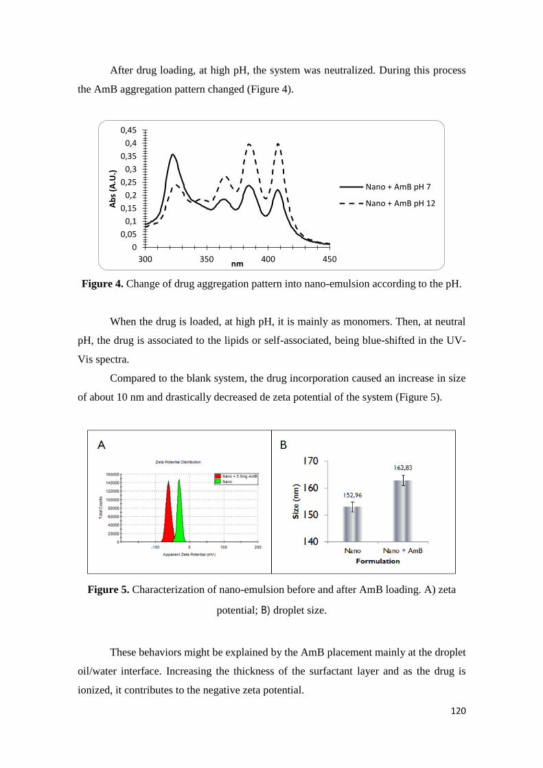
120
After drug loading, at high pH, the system was neutralized. During this process
the AmB aggregation pattern changed (Figure 4).
Figure 4. Change of drug aggregation pattern into nano-emulsion according to the pH.
When the drug is loaded, at high pH, it is mainly as monomers. Then, at neutral
pH, the drug is associated to the lipids or self-associated, being blue-shifted in the UV-
Vis spectra.
Compared to the blank system, the drug incorporation caused an increase in size
of about 10 nm and drastically decreased de zeta potential of the system (Figure 5).
Figure 5. Characterization of nano-emulsion before and after AmB loading. A) zeta
potential; B) droplet size.
These behaviors might be explained by the AmB placement mainly at the droplet
oil/water interface. Increasing the thickness of the surfactant layer and as the drug is
ionized, it contributes to the negative zeta potential.
0
0,05
0,1
0,15
0,2
0,25
0,3
0,35
0,4
0,45
300 350 400 450
Ab
s (A
.U.)
nm
Nano + AmB pH 7
Nano + AmB pH 12
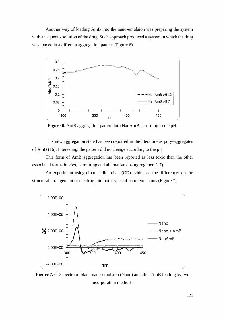
121
Another way of loading AmB into the nano-emulsion was preparing the system
with an aqueous solution of the drug. Such approach produced a system in which the drug
was loaded in a different aggregation pattern (Figure 6).
Figure 6. AmB aggregation pattern into NanAmB according to the pH.
This new aggregation state has been reported in the literature as poly-aggregates
of AmB (16). Interesting, the pattern did no change according to the pH.
This form of AmB aggregation has been reported as less toxic than the other
associated forms in vivo, permitting and alternative dosing regimen (17) .
An experiment using circular dichroism (CD) evidenced the differences on the
structural arrangement of the drug into both types of nano-emulsions (Figure 7).
Figure 7. CD spectra of blank nano-emulsion (Nano) and after AmB loading by two
incorporation methods.
0
0,05
0,1
0,15
0,2
0,25
0,3
300 350 400 450
Ab
s (A
.U.)
nm
NanAmB pH 12
NanAmB pH 7
-2,00E+06
0,00E+00
2,00E+06
4,00E+06
6,00E+06
300 350 400 450
ΔƐ
nm
Nano
Nano + AmB
NanAmB
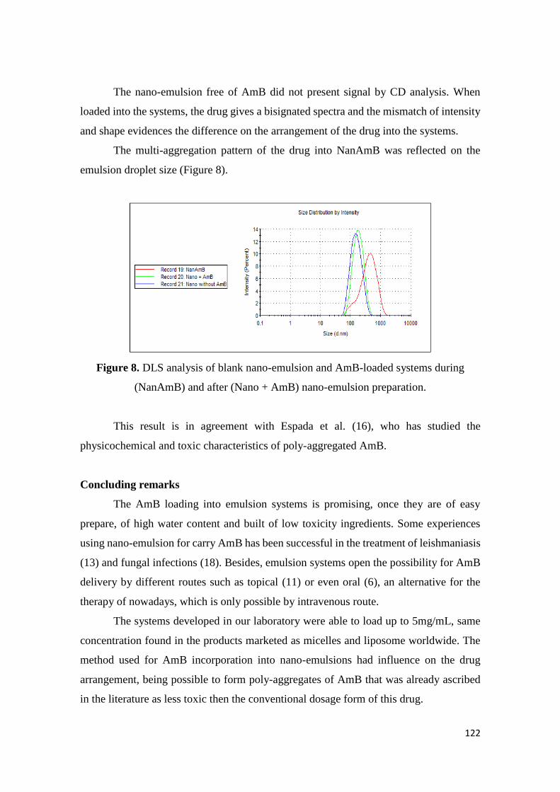
122
The nano-emulsion free of AmB did not present signal by CD analysis. When
loaded into the systems, the drug gives a bisignated spectra and the mismatch of intensity
and shape evidences the difference on the arrangement of the drug into the systems.
The multi-aggregation pattern of the drug into NanAmB was reflected on the
emulsion droplet size (Figure 8).
Figure 8. DLS analysis of blank nano-emulsion and AmB-loaded systems during
(NanAmB) and after (Nano + AmB) nano-emulsion preparation.
This result is in agreement with Espada et al. (16), who has studied the
physicochemical and toxic characteristics of poly-aggregated AmB.
Concluding remarks
The AmB loading into emulsion systems is promising, once they are of easy
prepare, of high water content and built of low toxicity ingredients. Some experiences
using nano-emulsion for carry AmB has been successful in the treatment of leishmaniasis
(13) and fungal infections (18). Besides, emulsion systems open the possibility for AmB
delivery by different routes such as topical (11) or even oral (6), an alternative for the
therapy of nowadays, which is only possible by intravenous route.
The systems developed in our laboratory were able to load up to 5mg/mL, same
concentration found in the products marketed as micelles and liposome worldwide. The
method used for AmB incorporation into nano-emulsions had influence on the drug
arrangement, being possible to form poly-aggregates of AmB that was already ascribed
in the literature as less toxic then the conventional dosage form of this drug.
123
The overall results reported in here show the possibilities provided by emulsion
systems on the search for an ideal carrier for the old molecule amphotericin B.
References
1. Silva AL, Júnior FA, Verissimo LM, Agnez-Lima LF, Egito LCM, de Oliveira
AG, et al. Physical Factors Affecting Plasmid DNA Compaction in Stearylamine-
Containing Nanoemulsions Intended for Gene Delivery. Pharmaceuticals. 2012;5(6):643-
54.
2. Silva AL, Marcelino HR, Verissimo LM, Araujo IB, Agnez-Lima LF, do Egito
ES. Stearylamine-Containing Cationic Nanoemulsion as a Promising Carrier for Gene
Delivery. J Nanosci Nanotechnol. 2016;16(2):1339-45.
3. Verissimo LM, Lima LF, Egito LC, de Oliveira AG, do Egito ES. Pharmaceutical
emulsions: a new approach for gene therapy. J Drug Target. 2010;18(5):333-42.
4. da Silveira WL, Damasceno BP, Ferreira LF, Ribeiro IL, Silva KS, Silva AL, et
al. Development and Characterization of a Microemulsion System Containing
Amphotericin B with Potential Ocular Applications. Curr Drug Deliv. 2016;13(6):982-
93.
5. Damasceno BP, Dominici VA, Urbano IA, Silva JA, Araujo IB, Santos-
Magalhaes NS, et al. Amphotericin B microemulsion reduces toxicity and maintains the
efficacy as an antifungal product. J Biomed Nanotechnol. 2012;8(2):290-300.
6. Silva AE, Barratt G, Chéron M, Egito EST. Development of oil-in-water
microemulsions for the oral delivery of amphotericin B. Int J Pharm. 2013;454(2):641-8.
7. Xavier Junior FH, Gueutin C, do Vale Morais AR, do Nascimento Alencar E, do
Egito EST, Vauthier C. HPLC Method for the Dosage of Paclitaxel in Copaiba Oil:
Development, Validation, Application to the Determination of the Solubility and Partition
Coefficients. Chromatographia. 2016;79(7):405-12.
8. McClements DJ. Nanoemulsions versus microemulsions: terminology,
differences, and similarities. Soft Matter. 2012;8(6):1719-29.
9. Lee PJ, Langer R, Shastri VP. Novel Microemulsion Enhancer Formulation for
Simultaneous Transdermal Delivery of Hydrophilic and Hydrophobic Drugs. Pharm Res.
2003;20(2):264-9.
10. Asher IM, Schwartzman G, USASRG. Amphotericin B. In: Florey K, editor.
Analytical Profile of Drug Substances. 6. New York: Academic Press; 1977. p. 1-42.
11. Butani D, Yewale C, Misra A. Amphotericin B topical microemulsion:
Formulation, characterization and evaluation. Colloids Surf B Biointerfaces.
2014;116:351-8.
124
12. Walker S, Tailor SAN, Lee M, Louie L, Louie M, Simor AE. Amphotericin B in
Lipid Emulsion: Stability, Compatibility, and In Vitro Antifungal Activity. Antimicrob
Agents Chemother. 1998;42(4):762-6.
13. Caldeira LR, Fernandes FR, Costa DF, Frézard F, Afonso LCC, Ferreira LAM.
Nanoemulsions loaded with amphotericin B: A new approach for the treatment of
leishmaniasis. Eur J Pharm Sci. 2015;70:125-31.
14. Mazerski J, Grzybowska J, Borowski E. Influence of net charge on the
aggregation and solubility behaviour of amphotericin B and its derivatives in aqueous
media. Eur Biophys J. 1990;18(3):159-64.
15. Zielinska J, Wieczor M, Baczek T, Gruszecki M, Czub J. Thermodynamics and
kinetics of amphotericin B self-association in aqueous solution characterized in molecular
detail. Sci Rep. 2016;6:19109.
16. Espada R, Valdespina S, Alfonso C, Rivas G, Ballesteros MP, Torrado JJ. Effect
of aggregation state on the toxicity of different amphotericin B preparations. Int J Pharm.
2008;361(1–2):64-9.
17. Espada R, Valdespina S, Molero G, Dea MA, Ballesteros MP, Torrado JJ.
Efficacy of alternative dosing regimens of poly-aggregated amphotericin B. Int J
Antimicrob Agents. 2008;32(1):55-61.
18. Filippin FB, Souza LC, Maranhão RC. Amphotericin B associated with
triglyceride-rich nanoemulsion: stability studies and in vitro antifungal activity. Quim
Nova. 2008;31:591-4.
125
CHAPTER V
Development and characterization of a microemulsion system containing
amphotericin B with potential ocular applications
126
127
128
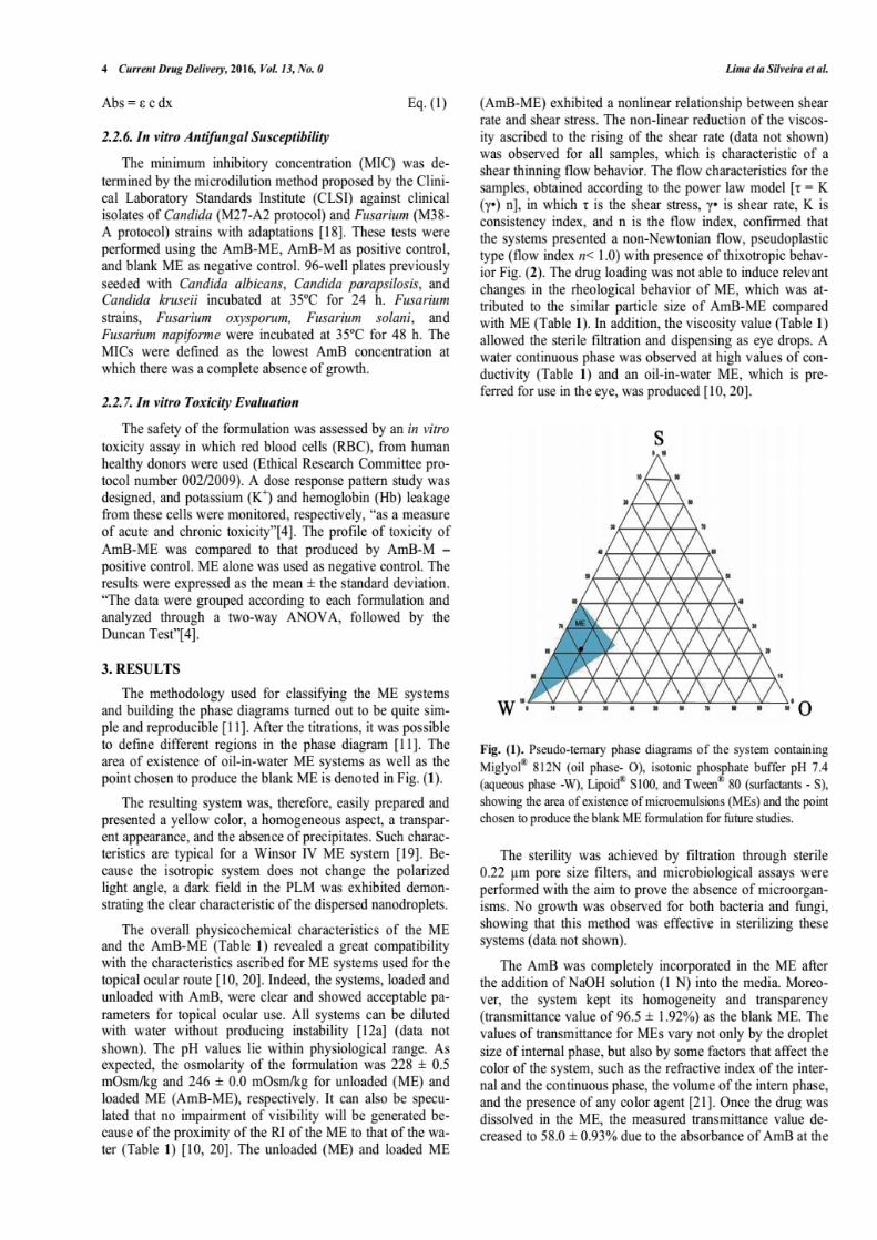
129
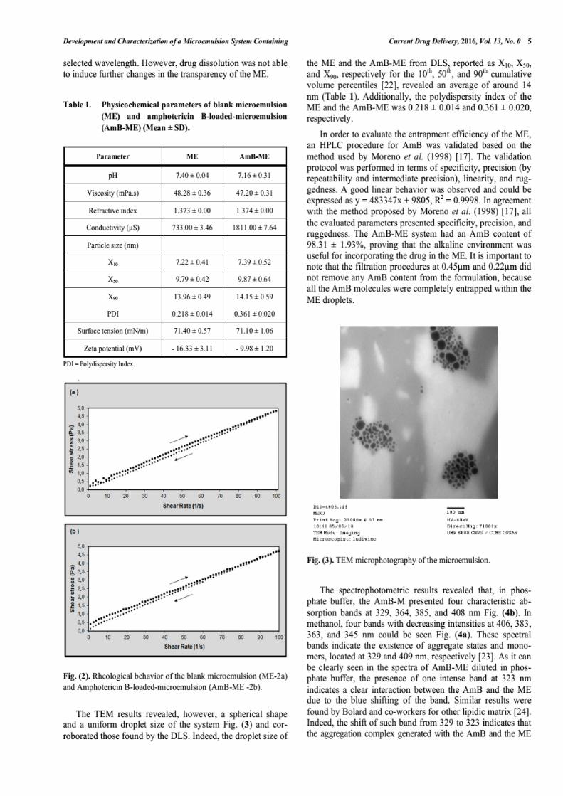
130
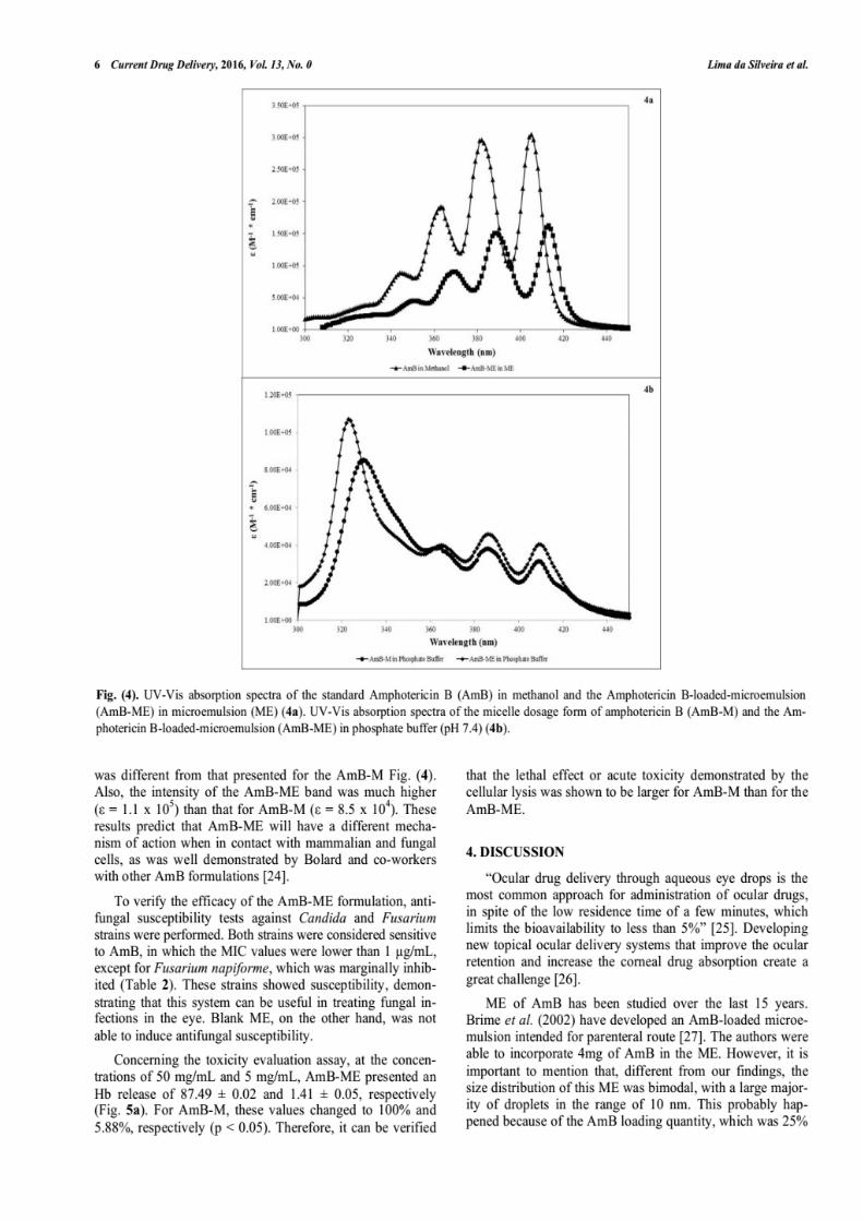
131
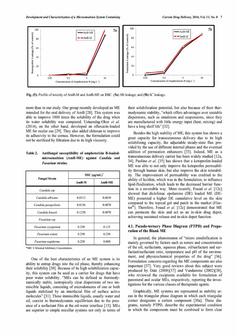
132
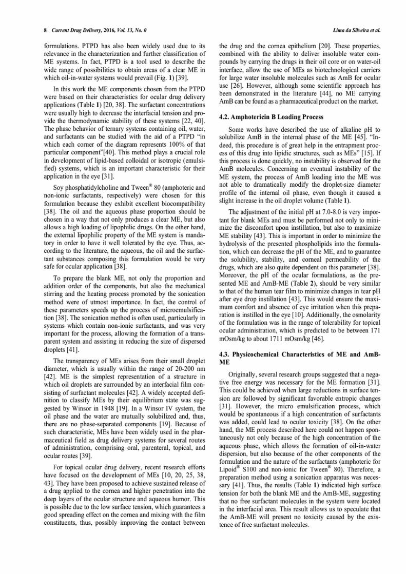
133
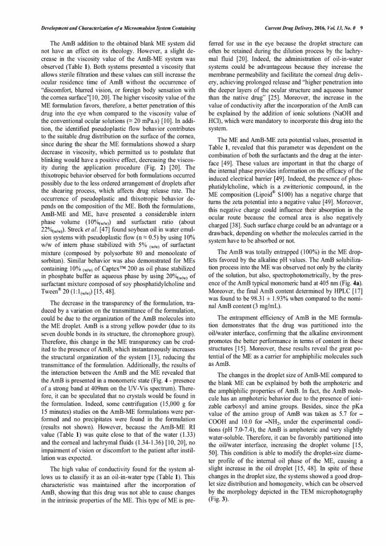
134
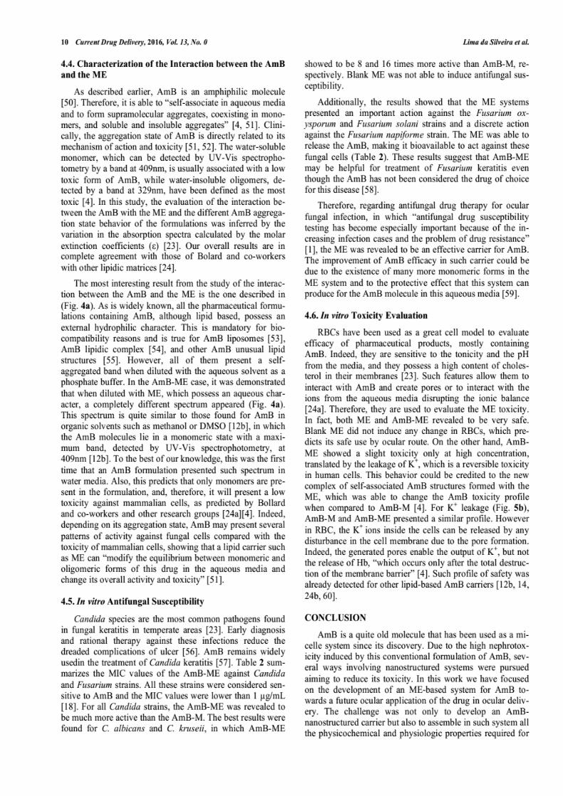
135
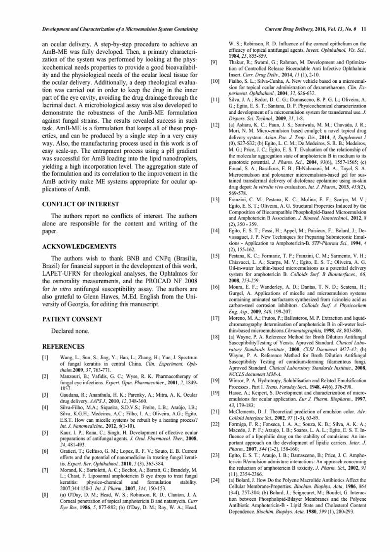
136
137
138
SECTION IV
VEHICULATION OF AN AmB-SOLUBLE DERIVATIVE INTO A
MICRO-EMULSION SYSTEM
____________________________________________________________
139
BRIEFING
The amphotericin B (AmB) physicochemical features cause the drug insolubility
in various media, mainly in water. Such an insolubility influences on the development of
new pharmaceutical products containing AmB, once the drug is soluble in a few types of
solvents that are, in most cases, incompatible with the human use.
We have observed the aggregation behavior of alkaline solubilized AmB under
the conditions of “super-aggregates” formation and developed a process in which the
insoluble molecule passes through out, becoming soluble in water at neutral or slightly
acid pH.
This section presents, in the chapter VI, a scientific report about the incorporation
of an AmB-derivative, developed by our group (under protection) into a micro-emulsion
system.

140
CHAPTER VI
Microemulsion encapsulation of an AmB soluble derivative
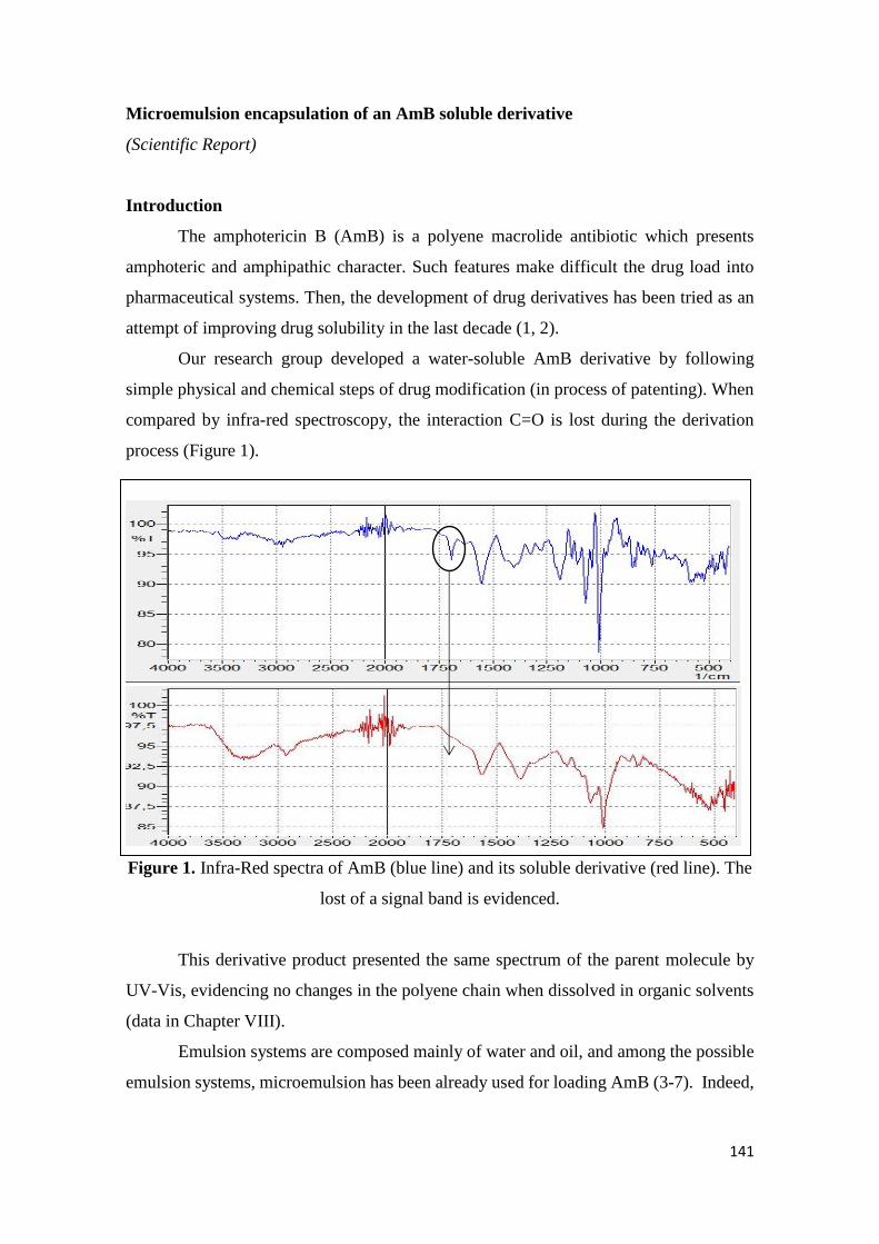
141
Microemulsion encapsulation of an AmB soluble derivative
(Scientific Report)
Introduction
The amphotericin B (AmB) is a polyene macrolide antibiotic which presents
amphoteric and amphipathic character. Such features make difficult the drug load into
pharmaceutical systems. Then, the development of drug derivatives has been tried as an
attempt of improving drug solubility in the last decade (1, 2).
Our research group developed a water-soluble AmB derivative by following
simple physical and chemical steps of drug modification (in process of patenting). When
compared by infra-red spectroscopy, the interaction C=O is lost during the derivation
process (Figure 1).
Figure 1. Infra-Red spectra of AmB (blue line) and its soluble derivative (red line). The
lost of a signal band is evidenced.
This derivative product presented the same spectrum of the parent molecule by
UV-Vis, evidencing no changes in the polyene chain when dissolved in organic solvents
(data in Chapter VIII).
Emulsion systems are composed mainly of water and oil, and among the possible
emulsion systems, microemulsion has been already used for loading AmB (3-7). Indeed,
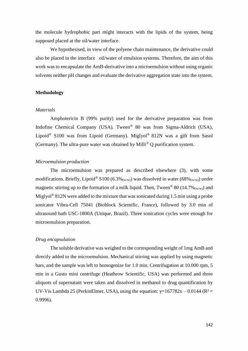
142
the molecule hydrophobic part might interacts with the lipids of the system, being
supposed placed at the oil/water interface.
We hypothesised, in view of the polyene chain maintenance, the derivative could
also be placed in the interface oil/water of emulsion systems. Therefore, the aim of this
work was to encapsulate the AmB-derivative into a microemulsion without using organic
solvents neither pH changes and evaluate the derivative aggregation state into the system.
Methodology
Materials
Amphotericin B (99% purity) used for the derivative preparation was from
Indofine Chemical Company (USA). Tween® 80 was from Sigma-Aldrich (USA),
Lipoid® S100 was from Lipoid (Germany). Miglyol® 812N was a gift from Sasol
(Germany). The ultra-pure water was obtained by Milli® Q purification system.
Microemulsion production
The microemulsion was prepared as described elsewhere (3), with some
modifications. Briefly, Lipoid® S100 (6.3%(w/w)) was dissolved in water (68%(w/w)) under
magnetic stirring up to the formation of a milk liquid. Then, Tween® 80 (14.7%(w/w)) and
Miglyol® 812N were added to the mixture that was sonicated during 1.5 min using a probe
sonicator Vibra-Cell 75041 (Bioblock Scientific, France), followed by 3.0 min of
ultrasound bath USC-1800A (Unique, Brazil). Three sonication cycles were enough for
microemulsion preparation.
Drug encapsulation
The soluble derivative was weighed to the corresponding weight of 1mg AmB and
directly added to the microemulsion. Mechanical stirring was applied by using magnetic
bars, and the sample was left to homogenize for 1.0 min. Centrifugation at 10.000 rpm, 5
min in a Gusto mini centrifuge (Heathrow Scientific, USA) was performed and three
aliquots of supernatant were taken and dissolved in methanol to drug quantification by
UV-Vis Lambda 25 (PerkinElmer, USA), using the equation: y=167782x – 0.0144 (R² =
0.9996).
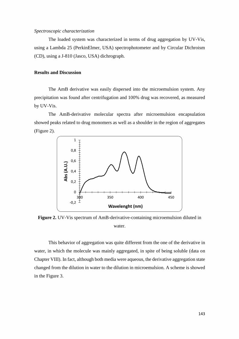
143
Spectroscopic characterization
The loaded system was characterized in terms of drug aggregation by UV-Vis,
using a Lambda 25 (PerkinElmer, USA) spectrophotometer and by Circular Dichroism
(CD), using a J-810 (Jasco, USA) dichrograph.
Results and Discussion
The AmB derivative was easily dispersed into the microemulsion system. Any
precipitation was found after centrifugation and 100% drug was recovered, as measured
by UV-Vis.
The AmB-derivative molecular spectra after microemulsion encapsulation
showed peaks related to drug monomers as well as a shoulder in the region of aggregates
(Figure 2).
Figure 2. UV-Vis spectrum of AmB-derivative-containing microemulsion diluted in
water.
This behavior of aggregation was quite different from the one of the derivative in
water, in which the molecule was mainly aggregated, in spite of being soluble (data on
Chapter VIII). In fact, although both media were aqueous, the derivative aggregation state
changed from the dilution in water to the dilution in microemulsion. A scheme is showed
in the Figure 3.
-0,2
0
0,2
0,4
0,6
0,8
1
300 350 400 450
Ab
s (A
.U.)
Wavelenght (nm)
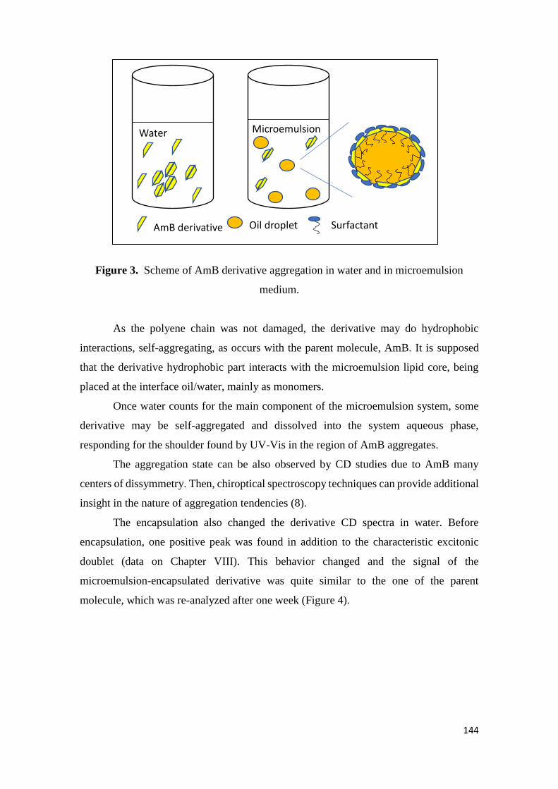
144
Figure 3. Scheme of AmB derivative aggregation in water and in microemulsion
medium.
As the polyene chain was not damaged, the derivative may do hydrophobic
interactions, self-aggregating, as occurs with the parent molecule, AmB. It is supposed
that the derivative hydrophobic part interacts with the microemulsion lipid core, being
placed at the interface oil/water, mainly as monomers.
Once water counts for the main component of the microemulsion system, some
derivative may be self-aggregated and dissolved into the system aqueous phase,
responding for the shoulder found by UV-Vis in the region of AmB aggregates.
The aggregation state can be also observed by CD studies due to AmB many
centers of dissymmetry. Then, chiroptical spectroscopy techniques can provide additional
insight in the nature of aggregation tendencies (8).
The encapsulation also changed the derivative CD spectra in water. Before
encapsulation, one positive peak was found in addition to the characteristic excitonic
doublet (data on Chapter VIII). This behavior changed and the signal of the
microemulsion-encapsulated derivative was quite similar to the one of the parent
molecule, which was re-analyzed after one week (Figure 4).
Water Microemulsion
AmB derivative Oil droplet Surfactant
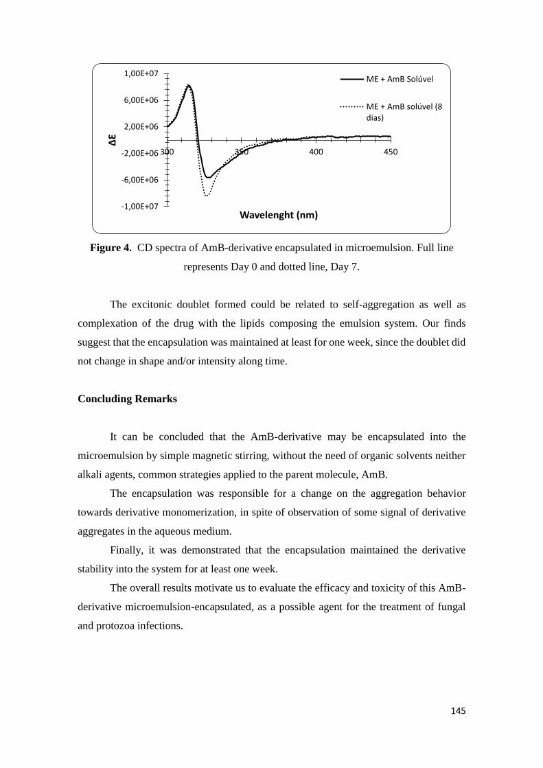
145
Figure 4. CD spectra of AmB-derivative encapsulated in microemulsion. Full line
represents Day 0 and dotted line, Day 7.
The excitonic doublet formed could be related to self-aggregation as well as
complexation of the drug with the lipids composing the emulsion system. Our finds
suggest that the encapsulation was maintained at least for one week, since the doublet did
not change in shape and/or intensity along time.
Concluding Remarks
It can be concluded that the AmB-derivative may be encapsulated into the
microemulsion by simple magnetic stirring, without the need of organic solvents neither
alkali agents, common strategies applied to the parent molecule, AmB.
The encapsulation was responsible for a change on the aggregation behavior
towards derivative monomerization, in spite of observation of some signal of derivative
aggregates in the aqueous medium.
Finally, it was demonstrated that the encapsulation maintained the derivative
stability into the system for at least one week.
The overall results motivate us to evaluate the efficacy and toxicity of this AmB-
derivative microemulsion-encapsulated, as a possible agent for the treatment of fungal
and protozoa infections.
-1,00E+07
-6,00E+06
-2,00E+06
2,00E+06
6,00E+06
1,00E+07
300 350 400 450
ΔƐ
Wavelenght (nm)
ME + AmB Solúvel
ME + AmB solúvel (8dias)
146
References
1. Adediran SA, Day TP, Sil D, Kimbrell MR, Warshakoon HJ, Malladi SS, et al.
Synthesis of a highly water-soluble derivative of amphotericin B with attenuated
proinflammatory activity. Mol Pharm. 2009;6(5):1582-90.
2. Thanathanee O, Miller D, Ringel DM, Schaffner CP, Alfonso EC, O'Brien TP.
Comparative In Vitro Antifungal Susceptibility Activity of Amphotericin B Versus
Amphotericin B Methyl Ester Against Candida albicans Ocular Isolates. J Ocular
Pharmacol Ther. 2012;28(6):589-92.
3. da Silveira WL, Damasceno BP, Ferreira LF, Ribeiro IL, Silva KS, Silva AL, et
al. Development and Characterization of a Microemulsion System Containing
Amphotericin B with Potential Ocular Applications. Curr Drug Deliv. 2016;13(6):982-
93.
4. Silva AE, Barratt G, Cheron M, Egito ES. Development of oil-in-water
microemulsions for the oral delivery of amphotericin B. Int J Pharm. 2013;454(2):641-8.
5. Pestana KC, Formariz TP, Franzini CM, Sarmento VH, Chiavacci LA, Scarpa
MV, et al. Oil-in-water lecithin-based microemulsions as a potential delivery system for
amphotericin B. Colloids and Surfaces B, Biointerfaces. 2008;66(2):253-9.
6. Brime B, Moreno MA, Frutos G, Ballesteros MP, Frutos P. Amphotericin B in
oil-water lecithin-based microemulsions: formulation and toxicity evaluation. J Pharm
Sci. 2002;91(4):1178-85.
7. Damasceno BP, Dominici VA, Urbano IA, Silva JA, Araujo IB, Santos-
Magalhaes NS, et al. Amphotericin B microemulsion reduces toxicity and maintains the
efficacy as an antifungal product. J Biomed Nanotechnol. 2012;8(2):290-300.
8. Britain, HG. Circular dichroism studies of the self-association of amphotericin B.
Chirality. 1994, 6 (8): p.665 – 669.
147
General Discussion
GENERAL DISCUSSION
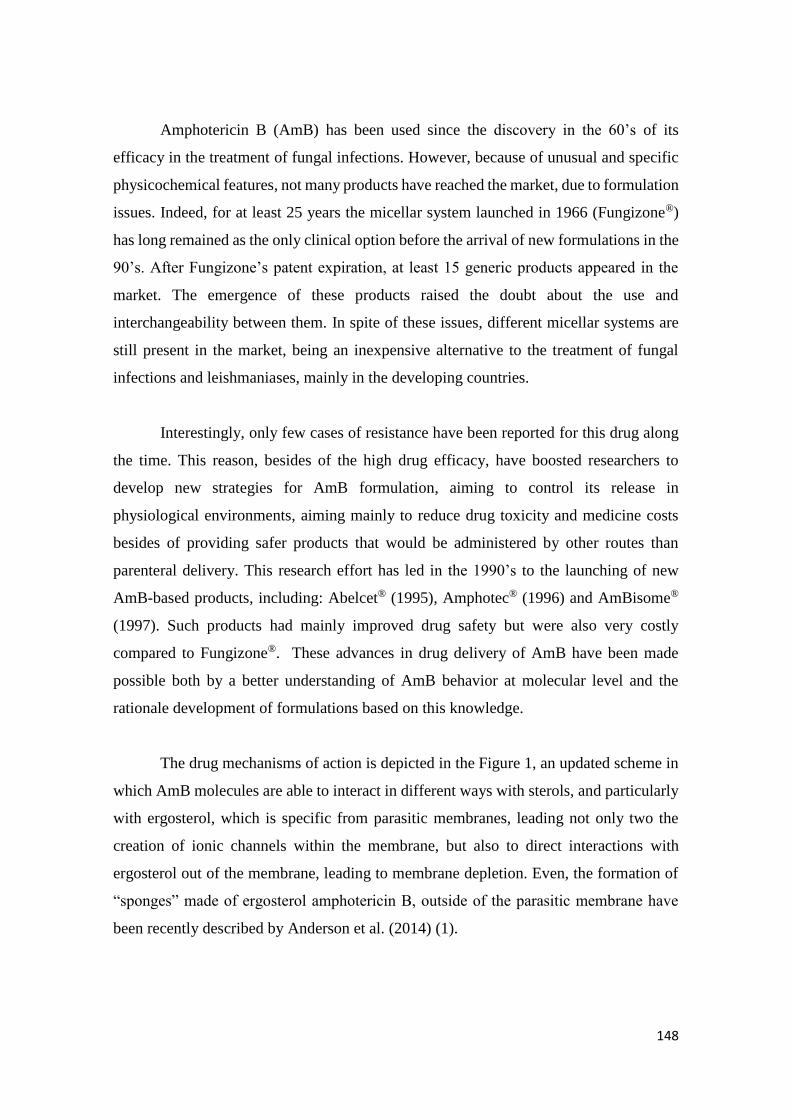
148
Amphotericin B (AmB) has been used since the discovery in the 60’s of its
efficacy in the treatment of fungal infections. However, because of unusual and specific
physicochemical features, not many products have reached the market, due to formulation
issues. Indeed, for at least 25 years the micellar system launched in 1966 (Fungizone®)
has long remained as the only clinical option before the arrival of new formulations in the
90’s. After Fungizone’s patent expiration, at least 15 generic products appeared in the
market. The emergence of these products raised the doubt about the use and
interchangeability between them. In spite of these issues, different micellar systems are
still present in the market, being an inexpensive alternative to the treatment of fungal
infections and leishmaniases, mainly in the developing countries.
Interestingly, only few cases of resistance have been reported for this drug along
the time. This reason, besides of the high drug efficacy, have boosted researchers to
develop new strategies for AmB formulation, aiming to control its release in
physiological environments, aiming mainly to reduce drug toxicity and medicine costs
besides of providing safer products that would be administered by other routes than
parenteral delivery. This research effort has led in the 1990’s to the launching of new
AmB-based products, including: Abelcet® (1995), Amphotec® (1996) and AmBisome®
(1997). Such products had mainly improved drug safety but were also very costly
compared to Fungizone®. These advances in drug delivery of AmB have been made
possible both by a better understanding of AmB behavior at molecular level and the
rationale development of formulations based on this knowledge.
The drug mechanisms of action is depicted in the Figure 1, an updated scheme in
which AmB molecules are able to interact in different ways with sterols, and particularly
with ergosterol, which is specific from parasitic membranes, leading not only two the
creation of ionic channels within the membrane, but also to direct interactions with
ergosterol out of the membrane, leading to membrane depletion. Even, the formation of
“sponges” made of ergosterol amphotericin B, outside of the parasitic membrane have
been recently described by Anderson et al. (2014) (1).
149
Figure 1. Molecular structure of amphotericin B and ergosterol and proposed unifying
model for amphotericin B antifungal activity involving two complementary AmB
mechanisms of antifungal activity including membrane permeabilization (1), AmB
ergosterol binding (2), and sterol sponge formation (3).
In this revised model, these authors wrote that: “In the widely accepted ion
channel model for its mechanism of cytocidal action, amphotericin forms aggregates
inside lipid bilayers that permeabilize and kill cells. In contrast, (the authors) report that
amphotericin exists primarily in the form of large, extra membranous aggregates that kill
yeast by extracting ergosterol from lipid bilayers.”(1)
It is important to note that membrane permeabilization leads to different types of
ion channels formation in ergosterol containing membranes (2), as depicted in Figure 2.
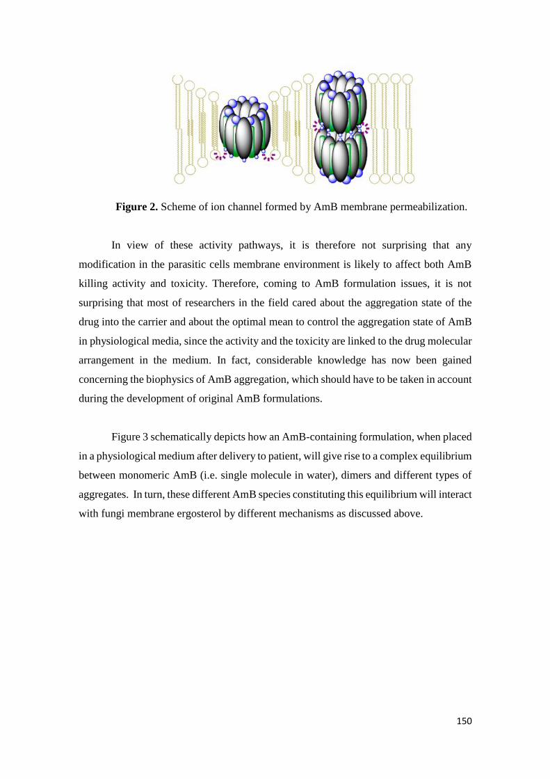
150
Figure 2. Scheme of ion channel formed by AmB membrane permeabilization.
In view of these activity pathways, it is therefore not surprising that any
modification in the parasitic cells membrane environment is likely to affect both AmB
killing activity and toxicity. Therefore, coming to AmB formulation issues, it is not
surprising that most of researchers in the field cared about the aggregation state of the
drug into the carrier and about the optimal mean to control the aggregation state of AmB
in physiological media, since the activity and the toxicity are linked to the drug molecular
arrangement in the medium. In fact, considerable knowledge has now been gained
concerning the biophysics of AmB aggregation, which should have to be taken in account
during the development of original AmB formulations.
Figure 3 schematically depicts how an AmB-containing formulation, when placed
in a physiological medium after delivery to patient, will give rise to a complex equilibrium
between monomeric AmB (i.e. single molecule in water), dimers and different types of
aggregates. In turn, these different AmB species constituting this equilibrium will interact
with fungi membrane ergosterol by different mechanisms as discussed above.
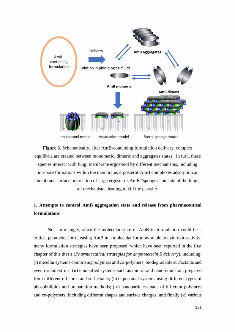
151
Figure 3. Schematically, after AmB-containing formulation delivery, complex
equilibria are created between monomeric, dimeric and aggregates states. In turn, these
species interact with fungi membrane ergosterol by different mechanisms, including
ion-pore formations within the membrane, ergosterol-AmB complexes adsorption at
membrane surface or creation of large ergosterol-AmB “sponges” outside of the fungi,
all mechanisms leading to kill the parasite.
1. Attempts to control AmB aggregation state and release from pharmaceutical
formulations
Not surprisingly, since the molecular state of AmB in formulation could be a
critical parameter for releasing AmB in a molecular form favorable to cytotoxic activity,
many formulation strategies have been proposed, which have been reported in the first
chapter of this thesis (Pharmaceutical strategies for amphotericin B delivery), including:
(i) micellar systems comprising polymers and co-polymers, biodegradable surfactants and
even cyclodextrins; (ii) emulsified systems such as micro- and nano-emulsion, prepared
from different oil cores and surfactants; (iii) liposomal systems using different types of
phospholipids and preparation methods; (iv) nanoparticles made of different polymers
and co-polymers, including different shapes and surface charges; and finally (v) various
152
other colloidal systems including nanodisks, carbon nanotubes, nanogels and cochleates
(3).
In view of this variety of carrier systems, this thesis was dedicated to the study of
AmB aggregation state into both marketed systems and newly conceived delivery
systems.
2. Variability of AmB aggregation state in marketed micellar pharmaceutical
formulations
In the second chapter (Amphotericin B-containing sodium deoxycholate micellar
systems: a comparison of physicochemical characteristics of marketed products) we have
compared three micellar systems, having same composition and produced in three
different countries: Brazil, India and France (4).
The labeled concentration of all products was 50 mg AmB per flask, that has to
be diluted in 10 mL water (theoretical final concentration: 5mg AmB/mL). Quite
curiously, two out of the three formulations were over-dosed, leading to over-
concentrated solutions (more than 25%), which may have consequences on the
aggregation state after delivery.
Indeed, the increase in concentration was correlated to an increase on the drug
aggregation level, which was observed in UV-Vis spectra, highlighting the close
relationship between concentration and drug aggregation. Meanwhile, the other physico-
chemical parameters analyzed such as aggregates size distribution and circular dichroism
spectra showed no differences among products.
The coexistence in the molecule of an hydrophilic and an hydrophobic moiety of
the molecule makes that AmB molecules are prone to extensive self-aggregation.
Consequently, in aqueous media, AmB can be found simultaneously in the form of
monomers, dimers (head-head or head-tail) and aggregates, related by different equilibria.
Talking about micellar formulations, when diluted (e.g. in physiological media), the drug
is released in the medium and is likely to self-aggregate, accordingly to the concentration.
153
Circular dichroism (CD) helped to clarify two important points. At the first, the
reversibility of aggregation was demonstrated by diluting the different micellar systems,
leading to AmB dis-aggregation, proving the aforementioned link between concentration
and aggregation. The second point concerns the type and stoichiometry of oligomers
formed. Our results showed that they were of the same type in all the samples tested,
despite the differences in drug concentrations.
Once CD and UV-Vis spectra were recorded for these samples, it is important to
clarify that these methods no longer coincide. Electronic absorption measured by UV-Vis
monitors the exact amounts of the different AmB forms, while CD allows the detection
of associated species in minor amounts.
The nature of aggregates seemed to be the same, once the ratio between the
positive and negative peaks of AmB couplet did not vary under dilution and according to
the CD couplet signature, AmB molecules are aggregated probably as antiparallel dimers.
The size of the structures in solution is quite controversy since different values
were obtained when DLS technique was employed, depending if the data were expressed
in number or in intensity. It seems that the aggregation state played a role on the droplet
size. However, as mentioned, this kind of measurement could be cautiously examined, as
different bias could affect the estimation of the aggregates actual size.
3. Formation of AmB super-aggregated from marketed micellar pharmaceutical
formulations
AmB super aggregates are known from the 90s, which result from the extended
aggregation of the different species of the “classical” equilibrium between monomeric,
dimeric and aggregated species. Super aggregates have been initially produced by heating
different existing formulations.
It is well known that mild heating of AmB micellar solutions promotes AmB
super-aggregation. Such super-aggregates are less toxic than the original preparation
while keeping drug efficiency. Since, this behavior has been observed in other systems,
including nanodisks (5), and more recently, attempts to engineer AmB-based
154
nanoparticles has been described (6). The interest of these strategy is to create larger
objects, which are known to demonstrate less toxicity against mamallian cells, while
keeping antiparasitic activity.
During the comparative study of different commercial formulations undertaken
during this work, significant differences in the labelled drug concentration were observed,
which were likely to directly impact AmB aggregation state. In the present work, we have
studied the super-aggregation process of AmB-sodium deoxycholeate (SD) micelles and
also evaluated the possibility of super-aggregation of the drug contained in a liposomal
suspension (7).
In the case of the micellar AmB-SD solution, we have confirmed by UV-Vis
spectrophotometry that the formation of super-aggregates was accompanied of an
electronic blue-shift. The main peak centered at 326 nm moved towards 323 nm for the
sodium-deoxycholate micellar system, confirming super-aggregation. In fact, the super-
aggregates formation could also be observed by naked eye, since the solution become
blurred. Such an increase in turbidity was confirmed by DLS measurements.
From clinical experience, AmB liposomal suspension is a safe product and maybe,
because of that, there are no reports in the literature describing super-aggregation
formation with this kind of lipidic colloidal systems. Not very surprisingly, we observed
no changes in spectral UV-Vis and CD signatures, suggesting that this system do not form
super-aggregates on heating. We can hypothesize that super-aggregates heating of
liposomal systems cannot lead to super-aggregation because the presence of oligomers
and monomers in solution seems to be mandatory for the new entity formation. In the
liposomes, AmB is strongly associated to the phospholipids and not released by dilution,
as demonstrated by UV-Vis and CD, even along the time. Interestingly, this case study
demonstrates that the physico-chemical state of aggregation within the formulation is able
to control the nature of AmB equilibrium outside of the formulation. In the present case,
if the reagents are not available, the new product cannot be produced.
An interesting point is that following the spectral changes of heated and unheated
systems along the time, it was observed that the unheated systems balanced the ratio
among dimers and monomers as a function of drug concentration. Then, the spectral shape
155
changed along the time. Such behavior was not observed when the system micellar system
was heated. In this case there was a maintenance of the spectral shape along the time. A
decrease in intensity was described for all the species, super-aggregates and monomers.
This experience allowed us to suggest that the super-aggregates had a more cohesive
structure, which resulted in an improvement of drug protection along the time. Heated
systems after 28 days of shelf-storage, contained at least 10% AmB more than the
unheated counterparts.
4. Consequences of super-aggregates delivery in the body by parenteral routes.
When AmB formulations are to be delivered by intravenous route of delivery, the
behavior of super aggregates in the blood stream and possibly their recognition by the
immune system may be an issue. Indeed, the possible recognition and capture of
polymeric nanoparticles is a well know phenomenon. As described by Coty et al. (2017)
(8), the adsorption of specific plasmatic proteins at the surface of the particles may lead,
or not, to their rapid recognition by specialized phagocytic cells in different organs, not
only leading to fast clearance of the particles out of the blood, but also to unadequate
organs/cells distribution within the body.
For this reason, we attempted first to get a better understanding of the architecture
of AmB super aggregates, by attempting to measure the energy necessary to self-
aggregation by using an isothermal titration calorimetry equipment (ITC). However, the
signals were quite low, just suggesting qualitatively that molecular re-organization
generated heat but making impossible to gain any insight on the aggregation process
itself.
However, we used a test known as the complement activation test, to evaluate the
possible impact of AmB aggregation on the immune response. This part of the work was
conducted mainly by Dr. Marcelino, in which we collaborated on the aggregates
characterization. Briefly, preliminary results were obtained, which showed a correlation
between the presence of AmB aggregates in (human) plasma and the activation of the
immunological system (9). Independent of the formulation administered, when AmB is
delivered by the intravenous route, AmB molecules are likely to bind to serum proteins,
as well as self-aggregate, all phenomena which may considerably affect its uptake by
156
macrophage cell-like. From a practical point of view, immune system activation would
be detrimental to safety of self-aggregated formulations and we suggest that these aspects
should be explored in more detail for a better understanding.
5. AmB incorporation into micro- and nano- emulsions and aggregation state control
Emulsions are systems able to carry lipophilic and hydrophilic molecules once
they are composed mainly of oil and water. According to the composition, way of
production and final characteristics, emulsions can be classified as micro-, nano- or
regular-emulsion. Having in mind that drug aggregation can occur in very different carrier
systems, we focused on AmB incorporation into micro- and nano-emulsion systems and
followed the drug aggregation patterns into these systems (10).
We reported AmB incorporation into micro- and nano-emulsion systems, trying
at first to figure out the solubility of the drug in the media by using pH alkalinization. In
order to avoid the use of organic solvents, researchers take advantage of the alkaline pKa
of the drug. Once the pH is higher than 10, AmB is mainly ionized and becomes soluble
in the medium. However, up to the moment, there was not a study concerning the best
alkali concentration in order to have AmB totally soluble or in the monomeric state.
In a first step, 1,48 .10-2 N NaOH alkalinization to pH > 11 helped to solubilize
AmB, which existed mainly as dimers as demonstrated by UV-Vis spectrophotometry. A
further increase in alkali (to 3,85 .10-2 N) was able to displace the UV-Vis spectra towards
monomeric bands, indicating that not only the pH but the alkali agent concentration
played a role on the drug solubility and aggregation.
In a second step, AmB solubility in nano- and micro-emulsion media was also
evaluated. These emulsions consisted of medium chain triglycerides associated to
different surfactants and water and had a constant composition in the whole study. It was
found that the nano-emulsion was able to solubilize a higher amount of AmB (5 mg AmB)
, when compared to the micro-emulsion (3 mg AmB).
Drug aggregation of AmB into the nano-emulsion increased as the drug
concentration was increased similarly to the behavior of already reported for micellar
157
systems. Interestingly, the increase in droplet size when comparing loaded and drug-free
droplets did not change in line with drug concentration.
When AmB was incorporated as a powder in the preparation, AmB was present
into the nano-emulsion both as monomers and aggregates. Changing the incorporation
process, when the drug was first solubilized into an alkaline aqueous phase, the drug was
loaded as poly-aggregates, an AmB species already described in the literature and which
have been described as being less toxic than the other AmB species.
When AmB was loaded into the micro-emulsion, AmB existed mainly in the
monomeric form. The studies conducted in collaboration with Dr. Silveira (11) and Dr.
Morais (12) showed how promising the AmB-containing micro-emulsion are. In Dr.
Silveira’s study the development of the micro-emulsion and all the characterization for
its use in ocular infections are showed. The system was less toxic than the micellar
formulation, having efficacy saved and even improved for the treatment of many fungal
species.
Dr. Morais and co-workers have tested, in vitro and in vivo, formulations which
would be efficient and inexpensive to treat leishmaniasis. They have tested heated and
unheated micellar systems as well as the AmB-loaded micro-emulsion, in comparison to
the liposomal system and no differences among the inexpensive systems (micro-emulsion
and heated micellar system) and the liposomal products were found in toxicity and
efficacy against L. donovani.
Up to this moment we have been dealing with AmB into different carrier systems:
micelles, liposomes, micro- and nano-emulsion. Caring mainly about characterizing the
aggregation state of the drug into the system and on how the aggregation pattern could
influence the drug toxicity and we have reported at least two successful uses of the micro-
emulsion system as an inexpensive alternative to the toxic micellar system. Then, we have
loaded the AmB derivative into a micro-emulsion system(13). The new AmB species was
easily dispersed in the micro-emulsion media, presenting UV-Vis spectra of monomers
and a slight shoulder corresponding to AmB aggregates.
7. Conclusions
158
Issues concerning AmB loading into pharmaceutical systems stems from the
unique, and unfavorable physicochemical features of this molecule. We hope that the
present work will contribute to bringing new strategies for its delivery and opening
possibilities for other administration routes, based on a better understanding of the
relationships between AmB aggregation and formulation characteristics.
REFERENCES
1. Anderson TM, Clay MC, Cioffi AG, Diaz KA, Hisao GS, Tuttle MD, et al.
Amphotericin forms an extramembranous and fungicidal sterol sponge. Nature Chemical
Biology. 2014;10(5):400-U121.
2. Gray KC, Palacios DS, Dailey I, Endo MM, Uno BE, Wilcock BC, et al.
Amphotericin primarily kills yeast by simply binding ergosterol. Proceedings of the
National Academy of Sciences of the United States of America. 2012;109(7):2234-9.
3. Silva AL, Ponchel G, Egito ESTd. Pharmaceutical strategies for amphotericin B
delivery. [Unpublished work]. In press 2017.
4. Silva AL, Chéron M, Ponchel G, Egito ESTd. Amphotericin B-containing sodium
deoxycholate micellar systems: a comparison of physicochemical characteristics of
marketed products. [Unpublished work]. In press 2017.
5. Burgess BL, He YM, Baker MM, Luo B, Carroll SF, Forte TM, et al. NanoDisk
containing super aggregated amphotericin B: a high therapeutic index antifungal
formulation with enhanced potency. International Journal of Nanomedicine.
2013;8:4733-42.
6. Zia Q, Mohammad O, Rauf MA, Khan W, Zubair S. Biomimetically engineered
Amphotericin B nano-aggregates circumvent toxicity constraints and treat systemic
fungal infection in experimental animals. Scientific Reports. 2017;7.
7. Silva AL, Castillo AYSTd, Chéron M, Ponchel G, Egito ESTd. Amphotericin B
super-aggregates: formation and influence on the drug stability. [Unpublished work]. In
press 2017.

159
8. Coty JB, Oliveira EE, Vauthier C. Tuning complement activation and pathway
through controlled molecular architecture of dextran chains in nanoparticle corona.
International Journal of Pharmaceutics. 2017;532(2):769-78.
9. Marcelino HR, Coty J-B, Silva AL, Chéron M, Vauthier C, Egito ESTd, et al.
Alternative mechanism for amphotericin B uptake by macrophage cell-like: the possible
role of the complement immune system. [Unpublished work]. In press 2016.
10. Silva AL. AmB loading into micro- and nano-emulsion systems. Unpublished
work. 2017.
11. Silveira WLLd, Damasceno BPGL, Ferreira LF, Ribeiro ILS, Silva KS, Silva AL,
et al. Development and characterization of a microemulsion system containing
amphotericin B with potential ocular applications. Current Drug Delivery.
2016;13(6):982-93.
12. Morais ARdV, Silva AL, Cojean S, Balaraman K, Bories C, Pomel S, et al. In
vitro and in vivo antileishmanial activity of cheap amphotericin B formulations: heated
amphotericin B and amphotericin B-loaded microemulsion. [Unpublished work]. In press
2017.
13. Silva AL. Microemulsion encapsulation of an AmB soluble derivative.
Unpublished work. 2017.
160
Conclusions
161
CONCLUSIONS
The amphotericin B (AmB) molecule has been studied since its discovery, in the
1950’s. Since that time many strategies have been considered aiming to improve drug
availability for the clinics. The first developed product containing this drug was a micellar
system produced as a mix of the biliary salt sodium deoxycholate and AmB. This system,
available up to now, is of easy prepare and was largely produced for many pharmaceutical
companies around the world, after Fungizone’s patent expiration.
In this work we made a comparison among Fungizone’s similar products,
concluding that they are not equivalents physic-chemically. The ratio among monomers
and aggregates is not reproducible on the different brands, even the labeled content was
not respected in some cases, evidencing no interchangeability of tested similar products.
Such result might be due to the difference on the production process or event to the raw
material employed for the different pharmaceutical companies.
The AmB molecular re-organization by mild heating is responsible for the
formation of the called “super-aggregates”. This procedure is already known for the
scientific community and the decrease on the drug toxicity is attributed to the “super-
aggregated” conformation. Many researchers have demonstrated the possibility of re-
arranging the drug from micellar systems but no reports are found about forming “super-
aggregates” from AmB-containing liposomes. We have showed that the “super-
aggregates” are AmB species more stable than the ones of the original product and, for
the first time, it was showed that is not possible to form “super-aggregates” from the
liposomal system AmBisome®.
Lipid systems loaded with AmB are known for being safer than micellar systems.
In fact, lipid complex and liposomes are available in the market and can be administered
in higher doses without inducement of nephrotoxicity or other side-effects related to the
infusion of AmB-micelles. Nevertheless, those are quite expensive products that impairs
their broadly use, mainly in developing countries.
We have showed that AmB can be loaded into emulsion system of easy production
and low cost, such are micro- and nano-emulsion. We have deepen the discussion about
drug incorporation dependence on pH medium, highlighting the importance of the alkali
162
agent concentration. Besides, applicability of microemulsions as AmB-carrier was
demonstrated as an alternative treatment for ocular infections and visceral leishmaniasis.
Finally, we understand that the main problems concerning AmB loading are linked
to its low solubility. Then, we reported the production process of an AmB-soluble
derivative that can be loaded into a micro-emulsion system, opening the possibility of
administering the drug for the treatment not only for various fungal and protozoa diseases,
but also for new administration routes.
The overall results presented in this Ph.D thesis are very important for the built of
the scientific knowledge about antibiotics, mainly the amphotericin B, in which was
evidenced that if one hand the drug aggregation state changes according to the system, on
the other hand one can control these changes towards the aggregation states supposed to
be less toxic and more efficient for the patients.
163
Synthèse de la Thèse ______________________________________________________________________
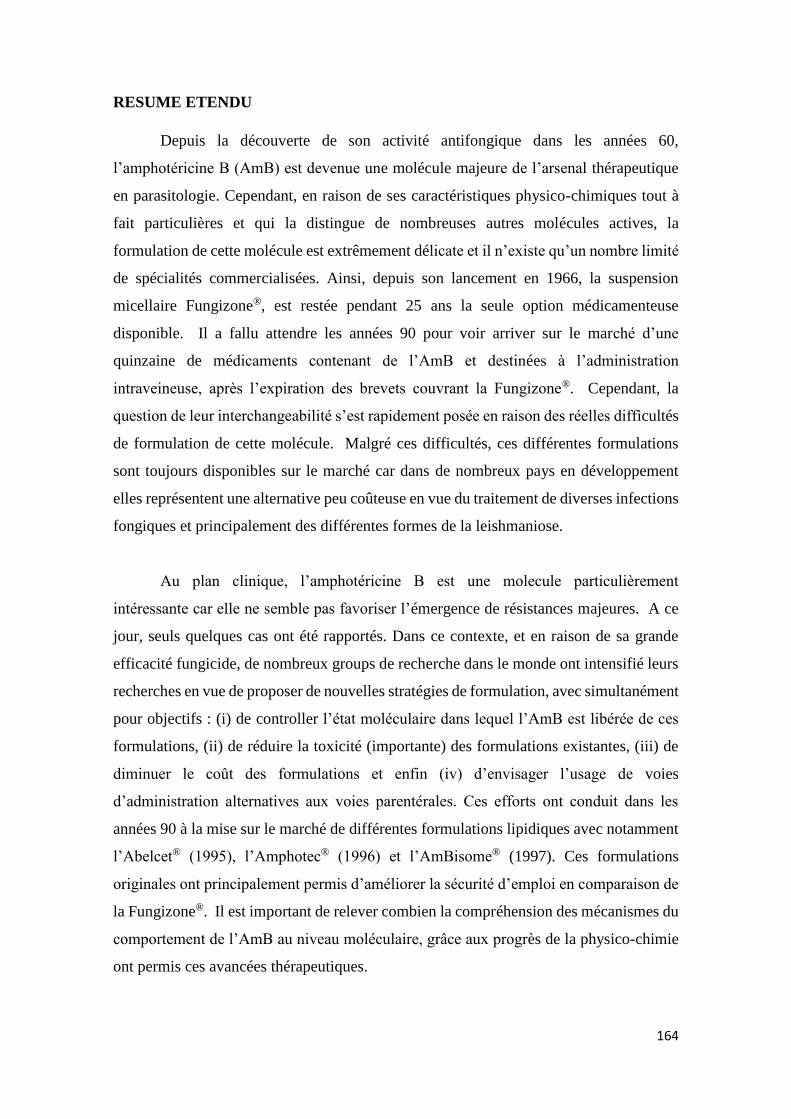
164
RESUME ETENDU
Depuis la découverte de son activité antifongique dans les années 60,
l’amphotéricine B (AmB) est devenue une molécule majeure de l’arsenal thérapeutique
en parasitologie. Cependant, en raison de ses caractéristiques physico-chimiques tout à
fait particulières et qui la distingue de nombreuses autres molécules actives, la
formulation de cette molécule est extrêmement délicate et il n’existe qu’un nombre limité
de spécialités commercialisées. Ainsi, depuis son lancement en 1966, la suspension
micellaire Fungizone®, est restée pendant 25 ans la seule option médicamenteuse
disponible. Il a fallu attendre les années 90 pour voir arriver sur le marché d’une
quinzaine de médicaments contenant de l’AmB et destinées à l’administration
intraveineuse, après l’expiration des brevets couvrant la Fungizone®. Cependant, la
question de leur interchangeabilité s’est rapidement posée en raison des réelles difficultés
de formulation de cette molécule. Malgré ces difficultés, ces différentes formulations
sont toujours disponibles sur le marché car dans de nombreux pays en développement
elles représentent une alternative peu coûteuse en vue du traitement de diverses infections
fongiques et principalement des différentes formes de la leishmaniose.
Au plan clinique, l’amphotéricine B est une molecule particulièrement
intéressante car elle ne semble pas favoriser l’émergence de résistances majeures. A ce
jour, seuls quelques cas ont été rapportés. Dans ce contexte, et en raison de sa grande
efficacité fungicide, de nombreux groups de recherche dans le monde ont intensifié leurs
recherches en vue de proposer de nouvelles stratégies de formulation, avec simultanément
pour objectifs : (i) de controller l’état moléculaire dans lequel l’AmB est libérée de ces
formulations, (ii) de réduire la toxicité (importante) des formulations existantes, (iii) de
diminuer le coût des formulations et enfin (iv) d’envisager l’usage de voies
d’administration alternatives aux voies parentérales. Ces efforts ont conduit dans les
années 90 à la mise sur le marché de différentes formulations lipidiques avec notamment
l’Abelcet® (1995), l’Amphotec® (1996) et l’AmBisome® (1997). Ces formulations
originales ont principalement permis d’améliorer la sécurité d’emploi en comparaison de
la Fungizone®. Il est important de relever combien la compréhension des mécanismes du
comportement de l’AmB au niveau moléculaire, grâce aux progrès de la physico-chimie
ont permis ces avancées thérapeutiques.

165
Afin d’illustrer cela, la Figure 1 décrit schématiquement les mécanismes
actuellement connus et par lesquels les molécules d’AmB interagissent avec les stérols et
tout particulièrement l’ergostérol, qui est un stérol spécifiquement constitutif des
membranes des cellules des parasites. Pour expliquer l’action antifungique de l’AmB,
les progrès récents en physico-chimie font appel, non seulement à la formation de pores
ioniques transmembranaires, mais aussi à d’autres mécanismes impliquant une interaction
avec l’ergostérol d’origine parasitaire et sa déplétion progressive de la membrane
cellulaire, suivie de la mort du parasite.
Dans ce dernier modèle, la production de véritables éponges constituées par l’agrégation
de molécules d’ergostérol et d’AmB a récemment été décrite par Thomas M Anderson et
al. 2014 (1).
Figure 1 : Structures moléculaires de l’amphotéricine B et de l’ergosterol et modèle
unifié décrivant les mécanismes de l’action antifongique par trois mécanismes
complémentaires, avec : (i) la perméabilisation membranaire par l’insertion de canaux
ioniques néoformés, (ii) la liaison d’une molécule d’AmB à une molécule d’ergosterol
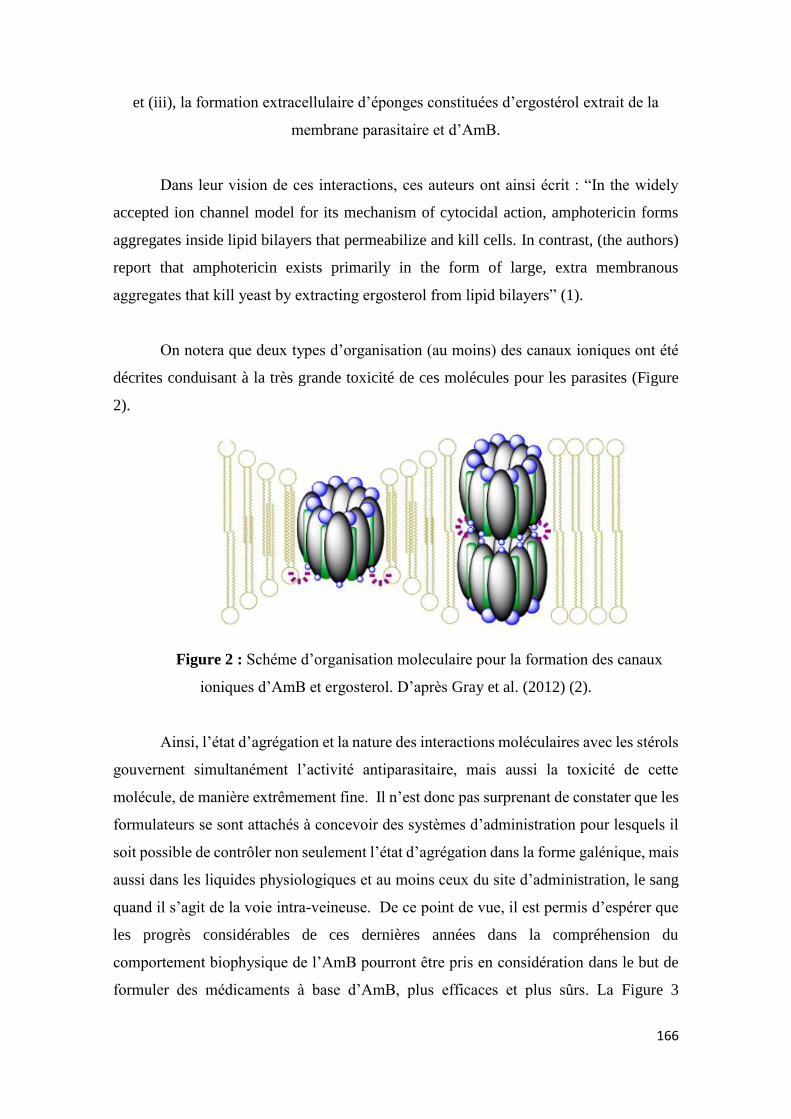
166
et (iii), la formation extracellulaire d’éponges constituées d’ergostérol extrait de la
membrane parasitaire et d’AmB.
Dans leur vision de ces interactions, ces auteurs ont ainsi écrit : “In the widely
accepted ion channel model for its mechanism of cytocidal action, amphotericin forms
aggregates inside lipid bilayers that permeabilize and kill cells. In contrast, (the authors)
report that amphotericin exists primarily in the form of large, extra membranous
aggregates that kill yeast by extracting ergosterol from lipid bilayers” (1).
On notera que deux types d’organisation (au moins) des canaux ioniques ont été
décrites conduisant à la très grande toxicité de ces molécules pour les parasites (Figure
2).
Figure 2 : Schéme d’organisation moleculaire pour la formation des canaux
ioniques d’AmB et ergosterol. D’après Gray et al. (2012) (2).
Ainsi, l’état d’agrégation et la nature des interactions moléculaires avec les stérols
gouvernent simultanément l’activité antiparasitaire, mais aussi la toxicité de cette
molécule, de manière extrêmement fine. Il n’est donc pas surprenant de constater que les
formulateurs se sont attachés à concevoir des systèmes d’administration pour lesquels il
soit possible de contrôler non seulement l’état d’agrégation dans la forme galénique, mais
aussi dans les liquides physiologiques et au moins ceux du site d’administration, le sang
quand il s’agit de la voie intra-veineuse. De ce point de vue, il est permis d’espérer que
les progrès considérables de ces dernières années dans la compréhension du
comportement biophysique de l’AmB pourront être pris en considération dans le but de
formuler des médicaments à base d’AmB, plus efficaces et plus sûrs. La Figure 3
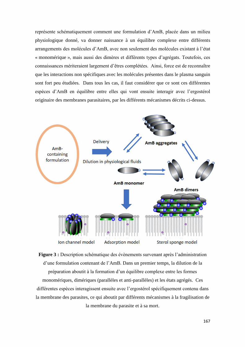
167
représente schématiquement comment une formulation d’AmB, placée dans un milieu
physiologique donné, va donner naissance à un équilibre complexe entre différents
arrangements des molécules d’AmB, avec non seulement des molécules existant à l’état
« monomérique », mais aussi des dimères et différents types d’agrégats. Toutefois, ces
connaissances mériteraient largement d’êtres complétées. Ainsi, force est de reconnaître
que les interactions non spécifiques avec les molécules présentes dans le plasma sanguin
sont fort peu étudiées. Dans tous les cas, il faut considérer que ce sont ces différentes
espèces d’AmB en équilibre entre elles qui vont ensuite interagir avec l’ergostérol
originaire des membranes parasitaires, par les différents mécanismes décrits ci-dessus.
Figure 3 : Description schématique des évènements survenant après l’administration
d’une formulation contenant de l’AmB. Dans un premier temps, la dilution de la
préparation aboutit à la formation d’un équilibre complexe entre les formes
monomériques, dimériques (parallèles et anti-parallèles) et les états agrégés. Ces
différentes espèces interagissent ensuite avec l’ergostérol spécifiquement contenu dans
la membrane des parasites, ce qui aboutit par différents mécanismes à la fragilisation de
la membrane du parasite et à sa mort.
168
1. Contrôle de l’état d’agrégation de l’AmB dans les formulations pharmaceutiques
et des modalités de sa libération.
Dans ce contexte, il n’est pas surprenant de constater que de nombreuses stratégies
de formulation de l’AmB ont été proposées. Pour cette raison, le premier chapitre de ce
travail a consisté à passer en revue ces différentes stratégies (Pharmaceutical strategies
for amphotericin B delivery), et plus particulièrement : (i) les systèmes micellaires
mettant en œuvre des polymères et des copolymères, des agents tensioactifs
biodégradables et même des cyclodextrines, (ii) des systèmes émulsifiés du type micro-
et nano-emulsions, préparées à partir de différentes huiles et différentes agents
tensioactifs; (iii) des liposomes préparés à partir de différentes phospholipides et par
différentes méthodes de préparation; (iv) des nanoparticules polymères à morphologie
contrôlée (géométrie, taille, charge de surface) et enfin (v) divers systèmes colloidaux tels
que des nanodisques, des cochleates, des nanotubes de carbone, des nanogels (3).
La grande variété de ces systèmes conduit à s’intéresser à l’état d’agrégation
permis dans chacun d’eux. Toutefois, pour une question d’efficacité, nous nous sommes
focalisés dans un premier temps sur la caractérisation de cet état dans les différents
médicaments actuellement sur le marché et dans un second temps au développement de
nouveaux systèmes.
2. Variabilité de l’état d’agrégation de l’AmB dans les formulations micellaires
commercialement disponibles.
Le second chapitre de ce travail (Amphotericin B-containing sodium deoxycholate
micellar systems: a comparison of physicochemical characteristics of marketed products)
a consisté à comparer trois système micellaires possédant la même composition et
produits au Brésil, en Inde et en France (4). La dose nominale d’AmB affichée par flacon
était de 50 mg, ce qui correspond à une concentration finale de 5mg AmB/mL après
dilution dans 10 mL d’eau.
Assez curieusement, deux de ces préparations se sont révélées sur-dosées, conduisant à
des solutions sur-concentrées d’environ 25%, ce qui pourrait avoir des conséquences en
terme d’agrégation après leur administration. Expérimentalement, l’augmentation de
concentration constatée a conduit à une augmentation du niveau d’agrégation de l’AmB,
169
tandis que d’autres paramètres physico-chimiques tels que la taille et la distribution des
agrégats, les spectres en dichroïsme circulaire (DC), ne présentaient pas de différences.
Toutefois, les études de dichroïsme circulaire (DC) ont permis de préciser les
points suivants. En premier lieu, la réversibilité de l’agrégation a été mise en évidence
lors d’essais de dilution des différents systèmes micellaires, conduisant à la dissociation
des agrégats d’AmB, confirmant l’existence d’une relation entre concentration et
agrégation. Dans un second temps, les états d’agrégation obtenu à partir des trois systèmes
ont été comparés mais n’ont pas montré de différences appréciables en dépit des
différences de concentration.in drug concentrations. Cependant, la comparaison des
spectres UV-Vis a permis de mettre en évidence des modifications que le DC n’était pas
capable de révéler. La combinaison de ces études suggère néanmoins que les agrégats
formés paraissent de même nature. Par ailleurs, le rapport des pics positifs et négatifs du
couplet observé en DC correspondant à l’AmB ne variait pas au cours de la dilution,
suggérant que les molécules d’AmB étaient probablement associées sous forme de
dimères antiparallèles.
La taille des différentes structures formées en solution est assez mal connue et les
données sont difficilement comparables aux données de la littérature dans la mesure où
les mesures de diffusion dynamique de la lumière (DLS) sont parfois exprimées en
nombre et parfois en intensité.
3. Formation de super agrégats d’AmB à partir des formes pharmaceutiques
commercialement disponibles
L’existence de super agrégats d’AmB a été mise en évidence dans le courant des
années 90. Ces structures paraissent résulter de l’agrégation d’agrégats préexistants et des
autres espèces d’AmB présentent dans l’équilibre « classique » entre les formes
monomériques, dimériques et agrégées. Les super agrégats ont d’abord été produits par
simple chauffage modéré des formulations micellaires existantes, notamment la
Fungizone. La préparation obtenue est alors moins toxique alors qu’elle garde son
efficacité thérapeutique. Par la suite, ce comportement a également été rapporté pour
d’autres systèmes, tels que les nanodisques (5), et plus récemment, différents travaux ont
été rapportés, consistant à préparer des « nanoparticules » d’AmB (6), ouvrant la voie à

170
diverses stratégies dans ce domaine, avec l’objectif de diminuer la toxicité de ces
médicaments vis-à-vis des cellules des mammifères, tout en conservant l’activité
antifongique.
Ainsi, durant ce travail, nous avons étudier la capacité de différentes formulations
commerciales constituées de micelles d’AmB et de sodium deoxycholeate (SD) et aussi
de formulations liposomales à former des super-agrégats en les soumettant à un chauffage
modéré (7).
Dans le cas des solutions micellaires d’AmB et de déoxycholate de sodium,
l’étude des spectres UV-vis a permis de mettre en évidence une dérive du pic centré à 326
nm vers 323. Cette dérive (dite « dérive vers le bleu ») a ainsi confirmé l’obtention d’un
état de super agrégation , d’ailleurs confirmé par une étude en DLS.
L’expérience acquise en clinique a montré que l’AmB liposomale est un produit
sûr. C’est peut-être la raison pour laquelle jusqu’à présent leur capacité à former des
super agrégats n’avait pas été évaluée. Quoi qu’il en soit, le chauffage modéré de la
suspension liposomale n’a pas abouti à la formation de super agrégats détectables par
spectrophotométrie UV Vis et/iu DC. Ainsi, la présence de monomères et de dimères
paraît indispensable à la formation des super aggrégats, alors que dans les formulations
liposomales, les molécules d’AmB sont fortement associées aux phospholipides
constitutifs du liposome, comme le montrent d’ailleurs des essais de libération par
dilution des suspensions et au cours desquels la spectrophotométrie UV-Vis et le DC ne
montrent jamais de libération d’AmB dans ces conditions. De façon intéressante, cette
étude du cas des formulation liposomales suggère donc que l’état de l’agrégation au sein
de la formulation est parfaitement capable de contrôler la nature des espèces d’AmB
disponibles à l’extérieur des objets, ainsi que leurs équilibres.
En suivant les changements spectraux des systèmes chauffés et non chauffés au
cours du temps, il a été observé dans le cas des systèmes non chauffés que le rapport des
dimères et des monomères s’équilibrait en fonction de la concentration en AmB. Alors
que des modifications de la forme des spectres ont été observées en fonction du temps,
dans le cas des liposomes un tel comportement n’a pas été observé. Dans le cas des
systèmes micellaires, une diminution des quantités de toutes les espèces décrites a été
171
observée. Ce réultat suggère que les super agrégats pourraient avoir une structure plus
cohésives que les autres espèces connues. D’un point de vue pratique, le chauffage
présentat égaleemnt un avantage en terme de conservation de l’AmB puisque, après 28
jours de stockage, les préparations contenant environ 10% d’AmB en plus,
comparativement aux contrôles non chauffés.
4. Conséquences de la présence de super agrégats lors de l’administration
parentérale des préparations.
Lors de l’administration intraveineuse des formulations d’AmB contenant des
agrégats ou des super-agrégats, la possible reconnaissance par le système immunitaire de
ces particules étrangères à l’organisme doit être prise en considération. Ce phénomène
connu dans le cas des microorganismes bien sûr, mais plus largement des nanoparticules
administrées par voie IV résulte de l’adsorption de certaines catégories de protéines
plasmatiques à la surface de ces objets qui sont alors rapidement phagocytées par diverses
cellules spécialisées présentes dans différents organes. Bien évidemment, il en résulte
non seulement une diminution des quantités de molécules actives réellement disponibles
aux sites d’action, mais aussi des modifications importantes de la distribution dans les
organes ou les cellules concernées par l’ction ( ou la toxicité d’ailleurs).
Afin d’étudier la possibilité de ce phénomène, nous avons tenté dans un premier
temps de mieux connaître la structure des agrégats de molécules d’AmB en utilisant la
technique calorimétrique de titration isotherme (ITC). Cependant, les signaux étaient trop
faibles pour être quantitativement exploitables.
En revanche, la mise en œuvre d’un test d’activation du complément
classiquement utilisé dans le cas des nanoparticules polymères, a été menée avec les
différentes formulations étudiées afin d’évaluer l’impact de l’agrégation sur en terme de
réponse immunitaire (8). Cette partie du travail a été menée en collaboration avec le Dr
Marcelino et ma contribution à cette partie du travail a consisté à caractériser les états
d’agrégation dans les préparations testées. Bien que préliminaires, ces premiers essais
suggèrent que lorsqu’on place les différentes préparations contenant des agrégats au
contact de plasma, on observe une activation du système du complément. Cette partie du
travail mériterait d’être développée dans la mesure où le déclenchement d’une réponse
172
immunitaire du fait de l’agrégation constituerait un effet négatif, en dépit d’autres
avantages observés par ailleurs.
5. Incorporation de l’AmB dans des micro- et des nano- émulsions et contrôle de
l’état d’agrégation obtenu
En fonction de leur composition, les émulsions constituées d’eau, d’huile et
d’agents tensioactifs, peuvent donner naissance, non seulement aux émulsions
conventionnelles, mais aussi aux micro-émulsions et aux nano-émulsions, selon leur
nature et leur mode de fabrication. Dans la mesure où l’agrégation de l’AmB dépend
fortement de la nature des systèmes auxquels elle est associée, nous nous sommes ainsi
focalisés sur le cas des micro- et des nano-émulsions puisque ces deux systèmes
constituent des systèmes différents de ceux étudiés précédemment, et avec pour objectif
d’étudier l’état d’agrégation de l’AmB qu’ils permettent d’obtenir (9).
Afin d’associer l’AmB à ces systèmes et alors qu’elle est naturellement insoluble
dans les excipients qui les composent, nous avons étudié dans un premier temps la
possibilité d’obtenir des solutions aqueuses alcalines d’AmB, facilement neutralisables,
et de préférence à l’emploi de solvants organiques difficilement éliminables. Ainsi, nous
avons montré qu’à pH = 11, et concentration 1,48 .10-2 N, l’AmB existait majoritairement
à l’état de dimères. Une alcalinisation supplémentaire (jusqu’à 3,85 .10-2 N) permet
d’observer un déplacement supplémentaire du spectre UV-Vis indiquant l’apparition de
monomères. Il s’est ainsi révélé possible de trouver une concentration en base permettant
d’obtenir la dissolution totale de l’AmB dans un état monomérique.
Dans une deuxième étape, l’incorporation de l’AmB dans des micro- et des nano-
émulsions de composition équivalente et classiquement préparées à partir de triglycérides
à chaîne moyenne (Mygliol 812) et de différents tensioactifs a été réalisée d’une part en
incorporant l’AmB directement sous forme de poudre dans les préparations et d’autre
part, par ajout de solutions alcalines d’AmB.
Ainsi, quand l’AmB était ajoutée sous forme de poudre dans les nano-émulsions,
l’AmB existait dans ces préparations simultanément à l’état de monomères et d’agrégats.
En revanche, lorsque l’AmB était ajoutée sous forme de solution alcaline lors de la
173
préparation de la nano-émulsion, l’AmB apparaissait principalement sous forme de poly-
agrégats. Comme cela a été discuté plus haut, l’existence de l’AmB sous forme
d’agrégats est de nature à diminuer la toxicité des préparations. Toutefois, le maintien de
l’efficacité de ces préparations reste à démontrer.
L’association de l’AmB aux micro-émulsions, permet d’observer qu’elle existait
principalement sous forme monomère. Plus prometteuses, l’efficacité de ces micro-
émulsions a été évaluée en collaboration avec les Dr. Silveira (10) et Dr. Morais (11).
Ainsi, l’étude du Dr. Silveira qui décrit le développement pharmaceutique et la
caractérisation de ces systèmes, a montré le grand intérêt de cette formulation dans le
traitement d’infections fongiques à localisation oculaire. Ainsi, cette formulation s’est
montrée moins toxique que les formulations micellaires commerciales, tout en ayant une
efficacité maintenue, voire accrue selon la nature des espèces fongiques étudiées. Par
ailleurs, le Dr. Morais et ses collaborateurs ont testé ces formulations, in vitro and in vivo,
dans le traitement de la leishmaniose en leur comparant les systèmes micellaires non
chauffés et chauffés, en utilisant les suspensions liposomales comme contrôle. Dans ces
conditions, les microémulsions et les systèmes micellaire chauffés avaient un profil
d’efficacité et de toxicité vis-à-vis de L. donovani comparable à celui des préparations
liposomales. Ainsi, ces micro-émulsions d’un coût très inférieur à celui des préparations
liposomales, pourraient représenter une alternative très prometteuse aux formulations
micellaires commerciales grâce à un profil de toxicité plus favorable (12).
7. Conclusions
Les caractéristiques physico-chimiques de l’AmB sont à la fois à l’origine de ses
propriétés fongicides remarquables, mais simultanément sont la source de sérieux
problèmes en terme de formulation pharmaceutique. Au cours de ce travail, en nous
basant sur une meilleure compréhension des phénomènes d’agrégation de l’AmB en lien
avec les caractéristiques des formulations étudiées, nous espérons avoir contribué à
clarifier les différentes stratégies d’administration actuellement développées, notamment
la technique du chauffage modéré. Ce travail pourrait également ouvrir la voie au
développement de stratégies d’administration par des voies d’administration alternatives
aux voies parentérales
174
REFERENCES
1. Anderson TM, Clay MC, Cioffi AG, Diaz KA, Hisao GS, Tuttle MD, et al.
Amphotericin forms an extramembranous and fungicidal sterol sponge. Nature Chemical
Biology. 2014;10(5):400-U121.
2. Gray KC, Palacios DS, Dailey I, Endo MM, Uno BE, Wilcock BC, et al.
Amphotericin primarily kills yeast by simply binding ergosterol. Proceedings of the
National Academy of Sciences of the United States of America. 2012;109(7):2234-9.
3. Silva AL, Ponchel G, Egito ESTd. Pharmaceutical strategies for amphotericin B
delivery. [Unpublished work]. In press 2017.
4. Silva AL, Chéron M, Ponchel G, Egito ESTd. Amphotericin B-containing sodium
deoxycholate micellar systems: a comparison of physicochemical characteristics of
marketed products. [Unpublished work]. In press 2017.
5. Burgess BL, He YM, Baker MM, Luo B, Carroll SF, Forte TM, et al. NanoDisk
containing super aggregated amphotericin B: a high therapeutic index antifungal
formulation with enhanced potency. International Journal of Nanomedicine.
2013;8:4733-42.
6. Zia Q, Mohammad O, Rauf MA, Khan W, Zubair S. Biomimetically engineered
Amphotericin B nano-aggregates circumvent toxicity constraints and treat systemic
fungal infection in experimental animals. Scientific Reports. 2017;7.
7. Silva AL, Castillo AYSTd, Chéron M, Ponchel G, Egito ESTd. Amphotericin B
super-aggregates: formation and influence on the drug stability. [Unpublished work]. In
press 2017.
8. Marcelino HR, Coty J-B, Silva AL, Chéron M, Vauthier C, Egito ESTd, et al.
Alternative mechanism for amphotericin B uptake by macrophage cell-like: the possible
role of the complement immune system. [Unpublished work]. In press 2016.
9. Silva AL. AmB loading into micro- and nano-emulsion systems. Unpublished
work. 2017.
175
10. Silveira WLLd, Damasceno BPGL, Ferreira LF, Ribeiro ILS, Silva KS, Silva AL,
et al. Development and characterization of a microemulsion system containing
amphotericin B with potential ocular applications. Current Drug Delivery.
2016;13(6):982-93.
11. Morais ARdV, Silva AL, Cojean S, Balaraman K, Bories C, Pomel S, et al. In
vitro and in vivo antileishmanial activity of cheap amphotericin B formulations: heated
amphotericin B and amphotericin B-loaded microemulsion. [Unpublished work]. In press
2017.
12. Silva AL. Microemulsion encapsulation of an AmB soluble derivative.
Unpublished work. 2017.
176
Enclosures
177
LIST OF ABSTRACTS PRESENTED IN CONFERENCES
SILVA, A. L.; do EGITO, E. S. T. ; PONCHEL, G. . Emulsion systems as a carrier for
oral delivery of amphotericin B. In: 14èmes Journées de l'École Doctorale Innovation
Thérapeutiquque, 2014, Châtenay-Malabry. XIV JED Innovation Thérapeutique, 2014.
v. U. p. 66-66.
SILVA, A. L.; do EGITO, E. S. T. ; PONCHEL, G. Evaluation of the aggregation state
of amphotericin B loaded in nanoemulsions intended for oral delivery. In: 17th
International Pharmaceutical Technology Symposium, 2014, Antalya. Pharmaceutical
Nanotechnology: Innovations, Therapeutic Possibilities, technological Challenges -
Abstract Book, 2014. p. 314-316.
MORAIS, A. R. V. ; SILVA, A. L. ; BALARAMAN, K. ; HUANG, N. ; CHÉRON, M.
; POMEL, S. ; COJEAN, S. ; LOISEAU, P. ; do EGITO, E. S. T. ; BARRATT, G. . New
formulations of amphotericin B to improve its therapeutic index for leishmaniasis
treatment. In: COST Action CM1307 2nd Conference, 2015, Belgrade. Chemoterapy
Towards Diseases Caused by Endoparasites, 2015. p. 27.
SILVA, A. L.; GENRE, J. ; PONCHEL, G. ; do EGITO, E. S. T. . Incorporação de
anfotericina B em um sistema emulsionado: influência da concentração do fármaco em
seu estado de agregação. In: XVII Congresso Científico e XVI Mostra de Extensão da
UnP, 2015, Natal. Tecnologia e Educação: A Construção do Futuro. Natal, 2015.
SILVA, A. L.; GENRE, J. ; PONCHEL, G. ; do EGITO, E. S. T. . Nanossistema de
anfotericina B para via oral: avaliação do estado de agregação do fármaco em meios
siimulados. In: XVII Congresso Científico e XVI Mostra de Extensão da UnP, 2015,
Natal. Tecnologia e Educação: A Construção do Futuro. Natal, 2015.
SILVA, A. L.; CHÉRON, M. ; PONCHEL, G. ; do EGITO, E. S. T. . Stability evaluation
of Amphotericin B-containing micellar systems after heating process. In: 76th
International Congress of FIP, 2016, Buenos Aires. Reducing the global burden of disease
- Rising to the challenge. Buenos Aires, 2016.
SILVA, A. L.; CHÉRON, M. ; PONCHEL, G. ; do EGITO, E. S. T. . Is a liposomal
system able to form amphotericin B super-aggregates?. In: 11th International Congress
178
of Pharmaceutical Sciences, 2017, Ribeirão Preto. Pharmaceutical sciences in an
emerging economy: Challenges for a sustainable world, 2017.
179
Stearylamine-containing cationic nanoemulsions as a promising carrier
for gene delivery
In 2013, when I have started my Ph.D studies, my mentor and I presented to the
RENORBIO a project in biotechnology entitled “Cationic nanoemulsions for gene
delivery: intracellular process and in vitro/in vivo evaluation of gene expression”, a
natural continuation of the work developed for my master degree, in which we have
developed a cationic system able to pack a plasmid DNA, clarifying the physical factors
influencing the genetic material compaction into a pharmaceutical system.
Still in the first months of Ph.D, due to collaboration with the Université Paris-
Sud, by means of a Capes/COFECUB cooperation project, we have modified my thesis
project resulting in the works presented in the previous nine chapters.
Although in a short period of time (around four months), summed to some results
from the master period, the previous Ph.D project resulted in paper entitled
“Stearylamine-Containing Cationic Nanoemulsion as a Promising Carrier to Gene
Delivery”, published in the Journal of Nanoscience and Nanotechology.
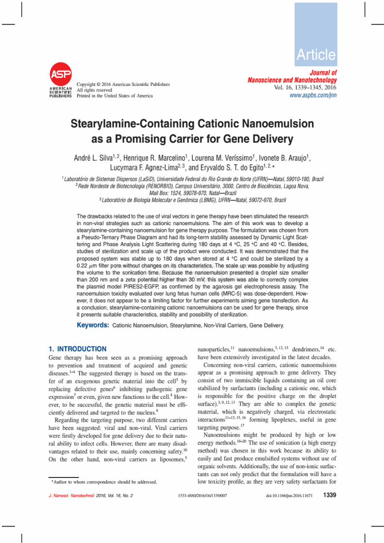
180

181
Short Curriculum vitae
182
PERSONAL INFORMATION
Name: André Leandro Silva
Date birth: 14 – 09 – 1988; At São Vicente, Brazil.
Address: Rua 21 de Setembro, 41A
Renato Gonçalves, Barreiras, Bahia - Brazil (47.806-004)
Phone: +55 77 3614 3223
Cellphone: + 55 84 9 9681 7596
e-mail: [email protected] or [email protected]
FORMAL EDUCATION
2006 – 2010 Bachelor in Pharmacy
Universidade Potiguar (with interchange period at Universidad
Nacional Andrés Bello – Santiago de Chile, Chile).
Scholarship: Programa Universidade para Todos (ProUni)
2011 – 2013 MSc. in Pharmaceutical Sciences
Universidade Federal do Rio Grande do Norte
Scholarship: Capes
2013 – 2017 PhD in Biotechnology / Biopharmacie et Pharmacotechnie
Universidade Federal do Rio Grande do Norte (thesis in co-
direction with the Université Paris-Sud - Châtenay-Malabry,
France)
Scholarship: Capes and Capes/Cofecub
PROFESSIONAL HISTORY
1. Universidade Federal do Oeste da Bahia
2017 – Current Adjunct Professor
Professional address:
R. Prof. José Seabra Lemos, 316, Recanto dos Pássaros.
UFOB – Campus Reitor Edgard Santos
Centro das Ciências Biológicas e da Saúde
Curso de Farmácia – Gabinete PU 23
CEP: 47.808 – 021
2. Universidade Federal do Rio Grande do Norte
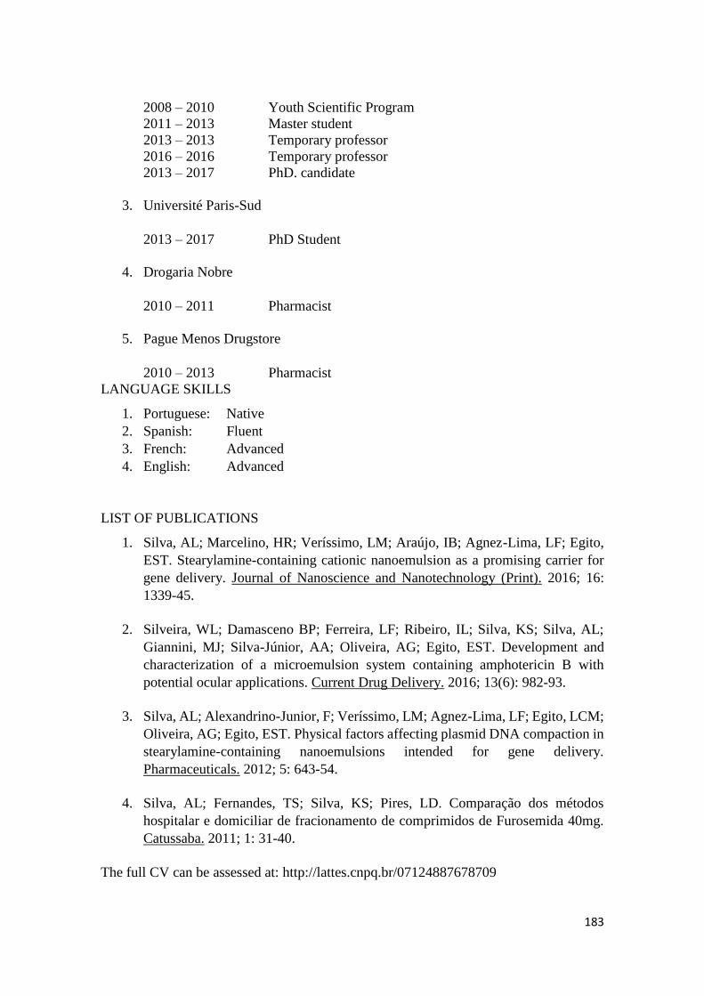
183
2008 – 2010 Youth Scientific Program
2011 – 2013 Master student
2013 – 2013 Temporary professor
2016 – 2016 Temporary professor
2013 – 2017 PhD. candidate
3. Université Paris-Sud
2013 – 2017 PhD Student
4. Drogaria Nobre
2010 – 2011 Pharmacist
5. Pague Menos Drugstore
2010 – 2013 Pharmacist
LANGUAGE SKILLS
1. Portuguese: Native
2. Spanish: Fluent
3. French: Advanced
4. English: Advanced
LIST OF PUBLICATIONS
1. Silva, AL; Marcelino, HR; Veríssimo, LM; Araújo, IB; Agnez-Lima, LF; Egito,
EST. Stearylamine-containing cationic nanoemulsion as a promising carrier for
gene delivery. Journal of Nanoscience and Nanotechnology (Print). 2016; 16:
1339-45.
2. Silveira, WL; Damasceno BP; Ferreira, LF; Ribeiro, IL; Silva, KS; Silva, AL;
Giannini, MJ; Silva-Júnior, AA; Oliveira, AG; Egito, EST. Development and
characterization of a microemulsion system containing amphotericin B with
potential ocular applications. Current Drug Delivery. 2016; 13(6): 982-93.
3. Silva, AL; Alexandrino-Junior, F; Veríssimo, LM; Agnez-Lima, LF; Egito, LCM;
Oliveira, AG; Egito, EST. Physical factors affecting plasmid DNA compaction in
stearylamine-containing nanoemulsions intended for gene delivery.
Pharmaceuticals. 2012; 5: 643-54.
4. Silva, AL; Fernandes, TS; Silva, KS; Pires, LD. Comparação dos métodos
hospitalar e domiciliar de fracionamento de comprimidos de Furosemida 40mg.
Catussaba. 2011; 1: 31-40.
The full CV can be assessed at: http://lattes.cnpq.br/07124887678709
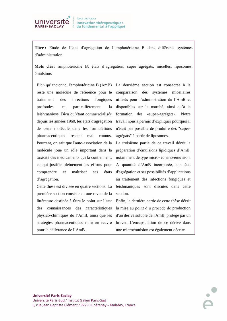
Université Paris-Saclay
Université Paris-Sud / Institut Galien Paris-Sud
5, rue Jean Baptiste Clément / 92290 Châtenay – Malabry, France
Titre : Etude de l’état d’agrégation de l’amphotéricine B dans différents systèmes
d’administration
Mots clés : amphotéricine B, états d’agrégation, super agrégats, micelles, liposomes,
émulsions
Bien qu’ancienne, l'amphotéricine B (AmB)
reste une molécule de référence pour le
traitement des infections fongiques
profondes et particulièrement la
leishmaniose. Bien qu’étant commercialisée
depuis les années 1960, les états d'agrégation
de cette molécule dans les formulations
pharmaceutiques restent mal connus.
Pourtant, on sait que l'auto-association de la
molécule joue un rôle important dans la
toxicité des médicaments qui la contiennent,
ce qui justifie pleinement les efforts pour
comprendre et maîtriser ses états
d’agrégation.
Cette thèse est divisée en quatre sections. La
première section consiste en une revue de la
littérature destinée à faire le point sur l’état
des connaissances des caractéristiques
physico-chimiques de l’AmB, ainsi que les
stratégies pharmaceutiques mise en œuvre
pour la délivrance de l’AmB.
La deuxième section est consacrée à la
comparaison des systèmes micellaires
utilisés pour l’administration de l’AmB et
disponibles sur le marché, ainsi qu’à la
formation des «super-agrégats». Notre
travail nous a permis d’expliquer pourquoi il
n'était pas possible de produire des "super-
agrégats" à partir de liposomes.
La troisième partie de ce travail décrit la
préparation d’émulsions lipidiques d’AmB,
notamment de type micro- et nano-émulsion.
A quantité d’AmB incorporée, son état
d'agrégation et ses possibilités d’applications
au traitement des infections fongiques et
leishmaniques sont discutés dans cette
section.
Enfin, la dernière partie de cette thèse décrit
la mise au point d’u procédé de production
d'un dérivé soluble de l'AmB, protégé par un
brevet. L'encapsulation de ce dérivé dans
une microémulsion est également décrite.
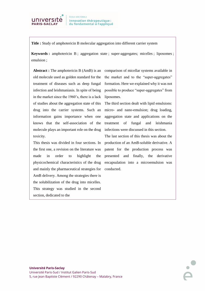
Université Paris-Saclay
Université Paris-Sud / Institut Galien Paris-Sud
5, rue Jean Baptiste Clément / 92290 Châtenay – Malabry, France
Title : Study of amphotericin B molecular aggregation into different carrier system
Keywords : amphotericin B ; aggregation state ; super-aggregates; micelles ; liposomes ;
emulsion ;
Abstract : The amphotericin B (AmB) is an
old molecule used as golden standard for the
treatment of diseases such as deep fungal
infection and leishmaniasis. In spite of being
in the market since the 1960’s, there is a lack
of studies about the aggregation state of this
drug into the carrier systems. Such an
information gains importance when one
knows that the self-association of the
molecule plays an important role on the drug
toxicity.
This thesis was divided in four sections. In
the first one, a revision on the literature was
made in order to highlight the
physicochemical characteristics of the drug
and mainly the pharmaceutical strategies for
AmB delivery. Among the strategies there is
the solubilization of the drug into micelles.
This strategy was studied in the second
section, dedicated to the
comparison of micellar systems available in
the market and to the “super-aggregates”
formation. Here we explained why it was not
possible to produce “super-aggregates” from
liposomes.
The third section dealt with lipid emulsions:
micro- and nano-emulsion; drug loading,
aggregation state and applications on the
treatment of fungal and leishmania
infections were discussed in this section.
The last section of this thesis was about the
production of an AmB-soluble derivative. A
patent for the production process was
presented and finally, the derivative
encapsulation into a microemulsion was
conducted.