Embed Size (px)
Citation preview
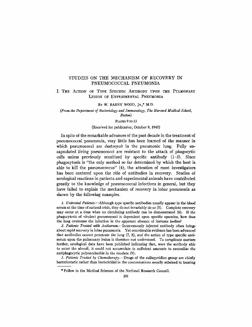
STUDIES ON THE MECHANISM OF RECOVERY INPNEUMOCOCCAL PNEUMONIA
I. THE ACTION OF TYPE SPECIFIC ANTIBODY UPON THE PULMONARYLESION OF EXPERIMENTAL PNEUMONIA
BY W. BARRY WOOD, JR.,* M.D.
(From the Department of Bacteriology and Immunology, The Harvard Medical School,Boston)
PLATES 9 TO 13
(Received for publication, October 9, 1940)
In spite of the remarkable advances of the past decade in the treatment ofpneumococcal pneumonia, very little has been learned of the manner inwhich pneumococci are destroyed in the pneumonic lung. Fully en-capsulated living pneumococci are resistant to the attack of phagocyticcells unless previously sensitized by specific antibody (1-3). Sincephagocytosis is "the only method so far determined by which the host isable to kill the pneumococcus" (4), the attention of most investigatorshas been centered upon the rle of antibodies in recovery. Studies ofserological reactions in patients and experimental animals have contributedgreatly to the knowledge of pneumococcal infections in general, but theyhave failed to explain the mechanism of recovery in lobar pneumonia asshown by the following examples.
1. Untreated Patients.-Although type specific antibodies usually appear in the bloodserum at the time of natural crisis, they do not invariably do so (5). Complete recoverymay occur at a time when no circulating antibody can be demonstrated (6). If thephagocytosis of virulent pneumococci is dependent upon specific opsonins, how doesthe lung overcome the infection in the apparent absence of immune bodies?
2. Patients Treated with Antiserum.-Intravenously injected antibody often bringsabout rapid recovery in lobar pneumonia. Yet considerable evidence has been advancedthat antibodies cannot penetrate the lung (7, 8), and the action of type specific anti-serum upon the pulmonary lesion is therefore not understood. To complicate mattersfurther, serological data have been published indicating that, were the antibody ableto enter the alveoli, it could not accumulate in sufficient amounts to neutralize theantiphagocytic polysaccharide in the exudate (9).
3. Patients Treated by Chemotherapy.--Drugs of the sulfapyridine group are chieflybacteriostatic rather than bactericidal in the concentrations usually attained in treating
* Fellow in the Medical Sciences of the National Research Council.201
202 MECHANISM O RECOVERY IN PNEUMOCOCCAL PNEUMONIA. I
human patients (10-12). Nevertheless, following treatment with sulfapyridine, pa-tients often recover without the aid of circulating antibodies (13, 14). Since phagocy-tosis is thought to depend upon specific opsonins, and the sulfapyridine itself killsrelatively few organisms, it is not at all clear how chemotherapy brings about the finaldestruction of pneumococci in the lesion.
The purpose of the present series of studies has been to investigate the problem ofrecovery in lobar pneumonia by the more direct methods of pathology. The patho-genesis of experimental pneumonia has been studied in a suitable laboratory animal,and the lungs of animals treated with antiserum and sulfapyridine have been examinedin detail at various stages of the disease. By correlating the observed histologicalchanges with the progress of the infection and the immune reactions of the host, datahave been obtained which appear to have a direct bearing upon the mechanism ofrecovery.
This paper deals with the histopathology and pathogenesis of a lobarpneumonia uniformly fatal in untreated rats and describes the effect oftype specific antibody upon the pulmonary lesion following treatment withantipneumococcal serum., Observations relating to the process of recoveryin rats treated by chemotherapy will be reported in a subsequent paper.
Methods
In 1935, Nungester and Jourdonais (15) produced lobar pneumonia in white rats byinoculating them intrabronchially with pneumococci suspended in a viscous mixture ofmucin. The disease produced was not uniformly fatal, and for that reason, preliminaryexperiments were undertaken at the start of the present investigation to increase theseverity of the infection. A group of 100 rats was inoculated by various modificationsof the Nungester technique, and although at first approximately one in every four ratssurvived, a more malignant pneumonia was finally produced which killed all of theuntreated animals. Changes in the method which seemed to be of importance included:(a) inoculation with a younger (6 hour) culture of pneumococci, (b) the use of a moreconcentrated (6 per cent) mixture of mucin,2 and (c) a prolonged, light anesthesia underether during which the rats were maintained in a vertical position to allow the mucinto flow further into the terminal bronchi. Elimination of the chance of spontaneousrecovery greatly clarified the results of subsequent experiments designed to demonstratethe effect of specific therapy.
Animals.-White rats, varying in weight from 180 to 250 gm., were obtained fromseveral breeders. Chronic pulmonary lesions of undetermined etiology were occa-sionally encountered. Whenever a chronic lesion was found at autopsy and its presenceinterfered with the interpretation of the findings in the pneumonic lung, the rat wasdiscarded and the experiment repeated. No spontaneous lesions resembling the acuteexperimental pneumonia were encountered in any of the 350 rats examined.
Pneumococcus Cultures.-A Type I pneumococcus of the A6 strain, generously sup-
1 A preliminary report of these studies was published in Science, 1940, 92, 15.2 Kepl and Gunn (16) have recently obtained a high mortality using 5 per cent
mucin.
W. BARRY WOOD, JR.
plied by Dr. O. H. Robertson, was used in all experiments. Cultures of the heart'sblood of rats dying with bacteriemia were seeded in defibrinated rabbit blood undervaseline, incubated 24 hours, and stored in the ice box. Under these conditions pneumo-cocci have been found to retain their virulence for several months (17), but to insuremaintenance of maximum virulence the organism was passed through rats at least onceevery 3 weeks.
For each experiment the inoculum was prepared as follows: From the stock culturein rabbit's blood, a transfer was made to blood broth and incubated 18 hours. 1 cc. ofthe blood broth culture was then transferred to approximately 10 cc. of 0.05 per centdextrose beef infusion broth to which defibrinated rabbit blood had been added in aconcentration of 1 per cent. After 6 hours of incubation at 37°C., 1 cc. of the dextroseblood broth culture was diluted 1:1000 in beef infusion broth and 1 cc. of the dilutedculture was added to 9 cc. of a mucin-saline mixture. 0.1 cc. of the resulting suspensionwas used for intrabronchial inoculation.
Mucin.-Finely powdered, commercial mucin was employed as routine. It was foundunnecessary to sterilize the mucin or grind it further before adding it to normal saltsolution. Most consistent results were obtained when a relatively concentrated mixtureof mucin was used in suspending the pneumococci; the concentration finally adoptedwas 6 per cent.
Intrabronchial Inoculation.-The rats were inoculated according to the techniquedescribed by Jourdonais and Nungester (18).
Size of Inoculum.-Bacterial counts of the inoculum were carried out in each experi-ment by the pour plate method. The number of organisms in the 0.1 cc. of mucinmixture varied from approximately 4,000 to 7,000. In preliminary experiments it wasfound that if fewer pneumococci were used, rats would occasionally recover spon-taneously.
Prolonged Anesthesia.-To insure the penetration of the inoculum into the terminalbronchi, the rats were hung (by the upper incisor teeth) in a vertical position for 30minutes while still under light ether anesthesia. This procedure seemed to increasethe severity of the pneumonia produced.
Blood Cultures.-Blood cultures were taken from the tail at frequent intervals duringthe course of the pneumonia, and in rats treated with antiserum, cultures were madejust before treatment and 5 minutes after the injection was completed. A loopful ofblood was streaked on a blood agar plate and incubated for 24 hours at 37C. Theidentity of the organism cultured was frequently checked by the Neufeld Quellungreaction. Similar cultures were made as routine from the heart at autopsy and werealso taken from the pleura, pericardium, and lungs when indicated.
Methods of Studying Pathology.-All surviving rats were killed with ether. Becauseof the possibility of post mortem growth of pneumococci in the lesion, the lungs of ratsautopsied more than one hour after death were not considered suitable for microscopicstudy. The lungs were fixed in Zenker-formol solution (5 per cent formalin) by themethod of Loosli (19). The thorax was opened and the aorta was immediately clamped.If the heart was still beating, it was allowed to do so for a moment, and then a tightligature was placed about its base to isolate the pulmonary circulation. If the hearthad already stopped beating, blood was forced on into the pulmonary vessels by clamping
3 Granular mucin, type 1701-W, prepared by the Wilson Laboratories, Chicago.
203
204 MECHANISM OF RECOVERY IN PNEUtOCOCCAL PNEUMONIA. I
the right ventricle. In either case the pulmonary capillaries were sufficiently distendedwith blood to facilitate identification of the alveolar walls in the microscopic sections(see Figs. 2, 3, 4, 6, etc.). After a blood culture had been taken from the heart, thelungs were fixed in situ by injecting the trachea with Zenker-formol solution. Thefixative was introduced slowly under a pressure of not more than 15 cm. of water, andwas allowed to re-expand the lungs until they again filled the thorax. The trachea wasthen tied off and the thoracic organs removed and placed in Zenker-formol solution for12 to 18 hours. The fixed specimens were washed for 24 hours in tap water and dehy-drated in 80 per cent alcohol. Sagittal blocks were cut from the single-lobed left lung,passing through the pneumonic lesion, and each block was further dehydrated in alcohol,imbedded in paraffin, sectioned, and stained by the Gram-Weigert technique.4 Directinjection of the lung was found to fix the exudate in place, and by redistending thealveoli to their normal size, greatly clarify the histology of the pneumonic lesion (Fig. 6).
Treatment.-The rats were treated with Type I antipneumococcal serum obtainedthrough the courtesy of Dr. W. G. Malcolm of Lederle Laboratories. Refined rabbitserum was used throughout these studies, except in one experiment where horse serumwas employed. The rabbit serum contained 6,000 units of antibody per cc.; the horseserum 2,500 units per cc. All rats were treated with a single intravenous injection ofantiserum. A dose of 1 cc. (6,000 units) was administered as routine, but in a fewexperiments smaller doses were given, and as little as 0.02 cc. (120 units) was foundto be effective.
The technique employed in injecting serum intravenously was as follows: The groinwas shaved and the skin was cleaned with iodine and alcohol while the rat was underlight ether anesthesia. An incision about 3 cm. long was made over the femoral vessels,and the vein was exposed by blunt dissection. A No. 26 needle attached to a 1 cc.tuberculin syringe containing the antiserum was inserted into the femoral vein, and theserum was injected slowly over a period of approximately 5 minutes. Following theintravenous injection the incision was closed with a continuous suture of silk. Althoughsuperficial infection occurred frequently about the skin sutures, no serious deep infectionswere encountered when the wound was properly closed.
If the antiserum was injected rapidly, the rats sometimes died of sudden respiratoryfailure. The slow injection of serum caused an increase in respiratory rate, but no otherreactions were noted except in extremely ill rats treated late in the course of the disease.The reaction observed in this latter group is described below.
RESULTS
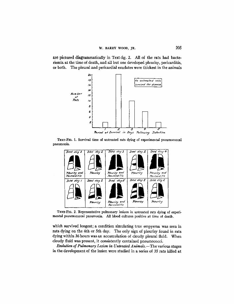
Fatality Rate among Untreated Rats.-The pneumococcal pneumonia
produced experimentally by the method outlined was uniformly fatal.
All of 40 untreated rats succumbed to the disease in less than 5 days, the
majority dying within 48 hours (see Text-fig. 1).
Extent of Lesion in Rats Dying of Pneumonia.-In only 5 rats was the
pneumonia confined to the left lung. Representative pulmonary lesions
4In order to accentuate the cellular elements of the exudate, the Gram-Weigertmethod was slightly modified by overstaining the sections with hematoxylin and de-colorizing in acid alcohol before staining with eosin.
W. BARRY WOOD, JR. 205
are pictured diagrammatically in Text-fig. 2. All of the rats had bacte-riemia at the time of death, and all but one developed pleurisy, pericarditis,or both. The pleural and pericardial exudates were thickest in the animals
Zo
/6
/4Alum be r 2
ofRa ts /0
a
4
£
,no v treated ats
osuvtr,/ed tfe derje.
-Th/ 2 J3 4- ,
?efrd orr Sv,! in D' Y FoDa oiV Tfenrn
TEXT-FIG. 1. Survival time of untreated rats dying of experimental pneumococcalpneumonia.
./ec dY dy day2 ,d day,3 .Ded d 2 Ddday 4
pku'r/ty 0ta' P/evrisy ;/ecorj'Jy and Pleur/ J PDler/sy a'
_D/ d / Dd y 2 .DJ-d dy. .D/ed day2 .Ded c/ayZ
P/eLr;.y PleCVrsy and P/eor/&Y Peu syPert card f s
TEXT-FIG. 2. Representative pulmonary lesions in untreated rats dying of experi-mental pneumococcal pneumonia. All blood cultures positive at time of death.
which survived longest; a condition simulating true empyema was seen inrats dying on the 4th or 5th day. The only sign of pleurisy found in ratsdying within 36 hours was an accumulation of cloudy pleural fluid. Whencloudy fluid was present, it consistently contained pneumococci.
Evolution of Pulmonary Lesion in Untreated Animals.-The various stagesin the development of the lesion were studied in a series of 35 rats killed at
n 1
206 MECHANISM OF RECOVERY IN PNEUMOCOCCAL PNEUMONIA. I
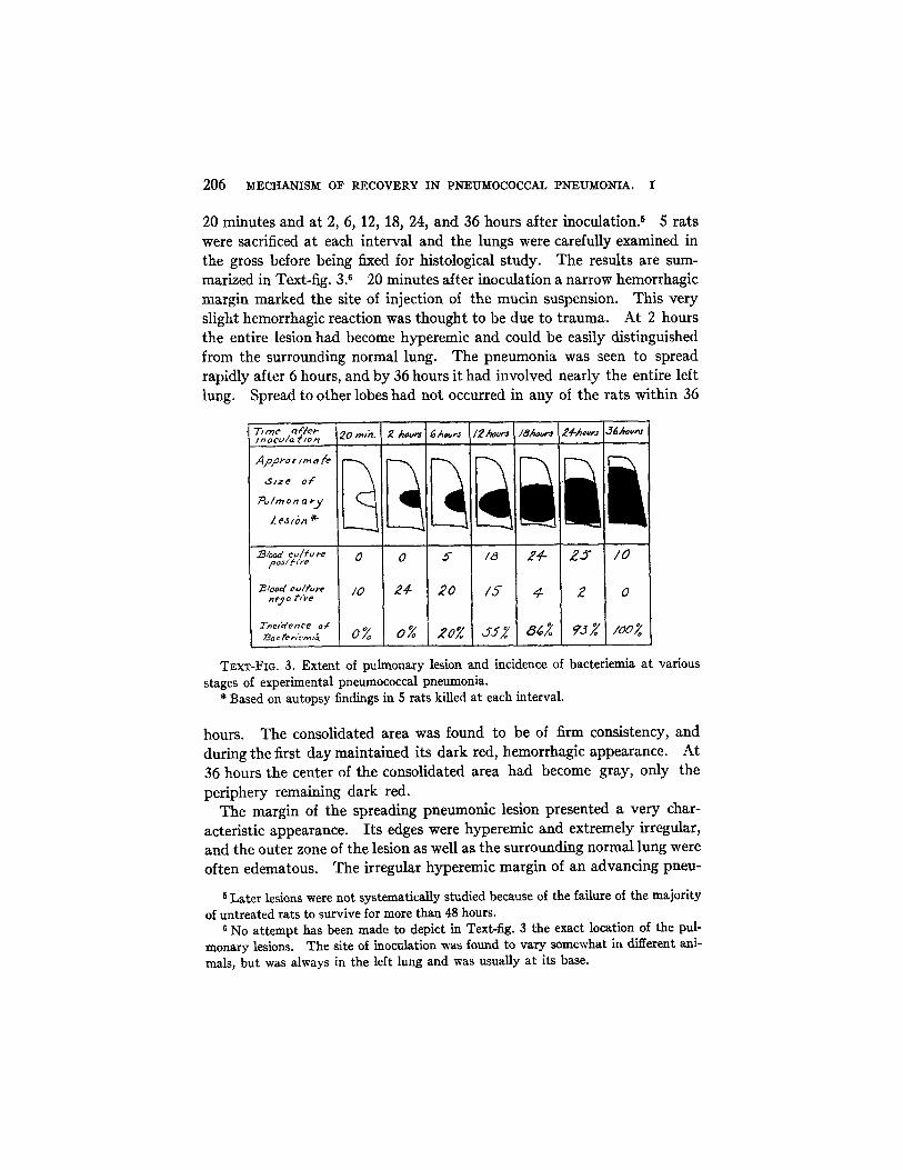
20 minutes and at 2, 6, 12, 18, 24, and 36 hours after inoculation. 6 5 ratswere sacrificed at each interval and the lungs were carefully examined inthe gross before being fixed for histological study. The results are sum-marized in Text-fig. 3.6 20 minutes after inoculation a narrow hemorrhagicmargin marked the site of injection of the mucin suspension. This veryslight hemorrhagic reaction was thought to be due to trauma. At 2 hoursthe entire lesion had become hyperemic and could be easily distinguishedfrom the surrounding normal lung. The pneumonia was seen to spreadrapidly after 6 hours, and by 36 hours it had involved nearly the entire leftlung. Spread to other lobes had not occurred in any of the rats within 36
I/nocu a /o jr|l20 . 2 our hrJ /2Ahovrs I8 J'2hAo r |
App-roxn/er-- I r-7 \ IF\I I . \ ' \I| \\| \ II \
alon a
Z jIA* L L L
B/odc,/f,re ,0 0 /5- 18 24- /0,PoJ 11re
B/ood c,/frr /0 l/ e 20 /5 4 2 0
|crene, o, %' 2o% aa 0% 0% 20% J5~ 8~ 93% /a007
TEXT-FIG. 3. Extent of pulmonary lesion and incidence of bacteriemia at various
stages of experimental pneumococcal pneumonia.* Based on autopsy findings in 5 rats killed at each interval.
hours. The consolidated area was found to be of firm consistency, andduring the first day maintained its dark red, hemorrhagic appearance. At36 hours the center of the consolidated area had become gray, only theperiphery remaining dark red.
The margin of the spreading pneumonic lesion presented a very char-acteristic appearance. Its edges were hyperemic and extremely irregular,and the outer zone of the lesion as well as the surrounding normal lung were
often edematous. The irregular hyperemic margin of an advancing pneu-
6 Later lesions were not systematically studied because of the failure of the majority
of untreated rats to survive for more than 48 hours.6 No attempt has been made to depict in Text-fig. 3 the exact location of the pul-
monary lesions. The site of inoculation was found to vary somewhat in different ani-
mals, but was always in the left lung and was usually at its base.
I _i- Io I % I I I I % I
W. BARRY WOOD, JR.
monia could easily be distinguished from the clearly demarcated edge ofarrested lesions seen in treated rats recovering from the disease. The siteof inoculation of the mucin remained visible in most cases as a sharplycircumscribed dark red patch. In rats inoculated with mucin alone theinjected area was found to maintain this same characteristic appearance fornearly a week and, of course, differed from the pneumococcal lesion inthat it failed to spread.
Invasion of the Blood Stream and Pleural Cavity.-Frequent blood cul-tures were taken during the course of pneumonia in untreated rats andrepresentative results are recorded in Text-fig. 3. Bacteriemia was notencountered during the first 2 hours, but 5 out of 25 blood cultures werepositive at 6 hours. The blood stream was invaded in roughly half of theanimals after 12 hours, and in over 90 per cent at the end of the first day.Rats with early bacteriemia showed the most extensive pulmonary lesions.
Cloudy pleural fluid was present occasionally as early as 6 hours in ratshaving positive blood cultures. The first signs of pleurisy usually occurred,however, at 18 or 24 hours, when most of the animals had developed bacteri-emia. Pleurisy in the absence of bacteriemia was extremely uncommon.
Histopathology and Pathogenesis of Pneumonia in Untreated Rats.-Theearly changes occurring in the lung following the intrabronchial injection ofpneumococci suspended in mucin have been carefully described by Gunnand Nungester (20). A lag of several hours occurs before the pneumococcibecome numerous enough to be seen in the alveoli, and the lesion does nottake on the characteristic appearance of a spreading pneumonia untilafter approximately 12 hours. The histology of later lesions observed inthe present study differed somewhat from that described by Gunn andNungester, probably because different methods were used in producing thepneumonia and in fixing the lungs. Certain essential features of the presenthistological findings are therefore briefly described, since they have a directbearing upon the interpretation of later experiments.
12 to 18 hours after inoculation three definite zones became recognizable in thepneumonic lesion. These zones were even more clearly defined after 24 and 36 hours,and although the transition between them was gradual, the histology of each was suffi-ciently characteristic to bear particular emphasis.
1. Outer Edema Zone.-The margin of the advancing pneumonic lesion was foundto be characterized by the presence of edema fluid in the alveoli (Fig. 2). The edemafluid, which stained a light pink, contained a great many pneumococci apparently multi-plying freely in this favorable medium (Fig. 3). The presence of the fluid also appearedto afford the pneumococci an excellent means of spreading into adjacent areas of normallung. In the outer zone very few, if any, leucocytes were present in the edema-filledalveoli.
207

208 MECHANISM OF RECOVERY IN PNEUMOCOCCAL PNEUMONIA. I
2. Zone of Early Consolidation and Phagocytosis.-Inside the outer edema zone asecond zone could be distinguished in which the alveoli contained both leucocytes andorganisms. In the outer portion of this area, where the leucocytes were few, pneumo-cocci were numerous (Fig. 6); and in the inner portion the leucocytes were more prevalentand the bacteria scarce. Phagocytosis of the organisms by polymorphonuclear leuco-cytes was a conspicuous feature of this portion of the lesion. In connection with laterexperiments, it should be emphasized that the phagocytic reaction, though definitelypresent, was less marked than that seen in rats treated with type specific antibody,there being many fewer organisms ingested per leucocyte in the untreated animals.
3. Inner Zone of Advanced Consolidation.-The thoroughness with which the poly-morphonuclear leucocytes of the alveolar exudate are able to destroy pneumococci wasclearly shown by the complete absence of organisms within the alveoli of the centralportion of the lesion (Figs. 7 and 8). The alveoli and many of the bronchi in this innerzone were packed with leucocytes. Only occasional organisms could still be detectedin the exudate, and these were easily distinguished from the large mucin granules seenin a few areas within the alveolar phagocytes. Heavy deposits of fibrin were notuncommonly observed after 18 hours (Fig. 9). In the older parts of the lesion, localareas of clearing were prominent, macrophages having appeared in the resolving alveolarexudate (Fig. 8).
Two additional histological findings deserve special emphasis, since they suggest themode of spread of pneumococci to other lobes and to the pleural cavity. The presencein large bronchi of appreciable amounts of pneumococcus-laden edema fluid was re-peatedly noted in sections from rats with rapidly advancing pneumonia (Fig. 5). Ham-burger and Robertson have demonstrated (21) in dogs that this watery bronchial exudatecontains myriads of organisms and is responsible for the spread of the infection fromone lobe to another. The frequency with which the bronchial edema fluid has beennoted in the present study indicates that the same mechanism may operate in rats.The presence of large numbers of pneumococci in edema-filled alveoli bordering on thepleural cavity (Fig. 4) suggests that pleurisy may be caused by the direct penetrationof pneumococci through the visceral pleura. It is conceivable that the organisms invadethe pleura by way of the lymphatics, draining outward at the periphery of the lung,but in the face of the present histological findings, the possibility of pneumococci passingdirectly into the pleural cavity cannot be excluded.
The histopathology of the 24 and 36 hour pneumonia was essentially the same asat 18 hours, except for the extent of the lesion. Rats dying after 2 or more days alsoshowed the same histological changes in the lungs, except that the older lesions werecharacterized by a more extensive zone of advanced consolidation in which local areasof clearing were relatively numerous.
Experiments on the Action of Antiserum.-Five separate experiments werecarried out in which rats were treated 2, 6, 12, 18, and 24 hours after inocu-lation. 21 rats were inoculated in each experiment, 3 serving as untreatedcontrols. The 18 treated animals were sacrificed in groups of 3 at differentintervals following treatment. The lungs of each rat were carefully ex-amined in the gross before being fixed for histological study. The resultsof the first four experiments are summarized in Text-figs. 4 to 7; the fifth

W. BARRY WOOD, JR.
experiment, in which rats were treated after 24 hours, is described laterunder a separate heading.
A )ne /Ao j sAo rs j/8Mo-vs |x Aoors s 1 9oOI T"ee f en"f's - |I I I I I I I I I A4?-tPoX11a *
slofie /zcyS _
Pvlm e 3 aS ) ;
NA/vmb,- of
·~arertevlt
P' e a" 0 0 0 0 0 0at rf- U 00 0 O 0 Ja f vfbyr° ° °0
TEXT-FIG. 4. Effect upon pulmonary lesion of type specific antiserum administeredintravenously 2 hours after inoculation.
* Based on autopsy findings in 3 rats killed at each interval.** Untreated rats died in less than 70 hours.
Lb*
ar /mncn |/hor I6 rs I/&os 0 e- ~2AaorasI ?6Avr/.R* If"r itA.Treo~~~~~~~~m ~ ~ ~ ?Aar eee' lnraett ~
fanre/,m rt
fatme 0 0 0 0 I
affet,%eat,~t a 0 0 0 0 0O
t tp 0 0 0 0 0 0 J
TEXT-FIG. 5. Effect upon pulmonary lesion and bacteriemia ofserum administered intravenously 6 hours after inoculation.
* Based on autopsy findings in 3 rats killed at each interval.** Untreated rats died in less than 70 hours.
type specific anti-
The Effect of Serum Treatment upon Fatality Rate.-No deaths due topneumonia occurred among the 76 animals treated within 18 hours afterinoculation, although many showed bacteriemia at the time of treatment.12 rats were allowed to live for one week and 12 more for 4 days before beingsacrificed. Histological examination of the lungs indicated in each casethat the pneumococcal infection had completely subsided.
209
210 MECHANISM OF RECOVERY IN PNEUMOCOCCAL PNEUMONIA. I
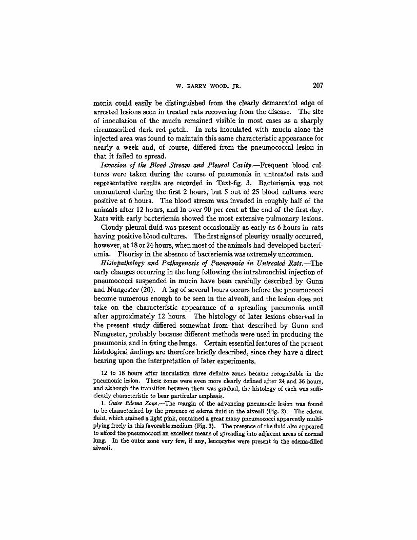
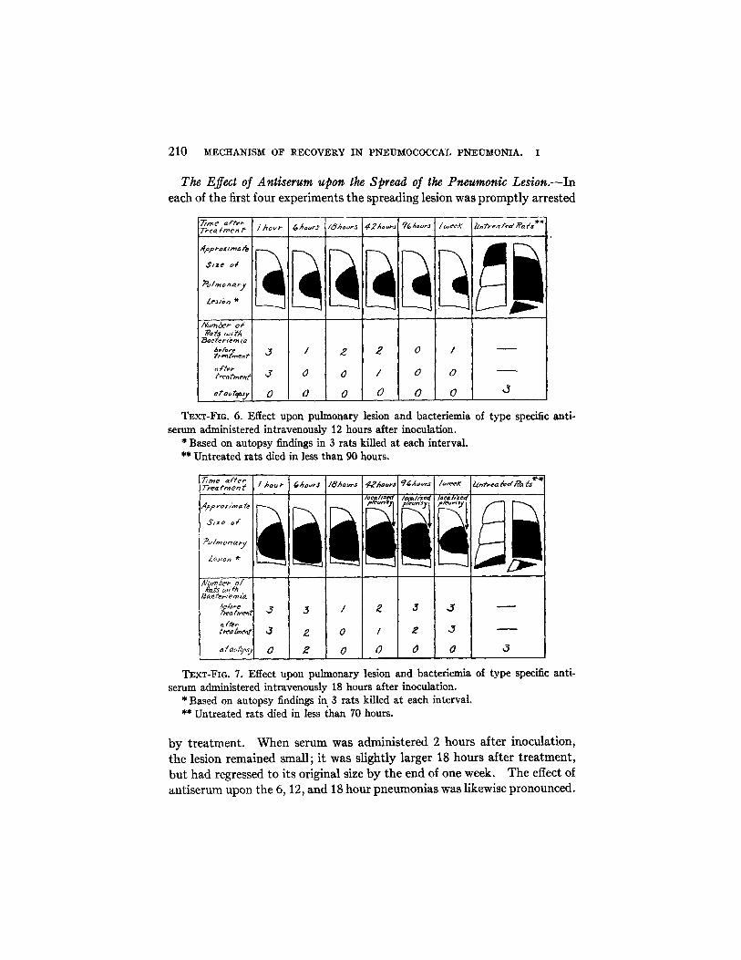
The Effect of Antiserum upon the Spread of the Pneumonic Lesion.-Ineach of the first four experiments the spreading lesion was promptly arrested
r1 -r1a,'m 1 Aeours1/weeX 1 _Thea irme'I /hour Ars /8eurJ 14 -- 9 2-t'e s .
-abr1md _ I _ I'
I /mOp'a ~yLie .* i
Number ofaC's D 'A
ream f J/
atauosy 0 0 0 0 0 ( 3
TEXT-FIG. 6. Effect upon pulmonary lesion and bacteriemiaserum administered intravenously 12 hours after inoculation.
* Based on autopsy findings in 3 rats killed at each interval.** Untreated rats died in less than 90 hours.
of type specific anti-
1rt: et i /h., r I CAuirj /ghors I Ahrw|piIevt4| /afe¢ I fea.a fsl
, r o rrra ;
,/zon o£e
?~/o a W
r/a I>
,r I /' ,1
A/o' b,- o/eats wI hac feTroz J
dAjereeriA j 3 / 2 3 -
tr tt 3 2 0 / 2 J
aaopsv 0 2 0 0 0 0 .3
TEXT-FIG. 7. Effect upon pulmonary lesion and bacteriemia of type specific anti-serum administered intravenously 18 hours after inoculation.
* Based on autopsy findings in 3 rats killed at each interval.** Untreated rats died in less than 70 hours.
by treatment. When serum was administered 2 hours after inoculation,the lesion remained small; it was slightly larger 18 hours after treatment,but had regressed to its original size by the end of one week. The effect ofantiserum upon the 6, 12, and 18 hour pneumonias was likewise pronounced.
W. BARRY WOOD, JR.
Although the area of consolidation was somewhat larger at the time of treat-ment in each succeeding experiment, the infection was always quicklycontrolled by serum therapy.
The gross appearance of the spreading lesion changed noticeably followingtreatment. The margin of the consolidated area became sharply de-marcated and lost the irregular hemorrhagic border characteristic of anadvancing pneumonia. The entire lesion gradually became gray in colorand in its center there appeared small patches of soft normal lung indicatingthat resolution had begun. These changes in the gross appearance of thelesion made it a simple matter to differentiate at autopsy between an ac-tively spreading pneumonia and one arrested by serum therapy.
The original area of lung injected with mucin remained firm and dark red,simulating exactly the lesion observed in rats inoculated with mucin alone.The mucin area showed no signs of spreading and could be clearly dis-tinguished from the surrounding pneumococcal lesion.
The Effect of Antiserum upon Bacteriemia and Pleurisy.-As shown inText-figs. 5, 6, and 7 the blood stream of bacteriemic rats cleared rapidlyfollowing serum therapy. None of the animals treated at 2 hours hadbacteriemia, but 2 at 6 hours, exactly half at 12 hours, and roughly 80 percent at 18 hours had positive blood cultures just before treatment. Whenthe bacteriemia was light, the blood became sterile a few minutes after theinjection of serum. When the initial blood culture showed many coloniesper cc., the second culture taken 5 minutes after treatment usually containedmany fewer organisms but seldom was sterile. In all but 2 rats the bloodcultures were negative at the time of autopsy. These 2 showed positivecultures 6 hours after treatment, although in both cases the number ofcolonies per cc. was markedly diminished. No rat developed bacteriemiafollowing therapy.
Signs of pleurisy were noted only in animals treated 18 hours after beinginfected. In this group all 9 rats sacrificed 42 hours or more after treatmentshowed between the visceral and parietal pleura small areas of dense ad-hesions indicative of localized pleural infections (see Text-fig. 7). Nogeneralized pleurisy or empyema, as seen in untreated rats, was observedin any of these animals, the serum treatment having apparently caused alocalization of the pleural infection.
The Action of Type Specific Antibody upon the Pulmonary Lesion.-Inorder to investigate the effect of antibody upon the pneumococci in thelesion, microscopic sections were made of the lungs of all rats killed at inter-vals after treatment. In rats treated 2 and 6 hours after inoculation thepneumonic process had not developed sufficiently to make histological study
211
212 MECHANISM OF RECOVERY IN PNEUMOCOCCAL PNEUMONIA. I
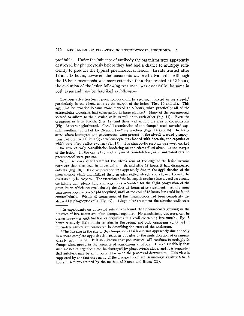
profitable. Under the influence of antibody the organisms were apparentlydestroyed by phagocytosis before they had had a chance to multiply suffi-ciently to produce the typical pneumococcal lesion. In rats treated after12 and 18 hours, however, the pneumonia was well advanced. Althoughthe 18 hour pneumonia was more extensive than that treated at 12 hours,the evolution of the lesion following treatment was essentially the same inboth cases and may be described as follows:-
One hour after treatment pneumococci could be seen agglutinated in the alveoli, 7
particularly in the edema zone at the margin of the lesion (Figs. 10 and 11). Thisagglutination reaction became more marked at 6 hours, when practically all of theextracellular organisms had congregated in large clumps. 8 Many of the pneumococciseemed to adhere to the alveolar walls as well as to each other (Fig. 11). Even theorganisms in large bronchi (Fig. 12) and those well within the area of consolidation(Fig. 13) were agglutinated. Careful examination of the clumped cocci revealed cap-sular swelling typical of the Neufeld Quellung reaction (Figs. 14 and 15). In manyareas where leucocytes and pneumococci were present in the alveoli marked phagocy-tosis had occurred (Fig. 16); each leucocyte was loaded with bacteria, the capsules ofwhich were often visibly swollen (Fig. 17). The phagocytic reaction was most markedin the zone of early consolidation bordering on the edema-filled alveoli at the marginof the lesion. In the central zone of advanced consolidation, as in untreated rats nopneumococci were present.
Within 6 hours after treatment the edema zone at the edge of the lesion becamenarrower than that seen in untreated animals and after 18 hours it had disappearedentirely (Fig. 18). Its disappearance was apparently due to the agglutination of thepneumococci which immobilized them in edema-filled alveoli and allowed them to beovertaken by leucocytes. The extension of the leucocytic exudate into alveoli previouslycontaining only edema fluid and organisms accounted for the slight progression of thegross lesion which occurred during the first 18 hours after treatment. At the sametime more organisms were phagocytized, until at the end of 18 hours few could be foundextracellularly. Within 42 hours most of the pneumococci had been completely de-stroyed by phagocytic cells (Fig. 19). 4 days after treatment the alveolar walls were
7 In experiments on untreated rats it was found that pneumococci growing in thepresence of free mucin are often clumped together. No conclusions, therefore, can bedrawn regarding agglutination of organisms in alveoli containing free mucin. By 18hours relatively little mucin remains in the lesion, and only organisms contained inmucin-free alveoli are considered in describing the effect of the antiserum.
s The increase in the size of the clumps seen at 6 hours was apparently due not onlyto a more complete agglutination reaction but also to the multiplication of organismsalready agglutinated. It is well known that pneumococci will continue to multiply inclumps when grown in the presence of homologous antibody. It seems unlikely thatsuch masses of organisms can be destroyed by phagocytosis alone, and it is suggestedthat autolysis may be an important factor in the process of destruction. This view issupported by the fact that many of the clumped cocci are Gram-negative after 6 to 18hours in sections stained by the method of Brown and Brenn (22).
W. BARRY WOOD, JR.
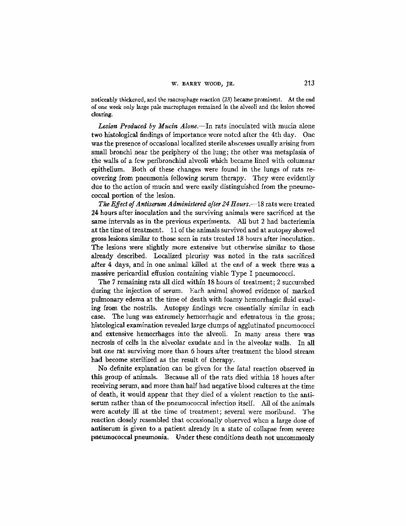
noticeably thickened, and the macrophage reaction (23) became prominent. At the endof one week only large pale macrophages remained in the alveoli and the lesion showedclearing.
Lesion Produced by Mucin Alone.-In rats inoculated with mucin alonetwo histological findings of importance were noted after the 4th day. Onewas the presence of occasional localized sterile abscesses usually arising fromsmall bronchi near the periphery of the lung; the other was metaplasia oftlhe walls of a few peribronchial alveoli which became lined with columnarepithelium. Both of these changes were found in the lungs of rats re-covering from pneumonia following serum therapy. They were evidentlydue to the action of mucin and were easily distinguished from the pneumo-coccal portion of the lesion.
The Effect of Antiserum Administered after 24 Hours.-18 rats were treated24 hours after inoculation and the surviving animals were sacrificed at thesame intervals as in the previous experiments. All but 2 had bacteriemiaat the time of treatment. 11 of the animals survived and at autopsy showedgross lesions similar to those seen in rats treated 18 hours after inoculation.The lesions were slightly more extensive but otherwise similar to thosealready described. Localized pleurisy was noted in the rats sacrificedafter 4 days, and in one animal killed at the end of a week there was amassive pericardial effusion containing viable Type I pneumococci.
The 7 remaining rats all died within 18 hours of treatment; 2 succumbedduring the injection of serum. Each animal showed evidence of markedpulmonary edema at the time of death with foamy hemorrhagic fluid exud-ing from the nostrils. Autopsy findings were essentially similar in eachcase. The lung was extremely hemorrhagic and edematous in the gross;histological examination revealed large clumps of agglutinated pneumococciand extensive hemorrhages into the alveoli. In many areas there wasnecrosis of cells in the alveolar exudate and in the alveolar walls. In allbut one rat surviving more than 6 hours after treatment the blood streamhad become sterilized as the result of therapy.
No definite explanation can be given for the fatal reaction observed inthis group of animals. Because all of the rats died within 18 hours afterreceiving serum, and more than half had negative blood cultures at the timeof death, it would appear that they died of a violent reaction to the anti-serum rather than of the pneumococcal infection itself. All of the animalswere acutely ill at the time of treatment; several were moribund. Thereaction closely resembled that occasionally observed when a large dose ofantiserum is given to a patient already in a state of collapse from severepneumococcal pneumonia. Under these conditions death not uncommonly
213

214 MECHANISM OF RECOVERY IN PNEUMOCOCCAL PNEUMONIA. I
results from a reaction characterized by pronounced pulmonary edema(24). The mechanism of the reaction is not understood, and an attempt isbeing made to study it further.
Because of the failure of the majority of infected rats to live more than 48hours, it was impossible to investigate systematically the effect of antiserumupon later lesions. Since there had invariably developed by the end of the2nd day an advanced pleural or pericardial infection, which was unaffectedby antiserum, accurate evaluation of late treatment was not possible. Aswill be pointed out below, however, there is little reason to believe that theaction of antibody upon the intrapulmonary infection is significantlychanged by the age of the lesion.
Promptness of Agglutination Reaction.-Agglutination of pneumococciwas observed in lungs fixed as soon as 10 minutes after the start of serumtherapy. The reaction began quickly but was not complete until sometimebetween 1 and 6 hours after treatment.
Necessary Dosage of Antiserum.-Although a dose of 6,000 units of anti-body was used in all of the experiments described above, as little as 120units was found to be effective in treating rats with 12 hour pneumonia.The rats not only survived the infection when given this small dose but thelungs of animals sacrificed 6 hours after treatment contained agglutinatedand opsonized pneumococci in the alveoli. When only 12 units of antibodywere administered, the rats failed to survive and no agglutination or opsoniceffect was observed in the lungs.
The Action of Horse Serum upon the Pulmonary Lesion.-The antibodycontained in antipneumococcal horse serum is known to be of larger molec-ular size than the corresponding rabbit antibody (25, 26). It has beensuggested that the antibody of rabbit serum may be able to penetrate tissuesmore readily (27) and may, therefore, be more effective as a therapeuticagent. In the present study antipneumococcal horse serum was found toexert the same effect upon thepulmonary lesion as did rabbit serum. Thespecific antibodies contained in horse serum penetrated the lung, as evi-denced by the presence of agglutinated and opsonized pneumococci withinthe alveoli.
The Type Specificity of the Antibody Effect.-Histological examination ofthe lungs of rats treated with Type II antiserum showed that heterologousantibody had no effect upon the pneumonic lesion.
COMMENT
The pathology of experimental pneumonia in rats was found to simulateclosely that of spontaneous lobar pneumonia in man. The histology of the
W. BARRY WOOD, JR.
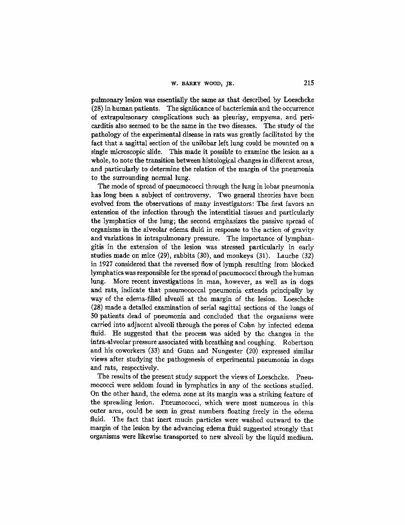
pulmonary lesion was essentially the same as that described by Loeschcke(28) in human patients. The significance of bacteriemia and the occurrenceof extrapulmonary complications such as pleurisy, empyema, and peri-carditis also seemed to be the same in the two diseases. The study of thepathology of the experimental disease in rats was greatly facilitated by thefact that a sagittal section of the unilobar left lung could be mounted on asingle microscopic slide. This made it possible to examine the lesion as awhole, to note the transition between histological changes in different areas,and particularly to determine the relation of the margin of the pneumoniato the surrounding normal lung.
The mode of spread of pneumococci through the lung in lobar pneumoniahas long been a subject of controversy. Two general theories have beenevolved from the observations of many investigators: The first favors anextension of the infection through the interstitial tissues and particularlythe lymphatics of the lung; the second emphasizes the passive spread oforganisms in the alveolar edema fluid in response to the action of gravityand variations in intrapulmonary pressure. The importance of lymphan-gitis in the extension of the lesion was stressed particularly in earlystudies made on mice (29), rabbits (30), and monkeys (31). Lauche (32)in 1927 considered that the reversed flow of lymph resulting from blockedlymphatics was responsible for the spread of pneumococci through the humanlung. More recent investigations in man, however, as well as in dogsand rats, indicate that pneumococcal pneumonia extends principally byway of the edema-filled alveoli at the margin of the lesion. Loeschcke(28) made a detailed examination of serial sagittal sections of the lungs of50 patients dead of pneumonia and concluded that the organisms werecarried into adjacent alveoli through the pores of Cohn by infected edemafluid. He suggested that the process was aided by the changes in theintra-alveolar pressure associated with breathing and coughing. Robertsonand his coworkers (33) and Gunn and Nungester (20) expressed similarviews after studying the pathogenesis of experimental pneumonia in dogsand rats, respectively.
The results of the present study support the views of Loeschcke. Pneu-mococci were seldom found in lymphatics in any of the sections studied.On the other hand, the edema zone at its margin was a striking feature ofthe spreading lesion. Pneumococci, which were most numerous in thisouter area, could be seen in great numbers floating freely in the edemafluid. The fact that inert mucin particles were washed outward to themargin of the lesion by the advancing edema fluid suggested strongly thatorganisms were likewise transported to new alveoli by the liquid medium.
215
216 MECHANISM OF RECOVERY IN PNEUMOCOCCAL PNEUMONIA. I
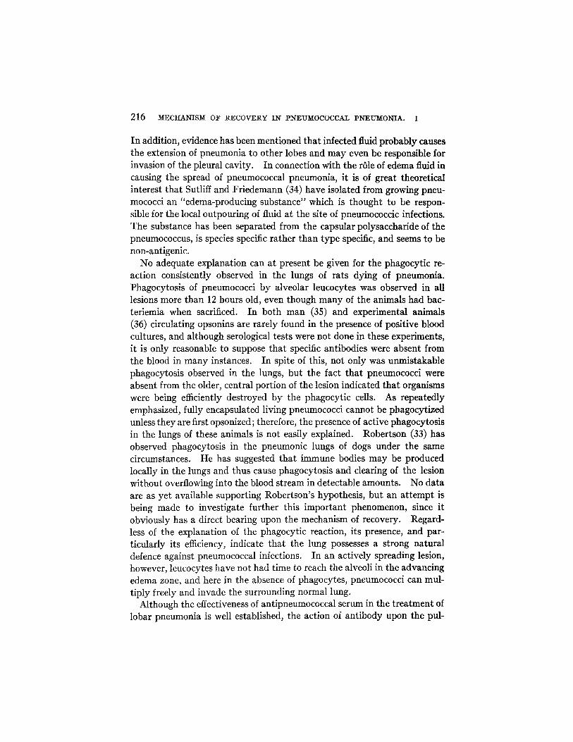
In addition, evidence has been mentioned that infected fluid probably causesthe extension of pneumonia to other lobes and may even be responsible forinvasion of the pleural cavity. In connection with the role of edema fluid incausing the spread of pneumococcal pneumonia, it is of great theoreticalinterest that Sutliff and Friedemann (34) have isolated from growing pneu-mococci an "edema-producing substance" which is thought to be respon-sible for the local outpouring of fluid at the site of pneumococcic infections.The substance has been separated from the capsular polysaccharide of thepneumococcus, is species specific rather than type specific, and seems to benon-antigenic.
No adequate explanation can at present be given for the phagocytic re-action consistently observed in the lungs of rats dying of pneumonia.Phagocytosis of pneumococci by alveolar leucocytes was observed in alllesions more than 12 hours old, even though many of the animals had bac-teriemia when sacrificed. In both man (35) and experimental animals(36) circulating opsonins are rarely found in the presence of positive bloodcultures, and although serological tests were not done in these experiments,it is only reasonable to suppose that specific antibodies were absent fromthe blood in many instances. In spite of this, not only was unmistakablephagocytosis observed in the lungs, but the fact that pneumococci wereabsent from the older, central portion of the lesion indicated that organismswere being efficiently destroyed by the phagocytic cells. As repeatedlyemphasized, fully encapsulated living pneumococci cannot be phagocytizedunless they are first opsonized; therefore, the presence of active phagocytosisin the lungs of these animals is not easily explained. Robertson (33) hasobserved phagocytosis in the pneumonic lungs of dogs under the samecircumstances. He has suggested that immune bodies may be producedlocally in the lungs and thus cause phagocytosis and clearing of the lesionwithout overflowing into the blood stream in detectable amounts. No dataare as yet available supporting Robertson's hypothesis, but an attempt isbeing made to investigate further this important phenomenon, since itobviously has a direct bearing upon the mechanism of recovery. Regard-less of the explanation of the phagocytic reaction, its presence, and par-ticularly its efficiency, indicate that the lung possesses a strong naturaldefence against pneumococcal infections. In an actively spreading lesion,however, leucocytes have not had time to reach the alveoli in the advancingedema zone, and here in the absence of phagocytes, pneumococci can mul-tiply freely and invade the surrounding normal lung.
Although the effectiveness of antipneumococcal serum in the treatment oflobar pneumonia is well established, the action of antibody upon the pul-
W. BARRY WOOD, JR.
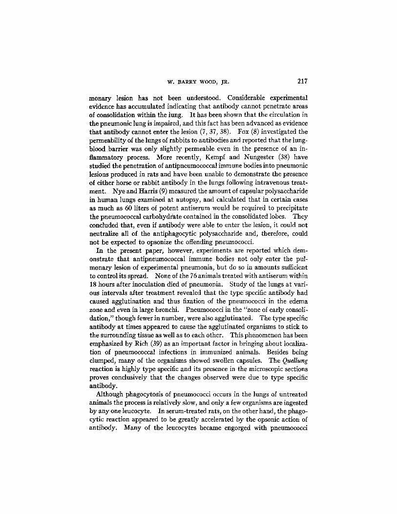
monary lesion has not been understood. Considerable experimentalevidence has accumulated indicating that antibody cannot penetrate areasof consolidation within the lung. It has been shown that the circulation inthe pneumonic lung is impaired, and this fact has been advanced as evidencethat antibody cannot enter the lesion (7, 37, 38). Fox (8) investigated thepermeability of the lungs of rabbits to antibodies and reported that the lung-blood barrier was only slightly permeable even in the presence of an in-flammatory process. More recently, Kempf and Nungester (38) havestudied the penetration of antipneumococcal immune bodies into pneumoniclesions produced in rats and have been unable to demonstrate the presenceof either horse or rabbit antibody in the lungs following intravenous treat-ment. Nye and Harris (9) measured the amount of capsular polysaccharidein human lungs examined at autopsy, and calculated that in certain casesas much as 60 liters of potent antiserum would be required to precipitatethe pneumococcal carbohydrate contained in the consolidated lobes. Theyconcluded that, even if antibody were able to enter the lesion, it could notneutralize all of the antiphagocytic polysaccharide and, therefore, couldnot be expected to opsonize the offending pneumococci.
In the present paper, however, experiments are reported which dem-onstrate that antipneumococcal immune bodies not only enter the pul-monary lesion of experimental pneumonia, but do so in amounts sufficientto control its spread. None of the 76 animals treated with antiserum within18 hours after inoculation died of pneumonia. Study of the lungs at vari-ous intervals after treatment revealed that the type specific antibody hadcaused agglutination and thus fixation of the pneumococci in the edemazone and even in large bronchi. Pneumococci in the "zone of early consoli-dation," though fewer in number, were also agglutinated. The type specificantibody at times appeared to cause the agglutinated organisms to stick tothe surrounding tissue as well as to each other. This phenomenon has beenemphasized by Rich (39) as an important factor in bringing about localiza-tion of pneumococcal infections in immunized animals. Besides beingclumped, many of the organisms showed swollen capsules. The Quellungreaction is highly type specific and its presence in the microscopic sectionsproves conclusively that the changes observed were due to type specificantibody.
Although phagocytosis of pneumococci occurs in the lungs of untreatedanimals the process is relatively slow, and only a few organisms are ingestedby any one leucocyte. In serum-treated rats, on the other hand, the phago-cytic reaction appeared to be greatly accelerated by the opsonic action ofantibody. Many of the leucocytes became engorged with pneumococci
217
218 MECHANISM OF RECOVERY IN PNEUMOCOCCAL PNEUMONIA. I
and within 42 hours after treatment practically all of the organisms in thelesion had been destroyed by phagocytic cells. It is of interest that mostof the pneumococci were phagocytized and digested by polymorphonuclearleucocytes and that the process was completed before any appreciablenumber of macrophages had appeared in the alveolar exudate. Robertson(33) has emphasized the importance of the macrophage reaction in bringingabout the final destruction of pneumococci in the lungs of untreated humanpatients and also of untreated dogs with experimental pneumonia. Thefact that the polymorphonuclear leucocytes appear to play a more importantrole in the present experiments may well be explained by the acceleratedphagocytic reaction caused by the specific psonins. Evidence will bepresented in a subsequent report that macrophages take a more activepart in the destruction of pneumococci when infected rats are treated withsulfapyridine.
The blood of animals naturally resistant to pneumococcal infections iscapable of destroying large numbers of pneumococci (40). Bacteriemiaresults only when the number of organisms entering the circulation fromthe pulmonary lesion exceeds that which can be handled by the pneumo-coccidal properties of the blood. Bacteriemia may be controlled by de-pressing the source of pneumococci in the lung, or by increasing the killingpower of the blood. Both mechanisms are involved in the action of anti-serum. The intravenous injection of specific antibody has been shown tocause immediate agglutination of circulating pneumococci; the clumpedorganisms become lodged in the capillaries of the liver, spleen, lungs, andpossibly other organs where they are destroyed by phagocytic cells (41).In the course of the present study antibody has also been found to causeagglutination and immobilization of pneumococci within the alveoli of thepneumonic lesion. Thus it becomes evident that antiserum exerts itsdramatic effect upon bacteriemia not only by hastening the destruction oforganisms already in the blood stream, but also by preventing pneumococcistill multiplying in the lung from invading the circulation. The resultanteffect is rapid sterilization of the blood.
No definite explanation can as yet be offered for the effect of antiserumupon early pleurisy. Since organisms could not be found in any of the areasof localized pleurisy observed in these experiments, no conclusions can bedrawn regarding the action of antibody upon the pleural infection. Themere fact, however, that serum treatment should lead to the localization ofan infection already involving the pleural cavity would seem of itself to beof considerable importance.

W. BARRY WOOD, JR.
The results of the present experiments leave no doubt as to the ability oftype specific antibody to penetrate the pulmonary lesion of experimentalpneumonia. The presence of agglutinated pneumococci in the alveoli, theswelling of their capsules, and the marked increase in the phagocytosis oforganisms by the cells of the alveolar exudate all serve as ample evidencethat antibody actually entered the alveoli. A study of the histologicalchanges occurring during recovery also indicates that antipneumococcalserum exerts its curative action in experimental pneumonia by halting thepneumococci in the advancing edema zone through agglutination thus caus-ing them to be overtaken and phagocytized by leucocytes. Experiments tobe reported in a subsequent paper have shown that sulfapyridine likewisestops the spread of the organisms at the margin of the lesion but does sothrough a different mechanism.
It has been stated that serum therapy is relatively ineffective in lobarpneumonia if instituted after the 4th day of the disease. The explanationsoffered for this commonly accepted dictum are that antibody cannot pene-trate the densely consolidated lesion of advanced pneumonia (42) and itcannot neutralize all of the antiphagocytic polysaccharide which has ac-cumulated in the pneumonic exudate (9). Neither of these explanationsappears to be valid in the light of the present results. It has been shownthat type specific antibody acts principally upon the advancing margin ofthe lesion in experimental pneumonia. Since there is good evidence thatmost of the pneumococci in both human and experimental pneumonia areat the border of the lesion, there is no reason to believe that intravenouslyadministered antiserum must penetrate areas of advanced consolidation inorder to control the infection. On the contrary, it has been repeatedly ob-served in both man and laboratory animals (33) that the cells of the alveolarexudate in densely consolidated areas are themselves capable of destroyingpneumococci without the aid of serotherapy. Were this not the case, lungabscesses would be frequent rather than extremely rare sequelae of untreatedpneumonia. Neither is there any reason to believe that intravenouslyinjected antibody must neutralize all of the polysaccharide in the pneumoniclesion. Much of the polysaccharide is contained within the consolidatedportion of the lesion where it cannot possibly offset the action of antibodyat the margin. The apparent ineffectiveness of late serum therapy, there-fore, cannot be explained either by the advanced stage of consolidation orby the amount of polysaccharide in the lung. Admitting that the exactcause of death, as in any severe infection, is often obscure, it would seemprobable that many failures have been due in the past to inadequate dosesof antiserum (43) rather than to the inability of antibody to affect the lesion.
219
220 MECHANISM OF RECOVERY IN PNEUMOCOCCAL PNEUMONIA. I
SUMMARY
A uniformly fatal lobar pneumonia was produced in white rats by inocula-tion of the left main bronchus with virulent Type I pneumococci suspendedin mucin. All of the animals succumbed in less than 5 days, half of themdying within 48 hours. In only 5 of 40 rats was the lesion confined to theleft lung, and all but one developed pleurisy, pericarditis, or both. All hadbacteriemia at the time of death.
The pathogenesis of the pulmonary lesion was studied by examining thelungs of 35 rats killed at various intervals following inoculation. Thepneumonic process spread rapidly until most of the left lung was involved in36 hours. Frequent blood cultures showed invasion of the blood stream ina few rats at 6 hours and in over 90 per cent at the end of the first day.The first signs of pleurisy usually appeared at 18 hours.
Microscopic examination of the actively spreading lesion revealed threecharacteristic zones: (1) an outer "edema zone" in which the alveoli con-tained many pneumococci floating freely in edema fluid, (2) a middle zonewhere both leucocytes and organisms were present, many of the latter beingphagocytized, and (3) an inner zone of advanced consolidation in which thealveoli contained many leucocytes but no organisms and where there werealready local areas of early resolution. Study of numerous lesions, at in-tervals of from 12 to 36 hours after inoculation, indicated that the pneumo-cocci spread into normal alveoli principally by way of the infected edemafluid in the outer zone. Pneumococcus-laden edema fluid in large bronchiand in alveoli beneath the pleura suggested the mode of spread of the in-fection to other lobes and possibly to the pleural cavity. No adequateexplanation could be found for the presence of active phagocytosis in thelungs of animals with bacteriemia and presumably without circulating anti-bodies, but this conspicuous phagocytic reaction was obviously responsiblefor the clearing of the central part of the spreading lesion.
The action of type specific antibody upon the pulmonary lesion of ex-perimental lobar pneumonia was studied in rats similarly infected buttreated with antipneumococcal serum. When injected intravenously in asingle dose within 18 hours after inoculation the antiserum was found toprotect all of the rats against the otherwise fatal pneumonia. It stoppedthe spread of the pneumonic lesion, cleared the blood stream of organisms,and prevented the extension of early pleurisy. The antibody caused ag-glutination and capsular swelling of the pneumococci in the lung, particu-larly in the edema zone at the margin of the lesion where they were mostnumerous. Apparently immobilized by agglutination the organisms were
W. BARRY WOOD, JR.
overtaken by leucocytes and destroyed by phagocytosis. The phagocyticreaction was greatly accelerated by the specific opsonins of the antiserum,and the pneumococci were destroyed by polymorphonuclear leucocytesbefore many macrophages appeared in the alveolar exudate. Within aweek after treatment resolution of the pulmonary lesion was well in prog-ress. Both horse and rabbit antibody were shown to penetrate the lung,and immune bodies were demonstrated in the alveoli within 10 minutesafter the start of treatment.
The relation of the observed phenomena to the curative action of anti-pneumococcal serum has been briefly discussed, and it is pointed out thatthe principal effect of antiserum is to cause immobilization of the pneumo-cocci in the advancing edema zone. Experiments to be reported in a laterpublication have shown that sulfapyridine exerts a similar effect through adifferent mechanism.
The author wishes to thank Dr. John Enders for much helpful advice, Mr. JohnBurke for preparation of the microscopic sections, and Dr. G. O. Favorite and Mr.Leo Talbert for the photomicrographs.
BIBLIOGRAPHY
1. Neufeld, F., and Rimpau, W., Deutsch. med. Woch., 1904, 30, 1458.2. Ward, H. K., and Enders, J. F., J. Exp. Med., 1933, 57, 527.3. Robertson, O. H., and Van Sant, H., J. Immunol., 1939, 37, 571.4. Heffron, R., Pneumonia with special reference to pneumococcus lobar pneumonia,
New York, The Commonwealth Fund, 1939, 151.5. Robertson, O. H., Graeser, J. B., Coggeshall, L. J., and Harrison, M. A., J. Clin.
Inv., 1934, 13, 633.6. Lord, F. T., and Parsons, E. L., J. Exp. Med., 1931, 53, 151.7. Kline, B. S., and Winternitz, M. C., J. Exp. Med., 1915, 21, 311.8. Fox, J. P., J. Immunol., 1936, 31, 7.9. Nye, R. N., and Harris, A. H., Am. J. Path., 1937, 13, 749.
10. Long, P. H., Bliss, E. A., and Feinstone, W. H., Pennsylvania Med. J., 1939, 42, 483.11. Reid, R. D., Proc. Soc. Exp. Biol. and Med., 1939, 41, 437.12. Finland, M., Spring, W. C., Jr., and Lowell, F. C., J. Clin. Inv., 1940, 19, 163.13. Wood, W. B., Jr., and Long, P. H., Ann. Int. Med., 1939, 13, 612.14. Finland, M., Spring, W. C., Jr., and Lowell, F. C., J. Clin. Inv., 1940, 19, 179.15. Nungester, W. J., and Jourdonais, L. F., J. Infect. Dis., 1936, 59, 258.16. Kepl, M., and Gunn, F. D., Proc. Soc. Exp. Biol. and Med., 1939, 40, 529.17. Enders, J. F., unpublished data.18. Jourdonais, L. F., and Nungester, W. J., Science, 1935, 81, 74.19. Loosli, C. G., Arch. Path., 1937, 24, 743.20. Gunn, F. D., and Nungester, W. J., Arch. Path., 1936, 21, 813.21. Hamburger, M., and Robertson, O. H., J. Exp. Med., 1940, 72, 261.22. Brown, J. H., and Brenn, L., Bull. Johns Hopkins Hosp., 1931, 48, 69.
221
222 MECHANISM OF RECOVERY IN PNEUMOCOCCAL PNEUMONIA. I
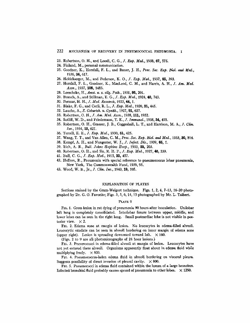
23. Robertson, O. H., and Loosli, C. G., J. Exp. Med., 1938, 67, 575.24. Finland, M., personal communication.25. Goodner, K., Horsfall, F. L., and Bauer, J. H., Proc. Soc. Exp. Biol. and Med.,
1936, 34, 617.26. Heidelberger, M., and Pederson, K. O., J. Exp. Med., 1937, 65, 393.27. Horsfall, F. L., Goodner, K., MacLeod, C. M., and Harris, A. H., J. Am. Med.
Assn., 1937, 108, 1483.28. Loeschcke, H., Anat. u. z. allg. Path., 1931, 86, 201.29. Branch, A., and Stillman, E. G., J. Exp. Med., 1924, 40, 743.30. Permar, H. H., J. Med. Research, 1923, 44, 1.31. Blake, F. G., and Cecil, R. L., J. Exp. Med., 1920, 31, 445.32. Lauche, A., Z. Geburtsh. u. Gynak., 1927, 91, 627.33. Robertson, O. H., J. Am. Med. Assn., 1938, 111, 1932.34. Sutliff, W. D., and Friedemann, T. E., J. Immunol., 1938, 34, 455.35. Robertson, O. H., Graeser, J. B., Coggeshall, L. T., and Harrison, M. A., J. Clin.
Inv., 1934, 13, 621.36. Terrell, E. E., J. Exp. Med., 1930, 51, 425.37. Wang, T. T., and Van Allen, C. M., Proc. Soc. Exp. Biol. and Med., 1933, 30, 814.38. Kempf, A. H., and Nungester, W. J., J. Infect. Dis., 1939, 66, 1.39. Rich, A. R., Bull. Johns Hopkins Hosp., 1933, 52, 203.40. Robertson, O. H., and Sia, R. H. P., J. Exp. Med., 1927, 46, 239.41. Bull, C. G., J. Exp. Med., 1915, 22, 457.42. Heffron, R., Pneumonia with special reference to pneumococcus lobar pneumonia,
New York, The Commonwealth Fund, 1939, 95.43. Wood, W. B., Jr., J. Clin. Inv., 1940, 19, 105.
EXPLANATION OF PLATES
Sections stained by the Gram-Weigert technique. Figs. 1, 2, 4, 7-13, 16-20 photo-graphed by Dr. G. O. Favorite; Figs. 3, 5, 6, 14, 15 photographed by Mr. L. Talbert.
PLATE 9
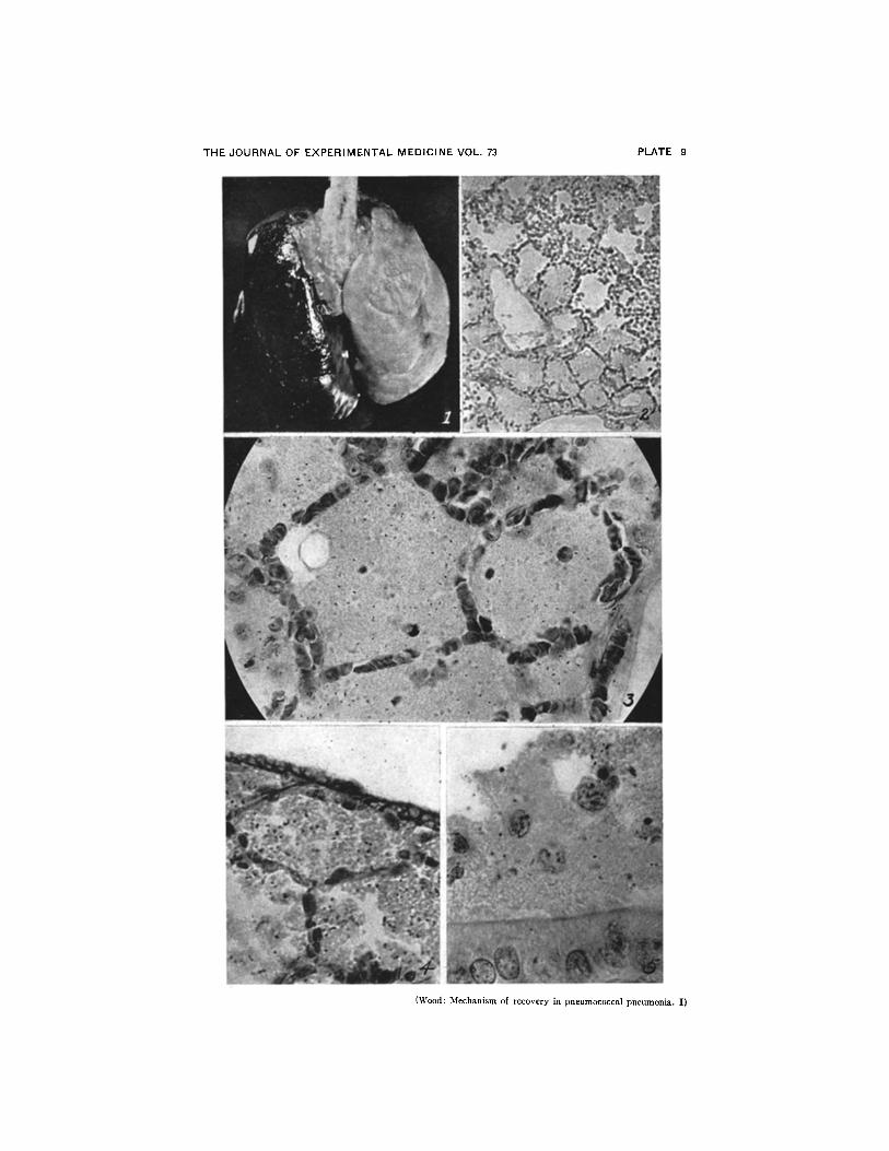
FIG. 1. Gross lesion in rat dying of pneumonia 90 hours after inoculation. Unilobarleft lung is completely consolidated. Interlobar fissure between upper, middle, andlower lobes can be seen in the right lung. Small postcardiac lobe is not visible in pos-terior view. X 2.
FIG. 2. Edema zone at margin of lesion. No leucocytes in edema-filled alveoli.Leucocytic exudate can be seen in alveoli bordering on inner margin of edema zone(upper right). Lesion is spreading downward toward left. X 160.
(Figs. 2 to 9 are all photomicrographs of 24 hour lesions.)FIG. 3. Pneumococci in edema-filled alveoli at margin of lesion. Leucocytes have
not yet entered these alveoli. Organisms apparently float about in edema fluid whilemultiplying freely. X 800.
FIG. 4. Pneumococcus-laden edema fluid in alveoli bordering on visceral pleura.Suggests possibility of direct invasion of pleural cavity. X 800.
FIG. 5. Pneumococci in edema fluid contained within the lumen of a large bronchus.Infected bronchial fluid probably causes spread of pneumonia to other lobes. X 1250.
THE JOURNAL OF EXPERIMENTAL MEDICINE VOL. 73
(Wood: Mechanism of recovery in pneumococcal pneumonia. I)
PLATE 9
PLATE 10
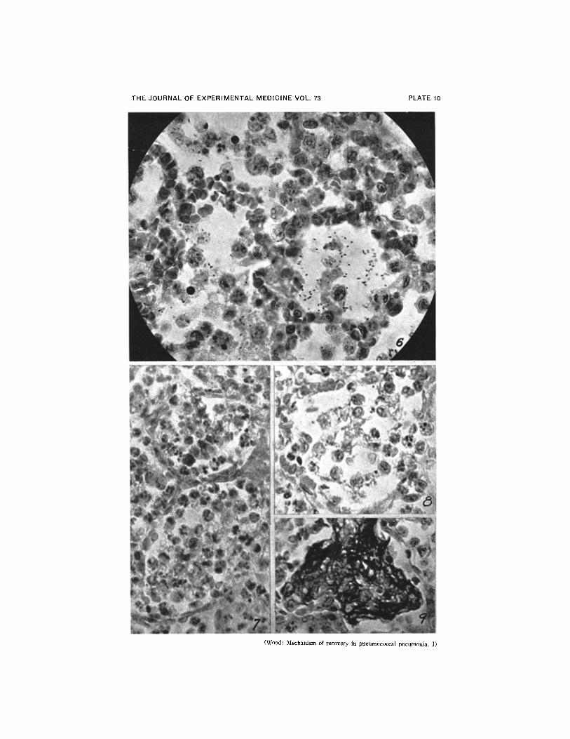
FIG. 6. Both pneumococci and leucocytes in alveoli within zone of early consolida-tion. This area borders on outer edema zone (see Fig. 2). Organisms are plentiful,but a few are already being phagocytized by leucocytes, although rat had bacteriemiawhen sacrificed. X 800.
FIG. 7. Leucocytic exudate in inner zone of advanced consolidation. Pneumococcihave been phagocytized and are no longer visible. Cells of exudate are still predomi-nantly polymorphonuclear. X 800.
FIG. 8. Early macrophage reaction and clearing of exudate in central area of ad-vanced consolidation. Rat had heavy bacteriemia when sacrificed. X 800.
FIG. 9. Fibrin deposits in alveoli in zone of consolidation near center of lesion.X 800.
THE JOURNAL OF EXPERIMENTAL MEDICINE VOL. 73
(Wood: Mechanism of recovery in pneumococcal pneumonia. I)
PLATE 10
PLATE 11
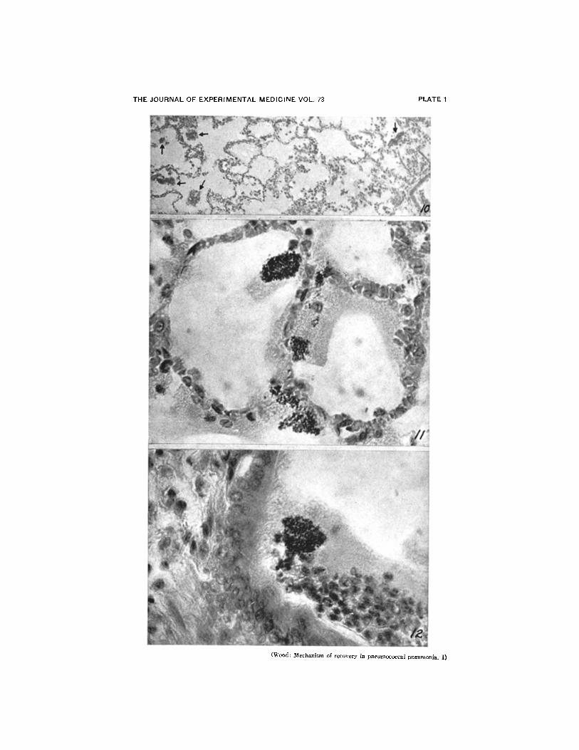
FIG. 10. Clumps of agglutinated pneumococci (see arrows) in edema zone at marginof lesion. Note small vacuoles in edema fluid. The rat was sacrificed 6 hours aftertreatment. X 160.
(All figures except 16, 17, and 20 are photomicrographs of lungs taken from ratstreated 18 hours after inoculation. Fig. 16, 17, and 20 show lesions in animals treatedat 12 hours.)
FIG. 11. Agglutination of pneumococci within the alveoli 6 hours after treatment.No leucocytes have as yet reached these alveoli in the outer edema zone. Many of theorganisms appear to cling to the alveolar walls as well as to each other. X 800.
FIG. 12. Agglutinated pneumococci within the lumen of a large bronchus. Althoughmany leucocytes are present in the bronchial exudate few contain organisms. X 800.
THE JOURNAL OF EXPERIMENTAL MEDICINE VOL. 73
(Wood: Mechanism of recovery in pneumococcal pneumonia. I)
PLATE 1
PLATE 12
FIG. 13. Large clumps of agglutinated pneumococci in an area of early consolidation6 hours after treatment. The large size of the clumps may be due in part to the con-tinued multiplication of the agglutinated organisms. It seems unlikely that leucocytescan attack such large masses of bacteria directly, and it is conceivable that autolysisplays an important r6le in their final destruction. Agglutination of pneumococci insuch areas indicates that antibody can penetrate the zone of early consolidation as wellas the edema zone at the margin of the lesion. X 800.
FIG. 14. Agglutinated pneumococci with swollen capsules characteristic of NeufeldQuellung reaction. Rat sacrificed 6 hours after treatment. X 1500.
FIG. 15. Higher magnification of Quellung reaction. X 3750.FIG. 16. Phagocytosis of pneumococci by polymorphonuclear leucocytes in alveolar
exudate 6 hours after treatment. X 800.FIG. 17. Higher magnification of leucocytes containing pneumococci with swollen
capsules. X 1800.

THE JOURNAL OF EXPERIMENTAL MEDICINE VOL. 73
(Wood: Mechanism of recovery in pneumococcal pneumonia. I)
PLATE 12
PLATE 13
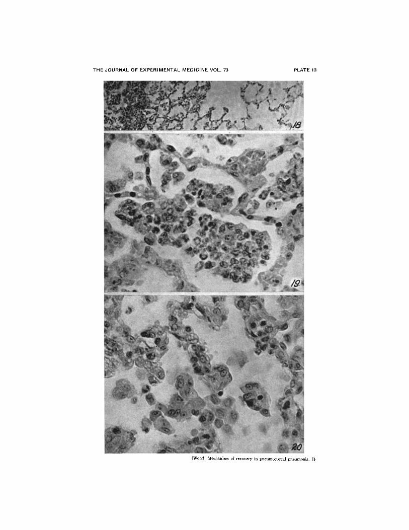
FIG. 18. Margin of lesion 18 hours after treatment showing complete disappearanceof edema zone. X 160.
FIG. 19. Alveolar exudate near margin of lesion 42 hours after treatment. Thepneumococci have been destroyed by the phagocytic cells. Although macrophages arepresent the exudate still contains many polymorphonuclear leucocytes. X 800.
FIG. 20. Advanced macrophage reaction with marked clearing of the lesion 1 weekafter treatment. The alveolar walls are noticeably thickened and a few large palemacrophages can be seen free in the alveoli. X 800.
THE JOURNAL OF EXPERIMENTAL MEDICINE VOL. 73
(Wood: Mechanism of recovery in pneumococcal pneumonia. I)
PLATE 13