Embed Size (px)
Citation preview

i
STUDIES ON THE CYTOTOXICITY AND ANTIOXIDANT ACTIVITY OF TEA
KOMBUCHA
A RESEARCH PROJECT REPORT SUBMITTED
BY
EUGENE AIDOO
(10069898)
THIS DISSERTATION IS SUBMITTED TO THE UNIVERSITY OF GHANA,
LEGON IN PARTIAL FULFILLMENT OF THE REQUIREMENT FOR THE
AWARD OF MSC. BIOCHEMISTRY DEGREE.
AUGUST, 2015
University of Ghana http://ugspace.ug.edu.gh

ii
DECLARATION
I, Eugene Aidoo, do declare that the experimental work described in this project report was
performed by me in the Department of Biochemistry, Cell and Molecular Biology, University
of Ghana, Legon and the Department of Clinical Pathology, Noguchi Memorial Institute for
Medical Research, College of Health Sciences, University of Ghana, Legon, under the
supervision of Prof. L.K.N Okine, Rev. Dr. W.S.K. Gbewonyo and Dr. Regina Appiah-
Opong.
………………………………………. Date ……………………………………
(Eugene Aidoo)
Candidate
………………………………………… Date ………………………………………
(Prof. L.K.N. Okine)
Supervisor
………………………………………… Date ………………………………………
(Rev. Dr. W.S.K. Gbewonyo)
Supervisor
………………………………………… Date ………………………………………..
(Dr. Regina Appiah-Opong)
Supervisor
University of Ghana http://ugspace.ug.edu.gh

iii
DEDICATION
I dedicate this project to The Almighty God, My family, especially my wife, Gloria O. K.
Aidoo, Mr. and Mrs. Adom-Oduro, my mum, Elizabeth S. Asiamah and dad, Gilbert K.
Aidoo, as well as my wonderful siblings for their immense support and encouragement.
I also thank the Board and members of Emmanuel Assemblies of God Church, Bubiashie, for
their prayers and encouragements.
University of Ghana http://ugspace.ug.edu.gh

iv
ACKNOWLEDGEMENT
Thank God for the gift of life. I wish to express my profound gratitude to my supervisors
Prof. L.K.N Okine, Rev. Dr. W.S.K Gbewonyo and Dr. Regina Appiah-Opong for their
guidance which contributed to the success of this study.
My dear wife Gloria O.K. Aidoo’s financial, spiritual, emotional and mechanical support was
overwhelming. She has immensely contributed to the success of this study; as a result I wish
to express my deepest gratitude to her.
I also wish to express my appreciation to all staff of the Department of Clinical Pathology of
the Noguchi Memorial Institute for Medical Research, especially Mrs. Eunice Apenteng
(Dotse), Ms. Abigail Aning, Mr. Ebenezer Ofori-Attah, Ms. Abena Kissi-Twum and Mr.
Justice Kumi for their guidance and great support.
I would like to thank my friends, Ms. Dorcas Frimpong, Mr. Christian Kweku Arhin, Mr.
Richmond Adzadogo and Mr. Fred Ayertey for their support and prayers.
May the good Lord bless all those who contributed to the success of this study.
University of Ghana http://ugspace.ug.edu.gh

v
CONTENTS
CHAPTER ONE ……………………………………………………………………….........1
1.0 INTRODUCTION AND LITERATURE REVIEW……………………………………1
1.1 INTRODUCTION……………………………………………………………….1
Aim……………………………………………………………………………….4
Specific Objectives……………………………………………………………….5
1.2 LITERATURE REVIEW………………………………………………………..6
1.2.1 Tea Kombucha………………………………………………………..6
1.2.2 Chemical Composition of Tea Kombucha……………………………7
1.2.3 Beneficial Effects of Tea Kombucha……………………………….. 9
1.2.4 Cancer……………………………………………………………......11
1.2.5 Causes of Cancer…………………………………………………….13
1.2.5.1 Chemical Carcinogens………………………………………..13
1.2.5.2 Effects of Age on cancer development………………………14
1.2.5.3 Lifestyle Factors……………………………………………..14
1.2.6 Types of Cancer……………………………………………………...15
1.2.6.1 Blood Cancers………………………………………………..15
1.2.6.2 Symptoms of Blood Cancer(Leukemia)……………………..16
1.2.6.3 Treatment of Cancers………………………………………..16
1.2.7 Phytochemicals……………………………………………………….18
1.2.7.1 Curcumin……………………………………………………..19
1.2.7.2 Medicinal Plants……………...……………………………....20
1.2.8 Importance of Antioxidants…….……………………………………25
1.2.9 Tea Kombucha as Antioxidant Source……………………………....26
1.2.10 Non-Toxic Nature of Tea Kombucha………………………………...27
University of Ghana http://ugspace.ug.edu.gh

vi
1.2.11 Anti-Proliferative Activity(Viability of Cells)………………………28
1.2.11.1 MTT Assay………………………………………….28
1.2.12 DPPH Assay………………………………………………………....30
1.2.13 Reducing Power……………………………………………………..30
CHAPTER TWO………………………………………………………………..32
2.0 MATERIALS AND METHODS…………………………………………...32
2.1 MATERIALS……………………………………………………………….32
2.1.1 Chemicals and Reagents…………………………………………………..32
2.1.2 Kombucha Starter Culture………………………………………………...32
2.1.3 Leukaemia Cell Line………………………………………………………32
2.2 METHODS………………………………………………………………….32
2.2.1 Preparation of Tea Kombucha……………………………………………..32
2.2.2 Freeze –drying of Tea Kombucha…………………………………………33
2.2.3 MTT Assay for Cell Viability……………………………………………...33
2.2.4 Antioxidant Determination Using DPPH Assay…………………………..34
2.2.5 Reducing Power……………………………………………………………34
2.2.6 Total Phenolic Content…………………………………………………….35
2.2.7 Statistical Analysis…………………………………………………………35
CHAPTER THREE……………………………………………………………...36
3.0 RESULTS……………………………………………………………………36
3.1 Cell Viability………………………………………………………...............36
3.2 DPPH Antioxidant Activity…………………………………………………38
3.3 Total Phenolic Content………………………………………………………40
3.4 Reducing Power……………………………………………………………...41
University of Ghana http://ugspace.ug.edu.gh

vii
CHAPTER FOUR……………………………………………………………….43
4.0 DISCUSSION, CONCLUSION AND RECOMMENDATIONS…………43
4.1 DISCUSSION…………………………………………………………….....43
4.2 CONCLUSION……………………………………………………………...45
4.3 RECOMMENDATION………………………………………………...........46
REFERENCES…………………………………………………………………..48
Appendix ………………………………………………………………………...55
University of Ghana http://ugspace.ug.edu.gh

viii
LIST OF FIGURES
Fig. 1.1: Structures of some organic acids present in tea kombucha…………………………7
Fig. 1.2: Chemical structures of some tea constituents………………………………………8
Fig. 1.3: Molecular targets of curcumin and/or its chemically-related analogues…………...19
Fig. 1.4: Chemical structure of curcumin and related derivatives/analogues………………..22
Fig. 1.5: Chemical structures of some anti-cancer agents……………………………………25
Fig. 1.6: The reduction of MTT to formazan………………………………………………...29
Fig. 3.1: Effect of varying concentrations of curcumin, tea kombucha and unfermented tea on
viability of Jurkat P9 leukemia cells…………………………………………………………37
Fig. 3.2: Antioxidant activity of varying concentrations of BHT, tea kombucha and
unfermented tea………………………………………………………………………………39
Fig. 3.3: Total phenolic content of tea kombucha and unfermented tea…………………….40
Fig. 3.4: Reducing power of varying concentrations of ascorbic acid, tea kombucha and
unfermented tea. …………………………………………………………………………….42
University of Ghana http://ugspace.ug.edu.gh

ix
LIST OF TABLES
Table 1.1: Various biological activities of tea kombucha…………………………………10
University of Ghana http://ugspace.ug.edu.gh

x
ABSTRACT
Fermentation of sugared tea with a symbiotic culture of acetic acid bacteria and yeast (tea
fungus) yields kombucha tea which is consumed worldwide for its refreshing taste and
beneficial effects on human health. It is claimed to prevent various types of cancer and
cardiovascular diseases, promote liver function, and stimulate the immune system. The anti-
proliferative and antioxidant activities of tea kombucha and unfermented tea were, therefore,
investigated in this study. The cytotoxic effect of the tea kombucha was studied using an
MTT (3-(4,5-dimethylthiazole-2-yl)-2,5-diphenyltetrazolium) assay while its antioxidant
activity was performed using 2,2-diphenyl-1-picrylhydrazyl (DPPH) and reducing power (the
ferric reducing/antioxidant power (FRAP)) assays. The total phenolic content of the tea
kombucha was also studied using the modified Folin-Ciocalteau colorimetric assay. It was
observed that the antioxidant activity of tea kombucha was higher compared to the
unfermented tea and this reflected in the total phenolics contents. Tea kombucha had 2.4-fold
and 7.3-fold significantly (p<0.05) higher phenolic content at concentrations of 2.5 and 5.0
mg/ml, respectively than the unfermented tea. The tea kombucha showed insignificantly low
cytotoxicity against the Jurkat P9 leukemia cells many orders of magnitude below that of the
standard drug, curcumin, which had an IC50 = 7.22 mg/ml whilst the unfermented tea was
without such effect. These results suggest that kombucha tea with its high antioxidant
activity may help protect cells against oxidative damage and possibly cancer.
University of Ghana http://ugspace.ug.edu.gh

1
CHAPTER ONE
1.0 INTRODUCTION AND LITERATURE REVIEW
1.1 INTRODUCTION
Cancer is a multifactorial disease that involves modulation of multiple pathways and targets.
In India, breast cancer is the second leading cause of cancer deaths in women and the risk of
its incidence is increasing every year. Several researches are going on to identify
chemotherapeutic and chemopreventive agents that can act on multiple signaling targets
(Aggarwal et al., 2004).
Evidence suggests that the plant kingdom is considered a good candidate for
chemoprevention and cancer therapy due to the high concentration and wide variety of
antioxidants such as resveratrol, genestein, beicalein, vitamin A, vitamin C, polyphenols,
(‒)‒Epigallocatechin 3-gallate, flavonoids, polyphenols, gallic acid, glycosides, verbascoside,
calceorioside, epicatechin, quercetin, curcumin, lovastatin, and many other types of
compounds with the capability to inhibit the cell proliferation of different cancer cells in vitro
and in vivo, such as colon cancer (HT-29, SW48, HCT116), breast (MCF7, MDA), cervix
(HeLa, SiHa, Ca-Ski, C33-A), liver (Hep G2), skin (A 431), fibroblasts (3T3 SV40), and
many other malignant cells (Grover et al., 2003; Cetojević-Simin et al., 2008). Studies have
indicated that antioxidants can be employed efficiently as chemopreventives and as effective
inhibitors of cell proliferation, promoting cell apoptosis, and increasing detoxification
enzymes, and inhibiting gene expression and scavenger reactive oxygen species (ROS). Thus,
many researchers are working with different types of natural antioxidants with the aim of
finding those with the greatest capacity to inhibit the development of cancer both in vitro as
well as in vivo. This is because these compounds have exhibited high potential for use not
University of Ghana http://ugspace.ug.edu.gh

2
only in the treatment of this disease, but they also act as good chemoprotective agents.
Identification of pharmacologically safe phytochemicals that have multitargeting effect is a
‘hotspot’ in cancer research. One such phytochemical is curcumin, the principle ingredient
from the yellow spice turmeric. It is a well-known plant derived chemopreventive agent,
common in Southeast Asian countries and used as a folk medicine and traditional food for
several centuries (Mohandas and Desai, 1999; Barclay et al., 2000).
It has been shown that dietary phytochemicals can interfere with each stage of the
development of carcinogenesis (Manson, 2003). As in the case of direct antioxidant effects,
dietary polyphenols are most likely to exert their chemopreventive effects on the
gastrointestinal tract, where they are present at highest concentrations (Aggarwal et al.,
2004). Indeed, studies have shown that various polyphenol-rich fruits and vegetables are
particularly effective in protecting against several types of cancer development (Mouria,
2002). Dietary polyphenols may exert their anticancer effects through several possible
mechanisms, such as removal of carcinogenic agents, modulation of cancer cell signaling and
antioxidant enzymatic activities, and induction of apoptosis as well as of cell cycle arrest.
Some of these effects may be related, at least partly, with their antioxidant activities
(Greenwald, 1996).
At the cellular level, there is good evidence that polyphenols present in tea, red wine, cocoa,
fruit juices, and olive oil; at some level, they are able to stimulate carcinogenesis and tumor
development (Mouria, 2002). For example, they may interact with reactive intermediates and
activated carcinogens and mutagens, they may modulate the activity of the key proteins
involved in controlling cell cycle progression, and they may influence the expression of many
cancer-associated genes. Perhaps most notably, the anticancer properties of green tea
University of Ghana http://ugspace.ug.edu.gh

3
flavanols have been reported in animal models and in human cell lines, as well as in human
intervention studies. On the other hand, green tea consumption has been proposed as
significantly reducing the risk of cancer of the biliary tract, bladder, breast, and colon
(Patterson et al., 1997).
Tea kombucha is a traditional drink made from a particular fermentation of sugared-black tea
and a symbiosis of yeast species (fungi) and acetic acid bacteria. It is commonly consumed
throughout the world as a medicinal health-promoting beverage (Dutta and Gachhui, 2007).
Tea kombucha is prepared by placing the kombucha culture (tea fungus) in a sugared tea
broth for fermentation (Chanda and Dave, 2009). If the kombucha culture is cultivated
according to the standard recipe with black tea, sweetened with sucrose, it turns this substrate
into a refreshing beverage called tea fungus beverage with high nutritive value and medicinal
properties (Lončar et al., 2000). The popularity of kombucha expanded like many other
traditional beverages due to its beneficial effects on human health and its ease in home
preparation. The amounts of tea, sugar, and tea fungus differ in different places (Chen and
Liu, 2000).
The taste of the kombucha changes during fermentation from a pleasantly fruity sour-like
sparkling flavour after a few days to a mild vinegar-like taste after a long incubation period.
Currently, tea kombucha is praised as “the ultimate health drink” or damned as “unsafe
medicinal tea” (Blanc, 1996; Hartmann et al., 2000). There are many opinions regarding the
health benefits and toxicity of kombucha beverage. Though it is claimed to be beneficial for
several medical ailments, very little or no clinical evidence is available for that. Studies on
tea kombucha were reviewed earlier (Dufresne and Farnworth, 2000; Ernst, 2003). Research
on tea kombucha was highly boosted during the past decade, but there were no review reports
University of Ghana http://ugspace.ug.edu.gh
4
published during the period. Several investigations have been conducted under static
conditions on the beverage (Kumar et al., 2008; Wang et al., 2010; Yang et al., 2010). Yeasts
and bacteria in tea kombucha are involved in metabolic activities that utilize substrates by
different and in complementary ways (Blanc, 1996).
Many in vivo and in vitro studies performed to evaluate the capability of antioxidants against
cancer, such as chemopreventive or therapeutic agents, were conducted employing natural
antioxidants from fruits and vegetables (Block et al., 1992; Helbock et al., 1998). Thus,
humans are forced to consume antioxidants in a more direct manner, either in the form of a
tablet, a pill, or any other form in order to supply the levels that the body requires of these
compounds to protect it against cell damage caused by oxidation reactions, thus reducing the
risk of certain cancer types, especially those of the epithelial surface and in the upper part of
the body, such as breast, lung, kidney, liver, intestine, and many others that have been well
documented (Patterson et al., 1997). However, further investigations are expected before a
better understanding of the function of many antioxidants and their utilization in the
prevention and treatment of cancer and other degenerative diseases could be obtained. Thus
this study investigated whether tea kombucha has anti-proliferative and antioxidant activities
with the view to establishing its potential for cancer prevention or treatment.
Aim
The main aim of the study was to evaluate the cytotoxicity and antioxidant activities of tea
kombucha.
University of Ghana http://ugspace.ug.edu.gh

5
Specific objectives
To determine the cytotoxic effect of tea kombucha on Jurkat P9 leukemia cell lines
using the MTT assay.
To evaluate the antioxidant activity of tea kombucha using DPPH and reducing power
assays.
To assess the total phenolic content of tea kombucha using Folin-Ciocalteau
colorimetric assay.
University of Ghana http://ugspace.ug.edu.gh

6
1.2 LITERATURE REVIEW
1.2.1 Tea Kombucha
Tea kombucha is a slightly sweet, slightly acidic refreshing beverage consumed worldwide. It
is obtained from infusion of tea leaves by the fermentation of a symbiotic association of
bacteria and yeasts forming “tea fungus” (Chen and Liu, 2000; Goh et al., 2012). A floating
cellulosic pellicle layer and the sour liquid broth are the 2 portions of tea kombucha. It tastes
like sparkling apple cider. Though green tea can be used for kombucha preparation, black tea
and white sugar are considered the finest substrates. Kombucha is the internationally used
Germanized form of the Japanese name for this slightly fermented tea beverage. It was first
used in East Asia for its healing benefits. Kombucha originated in northeast China
(Manchuria) where it was prized during the Tsin Dynasty (“Ling Chi”), about 220 B.C., for
its detoxifying and energizing properties. In 414 A.D., the physician Kombu brought the tea
fungus to Japan and he used it to cure the digestive problems of the Emperor Inkyo. It is
presently cultivated in at least 30 countries around the world (Stoner and Mukhtar, 1995).
Reiss (1994) has suggested that 50 g sucrose/L provides the optimal concentrations of ethanol
and lactic acid, and this sugar concentration has been used in traditional recipes for the
preparation of “teakwass” (another name for kombucha) for a long time. An optimum
fermentation time of eight days is required for the production of kombucha with pleasant
flavour and taste. Longer fermentation periods produce high levels of acids (like mild
vinegar) that may pose potential risks when consumed (Sreeramulu et al., 2000)
University of Ghana http://ugspace.ug.edu.gh

7
1.2.2 Chemical Composition of Tea Kombucha
Chemical analysis of tea kombucha showed the presence of various organic acids (Fig. 1.1),
such as acetic, gluconic, glucuronic, citric, L-lactic, malic, tartaric, malonic, oxalic, succinic,
pyruvic, usnic; also sugars, such as sucrose, glucose, and fructose; the vitamins B1, B2, B6,
B12, and C; 14 amino acids, biogenic amines, purines, pigments, lipids, proteins, some
hydrolytic enzymes, ethanol, antibiotically active matter, carbon dioxide, phenol, as well as
some tea polyphenols, minerals, anions, as well as insufficiently known products of yeast and
bacterial metabolites.
Fig. 1.1: Structures of some organic acids present in tea kombucha.
Yeasts hydrolyze sucrose into glucose and fructose by invertase activity and produce ethanol
via glycolysis, with a preference for fructose as a substrate. Acetic acid bacteria make use of
glucose to produce gluconic acid and ethanol to produce acetic acid. The pH value of tea
University of Ghana http://ugspace.ug.edu.gh

8
kombucha beverage decreases due to the production of organic acids during fermentation
(Dufresne and Farnworth, 2000; Jayabalan et al., 2007).
Tea contains polyphenols and flavonoids (theaflavins and thearubigins), catechin,
theobromine , caffeine, gallic acid, catechin gallates, tannins, gallotannin, small amounts of
aminophylline (Fig. 1.2) and a yellow volatile oil that is solid at ordinary temperatures and
has strong aromatic odour and taste (Jayabalan et al., 2008).
Fig. 1.2: Chemical structure of some tea constituents (Adapted from Lambert and
Yang, 2003)
The catechins and gallic acid complexes such as theaflavins, theaflavinic acids, thearubigins
or theasinensis present in the kombucha prepared from the black tea, possess a significant
University of Ghana http://ugspace.ug.edu.gh

9
degree of bioavailability. The composition of kombucha beverage indicates the presence of
numerous compounds and it depends on cultivation substrate, time and temperature of
fermentation process, as well as the microorganisms present in the culture (Chen and Liu,
2000). It also depends on the applied method of analysis.
1.2.3 Beneficial Effects of Tea Kombucha
Tea kombucha has been claimed by kombucha drinkers all over the world to have many
beneficial effects on human health. However, most of the benefits have been studied in
animal models only and there is a lack of scientific evidence based on clinical trials. Non-
human studies regarding antimicrobial, antioxidant, hepatoprotective, and anticancer
properties of tea kombucha have been carried out and the biological activities have been
reported (Table 1.1).
University of Ghana http://ugspace.ug.edu.gh

10
Table 1.1: Various Biological Activities of Tea Kombucha.
Biological
activity
Experimental
animal/cells
Treatment
period/dose
Parameters studied
Reference
Hypoglycaemic
activity
Mice 3 days and 1.71
mg/kg body weight
Blood sugar level Shenoy, 2000
Antioxidative
stress against
chromate
Rat 30 days and 0.6
ml/200 g body
weight
Plasma and tissue MDA
levels, delayed type
hypersensitivity response,
GSH, peroxidise, catalase
Sai et al., 2000
Longevity Mice 3 years and free
access
Longevity, general health,
and open-field exploratory
behavioural outcomes
Hartmann et
al., 2000
Antistress activity
against cold and
hypoxia
Rat 15 days and 1.6, 8.0
and 16 ml/kg body
weight
plasma/blood MDA and
reduced GSH, fecal output
Pauline et al.,
2001
Antioxidative
stress against lead
Rat 45 days and 1 ml/kg
body weight
Lipid peroxidation, creatine
phosphokinase, GSH, DNA
fragmentation in liver
Dipti et al.,
2003
Cytogenic activity Human
peripheral blood
lymphocytes
1 hour and 40 μg/ml Frequencies of sister
chromatid exchange and
micronuclei formation
Mrđanović et
al., 2007
Antihyperglycae
mic efficacy
Streptozotocin-
induced rats
45 days and 6 mg/kg
body weight
Plasma insulin,
haemoglobin, tissue
glycogen
Srihari et al.,
2013b
MDA= Malondialdehyde; GSH= reduced glutathione
University of Ghana http://ugspace.ug.edu.gh
11
Tea kombucha reduces cell damage induced by oxidative stress. It constitutes a potent
therapeutic supplement that improves resistance against cancer, cardiovascular diseases,
promotes digestive functions, stimulates the immune system and reduces inflammatory
problems (Greenwalt et al., 1998, Jayabalan et al., 2008; Banerjee et al., 2011; El-Taher,
2011). The putative health benefits of tea kombucha have been largely attributed to the
polyphenolic components of kombucha that have been transformed from the black tea (Bors
et al., 1996). In addition, organic acids, vitamins, amino acids, antibiotics and a variety of
micronutrients produced during fermentation of kombucha may also have a role in the health
benefits to some extent.
1.2.4 Cancer
Cancer is the name given to a collection of related diseases. In all types of cancer, some of
the body’s cells begin to divide without stopping and spread into surrounding tissues. Cancer
can start almost anywhere in the human body, which is made up of trillions of cells.
Normally, human cells grow and divide to form new cells as the body needs them. When
cells grow old or become damaged, they die, and new cells take their place.
When cancer develops, however, this orderly process breaks down. As cells become more
and more abnormal, old or damaged cells survive when they should die, and new cells form
when they are not needed. These extra cells can divide without stopping and may form
growths called tumors. Many cancers form solid tumors, which are masses of tissue. Cancers
of the blood, such as leukemias, generally do not form solid tumors.
Cancerous tumors are malignant, which means they can spread into, or invade nearby tissues.
In addition, as these tumors grow, some cancer cells can break off and travel to distant places
University of Ghana http://ugspace.ug.edu.gh

12
in the body through the blood or the lymph system and form new tumors far from the original
tumor. Unlike malignant tumors, benign tumors do not spread into, or invade, nearby tissues.
Benign tumors can sometimes be quite large. However, when removed, they usually do not
grow back, whereas malignant tumors sometimes do. Unlike most benign tumors elsewhere
in the body, benign brain tumors can be life threatening.
Cancer, after cardiovascular disease, is the second leading cause of death (Turgay and Sar,
2005). Worldwide about 10 million people per year are diagnosed with cancer and more than
6 million die of the disease and over 22 million people in the world are cancer patients (Pinar,
1998). When cancer is diagnosed, therapists face a formidable range of challenges. Treatment
usually consists of various combinations of surgery, radiation therapy, and chemotherapy but
despite these therapeutic options, cancer remains associated with high mortality.
Cancer cells differ from normal cells in many ways that allow them to grow out of control
and become invasive (Ertel et al., 2006). One important difference is that cancer cells are less
specialized than normal cells. That is, whereas normal cells mature into very distinct cell
types with specific functions, cancer cells do not. In addition, cancer cells are able to ignore
signals that normally tell cells to stop dividing or that begin a process known as programmed
cell death, or apoptosis, which the body uses to get rid of unneeded cells.
Cancer cells may be able to influence the normal cells, molecules, and blood vessels that
surround and feed a tumor—an area known as the microenvironment. For instance, cancer
cells can induce nearby normal cells to form blood vessels that supply tumors with oxygen
and nutrients, which they need to grow. These blood vessels also remove waste products from
tumors. Cancer cells are also often able to evade the immune system, a network of organs,
tissues, and specialized cells that protects the body from infections and other conditions
University of Ghana http://ugspace.ug.edu.gh

13
(Parkin, 2006). Although the immune system normally removes damaged or abnormal cells
from the body, some cancer cells are able to “hide” from the immune system. Tumors can
also use the immune system to stay alive and grow. For example, with the help of certain
immune system cells that normally prevent a runaway immune response, cancer cells can
actually keep the immune system from killing cancer cells.
1.2.5 Causes of Cancer
Cancer is a genetic disease—that is, it is caused by changes to genes that control the way our
cells function, especially how they grow and divide. Genetic changes that cause cancer can be
inherited from our parents. They can also arise during a person’s lifetime as a result of errors
that occur as cells divide or because of damage to DNA caused by certain environmental
exposures.
We all have a risk of developing cancer. Many cancers seem to develop for no apparent
reason. However, certain risk factors are known to increase the chance that one or more of
your cells will become abnormal and lead to cancer. Risk factors include the following:
1.2.5.1 Chemical carcinogens
A carcinogen is something (chemical, radiation, etc) that can damage a cell and make it more
likely to turn into a cancerous cell. As a general rule, the more the exposure to a carcinogen,
the greater the risk. Well known examples of carcinogens include:
Tobacco smoke.
Smokers are more likely to develop lung cancer, mouth cancer, throat cancer, oesophageal
cancer, bladder cancer and pancreas cancer. Smoking is thought to cause about a quarter of
University of Ghana http://ugspace.ug.edu.gh
14
all cancers. About 1 in 10 smokers die from lung cancer. The heavier you smoke, the greater
the risk. If you stop smoking, your risk goes down considerably.
Workplace chemicals
This includes chemicals such as asbestos, benzene, formaldehyde, etc. If you have worked
with these without protection you have an increased risk of developing certain cancers. For
example, a cancer called mesothelioma is linked to past exposure to asbestos.
1.2.5.2 Effect of Age on cancer development
The older you become, the more likely you will develop a cancer. This is probably due to an
accumulation of damage to cells in the body over time. Also, the body's defences against
abnormal cells may become less good as you become older. For example, the ability to repair
damaged cells, and the immune system which may destroy abnormal cells, may become less
efficient with age. So, eventually one damaged cell may manage to survive and multiply out
of control into a cancer. Most cancers develop in older people.
1.2.5.3 Lifestyle factors
Diet and other lifestyle factors can alter the risk of developing cancer. For example:
If you eat a lot of fruit and vegetables you have a reduced risk of developing certain
cancers. The exact way in which they protect against cancer is not fully understood.
These foods are rich in vitamins and minerals, and also contain chemicals called
antioxidants. They may protect against damaging chemicals that get into the body. We
should all eat at least five portions of fruit and vegetables per day (some experts
recommend even more).
Eating too much fatty food possibly increases the risk of developing certain cancers.
University of Ghana http://ugspace.ug.edu.gh

15
The risk of developing certain cancers is increased by obesity, lack of regular
exercise (physical activity) and drinking a lot of alcohol.
In general, cancer cells have more genetic changes, such as mutations in DNA, than normal
cells. Some of these changes may have nothing to do with the cancer; they may be the result
of the cancer, rather than its cause (Harlin and Gajewski, 2008).
1.2.6 Types of Cancer
There are more than 100 types of cancer. Cancers are usually named after the organs or
tissues where the cancers form. For example, lung cancer starts in cells of the lung, and brain
cancer starts in cells of the brain. Cancers also may be described by the type of cell that
formed them, such as an epithelial cell or a squamous cell (Worsham et al ., 2009). Here are
some categories of cancers that begin in specific types of cells: carcinoma, sarcomas,
leukemia, lymphoma etc.
1.2.6.1 Blood cancers
Primarily, there are three basic types of blood cancer. Each of the variety may also include
several variations, but in general this cancer is categorized into the following kinds
Leukemia - With spurt in the multiplicity of cancerous cells affecting either the
marrow or the blood; the ability of the circulatory system to produce blood is severely
impaired.
Lymphoma - The cancerous formation affecting the lymphocytes is referred to as
the lymphoma. Lymphocytes are one of the varieties of white blood corpuscles.
Myeloma – it is a cancer arising from plasma cells, a type of white blood cell which is
made in the bone marrow. Bone marrow is the ‘spongy’ material found in the centre
of the larger bones in the body. The bone marrow is where all blood cells are made.
University of Ghana http://ugspace.ug.edu.gh

16
1.2.6.2 Symptoms of blood cancer (leukemia).
Leukemia marked by an acute destruction of health sustaining red blood cells includes the
symptoms of anemia, weakness and extreme fatigue. Consequently one affected by it is likely
to sweat and come under bouts of breath shortness in course of performing day to day
activities of the regular kind. Vulnerability to infection and swelling of the lymph nodes are
some of the other fallouts of leukemia. Blood tests are likely to present higher counts of white
blood corpuscles.
Leukemia can be chronic or acute. A person afflicted with the chronic type may not exhibit
any of these symptoms. On the other hand, in leukemia of the acute type, the symptoms are
likely to manifest with rapid intensity.
1.2.6.3 Treatment of Cancers
Treatment options vary, depending on the type of cancer and how far it has grown and
spread. Briefly, the three most common treatments are:
Surgery: It may be possible to cut out a cancerous (malignant) tumour.
Chemotherapy: This is a treatment that uses anti-cancer medicines to kill cancer
cells, or to stop them from multiplying. There are various different types of medicines
used for chemotherapy. The medicine or combination of medicines selected depends
on the type of cancer being treated.
Radiotherapy: This is a treatment that uses high-energy beams of radiation which are
focused on cancerous tissue. This kills cancer cells, or stops cancer cells from
multiplying.
More recently, other treatments have been introduced which include:
University of Ghana http://ugspace.ug.edu.gh

17
Stem cell transplant: High-dose chemotherapy may damage bone marrow cells
and lead to blood problems. However, if you receive healthy bone marrow after the
chemotherapy then this helps to overcome this problem.
Hormone therapy: This is the use of medicines to block the effects of hormones.
This treatment may be used for cancers that are hormone-sensitive such as some
cancers of the breast, prostate and womb (uterus).
Immunotherapy: Some treatments can boost the immune system to help to fight
cancer. More specific immunotherapy involves injections of antibodies which aim to
attack and destroy certain types of cancer cells. Research is underway to try to find
vaccines that would stimulate your own immune system to make antibodies against
cancer cells.
Gene therapy: This is a new area of possible treatments. Research is underway to
find ways of blocking, repairing or replacing abnormal genes in cancer cells.
Special techniques: These can sometimes be used to cut off the blood supply to
tumours. The tumour then dies.
For some cancers, a combination of two or more treatments may be used. A range of other
treatments may also be used to ease cancer-related symptoms such as pain.
Certain herbs, at least two varieties of herbs known by the names of 'Garcinia Mangostana'
and 'xanothenes' have been found to be effective with respect of leukemia. The herbs and
compounds based on them have reflected intrinsic potential of growth inhibiting features
(Mohandas and Desai, 1999).
1.2.7 Phytochemicals
University of Ghana http://ugspace.ug.edu.gh

18
Phytochemicals and their synthetic derivatives have, over the decades, attracted huge
attention and made significant contribution in modern drug discovery programs for their
relevance in leveraging the severity or cure of several human diseases, including cancer.
These natural products and their derivatives thereof have demonstrated immense
pharmacological and biological properties. Although the molecular mechanisms of action of a
majority of these phytochemicals are yet to be elucidated, cumulative evidence and the
continued generation of new scientific data on their health benefits in disease prevention and
cure have accrued over the years. Recent advancement in molecular biology, high throughput
screening, biomarker identifications, target selection and genomic approaches have enabled
researchers to understand salient interactions of natural products or their derivatives with
cancer cells. Most phytochemicals exhibit their pharmacologic effects in nature through a
multi-targeted approach; a property that is highly desirable since therapy for carcinomas
invariably involves dysregulation of multiple genes and associated cell-signalling pathways at
various stages of initiation, progression and metastasis. On the other hand, in cancer initiation
and progression, acquired genetic alterations, microenvironment-mediated epigenetic
(heritable changes in gene activity and expression that occur without alteration in DNA
sequences and are sufficiently powerful to regulate the dynamics of gene expression)
perturbations have primarily been considered to play an important role in neoplastic
development (Ashendel, 1995). One of the most widely studied phytochemical with
anticancer properties is curcumin. Indeed, curcumin together with a number of related
chemically-defined derivatives have been used extensively in the treatment of a number of
malignant growths, such as breast cancer.
University of Ghana http://ugspace.ug.edu.gh

19
1.2.7.1Curcumin
The rhizome of the plant Curcuma longa L., commonly known as turmeric, has been used for
centuries as a spice and colouring agent. The dry rhizome of turmeric contains curcumin, the
main bioactive component. Curcumin displays a diverse range of molecular targets,
supporting the concept that it acts upon numerous biochemical and molecular cascades (Fig.
1.3).
Fig. 1.3: Molecular targets of curcumin and/or its chemically-related analogues and possible
mechanisms of action in various types of malignant growths (Huang et al., 1997).
University of Ghana http://ugspace.ug.edu.gh

20
Although the precise mode of action of this compound is yet to be fully elucidated, studies
have shown that the chemopreventive action of curcumin might be due to its ability to induce
apoptosis by several pathways (Kuo et al., 1996). This is a pleiotropic molecule which has
many pharmacological properties and popularly known for its anticarcinogenic, antioxidant
and anti inflammatory activities (Aggarwal et al., 2004; Ziech et al., 2010).
Since phytochemicals exhibit their therapeutic effect through multi-mechanism of action,
research into the mechanism of action of curcumin in cancer has demonstrated its relevance
in various biochemical pathways. The modulation of anti-apoptotic and survival pathways by
curcumin as a strategy to induce apoptosis in cancer cells is well documented as well as its
ability to inhibit proliferation, invasion, angiogenesis and metastasis of different cancers
through interaction with multiple cell-signalling proteins (Khar et al., 2001). A list of some of
apoptotic and growth inhibitory pathways activated by curcumin in tumour cells has been
well documented (Piwocka et al., 2001).
1.2.7.2 Medicinal plants
Natural products, especially plants, have been used for the treatment of various diseases for
thousands of years. Terrestrial plants have been used as medicines in Egypt, China, India and
Greece from ancient times and an impressive number of modern drugs have been developed
from them. The first written records on the medicinal uses of plants appeared in about 2600
BC from the Sumerians and Akkaidians (Samuelson, 1999). The “Ebers Papyrus”, the best
known Egyptian pharmaceutical record, which documented over 700 drugs, represents the
history of Egyptian medicine dated from 1500 BC. The Chinese Materia Medica, which
University of Ghana http://ugspace.ug.edu.gh

21
describes more than 600 medicinal plants, has been well documented with the first record
dating from about 1100 BC (Cragg et al., 1997). Documentation of the Ayurvedic system
recorded in Susruta and Charaka dates from about 1000 BC (Kappor, 1990). The Greeks also
contributed substantially to the rational development of the herbal drugs. Dioscorides, the
Greek physician (100 A.D.), described in his work “De Materia Medica” more than 600
medicinal plants. Phytochemicals have been proposed to offer protection against a variety of
chronic ailments including cardiovascular diseases, obesity, diabetes, and cancer. As for
cancer protection, it has been estimated that diets rich in phytochemicals can reduce cancer
risk by 20%.The compounds that are responsible for medicinal property of the drug are
usually secondary metabolites.
University of Ghana http://ugspace.ug.edu.gh

22
Fig. 1.4: Chemical structure of curcumin and related derivatives/analogues widely used
in the treatment of various forms of cancers (Fang et al., 2013)
University of Ghana http://ugspace.ug.edu.gh

23
Plants have a long history of use in the treatment of cancer. Hartwell, in his review of plants
used against cancer, lists more than 3000 plant species that have reportedly been used in the
treatment of cancer. It is significant that over 60% of currently used anticancer agents are
derived in one way or another from natural sources, including plants, marine organisms and
micro-organisms. Indeed, molecules derived from natural sources (so called natural
products), including plants, marine organisms and micro-organisms have played and continue
to play, a dominant role in the discovery of leads for the development of conventional drugs
for the treatment of most human diseases. The search for anti-cancer agents from plant
sources started in earnest in the 1950s with the discovery and development of the vinca
alkaloids, vinblastine and vincristine, and the isolation of the cytotoxic podophyllotoxins.
These discoveries prompted the United States National Cancer Institute (NCI) to initiate an
extensive plant collection program in 1960. This led to the discovery of many novel
chemotypes showing a range of cytotoxic activities, including the taxanes and camptothecins
(Cragg and Newmann, 2005).
Natural and some synthetic compounds can prevent, suppress, or reverse the progression of
cancer. Although tumors have traditionally been treated with chemotherapeutic agents, the
advents of compounds which prevent malignancies represent an emerging field and offer new
options. The first agents to advance into clinical use were the isolation of the vinca alkaloids,
vinblastine and vincristine (Fig. 1.5) from the Madagascar periwinkle, Catharanthus roseus
(Apo-cynaceae) introduced a new era of the use of plant material as anticancer agents. They
were the first agents to advance into clinical use for the treatment of cancer. Vinblastine and
vincristine are primarily used in combination with other cancer chemotherapeutic drugs for
the treatment of a variety of cancers, including leukemias, lymphomas, advanced testicular
University of Ghana http://ugspace.ug.edu.gh

24
cancer, breast and lung cancers, and Kaposi’s sarcoma. The discovery of paclitaxel from the
bark of the Pacific Yew, Taxus brevifolia Nutt. (Taxaceae), is another evidence of the success
in natural product drug discovery. Various parts of Taxus brevifolia and other Taxus species
(e.g., Taxus Canadensis, Taxus baccata ) have been used by several Native American Tribes
for the treatment of some noncancerous cases (Cragg and Newmann, 2005). Paclitaxel is
significantly active against ovarian cancer, advanced breast cancer, small and non-small cell
lung cancer. Camptothecin, isolated from the Chinese ornamental tree Camptotheca
acuminate (Nyssaceae), was advanced to clinical trials by NCI in the 1970s but was dropped
because of severe bladder toxicity. Topotecan and irinotecan are semi-synthetic derivatives of
camptothecin and are used for the treatment of ovarian and small cell lung cancers, and
colorectal cancers, respectively. Epipodophyllotoxin is an isomer of podophyllotoxin which
was isolated as the active antitumor agent from the roots of Podophyllum species,
Podophyllum peltatum and Podophyllum emodi (Berberidaceae). Etoposide and teniposide
are two semi-synthetic derivatives of epipodophyllotoxin and are used in the treatment of
lymphomas and bronchial and testicular cancers.
University of Ghana http://ugspace.ug.edu.gh

25
Fig. 1.5: Chemical structures of some anti-cancer agents (Gordon and David, 2005).
1.2.8 Importance of Antioxidants.
It has commonly been observed that people, particularly kids undergoing treatment for blood
cancer, respond better to curative measures if there is no drastic cut in the supply of
antioxidants. So, even with allopathic remedial options, it is important to go for a diet rich in
greens and antioxidants, in order to add to the supportive base of the treatment (Block et al .,
1992; Helbock et al ., 1998). Children oriented to a balanced diet with an adequate supply of
greens and raw fruits are less likely to develop blood cancer/leukemia.
University of Ghana http://ugspace.ug.edu.gh
26
1.2.9 Tea Kombucha as Antioxidant Source
There has been a global trend toward the use of phytochemicals as antioxidants and
functional foods. Bioactive molecules of natural sources are being utilized in the food
industry, and there is evidence that these molecules can act as anti-oxidants within the human
body. Antioxidant activity of tea kombucha may be correlated with its many claimed
beneficial effects like cancer prevention, immunity enhancement, and alleviation of
inflammation and arthritis. Jayabalan et al. (2008) reported on the free radical scavenging
ability of tea kombucha prepared from green tea, black tea, and tea waste material. They have
shown that total phenolic compounds, scavenging activity on DPPH radical, superoxide
radical, and inhibitory activity against hydroxyl radical-mediated linoleic acid were increased
with an increase in fermentation time, whereas reducing power, hydroxyl radical scavenging
ability and anti-lipid peroxidation ability were decreased.
Malbaša et al. (2011) studied the influence of 3 starter cultures (mixed culture of acetic acid)
bacteria and Zygosaccharomyces sp., mixed culture of acetic acid bacteria and S. cerevisiae,
and native local kombucha) on the antioxidant activities of green tea and black tea kombucha
beverage. They observed the highest antioxidant activity with native kombucha on green tea
beverage and acetic acid bacteria with Zygosaccharomyces sp. culture on black tea beverage.
The antioxidant property of kombucha tea was tested against tertiary butyl hydroperoxide
(TBHP)-induced cytotoxicity using murine hepatocytes. The results obtained showed that tea
kombucha neutralized the TBHP-induced changes and prevented cell death. These counter
effects were also shown by the unfermented black tea, but the tea kombucha was found to be
more efficient (Malbaša, 2004; Bhattacharya et al., 2011). Kombucha exhibited increased
free radical scavenging activities during fermentation. The extent of the activity depended
University of Ghana http://ugspace.ug.edu.gh

27
upon the fermentation time, type of tea material, and the normal microbiota of the kombucha
culture, which in turn determined the nature of their metabolites. Although free radical
scavenging properties of kombucha showed time-dependent profiles, prolonged fermentation
is not recommended because of accumulation of organic acids, which might reach harmful
levels for direct consumption. The identification of extracellular key enzymes responsible for
the structural modification of components during kombucha fermentation and potent
metabolites responsible for the free radical scavenging abilities are necessary to elucidate the
metabolic pathway leading to the products of kombucha fermentation. Metabolic
manipulations may be one of the effective methods to enhance the antioxidant activities and
fermentation efficiency of kombucha.
The possible anticancer mechanisms of tea polyphenols accepted by most researchers now
are as follows:
inhibition of gene mutation;
inhibition of cancer-cell proliferation;
induction of cancer-cell apoptosis and
termination of metastasis (Conney et al., 2002).
The anticancer properties of tea kombucha might be due to the presence of tea
polyphenols and their degradation products formed during fermentation.
1.2.10 Nontoxic Nature of Tea Kombucha
The U.S. Food and Drug Administration and Kappa Laboratories, Miami, Florida, U.S.A.
(1995), have carried out microbiological and biochemical tests and reported that tea
kombucha is safe for human consumption. Vijayaraghavan et al., (2000) studied the subacute
University of Ghana http://ugspace.ug.edu.gh

28
(90 days) oral toxicity potency of tea kombucha using rats by recording body weight, feed
intake, water intake, general behaviour, and histological examinations. They concluded that
kombucha feeding for 90 days to rats did not show any toxic signs. Hematological and
biochemical variables of rats studied were within clinical limits. Their study indicated that
rats fed tea kombucha for 90 days did not show any toxic effects. Pauline et al. (2001)
studied the toxicity of tea kombucha by feeding the rats orally for 15 days using 3 different
doses of tea kombucha (normal dose and 5 and 10 times that dose) and by measuring various
biochemical and histopathological parameters. They observed that tea kombucha displayed
no significant toxicity.
1.2.11 Anti-proliferative Activity (Viability of Cells)
Cytotoxicity assays are widely used in in vitro toxicology studies. The LDH leakage assay, a
protein assay, the neutral red and the MTT assays are the most commonly employed for the
detection of cytotoxicity or cell viability, following exposure to toxic substances (Conney et
al., 2002, Jayabalan et al., 2011)
1.2.11.1 3-(4,5-dimethylthiazole-2-yl)-2,5-diphenyltetrazolium (MTT) Assay
MTT is a water soluble tetrazolium salt, which is converted to an insoluble purple formazan
(Fig. 1.6) by cleavage of the tetrazolium ring by succinate dehydrogenase within the
mitochondria. The formazan produced is impermeable to the cell membranes and therefore
accumulates in healthy cells (Barltrop and Owen, 1991).
The MTT reduction assay was the first homogeneous cell viability assay developed for a 96-
well plate format that was suitable for high throughput screening (HTS) (Mosmann, 1983).
University of Ghana http://ugspace.ug.edu.gh
29
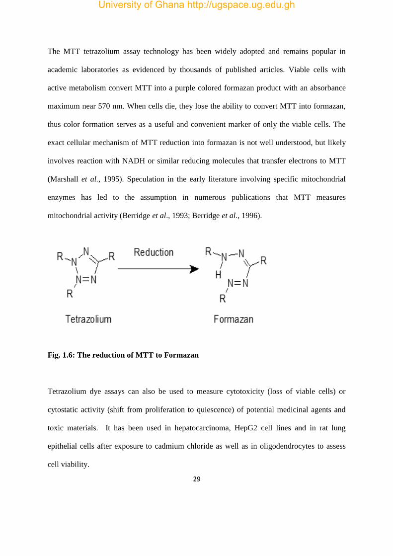
The MTT tetrazolium assay technology has been widely adopted and remains popular in
academic laboratories as evidenced by thousands of published articles. Viable cells with
active metabolism convert MTT into a purple colored formazan product with an absorbance
maximum near 570 nm. When cells die, they lose the ability to convert MTT into formazan,
thus color formation serves as a useful and convenient marker of only the viable cells. The
exact cellular mechanism of MTT reduction into formazan is not well understood, but likely
involves reaction with NADH or similar reducing molecules that transfer electrons to MTT
(Marshall et al., 1995). Speculation in the early literature involving specific mitochondrial
enzymes has led to the assumption in numerous publications that MTT measures
mitochondrial activity (Berridge et al., 1993; Berridge et al., 1996).
Fig. 1.6: The reduction of MTT to Formazan
Tetrazolium dye assays can also be used to measure cytotoxicity (loss of viable cells) or
cytostatic activity (shift from proliferation to quiescence) of potential medicinal agents and
toxic materials. It has been used in hepatocarcinoma, HepG2 cell lines and in rat lung
epithelial cells after exposure to cadmium chloride as well as in oligodendrocytes to assess
cell viability.
University of Ghana http://ugspace.ug.edu.gh

30
1.2.12 2, 2-diphenyl-1-picrylhydrazyl (DPPH) Assay
Determination of antioxidant activity of various types of foods using DPPH is comparable to
other methods. It is very difficult to select a suitable antioxidant assay method since anti-
oxidants act by several mechanisms and no one assay can capture the different modes of
action of antioxidant. Conventional cuvette assay of radical scavenging activity has been
replaced by 96-well plate microtitre assay for the past couple of years. Cuvette assay method
uses UV-Visible spectrophotometer to obtain the absorbance, where as 96-well plate method
uses ELISA plate reader for absorbance. The first method (cuvette assay) is very tedious,
time consuming and allows only one sample to be read at a time and requires high quantity of
reagent; whereas the second method (96 – well plate micro litre) is time saving and reads
about 96 samples at a time, with small amounts of reagent.
There are several advantages of the use of DPPH assay over other methods. These include
DPPH is allowed to react with the whole sample and sufficient time is given to allow
DPPH to react slowly even with weak anti-oxidants.
DPPH method may be utilized in aqueous and non-polar organic solvents and can be
used to examine both hydrophilic and lipophilic antioxidants.
DPPH assay is considered a valid accurate, easy and economic method for evaluation
of radical scavenging activity of antioxidants, since the radical compound is stable
and need not be generated.
1.2.13 Reducing Power
Reducing power is associated with antioxidant activity and may serve as a significant
reflection of the antioxidant activity (Denizot and Lang, 1986). Compounds with reducing
University of Ghana http://ugspace.ug.edu.gh

31
power indicate that they are electron donors and can reduce the oxidized intermediates of
lipid peroxidation processes, so that they can act as primary and secondary antioxidants
(Plumb et al., 1989). In this assay, the yellow colour of the test solution changes to various
shades of green and blue depending on the reducing power of each compound. Presence of
reducers causes the conversion of the Fe3+/ferricyanide complex used in this method to the
ferrous form. By measuring the formation of Pearl’s Prussian blue at 700 nm, it is possible to
determine the concentration of Fe3+ ion.
University of Ghana http://ugspace.ug.edu.gh

32
CHAPTER TWO
2.0 MATERIALS AND METHODS
2.1 MATERIALS
2.1.1 Chemicals and Reagents
RPMI-1640 culture medium, absolute isopropanol, and trypan blue solutions were obtained
from Sigma Aldrich Chemicals Co., Ltd. (St. Louis, MO, USA). Butylated hydroxytoluene
(BHT) and 1, 1-diphenyl-2-picryl hydrazyl (DPPH) free radical reagents were purchased
from Wako Pure Chemical Industries Ltd (Japan).
2.1.2 Kombucha Starter Culture
Kombucha starter culture was provided by Rev. Dr. W.S.K. Gbewonyo of the department of
biochemistry, cell and molecular biology, University of Ghana, Legon.
2.1.3 Leukemia cell line
Jurkat P9 cells were obtained from Shanghai Institute of Materia Medica, Chinese Academy
of Sciences (Shangai, China).
2.2 METHODS
2.2.1 Preparation of Tea Kombucha
Tea kombucha was prepared according to the method by Malbaša et al. (2011).
Sugared-water was prepared with 40 grams sugar (sucrose) per litre of boiled drinkable
water. The boiled sugared-water was then infused with one bag of Lipton tea (thread hanging
outside) and boiled again for 2 minutes after which the tea bag was removed. The boiled
University of Ghana http://ugspace.ug.edu.gh

33
sugared-tea was transferred into a clean transparent plastic container and allowed to cool to
room temperature. It was inoculated with 3% (w/v) of freshly grown tea fungus that had been
cultured in the same medium for 14 days and 10% (v/v) of previously fermented liquid tea
broth aseptically. The fermentation container was then covered, leaving a little opening for
aeration, which is required for proper fermentation of the tea. The set up was then kept in a
clean environment and left to grow in the dark at room temperature over a period of 14 days.
2.2.2 Freeze-Drying of Tea Kombucha
About 600 ml each of tea kombucha and unfermented tea were freeze-dried. The beverage
was transferred into small containers (150 ml) and placed in a Super Modulyo freeze-drying
machine (Thermo Scientific, USA) for 7 days to obtain dried extracts. The extracts were
weighed and stored in a cool dry place in the dark. They were reconstituted in distilled water
before use.
2.2.3 MTT Assay for Cell Viability
Cell viability was determined according to the method of Ayisi et al., (1991). This assay
measures the reduction of yellow MTT by succinate dehydrogenase to an insoluble purple
formazan product. Aliquots (100 µl) of Jurkat P9 leukemia cells containing 1 x 10⁵ cells/ml
were added to each well of a 96-well flat-microtiter plate and incubated (24 hours at room
temperature) with various concentrations of two-fold dilution of both the tea kombucha and
unfermented tea extract (highest concentration:1000 μg/ml). Three replicates were used for
each concentration in the experiment. After 24 hour incubation, 20 µl of MTT was added to
each well, mixed and then incubated for 4 hours at room temperature. A 150 µl of acidified
University of Ghana http://ugspace.ug.edu.gh

34
isopropanol was added. After an overnight incubation at 37ºC, absorbance was measured at
570 nm using a spectrophotometer. The inhibition rates were calculated as follows:
% inhibition = Mean absorbance of treated sample- Mean absorbance of c.c × 100%
Mean absorbance of untreated sample –Mean absorbance of c.c
c.c means colour control
2.2.4 Antioxidant Determination using DPPH Assay
The antioxidant activity was determined using the DPPH free radical scavenging assay
described by Brand-Williams et al. (1995). The tea kombucha or unfermented tea was diluted
in distilled water to a final concentration of 0.5 mg/ml. A 100 µl of 0.5 mM methanolic
DPPH was added to 100 µl of extract. This was done in triplicate and the reaction mixture
incubated in the dark for 20 minutes. The absorbance was measured at 517 nm using
spectrophotometer. Butylated hydroxyltuolene (BHT) served as a positive standard control,
and absolute methanol as the blank. Percentage antioxidant activity was determined as:
% Antioxidant Activity= (Absorbance of blank well-(Abs. of sample - Abs. of c.c)) × 100%
Absorbance of Blank
c.c means colour control and Abs. means Absorbance
2.2.5 Reducing Power
This is an assay that measures the reduction of Fe3+ to Fe2+ as described by Plumb et al.
(1989). Various concentrations (0-5 mg/ml) of both tea kombucha and unfermented tea
extracts (200 µl each) were mixed with 0.2 M phosphate buffer (2.5 ml) and 0.5 M potassium
ferricyanide (200 µl). The mixture was kept at 50°C in a water bath for 20 minutes. After
University of Ghana http://ugspace.ug.edu.gh

35
cooling, 200 µl of 10% (w/v) trichloroacetic acid was added and centrifuged at 3000 rpm for
10 minutes. The upper layer of solution (200 µl) was mixed with distilled water (200 µl) and
a freshly prepared 0.1M ferric chloride solution (40 µl) and incubated for 30 minutes at room
temperature. The absorbance was measured at 700 nm. Ascorbic acid at various
concentrations (1-5 mg/ml) was used as standard.
2.2.6 Total Phenolic Content
It was evaluated using a modified colorimetric method described previously by Singleton and
Rossi (1965). An Aliquot of 10 µl of tea kombucha/unfermented tea or gallic acid (as
calibration standard) was placed into an Eppendorf tube. Distilled water (0.79 ml) was then
added. After that 50 µl of Folin-Ciocalteau reagent was added and mixed thoroughly. The
mixture was then incubated at room temperature for 8 min. Sodium carbonate (20 g/100ml)
solution (150 µl) was added, mixed and incubated at room temperature for 2 hours. An
aliquot of 100 µl was placed into a 96 – well plate. The absorbance was measured at 750 nm
using a plate reader.
2.2.7 Statistical Analysis:
The statistical analysis was carried out using SPSS for Windows version 10.0 statistical
software (SPSS Inc, Chicago, USA). Statistically significant differences between the groups
were compared using one-way analysis of variance (ANOVA). The data are displayed as
means ± standard deviation (SD) and p-values less than 0.05 are considered “statistically
significant”.
University of Ghana http://ugspace.ug.edu.gh

36
CHAPTER THREE
3.0 RESULTS
3.1 Cell Viability
Curcumin, the standard positive control, showed decreasing % cell viability with increasing
concentration (Fig. 3.1A). However, the cell viability of the tea kombucha and the
unfermented tea used as normal control, increased slightly at lower concentration to about
120 % but slightly decreased upon increasing the concentration albeit to a higher degree in
the tea kombucha. Curcumin had an IC50 value of 7.22 mg/ml while that of the tea kombucha
and unfermented tea could not be determined within the concentration range tested (since the
% cell viability was above 70% at the highest concentration of 1000 µM). Curcumin showed
cytotoxic effect but the tea kombucha and the unfermented tea showed no cytotoxic effects
on Jurkat cells (Fig. 3.1).
University of Ghana http://ugspace.ug.edu.gh

37
CONCENTRATION (mg/ml)
(A)
(B)
Fig. 3. 1: Effect of varying concentrations of curcumin (A), tea kombucha and unfermented
tea (B) on viability of Jurkat P9 leukemia cells. Results are means of ± SEM of n= 3
IC50 = 7.22 mg/ml
University of Ghana http://ugspace.ug.edu.gh

38
3.2 DPPH Antioxidant Activity
DPPH free radical-scavenging ability of the tea kombucha (TK) was measured together with
butylated hydroxytoluene (BHT) was used as a standard positive control and unfermented tea
(BT) as a normal control (Fig. 3.2). The plot of concentration against % antioxidant activity
gave sigmoid curves from which EC50 values were obtained.
University of Ghana http://ugspace.ug.edu.gh

39
(A)
(B)
Fig. 3.2: Antioxidant activity of varying concentrations of BHT (A), tea kombucha and
unfermented tea (B). Results are means of ± SEM of n= 3
EC50 = 0.148 mg/ml
University of Ghana http://ugspace.ug.edu.gh

40
3.3 Total Phenolic Content
The total phenolic content of the tea kombucha (TK) and the unfermented tea (BT) increased
with increasing concentration (Fig. 3.3). At a concentration of 2.5 mg/ml the total phenolic
content expressed as mg/100 mg gallic acid equivalent (GAE) was 1016.0 and 139.2 for TK
and BT, respectively; and at 5 mg/ml the total phenolic content was 1401.1 and 577.6 for TK
and BT, respectively. Thus at extract concentration of 2.5 mg/ml, the total phenolic content of
TK was 7-fold higher than the BT, whilst at 5.0 mg/ml total phenolic content of the TK was
2.5-fold higher than the BT.
Extract conc. 1= 2.5 mg/ml and 2= 5.0 mg/ml GAE = gallic acid equivalent
Fig. 3.3: Total phenolic content of tea kombucha (TK) and unfermented tea (BT) (A)
and calibration curve for gallic acid (B). Results are means ± SEM of n= 3 for (A) and
means of n=2 for (B). *value significantly different from BT control; p < 0.05; # value
significantly different from 2.5 mg/ml extract; p < 0.05
*
*
University of Ghana http://ugspace.ug.edu.gh

41
3.4 Reducing Power
The Fe3+ reducing power of the standard ascorbic acid reached a maximum at 1.0 mg/ml at a
mean absorbance of 0.70 while that of the tea kombucha reached a maximum at 20 mg/ml
with a mean absorbance of 0.70. The reducing power of the unfermented tea, however,
showed an absorbance of 0.5 at 20 mg/ml which continued to increase to about 0.70 at 40
mg/ml. Thus the tea kombucha has about 2-fold higher reducing power than the unfermented
tea (Fig. 3.4).
University of Ghana http://ugspace.ug.edu.gh

42
UNFERMENTED TEA
TEA KOMBUCHA
(A)
B)
Fig. 3.4: Reducing power of varying concentrations of ascorbic acid (A), tea kombucha
and unfermented tea (B). Results are means ± SEM of n=3
University of Ghana http://ugspace.ug.edu.gh

43
CHAPTER FOUR
4.0 DISCUSSION, CONCLUSION AND RECOMMENDATION
4.1 DISCUSSION
Cell-based assays are often used for screening collections of compounds to determine if the
test molecules have effects on cell proliferation or show direct cytotoxic effects that
eventually lead to cell death. Regardless of the type of cell-based assay being used, it is
important to know how many viable cells are remaining at the end of the experiment. There
are a variety of assay methods that can be used to estimate the number of viable eukaryotic
cells. In this study MTT assay was used.
The half maximal inhibitory concentration, IC50, for the tea samples compared to the
standard, curcumin, were very high. Thus, the tea samples showed very low inhibitory
activities against the growth of the Jurkat P9 leukemia cells. It is possible that much higher
concentrations of the samples may be required for inhibition. According to the U.S. Food and
Drug Administration and Kappa Laboratories, Miami, Florida, U.S.A. (1995) strong
cytotoxic agents must have IC50 values less than 30 µg/ml. The percentage cell viability at
the highest concentration tested for the tea kombucha was lower than that of the unfermented
tea (Fig. 3.1). This suggests that some cytotoxic substances may be produced as a result of
the fermentation process. The weak anti-proliferative activity of the tea samples against the
Jurkat P9 leukemia cells as compared to the standard curcumin, which showed strong
inhibitory effects, suggests that the samples are less likely to inhibit cell growth. This finding
supports the observations made by Vijayaraghavan et al. (2000) and Pauline et al. (2001) in
which tea kombucha displayed no significant toxicity.
University of Ghana http://ugspace.ug.edu.gh

44
Total antioxidant capacity of tea kombucha and unfermented tea were tested using the DPPH
free radical scavenging activity. The EC50 values of 1.19 mg/ml and 2.66 mg/ml for tea
kombucha and unfermented tea, respectively indicates that tea kombucha has a 2-fold
stronger antioxidant activity than the unfermented tea. This suggests that the tea kombucha
contained more antioxidants than the unfermented tea probably as a result of the fermentation
process. This corresponds to work done by Chu and Chen (2006) that examined tea
kombucha and established that total phenol content of all kombucha samples showed a linear
increase with fermentation time. And that its antioxidant activity correlates with its total
phenolic content. Antioxidants, especially polyphenols, have been found to be promising
agents against cervical cancer, including induction of apoptosis, growth arrest, inhibition of
DNA synthesis, and modulation of signal transduction pathway; additionally, polyphenols
can interfere with each stage of carcinogenesis initiation, promotion, and progression for the
prevention of cancer development (Aggarwal et al., 2004). Reducing power is associated
with antioxidant activity and may serve as a significant reflection of the antioxidant activity
of a compound (Denizot and Lang, 1986). Compounds with reducing power indicate that they
are electron donors and can reduce the oxidized intermediates of lipid peroxidation processes,
so that they can act as primary and secondary antioxidants (Plumb et al., 1989). In this assay,
there were more Fe3+ reducers in the tea kombucha than the unfermented tea (Fig. 3.3)
suggesting that the fermentation process caused the production of substances with reducing
power thus increasing antioxidant activity. This corresponds to findings by Jayabalan et al.
(2008) in which they reported on the free radical scavenging abilities of tea kombucha
prepared from green tea, black tea and tea waste.
University of Ghana http://ugspace.ug.edu.gh

45
The total phenolic content expressed as mg/ 100 g GAE at 2.5 mg/ml and 5.0 mg/ml of tea
extracts were 7.3 – fold and 2.4 – fold, respectively higher in tea kombucha than unfermented
tea (Fig. 3.3). This suggests that at either concentration of extracts the total phenolic content
of the tea kombucha was significantly higher than the unfermented tea. This corroborates
with the higher reducing power or radical scavenging activity of the tea kombucha vis-à-vis
the unfermented tea indicating that the total phenolic content may be responsible for
antioxidant activity expressed by both extracts (Vissoto et al., 2013). This was established by
Jayabalan et al. (2007) in which a highly pronounced increase of total phenol content in all
kombucha samples was recorded. Also Chu and Chen (2006) proved that the content was up
to 7.8 mM gallic acid equivalent (GAE; 15th day of fermentation) and only around 4 mM
GAE for black tea.
Tea kombucha showed a higher antioxidant activity compared to the unfermented tea due to
compounds such as glucuronic acids, polyphenols and other organic acids produced during
the fermentation process, hence its ability to shield the Jurkat P9 leukemia cells from
oxidative damage. The antioxidants present in the tea kombucha may mop up the oxyradicals
and neutralize their effects thereby protecting the cells from damage. This may explain why
the tea kombucha showed no cytotoxic effect on the Jurkat P9 leukemia cells (Jayabalan et
al., 2008).
4.2 CONCLUSION
Tea kombucha was observed to have higher antioxidant activity than unfermented tea and it
slightly reduced Jurkat P9 leukemia cell viability many orders of magnitude below that of the
standard drug, curcumin, a potent antioxidant, whilst the unfermented tea was without effect.
University of Ghana http://ugspace.ug.edu.gh

46
4.3 RECOMMENDATION
Tea kombucha extracts of different concentrations should be investigated compared to
unfermented tea extract.
The tea extracts concentrations exhibiting cytotoxicity should be tested in normal and
other cancer cell lines to establish selectivity in cytotoxicity.
The effects of the tea extracts on lipid peroxidation should be determined.
The apoptotic activity of the tea extracts should also be investigated.
University of Ghana http://ugspace.ug.edu.gh

47
REFERENCES
Aggarwal S, Takada Y, Singh S, Myers JN, Aggarwal BB. (2004). Inhibition of growth and
survival of human head and neck squamous cell carcinoma cells by curcumin via modulation
of nuclear factor-kB signaling. Int JCancer. 111:679-692.
Ashendel CL. (1995). Signal transduction and carcinogenesis. J. Nutr. 125, 6868-6918
Ayisi NK, Gupta SV, Qualtiere LF. (1991). Modified tetrazolium-based colorimetric method for
determining the activities of anti-HIV compounds. Journal of Virological Methods 33(3):
335-344.
Banerjee D, Hassarajani SA, Maity B, Narayan G, Bandyopadhyay SK, Chattopadhyay S. (2011).
Comparative healing property of kombucha tea and black tea against indomethacin-induced
gastric ulceration in mice: possible mechanism of action. Food Function 1:284–293.
Barclay L, Vinqvist M, Mukai K, Goto H, Hashimoto Y, Tokunaga A, Uno H. (2000). The
antioxidant mechanism of curcumin: classical methods are needed to determine
antioxidant mechanism and activity. Org. Lett. 2(18): 2841-2843.
Barltrop J, Owen T. (1991). 5-(3-carboxymethoxyphenyl)-2-(4,5-dimethylthiazoly)-3-(4-
sulfophenyl)tetrazolium, inner salt (MTS) and related analogs of 3-(4,5-dimethylthiazolyl)-
2,5-diphenyltetrazolium bromide (MTT) reducing to purple water-soluble formazans as cell-
viability indicators. Bioorg Med Chem Lett.; 1:611–614.
Berridge M, Tan A, McCoy K, Wang R. (1996). The Biochemical and Cellular Basis of Cell
Proliferation Assays that Use Tetrazolium Salts. Biochemica. ; 4:14–19.
Berridge MV, Tan AS. (1993). Characterization of the cellular reduction of 3-(4,5-dimethylthiazol-2-
yl)-2,5-diphenyltetrazolium bromide (MTT): subcellular localization, substrate dependence,
University of Ghana http://ugspace.ug.edu.gh
48
and involvement of mitochondrial electron transport in MTT reduction. Arch Biochem
Biophys. 303(2):474–482.
Bertino JR. (1997). Oncology, 24, S18-S23.
Bhattacharya S, Gachhui R, Sil PC. (2011). Hepatoprotective properties of kombucha tea against
TBHP-induced oxidative stress via suppression of mitochondria-dependent apoptosis.
Pathophysiology 18:221–234.
Blanc PJ. (1996). Characterization of the tea fungus metabolites. Biotechnol Lett 18:139–142.
Block G, Patterson B, Subar A. (1992). Fruit, vegetables and cancer prevention: A review of the
epidemiologic evidence. Nutr Cancer. 18: 1–29.
Bors W, Heller W, Michel C. (1996). Flavonoids and Polyphenols: chemistry and biology. In
Handbook of Antioxidants. 409: 36-40.
Brand-Williams W, Cuvelier M, Berset C. (1995). Lebensm Wiss Technol. 28: 25-30.
Cetojević-Simin DD, Bogdanovic GM, Cvetkovic DD, Velicanski AS. (2008). Antiproliferative and
antimicrobial activity of traditional kombucha and Satureja montana L. Kombucha. J BUON
133:395–401.
Chanda S, Dave R. (2009). In vitro models for antioxidant activity evaluation and some medicinal
plants possessing antioxidant properties: An overview. African Journal of Microbiology
Research. 3(13):981‐996.
Chen C, Liu BY. (2000). Changes in major components of tea fungus metabolites during prolonged
fermentation. J Appl Microbiol 89:834–839.
Chu SC, Chen C. (2006). Effects of origins and fermentation time on the antioxidant activities of
kombucha. Food Chem.98: 502-507.
University of Ghana http://ugspace.ug.edu.gh
49
Conney AH, Lu YP, Lou YR, Huang MT. (2002). Inhibitory effects of tea and caffeine on UV-
induced carcinogenesis: relationship to enhanced apoptosis and decreased tissue fat. Eur. J
Cancer Prev. 2:28–36.
Cragg GM and Newmann DJ. (2005). Plants as source of anticancer agents. J Ethnopharmacol 100:
72-79.
Cragg GM, Newmann DJ, Holbeck S. (2002). Natural products and derivatives as leads to cell cycle
pathway targets in cancer chemotherapy. Curent Cancer drug targets, 2, 279-308.
Cragg GM, Newmann DJ, Snader KM. (1997). Natural products in drug discovery and development.
Journal of Natural Products, 60, 52-60.
Denizot F, and Lang R. (1986). Rapid colorimetric assay for cell growth and survival. Modifications
to the tetrazolium dye procedure giving improved sensitivity and reliability. J. Immunol.
Meth.; 89:271–277.
Dipti P, Yogesh B, Kain AK, Pauline T, Anju B, Sairam M, Singh B, Mongia SS, Kumar GI,
Selvamurthy W. (2003). Lead-induced oxidative stress: beneficial effects of kombucha tea.
Biomed Environ Sci. 16:276–282.
Dufresne C, Farnworth E. (2000). Tea, kombucha, and health: a review. Food Res Int 33:409–421.
Dutta D, Gachhui R. (2007). Nitrogen-fixing and cellulose-producing Gluconacetobacter kombuchae
sp. nov., isolated from kombucha tea. Int J Syst Evol Microbiol. 57:353–357.
El-Taher EM. (2011). Kombucha: a new microbial phenomenon and industrial benefits. African J
Biol Sci 7:41–60.
Ernst E. (2003). Kombucha: a systematic review of the clinical evidence. Forsch Komplementarmed
Klass Naturheilkd 10:85–87.
University of Ghana http://ugspace.ug.edu.gh

50
Ertel A, Verghese A, Byers SW, Ochs M, Tozeren A. (2006). Pathway-specific differences between
tumor cell lines and normal and tumor tissue cells. Molecular Cancer 5: 55
Fang X, Fang L, Gou S, Chang L. (2013). Bioorganic and Medicinal Chemistry Letters, Elsevier.
Fernanda BJ, Daniela J. (2005). Toxicon, 45, 459-66.
Food and Drug Administration (1995). FDA cautions consumers on “Kombucha Mushroom Tea”
(News release). Department of Health and Human Services, Public Health Services, Food and
Drug Administration, Washington, DC.
Goh WN, Rosma A, Kaur B, Eazilah A, Karim AA, Bhat R. (2012a). Fermentation of black tea broth
(kombucha): I. Effects of sucrose concentration and fermentation time on the yield of
microbial cellulose. Int Food Res J 19:109–107.
Gordon MC and David J. (2005). Plants as a source of anti-cancer agents. Journal of
Ethnopharmacology. 100: 72
Greenwald P. (1996). Chemoprevention of cancer. Sci. Am. 275, 96-99.
Greenwalt CJ, Ledford RA, Steinkraus KH. (1998). Determination and characterization of the
antimicrobial activity of the fermented tea kombucha. LWT Food Sci Technol 31:291–296.
Grover P, Danadevi K, Mahboob M, Rozati R, Saleha Banu B, Rahman MF. (2003): Evaluation of
genetic damage in workers employed in pesticide production utilizing the comet assay.
Mutagenesis, 18, 201-205.
Harlin H, Gajewski TF. (2008). Diagnosis and treatment of Mycoplasma-contaminated cell cultures.
Curr. Protoc. Cytom. 43, A.3C.1-A.3C.7
Hartmann AM, Burleson LE, Holmes AK, Geist CR. (2000). Effects of chronic kombucha ingestion
on open-field behaviors, longevity, appetitive behaviors, and organs in C57-BL/6 mice: a
pilot study. Nutrition 16:755–761.
University of Ghana http://ugspace.ug.edu.gh

51
Helbock H, Beckman K, Shigenaga M, Walter P, Woodall A, Yeo H, Ames B. (1998). DNA
oxidation matters: The HPLC-electrochemical detection assay of 8-
oxodeoxyguanosine and 8-oxo-guanine. Proc Natl Acad Sci. 95: 288–293.
Huang MT, Newmar HL, Fenkel K. (1997). Inhibitory effects of curcumin on tumorigenesis in mice.
J Cell Biochem;27:26–34.
Jayabalan R, Chen PN, Hsieh YS, Prabhakaran K, Pitchai P, Marimuthu S, Thangaraj P,
Swaminathan K, Yun SE. (2011). Effect of solvent fractions of kombucha tea on viability and
invasiveness of cancer cells—characterization of dimethyl 2-(2-hydroxy-2-
methoxypropylidine) malonate and vitexin. Indian J Biotechnol 10:75–82.
Jayabalan R, Marimuthu S, Thangaraj P, Sathishkumar M, Binupriya AR, Swaminathan K, Sei EY.
(2008). Preservation of kombucha tea effect of temperature on tea components and free
radical scavenging properties. J Agri Food Chem 56:9064–9071.
Jayabalan R, Marimuthu S, Swaminathan K. (2007). Changes in content of organic acids and tea
polyphenols during kombucha tea fermentation. Food Chem 102:392–398.
Kappor LD. (1990). CRC Handbook of Ayurvedic medicinal plants. Boca Raton, Florida, CRC
Press, 416-417.
Khar A, Ali AM, Pardhasaradhi BVV, Varalakshmi CH, Anjum R, Kumari AL. (2001). Induction of
stress response renders human tumor cell lines resistant to curcumin-mediated apoptosis: role
of reactive oxygen intermediates. Cell Stress Chaperones; 6:368–76.
Kumar SD, Narayan G, Hassarajani S. (2008). Determination of anionic minerals in black and
kombucha tea using ion chromatography. Food Chem 111:784–788.
Kuo ML, Huang TS, Lin JK. (1996). Curcumin, an antioxidant and antitumor promoter, induces
apoptosis in human leukemia cells. Biochem Biophys Acta ;1317:95–100.
University of Ghana http://ugspace.ug.edu.gh

52
Lambert JD, Yang CS. (2003). Cancer Chemopreventive activity and bioavailability of tea and tea
polyphenols. Mutat Res. 523-524, 201-208
Lončar ES, Petrović SE, Malbaša RV, Verac RM. (2000). Biosynthesis of glucuronic acid by means
of tea fungus. Nahrung 44:138–9.
Malbaša RV, Lončar ES, Vitas JS, Čanadanović-Brunet JM. (2011). Influence of starter cultures on
the antioxidant activity of kombucha beverage. Food Chem 127:1727–1731.
Malbaša RV. (2004). Investigation of antioxidant activity of beverage from tea fungus fermentation
[Ph.D. Thesis], University of Novi Sad, Faculty of Technology, Novi Sad, Serbia.
Manson MM. (2003). Cancer prevention: the potential for diet to modulate molecular signalling.
Trends Mol. Med. 9, 11-18.
Marshall NJ, Goodwin CJ, Holt SJ. (1995). A critical assessment of the use of microculture
tetrazolium assays to measure cell growth and function. Growth Regul. 5(2):69–84.
Mohandas K, Desai D. (1999). Epidemiology of digestive tract cancers in India. V. Large and small
bowel. Indian J Gastroenterol. 18:118-121
Mosmann, T. (1983). Rapid colorimetric assay for cellular growth and survival: Application to
proliferation and cytotoxicity assays. J. Immunol. Meth. ; 65:55–63.
Mouria M. (2002). Food-derived polyphenols inhibit pancreatic cancer growth through
mitochondrial cytochrome C release and apoptosis. Int. J. Cancer; 98: 761-769
Mrdanovic J, Bogdanovic G, Cvetkovic D, Velicanski A, Cetojevic-Simin D. (2007). The frequency
of sister chromatid exchange and micronuclei in evaluation of cytogenetic activity of
Kombucha on human peripheral blood lymphocytes. Arch. Oncol., 15, 85-88.
Parkin, D.(2006) .The global health burden of infection-associated cancers in the year 2002. Int J
Cancer. 118:3030-3044.
University of Ghana http://ugspace.ug.edu.gh

53
Patterson RE, White E, Kristal AR. (1997). Vitamin supplements and cancer risk: the epidemiologic
evidence. Cancer Causes and Control; 8(5):786-802.
Pauline T, Dipti P, Anju B, Kavimani S, Sharma SK, Kain AK, Sarada SK, Sairam M, Ilavazhagan
G, Devendra K, Selvamurthy W. (2001). Studies on toxicity, anti-stress and hepato-protective
properties of kombucha tea. Biomed Environ Sci 14:207–213.
Pinar R. (1998). To investigate knowledge of Turkish people about cancer. Cancer agenda, 2, 66-73.
Piwocka K, Jaruga E, Skierski J, Gradzka I, Sikora E. (2001). Effect of glutathione depletion on
caspase-3 independent apoptosis pathway induced by curcumin in Jurkat cells. Free Radic
Biol Med; 31:670–8.
Plumb JA, Milroy R, Kaye SB. (1989). Effects of the pH dependence of 3-(4,5-dimethylthiazol-2-
yl)-2,5-diphenyl-tetrazolium bromide-formazan absorption on chemosensitivity determined
by a novel tetrazolium-based assay. Cancer Res 15; 49(16):4435-4440.
Reiss J. (1994). Influence of different sugars on the metabolism of the tea fungus. Z Lebensm Unters
For 198: 258-261.
Sai Ram M, Anju B, Pauline T, Dipti P, Kain AK, Mongia SS, Sharma SK, Singh B, Singh R,
Ilavazhagan G, Kumar D, Selvamurthy W. (2000). Effect of kombucha tea on chromate (VI)-
induced oxidative stress in albino rats. J Ethnopharma 71:235–240.
Samuelsson G. (1999). Drugs of natural origin: a textbook of Pharmacognosy. 4th edition,
Stockholm, Swedish Pharmaceutical Press. Plant Physiol 124: 507-514.
Shenoy KC. (2000). Hypoglycemic activity of bio-tea in mice. Indian J Exp Biol 38:278–279.
Singleton V and Rossi J. (1965). Colorimetry of total phenolics with phosphomolybdic
phosphotungstic acid reagents. American Journal of Enology and Viticulture 16:144-158.
University of Ghana http://ugspace.ug.edu.gh

54
Sreeramulu G, Zhu Y, Knol W. (2000). Kombucha fermentation and its antimicrobial activity. J Agri
Food Chem 48:2589–2594.
Srihari T, Karthikesan K, Ashokkumar N, Satyanarayana U. (2013b). Antihyperglycaemic efficacy
of kombucha in streptozotocin-induced rats. J. Funct. Foods 3:1794–802.
Stoner GD, Mukhtar H. (1995). Polyphenols as cancer chemopreventive agents. J Cell Biochem
22(supp 1): 169-180.
Turgay S and Sar D. (2005). Plants as a source of anticancer agents. Turkistan, Premethodol vol:
436-441.
Vijayaraghavan R, Singh M, Rao P, Bhattacharya R, Kumar P, Sugendran K. (2000). Biomed.
Environ. Sci.13: 293-299.
Vissotto LC, Rodrigues E, Chisté RC, Benassi MT, Mercadante AZ. (2013). Correlation, by
multivariate statistical analysis, between the scavenging capacity against reactive oxygen
species and the bioactive compounds from frozen fruit pulps. Ciência e Tecnologia de
Alimentos, 33(Supl. 1), 57-65.
Wang H, Provan G, Helliwell K. (2010). Tea flavonoids, their functions, utilization and
analysis. Food Sci. Tech. 11: 152–160.
Worsham M, Raju U, Lu M. (2009). Risk factors for breast cancer from benign breast disease in
a diverse population. Breast Cancer Res Treat. 118(1):1-7.
Yang Z, Zhou F, Ji B, Li B, Luo Y, Yang L, Li T. (2010). Symbiosis between microorganisms from
kombucha and kefir: Potential significance to the enhancement of kombucha function.
Applied Biochemistry and Biotechnology, 160(2), 446-455.
University of Ghana http://ugspace.ug.edu.gh

55
Ziech D, Franco R, Georgkalikas A, Georgakila S, Malamou-Mitsi V, Schoneveld O.
(2010). The role of reactive oxygen species and oxidative stress in environmental
carcinogenesis and biomarker development. Chem Biol. Interact.188:334–339.
APPENDIX
CALIBRATION CURVE FOR GALLIC ACID
Gallic acid (calibration standards)
(a) Stock [5 mg/ml]
Gallic acid (50 mg) was dissolved in 1 ml of absolute ethanol. Distilled water was
then added to dilute it to 10ml. Solution was then stored at 4 oC.
(b) Dilutions
(c) Serial dilutions of stock using distilled water were done to obtain concentrations for
calibration curve.
0.5, 0.25, 0.125
University of Ghana http://ugspace.ug.edu.gh