Embed Size (px)
Citation preview

Rockefeller UniversityDigital Commons @ RU
Student Theses and Dissertations
2014
Studies of a Novel Phage Lytic Enzyme, PlySs2Daniel B. Gilmer
Follow this and additional works at: http://digitalcommons.rockefeller.edu/student_theses_and_dissertations
Part of the Life Sciences Commons
This Thesis is brought to you for free and open access by Digital Commons @ RU. It has been accepted for inclusion in Student Theses andDissertations by an authorized administrator of Digital Commons @ RU. For more information, please contact [email protected].
Recommended CitationGilmer, Daniel B., "Studies of a Novel Phage Lytic Enzyme, PlySs2" (2014). Student Theses and Dissertations. Paper 214.

STUDIES OF A NOVEL PHAGE LYTIC ENZYME, PlySs2
A Thesis Presented to the Faculty of
The Rockefeller University
in Partial Fulfillment of the Requirements for
the Degree of Doctor of Philosophy
by
Daniel B. Gilmer
June 2014

© Copyright by Daniel B. Gilmer 2014

STUDIES OF A NOVEL PHAGE LYTIC ENZYME, PlySs2
Daniel B. Gilmer, Ph.D.
The Rockefeller University 2014
Streptococcus suis infects pigs worldwide and may be zoonotically transmitted to
humans with a mortality rate of up to 20%. Methicillin-resistant Staphylococcus aureus
(MRSA) and Streptococcus pyogenes (group A streptococci – GrAS) cause potentially
fatal human diseases. These are just three of the many Gram-positive pathogens for
which resistance to leading antibiotics has emerged. The goal of this work was to develop
a novel antimicrobial treatment to combat these and other antibiotic-resistant pathogens.
We identified a novel bacteriophage lysin, derived from an S. suis phage termed
PlySs2 (phage lysin from S. suis 2). This thesis is divided into four main sections
detailing PlySs2: characterization (chapter 2); activity against S. suis (chapter 3); broad
lytic activity (chapter 4); and efficacy in vivo (chapter 5).
PlySs2 has an N-terminal CHAP catalytic domain and a C-terminal SH3b binding
domain. It is stable at 50°C for 30 min, 37°C for >24 h, 4°C for 15 days, and -80°C for
>7 months; it maintained full activity after 10 freeze-thaw cycles.
PlySs2 displays potent lytic activity against most strains of S. suis including the
type strain S735, the pathogenic serotype 2, strain 10, and the pathogenic serotype 9
strain 7997. At 64 μg/ml, PlySs2 reduced multiple strains of S. suis by 6-logs within 1
hour in vitro. PlySs2 exhibited a minimum inhibitory concentration (MIC) of 32 μg/ml
for S. suis strain S735 and 64 μg/ml for strain 7997. While resistance to gentamicin was
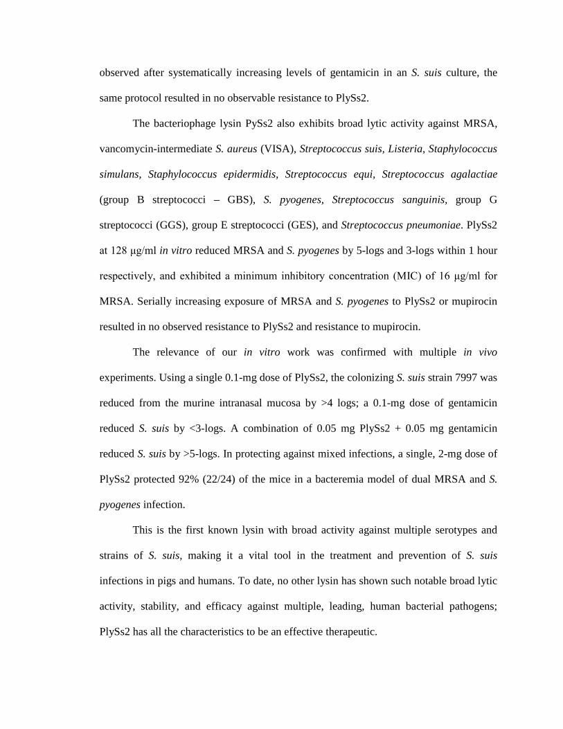
observed after systematically increasing levels of gentamicin in an S. suis culture, the
same protocol resulted in no observable resistance to PlySs2.
The bacteriophage lysin PySs2 also exhibits broad lytic activity against MRSA,
vancomycin-intermediate S. aureus (VISA), Streptococcus suis, Listeria, Staphylococcus
simulans, Staphylococcus epidermidis, Streptococcus equi, Streptococcus agalactiae
(group B streptococci – GBS), S. pyogenes, Streptococcus sanguinis, group G
streptococci (GGS), group E streptococci (GES), and Streptococcus pneumoniae. PlySs2
at 128 μg/ml in vitro reduced MRSA and S. pyogenes by 5-logs and 3-logs within 1 hour
respectively, and exhibited a minimum inhibitory concentration (MIC) of 16 μg/ml for
MRSA. Serially increasing exposure of MRSA and S. pyogenes to PlySs2 or mupirocin
resulted in no observed resistance to PlySs2 and resistance to mupirocin.
The relevance of our in vitro work was confirmed with multiple in vivo
experiments. Using a single 0.1-mg dose of PlySs2, the colonizing S. suis strain 7997 was
reduced from the murine intranasal mucosa by >4 logs; a 0.1-mg dose of gentamicin
reduced S. suis by <3-logs. A combination of 0.05 mg PlySs2 + 0.05 mg gentamicin
reduced S. suis by >5-logs. In protecting against mixed infections, a single, 2-mg dose of
PlySs2 protected 92% (22/24) of the mice in a bacteremia model of dual MRSA and S.
pyogenes infection.
This is the first known lysin with broad activity against multiple serotypes and
strains of S. suis, making it a vital tool in the treatment and prevention of S. suis
infections in pigs and humans. To date, no other lysin has shown such notable broad lytic
activity, stability, and efficacy against multiple, leading, human bacterial pathogens;
PlySs2 has all the characteristics to be an effective therapeutic.

iii
To my wife, Chanel
To my children
To my parents

iv
ACKNOWLEDGEMENTS
I am especially grateful to my advisor and mentor, Dr. Vincent Fischetti. He fully
empowered me to freely execute all of the work in this thesis. As a mentor, he guided
with patience, while always demanding the highest standard of scientific investigation. In
addition, he valued my nonscientific endeavors, which afforded me an exceptionally
fulfilling graduate experience.
I am grateful to my faculty advisory committee, comprised of Dr. Erec Stebbins
and Dr. Howard Hang, for their wise insights and consistent guidance. Also, I am
thankful for Dr. David Donovan graciously accepting a position on my committee as the
external examiner.
I am incredibly thankful for Dr. Jonathan Schmitz. From my first day in the
laboratory, he helped me launch my thesis project – even while writing his own thesis
during my first months. Through the years, Dr. Chad Euler provided invaluable
contributions to the in vivo work. I am also grateful for Nathan Franck for his excellent
technical expertise. Dennis Spencer was both a sounding board and friend in times of
uncertainty. I was amazed by each of the excellent summer students I mentored here at
Rockefeller. Magid Mohamed, Khoi Nguyen, Karen Tong, and Haaris Khan each
performed at the level of a PhD candidate. I am indebted to all the members of the
Fischetti Laboratory who collectively established a collaborative, constructive
environment where I was comfortable proposing any study.
I was fortunate to work with Dr. Jaap Wagenaar and Dr. Niels Dekker of Utrecth
University, as well as Dr. Sünje Pamp and Dr. David Relman of Stanford University in
collaborations. It has been an honor to work with Dr. Raymond Schuch of ContraFect

v
Corporation, and I am eager to see how this scientific story continues to develop. I
highlight specific, scientific contributions at the end of each chapter.
I am thankful to the Rockefeller community for providing: scientific expertise; a
comfortable living environment; and safety for my family. This was largely due to my
exceptional fellow coworkers in housing, security, the mailroom, and the Child and
Family Center. In the Dean’s Office, Dr. Sidney Strickland, Dr. Emily Harms, Marta
Delgado, Kristen Cullen, Cris Rosario, Courtney McBride, and Stephanie Fernandez have
been incredibly supportive from my application to the program through my thesis
defense, enabling me to focus solely on science. To my friends throughout New York
City, I am thankful for how you have helped me view my work in ways I had never
considered.
This thesis is the culmination of education spanning three decades. I would never
have sought a PhD, if it had not been for Mr. Terry Nusbaum teaching me biology in high
school years ago. At Howard University, Dr. Winston Anderson inspired me to make
discoveries that had never been made; potentially benefitting thousands. Dr. Cathy
Drennan of Massachusetts Institute of Technology revealed the full process of science to
me – from writing grants to reviewing others’ publications.
I am grateful to the Howard Hughes Medical Institute (HHMI) Gilliam fellowship
for funding all five years of my graduate study. The supplemental training and
networking at HHMI conferences immeasurably augmented the generous financial
support. This work was also made possible by multiple grants from the US National
Institutes of Health.

vi
Most of all, I would like to thank my wife, Chanel Gilmer, who has journeyed
with me through every day of this doctoral work. From applying to graduate school to
writing my thesis, she has been an unwavering source of passionate encouragement and
insightful perspective. My children have been wonderful joys throughout my study
bringing meaning to my every day. And finally, I am forever indebted to my parents who
taught me that my purpose comes before my profession in light of Matthew 6:33.

vii
TABLE OF CONTENTS
DEDICATION……..…………………………………………………………………....iii
ACKNOWLEDGEMENTS ............................................................................................ iv
TABLE OF CONTENTS ............................................................................................... vii
LIST OF FIGURES ....................................................................................................... xiv
LIST OF TABLES ......................................................................................................... xvi
LIST OF ABBREVIATIONS ...................................................................................... xvii
1 CHAPTER 1 – INTRODUCTION ............................................................................ 1
1.1 Gram-positive pathogens ....................................................................................... 1
1.1.1 Streptococcus suis ........................................................................................... 1
1.1.1.1 Pathogenesis ............................................................................................. 1
1.1.1.2 Zoonosis ................................................................................................... 2
1.1.2 Streptococcus pyogenes - GrAS ...................................................................... 4
1.1.2.1 Colonization ............................................................................................. 4
1.1.2.2 Pathogenesis ............................................................................................. 4
1.1.2.3 Mortality .................................................................................................. 5
1.1.3 Staphylococcus aureus .................................................................................... 5
1.1.3.1 Pathogenesis ............................................................................................. 5
1.1.3.2 Transmission ............................................................................................ 6
1.1.4 Other Gram-positive pathogens ...................................................................... 6
1.1.4.1 Streptococcus agalactiae – GBS ............................................................. 7
1.1.4.2 Listeria monocytogenes ........................................................................... 7
1.2 Antibiotics .............................................................................................................. 8

viii
1.2.1 Treatment ........................................................................................................ 8
1.2.2 Resistance ....................................................................................................... 9
1.3 Phage Lytic Enzymes ........................................................................................... 13
1.3.1 Phage ............................................................................................................. 13
1.3.1.1 Life cycles .............................................................................................. 13
1.3.1.1.1 Lysogenic cycle .............................................................................. 14
1.3.1.1.2 Lytic cycle ....................................................................................... 14
1.3.1.2 Role in nature ......................................................................................... 15
1.3.1.3 Shaping molecular biology .................................................................... 15
1.3.1.4 Therapeutic application .......................................................................... 16
1.3.2 Lysins ............................................................................................................ 17
1.3.2.1 Discovery ............................................................................................... 18
1.3.2.2 Structure ................................................................................................. 20
1.3.2.3 Activity .................................................................................................. 20
1.3.2.4 Current Lysins ........................................................................................ 24
1.3.2.4.1 LySMP – S. suis phage enzyme ...................................................... 25
1.3.2.4.2 ClyS – staphylococcal phage enzyme ............................................. 27
1.3.2.4.3 PlyC – streptococcal phage lysin .................................................... 27
1.4 Peptidoglycan ....................................................................................................... 28
1.5 AIMS.................................................................................................................... 33
2 CHAPTER 2 – LYSIN CHARACTERIZATION .................................................. 34
2.1 MATERIALS AND METHODS ......................................................................... 34
2.1.1 Discovery ...................................................................................................... 34

ix
2.1.1.1 Genomic sequence analysis ................................................................... 34
2.1.1.2 Cloning ................................................................................................... 34
2.1.1.3 Candidate assay ...................................................................................... 34
2.1.2 Expression ..................................................................................................... 35
2.1.3 Purification .................................................................................................... 35
2.1.4 Characterization ............................................................................................ 36
2.1.4.1 Optimization .......................................................................................... 36
2.1.4.2 Stability .................................................................................................. 36
2.2 RESULTS ............................................................................................................ 37
2.2.1 Identification ................................................................................................. 37
2.2.2 Purification .................................................................................................... 40
2.2.3 Characterization ............................................................................................ 42
2.2.3.1 Optimization .......................................................................................... 42
2.2.3.2 Stability .................................................................................................. 48
2.3 ACKNOWLEDGEMENTS ................................................................................. 53
3 CHAPTER 3 – STREPTOCOCCUS SUIS SUSCEPTIBILITY ............................ 54
3.1 MATERIALS AND METHODS ......................................................................... 54
3.1.1 Bacterial strains ............................................................................................. 54
3.1.2 Lytic activity against S. suis.......................................................................... 57
3.1.3 Bactericidal assay.......................................................................................... 57
3.1.4 MIC assay ..................................................................................................... 58
3.1.5 Resistance ..................................................................................................... 58
3.2 RESULTS ............................................................................................................ 59

x
3.2.1 Lytic activity against S. suis.......................................................................... 59
3.2.2 Bactericidal assay.......................................................................................... 65
3.2.3 MIC assay ..................................................................................................... 67
3.2.4 Resistance ..................................................................................................... 69
3.3 ACKNOWLEDGEMENTS ................................................................................. 71
4 CHAPTER 4 – BROAD GRAM-POSITIVE SUSCEPTIBILITY ....................... 72
4.1 MATERIALS AND METHODS ......................................................................... 72
4.1.1 Bacterial strains ............................................................................................. 72
4.1.2 Lytic activity ................................................................................................. 78
4.1.3 Bactericidal assay.......................................................................................... 78
4.1.4 MIC assay ..................................................................................................... 78
4.1.5 Resistance ..................................................................................................... 79
4.1.6 PlySs2 catalytic domain lytic assay .............................................................. 79
4.1.7 Fluorescent binding assay ............................................................................. 80
4.2 RESULTS ............................................................................................................ 81
4.2.1 Lytic activity ................................................................................................. 81
4.2.2 Bactericidal assay.......................................................................................... 87
4.2.3 MIC Assay .................................................................................................... 90
4.2.4 Resistance ..................................................................................................... 92
4.2.5 PlySs2 catalytic domain lytic assay .............................................................. 94
4.2.6 Fluorescent binding assay ............................................................................. 96
4.3 ACKNOWLEDGEMENTS ................................................................................. 99
5 CHAPTER 5 – IN VIVO MODELS ...................................................................... 100

xi
5.1 MATERIALS AND METHODS ....................................................................... 100
5.1.1 Intranasal mucosa model............................................................................. 100
5.1.1.1 S. suis colonization............................................................................... 100
5.1.1.2 Treatment ............................................................................................. 100
5.1.2 Mixed bacteremia model............................................................................. 101
5.1.2.1 MRSA + GrAS Infection ..................................................................... 101
5.1.2.2 Treatment ............................................................................................. 101
5.2 RESULTS .......................................................................................................... 102
5.2.1 Intranasal mucosa decolonization ............................................................... 102
5.2.2 Mixed bacteremia protection ...................................................................... 105
5.3 ACKNOWLEDGEMENTS ............................................................................... 109
6 CHAPTER 6 – DISCUSSION................................................................................ 111
6.1 Lysin Characterization ....................................................................................... 111
6.1.1 Identification ............................................................................................... 111
6.1.2 Purification and Stability ............................................................................ 111
6.1.3 Catalytic Domain ........................................................................................ 112
6.1.4 Binding Domain .......................................................................................... 112
6.2 S. suis susceptibility to PlySs2 ........................................................................... 113
6.2.1 Lytic activity ............................................................................................... 113
6.2.2 Implications of lytic activity ....................................................................... 114
6.3 Broad Gram-positive susceptibility ................................................................... 115
6.3.1 Lysin specificity .......................................................................................... 115
6.3.2 Broad PlySs2 lytic activity.......................................................................... 116

xii
6.3.2.1 S. aureus susceptibility to PlySs2 ........................................................ 117
6.3.2.2 Streptococcal susceptibility to PlySs2 ................................................. 117
6.3.3 Broad bactericidal activity against streptococci and staphylococci ............ 118
6.4 Absence of in vitro resistance to PlySs2 ............................................................ 118
6.4.1 No observable in vitro resistance to PlySs2 in S. suis ................................ 118
6.4.2 No observable in vitro resistance to PlySs2 in MRSA or S. pyogenes ....... 119
6.5 Intranasal mucosa decolonization ...................................................................... 119
6.5.1 Lysin alone or with antibiotic ..................................................................... 120
6.5.2 Prophylaxis ................................................................................................. 121
6.6 Mixed bacteremia protection ............................................................................. 121
6.6.1 Implications................................................................................................. 122
6.6.2 PlySs2 applications ..................................................................................... 124
6.7 Other tests .......................................................................................................... 125
6.8 Importance ......................................................................................................... 125
7 CHAPTER 7 – CONCLUSIONS ........................................................................... 127
8 APPENDIX .............................................................................................................. 129
8.1 PlySs2 minipig protection from S. suis .............................................................. 129
8.1.1 BACKGROUND ........................................................................................ 129
8.1.2 MATERIALS AND METHODS ................................................................ 129
8.1.2.1 PlySs1 + PlySs2 preliminary drug interaction ..................................... 129
8.1.2.2 Minipig treatment................................................................................. 129
8.1.3 RESULTS ................................................................................................... 130
8.1.3.1 PlySs1 + PlySs2 preliminary drug interaction ..................................... 130

xiii
8.1.3.2 Minipig treatment................................................................................. 132
8.1.4 DISCUSSION ............................................................................................. 134
8.1.5 ACKNOWLEDGEMENTS ........................................................................ 134
8.2 Rat oral cavity metagenomics ............................................................................ 134
8.2.1 BACKGROUND ........................................................................................ 135
8.2.2 MATERIALS AND METHODS ................................................................ 135
8.2.3 RESULTS ................................................................................................... 137
8.2.4 DISCUSSION ............................................................................................. 141
8.2.5 ACKNOWLEDGEMENTS ........................................................................ 143
8.3 Structural studies ................................................................................................ 143
8.3.1 BACKGROUND ........................................................................................ 143
8.3.2 METHODS ................................................................................................. 143
8.3.3 RESULTS ................................................................................................... 144
8.3.4 ACKNOWLEDGEMENTS ........................................................................ 146
8.4 DNA AND AMINO ACID SEQUENCES ........................................................ 147
9 REFERENCES ........................................................................................................ 149

xiv
LIST OF FIGURES
Figure 1.1 Reports of individual human S. suis infections have recently increased. .......... 3
Figure 1.2 Periodic emergence of S. aureus antibiotic resistance. ................................... 10
Figure 1.3 New systemic antibacterial agents approved by the US FDA. ........................ 12
Figure 1.4 Lytic cycle versus lysin treatment. .................................................................. 22
Figure 1.5 Gram-positive cell wall cross-section diagram. .............................................. 29
Figure 1.6 Peptidoglycan molecular composition. ........................................................... 31
Figure 2.1 PlySs2 amino acid sequence and structure. ..................................................... 38
Figure 2.2 PlySs2 enzymatic domain alignment............................................................... 39
Figure 2.3 PlySs2 corresponded to bands at ~26 kDa on SDS-PAGE. ............................ 41
Figure 2.4 PlySs2 was found to have the most acute activity in basic pH levels. ............ 43
Figure 2.5 NaCl does not augment PlySs2 activity. ......................................................... 45
Figure 2.6 Dithiothreitol (DTT) does not inhibit PlySs2 activity. .................................... 46
Figure 2.7 Minimal ion depletion from EDTA inhibits PlySs2 activity. .......................... 47
Figure 2.8 PlySs2 is stable under a variety of conditions. ................................................ 49
Figure 2.9 PlySs2 is stable and active after ten, consecutive freeze-thaws. ..................... 52
Figure 3.1 PlySs2 lysed almost all strains of S. suis over 30 minutes. ............................. 60
Figure 3.2 PlySs2 lysed almost all strains of S. suis over 60 minutes. ............................. 61
Figure 3.3 S. suis strain S735 exposed to various concentrations of PlySs2. ................... 63
Figure 3.4 S. suis strain 7997 exposed to various concentrations of PlySs2. ................... 64
Figure 3.5 PlySs2 was bactericidal to nearly all strains of S. suis. ................................... 66
Figure 3.6 S. suis 7997 and S735 did not develop resistance to PlySs2 in vitro. ............. 70

xv
Figure 4.1 PlySs2 displayed activity against various species. .......................................... 83
Figure 4.2 PlySs2 displayed activity against various species over 60 minutes. ............... 85
Figure 4.3 PlySs2 was bactericidal across multiple species of bacteria. .......................... 89
Figure 4.4 MRSA, MSSA, and GrAS did not acquire resistance to PlySs2 in vitro. ....... 93
Figure 4.5 Lytic effect of PlySs2 Full Length VS Catalytic Domain ............................... 95
Figure 4.6 PlySs2-BD Flurescence and comparison to PlySs2-CD activity. ................... 97
Figure 5.1 PlySs2 and gentamicin may act additively to reduce S. suis in vivo. ............ 103
Figure 5.2 PlySs2 protected mice from mixed MRSA and GrAS infection. .................. 106
Figure 8.1 PlySs1 + PlySs2 drug interaction. ................................................................. 131
Figure 8.2 Pig deaths over two weeks. ........................................................................... 133
Figure 8.3 There were two predominant phyla in the oral cavity. .................................. 139
Figure 8.4 Crystal of PlySs2. .......................................................................................... 145

xvi
LIST OF TABLES
Table 1.1 Timeline of lysin discovery .............................................................................. 19
Table 3.1 Strains used in the S. suis susceptibility study .................................................. 55
Table 3.2 The MIC of PlySs2 for S. suis serotypes and strainsa ....................................... 68
Table 4.1 Strains used in this study .................................................................................. 73
Table 4.2 The MIC of PlySs2 for various Gram-positive speciesa ................................... 91

xvii
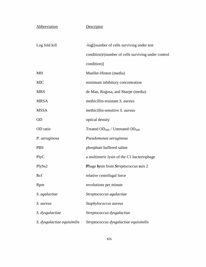
LIST OF ABBREVIATIONS
Abbreviation Descriptor
B. anthracis Bacillus anthracis
B. cereus Bacillus cereus
B. subtilis Bacillus subtilis
B. thuringiensis Bacillus thuringiensis
BCA bicinchoninic acid (assay)
BD binding domain
BHI brain heart infusion (media)
Buffer A 15 mM PB, pH 8.0
Buffer B 15 mM PB, pH 6.7
Buffer C 50 mM PB, pH 7.4
Buffer D
Buffer E
20 mM PB, pH 7.4
20 mM glycine-NaOH, pH 9.3
CA-MRSA community-acquired methicillin-resistant S. aureus
CD catalytic domain
CFU colony forming unit
CHAP cysteine-histidine amidohydrolase/peptidase (domain)
ClyS chimeric lysin for staphylococci
CM carboxymethyl

xviii
Abbreviation Descriptor
DEAE diethylaminoethyl
DTT dithiothreitol
E. coli Escherichia coli
E. faecalis Enterococcus faecalis
E. faecium Enterococcus faecium
EDTA ethylenediaminetetraacetic acid
FDA Food and Drug Administration (United States)
FL full-length (enzyme)
GBS Streptococcus agalactiae
GCS group C streptococci
GES group E streptococci
GGS group G streptococci
GrAS group A streptococci, S. pyogenes
kD kilodaltons
L. acidophilus Lactobacillus acidophilus
L. gasseri Lactobacillus gasseri
L. monocytogenes Listeria monocytogenes
L. rhamnosus Lactobacillus rhamnosus
LB Luria-Bertani (media)
xix
Abbreviation Descriptor
Log fold kill -log[(number of cells surviving under test
condition)/(number of cells surviving under control
condition)]
MH Mueller-Hinton (media)
MIC minimum inhibitory concentration
MRS de Man, Rogosa, and Sharpe (media)
MRSA methicillin-resistant S. aureus
MSSA methicillin-sensitive S. aureus
OD optical density
OD ratio Treated OD600 / Untreated OD600
P. aeruginosa Pseudomonas aeruginosa
PBS phosphate buffered saline
PlyC a multimeric lysin of the C1 bacteriophage
PlySs2 Phage lysin from Streptococcus suis 2
Rcf relative centrifugal force
Rpm revolutions per minute
S. agalactiae Streptococcus agalactiae
S. aureus Staphylococcus aureus
S. dysgalactiae Streptococcus dysgalactiae
S. dysgalactiae equisimilis Streptococcus dysgalactiae equisimilis

xx
Abbreviation Descriptor
S. epidermidis Staphylococcus epidermidis
S. equi Streptococcus equi
S. equi zooepidemicus Streptococcus equi zooepidemicus
S. gordonii Streptococcus gordonii
S. mutans Streptococcus mutans
S. oralis Streptococcus oralis
S. pneumoniae Streptococcus pneumoniae
S. pyogenes Streptococcus pyogenes
S. rattus Streptococcus rattus
S. sanguinis Streptococcus sanguinis
S. simulans Staphylococcus simulans
S. sobrinus Streptococcus sobrinus
S. suis Streptococcus suis
ST serotype
VISA vancomycin-intermediate S. aureus
VRSA vancomycin-resistant S. aureus
1
1 CHAPTER 1 – INTRODUCTION
1.1 Gram-positive pathogens
Gram-positive pathogens such as Streptococcus pyogenes (group A streptococci –
GrAS), Staphylococcus aureus, Streptococcus suis, Streptococcus agalactiae (group B
streptococci – GBS), and Listeria monocytogenes are responsible for millions of serious
and sometimes fatal infections worldwide. Additionally, resistance to conventional
antibiotics has been on the rise, resulting in increased rates of infection, morbidity,
mortality, and treatment costs. Consequently, new therapeutic methods need to be
developed to reduce the antibiotic pressure on these pathogens. Our laboratory studies
many different genus and species of these bacteria in hopes of finding novel therapeutics
1.1.1 Streptococcus suis
1.1.1.1 Pathogenesis
The zoonotic Gram-positive pathogen S. suis causes sepsis and meningitis in pigs
and humans. S. suis was first isolated from septicemic pigs and subsequently found to be
biochemically distinct from other streptococci (de Moor 1963). After recent
reclassification (Hill et al. 2005), there are 33 serotypes distinguished by variations in the
capsular polysaccharide synthesis (cps) locus (Liu et al. 2013). Serotype 2, strain 10 and
serotype 9, strain 7997 of S. suis have been most often associated with disease, with
strain 7997 causing an increasing proportion of the infections worldwide (Silva et al.
2006, Wu, Zhang, and Lu 2008, Gottschalk et al. 2010). Pigs intranasally colonized with
S. suis may transmit the pathogen to humans and piglets causing pneumonia, septicemia,
or meningitis within days (Dekker et al. 2013, Arends and Zanen 1988, Gottschalk et al.
2010). While the pig carriage rate can reach 100% (Rasmussen et al. 1999), only a subset

2
of pigs develop meningitis or septicemia.
1.1.1.2 Zoonosis
In particular, the serotype 9 strain 7997 has been associated with increasing
reports of zoonotic transmission from pigs to humans (Sriskandan and Slater 2006). The
earliest human case was reported in 1968; since then, over 700 human cases have been
reported in multiple continents with a 5-20% mortality rate (Figure 1.1) (Perch,
Kristjansen, and Skadhauge 1968, Sriskandan and Slater 2006, Yu et al. 2006, Trottier et
al. 1991). In a 2-month 2005 outbreak in China, among 203 human cases reported, the
median time from exposure to infection was 2.2 days, but ranged from 3 hours to 14 days
(Yu et al. 2006).

3
†The 215 cases officially reported during the 2005 outbreak in Sichuan Province and the
25 cases of 1998 outbreak in Jiangsu Province, both in China, were excluded from the
analysis to accurately follow the evolution of S. suis human disease.
‡Only 11 months of 2009 were included.
Figure 1.1 Reports of individual human S. suis infections have
recently increased.
Annual, confirmed, reported human cases of S. suis infection have increased over the past
decade. This may be due, in part, to increased reporting rather than an actual increase in
S. suis infection. Adapted from Future Microbiology (Gottschalk et al. 2010) with
permission of Future Medicine Ltd.

4
Humans and pigs can be topically and systemically treated with penicillin or
gentamicin with success, but S. suis isolates resistant to these antibiotics have emerged
(Cantin et al. 1992, Varela et al. 2013, Gottschalk et al. 2010). Further, there is no
vaccine for S. suis (Fittipaldi et al. 2012). As such, S. suis appears to be developing a
more consistent presence in human populations and these infections may become more
difficult to treat.
1.1.2 Streptococcus pyogenes - GrAS
1.1.2.1 Colonization
Streptococcus pyogenes is a group A beta hemolytic streptococci. Over 30% of the
human population may be colonized with Streptococcus pyogenes in the upper
respiratory tract – the only known site of benign colonization (Mandell et al. 2005).
Colonized individuals are much less likely than severely sick persons to transmit illness
(Mandell et al. 2005).
1.1.2.2 Pathogenesis
S. pyogenes annually infects over 750 million people (Carapetis et al. 2005),
resulting in 25% mortality among the ~650,000 cases that progress to severe infection
(Carapetis et al. 2005, Cunningham 2000, Bessen et al. 2011). This pathogen is
responsible for a broad range of infections such as pharyngitis, impetigo, scarlet fever,
erysipelas, cellulitis, toxic-shock syndrome, and necrotizing fasciitis; and it can lead to
serious sequelae such as rheumatic fever, and acute glomerulonephritis (Swedo et al.
1997, Parker, Tomlinson, and Williams 1955, Bisno, Brito, and Collins 2003).

5
1.1.2.3 Mortality
The mortality rates can be very high for these infections, including 20% for
necrotizing fasciitis, and 50% for toxic-shock syndrome (Bisno, Brito, and Collins 2003).
Rheumatic fever, acute glomerulonephritis, and forms of obsessive-compulsive disorder
are non-suppurative sequelae associated with an S. pyogenes infection (Swedo et al.
1997). Invasive streptococcal infections and rheumatic fever outbreaks have seen a rise
worldwide since the 1980’s (Kavey and Kaplan 1989).
1.1.3 Staphylococcus aureus
1.1.3.1 Pathogenesis
Of all the Gram-positive pathogens, Staphylococcus aureus has become the most
difficult to treat. S. aureus is a Gram-positive facultative anaerobe that causes most
Staphylococcus infections in man. Human anterior nares (nostrils) are typically the
primary sites of S. aureus colonization, along with other moist openings on the body
serving as additional sites for entry (White and Smith 1963, Kluytmans, van Belkum, and
Verbrugh 1997, von Eiff et al. 2001, Wertheim et al. 2005a).
S. aureus is capable of producing severe, secondary infections in
immunocompromised individuals, as well as causing disease in otherwise healthy people.
Besides skin and soft tissue infections (SSTIs), S. aureus can cause sepsis, pneumonia,
necrotizing fasciitis, pyomyositis, endocarditis, toxic shock syndrome, and scalded skin
syndrome (White and Smith 1963, Wertheim et al. 2005b). These infections are usually
treated with methicillin, mupirocin, or vancomycin. Unfortunately, many S. aureus
strains, such as methicillin-resistant S. aureus (MRSA) and (less often) vancomycin-
resistant S. aureus (VRSA), have acquired resistance to one or more antibiotics used as

6
standard treatment (Howden et al. 2010), making them even more difficult to treat with
available antimicrobials (Howden et al. 2010).
1.1.3.2 Transmission
Further exacerbating the problem, MRSA is readily transmitted between patients in
hospitals (Coates, Bax, and Coates 2009). MRSA account for more than 50% of hospital
isolates causing pneumonia and septicemia (Klein, Smith, and Laxminarayan 2007),
particularly in intensive care units, resulting in 30-40% mortality (Tiemersma et al.
2004, Laupland, Ross, and Gregson 2008). It is the primary cause of lower respiratory
tract infections, surgical site infections, and ~19,000 deaths/year in the US alone
(Fischetti 2008, Klein, Smith, and Laxminarayan 2007).
While health-care-associated MRSA strains usually infect susceptible patients,
community-associated MRSA (CA-MRSA) can infect healthy individuals (CDC
1999, Herold et al. 1998, Zetola et al. 2005, David and Daum 2010). CA-MRSA strains
are often more virulent and are capable of causing more severe diseases in humans and
animal models (Adem et al. 2005, Miller et al. 2005, Li et al. 2009, Voyich et al. 2005).
Distinct strains of CA-MRSA are epidemic in Europe, North America, Oceania, and
other regions (Herold et al. 1998, Vandenesch et al. 2003, Tristan et al. 2007). The MW2
strain (pulsed-field type USA400) is the prototypical CA-MRSA, having contributed to
the incipient outbreak of CA-MRSA in the USA, which led to an epidemic (CDC
1999, Deleo et al. 2010). There is currently no vaccine for S. aureus (Middleton 2008).
1.1.4 Other Gram-positive pathogens
Zoonotic Gram-positive pathogens include: Streptococcus equi (strangles – an
upper respiratory tract infection – in equines, e.g. horses); and Streptococcus suis (sepsis

7
and meningitis in pigs and humans). The pathogenic S. suis serotype 9 strain 7997 has
been associated with increasing reports of zoonotic transmission from pigs to humans
(Sriskandan and Slater 2006). Humans and pigs have been treated with penicillin or
gentamicin, but S. suis isolates resistant to these antibiotics exist (Cantin et al. 1992). S.
suis may develop a consistent presence in human populations in years to come.
There are many other Gram-positive human pathogens, including: Streptococcus
sanguinis (dental plaque and caries); S. sanguinis (endocarditis); Group G Streptococcus;
group E Streptococcus; and S. pneumoniae (pneumonia, otitis media, meningitis,
bacteremia, sepsis, endocarditis, peritonitis, and cellulitis).
1.1.4.1 Streptococcus agalactiae – GBS
Another beta-hemolytic Gram-positive streptococcus, Streptococcus agalactiae
(Group B streptococci – GBS) contains an antiphagocytic capsule as its primary virulence
factor (Yeung and Mattingly 1984, Rubens et al. 1987). S. agalactiae can exist in the
human gastrointestinal system, occasionally colonizing secondary sites like the vagina in
over 33% of women (Boyer et al. 1983, Meyn, Krohn, and Hillier 2009). The colonizing
S. agalactiae can infect a neonate during birth resulting in bacterial septicemia, making
early-onset S. agalactiae the primary cause of death in newborns for over 4 decades
(Lancefield and Hare 1935, Fry 1938, Hare and Colebrook 1934),(Zangwill, Schuchat,
and Wenger 1992). The current standard of practice exposes the mother to antibiotics that
further the likelihood of resistance.
1.1.4.2 Listeria monocytogenes
A recent Gram-positive pathogen outbreak involving Listeria monocytogenes
killed 30 in the United States from July to December 2011 making it the most deadly

8
food-borne illness outbreak in the US since the 1970’s (Baertlein 2011). Most individuals
contract listeriosis after consumption of contaminated food, facing a mortality rate of 20-
30%, even with antibiotic therapy (Schuppler and Loessner 2010, Hof, Szabo, and Becker
2007). Listeria survives well in food processing systems and the human gastrointestinal
tract, readily adjusting to swift changes in pH, salinity, and temperature (Schuppler and
Loessner 2010, Ramaswamy et al. 2007, Dieterich et al. 2006).
1.2 Antibiotics
1.2.1 Treatment
Many bacteria interact with molds, plants, or other bacteria. As a defense, these
hosts have developed antibiotics – relatively small molecules that impede bacterial
proliferation. Antibiotics may be bacteriostatic or bactericidal. A minimum inhibitory
concentration (MIC) assay determines the bacteriostatic concentration of an antibiotic. A
colony forming unit assay evaluates log fold killing to determine bactericidal efficacy
(MBC).
Derivatives of molds and plants have been used to treat bacterial infections for
millennia. The antiseptic properties of these antibiotic molecules began to be formally
described and empirically evaluated throughout the late 19th century. Scientists began to
focus on a small molecule excreted by mold that seemed to inhibit or kill bacteria. In
1923, Sir Alexander Fleming worked with a chemist to successfully purify penicillin. By
1945, in his Nobel Lecture, Fleming was already warning of antibiotic resistance.
Antibiotics can be used to treat a wide range of bacterial pathogens in humans.
Nonetheless, antibiotics can present undesirable side-effects. An example of antibiotic-

9
associated illnesses is Clostridium difficile-associated enteritis (Kuijper et al. 2006).
Further, antibiotics can be rendered ineffective when bacteria develop resistance.
1.2.2 Resistance
Today, there are hundreds of antibiotics available to treat numerous pathogens.
Nonetheless, over-prescription of these drugs where they are not needed or beneficial is
an issue. For example, viruses account for the vast majority of pharyngitis (sore throat)
infections. Nonetheless, doctors prescribe antibiotics for sore throat nearly 60% of the
time (Barnett and Linder 2014). While wiser use of antibiotics may decrease rates of
resistance, antibiotic resistance existed before widespread use of antibiotics (Lederberg
and Lederberg 1952). Unfortunately, the US Centers for Disease Control and Prevention
did not begin tracking antibiotic resistance in a national report until 2013 (CDC 2013).
Therefore, it is still difficult to evaluate just how severe antibiotic resistance has become
and for how long.
Through mutation and horizontal gene transfer, bacteria are consistently acquiring
resistance to leading antibiotics. In the case of S. aureus, there has been a consistent
acquisition of resistance over time (Figure 1.2). Antibiotics act by inhibiting the synthesis
of the bacterial cell wall, proteins, or nucleic acids. They may also alter the plasma
membrane or metabolite activity to assert a lethal effect. Bacteria may be impervious to
antibiotics due to innate, acquired, or adaptive resistance.

10
Figure 1.2 Periodic emergence of S. aureus antibiotic resistance.
For S. aureus, clinical resistance tends to emerge 5-10 years after the introduction of a
novel antibiotic. There has been a steady decrease in the number of effective antibiotics
to treat both community- and hospital-associated strains. S. aureus clone “phage type
80/81” was the most prevalent of the penicillin-resistant strains (which expressed a
plasmid-encoded penicillinase and caused the first wave of resistance). Adapted from
(Chambers and Deleo 2009) with permission from Nature Publishing Group.

11
Innate resistance refers to the inherent characteristics of a species of bacteria that
enable it to resist the action of an antibiotic. For instance Pseudomonas aeruginosa and
Acinetobacter baumannii have uniquely impervious outer membranes that decrease the
influx of many antibiotics (Fernandez and Hancock 2012).
Acquired resistance occurs when bacteria incorporate new genetic sequences by
acquiring new DNA (via plasmids, integrons, etc.) or mutations. If bacteria acquire
genetic material encoding an antibiotic inhibitor, they will express that inhibitor, and thus
be resistant.
Adaptive resistance occurs when bacteria change the expression of genes and/or
proteins to withstand the presence of an antibiotic. For instance, up-regulating the
expression of efflux pumps may keep intracellular concentrations of the antibiotic below
bacteriostatic levels.
Antibiotic resistant bacteria could cause pandemics similar to those seen before
the advent of antibiotic therapeutics. Compounding the problem, fewer systemic
antibiotics are being approved by the US FDA (Figure 1.3). There are three main
approaches to address the development of resistant bacteria: preventing antibiotic-
resistant infections; tracking resistant bacteria with improved diagnostics and
surveillance; and developing new antimicrobials. One new antimicrobial approach
involves the oldest infectious agents discovered, bacteriophages, along with their
enzymatic components – novel antimicrobial enzyme-based antibiotics termed
“enzybiotics”.

12
Figure 1.3 New systemic antibacterial agents approved by the US
FDA.
There has been a steady decrease in the number of approved systemic antimicrobials over
the past 5 years. This is partially due to a decreasing reservoir of antimicrobials, but
economics and regulation have also affected recent output. Image taken from (Boucher et
al. 2013) with permission from Oxford University Press.

13
1.3 Phage Lytic Enzymes
1.3.1 Phage
Bacteriophages (phages) are viruses that infect bacteria. They are also the most
numerous genetic elements on Earth, numbering 1031. Every 48 hours, half of the bacteria
on Earth are destroyed by phages (Rohwer, Prangishvili, and Lindell 2009, Hendrix
2002). Having evolved over eons to infect and lyse bacteria, phages have been proposed
as therapeutics ever since they were observed by Frederick Twort in 1915 and fully
discovered by Félix d'Hérelle in 1917. Bacteria have evolved numerous defenses against
viruses, exhibiting resistance to phages so rapidly that a cocktail of phages is required for
treatment. Further, the cocktail must be changed during a treatment regimen.
Nevertheless, the therapeutic effect of phages is found in their ability to lyse bacteria.
This involves molecules to which bacteria are not able to easily evolve resistance.
1.3.1.1 Life cycles
As viruses, all phages are structured with nucleic acid packaged in protein. In
order to replicate and assemble progeny, they must infect a host. Replication and the
release of viral progeny can take place without killing the bacterial host (e.g. filamentous
bacteriophages called inoviruses) (Waldor, Friedman, and Adhya 2005). Nonetheless,
nearly all discovered viruses exhibit two life cycles - lysogenic and lytic. Phages have the
potential to enter a particular life cycle upon initial infection based on the number of viral
particles, the state of those particles, and the metabolic state within the host (Zeng et al.
2010).

14
1.3.1.1.1 Lysogenic cycle
In the lysogenic (i.e. temperate or benign) phase, the proviral nucleic acid remains
in the host as a dormant episome or as a sequence integrated into the host’s genomic
DNA. Two important processes are required for lysogeny: integration into a specific
location in the bacterial genome (Van Duyne 2005), and repression of lytic-pathway
protein transcription (Waldor, Friedman, and Adhya 2005).
Prophage may replicate along with the host for many cycles (with copies of the
prophage being passed on to host progeny) during the lysogenic phase. Reactivation of
the phage occurs when it excises from the genome and lytic pathway proteins commence
translation. Phages reactivate when their host is compromised due to DNA damage,
environmental stress, or other host factors (Broudy, Pancholi, and Fischetti 2002).
However, phages may spontaneously reactivate without stressors at a rate of ~10-4 to 10-5
cells (Waldor, Friedman, and Adhya 2005).
If they remain integrated for a long period, prophage can be damaged or
attenuated. If prophage lose the ability to reactivate, they may interminably persist as a
part of the bacterium’s prophage genome (Casjens 2003). Strains of bacteria will carry
and express attenuated prophage genes if those genes prove advantageous. Although this
is deleterious to the original phage, it represents another way that phages play a vital role
in the evolution of bacteria.
1.3.1.1.2 Lytic cycle
The lytic (i.e. virulent) phase includes phage replication, viral assembly, and
progeny release. In order to replicate, the phage takes over its host’s transcriptional
processes. After replication, the phage induces the host to transcribe proteins to package

15
the replicated phage nucleic acids. In most characterized pathways, after assembly, the
phage induces lysis of the bacterial host, releasing phage progeny. Macroscopically, this
appears as a clearing zone, or plaque, when phages are overlaid with susceptible bacteria
on agar.
1.3.1.2 Role in nature
As discussed above phages play numerous, vital roles in the global microbiome.
As the largest reservoir of genomic information, phages are responsible for most of the
genetic exchange between living organisms. As previously alluded, phages encode many
genes including virulence factors that can be advantageous to the host during viral
lysogeny (Breitbart et al. 2004). Examples of such virulence factors are the S. pyogenes
superantigens causing toxic shock, which are derived from prophage (Novick, Christie,
and Penades 2010).
1.3.1.3 Shaping molecular biology
The ability of phages to transmit DNA has enabled experiments involving phages
to guide the field of molecular biology from its inception as a discipline. The process of
genetic mutation was elucidated by observing the development of phage-resistant E. coli
(Luria and Delbruck 1943, Lederberg and Lederberg 1952). Further, in 1952, Hershey
and Chase used T2 phage to confirm Avery’s finding that DNA is the heritable genetic
macromolecule for all life (Avery, Macleod, and McCarty 1944). Phage subcomponents
currently play a daily role in molecular biology as ligases, kinases, and polymerases. Due
to their narrow host-specificity, phages are currently used to identify and type many
different bacterial isolates (Brown and Cherry 1955, Abshire, Brown, and Ezzell 2005).

16
1.3.1.4 Therapeutic application
Phages have been used to treat infections in humans since their discovery in 1917.
Due to a variety of factors, biologists and clinicians had trouble optimizing phage therapy
for widespread use. There were successful trials against pathogens in Eastern Europe, but
the results did not gain traction in the West (Sulakvelidze, Alavidze, and Morris 2001).
With the arrival of antibiotics in the 1940’s, US research in therapeutic phages subsided.
The pervasive emergence of antibiotic-resistant bacteria forced a reevaluation of
phage therapy application toward the end of the 20th century. Early work in the US
included animal models of E. coli and Pseudomonas aeruginosa infections (Smith and
Huggins 1982, 1983, Soothill 1994). Phages have now been tested through double-
blinded phase II clinical trials against P. aeruginosa causing chronic otitis (Wright et al.
2009) with safety in all patients and efficacy in those treated. A single, 2.4 ng dose of 6
bacteriophages were able to improve the condition of 92% of the patients over 6 weeks,
and led to the infection subsiding altogether in 25% of the patients (Wright et al. 2009).
Phase I safety trials have also begun in the US (Rhoads et al. 2009, Bruttin and Brussow
2005, Merabishvili et al. 2009).
It has also been shown that the same benefits of phage therapy in humans translate
to veterinary applications as well (Johnson et al. 2008). Finally, phage may also treat
plant pathogens, which the phage already target and lyse in nature (Balogh et al. 2010).
The US EPA has already approved phage cocktails to control Xanthomonas and
Pseudomonas infections in tomatoes and pepper. A cocktail of phages has been approved
by the US FDA to be included in packaged meat and cheese to prevent/control
contamination by listeria (FDA Code of Federal Regulations 21CFR172.785).

17
Interestingly, phages are the only antimicrobial agent that amplifies itself during
therapeutic delivery.
Phage specificity is also a limitation, as physicians rarely know the pathogen
causing an infection before recommending treatment. Also, bacteria readily develop
resistance to a single phage lineage, necessitating a cocktail for rapid bacterial treatment.
The US FDA prefers to approve homogenous treatments rather than heterogeneous
cocktails.
Bacteria have co-evolved with phages, building up multiple defenses against
them. Alternatively, exogenous bacteriophage lysins mediate lysis in a pathway that
differs from phages (as discussed below), so bacteria should not readily develop
resistance. This has been confirmed in (Loeffler, Nelson, and Fischetti 2001, Schuch,
Nelson, and Fischetti 2002, Pastagia et al. 2011).
1.3.2 Lysins
Alternative therapies must be developed to mitigate the sharp increase in
antibiotic resistance among Gram-positive bacteria including S. suis and S. aureus. Novel
antimicrobial sources include enzyme-based antibiotics (“enzybiotics”) such as phage
lytic enzymes (endolysins, or simply “lysins”) (reviewed in (Fischetti 2010) and
(O'Flaherty, Ross, and Coffey 2009). These peptidoglycan hydrolases (catalyzing a
variety of specific bonds) are encoded by virtually all double-stranded DNA phages.
Bacteriophages encode lysins to hydrolyze the peptidoglycan bonds in the
bacterial cell wall after phage progeny replicate inside the infected host bacterium
(Wang, Smith, and Young 2000). Disruption of the cell wall leads to osmotic lysis of the
bacteria and release of viral progeny (Fischetti 2008).

18
When applied exogenously, purified lysins are able to access and degrade the
bonds in the cell wall of Gram-positive bacteria, because they lack the outer membrane
found in Gram-negative bacteria (Fischetti 2008). Besides chemical agents, lysins kill
bacteria more rapidly than any known biological compound (Nelson, Loomis, and
Fischetti 2001, Loeffler, Nelson, and Fischetti 2001, Fischetti 2005). Lysins typically
demonstrate high specificity, with lethal activity directed against the species that the
lysin-encoding phage infects (Fischetti 2008, Nelson, Loomis, and Fischetti 2001, Daniel
et al. 2010, Loeffler, Nelson, and Fischetti 2001, Cheng et al. 2005). Therefore, lysins
should not perturb the host’s normal flora as would broader-acting antibiotics (Fischetti
2008).
1.3.2.1 Discovery
It was nearly a century after their initial identification and extraction before lysins
were tested in vivo. This delay is due primarily to the emergence of antibiotics and
technological challenges. The identification, purification, and production of enough
highly active lysin for in vivo trials took decades (Table 1.1).

19
Table 1.1 Timeline of lysin discovery
Discovery Reference
Phage lytic agent first extracted Felix d‘Hérelle 1921
Phage virolysin differs from host lytic enzyme (Ralston et al. 1955)
Isolated antigenically-distinct lytic enzymes (Ralston and McIvor 1964)
First lysin amino acid sequence (Tsugita and Inouye 1968)
Lysin purified by ion exchange chromatography (Doughty and Mann 1967)
First lysin nucleotide sequence cloned (Owen et al. 1983)
First therapeutic use of a lysin in an animal model (Nelson, Loomis, and Fischetti
2001)

20
1.3.2.2 Structure
Gram-positive lysins usually have two domains, a catalytic N-terminal domain,
and a C-terminal binding domain. This structural orientation is discussed in more detail in
section 2.2.1 below. Some lysins have multiple units made of one subcomponent, such as
PlyC. Others, usually staphylococcal lysins, will have multiple catalytic domains linked
to one binding domain (Navarre et al. 1999). In most characterized lysins, the binding
domain confers specificity, making the first interaction with cell wall components. This
enables the catalytic domain to hydrolyze bonds in the cell wall. The full structure for
Cpl-1 free and bound to choline indicate that the binding domain attaching to choline
induces a conformational change orienting the catalytic domain for efficient
peptidoglycan hydrolysis (Hermoso et al. 2003).
1.3.2.3 Activity
Today, lysins can be cloned from viral prophage sequences within bacterial
genomes, recombinantly expressed in Escherichia coli, purified, and used for treatment
(Beres and Musser 2007, Nelson, Loomis, and Fischetti 2001, Fischetti 2010). With
advances in sequencing, one can screen the published bacterial sequences and identify
candidate lysin sequences found in lysogens through homology analysis with known
lysin sequences with standard algorithms (e.g. Blast, Pfam). Furthermore, complete
phage genomes are being published more frequently than ever totaling 600+ in the NCBI
database as of early 2014. As described by (Schmitz 2011), bioinformatic analyses can
utilize metadata from public databases for lysin gene localization, identification,
mechanism, phylogeny, and catalytic residues.

21
When applied exogenously, these enzymes are able to access the peptidoglycan
layer in the Gram-positive cell envelope (due to the lack of an outer membrane) and
produce the same lytic effect as when they are expressed inside the bacterial cell for
phage progeny release (Figure 1.4) (Fischetti 2008).

22
Figure 1.4 Lytic cycle versus lysin treatment.
(A) Left: Phage-mediated host lysis enables the virions (in red) to escape after
replication. Right: Recombinant lysins are able to exogenously lyse Gram-positive
bacteria just as lysins expressed within their host. (B) Electron micrograph (EM) of cells
after phage-mediated lysis. (C) EM of cells after lysin-mediated lysis. Image modified
from (Fischetti, Nelson, and Schuch 2006) with permission from Nature Publishing
Group.

23
Lytic cycle Lysin treatment
B C
A

24
Unlike antibiotics, an important feature of phage lysins is their rapid, lethal effect
on bacteria (Nelson, Loomis, and Fischetti 2001, Loeffler, Nelson, and Fischetti
2001, Fischetti 2005). Lysins are notable for the potency and specificity they demonstrate
– generally, toward the species that the encoding phage infects or closely related
organisms (Daniel et al. 2010, Nelson, Loomis, and Fischetti 2001, Loeffler, Nelson, and
Fischetti 2001, Cheng et al. 2005, Fischetti 2008). As such, they presumably exert a less-
dramatic affect on the normal nonpathogenic flora in the host than broad spectrum
antibiotics (Fischetti 2008). Lysins kill bacteria quicker than any known non-chemical
agents. While no lysin has yet been FDA-approved, these enzymes could be used to treat
antibiotic-resistant bacteria. To date, no lysin has shown broad in vivo activity against
multiple species of bacterial pathogens.
1.3.2.4 Current Lysins
Numerous lysins have been tested in vitro. They’ve shown activity against
Bacillus anthracis, Bacillus cereus, Listeria monocytogenes, Staphyloccous aureus,
Streptococcus agalactiae, Streptococcus pneumonia, Streptococcus pyogenes,
Streptoccus suis, and Streptococcus uberis among other species (Garcia et al.
1987, Navarre et al. 1999, Gaeng et al. 2000, Loeffler, Nelson, and Fischetti
2001, Nelson, Loomis, and Fischetti 2001, Schuch, Nelson, and Fischetti 2002, Pritchard
et al. 2004, Cheng et al. 2005, O'Flaherty et al. 2005, Korndorfer et al. 2006, Pritchard et
al. 2007, Porter et al. 2007, Celia, Nelson, and Kerr 2008, Daniel et al. 2010, Schmelcher,
Tchang, and Loessner 2011, Donovan et al. 2006, Donovan, Lardeo, and Foster-Frey
2006, Mao et al. 2013, Rodriguez et al. 2011). Most are N-acetylmuramoyl-L-alanine
amidases and/or endopeptidases.

25
Of these, many have been tested in vivo including PlyC, PlyGBS, PlyPH, Cpl-1,
PAL, MV-L, CHAPK, LysGH15, ClyS, P-27/HP, and a few chimeras (Nelson, Loomis,
and Fischetti 2001, Cheng et al. 2005, Schuch, Nelson, and Fischetti 2002, Yoong et al.
2006, Loeffler, Nelson, and Fischetti 2001, Loeffler and Fischetti 2003, Rashel et al.
2007, Fenton et al. 2011, Daniel et al. 2010, Gu et al. 2011, Gupta and Prasad
2011, Schmelcher et al. 2012). Most lysins have been tested in murine decolonization
models. Further, Cpl-1 (a pneumococcal lysin) has been shown to be synergistic with
another pneumococcal lysin Pal in vivo (Loeffler and Fischetti 2003, Jado et al. 2003).
Likewise, ClyS, a staphylococcal lysin displays synergy with the antibiotic oxacillin
(Daniel et al. 2010).
It has been shown that lysins delivered to animals systemically remain active for
approximately 20 minutes (Loeffler, Djurkovic, and Fischetti 2003). Delivering a foreign
protein to the bloodstream of animals will elicit an immunogenic effect. Nonetheless,
rabbit hyperimmune serum raised against Cpl-1 does not inhibit Cpl-1 lytic activity
(Loeffler, Djurkovic, and Fischetti 2003). The activity of Cpl-1 does not decrease as it
remains active within the highly immune serum. This was also seen in experiments using
a staphylococcal-specific lysin (Rashel et al. 2007). Loessner (Loessner et al. 2002)
found that the binding domain of a listeria-specific phage enzyme has the nanomolar
substrate affinity of an IgG molecule. This may partially explain why the action of the
enzyme is not inhibited even in highly immune serum.
1.3.2.4.1 LySMP – S. suis phage enzyme
Two phages (Ss1 and SMP) infecting S. suis have been previously isolated. The
first isolated from S. suis was Ss1, a siphoviral prophage induced from the genome of

26
serotype 2 strain 89-999 (Harel et al. 2003). An Ss1 lysin has not been identified, but the
lysin of another S. suis phage has been developed. Ma and Lu isolated a lytic phage
(SMP) after sequencing the 36 kb genome of S. suis retrieved from nasal swabs of
healthy pigs (Ma and Lu 2008). SMP displayed narrow specificity, targeting just 2/24 S.
suis serotype 2 strains. From SMP, Ma and Lu cloned and recombinantly expressed the
SMP lysin (LySMP), which contains five cysteine residues thought to form
intramolecular disulfide bridges. LySMP displayed in vitro bacteriolytic activity against
many S. suis serotypes. Unfortunately, the recombinant LySMP only folds properly after
the addition of reducing agents, which may limit its potential for in vivo trials (Wang,
Sun, and Lu 2009). Since then, it has been tested against biofilms in vitro, but not in vivo
(Meng et al. 2011).
Of the currently reported S. suis lysins, none have been shown to have activity
against more than 3 serotypes of S. suis, nor have they been shown to decolonize animals
in vivo (Harel et al. 2003, Ma and Lu 2008, Wang, Sun, and Lu 2009, Meng et al. 2011).
Recently, our lab discovered a phage lytic enzyme from an S. suis prophage with broad
activity against various pathogenic Gram-positives, which was named PlySs2 (Phage
lysin from S. suis 2) (Gilmer et al. 2013). In this thesis, we characterize the activity of
PlySs2 against S. suis and test this lysin’s ability to decolonize S. suis from murine nasal
passages. Further, we show that this enzyme protected mice from a mixed bacteremic
infection of methicillin-resistant Staphylococcus aureus (MRSA) and Streptococcus
pyogenes, neither of which were found to develop resistance to PlySs2 in vitro (Gilmer et
al. 2013).

27
1.3.2.4.2 ClyS – staphylococcal phage enzyme
Several lysins have also been developed against MRSA (Daniel et al.
2010, O'Flaherty et al. 2005, Rashel et al. 2007). A staphylococcal-specific chimeric
lysin, ClyS was previously developed in our lab from the bacteriophages Twort and Φ
NM3 lysins (Daniel et al. 2010). ClyS displays activity against S. aureus, MSSA, MRSA,
S. epidermidis, S. simulans, and S. sciuri. It is very specific to staphylococci, as it
displays no activity against species of streptococci or bacilli. In vivo, it has been used to
remove MRSA from C57BL/6J mice nasally colonized with ~5 × 10⁹ CFU by ~2 logs. It
has also been shown to protect mice from septicemia-induced death alone and
synergistically with oxacillin.
1.3.2.4.3 PlyC – streptococcal phage lysin
A lytic agent from C1 phage (Krause 1957), PlyC is one of the most effective
lysins discovered. PlyC displays lytic activity against groups A, C, and E streptococci,
but registers essentially no lytic activity against other bacteria. Just 10 ng is able to
sterilize ~107 live S. pyogenes (Nelson, Loomis, and Fischetti 2001). Further, it has been
shown to protect mice inoculated with 107 CFU S. pyogenes from colonization (Nelson,
Loomis, and Fischetti 2001). This lysin also differs from all other published lysins in its
114 kD size – much larger than the 25-40 kD size of most phage lysins. Eight PlyCB (~8
kD) subunits form a nonamer with a single, distinct PlyCA (~50 kD) gene product to
form the active PlyC molecule (Nelson et al. 2006).
To date, however, no lysin has shown high lytic activity against multiple species
of different bacterial pathogens. While developing a lysin with activity against the
zoonotic pathogen Streptococcus suis, we discovered that the enzyme PlySs2 has activity

28
against a wide range of Gram-positive pathogens, and in vivo efficacy against S. suis,
MRSA and S. pyogenes.
1.4 Peptidoglycan
Bacterial cell walls provide shape, size, and defense. All of the nutrients that
come into a bacterial cell traverse the plasma membrane, so an amenable surface area to
volume ratio is essential. If the cell is too large, there is not enough surface area for the
exchange of nutrients to maintain metabolic equilibrium inside the cell. If the cell is too
small, there is too much surface area without enough volume for intracellular processes.
The role of the cell wall in maintaining cell shape also affects surface area to volume
ratio. A circular shape favors volume, and an elongated shape favors surface area.
Bacteria are often hypotonic to their surroundings. This creates up to 10-25 atm of
pressure, which would easily destroy the fluid plasma membrane if the cell were not
encased by a wall. Finally, the cell wall acts as a physical barrier protecting the cell from
physical and chemical stress.
Bacteria are divided into two, large, taxonomic categories according to their cell
wall layers. Gram-negative bacteria have a plasma membrane, peptidoglycan layer, cell
envelope and possibly a capsule. Gram-positive bacteria lack an outer envelope, so their
thicker peptidoglycan is exposed to the outer milieu (Figure 1.5).

29
Figure 1.5 Gram-positive cell wall cross-section diagram.
From the extracellular space to the cytoplasm, the Gram-positive cell wall includes layers
of peptidoglycan (blue) covering the plasma membrane composed of phospholipids
(heads in pink, tails in black spirals). This organization contrasts with the Gram-negative
cell wall, which has a thinner layer of peptidoglycan and a cell envelop that shields the
peptidoglycan from the extracellular space. Image modified and used with permission
from Wikimedia Commons.
30
The strongest (most rigid) structural feature of the bacterial cell wall is made of
peptidoglycan. This macromolecule is composed of a polymeric carbohydrate backbone
of repeating N-Acetylglucosamine (GlcNAc) N-Acetylmuramic acid (MurNAc)
disaccharides (Vollmer, Blanot, and de Pedro 2008). Penta-/tetra-peptide stems connect
to MurNAc on the backbone (Figure 1.6A). These stems can form cross-links or cross-
bridges adding structural stability to the peptidoglycan cell wall (Figure 1.6B). The
backbone is highly conserved among most bacteria, but the cross-bridge can widely vary
even within species.

31
Figure 1.6 Peptidoglycan molecular composition.
(A) The E. coli peptidoglycan structure repeats through the cell wall. The yellow
highlight inscribes the monomer, a disaccharide tetrapeptide. The red lettering highlights
a cross-linked peptide. (B) Comparison between E. coli (left) and S. aureus (right)
peptidoglycan dimers linked by their stems. From top to bottom, there is a disaccharide
backbone, tetrapeptide stem, cross-link (for E. coli) or cross-bridge (pentaglycine for S.
aureus), tetrapeptide stem, and disaccharide backbone. Images modified from (Vollmer,
Blanot, and de Pedro 2008) with permission from publisher John Wiley and Sons.

32
A
B

33
Lysins can catalyze any of the bonds in Figure 1.6. Endo-β-N-
acetylglucosaminidases and N-acetylmuramidases catalyze bonds between the
carbohydrate subunits. Endopeptidases hydrolyze stem or cross-bridge peptides, and N-
acetylmuramoyl-L-alanine amidases cut the bond joining the disaccharide to the peptide
stem ((Loessner 2005, Young 1992, Fischetti 2010)).
1.5 AIMS
For most of the 20th century, antibiotics effectively treated bacterial infections.
With the emergence of widespread antibiotic resistance, bacteriophages provide an
alternative antimicrobial. Rather than using the entire phage, lysins provide a simpler,
more effective treatment.
Our first objective was to find a novel lysin with activity against S. suis. In
characterizing this lysin in vitro, we discovered its efficacy against a variety of disparate
species including S. aureus, S. pyogenes, and other Gram-positive pathogens. We
biochemically characterized the lysin over ranges of temperature, salt, and pH. We also
tested this lysin to ensure that resistance would not readily emerge.
Given its utility, we proceeded to develop novel in vivo models to expand lysin
applications for decolonization, with antibiotics, treatment of bacteremia, against two
infections, and with two lysins against two infections. To lysin technology, we hope to
contribute a novel lysin candidate and novel in vivo models in light of emerging antibiotic
resistance.

34
2 CHAPTER 2 – LYSIN CHARACTERIZATION
2.1 MATERIALS AND METHODS
2.1.1 Discovery
2.1.1.1 Genomic sequence analysis
There are over 11 completed S. suis genomes in the NCBI database. Dr. Jonathan
Schmitz analyzed the published sequences of S. suis strains to locate potential phage
lysins within prophage regions. The theoretical translation of each open reading frame
(ORF) was evaluated with BlastP and Pfam, revealing a single candidate of interest.
Through sequence analysis and functional screening, we confirmed a new phage lysin
from S. suis (termed PlySs2).
2.1.1.2 Cloning
The candidate lysin gene (PlySs2 from S. suis strain 89/1591) was PCR-cloned
from genomic DNA with the following primers: AATGCTAGCCTGATACACAGTTAG
AGACC (forward) and CCTAAGCTTCTTTTCACAAATCATAATCCCCAG (reverse).
The underlined nucleotides represent engineered restriction sites (NheI and HindIII),
which were cut with the corresponding enzymes (NEB, Ipswich, MA) to clone PlySs2
into the pBAD24 expression plasmid (pBAD24_PlySs2) encoding ampicillin-selection
and arabinose-induction. The pBAD24_PlySs2 vector was transformed into E. coli
TOP10 cells (Invitrogen).
2.1.1.3 Candidate assay
The aforementioned E. coli clone was grown as a patch on LB-agar supplemented
with 0.2% arabinose, permeabilized by a 10-minute exposure to chloroform vapor, and
overlaid with soft-agar containing heat-killed S. suis bacteria. A streptococcal clearing
35
zone around the E. coli patch confirmed active recombinant expression of PlySs2 (Wang,
Sun, and Lu 2009).
2.1.2 Expression
For PlySs2 expression and purification, the above clone was propagated in LB
broth (37°C, 220 rpm aeration) with 100 μg/ml ampicillin. Recombinant expression was
induced at OD600 ~0.8 by addition of arabinose (0.2%, final concentration). Following
overnight incubation, the cells were pelleted at 10,722 rcf for 20 mins and resuspended in
15 mM phosphate buffer (PB), pH 8.0 (buffer A) supplemented with protease inhibitor
cocktail tablets (Roche). Cells were lysed with an EmulsiFlex C-5 homogenizer. The
homogenate was centrifuged at 1,723 rcf for 20 mins. After debris removal via
ultracentrifugation (35,000× G, 1 hr), the supernatant was adjusted to pH 7.4 with the
addition of ~4 volumes of buffer A.
2.1.3 Purification
The sample was passed through a HiTrap Fast Flow diethylaminoethyl (DEAE)
anion-exchange column (General Electric) equilibrated with 15 mM PB, pH 7.4. The
flow-through (which contained the desired PlySs2) was subjected to ammonium sulphate
precipitation at 225 g/l (40% saturation). The precipitated protein was resolubilized in 40
ml of 15 mM PB, pH 6.7 (buffer B) for every liter of initial E. coli culture to prevent
precipitation. This solution was dialyzed extensively against buffer B with 12-14 kD
dialysis tubing (Spectrum Laboratories) to remove any remaining ammonium sulphate.
Finally, the dialysate was passed through a HiTrap Fast Flow carboxymethyl (CM)
cation-exchange column (General Electric). The CM column was washed in buffer B +
17 mM NaCl, which resulted in an elution peak containing the purified PlySs2.
36
The presence of PlySs2 was confirmed by lytic activity (clearing zones on agar
plates containing embedded, autoclaved S. suis) and verified by sodium dodecyl sulfate
polyacrylamide gel electrophoresis (SDS-PAGE) analysis with Coomassie stain. All
fractions containing PlySs2 were pooled and stored at -80°C. For in vivo tests requiring
higher concentrations of 20 mg/ml, purified PlySs2 was dialyzed (into 15 mM NaCl, 5
mM PB, pH 7.4), frozen to -80°C, lyophilized overnight, and resuspended in
approximately one tenth of the initial volume before lyophilization. A bicinchoninic acid
(BCA) assay (Sigma) was used to determine the protein concentration.
2.1.4 Characterization
2.1.4.1 Optimization
The optimal biochemical conditions for PlySs2 enzymatic activity were screened
using spectrophotometric analysis. Log-phase S. suis strain 7997 were adjusted with
buffer to an OD600 of 0.8 to 1.0 in 96-well microtiter plates (Falcon). PlySs2, at 32 μg/ml,
or control vehicle was added to each sample well. At room temperature, a Spectramax
Plus 384 (Molecular Devices) took spectrophotometric readings (at λ = 600 nm, i.e.,
OD600) of each well every minute over an hour. The degree of S. suis turbidity reduction
(OD600) in the test wells indicated the amount of lysin activity. The pH-dependence of
the enzyme was first addressed using two buffer sets with overlapping pH ranges,
citrate/phosphate (4.6 – 8.0) and bis-Tris propane (7.0 – 9.7). Concentrations of NaCl,
ethylenediaminetetraacetic acid (EDTA), and dithiothreitol (DTT) were also varied.
2.1.4.2 Stability
The thermal stability of PlySs2 was studied by pre-exposing the enzyme to
temperature conditions for defined scales of time: various high temperatures for 30

37
minutes, 37°C for hours, 4°C for days, and -80°C for months. The activity of each aliquot
against S. suis 7997 was determined spectrophotometrically as described above. PlySs2
activity was also tested after consecutive freeze-thaw cycles between -80°C and room
temperature.
2.2 RESULTS
2.2.1 Identification
PlySs2 was identified in a prophage region of an S. suis serotype 2 strain 89/1591
(the ORF was originally annotated in GenBank as SH3-type 5 domain protein,
ZP_03625529 (Lucas 2004, Schmitz, Schuch, and Fischetti 2010, Schmitz 2011)). The
putative PlySs2 lysin had the greatest homology of only 35% identity over 53% coverage
with an E-value of <10-7 – this was to an S. suis surface antigen. E. coli transformed with
pBAD24_PlySs2 (a plasmid for PlySs2 expression) were grown on S. suis overlay plates.
The formation of clearing zones, or plaques, around these E. coli colonies confirmed
successful cloning and soluble expression.
According to a National Center for Biotechnology Information Basic Local
Alignment Search Tool (BLAST) search, PlySs2 likely encodes an N-terminal CHAP
catalytic domain (cysteine-histidine amidohydrolase/peptidase, PF05257) and a C-
terminal SH3-type 5 binding domain (PF08460) (Figure 2.1). By primary sequence, the
PlySs2 CHAP domain is divergent from other database CHAP domains (Figure 2.2,
(Schmitz 2011), data not shown).

38
Figure 2.1 PlySs2 amino acid sequence and structure.
With a molecular mass of ~26 kD, PlySs2 contains an amine-terminal catalytic domain
and a carboxy-terminal binding domain. (A) PlySs2 amino acid sequence. (B) The
catalytic domain corresponds to residues 8-146. The binding domain spans residues 162-
228.

39
Figure 2.2 PlySs2 enzymatic domain alignment.
(A) The CHAP domains of the streptococcal lysins PlySs2 and PlyC (subunit A,
GenBank no. AAP42310) are aligned here. Amino-acid identities are indicated with
underlying asterisks and highlighting. The positions of the presumptive catalytic residues
(cysteine and histidine, for which the domain is named) are indicated with arrows. (B)
The aligned catalytic domains of the staphylococcal lysins PlySs2 and ClyS. Adopted
with permission from (Schmitz 2011).

40
2.2.2 Purification
The aforementioned preparation led to a highly pure lysin preparation in just two
chromatographic steps. With a predicted pI of 9.01, PlySs2 flowed directly through a
DEAE column at pH 7.4 (Figure 2.3, lane 4) leaving the bulk of the contaminant proteins
on the DEAE column (compare Figure 2.3, lanes 3 and 4). Following an ammonium
sulfate-precipitation step, PlySs2 eluted cleanly both in the shoulder of the flow through
peak of a CM column and in the 17 mM NaCl wash (see methods for details), being
purified from proteins that rapidly flowed through the CM resin. The preparation yielded
∼60 mg of protein per liter of E. coli culture at ~1.5 mg/ml with >99% purity (Figure 2.3,
lane 6). The yield increased to ~150 mg per liter of E. coli culture at ~2.0 mg/ml with
>90% purity when the CM column step was omitted (Figure 2.3, lane 5). All experiments
were performed with this preparation. Concentrating PlySs2 via lyophilization and
reconstitution generated a soluble solution of PlySs2 at ~20 mg/ml, which retained the
concentration-adjusted activity of the lower concentration starting material.

41
Figure 2.3 PlySs2 corresponded to bands at ~26 kDa on SDS-PAGE.
All PlySs2 purification samples were run on 4-12% Bis-Tris gels at 200 V for ~40 mins
and stained with Coomassie. Lane 1: Whole cell lysate from E. coli. Lane 2: Supernatant
from lysed E. coli. Lane 3: Pellet from lysed E. coli. Lane 4: The DEAE column flow
through containing PlySs2. Lane 5: Resuspended pellet from 40% ammonium sulphate
precipitation. Lane 6: A single band at ~26 kDa indicating the purity of PlySs2 after the
CM column. Note: PlySs2 has a predicted molecular mass of ~26 kDa.
~26 kDa →
Lane . 1 2 3 4 5 6

42
2.2.3 Characterization
2.2.3.1 Optimization
The optimal biochemical conditions for PlySs2 activity were determined by
evaluating PlySs2 activity against log-phase pathogenic S. suis strain 7997 under
different conditions. The S. suis cells were prepared for spectrophotometric analysis. The
activity was determined by the degree of S. suis strain 7997 turbidity reduction (OD600)
following the addition of PlySs2 at 32 μg/ml. PlySs2 activity was tested through a range
of pH values to determine its optimum physiological buffering conditions. The lysin was
most active in citrate/phosphate buffer at pH 8.0 (Figure 2.4A) and in bis-Tris propane
buffer at pH 9.7 (Figure 2.4B). PlySs2 could be optimally active at higher pH levels, but
those levels would not be physiologically relevant. In the acidic range, there was strong
activity to pH 6.0. Unlike certain other lysins, salt did not augment PlySs2 activity
(Figure 2.5). Conversely, DTT did not inhibit PlySs2 function (Figure 2.6). Treatment
with >4 μM EDTA had an inhibitory effect on PlySs2-induced lysis of S. suis (Figure
2.7).

43
Figure 2.4 PlySs2 was found to have the most acute activity in basic
pH levels.
To test the optimal pH for PlySs2 activity, 32 μg/ml PlySs2 was mixed with log-phase S.
suis 7997 suspended to a final OD600 of 1.0 in phosphate/citrate buffer (A) or bis-tris
propane (B) at various pH levels. In controls (-), ddH2O replaced PlySs2.
Spectrophotometric readings were taken at OD600 every minute for an hour.
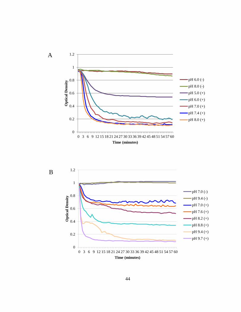
44
0
0.2
0.4
0.6
0.8
1
1.2
0 3 6 9 12 15 18 21 24 27 30 33 36 39 42 45 48 51 54 57 60
Opt
ical
Den
sity
Time (minutes)
pH 6.0 (-)
pH 8.0 (-)
pH 5.0 (+)
pH 6.0 (+)
pH 7.0 (+)
pH 7.4 (+)
pH 8.0 (+)
0
0.2
0.4
0.6
0.8
1
1.2
0 3 6 9 12 15 18 21 24 27 30 33 36 39 42 45 48 51 54 57 60
Opt
ical
Den
sity
Time (minutes)
pH 7.0 (-)
pH 9.4 (-)
pH 7.0 (+)
pH 7.6 (+)
pH 8.2 (+)
pH 8.8 (+)
pH 9.4 (+)
pH 9.7 (+)
A
B

45
Figure 2.5 NaCl does not augment PlySs2 activity.
Log-phase S. suis 7997 were centrifuged and resuspended in PB to OD600 ~1.0. To test
the optimal salinity for PlySs2 activity, 32 μg/ml PlySs2 was mixed with S. suis strain
7997 in phosphate buffer at different NaCl concentrations. PlySs2 is most active in the
absence of NaCl. In controls (-), PB replaced PlySs2. Spectrophotometric readings were
taken at OD600 every minute for an hour.
0
0.2
0.4
0.6
0.8
1
1.2
0 3 6 9 12 15 18 21 24 27 30 33 36 39 42 45 48 51 54 57 60
Opt
ical
Den
sity
Time (minutes)
150 mM NaCl (-)
0 mM NaCl (-)
150 mM NaCl (+)
50 mM NaCl (+)
20 mM NaCl (+)
10 mM NaCl (+)
0 mM NaCl (+)

46
Figure 2.6 Dithiothreitol (DTT) does not inhibit PlySs2 activity.
Log-phase S. suis 7997 were centrifuged and resuspended in PB to OD600 ~1.0. PlySs2
was incubated in 5 mM DTT at room temperature for 60 minutes. Incubated or
unincubated PlySs2 was added at 32 μg/ml. In controls (-), PB replaced PlySs2.
Spectrophotometric readings were taken at OD600 every minute for an hour.
0
0.1
0.2
0.3
0.4
0.5
0.6
0.7
0.8
0.9
1
0 3 6 9 12 15 18 21 24 27 30 33 36 39 42 45 48 51 54 57 60
Opt
ical
Den
sity
Time (minutes)
DTT+ddH2O
PlySs2
DTT-incubated PlySs2

47
Figure 2.7 Minimal ion depletion from EDTA inhibits PlySs2 activity.
Log-phase S. suis 7997 were centrifuged and resuspended in PB to OD600 ~1.0. PlySs2
was added at 32 μg/ml to cells with various concentrations of EDTA. In controls (-), PB
replaced PlySs2. Spectrophotometric readings were taken at OD600 every minute for an
hour.
0
0.2
0.4
0.6
0.8
1
1.2
1 4 7 10 13 16 19 22 25 28 31 34 37 40 43 46 49 52 55 58 61
Opt
ical
Den
sity
Time (minutes)
4 μM EDTA (-)
40 μM EDTA (-)
200 μM EDTA (+)
40 μM EDTA (+)
4 μM EDTA (+)
0 nM EDTA (+)

48
2.2.3.2 Stability
The activity of each PlySs2 aliquot incubated at different temperatures was tested
(at 32 μg/ml) against S. suis strain 7997 and spectrophotometrically evaluated, as outlined
above. When PlySs2 was incubated at different temperatures from 22°C – 85°C for 30
min, its activity was principally unaffected until 60°C (Figure 2.8A). Activity was
retained during incubation at 37°C for 24 hours, with activity starting to diminish after 48
hours (Figure 2.8, B). There was no observable decrease in activity after 15 days at 4°C
(Figure 2.8, C) or storage at -80°C for >7 months (Figure 2.8, D). The lysin also endured
10 consecutive freeze-thaws cycles (-80°C to room temperature) without any observable
effects (Figure 2.9).

49
Figure 2.8 PlySs2 is stable under a variety of conditions.
PlySs2 was incubated: (A) for 30 minutes at various temperatures, then cooled; (B) at
37°C for different increments of time; (C) at 4°C for different numbers of days; (D) at
-80°C for different numbers of months. For each test, 32 μg/ml PlySs2 was added to S.
suis 7997 cells at an OD600 of ~1.0. Spectrophotometric readings were taken at OD600
every minute for an hour.
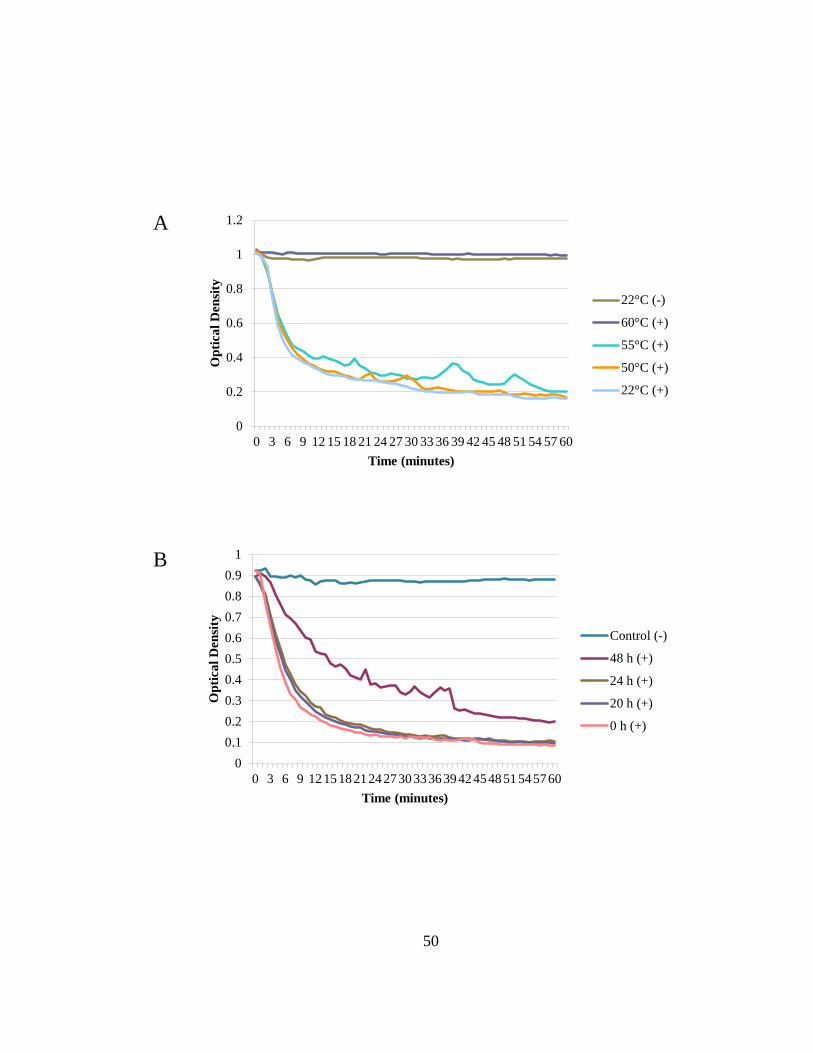
50
0
0.2
0.4
0.6
0.8
1
1.2
0 3 6 9 12 15 18 21 24 27 30 33 36 39 42 45 48 51 54 57 60
Opt
ical
Den
sity
Time (minutes)
22°C (-)
60°C (+)
55°C (+)
50°C (+)
22°C (+)
00.10.20.30.40.50.60.70.80.9
1
0 3 6 9 1215182124273033363942454851545760
Opt
ical
Den
sity
Time (minutes)
Control (-)
48 h (+)
24 h (+)
20 h (+)
0 h (+)
A
B

51
00.10.20.30.40.50.60.70.80.9
1
0 3 6 9 1215182124273033363942454851545760
Opt
ical
Den
sity
Time (minutes)
Control (-)
Day 15 (+)
Day 10 (+)
Day 5 (+)
Day 0 (+)
0
0.2
0.4
0.6
0.8
1
1.2
0 3 6 9 1215182124273033363942454851545760
Opt
ical
Den
sity
Time (minutes)
Control (-)
7 months (+)
4 months (+)
1 month (+)
C
D

52
Figure 2.9 PlySs2 is stable and active after ten, consecutive freeze-
thaws.
Log-phase S. suis 7997 were centrifuged and resuspended in PB to OD600 ~1.0. PlySs2
was taken from -80°C to room temperature and back to -80°C on subsequent days from
Day 1 to Day 10. A sample from each day was added to cells at 32 μg/ml. In controls (-),
PB replaced PlySs2. Spectrophotometric readings were taken at OD600 every minute for
an hour.
0
0.1
0.2
0.3
0.4
0.5
0.6
0.7
0.8
0.9
1
0 3 6 9 12 15 18 21 24 27 30 33 36 39 42 45 48 51 54 57 60
Opt
ical
Den
sity
Time (minutes)
Control (-)
Day 10 (+)
Day 7 (+)
Day 4 (+)
Day 1 (+)

53
2.3 ACKNOWLEDGEMENTS
I would like to thank Magid Mohamed for his technical assistance. Dr. Jonathan
E. Schmitz provided invaluable contributions throughout the aforementioned discovery
and characterization. I am also grateful to Dr. Jaap Wagenaar for providing the S. suis
strains for each test.

54
3 CHAPTER 3 – STREPTOCOCCUS SUIS SUSCEPTIBILITY
3.1 MATERIALS AND METHODS
3.1.1 Bacterial strains
All S. suis strains were stored at -80°C (Table 3.1). The strains were grown in
brain heart infusion (BHI) broth at 37°C for all tests. Luria Bertani (LB) broth was used
to cultivate Escherichia coli. All media was from Becton Dickinson, and Company
(Sparks, MD).

55
Table 3.1 Strains used in the S. suis susceptibility study
Species Serotype Strain ATCC Sourcea
Escherichia coli TOP10 1
Streptococcus suis 1
2
Streptococcus suis 1 6112 2
Streptococcus suis 1 6388 2
Streptococcus suis 2
2
Streptococcus suis 2 10 2
Streptococcus suis 2 S735 43765 2
Streptococcus suis 3
2
Streptococcus suis 4
2
Streptococcus suis 5
2
Streptococcus suis 6
2
Streptococcus suis 7
2
Streptococcus suis 7 7197 2
Streptococcus suis 7 7711 2
Streptococcus suis 8
2
Streptococcus suis 9
2
Streptococcus suis 9 7997 2
Streptococcus suis 9 8067 2
Streptococcus suis 10
2

56
Species Serotype Strain ATCC Sourcea
Streptococcus suis 11
2
Streptococcus suis 12
2
Streptococcus suis 13
2
Streptococcus suis 14
2
a 1, The Rockefeller University Collection; 2, Jaap A. Wagenaar, Utrecht University,
Utrecht, The Netherlands.

57
3.1.2 Lytic activity against S. suis
Strains of bacteria were grown to log-phase at 37°C and brought to an optical
density (OD600) of ~1.0 with 15 mM phosphate buffer (PB), pH 8.0 (buffer A) as
measured in 96-well microtiter plates (Falcon). From these bacterial stocks, 245 μl were
added to each well of a 96-well microtiter plate. In triplicate for every strain, each well
received 5 μl of PlySs2 at 1.6 μg/μl (8 μg, thus a final concentration of 32 μg/ml). [In
preliminary experiments, 32 μg/ml provided the best resolution in determining PlySs2
activity.] Corresponding triplicate wells received 5 µl of 15 mM PB, pH 6.7 (buffer B)
control vehicle. S. suis 7997 served as a positive control for each trial. As detailed above,
spectrophotometric readings were taken of each well every minute over an hour. The
degree of turbidity reduction indicated lysin activity.
3.1.3 Bactericidal assay
Log-phase bacteria were centrifuged and washed 1× in buffer A and adjusted to
an OD600 of 0.1 (= 0.5 McFarland, ~108 CFU/ml) in buffer A with a SmartSpec™ Plus
Spectrophotometer (Bio-Rad). A 100 µl aliquot of the cell suspension was distributed in
96-well polypropylene microtiter plate (Costar). PlySs2, at 64 μg/ml, or buffer B control
vehicle was added to wells in triplicate for each strain. Plates were sealed and shaken at
37°C every minute for 1 hour. At this time, 10-fold serial dilutions of each well were
plated on BHI agar and incubated at 37°C. Resultant colonies were enumerated after 18
h. The bactericidal effect was calculated as the difference between vehicle-treated and
PlySs2-treated CFU for each strain.

58
3.1.4 MIC assay
The protocol of Wiegand, et al. (Wiegand, Hilpert, and Hancock 2008) was used
with adjustments to determine minimum inhibitory concentrations (MICs). Briefly, each
strain was grown in BHI and adjusted to ~5 × 10⁵ cells/ml in BHI and distributed into 4
wells of a 96-well round bottom polystyrene microtiter plate. In each of two wells
corresponding to each strain, either sterile-filtered lysin or control vehicle was added
(Wiegand, Hilpert, and Hancock 2008). The lysin concentration varied from 0.5 – 1,024
μg/ml PlySs2. The plates were then incubated for 18 h at 37°C. The MIC was the highest
concentration of lysin that prevented the formation of a visible cell pellet (a measure of
growth) on the bottom of the wells. The MICs were also colorimetrically confirmed by
staining the bacteria in the wells with alamarBlue vital dye following the
manufacturer’s protocol (Invitrogen).
3.1.5 Resistance
A published protocol to test the in vitro development of antibiotic-resistance was
followed (Rouse et al. 2005, Pastagia et al. 2011, Gilmer et al. 2013). Briefly, S. suis was
grown in the presence of doubling concentrations of PlySs2 over 8 days in BHI broth,
and the PlySs2 MIC was tested daily to determine if resistance was acquired. On the first
day, bacteria at ~5 × 10⁸ CFU/ml were grown overnight at 37°C in 10 ml BHI containing
1/32× the PlySs2 MIC for the given strain. On the second day, the culture was split into
two equal portions. The cells of one aliquot were pelleted and resuspended in 10 ml fresh
BHI media with double the concentration of PlySs2 (i.e., 1/16× the PlySs2 MIC on day
2). This aliquot was re-incubated at 37°C overnight. Over 8 days, the concentration of

59
PlySs2 was serially doubled from 1/32× the initial MIC (on day 1) to 4× the initial MIC
(on day 8).
A sample of the second half of the aliquot was spread on BHI agar containing the
PlySs2 MIC for that strain. After incubation at 37°C, 4 colonies were selected from the
BHI agar plate to determine if a 4-fold increase in PlySs2 MIC was achieved for that
strain, indicating the emergence of resistance. The protocol was repeated with gentamicin
as an antibiotic resistance control for each S. suis strain, because S. suis strains develop
resistance to gentamicin in vitro (Varela et al. 2013).
3.2 RESULTS
3.2.1 Lytic activity against S. suis
Purified PlySs2 was tested against 22 strains representing 8 serotypes of S. suis to
assess its range of lytic activity. Over 30 minutes, 14 of 22 strains were reduced to an
OD600 ratio of ≤0.2 from a starting OD600 of ~1.0 (Figure 3.1). Readings taken after 60
minutes showed the same relative reduction in OD600 (Figure 3.2). This group of PlySs2-
sensitive S. suis strains included the type strain S735, and the pathogenic strains 10 and
7997. The reduction in OD600 ratio of other S. suis strains was reduced to ≤0.6. Serotype
12 was the only strain that exhibited a negligible decrease in optical density.

60
Figure 3.1 PlySs2 lysed almost all strains of S. suis over 30 minutes.
Bacteria in logarithmic growth were exposed to 32 μg/ml PlySs2 for 30 minutes in PB
(for 60-minute readings, see Figure 3.2). The activity was measured by OD600 reduction.
To normalize and combine values from multiple tests, the final OD600 of the treated
samples was divided by the final OD600 of the untreated samples. An OD600 ratio of 1.0
indicates no lysis, while an OD600 ratio of ~0.02 indicates complete lysis.
0 0.2 0.4 0.6 0.8 1 1.2
Serotype 12Serotype 4
Serotype 7, strain 7197Serotype 5
Serotype 7, strain 7711Serotype 9
Serotype 10Serotype 6Serotype 7
Serotype 9, strain 8067Serotype 3
Serotype 9, strain 7997Serotype 11Serotype 14Serotype 8Serotype 2
Serotype 13Serotype 2, strain 10
Serotype 1, strain 6112Serotype 1, strain 6388
Serotype 1Serotype 2, strain S735
Treated/Untreated OD600

61
Figure 3.2 PlySs2 lysed almost all strains of S. suis over 60 minutes.
Bacteria in logarithmic growth were exposed to 32 μg/ml PlySs2 for 60 minutes in PB
(for 30-minute readings, see Figure 3.1). The activity was measured by OD600 reduction.
To normalize and combine values from multiple tests, the final OD600 of the treated
samples was divided by the final OD600 of the untreated samples. An OD600 ratio of 1.0
indicates no lysis, while an OD600 ratio of ~0.02 indicates complete lysis.
0 0.2 0.4 0.6 0.8 1 1.2
Serotype 12Serotype 4
Serotype 7, strain 7197Serotype 5
Serotype 7, strain 7711Serotype 9
Serotype 10Serotype 7Serotype 6Serotype 3
Serotype 14Serotype 11
Serotype 9, strain 8067Serotype 2
Serotype 13Serotype 8
Serotype 9, strain 7997Serotype 2, strain 10
Serotype 1Serotype 1, strain 6388Serotype 1, strain 6112Serotype 2, strain S735
Treated/Untreated OD600

62
S. suis strains S735 and 7997 were also tested in a time-dependent lytic assay at
various PlySs2 dosages (Figure 3.3 and Figure 3.4), showing a significant drop in OD600
at >4 µg/ml for both strains. These time-dependent lytic assay results led us to use 32
µg/ml for the best resolution in testing all strains.

63
Figure 3.3 S. suis strain S735 exposed to various concentrations of
PlySs2.
S. suis strain S735 is sensitive to concentrations as low as 0.5 µg/ml PlySs2. S. suis strain
S735 in logarithmic growth was exposed to various concentrations of PlySs2 ranging
from 0.25 µg/ml – 128 µg/ml for 60 minutes in PB. Readings at OD600 were taken every
minute.
0
0.2
0.4
0.6
0.8
1
1.2
0 5 10 15 20 25 30 35 40 45 50 55 60
Opt
ical
Den
sity
Time (minutes)
0.25 μg/ml 0.5 μg/ml 2 μg/ml 4 μg/ml 8 μg/ml 16 μg/ml 32 μg/ml 64 μg/ml 128 μg/ml

64
Figure 3.4 S. suis strain 7997 exposed to various concentrations of
PlySs2.
S. suis strain 7997 is sensitive to concentrations as low as 1.0 µg/ml PlySs2. S. suis strain
7997 in logarithmic growth was exposed to various concentrations of PlySs2 ranging
from 0.25 µg/ml – 128 µg/ml for 60 minutes in PB. Readings at OD600 were taken every
minute.
0
0.2
0.4
0.6
0.8
1
1.2
0 5 10 15 20 25 30 35 40 45 50 55 60
Opt
ical
Den
sity
Time (minutes)
0.25 μg/ml 0.5 μg/ml 2 μg/ml 4 μg/ml 8 μg/ml 16 μg/ml 32 μg/ml 64 μg/ml 128 μg/ml

65
3.2.2 Bactericidal assay
The lethal effect of PlySs2 was quantified for a select set of S. suis serotypes and
strains. After 60 minutes of exposure to 64 μg/ml of PlySs2, all S. suis strains were
reduced by 5-6 logs, except serotype 5 (~2-logs) (Figure 3.5). The relative lethal effect
from one strain to another correlated with the lytic activity reported above. Strain 7197
was an outlier in that it was reduced by ~5.5 logs in the bactericidal assay, but was only
reduced in the lytic assay to an OD600 ratio of ~0.5.
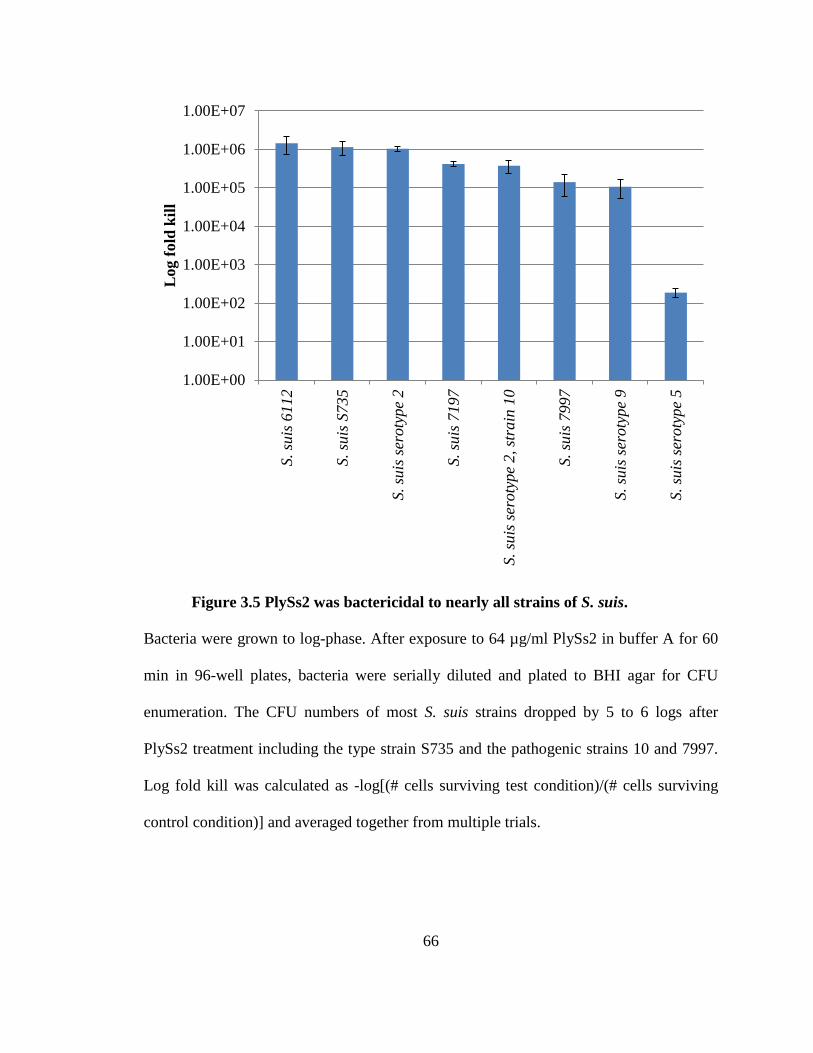
66
Figure 3.5 PlySs2 was bactericidal to nearly all strains of S. suis.
Bacteria were grown to log-phase. After exposure to 64 µg/ml PlySs2 in buffer A for 60
min in 96-well plates, bacteria were serially diluted and plated to BHI agar for CFU
enumeration. The CFU numbers of most S. suis strains dropped by 5 to 6 logs after
PlySs2 treatment including the type strain S735 and the pathogenic strains 10 and 7997.
Log fold kill was calculated as -log[(# cells surviving test condition)/(# cells surviving
control condition)] and averaged together from multiple trials.
1.00E+00
1.00E+01
1.00E+02
1.00E+03
1.00E+04
1.00E+05
1.00E+06
1.00E+07
S. su
is 6
112
S. su
is S
735
S. su
is se
roty
pe 2
S. su
is 7
197
S. su
is se
roty
pe 2
, str
ain
10
S. su
is 7
997
S. su
is se
roty
pe 9
S. su
is se
roty
pe 5
Log
fold
kill
67
3.2.3 MIC assay
The MIC of PlySs2 against these serotypes also qualitatively correlated with the
lytic and bactericidal assays. The PlySs2 MIC ranged from 32 to 512 μg/ml for all
PlySs2-sensitive strains. As expected, from earlier data, serotype 12 was not inhibited at
>1,024 μg/ml (Table 3.2).

68
Table 3.2 The MIC of PlySs2 for S. suis serotypes and strainsa
Species Serotype Strain MIC (μg/ml)
S. suis
2 S735 32 2
64
7 7197 128 9 7997 128 9
128
1 6112 256 2 10 256 5
512
12
>1,024 S. aureus
MW2 16
S. pyogenes M1 MGAS 5005 128
a Bacteria were examined for growth at each concentration of PlySs2 from 0.5 - 1,024
μg/ml. The lowest concentration preventing growth is the PlySs2 MIC (column 4) for
each strain (columns 2 and 3) of each species (column 1). Consistent with other tests, S.
suis type strain S735 registered a low MIC while there was a higher MIC observed for S.
suis strain 7997. The MIC of PlySs2 for S. suis serotype 12 was above the assay
parameters. The previously published PlySs2 MICs for S. aureus and S. pyogenes are
included for reference (Gilmer et al. 2013).

69
3.2.4 Resistance
According to an established protocol (Rouse et al. 2005, Pastagia et al.
2011, Gilmer et al. 2013), both S. suis strains S735 and 7997 were challenged with
incrementally doubling concentrations of PlySs2 to determine if they would develop
resistance. Neither strain developed PlySs2 resistance – defined as exhibiting a >4×
increase from the original PlySs2 MIC for each strain (Figure 3.6). Using the antibiotic
gentamicin in the same procedure led to both S. suis strains S735 and 7997 developing
resistance (Figure 3.6).

70
Figure 3.6 S. suis 7997 and S735 did not develop resistance to PlySs2
in vitro.
S. suis S735 and S. suis 7997 grew in media containing 1/32× (3.13%) to 4× (400%) the
MIC of PlySs2 or gentamicin over 8 days. Comparing the MICs of PlySs2 after each day
to the initial MIC of PlySs2 for each strain determined resistance. Neither developed
resistance to PlySs2. Both S. suis strain S735 and S. suis strain 7997 developed resistance
to the positive control, gentamicin.
0
4
8(M
IC) /
(Ini
tial M
IC)
Day (% MIC exposure)
S. suis 7997 - PlySs2
S. suis 7997 - Gentamicin
S. suis S735 - PlySs2
S. suis S735 - Gentamicin

71
3.3 ACKNOWLEDGEMENTS
This chapter is based on a submitted manuscript co-authored (in order) by: Dr.
Jonathan E. Schmitz, Dr. Chad W. Euler and Dr. Vincent A. Fischetti. We thank Haaris
Khan, Khoi Nyguen, Karen Tong, and Dr. Chad Euler for providing helpful advice and
excellent assistance. We are grateful to: Dr. Jaap Wagenaar for the S. suis serotypes and
strains. Daniel Gilmer is a Gilliam fellow of the Howard Hughes Medical Institute. This
work was specifically supported by USPHS grant AI11822 to VAF. Dr. Fischetti has
declared a potential conflict of interest.

72
4 CHAPTER 4 – BROAD GRAM-POSITIVE SUSCEPTIBILITY
4.1 MATERIALS AND METHODS
4.1.1 Bacterial strains
All strains were stored at -80°C (Table 1.1). Staphylococcus, Streptococcus,
Listeria, Enterococcus, Pseudomonas, Bacillus strains were cultivated in brain heart
infusion (BHI) broth unless substituted with Mueller Hinton (MH) medium during the
minimum inhibitory concentration (MIC) determination below. Lactobacillus strains
were cultivated in de Man, Rogosa and Sharpe (MRS) broth (Sigma). Escherichia coli
were grown in Luria Bertani (LB) broth. All media were acquired from Becton,
Dickinson, and Company (Sparks, MD), unless otherwise stated. Bacteria were
propagated at 37°C and shaken at 200 rpm, if necessary.

73
Table 4.1 Strains used in this study
Organism Serotype Strain ATCC Sourcea Notes
Bacillus anthracis
Δ Sterne 1
Bacillus cereus
14579 1
Bacillus subtilis
SL4 1
Bacillus thuringiensis
HD-73 1
Enterococcus faecalis
V583 1
Enterococcus faecium
1 EFSK-2
Escherichia coli
TOP10 1
Group E streptococci 2 K131 123191 2
Lactobacillus
acidophilus
Pak 4357 3
Lactobacillus
acidophilus
11975 3
Lactobacillus
acidophilus
4356 3
Lactobacillus gasseri
19992 3
Lactobacillus
rhamnosus
LMS2-1 3
Lactobacillus
rhamnosus
21052 3

74
Organism Serotype Strain ATCC Sourcea Notes
Listeria monocytogenes
HER 1184 1
Listeria monocytogenes 4b N3013 1
Listeria monocytogenes 3b FSLJ 1 1
Listeria monocytogenes
1 RS823
Listeria monocytogenes
1 RS820
Listeria monocytogenes
HER1083 1
Listeria monocytogenes
BAA-680 2
Pseudomonas
aeruginosa
RS1 1
Staphylococcus aureus
4
vancomycin intermediate resistance IV
Staphylococcus aureus
4
vancomycin intermediate resistance III
Staphylococcus aureus
RN4220 1
Staphylococcus aureus
Newman 5
methicillin sensitive - mutant LyrA
Staphylococcus aureus
Newman 5 methicillin sensitive
Staphylococcus aureus
MW2
BAA-1707 2
methicillin resistant - community acquired
Staphylococcus aureus
192 1 methicillin resistant
Staphylococcus aureus
1
methicillin resistant from patient DS
Staphylococcus aureus
1
highly mupirocin resistant
Staphylococcus aureus
1
D712 - daptomycin resistant
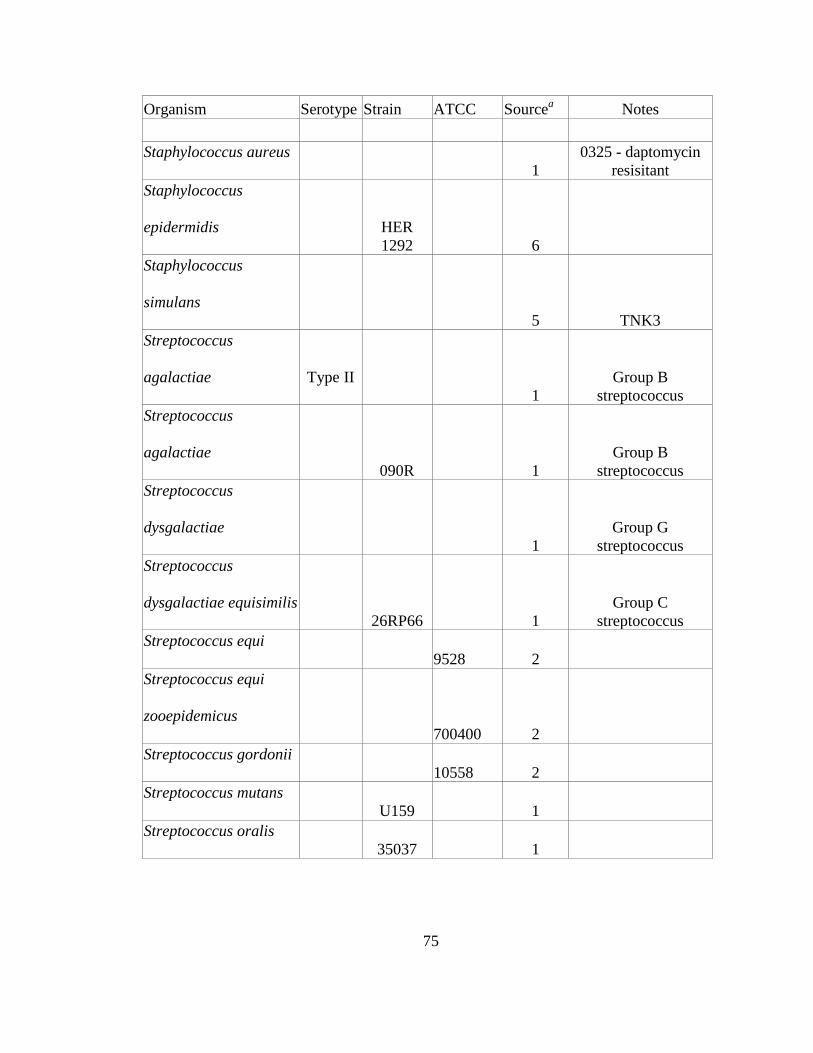
75
Organism Serotype Strain ATCC Sourcea Notes
Staphylococcus aureus
1
0325 - daptomycin resisitant
Staphylococcus
epidermidis
HER 1292 6
Staphylococcus
simulans
5 TNK3
Streptococcus
agalactiae Type II
1
Group B streptococcus
Streptococcus
agalactiae
090R 1
Group B streptococcus
Streptococcus
dysgalactiae
1
Group G streptococcus
Streptococcus
dysgalactiae equisimilis
26RP66 1
Group C streptococcus
Streptococcus equi
9528 2
Streptococcus equi
zooepidemicus
700400 2
Streptococcus gordonii
10558 2
Streptococcus mutans
U159 1
Streptococcus oralis
35037 1

76
Organism Serotype Strain ATCC Sourcea Notes
Streptococcus
pneumoniae 9V DCC1335 1
Streptococcus
pneumoniae 6 DCC1850 1
Streptococcus
pneumoniae 15 DCC1476 1
Streptococcus
pneumoniae 11
1
Streptococcus
pneumoniae
1 mutant Lyt 4-4
Streptococcus pyogenes M6 D471 1
Streptococcus pyogenes M-neg D471 1 mutant JRS75
Streptococcus pyogenes M6 MGAS 10394 BAA-946 2
Streptococcus pyogenes M49 NZ131 7
Streptococcus pyogenes M4 SmR 1
streptomycin resistant - mucoid
Streptococcus pyogenes M3 MGAS 315 BAA-595 2
Streptococcus pyogenes M18 MGAS 8232 BAA-572 2
Streptococcus pyogenes M1 CEM1ΔΦ 1
mucoid – mouse passaged
Streptococcus pyogenes M1 CEM1ΔΦ 1
Streptococcus pyogenes M1 SF370 1
mucoid – mouse passaged

77
Organism Serotype Strain ATCC Sourcea Notes
Streptococcus pyogenes M1 SF370 7
Streptococcus pyogenes M1 MGAS 5005 BAA-947 2
Streptococcus rattus
BHT 1
Streptococcus
sanguinis
10556 2
Streptococcus sobrinus
6715 1
Streptococcus suis 9 7997 8
a 1, The Rockefeller University Collection; 2, ATCC; 3, ContraFect Corporation,
Yonkers, NY; 4, Alexander Tomasz, The Rockefeller University; 5, Olaf Schneewind,
University of Chicago, Chicago, IL; 6, Barry Kreiswirth, Public Health Research
Institute, New Jersey, NJ; 7, Joseph Ferretti, University of Oklahoma Health Science
Center, Oklahoma City, OK; 8, Jaap A. Wagenaar, Utrecht University, Utrecht, The
Netherlands.

78
4.1.2 Lytic activity
Log-phase bacteria were adjusted with buffer A to an OD600 of 0.8 to 1.0 in 96-
well microtiter plates (Falcon). PlySs2, at 32 μg/ml, or buffer B control vehicle was
added to each sample well. In each run, S. suis 7997 was included as a positive control.
Spectrophotometric readings (OD600) of each well were taken by a Spectramax Plus 384
(Molecular Devices) every minute over 60 minutes at room temperature. Lysin activity
was gauged by the degree of turbidity reduction (OD600) following enzyme addition.
4.1.3 Bactericidal assay
Log-phase bacteria were resuspended in buffer A to an OD600 of 0.1 (= 0.5
McFarland, ~108 CFU/ml) and aliquoted into wells of a polypropylene microtiter plate
(Costar). Actual inoculum titers for each experiment were derived from plating serial
dilutions of each inoculum. For each organism, buffer B control vehicle or PlySs2 was
added at 128 μg/ml to wells in triplicate. Plates were sealed and incubated at 37°C with
agitation every 5 minutes for 1 hour. After incubation, cells were serially diluted in 10-
fold increments and plated on BHI agar. Death (log fold kill) was calculated as -
log[(CFUs in the test condition) ÷ (CFUs in the control condition)].
4.1.4 MIC assay
The protocol from Wiegand, et al. was followed to determine MICs, with
adjustments as detailed below (Wiegand, Hilpert, and Hancock 2008). Briefly, a final
suspension of ~5 × 10⁵ cells/ml in MHB (or BHI for S. pyogenes) plus sterile-filtered
lysin or control vehicle was distributed within a 96-well microtiter plate in triplicate
(Wiegand, Hilpert, and Hancock 2008). Cells were challenged with 0.5 – 1,024 μg/ml
PlySs2 in triplicate. MICs were determined by detection of cell pellet formation in the

79
bottom of rounded polysterene plate wells; they were corroborated colorimetrically with
alamarBlue vital dye (Invitrogen) following the manufacturer’s protocol.
4.1.5 Resistance
According to an established protocol for the in vitro development of mupirocin-
resistance (Rouse et al. 2005, Pastagia et al. 2011), S. aureus CA-MRSA MW2, S. aureus
MSSA 8325, and S. pyogenes MGAS 5005 were grown in the presence of PlySs2 in
liquid culture. Initially, bacterial cells at 5 × 10⁸ CFU/ml were grown overnight in the
presence of 1/32× the MIC of PlySs2 against each strain (37°C, BHI broth for S.
pyogenes with cap secured during gentle shaking, MHB broth for S. aureus with 220 rpm
aeration). The cells were pelleted by centrifugation for 10 min at 900 rcf and sub-divided
into two aliquots.
One aliquot was diluted 10-fold into fresh MHB media with double the
concentration of PlySs2; a portion of the other was spread onto the surface of MHA agar
containing the PlySs2 MIC for that species. The MIC of 4 resultant colonies was
recalculated by microdilution for each serial passage, as described above (Wiegand,
Hilpert, and Hancock 2008). This was done to determine if a resistant clone had emerged
(defined as a 4-fold increase in MIC). The above procedure was repeated over an 8-day
period, and the concentration of PlySs2 in the liquid culture was serially doubled from
1/32× to 4× the original MIC. This process was also conducted with mupirocin for each
MRSA strain, to serve as an antibiotic resistance positive control.
4.1.6 PlySs2 catalytic domain lytic assay
The PlySs2 catalytic domain sequence (PlySs2-CD, ~0.49 kb), containing some
residues from the linker, was inserted into pBAD24 vector with HindIII and NheI. This

80
vector was transformed into TOP10 E. coli (Invitrogen). For purification, E. coli cells
induced to express pBAD24_PlySs2-CD were pelleted and re-suspended in 20 mM PB,
pH 7.4 (buffer D). EDTA-free protease inhibitor tablets were added before homogenizing
the cells, which were subsequently spun at 16,000 rpm for 60 min. The volume was
adjusted with buffer D to raise pH.
The soluble lysate was run through a CM Sepharose® column equilibrated to
buffer D. The flow through containing PlySs2-CD was collected. This was dialyzed
overnight at 4ºC into 20 mM glycine-NaOH, pH 9.3 (buffer E). This material was run
over on Mono Q column equilibrated to buffer E. The material was eluted gradually from
1-100 mM NaCl, buffer E. This was repeated with elution by 30-100 mM NaCl, buffer E.
Pure fractions dialyzed into buffer D and pooled. Concentration was confirmed to be 1.7
mg/ml.
Following the aforementioned protocol for lytic assay (page 59), log-phase S. suis
strain 7997 cells were exposed to full length PlySs2 (PlySs2-FL) at 32 µg/ml, and
PlySs2-CD at 850 µg/ml. As before, the degree of turbidity reduction (OD600) in the test
wells indicated the degree of lysin activity.
4.1.7 Fluorescent binding assay
The PlySs2 binding domain sequence (PlySs2-BD, ~0.26 kb) was cloned from S.
suis strain 89/1591 with the forward primer (containing NheI site, underlined)
AATGCTAGCATGCGTTCCTATCGCGAGAC, and the reverse primer (containing
HindIII site, underlined) CCTAAGCTTCTTTTCACAAATCATAATCCCCAG. This
was cloned into the pBAD24 vector and transformed into TOP10 E. coli (Invitrogen).
The resultant protein, PlySs2-BD is expected to be 9.5 kD and have a theoretical pI 9.57.

81
E. coli cells induced to express pBAD24_PlySs2-BD were pelleted and re-
suspended them in 20 mM PB, pH 7.4 (buffer D). EDTA-free protease inhibitor tablets
were added before homogenizing the cells, which were subsequently spun at 15,000 rpm
for 80 min. The pH was reduced by adding 30 mM PB, pH 6.0. The lysate was run over a
CM Sepharose® FF column where it eluted at 250 mM NaCl, buffer D. The pure
fractions were pooled and dialysed overnight in 4 l of buffer D. A BCA protein assay
(Sigma) confirmed the concentration to be 8.9 mg/ml.
This PlySs2-BD was labeled with AlexaFluor 488 as per the manufacturer’s
instructions (Becton, Dickinson and Company – Sparks, MD). Subject strains were
grown overnight in BHI at 37°C, and subsequently pelleted and resuspended in PBS to
OD600 = 0.1. Of this dilution, 1 ml was pelleted and resuspended in 200 μl of 2%
paraformaldehyde in PBS for 30 minutes.
After washing with PBS, pellets were resuspended in 500 μl PBS + 5% blocking
solution for 1 hour to which 500 μl PBS + 0.5 μl labeled PlySs2-BD was added for
another hour. After three washes with PBS, pellets were resuspended in 50 μl PBS. Cells
(5 μl) were then pipetted onto a slide sealed with a cover slip. Fluorescence photographed
on an Eclipse E400 microscope (Nikon) using the QCapture Pro version 5.1 imaging
software. Images displayed high, medium, and low intensity.
4.2 RESULTS
4.2.1 Lytic activity
Purified PlySs2 was tested against a wide range of bacterial species and strains to
determine the range of lytic activity. Starting at an OD600 of ~1.0, all tested strains of S.
aureus – including strains resistant to methicillin, vancomycin, daptomycin, mupirocin,

82
and lysostaphin – were reduced to an OD600 ratio ≤ 0.3 after lysis by PlySs2 over 30
minutes of exposure (Figure 4.1). Readings were also taken after 60 minutes (Figure 4.2).
The OD600 ratio of other staphylococci, including Staphylococcus simulans and
Staphylococcus epidermidis, was reduced to ~0.2.

83
Figure 4.1 PlySs2 displayed activity against various species.
PlySs2 displayed activity against various species. Multiple strains of staphylococci
(including MRSA, MSSA, and VISA), streptococci, enterococci, Listeria, bacilli, and
lactobacilli were tested for susceptibility to PlySs2 activity. Escherichia and
Pseudomonas were tested as Gram-negative controls. Log-phase cultures were exposed
to 32 µg/ml PlySs2 for 30 min in PB (for 60-min readings) (see also Figure 4.2). The
final OD600 of the treated samples was divided by the final OD600 of the untreated
samples to generate the normalized values. Complete lysis registered a ratio of ~0.02. ST,
serotype.

84

85
Figure 4.2 PlySs2 displayed activity against various species over 60
minutes.
Multiple strains of staphylococci (including MRSA, MSSA, and VISA), streptococci,
enterococci, Listeria, bacilli, and lactobacilli were tested for susceptibility to PlySs2
activity. Escherichia and Pseudomonas were tested as Gram-negative controls. Log-
phase cultures were exposed to 32 μg/ml PlySs2 for 60 minutes in PB. The final OD600 of
the treated samples was divided by the final OD600 of the untreated samples to generate
the normalized values. Complete lysis registers a ratio of ~0.02.
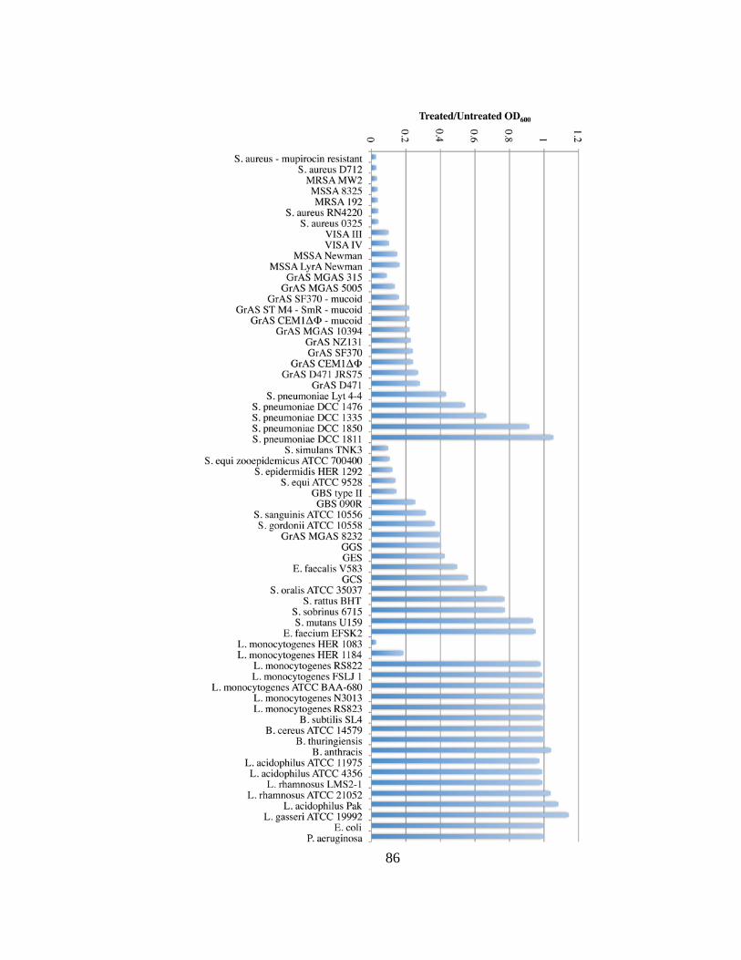
86

87
With streptococci, PlySs2 lysed most of the tested M-protein serotypes of S.
pyogenes, including M1, M3, M4, M6, M18, M49, an M-negative variant, as well as
unencapsulated and highly encapsulated strains, decreasing their OD600 ratio to ≤ 0.4.
PlySs2 also exhibited strong lytic activity against S. suis, Streptococcus equi
zooepidemicus, Streptococcus equi, S. agalactiae Type II (encapsulated), and S.
agalactiae 090R. The pathogenic Streptococcus sanguinis, and groups G and E
streptococci were moderately sensitive to PlySs2. Streptococcus mutans, group C
streptococci, Streptococcus oralis, Streptococcus rattus, and Streptococcus sobrinus were
only reduced to an OD600 ratio between 0.7 and 0.9. PlySs2 did not reduce the OD600
ratio for any Streptococcus pneumoniae strains below 0.5. Streptococcus gordonii was
the only commensal against which PlySs2 exhibited activity (Figures 4.1 and 4.2).
PlySs2 showed some activity against genera outside Staphylococcus and
Streptococcus. While two strains of Listeria were sensitive to PlySs2, other strains were
not. In the Enterococcus genus, which is associated with high levels of antibiotic
resistance, E. faecalis was sensitive to PlySs2 (although less than staphylococci or
streptococci), but E. faecium was not. No activity was seen against any of the different
species of Bacilli, strains of lactobacilli, or Gram-negatives.
4.2.2 Bactericidal assay
PlySs2 was tested for log-fold killing of several species and strains of susceptible
pathogenic organisms tested by OD600 decrease. At 128 μg/ml PlySs2 and 60 minutes
exposure, the vancomycin-intermediate S. aureus (VISA) strain was only reduced by 2-
logs. However, PlySs2 reduced the viability of L. monocytogenes, S. agalactiae, S.

88
aureus, and S. pyogenes from ~3 to >6-logs (Figure 4.3). The negative control E. coli was
not reduced in OD600 after PlySs2 treatment.

89
Figure 4.3 PlySs2 was bactericidal across multiple species of bacteria.
Log-phase bacteria were treated in 96-well plates with 128 µg/ml PlySs2 in buffer A for
60 min, then serially diluted and plated onto BHI agar for CFU enumeration. The log kill
was calculated by comparing the difference between vehicle-treated and PlySs2-treated
CFU results. Of note, PlySs2 dramatically reduced S. agalactiae and L. monocytogenes.
There was no reduction in number of the negative control E. coli.

90
4.2.3 MIC Assay
When the MIC of PlySs2 was tested against strains of S. aureus, S. pyogenes, L.
monocytogenes, S. agalactiae, and E. coli, most of the values qualitatively correlated
directly to the lytic and killing activity. MICs ranged from 8 to 256 μg/ml for all strains,
except the VISA strain, which was not inhibited at >1,024 μg/ml (Table 4.2). The MIC of
PlySs2 was relatively low for L. monocytogenes and S. aureus. S. pyogenes and S.
agalactiae registered similar PlySs2 MICs. The negative control E. coli did not register a
measurable PlySs2 MIC.

91
Table 4.2 The MIC of PlySs2 for various Gram-positive speciesa
Species Strain MIC μg/ml
Visual Colorimetric
L. monocytogenes HER 1184 8 16
HER 1083 8 16
S. aureus MSSA 8325 16 16
MRSA MW2 16 32
LyrA 32 32
VISA III 32 64
GrAS SF370 128 128
MGAS 5005 128 256
GBS 090R 256 256
type II 512 512
E. coli Top10 >1,024 >1,024
aAll MICs were evaluated visually for bacterial growth and with alamarBlue vital dye
(colorimetrically) at concentrations from 0.5 to 1,024 µg/ml of PlySs2 for each strain of
each species listed. There was a low MIC for MRSA MW2, as expected, and a higher
MIC for GrAS strain MGAS 5005. The MIC of PlySs2 for the negative control, E. coli,
was above the limits of the assay. For a reference, ClyS, LysK, and CHAPK lysins have
registered MICs against S. aureus strains from 30 to 80 µg/ml (Pastagia et al.
2011, Daniel et al. 2010, Fenton et al. 2011, Becker et al. 2009).

92
4.2.4 Resistance
Using a published, standardized method for calculating resistance, both
staphylococcal and streptococcal strains were analyzed for the development of resistance
against PlySs2 by serial exposure to incrementally doubling concentrations of the lysin.
Under these testing conditions, none of the S. aureus or S. pyogenes strains exposed to
PlySs2 over 8 days developed resistance (defined as a 4-fold increase from the original
MIC) (Figure 4.4). Following the same procedure, both S. aureus strains MW2 and 8325
developed resistance to the antibiotic mupirocin (Figure 4.4).

93
Figure 4.4 MRSA, MSSA, and GrAS did not acquire resistance to
PlySs2 in vitro.
MRSA strain MW2, MSSA strain 8325, and GrAS strain MGAS 5005 were exposed to
1/32× to 4× the MIC of PlySs2 and mupirocin (S. aureus strains) over 8 days. The daily
MICs of PlySs2 were compared to the starting MIC of PlySs2 for each strain of bacteria
to ascertain resistance. None developed resistance to PlySs2. Both MW2 and 8325
developed resistance to the positive control, mupirocin.
0
4
8
12
16
(MIC
) / (I
nitia
l MIC
)
Day (% MIC exposure)
MRSA MW2 - PlySs2
MRSA MW2 - Mupirocin
MSSA 8325 - PlySs2
MSSA 8325 - Mupirocin
GrAS MGAS 5005 - PlySs2

94
4.2.5 PlySs2 catalytic domain lytic assay
The catalytic domain is much less effective at mediating lysis of S. suis strain
7997 compared to the full length PlySs2 lysin. Therefore, we used 850 µg/ml in order to
establish enough resolution within its activity profile among various species and strains.
Nevertheless, at a high concentration, it is able to lyse certain strains similar to the effect
of PlySs2-FL at 32 µg/ml. The specificity of PlySs2-CD at high concentrations is
somewhat consistent with the specificity of PlySs2 at lower concentrations against most
streptococci. PlySs2-CD does not seem to target staphylococci even at high
concentrations.

95
Figure 4.5 Lytic effect of PlySs2 Full Length VS Catalytic Domain
Strains of staphylococci (including MRSA, MSSA, and VISA), streptococci, enterococci,
Listeria, bacilli, and lactobacilli were tested for susceptibility to PlySs2-FL or PlySs2-CD
activity. Escherichia was tested as a Gram-negative control. Log-phase cultures were
exposed to lysin for 60 minutes in PB. The final OD600 of the treated samples was
divided by the final OD600 of the untreated samples to generate the normalized values.
Complete lysis registers a ratio of ~0.02.
0 0.2 0.4 0.6 0.8 1
E. coliE. faecium EFSK2
S. sobrinus 6715S. pneumoniae DCC1335
GCSE. faecalis V583
S. sanguinis 10556GrAS D471GBS 090R
GBS type IIS. equi ATCC9528GrAS MGAS 5005
S. epidermidis HER1292S. suis 7997
VISA IIIGrAS MGAS 315
S. suis S735L. monocytogenes HER1083
Treated/Untreated OD600
PlySs2-FL (32 μg/ml)
PlySs2-CD (850 μg/ml)

96
4.2.6 Fluorescent binding assay
Cells were tested for binding fluorescent PlySs2-BD. Many species were tested,
but only three are presented here as representative results. PlySs2-BD bound best to S.
agalactiae type II, although PlySs2-FL has less activity against this strain than against S.
pyogenes strain 5005 or S. suis strain 7997.
There was little correlation between the species that PlySs2-BD bound and the in
vitro activity of PlySs2-FL or PlySs2-CD. This may be attributed to each domain
requiring the other to mediate their effect. Nevertheless, this preliminary study will
require further investigation to elucidate the specificity of PlySs2.

97
Figure 4.6 PlySs2-BD Flurescence and comparison to PlySs2-CD
activity.
1 – S. suis strain 7997
2 – S. pyogenes strain 5005
3 – S. agalactiae type II [1× exposure time (left bottom); ½× (right bottom)]
(A) This PlySs2-BD was labeled with AlexaFluor 488 as per the manufacturer’s
instructions (Becton, Dickinson and Company – Sparks, MD). Subject strains were
resuspended in 500 μl PBS + 5% blocking solution for 1 hour to which 500 μl PBS + 0.5
μl labeled PlySs2-BD was added for another hour. Cells (5 μl) were pipetted onto a slide
sealed with a cover slip. Fluorescence photographed on an Eclipse E400 microscope
(Nikon) using the QCapture Pro version 5.1 imaging software. Scale bars, 1 µm. (B)
Results for these three strains taken from Figure 4.5.

98
0 0.1 0.2 0.3 0.4 0.5 0.6 0.7 0.8 0.9 1
3
2
1
Treated/Untreated OD600
PlySs2-FL (32 μg/ml) PlySs2-CD (850 μg/ml)
1 2 3 A
B

99
4.3 ACKNOWLEDGEMENTS
This chapter is based on a manuscript (Gilmer et al. 2013) co-authored (in order)
by: Dr. Jonathan E. Schmitz, Dr. Chad W. Euler, and Dr. Vincent A. Fischetti. For their
adroit technical assistance, we thank Nathan Franck, Anna Serrano, Magid Mohamed,
Haaris Khan, and Karen Tong. We are grateful to: Olaf Schneewind for S. simulans, S.
aureus Newman, and S. aureus lyrA; Barry Kreiswirth for S. epidermidis; Alexander
Tomasz for MRSA, VISA, and other staphylococcal strains; ATCC for MRSA and
streptococcal strains; ContraFect Corporation for lactobacilli strains; Marcelo Gottschalk
for S. suis strain 89/1591; Jaap Wagenaar for S. suis strain 7997; and Joseph Ferretti for
S. pyogenes strains SF370 and NZ131. This work was specifically supported by USPHS
grant AI11822 to VAF. Dr. Fischetti has declared a potential conflict of interest.

100
5 CHAPTER 5 – IN VIVO MODELS
5.1 MATERIALS AND METHODS
5.1.1 Intranasal mucosa model
5.1.1.1 S. suis colonization
A nasal mucosal colonization model described by Seitz, M., et al. (Seitz et al.
2012), was used to test the in vivo efficacy of PlySs2 to decolonize S. suis strain 7997.
Each strain was made spontaneously resistant to 200 μg/ml streptomycin, through
passage of bacteria in media containing increasing concentrations of antibiotic, to
distinguish it from other organisms found in the murine nasal mucosa. Next, 4-week old
female CD-1® mice were obtained from Charles River (Wilmington, MA). After a week
of acclimation, mice were given water with 5 mg/ml streptomycin. After two days, mice
were anesthetized and 12.5 μl of 1% acetic acid was delivered to each nostril. An hour
later, mice were administered 10 μl (∼1 × 10⁹ CFU) of mid log-phase (OD600 of ~0.5)
bacteria in 50 mM PB, pH 7.4 (buffer C) in each nostril. An aliquot of each inoculation
stock was serially diluted and plated to Columbia blood agar plates to confirm the actual
bacterial inoculation titer.
5.1.1.2 Treatment
Twenty-four hours post-colonization, the animals were randomly divided into 4
treatment groups. To each nostril, we delivered 10 μl of either: buffer C alone (group 1),
5 mg/ml PlySs2 in buffer C (group 2), 5 mg/ml gentamicin in buffer C (group 3), or a
combination of 2.5 mg/ml PlySs2 and 2.5 mg/ml gentamicin in buffer C (group 4).
Twenty-four hours after treatment, all mice were euthanized by CO2-inhalation. The
nasal passage of each mouse was surgically removed post-mortem, bisected to expose the

101
sinuses, and vortexed in 500 μl of buffer C. Serial dilutions were then streaked on 5%
sheep blood plates (containing 200 μg/ml streptomycin) and incubated at 37°C for final
colony counts. All protocols in this in vivo study were approved by The Rockefeller
University’s Institutional Animal Care and Use Committee.
5.1.2 Mixed bacteremia model
5.1.2.1 MRSA + GrAS Infection
The Rockefeller University’s Institutional Animal Care and Use Committee
approved all in vivo protocols. A systemic infection model described by Daniel, A. et al.,
was used to test for the in vivo efficacy of PlySs2 against multiple Gram-positive bacteria
(Daniel et al. 2010). Briefly, 4-5 week old female FVB/NJ mice (weight range 15 to 20 g)
were obtained from The Jackson Laboratory (Bar Harbor, ME). After a period of
acclimation, mice were injected intraperitoneally (IP) with 0.5 ml of mid log-phase
(OD600 of 0.5) bacteria diluted with 5% hog gastric mucin (Sigma) in saline. Bacterial
suspensions contained ∼5 × 10⁵ CFU/ml of MW2, a PVL toxin-encoding MRSA strain,
∼1 × 10⁷ of MGAS 5005, an M1 serotype of S. pyogenes that is virulent in humans and
mice, or a simultaneous combination of both bacteria at the above concentrations for the
mixed infection experiments. Actual bacterial inoculation titers were calculated by serial
dilution and plating to Columbia blood agar plates for each experiment.
5.1.2.2 Treatment
Mice became bacteremic within 1-3 hours and contained MRSA and/or S.
pyogenes within multiple organs, including spleen, liver, kidney, and heart/blood
(reference (Daniel et al. 2010), and unpublished observations). Three hours post-
infection, the animals were divided into 4 to 5 treatment groups per infection type and

102
were administered IP 0.5 ml of either 20 mM phosphate buffer, 2 mg/ml of the
streptococcal-specific lysin PlyC (Nelson et al. 2006), 2 mg/ml of the staphylococcal-
specific lysin ClyS (Daniel et al. 2010), 2-4 mg/ml PlySs2, or a combination of 2 mg/ml
PlyC and 2 mg/ml ClyS. A PlySs2 stock of 4 mg/ml was used for S. pyogenes-infected
mice to increase survival over an initial 70% survival rate with treatment of 2 mg/ml of
PlySs2 (data not shown). While this dosage was possible with PlySs2, it was above our
obtainable PlyC or ClyS stock concentration at the time.
The survival rate for each experimental group was monitored every 12 hours for
the first 24 hours, then every 24 hours up to 10 days post-infection. The data were
statistically analyzed by Kaplan-Meier Survival curves with standard error, 95%
confidence intervals, and significance (Log-rank / Mantel-Cox test) calculated using the
Prism computer program (GraphPad Software; La Jolla, CA).
5.2 RESULTS
5.2.1 Intranasal mucosa decolonization
To determine if the in vitro activity of PlySs2 against S. suis predicts its ability to
remove S. suis colonizing the nasal passages in vivo, mice were intranasally colonized
with S. suis strain 7997, and subsequently treated intranasally with a single dose of
PlySs2 or buffer. The number of CFUs remaining in the nasal mucosa was determined by
plating serial dilutions on blood agar. The results from multiple, separate experiments
were combined and plotted (Figure 5.1). Relative to the buffer-treated control, the nasal
mucosa of mice were decolonized of S. suis by >3 logs after gentamicin treatment, >4
logs after PlySs2 treatment, and >5 logs after treatment with gentamicin + PlySs2.

103
Figure 5.1 PlySs2 and gentamicin may act additively to reduce S. suis
in vivo.
PlySs2 removed S. suis from the murine intranasal mucosa. CD-1® mice were nasally
colonized with the pathogenic S. suis strain 7997. Twenty-four hours after colonization,
in each nostril, mice received 10 μl of either 50 mM PB, pH 7.4 (buffer C), 5 mg/ml
PlySs2 in buffer C, 4 mg/ml gentamicin in buffer C, or a combination of 2.5 mg/ml
PlySs2 and 2.5 mg/ml gentamicin in buffer C. S. suis CFU counts were calculated for the
anterior nasal passage of each mouse.

104

105
5.2.2 Mixed bacteremia protection
To determine if the broad lytic activity of PlySs2 could provide in vivo protection
from Gram-positive pathogens, mice were infected IP with either MRSA (MW2) or S.
pyogenes (MGAS 5005) independently or simultaneously, and subsequently treated with
lysin(s). The results from 4 separate experiments were combined and mouse survival data
plotted with a Kaplan-Meier survival curve (Figure 5.2).

106
Figure 5.2 PlySs2 protected mice from mixed MRSA and GrAS
infection.
FVB/NJ mice were injected i.p. with 5% mucin containing the pathogen of interest. Three
hours post-infection, mice received one i.p. injection of either 20 mM phosphate buffer
(control) or lysin treatment. (A) Survival data for the MRSA infection. Mice were
infected with -5 × 10⁵ CFU of MRSA strain MW2 and treated with either 1 mg of ClyS, 1
mg of PlyC, or 1 mg of PlySs2. (B) Survival data for the GrAS infection. Mice were
infected with -1 × 10⁷ GrAS strain MGAS 5005 and treated with either 1 mg of ClyS, 1
mg of PlyC, or 2 mg of PlySs2. (C) Survival data for the mixed MRSA and GrAS
infection. Mice were infected with a combination of both bacteria from the above
inoculums at the same concentrations. Mice were treated with either 1 mg of ClyS, 1 mg
of PlyC, a combination of 1 mg of ClyS plus 1 mg of PlyC, or 2 mg of PlySs2. In all tests
(A to C), mice were monitored for survival over 10 days. The results from 4 independent
experiments were combined, and the data are plotted as a Kaplan-Meier survival curve;
bars indicate standard errors. All the PlySs2 treatment groups (A to C) showed
statistically significant differences (P < 0.0001) compared to the nonspecific single lysin
or buffer controls, based on the log rank (Mantel-Cox) test.

107
A
B

108
C

109
Mice infected with MRSA alone could be protected from death with either PlySs2
(89%; 16/18) or ClyS (86%; 24/28) lysins, but not the Streptococcus-specific PlyC (17%;
2/12) (Figure 5.2A); while mice infected with S. pyogenes alone could only be protected
with either PlySs2 (94%; 15/16) or PlyC (100%; 12/12), but not the Staphylococcus-
specific ClyS (0%; 0/12) (Figure 5.2B). Mice infected with S. pyogenes alone tended to
succumb at a slower rate. All deaths from the MRSA infection occurred within the first
two days; deaths from the S. pyogenes infection continued into the fourth day. In the first
24 hours of infection: 4/18 (22%) control mice survived MRSA and 11/15 (73%) control
mice survived S. pyogenes; with only 1/18 (6%) and 1/15 (7%) surviving to the end of the
experiment, respectively.
Likewise, only 1/23 (4%) of the buffer-treated control mice survived when
simultaneously infected with both MRSA and S. pyogenes. In these mixed infection
experiments, single-agent therapy with either ClyS (11%; 2/18) or PlyC (17%; 3/18) was
also not significantly protective. While the mix infection animals treated with ClyS
succumbed slower, with 14/16 dead by day 4, similar to mice infected with only S.
pyogenes. However, mice could be protected from death with either a combination of
ClyS and PlyC (80%; 16/20) or PlySs2 alone (92%; 22/24) over the 10-day course of the
experiment (Figure 5.2C). Most of the PlySs2-treated mice survive the mixed infection
yielding results comparable to, if not better than, the mice treated with both ClyS + PlyC
at the same time.
5.3 ACKNOWLEDGEMENTS
I would like to thank Dr. Chad Euler for assistance in devising the nasal
decolonization model as well as his design and execution of the murine mixed bacteremia

110
model with the assistance of Nathan Franck. I am grateful to: Alexander Tomasz for
MRSA, VISA, and other staphylococcal strains; ATCC for MRSA and streptococcal
strains; and Jaap Wagenaar for S. suis strains S735 and 7997.

111
6 CHAPTER 6 – DISCUSSION
6.1 Lysin Characterization
6.1.1 Identification
Previous lysins have been identified from bacteriophage lysate, metagenomic
screens, or shotgun-cloned phage DNA. After inspecting several S. suis genomic
sequences, we found a putative open reading frame that resembled a phage lytic enzyme.
This gene, PlySs2, was cloned, expressed, and found to have lytic activity against
peptidoglycan.
6.1.2 Purification and Stability
PlySs2 is more tractable and stable than many previously reported lysins (Wang,
Sun, and Lu 2009, Daniel et al. 2010, Nelson et al. 2006). PlySs2 preparation is relatively
straightforward, yielding very pure, high yields of product in just a few steps. It remains
soluble in concentrations exceeding 20 mg/ml (data not shown), and can be subjected to
high or low temperatures for prolonged periods with little affect on its activity, even
when repeatedly freeze-thawed. These features support PlySs2 as a suitable lysin for
further development.
Biochemical experiments analyzing the in vitro activity of PlySs2 showed that
DTT had no effect on PlySs2 activity. This indicates that: [1] the lysin does not form
dimers via disulfide bridges, and [2] recombinant PlySs2 folded properly – LySMP
requires reducing agents (Wang, Sun, and Lu 2009). The lysins may differ in this
requirement, because they vary in cysteine residues – LySMP has five, while PlySs2 has
just one. Alternatively, the EDTA-induced inhibition of PlySs2 suggests that it may rely
upon a divalent cation as a cofactor.
112
6.1.3 Catalytic Domain
The PlySs2 catalytic domain is less lytic than the full-length protein, but still
retains specificity. We had to use a much greater concentration of PlySs2-CD compared
to PlySs2-FL, to mediate lysis. Nonetheless, our results suggest that a degree of
specificity lies in the catalytic domain alone. Other phage lysins have CHAP domains
(Nelson et al. 2006, Baker et al. 2006, Daniel et al. 2010, Becker et al. 2009, Donovan et
al. 2006), but PlySs2 has a distinct primary sequence from other database CHAP domains
(all pairwise E-values >10-15) (Schmitz 2011). In Figure 2.2, the CHAP domain of PlySs2
is aligned to PlyC with little identity (28% sequence identity, E-value = 10-8) (Nelson et
al. 2006). This indicates that there are novel features in the PlySs2 catalytic domain that
may influence the lysins specificity, but the target is currently unknown.
CHAP domains may act as alanine-amidases (Nelson, Loomis, and Fischetti
2001) or cross-bridge endopeptidases (Daniel et al. 2010). Further, some strains of S. suis
have no cross-bridge while others do (Kilpperbalz and Schleifer 1987). Therefore, while
the PlySs2 CHAP domain mediates the catalytic activity of PlySs2, it probably does not
confer the entire specificity of the enzyme. Going forward, the active site will need to be
determined, possibly via the novel method involving mass spectrometry employed with
the newly identified, anti-streptococcal lysin PlyPy (Lood et al. 2014).
6.1.4 Binding Domain
The promiscuity of PlySs2 casts even more uncertainty on the target for this
binding domain. The cross-bridge varies greatly across all PlySs2-susceptible specimens,
so it is an unlikely target for the PlySs2 binding domain. These cross-bridges can acquire
variations leading to lysostaphin resistance, but PlySs2 displayed activity against both

113
lysostaphin-sensitive and lysostaphin-resistant (LyrA) S. aureus strains. Accordingly,
experiments are in progress to determine the binding and cleavage substrates for PlySs2.
In our work studying the binding domain, we’ve found that it differentially binds
species and even strains of the same species. While the activity of the binding domain
does not seem to correlate with that of the full-length enzyme (again suggesting some
specificity attributed to the catalytic domain), the binding domain of PlySs2 does seems
to contribute to its unique activity profile. Previous phage lysin binding domains have
been shown in general to determine lysin specificity (Hermoso et al. 2003, Grundling and
Schneewind 2006). As shown, PlySs2 has an SH3b domain, which has been shown to
increase endolysin lytic domain activity against staphylococci and streptococci (Donovan
et al. 2006). SH3 domains are commonly seen in viral and bacterial cell wall-binding
proteins, although the exact molecular target remains unknown (Xu et al. 2009). SH3b
(bacterial homologues of SH3 domains) have been shown to bind metals and
polypeptides (Pohl, Holmes, and Hol 1999, Wylie et al. 2005). As such, an SH3b domain
of a B. cereus endopeptidase has been shown to bind the free amino group of the N-
terminal alanine in the stem peptide of the peptidoglycan (Xu et al. 2010). The ubiquity
of this amino acid domain in peptidoglycans, suggests this amine could also be the
substrate for the broad binding of the PlySs2 SH3b domain.
6.2 S. suis susceptibility to PlySs2
6.2.1 Lytic activity
The majority of examined S. suis strains were very sensitive to the PlySs2 lysin as
seen in lytic, bactericidal, and MIC assays. As such, PlySs2 could alter the way
veterinarians and clinicians treat S. suis in pigs and humans. Neither the S. suis type strain

114
S735 nor the pathogenic S. suis strain 7997 developed resistance to PlySs2 when tested in
vitro. A critical strength of PlySs2 is its wider specificity to a subset of Gram-positive
bacterial pathogens, including S. suis (Gilmer et al. 2013), enabling broader protection
from these organisms.
There was consistency among the lytic, bactericidal, and MIC assays; each
returned qualitatively correlative results – i.e., the most sensitive strains displayed high
susceptibility in each test. The MIC of other S. suis lysins have not been published, but
the PlySs2 MIC for S. aureus and S. pyogenes are similar to those of S. suis (Table 1,
(Gilmer et al. 2013)). For many clinical strains of S. suis, the MICs of ampicillin,
amoxicillin, ciprofloxacin, kanamycin, and rifampin is >640 μg/ml (Meng et al. 2011).
This MIC level is higher than the PlySs2 MIC against all but one of the S. suis strains we
tested (Table 1). On a molar basis, with the molecular weight of PlySs2 being 26,060
g/mol, PlySs2 is several-fold more effective than antibiotics, which are usually ≤500
g/mol (e.g., gentamicin ~478 g/mol).
6.2.2 Implications of lytic activity
PlySs2 could have promise in vivo against nearly all S. suis strains (save serotype
12 – the only tested serotype that was not moderately susceptible to PlySs2) (Figure 3.1).
The reason for this innate resistance is unknown at this time, but could be due to
differences in the cell wall that prevent lysin access to the peptidoglycan. Modifications
in the binding or catalytic substrates of the S. suis serotype 12 cell wall could also nullify
PlySs2 activity. Nevertheless, all other strains including both pathogenic strains 10 and
7997 were highly sensitive to PlySs2. As with some of the S. suis strains, PlySs2 was
previously shown to display moderate activity against S. pyogenes strain MGAS 5005 in

115
vitro (Gilmer et al. 2013). However, despite this lower efficiency against S. pyogenes
5005, PlySs2 is able to protect mice from bacteremia by strain MGAS 5005, suggesting
that PlySs2 could also be protective in vivo to moderately susceptible S. suis strains
(Gilmer et al. 2013). Alternative reasons for the various levels of activity among S. suis
strains could also be due to a variety of differences including, but not limited to: i)
encapsulation ii) cell wall thickness iii) catalytic domain specificity and iv) expression of
the cell wall receptor to which the binding domain attaches.
6.3 Broad Gram-positive susceptibility
6.3.1 Lysin specificity
PlySs2, has demonstrated broad lytic activity against multiple Gram-positive
pathogens, including S. pyogenes and S. aureus, in vitro. Most previously characterized
lysins, by contrast, demonstrate activity against a narrow spectrum of species. Contrary to
the broad activity of PlySs2, a noted strength of many lysins is their target-specificity.
Antibiotics may kill commensal organisms along with target pathogens, potentially
leading to adverse sequelae (e.g. diarrhea, or more serious Clostridium difficile
complications) (Kuijper et al. 2006). By contrast, lysins might be used to treat a single
pathogen without disrupting the normal bacterial flora (Fischetti 2008), although this
specificity could admittedly be a limitation in treating multiple pathogens. Our results
show it is possible that a single lysin like PlySs2 could be used to treat multiple Gram-
positive pathogens, leaving many other Gram-positive and all Gram-negative
commensals unaffected.
PlySs2 did not lyse any commensal lactobacilli and showed significant activity
against other genera, killing some strains of Listeria and E. faecalis, but not E. faecium or

116
strains of bacilli. It is unlikely that PlySs2 could be used therapeutically for S. mutans, S.
oralis, S. rattus, and S. sobrinus as well as some strains of S. pneumonia, because of its
low activity against these pathogens.
6.3.2 Broad PlySs2 lytic activity
PlySs2 exhibits activity against members of two distinct phylogenetic orders:
Bacillales (Staphylococcus, Listeria, et al.) and Lactobacillales (Streptococcus,
Enterococcus, et al.). The peptidoglycan structures of these two orders are quite similar
except for their cross-bridges, which vary widely in composition and length (Schleifer
and Kandler 1972, Vollmer, Blanot, and de Pedro 2008). Phage lysins rarely act on both
Bacillales and Lactobacillales (Loeffler, Nelson, and Fischetti 2001, Schuch, Nelson, and
Fischetti 2002, O'Flaherty et al. 2005, Zimmer et al. 2002, Loessner et al. 1997, Daniel et
al. 2010). Furthermore, other lysins usually retain greater activity against the species
infected by the phage from which the lysin was cloned (Yoong et al. 2004, 2006),
whereas PlySs2 demonstrated more activity against S. aureus than S. suis. The lack of
activity against Gram-negatives like Escherichia coli is expected, because their outer-
membrane shields their peptidoglycan layer.
Lysing only 2 of 6 listeria strains tested, PlySs2 activity against listeria was less
determinate. The activity of PlySs2 against Staphylococcus simulans, Streptococcus equi
zooepidemicus, Staphylococcus equi, S. suis provides further evidence that the substrate
for the binding domain exists outside of the cross-bridge, because their cross-bridges vary
(with some having no cross-bridge at all). The polysaccharide capsule around S.
agalactiae enhances its virulence. Type II S. agalactiae has a thicker capsule than most,
and has a correspondingly higher level of virulence (Yeung and Mattingly 1984). S.

117
agalactiae strains with thinner capsules have been shown to be less virulent (Rubens et
al. 1987, Wessels et al. 1989). Importantly, PlySs2 has comparable activity against S.
agalactiae with and without a capsule, Type II, and 090R respectively.
6.3.2.1 S. aureus susceptibility to PlySs2
All tested strains of S. aureus were highly susceptible to lysis by PlySs2 (Figure
4.1), including strains resistant to methicillin, vancomycin, daptomycin, mupirocin, and
lysostaphin. Its lytic activity against vancomycin-intermediate S. aureus (VISA) and
Newman strains was somewhat less than its lytic activity against other staphylococcal
strains. The reduced activity against the VISA strains could be the result of the thicker
cell wall in these organisms (Sieradzki and Tomasz 2003), increasing the time necessary
to result in lysis. The strong activity of PlySs2 against S. simulans and S. epidermidis
supports its use to treat a wide array of other staphylococcal infections.
6.3.2.2 Streptococcal susceptibility to PlySs2
Many streptococci were susceptible to PlySs2 lysis in vitro. PlySs2 exhibited
potent lytic activity against its native species, S. suis, as well as S. equi, S. equi
zooepidemicus, and S. pyogenes. Of note, there was no difference in PlySs2 activity
against unencapsulated or highly encapsulated variants of S. pyogenes. There are greater
than 150 M types for S. pyogenes (Bessen et al. 2011). Remarkably, PlySs2 has activity
against all the tested M-types. Moderate activity was observed against groups C, E, and G
streptococci, suggesting in vivo experiments will be necessary to determine if PlySs2
could be used to treat infections by these organisms. PlySs2 activity against S. sanguinis
also indicates its potential to treat dental plaque. Further studies must investigate its
efficacy against S. sanguinis after biofilm development.

118
Although the in vitro activity of PlySs2 is more robust against MRSA than S.
pyogenes, our in vivo data demonstrates its efficacy as an effective therapeutic against
each. This may be due to differences in the structure or composition of the S. aureus cell
wall compared to that of S. pyogenes affecting substrate accessibility. It could also be a
result of the pathogenesis of each species and how they interact with the murine host.
6.3.3 Broad bactericidal activity against streptococci and staphylococci
As with the activity against S. suis, we found that the bactericidal assays
quantitatively confirm PlySs2’s ability to effectively kill strains that vary in drug
resistance and encapsulation; with correlations between the lytic, bactericidal, and MIC
assays. Thus, even though they were not tested, it is likely that pathogens that were found
to be more sensitive to in vitro PlySs2 lysis than S. pyogenes MGAS 5005, may also be
sensitive to PlySs2 in vivo. Of note, the MIC of PlySs2 against several S. aureus strains
was less than or equal to the MICs of other staphylococcal lysins including ClyS, LysK,
and CHAPK (Pastagia et al. 2011, Daniel et al. 2010, Fenton et al. 2011, Becker et al.
2009).
6.4 Absence of in vitro resistance to PlySs2
6.4.1 No observable in vitro resistance to PlySs2 in S. suis
Using the same conditions that led to gentamicin resistance, the pathogenic S. suis
strain 7997 and the type strain S735 were unable to establish resistance to PlySs2. This
result is consistent with other lysins, such as ClyS and PlyG (Schuch, Nelson, and
Fischetti 2002, Pastagia et al. 2011). Antibiotic resistance may occur when bacteria either
inactivate the drug or alter the target site. No molecule has been described to
extracellularly inactivate lysins before they cleave the cell wall. Because PlySs2 lyses

119
disparate bacterial species with either diverse peptidoglycan cross-bridge structures, or no
cross-bridge at all (Robinson, Hardman, and Sloan 1979), the PlySs2 cleavage site is
unlikely to be the cross-bridge, but a more common peptidoglycan structure. Since lysins
have evolved to target essential cell wall structures (Fischetti 2008), it may be difficult
for structural resistance to rapidly occur.
6.4.2 No observable in vitro resistance to PlySs2 in MRSA or S. pyogenes
All of the Gram-positive pathogens against which PlySs2 has activity have
developed resistance against conventional antibiotics. Neither S. pyogenes nor MRSA
were able to establish resistance to PlySs2 under conditions leading to mupirocin
resistance. As mentioned, a PlySs2-susceptible pathogen may not easily alter the PlySs2
peptidoglycan target. To date, the only known resistance to a lysin-like molecule involves
the insertion of a serine residue into an S. aureus pentaglycine cross-bridge to establish
resistance to lysostaphin (a non-phage endopeptidase from S. simulans). However, it is
unlikely that PlySs2 is a canonical cross-bridge endopeptidase, because of its activity
against disparate bacterial species with diverse cross-bridge structures (Robinson,
Hardman, and Sloan 1979), which included lysostaphin-resistant staphylococci.
6.5 Intranasal mucosa decolonization
PlySs2 is one of the most effective lysins used to decolonize the murine intranasal
mucosa of pathogenic bacteria (Cheng et al. 2005, Loeffler, Nelson, and Fischetti
2001, Nelson, Loomis, and Fischetti 2001, Rashel et al. 2007, Schuch, Nelson, and
Fischetti 2002, Daniel et al. 2010). It is also the first lysin tested in combination with an
antibiotic against S. suis in vivo and shown to be more effective than each compound
alone.

120
On a molar basis, our results show that PlySs2 can kill S. suis in vivo several-fold
more effectively than gentamicin, without killing as broadly as gentamicin. During
treatment of an S. suis infection with antibiotics, both commensal and pathogenic bacteria
would be affected, causing deleterious sequelae in the treated individuals. However,
PlySs2 used alone to treat an S. suis infections should have minimal effect on the normal
bacterial flora.
6.5.1 Lysin alone or with antibiotic
Colonization by S. suis could be reduced by PlySs2 alone or in combination with
gentamicin. An important finding in this thesis was that a single dose of PlySs2 could
reduce the pathogenic S. suis strain 7997 from murine intranasal mucosa by >4 logs
(Figure 5.1). This is significantly greater than the <3-log reduction of strain 7997 after
treatment with gentamicin. PlySs2 + gentamicin resulted in a >5-log decrease in
intranasal carriage of strain 7997 after a single dose, suggesting that such a combination
can be used together for increased effectiveness. This builds on previous studies reporting
beneficial interactions between lysins and antibiotics – the first being the in vitro synergy
of Cpl-1 with gentamicin and penicillin (Djurkovic, Loeffler, and Fischetti 2005). In vivo
synergy has been reported between ClyS and oxacillin (Daniel et al. 2010), and recently
also between PlySs2 and daptomycin, vancomycin, or oxacillin (Schuch et al. 2013).
Other lysins have been shown to decolonize staphylococci, streptococci, or
pneumococci in oral and nasal animal models (Cheng et al. 2005, Loeffler, Nelson, and
Fischetti 2001, Nelson, Loomis, and Fischetti 2001, Rashel et al. 2007, Schuch, Nelson,
and Fischetti 2002, Daniel et al. 2010); but these lysin decolonization models did not
include antibiotic combinations, and none were tested against S. suis. Further, bacteria

121
have developed resistance to mupirocin, the main anti-infective used for mucous
membrane decolonization or carriage reduction of staphylococci (Hudson 1994). Our
results indicate that lysins could be used in combination with antibiotics for mucosal
decolonization, capitalizing on the strengths of both lysins (rapid, specific killing) and
antibiotics (longer half life).
6.5.2 Prophylaxis
A pregnant sow carrying S. suis in her nasal mucosa may transmit the pathogen to
her piglets (Clifton-Hadley 1984, Robertson and Blackmore 1989), resulting in fatal S.
suis infections (Robertson et al. 1991, Amass, Wu, and Clark 1996, Torremorell,
Calsamiglia, and Pijoan 1998, Cloutier et al. 2003, Swildens et al. 2007). PlySs2 could be
used to prophylactically remove or significantly reduce S. suis from the nasal mucosa of
the pregnant or nursing sow until her offspring are beyond the age of acute S. suis
susceptibility. This could be done with a minimally invasive topical treatment, as shown
with our murine model, as lysins can be active in both liquid and gel formulations
(Pastagia et al. 2011, Gilmer et al. 2013).
6.6 Mixed bacteremia protection
No lysin has been used to clear a bacteremic infection in vivo by more than one
pathogenic organism; to our knowledge mixed infections have not been previously tested.
An important finding in this study was our ability to clear mixed MRSA and S. pyogenes
bacteremic infections from 92% of mice with a single dose of PlySs2. This survival rate
is better than the 80% survival of mice treated with the combination of ClyS + PlyC
(Figure 5.2C).

122
In a mixed infection, using either PlyC or ClyS alone eliminated only one of the
infecting pathogens, resulting in death by the other; this death occurred within the same
time frame and rate as the corresponding singly infected, non-treated controls, i.e.,
MRSA-infected animals died in 24 hours and S. pyogenes infected animals in 3 days. All
three curves of the infection experiments (Figure 5.2A, B, and C) come from the same
initial inoculums in 4 experiments, i.e. mixed infection experiments always paired with
both types of single infection experiments to show that the single infection alone would
kill animals if not treated properly. These results strongly support the idea that PlySs2 is
curing the animals of their infection by simultaneously killing both pathogens. Further,
stronger results may arise from increased or repeated dosage.
6.6.1 Implications
This study demonstrates that a combination of lysins (PlyC and ClyS) with
different specificities could be used effectively to treat mixed infections (Figure 5.2C).
The chimeric staphylococcal lysin ClyS, has been shown to clear MRSA from a murine
model of septicemia (Daniel et al. 2010). To date, besides our work on PlySs2, no lysin
has been shown to clear more than one organism from a septicemia infection, and none
has cleared a mixed infection of any kind.
Recent studies have indicated that secondary infections caused by colonizing
MRSA, S. pyogenes, or S. pneumoniae have accounted for up to 90% of deaths from
influenza pandemics (Morens, Taubenberger, and Fauci 2009, Brundage and Shanks
2007, 2008, Hussell, Wissinger, and Goulding 2009). The same pathogens caused
complications in nearly 30% of the 2010 H1N1 pandemic cases (2009). Prophylactic
usage could decrease the rate of these fatalities.

123
The moderate PlySs2 specificity for certain pathogens, and the lack of resistance
in those pathogens qualifies PlySs2 for prophylactic usage. Mupirocin and polysporin are
typically given to treat S. aureus on mucous membranes, but S. aureus can develop
resistance to each (Hudson 1994). Lysins with specific activities against either
staphylococci, streptococci, or pneumococci have each been shown to decolonize these
pathogens in animal models of oral and nasal mucosal colonization (Cheng et al.
2005, Loeffler, Nelson, and Fischetti 2001, Nelson, Loomis, and Fischetti 2001, Rashel et
al. 2007, Schuch, Nelson, and Fischetti 2002, Daniel et al. 2010), however, a mixture of
the three enzymes would need to be used to remove these pathogens. PlySs2 alone could
be used to decolonize susceptible populations of staphylococci, streptococci, and perhaps
certain pneumococcal isolates during flu season to reduce the possibility of a secondary
infection.
Furthermore, PlySs2 may also serve as a viable treatment for other infections and
diseases caused by S. aureus and/or S. pyogenes such as: scarlet fever, erysipelas,
cellulitis, necrotizing fasciitis, endocarditis, bacteremia/sepsis, a variety of abscess and
non-abscess forming SSTIs, and impetigo. It may also treat neonatal septicemia, because
it displays more in vitro activity against S. agalactiae than it does against S. pyogenes
5005 (Figure 4.1, Figure 4.3, Table 4.2). S. aureus and S. pyogenes cause diseases with
similar pathologies and sites of infection in man. Clinicians are often unsure which
organism is causing disease, which could be a mixed infection in severe trauma cases.
PlySs2 would be a valuable tool in their repertoire to treat such cases.
As a lysin, PlySs2 rapidly kills its target, far quicker than antibiotics. PlySs2 only
has to contact the bonds of the cell wall to mediate its effect; conventional antibiotics

124
must remain in contact with the bacteria for 1-3 hours before any effect is seen on
bacterial viability is observed. This feature has enabled phage lysins to treat systemic
infections in animal models (Loeffler, Nelson, and Fischetti 2001, Schuch, Nelson, and
Fischetti 2002).
6.6.2 PlySs2 applications
One could also use PlySs2 to prophylactically clear human mucous membrane
reservoirs of pathogenic bacteria resistant to antibiotics. Antibiotics are not used in this
manner, because resistant bacteria could emerge. Penicillin can be used to treat S.
pyogenes, which remains acutely sensitive; but if the impetigo is caused by MRSA,
penicillin may be ineffective. If PlySs2 were used prophylactically and resulted in even a
moderate drop in colonization of 10% or more, it could reduce the severity of this
disease.
PlySs2 may treat primary infections and prophylactically decrease the likelihood
of secondary infections. Given its rapid clearance of MRSA and S. pyogenes, PlySs2
could be used in the case of complications. This would be especially necessary for high-
risk individuals, which are allergic to leading antibiotics, have been exposed to a series of
antibiotics, or are immunocompromised (which leads to a higher rate of mortality from
bacterial infection). In the septic mice, PlySs2 acted quickly enough that it could have an
effect before being cleared from the animal. The results also indicate that PlySs2 remains
active in blood.
In humans, there are no known substances to which lysins bind. Neither
antibodies formed by mice nor humans are able to inactivate lysins (Loeffler, Djurkovic,
and Fischetti 2003, Loessner et al. 2002, Rashel et al. 2007, Jado et al. 2003). When
125
administered intraperitoneally, Cpl-1 (an anti-pneumococcal lysin) cured mice of
pneumonia (Grandgirard et al. 2008, Witzenrath et al. 2009). This provides further in vivo
evidence that lysins can act quickly and effectively at different sites from administration.
6.7 Other tests
In this report, we show PlySs2 was effective against S. suis, MRSA, and GrAS in
murine decolonization and bacteremia models. Other in vivo models have been executed
using PlySs2. This includes a MRSA rat wound infection model, which we will publish
soon. In the Appendix, we detail the preliminary work behind a set of swine S. suis
colonization and infection experiments. Our collaborators at Novartis have also executed
a rabbit MRSA endocarditis experiment, but there were technical issues, which
obfuscated the results. Finally, there are multiple promising in vivo models currently
underway, which were designed by ContraFect Corp. in an effort to enter Phase I trials
with PlySs2 (termed “CF-301” by ContraFect Corp.).
6.8 Importance
In summary, we have discovered a novel approach for treatment and prophylaxis
of S. suis infection and/or colonization, with remarkable activity against all but one tested
strain of S. suis. Pregnant sows treated prophylactically with PlySs2 prior to delivery, and
S. suis piglet infections treated with PlySs2 alone or in combination with antibiotic could
help control this disease on farms. Since we found that neither S. suis strain 7997 nor
S735 developed resistance to PlySs2 in vitro, PlySs2 could be developed as a vital
addition to the current approaches controlling S. suis spread in pigs, and zoonotic
transmission causing fatal infections in humans.

126
While pursuing a novel treatment for S. suis infection, we discovered a lysin with
broad lytic activity against strains of MRSA, VISA, S. suis, Listeria, S. simulans, S. equi
zooepidemicus, S. equi, S. agalactiae, S. pyogenes, S. sanguinis, S. gordonii, group G
streptococci, group E streptococci, E. faecalis, and S. pneumoniae. PlySs2 was relatively
simple to produce, tractable, and very stable. We have demonstrated here the ability of
PlySs2 to protect mice with mixed infection by MRSA and S. pyogenes. Neither of these
pathogens was observed to develop resistance to PlySs2 in vitro. PlySs2 could therefore
become a vital addition to the armamentarium against multi-drug resistant S. aureus, S.
pyogenes, and various other Gram-positive pathogens.
PlySs2 represents a novel breakthrough in the field of bacteriophage lysin
technology. It is now possible to envision other lysins with broad therapeutic activity
retaining specificity to a subset of mostly pathogenic Gram-positives. For PlySs2, this
novel capability possibly lies in the divergent PlySs2 CHAP domain, or SH3 binding
domain. PlySs2 occupies a vital space in the spectrum between strict lysin specificity and
unselective antibiotic activity. Ideally, a therapeutic should have activity against major
pathogens without affecting commensals; this report of PlySs2 is the first to indicate that
a lysin could serve that function.

127
7 CHAPTER 7 – CONCLUSIONS
Bacteriophages have evolved lysins to infect and destroy bacterial hosts over a
billion years. Lysins provide a novel source of antimicrobials as antibiotic resistance
expands. With technological advances, numerous lysins are being discovered. This study
expands the lysin armamentarium with a vital addition and new applications.
We have introduced one of the first lysins with in vivo efficacy against multiple,
leading, human bacterial pathogens. This is the first indication that a lysin with activity
against two pathogens could be used to treat a mixed or indistinguishable infection –
rather than using a broad-spectrum antibiotic. This study reinforces the strength of lysin
technology as none of the bacteria treated in vivo developed lysin resistance in vitro.
The results indicate that lysins could be used combinatorially or with antibiotics,
capitalizing on the strengths of both lysins and antibiotics. The latter approach could
reduce the rate of antibiotic resistance, extending antibiotic utility. Having used two
lysins to clear a mixed infection, we can now envision the application of a lysin
armamentarium. A repertoire of lysins targeted to specific pathogens could be used for
each pathogen independently, or in concert.
Many challenges remain. At this time, no lysins have been approved by the FDA
for human therapeutic use. Furthermore, there are no reports describing the clinical
development of a phage lysin, however these activities have begun for some enzymes.
Because of the narrow spectrum of activity for some lysins, diagnostics for rapid
pathogen identification would be useful for selecting the therapeutically relevant lysin.
Nevertheless, the need for novel antimicrobials and advances in technology should
mitigate these obstacles. Ideally, the development of therapeutic lysins will increase

128
faster than the prevalence of antibiotic resistance. This work is a contribution to that
effort.

129
8 APPENDIX
8.1 PlySs2 minipig protection from S. suis
8.1.1 BACKGROUND
The first in vivo PlySs2 test included PlySs1 and occurred in minipigs. Prior to the
discovery of PlySs2, Dr. Jonathan Schmitz discovered PlySs1 – a LySMP homologue –
by screening the unsequenced genome of clinical S. suis strain 7711 (Schmitz 2011).
After cloning and expression, PlySs1 was purified through ammonium sulphate
precipitation and DEAE anion-exchange column chromatography.
8.1.2 MATERIALS AND METHODS
8.1.2.1 PlySs1 + PlySs2 preliminary drug interaction
PlySs1 and PlySs2 were tested to determine whether they would have an
antagonistic, additive, or synergistic effect on their respective activity against S. suis
strain 7997. As in previous chapters, log-phase bacteria were adjusted with buffer A to an
OD600 of 0.8 to 1.0 in 96-well microtiter plates (Falcon). PlySs1 at 8 or 16 µg/ml, PlySs2
at 2 or 4 μg/ml, PlySs1 + PlySs2 (at 8 µg/ml and 2 µg/ml, respectively), or buffer B
control vehicle was added to each sample well. Spectrophotometric readings (OD600) of
each well were taken by a Spectramax Plus 384 (Molecular Devices) every minute over
one hour at room temperature. Lysin activity was gauged by the degree of turbidity
reduction (OD600) following enzyme addition.
8.1.2.2 Minipig treatment
To test PlySs1 and PlySs2 against S. suis in vivo, 22 minipigs were colonized with
~1×10⁹ CFU S. suis strain 7997; 12 were colonized by contact; 10 were colonized by

130
direct nasal inoculation. After 1 week, each group was separated into two equal
subgroups.
Twice daily, half of the contact-colonized, and half of the inoculation-colonized
pigs were treated. These treatment subgroups received, both nasally and orally, 5 ml
saline solutions containing 1,118 mg of PlySs1 and 430 mg of PlySs2 twice daily. A
higher dose of PlySs1 was given, because PlySs1 has is less active against S. suis than
PlySs2. The increased dose of PlySs1 compared to PlySs2 was inversely proportional to
their different S. suis activity. The untreated subgroups received 5 ml saline solution
nasally and orally twice daily. This fatal infection rate was tracked over two weeks.
8.1.3 RESULTS
8.1.3.1 PlySs1 + PlySs2 preliminary drug interaction
PlySs1 and PlySs2 seem to have a synergistic effect on S. suis. When used at half
the concentration together, they register a greater activity than either of them at double
those respective concentrations. This suggests that their divergent structures confer
binding and/or catalytic activity on different substrates within the cell wall.

131
Figure 8.1 PlySs1 + PlySs2 drug interaction.
Log-phase S. suis 7997 were centrifuged and resuspended in PB to OD600 ~1.0. To test
PlySs1 and PlySs2 interaction, 8 μg/ml PlySs1 and 2 µg/ml PlySs2 were mixed with S.
suis strain 7997 in phosphate buffer. Tests were also run with 8-16 μg/ml PlySs1 and 2-4
µg/ml PlySs2 alone to indicate whether PlySs1 + PlySs2 combined activity was merely
additive or synergistic. In controls (-), PB replaced lysin. Spectrophotometric readings
were taken at OD600 every minute for an hour.
0
0.2
0.4
0.6
0.8
1
0 3 6 9 12 15 18 21 24 27 30 33 36 39 42 45 48 51 54 57 60
Opt
ical
Den
sity
Time (minutes)
Control (no lysin) 2 μg/ml PlySs2 8 μg/ml PlySs1 16 μg/ml PlySs1 4 μg/ml PlySs2 8 μg/ml PlySs1 & 2 μg/ml PlySs2

132
8.1.3.2 Minipig treatment
None of the contact inoculated pigs died over two weeks; neither in the control
nor the test group. Of the minipigs directly inoculated with S. suis, 1/5 (20%) of the
control pigs survived, while 4/5 (80%) of the treated pigs survived.

133
Figure 8.2 Pig deaths over two weeks.
Minipigs were either inoculated with ~1×10⁹ CFU S. suis strain 7997 or put in contact
with the inoculated pigs. The treated pigs received, both nasally and orally, 5 ml saline
solutions containing 1,118 mg of PlySs1 and 430 mg of PlySs2 twice daily. Untreated
animals received vehicle of the same volume at the same intervals.
0
1
2
3
4
5
6
7
1 2 3 4 5 6 7 8 9 10 11 12 13
Ani
mal
s aliv
e
Days post colonization
Untreated, inoculatedUntreated, contactTreated, inoculatedTreated, contact

134
8.1.4 DISCUSSION
The PlySs1 + PlySs2 drug interaction test was preliminary, because we did not
execute a checkerboard broth microdilution assay to fully indicate synergism.
Nonetheless, in this test, the combination of PlySs1 + PlySs2 was greater than either used
at the same concentration individually (Figure 8.1), which is the definition of synergism.
From colony counts collected from individual minipigs (data not shown), it seems
as though the inoculation-colonized minipigs carried higher levels of S. suis strain 7997
than the contact-inoculated animals. This is the best explanation for all of the minipigs
contact-colonized surviving. Often, in situ, mother sows colonized with S. suis pass the
pathogen to her piglets through contact. This transmission did not occur to the same
extent in our study.
This study indicated very promising applications for PlySs1 + PlySs2 given their
ability to protect 80% of minipigs inoculated with S. suis strain 7997 where only 20% of
the controls survived. The results from this initial minipig study were very promising and
led to further studies slated for submission to a journal later this year. Given PlySs2’s
novel structure, and higher activity against S. suis and other pathogens, we proceeded to
focus on PlySs2 for all future in vivo S. suis studies after this minipig model.
8.1.5 ACKNOWLEDGEMENTS
Dr. Jonathan Schmitz purified all of the PlySs1 for the minipig study and aided in
the shipping of all material. I am very thankful for Dr. Niels Dekker and Dr. Jaap
Wagenaar of Utrecht University designed and executed the minipig S. suis infection
model.
8.2 Rat oral cavity metagenomics

135
8.2.1 BACKGROUND
Animals contain symbiotic bacterial microbiomes composed of both commensal
and pathogenic species. In humans, the cells of the gut microbiome outnumber human
cells 10:1 (Berg 1996). Commensal bacteria assist in many of our bodily functions
providing both digestive and immunologic benefits. They enable us to absorb nutrients
from fibers our cells are unable to metabolize. Further, bacteria provide an intricate
system of defenses within the body, suppressing pathogenic bacteria, and presenting
antigens against which we develop antibodies.
Broad-spectrum, systemic antibiotics kill many commensals in addition to the
pathogenic bacteria species causing the illness. This often causes diarrhea, which can lead
to dehydration. Prolonged use can allow pathogenic bacteria (lying outside of the
antibiotic’s spectrum of efficacy) to flourish such as C. difficile (Kuijper et al. 2006).
A key benefit to using phage lysins is their specificity. This maximizes their rapid
action against a single target. Lytic specificity should limit the perturbation to the
microbiome of the patient. In order to test this hypothesis, we attempted to reduce a
genus of bacteria and evaluate the effect on other bacterial levels in that environment.
ClyS was the best lysin for this study. All the other lysins are either too broad, or
act on bacteria not typically found in the rat oral cavity.
8.2.2 MATERIALS AND METHODS
The rats were male Sprague Dawley rats (Strain Code 400 from Charles River),
weighing 150-250 grams, born on the same date. Samples for culture were taken in
preliminary experiments. These samples confirmed that a 2-log drop occurred in vivo
among S. aureus. This was determined via CFU analysis of serial dilutions from swabs

136
taken before and after treatment (data not shows). Two morphologies were observed on
staph-selective mannitol plates - a small white one, and the other yellow and smooth.
Both the yellow and white staph were sensitive to PlySs2 (data not shown). I did a
straight-forward lytic assay (reduced OD600 absorbance) taking readings every minute
over an hour of exposure to varying concentrations of PlySs2 from 16-500 µg/ml. Each
was suspended in BHI media with 25% glycerol. Aliquots were shipped to collaborators
for sequencing to establish exactly which strains to expect to observe a reduction.
In the final test, all rats were given one week to acclimate in our animal facility
and environment. Then, across two weeks, the oral cavities of 14 rats were sampled daily,
day 1 through day 8. On day 8, nine rats were treated with lysin while the remaining rats
received an equal volume of vehicle. All rats were housed individually and numbered.
Each rat was treated with 200 µl of 1 mg/ml ClyS or its vehicle for 15 minutes (while
anesthetized) twice, over 2 hours. After the first treatment, rats were placed in new
bedding without food or water. Thirty minutes after the second treatment, fresh food and
water were provided. After treatment the rats were additionally swabbed 4 hours after
each being treated with ClyS or control vehicle. The oral cavities were then sampled
daily until day 15. Thereafter, rats were sampled weekly, day 22, and day 29.
Furthermore, at each sample collection, 1-2 fresh fecal pellets were also obtained with
sterile tweezers for additional microbiological analysis.
Samples were collected from the oral cavity by inserting a single swab and rolling
it over the back of the tongue (approx. 3-4 cm from the tip of the tongue). This single
swab samples the tongue, cheeks, roof of the mouth, and top of the throat. Separate swabs
were used for culture. Swabs for 16S rDNA profiling (deep sequencing) were tubed and

137
frozen at -80°C. Following the standard protocols from the Human Microbiome Project,
we employed sterile Catch-AllTM sample collection swabs from Epicentre
Biotechnologies (Madison, WI) for all oral sample collection (Aagaard et al. 2013).
All samples were collected in freezing vials that did not contain any preservatives.
They were subsequently stored at -80°C immediately after collection, to minimize any
growth that may alter the composition of the microbial samples. For transport, samples
were placed on dry ice in a styrofoam container with the lid secured and shipped
overnight by FedEx®. The samples never thawed during shipment. No culture samples
were taken from the animals in the test to minimize disruption from multiple swabs.
Two rats of the 14 rats died during the study due to suffocation during oral
treatment. Therefore, in the following results, the test rat numbers are 1, 2, 3, 4, 5, 7, 9,
10, 11. The controls rat numbers are 12, 13, 14. This nonlinear numbering comes from
the deceased rats being previously labeled 6 and 8. In total, 232 swabs and 164 pellets
were shipped; near 400 samples in total. To determine background DNA levels, our
collaborators extracted DNA from devices used for sample acquisition, storage and
extraction (e.g. tubes, swabs).
8.2.3 RESULTS
The test run yielded a 1-2 log drop (reducing the S. aureus to an average of
approximately 1-10% its original levels in 5 rats). In a couple rats, there was about a 100-
fold reduction, while a few had reduction was less than even 10-fold. In the three controls
given buffer rather than ClyS, S. aureus CFU increased slightly in two and reduced in
one.

138
The S. aureus 16S rDNA level was near the threshold of detection, so our
collaborators decided to re-sequence some of the samples to obtain more sequences in
total per sample. Our collaborators plan to set up a Staph qPCR assay. Interestingly, they
found a non-staphyloccal strain of interest, in which the operational taxonomic unit
(OUT) is significantly reduced within the oral microbiota 4 hours after treatment. It
shows no sensitivity to ClyS. Past work indicates that this strain may have a metabolic
dependence on other organisms such as S. aureus.
It seems as though ClyS has a subtle effect on the oral cavity. The two most
dominant genera in the rat oral cavity before treatment are Actinobacteria (Rothia) and
streptococci. Both are resistant to ClyS, and their relative levels do not seem to change
after treatment. There may be significant changes among low-abundant taxa, but
resequencing will be needed to provide more resolution. Initially, it seemed as though
some commensal staph strains increased short-term after lysin treatment although there
was no increase in S. aureus. Further testing will need to confirm whether these staph
species occupy the niche vacated by S. aureus lysis. On average, there was an increase of
Veillonella abundance after treatment.

139
Figure 8.3 There were two predominant phyla in the oral cavity.
Most of the bacteria, by genomic quantification, belong to the bacterial phyla
Actinobacteria and Firmicutes. These two phyla compose approximately 70% and 20%,
respectively, of the species by number. Combined, they accounted for at least 60% of the
species in all samples.

140
141
Our collaborators are currently re-sequencing samples for higher resolution to
gather more subtle changes that may have occurred after treatment. The two main issues
that must be addressed are: [1] if the staph in the test rats of the first test were
significantly reduced, and [2] had some consistent affect on other species as compared to
the controls.
8.2.4 DISCUSSION
Lysins should have less effect on any microbiome due to their specificity. ClyS
seems to have had a minimal effect on the rat oral cavity. More sequencing will be
necessary to confirm the degree to which ClyS removed S. aureus. Nonetheless, if S.
aureus was dramatically decreased, the minimal effect would support the theory that
lysins are less disruptive to a system than broad-spectrum antibiotics.
This is a very preliminary experiment, so there are a number of variables one
would want to mitigate upon repeating a similar study. From the start, we planned to
retroactively test ClyS against other species of interest after sequencing the rat oral
microbiota. It may also be worth also examining isoflurane effects by swabbing a few
new Sprague-Dawley rats before and after 15 minutes exposure to isoflurane. We could
also test any affect of the swabs as well. However, any perturbations to the microbiome
within the mouth due to daily swabbing, twice on day 7, and anesthetization should be
seen or accounted for in the control as well. In the future, it would be good to get swabs
before and after the test in order to test species for lysin sensitivity, morphology, or other
characteristics.
142
Complete reduction of S. aureus was not necessary, just a couple logs so that it
would show up in the sequencing of the microbiota in the test rats. One would expect
there to always be some S. aureus persisting in crevices of the oral cavity. It would also
be good to get all numbers (colony counts, sequencing reads, etc.) from the same
experiment (rats). A strength of this study is that either result from S. aureus removal
(other species affected OR not affected) would be important. A main concern i the
possible challenge of knocking down S. aureus with certainty in the tests versus controls.
It also seems that the coprophagous behavior of the rats contaminated a few of the oral
swabs and thus other methods to reduce this variable may be necessary.
Since the most dramatic shifts in number seem to be among taxa of relatively low
abundance, our collaborators have to execute very deep sequencing to resolve the
changes in number among these species. This work also has the importance of surveying
the microbiota of the rat oral cavity, which has not been well characterized to date (even
if specific to this distributor and these rats).
In the future it may be worth trying the following experimental design. Infect
mice with S. aureus as in the aforementioned bacteremia model. Then, treat with a very
broad spectrum antibiotic or ClyS injected IP, daily for 14 days. Sample retrieval would
be a challenge throughout the study, but collecting fecal pellets daily might reveal some
gut microbiota changes. At the end of the 14 days, the mice could be killed and their
entire intestinal tract could be removed (end to end, clamped, washed, and frozen, or even
homogenized and counted).

143
8.2.5 ACKNOWLEDGEMENTS
Thank you to Dr. Sünje Pamp and Dr. David Relman of Stanford University for
helping to design this study and for executing all of the sequencing.
8.3 Structural studies
8.3.1 BACKGROUND
A few lysin structures have been solved – there are approximately 10 in Protein
Data Bank (PDB) as of early 2014. Given PlySs2’s unique domain sequence, we sought
to solve the structure of this novel phage lysin. So far, the results have not led to a
diffraction pattern from which we can solve the structure. Nonetheless, extensive
screening of crystallization conditions has yielded crystals in multiple conditions.
8.3.2 METHODS
Purified PlySs2 protein was buffer exchanged into 25 mM Tris / 50 mM NaCl
during a polishing step using size exclusion chromatography (HiLoad 16/60 Superdex 75
pg, GE Healthcare). Fractions were analyzed using SDS-PAGE and those that contained
protein corresponding to the MW of PlySs2 were pooled and concentrated to 15 mgs/ml
for use in vapor-diffusion crystallization trials. Initial hits were obtained in sparse-matrix
screens using the sitting drop method at a 1:1 mixture of protein and mother liquor in a
drop volume of 0.6 µl. Subsequent fine screening of conditions and seeding experiments
were carried out using the hanging drop technique and larger drop and mother liquor
volumes (1-10 µl and 0.5-1 ml, respectively). Protein concentration was also screened as
a variable (9-30 mgs/ml).

144
8.3.3 RESULTS
For crystallization, the most promising conditions contained 40-50% (v/v) 2-
methyl-2,4-pentadiol and 0.1 M Tris buffer, in a pH range of 8-8.6 (Figure 8.4). Protein
crystals, confirmed by UV light imaging, were mounted in fiber loops ranging in size
from 0.05-0.5 mm, flash frozen in a nitrogen stream at 100 K, and subjected to diffraction
experiments using either a rotating anode X-ray generator at our home source or
synchrotron radiation at Brookhaven National Synchrotron Light Source beamline X29.
Crystallization and diffraction experiments are ongoing. To date, more than 200 crystals
have been screened, and although diffraction has been detected, none of the data has been
suitable for indexing.

145
Figure 8.4 Crystal of PlySs2.
This crystal, which is approximately 100 µm, resulted from PlySs2 at 9 mg/ml mixed
with a crystallization cocktail of 40-50% (v/v) 2-methyl-2,4-pentadiol and 0.1 M Tris
buffer, pH 8-8.6.

146
8.3.4 ACKNOWLEDGEMENTS
I am grateful for Dr. Whitney Macdonald who led the work to crystallize PlySs2.
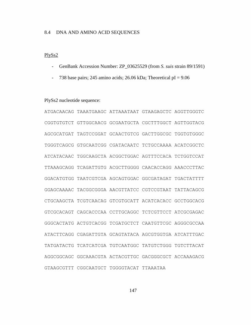
147
8.4 DNA AND AMINO ACID SEQUENCES
PlySs2
- GenBank Accession Number: ZP_03625529 (from S. suis strain 89/1591)
- 738 base pairs; 245 amino acids; 26.06 kDa; Theoretical pI = 9.06
PlySs2 nucleotide sequence:
ATGACAACAG TAAATGAAGC ATTAAATAAT GTAAGAGCTC AGGTTGGGTC
CGGTGTGTCT GTTGGCAACG GCGAATGCTA CGCTTTGGCT AGTTGGTACG
AGCGCATGAT TAGTCCGGAT GCAACTGTCG GACTTGGCGC TGGTGTGGGC
TGGGTCAGCG GTGCAATCGG CGATACAATC TCTGCCAAAA ACATCGGCTC
ATCATACAAC TGGCAAGCTA ACGGCTGGAC AGTTTCCACA TCTGGTCCAT
TTAAAGCAGG TCAGATTGTG ACGCTTGGGG CAACACCAGG AAACCCTTAC
GGACATGTGG TAATCGTCGA AGCAGTGGAC GGCGATAGAT TGACTATTTT
GGAGCAAAAC TACGGCGGGA AACGTTATCC CGTCCGTAAT TATTACAGCG
CTGCAAGCTA TCGTCAACAG GTCGTGCATT ACATCACACC GCCTGGCACG
GTCGCACAGT CAGCACCCAA CCTTGCAGGC TCTCGTTCCT ATCGCGAGAC
GGGCACTATG ACTGTCACGG TCGATGCTCT CAATGTTCGC AGGGCGCCAA
ATACTTCAGG CGAGATTGTA GCAGTATACA AGCGTGGTGA ATCATTTGAC
TATGATACTG TCATCATCGA TGTCAATGGC TATGTCTGGG TGTCTTACAT
AGGCGGCAGC GGCAAACGTA ACTACGTTGC GACGGGCGCT ACCAAAGACG
GTAAGCGTTT CGGCAATGCT TGGGGTACAT TTAAATAA

148
PlySs2 amino acid sequence:
MTTVNEALNN VRAQVGSGVS VGNGECYALA SWYERMISPD ATVGLGAGVG
WVSGAIGDTI SAKNIGSSYN WQANGWTVST SGPFKAGQIV TLGATPGNPY
GHVVIVEAVD GDRLTILEQN YGGKRYPVRN YYSAASYRQQ VVHYITPPGT
VAQSAPNLAG SRSYRETGTM TVTVDALNVR RAPNTSGEIV AVYKRGESFD
YDTVIIDVNG YVWVSYIGGS GKRNYVATGA TKDGKRFGNA WGTFK
- CHAP enzymatic domain (PF05257)
- SH3-Type 5 binding domain (PF08460)

149
9 REFERENCES
2009. "Bacterial coinfections in lung tissue specimens from fatal cases of 2009 pandemic
influenza A (H1N1) - United States, May-August 2009." MMWR Morb Mortal
Wkly Rep 58 (38):1071-4.
Aagaard, K., J. Petrosino, W. Keitel, M. Watson, J. Katancik, N. Garcia, S. Patel, M.
Cutting, T. Madden, H. Hamilton, E. Harris, D. Gevers, G. Simone, P. McInnes,
and J. Versalovic. 2013. "The Human Microbiome Project strategy for
comprehensive sampling of the human microbiome and why it matters." FASEB J
27 (3):1012-22.
Abshire, T. G., J. E. Brown, and J. W. Ezzell. 2005. "Production and validation of the use
of gamma phage for identification of Bacillus anthracis." J Clin Microbiol 43
(9):4780-8.
Adem, P. V., C. P. Montgomery, A. N. Husain, T. K. Koogler, V. Arangelovich, M.
Humilier, S. Boyle-Vavra, and R. S. Daum. 2005. "Staphylococcus aureus sepsis
and the Waterhouse-Friderichsen syndrome in children." N Engl J Med 353
(12):1245-51.
Amass, S. F., C. C. Wu, and L. K. Clark. 1996. "Evaluation of antibiotics for the
elimination of the tonsillar carrier state of Streptococcus suis in pigs." J Vet Diagn
Invest 8 (1):64-7.
Arends, J. P., and H. C. Zanen. 1988. "Meningitis caused by Streptococcus suis in
humans." Rev Infect Dis 10 (1):131-7.

150
Avery, O. T., C. M. Macleod, and M. McCarty. 1944. "Studies on the Chemical Nature of
the Substance Inducing Transformation of Pneumococcal Types : Induction of
Transformation by a Desoxyribonucleic Acid Fraction Isolated from
Pneumococcus Type Iii." J Exp Med 79 (2):137-58.
Baertlein, Lisa. 2011. Death toll from listeria outbreak rises to 29. edited by Cynthia
Johnston: Reuters. Original edition.
Baker, J. R., C. Liu, S. Dong, and D. G. Pritchard. 2006. "Endopeptidase and glycosidase
activities of the bacteriophage B30 lysin." Appl Environ Microbiol 72 (10):6825-
8.
Balogh, B., J. B. Jones, F. B. Iriarte, and M. T. Momol. 2010. "Phage therapy for plant
disease control." Curr Pharm Biotechnol 11 (1):48-57.
Barnett, M. L., and J. A. Linder. 2014. "Antibiotic prescribing to adults with sore throat
in the United States, 1997-2010." JAMA Intern Med 174 (1):138-40.
Becker, S. C., S. Dong, J. R. Baker, J. Foster-Frey, D. G. Pritchard, and D. M. Donovan.
2009. "LysK CHAP endopeptidase domain is required for lysis of live
staphylococcal cells." FEMS Microbiol Lett 294 (1):52-60.
Beres, S. B., and J. M. Musser. 2007. "Contribution of exogenous genetic elements to the
group A Streptococcus metagenome." PLoS One 2 (8):e800.
Berg, R. D. 1996. "The indigenous gastrointestinal microflora." Trends Microbiol 4
(11):430-5.
Bessen, D.E., N. Kumar, G.S. Hall, D.R. Riley, F. Luo, S. Lizano, C.N. Ford, W.M.
McShan, S.V. Nguyen, and J.C. Dunning Hotopp. 2011. "Whole Genome

151
Association Study on Tissue Tropism Phenotypes in Group A Streptococcus."
Journal of Bacteriology:JB. 05263-11v1.
Bisno, A. L., M. O. Brito, and C. M. Collins. 2003. "Molecular basis of group A
streptococcal virulence." Lancet Infect Dis 3 (4):191-200.
Boucher, H. W., G. H. Talbot, D. K. Benjamin, Jr., J. Bradley, R. J. Guidos, R. N. Jones,
B. E. Murray, R. A. Bonomo, D. Gilbert, and America Infectious Diseases
Society of. 2013. "10 x '20 Progress--development of new drugs active against
gram-negative bacilli: an update from the Infectious Diseases Society of
America." Clin Infect Dis 56 (12):1685-94.
Boyer, K.M., C.A. Gadzala, P.D. Kelly, L.I. Burd, and S.P. Gotoff. 1983. "Selective
intrapartum chemoprophylaxis of neonatal group B streptococcal early-onset
disease. II. Predictive value of prenatal cultures." Journal of Infectious Diseases
148 (5):802.
Breitbart, M., B. Felts, S. Kelley, J. M. Mahaffy, J. Nulton, P. Salamon, and F. Rohwer.
2004. "Diversity and population structure of a near-shore marine-sediment viral
community." Proc Biol Sci 271 (1539):565-74.
Broudy, T. B., V. Pancholi, and V. A. Fischetti. 2002. "The in vitro interaction of
Streptococcus pyogenes with human pharyngeal cells induces a phage-encoded
extracellular DNase." Infect Immun 70 (6):2805-11.
Brown, E. R., and W. B. Cherry. 1955. "Specific identification of Bacillus anthracis by
means of a variant bacteriophage." J Infect Dis 96 (1):34-9.

152
Brundage, J. F., and G. D. Shanks. 2007. "What really happened during the 1918
influenza pandemic? The importance of bacterial secondary infections." J Infect
Dis 196 (11):1717-8; author reply 1718-9.
Brundage, J. F., and G. D. Shanks. 2008. "Deaths from bacterial pneumonia during 1918-
19 influenza pandemic." Emerg Infect Dis 14 (8):1193-9.
Bruttin, A., and H. Brussow. 2005. "Human volunteers receiving Escherichia coli phage
T4 orally: a safety test of phage therapy." Antimicrob Agents Chemother 49
(7):2874-8.
Cantin, M., J. Harel, R. Higgins, and M. Gottschalk. 1992. "Antimicrobial resistance
patterns and plasmid profiles of Streptococcus suis isolates." J Vet Diagn Invest 4
(2):170-4.
Carapetis, J.R., A.C. Steer, E.K. Mulholland, and M. Weber. 2005. "The global burden of
group A streptococcal diseases." The Lancet infectious diseases 5 (11):685-694.
Casjens, S. 2003. "Prophages and bacterial genomics: what have we learned so far?" Mol
Microbiol 49 (2):277-300.
CDC. 1999. "From the Centers for Disease Control and Prevention. Four pediatric deaths
from community-acquired methicillin-resistant Staphylococcus aureus--
Minnesota and North Dakota, 1997-1999." JAMA 282 (12):1123-5.
CDC. 2013. Antibiotic Resistance Threats in the United States. U.S. Department of
Health and Human Services, Centers for Disease Control and Prevention. Original
edition.
Celia, L. K., D. Nelson, and D. E. Kerr. 2008. "Characterization of a bacteriophage lysin
(Ply700) from Streptococcus uberis." Vet Microbiol 130 (1-2):107-17.

153
Chambers, H. F., and F. R. Deleo. 2009. "Waves of resistance: Staphylococcus aureus in
the antibiotic era." Nat Rev Microbiol 7 (9):629-41.
Cheng, Q., D. Nelson, S. Zhu, and V. A. Fischetti. 2005. "Removal of group B
streptococci colonizing the vagina and oropharynx of mice with a bacteriophage
lytic enzyme." Antimicrob Agents Chemother 49 (1):111-7.
Clifton-Hadley, F. A. 1984. "Studies of Streptococcus suis type 2 infection in pigs." Vet
Res Commun 8 (3):217-27.
Cloutier, G., S. D'Allaire, G. Martinez, C. Surprenant, S. Lacouture, and M. Gottschalk.
2003. "Epidemiology of Streptococcus suis serotype 5 infection in a pig herd with
and without clinical disease." Vet Microbiol 97 (1-2):135-51.
Coates, T., R. Bax, and A. Coates. 2009. "Nasal decolonization of Staphylococcus aureus
with mupirocin: strengths, weaknesses and future prospects." J Antimicrob
Chemother 64 (1):9-15.
Cunningham, M. W. 2000. "Pathogenesis of group A streptococcal infections." Clin
Microbiol Rev 13 (3):470-511.
Daniel, A., C. Euler, M. Collin, P. Chahales, K. J. Gorelick, and V. A. Fischetti. 2010.
"Synergism between a novel chimeric lysin and oxacillin protects against
infection by methicillin-resistant Staphylococcus aureus." Antimicrob Agents
Chemother 54 (4):1603-12.
David, M. Z., and R. S. Daum. 2010. "Community-associated methicillin-resistant
Staphylococcus aureus: epidemiology and clinical consequences of an emerging
epidemic." Clin Microbiol Rev 23 (3):616-87.

154
de Moor, C. E. 1963. "Septicaemic Infections in Pigs, Caused by Haemolytic
Streptococci of New Lancefield Groups Designated R, S, and T." Antonie Van
Leeuwenhoek 29:272-80.
Dekker, N., A. Bouma, I. Daemen, D. Klinkenberg, L. van Leengoed, J. A. Wagenaar,
and A. Stegeman. 2013. "Effect of spatial separation of pigs on spread of
Streptococcus suis serotype 9." PLoS One 8 (4):e61339.
Deleo, F. R., M. Otto, B. N. Kreiswirth, and H. F. Chambers. 2010. "Community-
associated meticillin-resistant Staphylococcus aureus." Lancet 375 (9725):1557-
68.
Dieterich, G., U. K‰rst, E. Fischer, J. Wehland, and L. J‰nsch. 2006. "LEGER:
knowledge database and visualization tool for comparative genomics of
pathogenic and non-pathogenic Listeria species." Nucleic Acids Research 34
(suppl 1):D402.
Djurkovic, S., J. M. Loeffler, and V. A. Fischetti. 2005. "Synergistic killing of
Streptococcus pneumoniae with the bacteriophage lytic enzyme Cpl-1 and
penicillin or gentamicin depends on the level of penicillin resistance." Antimicrob
Agents Chemother 49 (3):1225-8.
Donovan, D. M., J. Foster-Frey, S. Dong, G. M. Rousseau, S. Moineau, and D. G.
Pritchard. 2006. "The cell lysis activity of the Streptococcus agalactiae
bacteriophage B30 endolysin relies on the cysteine, histidine-dependent
amidohydrolase/peptidase domain." Appl Environ Microbiol 72 (7):5108-12.

155
Donovan, D. M., M. Lardeo, and J. Foster-Frey. 2006. "Lysis of staphylococcal mastitis
pathogens by bacteriophage phi11 endolysin." FEMS Microbiol Lett 265 (1):133-
9.
Doughty, C. C., and J. A. Mann. 1967. "Purification and properties of a bacteriophage-
induced cell wall peptidase from Staphylococcus aureus." J Bacteriol 93
(3):1089-95.
Fenton, M., R. P. Ross, O. McAuliffe, J. O'Mahony, and A. Coffey. 2011.
"Characterization of the staphylococcal bacteriophage lysin CHAP(K)." J Appl
Microbiol 111 (4):1025-35.
Fernandez, L., and R. E. Hancock. 2012. "Adaptive and mutational resistance: role of
porins and efflux pumps in drug resistance." Clin Microbiol Rev 25 (4):661-81.
Fischetti, V. A. 2005. "Bacteriophage lytic enzymes: novel anti-infectives." Trends
Microbiol 13 (10):491-6.
Fischetti, V. A. 2008. "Bacteriophage lysins as effective antibacterials." Curr Opin
Microbiol 11 (5):393-400.
Fischetti, V. A. 2010. "Bacteriophage endolysins: a novel anti-infective to control Gram-
positive pathogens." Int J Med Microbiol 300 (6):357-62.
Fischetti, V. A., D. Nelson, and R. Schuch. 2006. "Reinventing phage therapy: are the
parts greater than the sum?" Nat Biotechnol 24 (12):1508-11.
Fittipaldi, N., M. Segura, D. Grenier, and M. Gottschalk. 2012. "Virulence factors
involved in the pathogenesis of the infection caused by the swine pathogen and
zoonotic agent Streptococcus suis." Future Microbiol 7 (2):259-79.

156
Fry, R. M. 1938. "Prevention and Control of Puerperal Sepsis: Bacteriological Aspects."
Br Med J 2 (4049):340-2.
Gaeng, S., S. Scherer, H. Neve, and M. J. Loessner. 2000. "Gene cloning and expression
and secretion of Listeria monocytogenes bacteriophage-lytic enzymes in
Lactococcus lactis." Appl Environ Microbiol 66 (7):2951-8.
Garcia, J. L., E. Garcia, A. Arraras, P. Garcia, C. Ronda, and R. Lopez. 1987. "Cloning,
purification, and biochemical characterization of the pneumococcal bacteriophage
Cp-1 lysin." J Virol 61 (8):2573-80.
Gilmer, D. B., J. E. Schmitz, C. W. Euler, and V. A. Fischetti. 2013. "Novel
bacteriophage lysin with broad lytic activity protects against mixed infection by
Streptococcus pyogenes and methicillin-resistant Staphylococcus aureus."
Antimicrob Agents Chemother 57 (6):2743-50.
Gottschalk, M., J. Xu, C. Calzas, and M. Segura. 2010. "Streptococcus suis: a new
emerging or an old neglected zoonotic pathogen?" Future Microbiol 5 (3):371-91.
Grandgirard, D., J. M. Loeffler, V. A. Fischetti, and S. L. Leib. 2008. "Phage lytic
enzyme Cpl-1 for antibacterial therapy in experimental pneumococcal
meningitis." J Infect Dis 197 (11):1519-22.
Grundling, A., and O. Schneewind. 2006. "Cross-linked peptidoglycan mediates
lysostaphin binding to the cell wall envelope of Staphylococcus aureus." Journal
of Bacteriology 188 (7):2463-72.
Gu, J., W. Xu, L. Lei, J. Huang, X. Feng, C. Sun, C. Du, J. Zuo, Y. Li, T. Du, L. Li, and
W. Han. 2011. "LysGH15, a novel bacteriophage lysin, protects a murine

157
bacteremia model efficiently against lethal methicillin-resistant Staphylococcus
aureus infection." J Clin Microbiol 49 (1):111-7.
Gupta, R., and Y. Prasad. 2011. "P-27/HP endolysin as antibacterial agent for antibiotic
resistant Staphylococcus aureus of human infections." Curr Microbiol 63 (1):39-
45.
Hare, R., and L. Colebrook. 1934. "The biochemical reactions of hÊmolytic streptococci
from the vagina of febeile and afebeile parturient women." The Journal of
Pathology and Bacteriology 39 (2):429-442.
Harel, J., G. Martinez, A. Nassar, H. Dezfulian, S. J. Labrie, R. Brousseau, S. Moineau,
and M. Gottschalk. 2003. "Identification of an inducible bacteriophage in a
virulent strain of Streptococcus suis serotype 2." Infect Immun 71 (10):6104-8.
Hendrix, R. W. 2002. "Bacteriophages: evolution of the majority." Theor Popul Biol 61
(4):471-80.
Hermoso, J. A., B. Monterroso, A. Albert, B. Galan, O. Ahrazem, P. Garcia, M.
Martinez-Ripoll, J. L. Garcia, and M. Menendez. 2003. "Structural basis for
selective recognition of pneumococcal cell wall by modular endolysin from phage
Cp-1." Structure 11 (10):1239-49.
Herold, B.C., L.C. Immergluck, M.C. Maranan, D.S. Lauderdale, R.E. Gaskin, S. Boyle-
Vavra, C.D. Leitch, and R.S. Daum. 1998. "Community-acquired methicillin-
resistant Staphylococcus aureus in children with no identified predisposing risk."
JAMA: the journal of the American Medical Association 279 (8):593-598.
Hill, J. E., M. Gottschalk, R. Brousseau, J. Harel, S. M. Hemmingsen, and S. H. Goh.
2005. "Biochemical analysis, cpn60 and 16S rDNA sequence data indicate that

158
Streptococcus suis serotypes 32 and 34, isolated from pigs, are Streptococcus
orisratti." Vet Microbiol 107 (1-2):63-9.
Hof, H., K. Szabo, and B. Becker. 2007. "Epidemiology of listeriosis in Germany: a
changing but ignored pattern]." Deutsche medizinische Wochenschrift (1946) 132
(24):1343.
Howden, B. P., J. K. Davies, P. D. Johnson, T. P. Stinear, and M. L. Grayson. 2010.
"Reduced vancomycin susceptibility in Staphylococcus aureus, including
vancomycin-intermediate and heterogeneous vancomycin-intermediate strains:
resistance mechanisms, laboratory detection, and clinical implications." Clin
Microbiol Rev 23 (1):99-139.
Hudson, I. R. 1994. "The efficacy of intranasal mupirocin in the prevention of
staphylococcal infections: a review of recent experience." J Hosp Infect 27 (2):81-
98.
Hussell, T., E. Wissinger, and J. Goulding. 2009. "Bacterial complications during
pandemic influenza infection." Future Microbiol 4 (3):269-72.
Jado, I., R. Lopez, E. Garcia, A. Fenoll, J. Casal, and P. Garcia. 2003. "Phage lytic
enzymes as therapy for antibiotic-resistant Streptococcus pneumoniae infection in
a murine sepsis model." J Antimicrob Chemother 52 (6):967-73.
Johnson, R. P., C. L. Gyles, W. E. Huff, S. Ojha, G. R. Huff, N. C. Rath, and A. M.
Donoghue. 2008. "Bacteriophages for prophylaxis and therapy in cattle, poultry
and pigs." Anim Health Res Rev 9 (2):201-15.
Kavey, Raeew, and E.L. Kaplan. 1989. "Resurgence of acute rheumatic fever." Pediatrics
84 (3):585.
159
Kilpperbalz, R., and K. H. Schleifer. 1987. "Streptococcus-Suis Sp-Nov, Nom-Rev."
International Journal of Systematic Bacteriology 37 (2):160-162.
Klein, E., D. L. Smith, and R. Laxminarayan. 2007. "Hospitalizations and deaths caused
by methicillin-resistant Staphylococcus aureus, United States, 1999-2005." Emerg
Infect Dis 13 (12):1840-6.
Kluytmans, J., A. van Belkum, and H. Verbrugh. 1997. "Nasal carriage of
Staphylococcus aureus: epidemiology, underlying mechanisms, and associated
risks." Clin Microbiol Rev 10 (3):505-20.
Korndorfer, I. P., J. Danzer, M. Schmelcher, M. Zimmer, A. Skerra, and M. J. Loessner.
2006. "The crystal structure of the bacteriophage PSA endolysin reveals a unique
fold responsible for specific recognition of Listeria cell walls." J Mol Biol 364
(4):678-89.
Krause, R. M. 1957. "Studies on bacteriophages of hemolytic streptococci. I. Factors
influencing the interaction of phage and susceptible host cell." J Exp Med 106
(3):365-84.
Kuijper, E. J., B. Coignard, P. Tull, Escmid Study Group for Clostridium difficile, E. U.
Member States, Prevention European Centre for Disease, and Control. 2006.
"Emergence of Clostridium difficile-associated disease in North America and
Europe." Clin Microbiol Infect 12 Suppl 6:2-18.
Lancefield, R.C., and R. Hare. 1935. "The serological differentiation of pathogenic and
non-pathogenic strains of hemolytic streptococci from parturient women." The
Journal of experimental medicine 61 (3):335.

160
Laupland, K.B., T. Ross, and D.B. Gregson. 2008. "Staphylococcus aureus bloodstream
infections: risk factors, outcomes, and the influence of methicillin resistance in
Calgary, Canada, 2000ñ2006." Journal of Infectious Diseases 198 (3):336.
Lederberg, J., and E. M. Lederberg. 1952. "Replica plating and indirect selection of
bacterial mutants." J Bacteriol 63 (3):399-406.
Li, M., B. A. Diep, A. E. Villaruz, K. R. Braughton, X. Jiang, F. R. DeLeo, H. F.
Chambers, Y. Lu, and M. Otto. 2009. "Evolution of virulence in epidemic
community-associated methicillin-resistant Staphylococcus aureus." Proc Natl
Acad Sci U S A 106 (14):5883-8.
Liu, Z., H. Zheng, M. Gottschalk, X. Bai, R. Lan, S. Ji, H. Liu, and J. Xu. 2013.
"Development of multiplex PCR assays for the identification of the 33 serotypes
of Streptococcus suis." PLoS One 8 (8):e72070.
Loeffler, J. M., S. Djurkovic, and V. A. Fischetti. 2003. "Phage lytic enzyme Cpl-1 as a
novel antimicrobial for pneumococcal bacteremia." Infect Immun 71 (11):6199-
204.
Loeffler, J. M., and V. A. Fischetti. 2003. "Synergistic lethal effect of a combination of
phage lytic enzymes with different activities on penicillin-sensitive and -resistant
Streptococcus pneumoniae strains." Antimicrob Agents Chemother 47 (1):375-7.
Loeffler, J. M., D. Nelson, and V. A. Fischetti. 2001. "Rapid killing of Streptococcus
pneumoniae with a bacteriophage cell wall hydrolase." Science 294 (5549):2170-
2.
Loessner, M. J. 2005. "Bacteriophage endolysins--current state of research and
applications." Curr Opin Microbiol 8 (4):480-7.
161
Loessner, M. J., K. Kramer, F. Ebel, and S. Scherer. 2002. "C-terminal domains of
Listeria monocytogenes bacteriophage murein hydrolases determine specific
recognition and high-affinity binding to bacterial cell wall carbohydrates." Mol
Microbiol 44 (2):335-49.
Loessner, M. J., S. K. Maier, H. Daubek-Puza, G. Wendlinger, and S. Scherer. 1997.
"Three Bacillus cereus bacteriophage endolysins are unrelated but reveal high
homology to cell wall hydrolases from different bacilli." J Bacteriol 179
(9):2845-51.
Lood, R., A. Raz, H. Molina, C. W. Euler, and V. A. Fischetti. 2014. "A highly active
and negatively charged Streptococcus pyogenes lysin with a rare D-alanyl-L-
alanine endopeptidase activity protects mice against streptococcal bacteremia."
Antimicrob Agents Chemother.
Lucas, S., A. Copeland, A. Lapidus, et al. 2004. Sequencing of the draft genome and
assembly of Streptococcus suis 89/1591. In Unpublished. Original edition.
Luria, S. E., and M. Delbruck. 1943. "Mutations of Bacteria from Virus Sensitivity to
Virus Resistance." Genetics 28 (6):491-511.
Ma, Y. L., and C. P. Lu. 2008. "Isolation and identification of a bacteriophage capable of
infecting Streptococcus suis type 2 strains." Vet Microbiol 132 (3-4):340-7.
Mandell, Gerald L., R. Gordon Douglas, John E. Bennett, and Raphael Dolin. 2005.
"Mandell, Douglas, and Bennett's principles and practice of infectious diseases."
[Book review (E-STREAMS)]. Elsevier/Churchill Livingstone. http://www.e-
streams.com/es0806_7/es0867_4196.html.

162
Mao, J., M. Schmelcher, W. J. Harty, J. Foster-Frey, and D. M. Donovan. 2013.
"Chimeric Ply187 endolysin kills Staphylococcus aureus more effectively than the
parental enzyme." FEMS Microbiol Lett 342 (1):30-6.
Meng, X., Y. Shi, W. Ji, X. Meng, J. Zhang, H. Wang, C. Lu, J. Sun, and Y. Yan. 2011.
"Application of a bacteriophage lysin to disrupt biofilms formed by the animal
pathogen Streptococcus suis." Appl Environ Microbiol 77 (23):8272-9.
Merabishvili, M., J. P. Pirnay, G. Verbeken, N. Chanishvili, M. Tediashvili, N. Lashkhi,
T. Glonti, V. Krylov, J. Mast, L. Van Parys, R. Lavigne, G. Volckaert, W.
Mattheus, G. Verween, P. De Corte, T. Rose, S. Jennes, M. Zizi, D. De Vos, and
M. Vaneechoutte. 2009. "Quality-controlled small-scale production of a well-
defined bacteriophage cocktail for use in human clinical trials." PLoS One 4
(3):e4944.
Meyn, L.A., M.A. Krohn, and S.L. Hillier. 2009. "Rectal colonization by group B
Streptococcus as a predictor of vaginal colonization." American journal of
obstetrics and gynecology 201 (1):76. e1-76. e7.
Middleton, J. R. 2008. "Staphylococcus aureus antigens and challenges in vaccine
development." Expert Rev Vaccines 7 (6):805-15.
Miller, L. G., F. Perdreau-Remington, G. Rieg, S. Mehdi, J. Perlroth, A. S. Bayer, A. W.
Tang, T. O. Phung, and B. Spellberg. 2005. "Necrotizing fasciitis caused by
community-associated methicillin-resistant Staphylococcus aureus in Los
Angeles." N Engl J Med 352 (14):1445-53.
Morens, D. M., J. K. Taubenberger, and A. S. Fauci. 2009. "The persistent legacy of the
1918 influenza virus." N Engl J Med 361 (3):225-9.

163
Navarre, W. W., H. Ton-That, K. F. Faull, and O. Schneewind. 1999. "Multiple
enzymatic activities of the murein hydrolase from staphylococcal phage phi11.
Identification of a D-alanyl-glycine endopeptidase activity." J Biol Chem 274
(22):15847-56.
Nelson, D., L. Loomis, and V. A. Fischetti. 2001. "Prevention and elimination of upper
respiratory colonization of mice by group A streptococci by using a bacteriophage
lytic enzyme." Proc Natl Acad Sci U S A 98 (7):4107-12.
Nelson, D., R. Schuch, P. Chahales, S. Zhu, and V. A. Fischetti. 2006. "PlyC: a
multimeric bacteriophage lysin." Proc Natl Acad Sci U S A 103 (28):10765-70.
Novick, R. P., G. E. Christie, and J. R. Penades. 2010. "The phage-related chromosomal
islands of Gram-positive bacteria." Nat Rev Microbiol 8 (8):541-51.
O'Flaherty, S., A. Coffey, W. Meaney, G. F. Fitzgerald, and R. P. Ross. 2005. "The
recombinant phage lysin LysK has a broad spectrum of lytic activity against
clinically relevant staphylococci, including methicillin-resistant Staphylococcus
aureus." Journal of Bacteriology 187 (20):7161-4.
O'Flaherty, S., R. P. Ross, and A. Coffey. 2009. "Bacteriophage and their lysins for
elimination of infectious bacteria." FEMS Microbiol Rev 33 (4):801-19.
Owen, J. E., D. W. Schultz, A. Taylor, and G. R. Smith. 1983. "Nucleotide sequence of
the lysozyme gene of bacteriophage T4. Analysis of mutations involving repeated
sequences." J Mol Biol 165 (2):229-48.
Parker, M. T., A. J. Tomlinson, and R. E. Williams. 1955. "Impetigo contagiosa; the
association of certain types of Staphylococcus aureus and of Streptococcus
pyogenes with superficial skin infections." J Hyg (Lond) 53 (4):458-73.

164
Pastagia, M., C. Euler, P. Chahales, J. Fuentes-Duculan, J. G. Krueger, and V. A.
Fischetti. 2011. "A novel chimeric lysin shows superiority to mupirocin for skin
decolonization of methicillin-resistant and -sensitive Staphylococcus aureus
strains." Antimicrob Agents Chemother 55 (2):738-44.
Perch, B., P. Kristjansen, and K. Skadhauge. 1968. "Group R streptococci pathogenic for
man. Two cases of meningitis and one fatal case of sepsis." Acta Pathol Microbiol
Scand 74 (1):69-76.
Pohl, E., R. K. Holmes, and W. G. Hol. 1999. "Crystal structure of a cobalt-activated
diphtheria toxin repressor-DNA complex reveals a metal-binding SH3-like
domain." J Mol Biol 292 (3):653-67.
Porter, C. J., R. Schuch, A. J. Pelzek, A. M. Buckle, S. McGowan, M. C. Wilce, J.
Rossjohn, R. Russell, D. Nelson, V. A. Fischetti, and J. C. Whisstock. 2007. "The
1.6 A crystal structure of the catalytic domain of PlyB, a bacteriophage lysin
active against Bacillus anthracis." J Mol Biol 366 (2):540-50.
Pritchard, D. G., S. Dong, J. R. Baker, and J. A. Engler. 2004. "The bifunctional
peptidoglycan lysin of Streptococcus agalactiae bacteriophage B30."
Microbiology 150 (Pt 7):2079-87.
Pritchard, D. G., S. Dong, M. C. Kirk, R. T. Cartee, and J. R. Baker. 2007. "LambdaSa1
and LambdaSa2 prophage lysins of Streptococcus agalactiae." Appl Environ
Microbiol 73 (22):7150-4.
Ralston, D. J., B. S. Baer, M. Lieberman, and A. P. Krueger. 1955. "Virolysin: a virus-
induced lysin from staphylococcal phage lysates." Proc Soc Exp Biol Med 89
(4):502-7.

165
Ralston, D. J., and M. McIvor. 1964. "Lysis-from-without of Staphylococcus Aureus
Strains by Combinations of Specific Phages and Phage-Induced Lytic Enzymes."
J Bacteriol 88:676-81.
Ramaswamy, V., V. M. Cresence, J. S. Rejitha, M. U. Lekshmi, K. S. Dharsana, S. P.
Prasad, and H. M. Vijila. 2007. "Listeria--review of epidemiology and
pathogenesis." J Microbiol Immunol Infect 40 (1):4-13.
Rashel, M., J. Uchiyama, T. Ujihara, Y. Uehara, S. Kuramoto, S. Sugihara, K. Yagyu, A.
Muraoka, M. Sugai, K. Hiramatsu, K. Honke, and S. Matsuzaki. 2007. "Efficient
elimination of multidrug-resistant Staphylococcus aureus by cloned lysin derived
from bacteriophage phi MR11." J Infect Dis 196 (8):1237-47.
Rasmussen, S. R., F. M. Aarestrup, N. E. Jensen, and S. E. Jorsal. 1999. "Associations of
Streptococcus suis serotype 2 ribotype profiles with clinical disease and
antimicrobial resistance." J Clin Microbiol 37 (2):404-8.
Rhoads, D. D., R. D. Wolcott, M. A. Kuskowski, B. M. Wolcott, L. S. Ward, and A.
Sulakvelidze. 2009. "Bacteriophage therapy of venous leg ulcers in humans:
results of a phase I safety trial." J Wound Care 18 (6):237-8, 240-3.
Robertson, I. D., and D. K. Blackmore. 1989. "Prevalence of Streptococcus suis types 1
and 2 in domestic pigs in Australia and New Zealand." Vet Rec 124 (15):391-4.
Robertson, I. D., D. K. Blackmore, D. J. Hampson, and Z. F. Fu. 1991. "A longitudinal
study of natural infection of piglets with Streptococcus suis types 1 and 2."
Epidemiol Infect 107 (1):119-26.

166
Robinson, J. M., J. K. Hardman, and G. L. Sloan. 1979. "Relationship between
lysostaphin endopeptidase production and cell wall composition in
Staphylococcus staphylolyticus." J Bacteriol 137 (3):1158-64.
Rodriguez, L., B. Martinez, Y. Zhou, A. Rodriguez, D. M. Donovan, and P. Garcia. 2011.
"Lytic activity of the virion-associated peptidoglycan hydrolase HydH5 of
Staphylococcus aureus bacteriophage vB_SauS-phiIPLA88." BMC Microbiol
11:138.
Rohwer, F., D. Prangishvili, and D. Lindell. 2009. "Roles of viruses in the environment."
Environ Microbiol 11 (11):2771-4.
Rouse, M. S., M. Rotger, K. E. Piper, J. M. Steckelberg, M. Scholz, J. Andrews, and R.
Patel. 2005. "In vitro and in vivo evaluations of the activities of lauric acid
monoester formulations against Staphylococcus aureus." Antimicrob Agents
Chemother 49 (8):3187-91.
Rubens, CE, MR Wessels, LM Heggen, and DL Kasper. 1987. "Transposon mutagenesis
of type III group B Streptococcus: correlation of capsule expression with
virulence." Proceedings of the National Academy of Sciences 84 (20):7208.
Schleifer, K. H., and O. Kandler. 1972. "Peptidoglycan types of bacterial cell walls and
their taxonomic implications." Bacteriol Rev 36 (4):407-77.
Schmelcher, M., A. M. Powell, S. C. Becker, M. J. Camp, and D. M. Donovan. 2012.
"Chimeric phage lysins act synergistically with lysostaphin to kill mastitis-
causing Staphylococcus aureus in murine mammary glands." Appl Environ
Microbiol 78 (7):2297-305.

167
Schmelcher, M., V. S. Tchang, and M. J. Loessner. 2011. "Domain shuffling and module
engineering of Listeria phage endolysins for enhanced lytic activity and binding
affinity." Microb Biotechnol 4 (5):651-62.
Schmitz, J. E., R. Schuch, and V. A. Fischetti. 2010. "Identifying active phage lysins
through functional viral metagenomics." Appl Environ Microbiol 76 (21):7181-7.
Schmitz, Jonthan. 2011. "Expanding the Horizons of Enzybiotic Identification." Doctor
of Philosophy, Laboratory of Bacterial Pathogenesis, The Rockefeller University.
Schuch, R., H. M. Lee, B. C. Schneider, K. L. Sauve, C. Law, B. K. Khan, J. A. Rotolo,
Y. Horiuchi, D. E. Couto, A. Raz, V. A. Fischetti, D. B. Huang, R. C. Nowinski,
and M. Wittekind. 2013. "Combination Therapy With Lysin CF-301 and
Antibiotic Is Superior to Antibiotic Alone for Treating Methicillin-Resistant
Staphylococcus aureus-Induced Murine Bacteremia." J Infect Dis.
Schuch, R., D. Nelson, and V. A. Fischetti. 2002. "A bacteriolytic agent that detects and
kills Bacillus anthracis." Nature 418 (6900):884-9.
Schuppler, M., and M. J. Loessner. 2010. "The Opportunistic Pathogen Listeria
monocytogenes: Pathogenicity and Interaction with the Mucosal Immune
System." Int J Inflam 2010:704321.
Seitz, M., A. Beineke, J. Seele, M. Fulde, P. Valentin-Weigand, and C. G. Baums. 2012.
"A novel intranasal mouse model for mucosal colonization by Streptococcus suis
serotype 2." J Med Microbiol 61 (Pt 9):1311-8.
Sieradzki, K., and A. Tomasz. 2003. "Alterations of cell wall structure and metabolism
accompany reduced susceptibility to vancomycin in an isogenic series of clinical
isolates of Staphylococcus aureus." J Bacteriol 185 (24):7103-10.

168
Silva, L. M., C. G. Baums, T. Rehm, H. J. Wisselink, R. Goethe, and P. Valentin-
Weigand. 2006. "Virulence-associated gene profiling of Streptococcus suis
isolates by PCR." Vet Microbiol 115 (1-3):117-27.
Smith, H. W., and M. B. Huggins. 1982. "Successful treatment of experimental
Escherichia coli infections in mice using phage: its general superiority over
antibiotics." J Gen Microbiol 128 (2):307-18.
Smith, H. W., and M. B. Huggins. 1983. "Effectiveness of phages in treating
experimental Escherichia coli diarrhoea in calves, piglets and lambs." J Gen
Microbiol 129 (8):2659-75.
Soothill, J. S. 1994. "Bacteriophage prevents destruction of skin grafts by Pseudomonas
aeruginosa." Burns 20 (3):209-11.
Sriskandan, S., and J. D. Slater. 2006. "Invasive disease and toxic shock due to zoonotic
Streptococcus suis: an emerging infection in the East?" PLoS Med 3 (5):e187.
Sulakvelidze, A., Z. Alavidze, and J. G. Morris, Jr. 2001. "Bacteriophage therapy."
Antimicrob Agents Chemother 45 (3):649-59.
Swedo, S. E., H. L. Leonard, B. B. Mittleman, A. J. Allen, J. L. Rapoport, S. P. Dow, M.
E. Kanter, F. Chapman, and J. Zabriskie. 1997. "Identification of children with
pediatric autoimmune neuropsychiatric disorders associated with streptococcal
infections by a marker associated with rheumatic fever." Am J Psychiatry 154
(1):110-2.
Swildens, B., M. Nielen, H. J. Wisselink, J. H. Verheijden, and J. A. Stegeman. 2007.
"Elimination of strains of Streptococcus suis serotype 2 from the tonsils of carrier
sows by combined medication and vaccination." Vet Rec 160 (18):619-21.

169
Tiemersma, E.W., SL Bronzwaer, O. Lyytikainen, J.E. Degener, P. Schrijnemakers, N.
Bruinsma, J. Monen, W. Witte, and H. Grundman. 2004. "Methicillin-resistant
Staphylococcus aureus in Europe, 1999ñ2002." Emerg Infect Dis 10 (9):1627-34.
Torremorell, M., M. Calsamiglia, and C. Pijoan. 1998. "Colonization of suckling pigs by
Streptococcus suis with particular reference to pathogenic serotype 2 strains." Can
J Vet Res 62 (1):21-6.
Tristan, A., M. Bes, H. Meugnier, G. Lina, B. Bozdogan, P. Courvalin, M. E. Reverdy,
M. C. Enright, F. Vandenesch, and J. Etienne. 2007. "Global distribution of
Panton-Valentine leukocidin--positive methicillin-resistant Staphylococcus
aureus, 2006." Emerg Infect Dis 13 (4):594-600.
Trottier, S., R. Higgins, G. Brochu, and M. Gottschalk. 1991. "A case of human
endocarditis due to Streptococcus suis in North America." Rev Infect Dis 13
(6):1251-2.
Tsugita, A., and M. Inouye. 1968. "Complete primary structure of phage lysozyme from
Escherichia coli T4." J Mol Biol 37 (1):201-12.
Van Duyne, G. D. 2005. "Lambda integrase: armed for recombination." Curr Biol 15
(17):R658-60.
Vandenesch, F., T. Naimi, M. C. Enright, G. Lina, G. R. Nimmo, H. Heffernan, N.
Liassine, M. Bes, T. Greenland, M. E. Reverdy, and J. Etienne. 2003.
"Community-acquired methicillin-resistant Staphylococcus aureus carrying
Panton-Valentine leukocidin genes: worldwide emergence." Emerg Infect Dis 9
(8):978-84.
170
Varela, N. P., P. Gadbois, C. Thibault, M. Gottschalk, P. Dick, and J. Wilson. 2013.
"Antimicrobial resistance and prudent drug use for Streptococcus suis." Anim
Health Res Rev 14 (1):68-77.
Vollmer, W., D. Blanot, and M. A. de Pedro. 2008. "Peptidoglycan structure and
architecture." FEMS Microbiol Rev 32 (2):149-67.
von Eiff, C., K. Becker, K. Machka, H. Stammer, G. Peters, and Study Grp. 2001. "Nasal
carriage as a source of Staphylococcus aureus bacteremia." New England Journal
of Medicine 344 (1):11-16.
Voyich, J. M., K. R. Braughton, D. E. Sturdevant, A. R. Whitney, B. Said-Salim, S. F.
Porcella, R. D. Long, D. W. Dorward, D. J. Gardner, B. N. Kreiswirth, J. M.
Musser, and F. R. DeLeo. 2005. "Insights into mechanisms used by
Staphylococcus aureus to avoid destruction by human neutrophils." J Immunol
175 (6):3907-19.
Waldor, Matthew K., David I. Friedman, and Sankar Lal Adhya. 2005. Phages : their
role in bacterial pathogenesis and biotechnology. Washington, D.C.: ASM Press.
Wang, I. N., D. L. Smith, and R. Young. 2000. "Holins: the protein clocks of
bacteriophage infections." Annu Rev Microbiol 54:799-825.
Wang, Y., J. H. Sun, and C. P. Lu. 2009. "Purified recombinant phage lysin LySMP: an
extensive spectrum of lytic activity for swine streptococci." Curr Microbiol 58
(6):609-15.
Wertheim, H. F. L., D. C. Melles, M. C. Vos, W. van Leeuwen, A. van Belkum, H. A.
Verbrugh, and J. L. Nouwen. 2005a. "The role of nasal carriage in
Staphylococcus aureus infections." Lancet Infectious Diseases 5 (12):751-762.
171
Wertheim, H. F., D. C. Melles, M. C. Vos, W. van Leeuwen, A. van Belkum, H. A.
Verbrugh, and J. L. Nouwen. 2005b. "The role of nasal carriage in
Staphylococcus aureus infections." Lancet Infect Dis 5 (12):751-62.
Wessels, M. R., C. E. Rubens, V. J. Benedi, and D. L. Kasper. 1989. "Definition of a
bacterial virulence factor: sialylation of the group B streptococcal capsule." Proc
Natl Acad Sci U S A 86 (22):8983-7.
White, A., and J. Smith. 1963. "Nasal Reservoir as the Source of Extranasal
Staphylococci." Antimicrob Agents Chemother (Bethesda) 161:679-83.
Wiegand, I., K. Hilpert, and R. E. Hancock. 2008. "Agar and broth dilution methods to
determine the minimal inhibitory concentration (MIC) of antimicrobial
substances." Nat Protoc 3 (2):163-75.
Witzenrath, M., B. Schmeck, J. M. Doehn, T. Tschernig, J. Zahlten, J. M. Loeffler, M.
Zemlin, H. Muller, B. Gutbier, H. Schutte, S. Hippenstiel, V. A. Fischetti, N.
Suttorp, and S. Rosseau. 2009. "Systemic use of the endolysin Cpl-1 rescues mice
with fatal pneumococcal pneumonia." Crit Care Med 37 (2):642-9.
Wright, A., C. H. Hawkins, E. E. Anggard, and D. R. Harper. 2009. "A controlled clinical
trial of a therapeutic bacteriophage preparation in chronic otitis due to antibiotic-
resistant Pseudomonas aeruginosa; a preliminary report of efficacy." Clin
Otolaryngol 34 (4):349-57.
Wu, Z., W. Zhang, and C. Lu. 2008. "Comparative proteome analysis of secreted proteins
of Streptococcus suis serotype 9 isolates from diseased and healthy pigs." Microb
Pathog 45 (3):159-66.

172
Wylie, G. P., V. Rangachari, E. A. Bienkiewicz, V. Marin, N. Bhattacharya, J. F. Love, J.
R. Murphy, and T. M. Logan. 2005. "Prolylpeptide binding by the prokaryotic
SH3-like domain of the diphtheria toxin repressor: a regulatory switch."
Biochemistry 44 (1):40-51.
Xu, Q., P. Abdubek, T. Astakhova, H. L. Axelrod, C. Bakolitsa, X. Cai, D. Carlton, C.
Chen, H. J. Chiu, M. Chiu, T. Clayton, D. Das, M. C. Deller, L. Duan, K. Ellrott,
C. L. Farr, J. Feuerhelm, J. C. Grant, A. Grzechnik, G. W. Han, L. Jaroszewski,
K. K. Jin, H. E. Klock, M. W. Knuth, P. Kozbial, S. S. Krishna, A. Kumar, W. W.
Lam, D. Marciano, M. D. Miller, A. T. Morse, E. Nigoghossian, A. Nopakun, L.
Okach, C. Puckett, R. Reyes, H. J. Tien, C. B. Trame, H. van den Bedem, D.
Weekes, T. Wooten, A. Yeh, K. O. Hodgson, J. Wooley, M. A. Elsliger, A. M.
Deacon, A. Godzik, S. A. Lesley, and I. A. Wilson. 2010. "Structure of the
gamma-D-glutamyl-L-diamino acid endopeptidase YkfC from Bacillus cereus in
complex with L-Ala-gamma-D-Glu: insights into substrate recognition by
NlpC/P60 cysteine peptidases." Acta Crystallogr Sect F Struct Biol Cryst
Commun 66 (Pt 10):1354-64.
Xu, Q., S. Sudek, D. McMullan, M. D. Miller, B. Geierstanger, D. H. Jones, S. S.
Krishna, G. Spraggon, B. Bursalay, P. Abdubek, C. Acosta, E. Ambing, T.
Astakhova, H. L. Axelrod, D. Carlton, J. Caruthers, H. J. Chiu, T. Clayton, M. C.
Deller, L. Duan, Y. Elias, M. A. Elsliger, J. Feuerhelm, S. K. Grzechnik, J. Hale,
G. W. Han, J. Haugen, L. Jaroszewski, K. K. Jin, H. E. Klock, M. W. Knuth, P.
Kozbial, A. Kumar, D. Marciano, A. T. Morse, E. Nigoghossian, L. Okach, S.
Oommachen, J. Paulsen, R. Reyes, C. L. Rife, C. V. Trout, H. van den Bedem, D.

173
Weekes, A. White, G. Wolf, C. Zubieta, K. O. Hodgson, J. Wooley, A. M.
Deacon, A. Godzik, S. A. Lesley, and I. A. Wilson. 2009. "Structural basis of
murein peptide specificity of a gamma-D-glutamyl-l-diamino acid
endopeptidase." Structure 17 (2):303-13.
Yeung, MK, and SJ Mattingly. 1984. "Biosynthetic capacity for type-specific antigen
synthesis determines the virulence of serotype III strains of group B streptococci."
Infection and immunity 44 (2):217.
Yoong, P., R. Schuch, D. Nelson, and V. A. Fischetti. 2004. "Identification of a broadly
active phage lytic enzyme with lethal activity against antibiotic-resistant
Enterococcus faecalis and Enterococcus faecium." J Bacteriol 186 (14):4808-12.
Yoong, P., R. Schuch, D. Nelson, and V. A. Fischetti. 2006. "PlyPH, a bacteriolytic
enzyme with a broad pH range of activity and lytic action against Bacillus
anthracis." J Bacteriol 188 (7):2711-4.
Young, R. 1992. "Bacteriophage lysis: mechanism and regulation." Microbiol Rev 56
(3):430-81.
Yu, H., H. Jing, Z. Chen, H. Zheng, X. Zhu, H. Wang, S. Wang, L. Liu, R. Zu, L. Luo, N.
Xiang, H. Liu, X. Liu, Y. Shu, S. S. Lee, S. K. Chuang, Y. Wang, J. Xu, W.
Yang, and groups Streptococcus suis study. 2006. "Human Streptococcus suis
outbreak, Sichuan, China." Emerg Infect Dis 12 (6):914-20.
Zangwill, KM, A. Schuchat, and JD Wenger. 1992. "Group B streptococcal disease in the
United States, 1990: report from a multistate active surveillance system." MMWR.
CDC surveillance summaries: Morbidity and mortality weekly report. CDC
surveillance summaries/Centers for Disease Control 41 (6):25.

174
Zeng, L., S. O. Skinner, C. Zong, J. Sippy, M. Feiss, and I. Golding. 2010. "Decision
making at a subcellular level determines the outcome of bacteriophage infection."
Cell 141 (4):682-91.
Zetola, N., J. S. Francis, E. L. Nuermberger, and W. R. Bishai. 2005. "Community-
acquired meticillin-resistant Staphylococcus aureus: an emerging threat." Lancet
Infect Dis 5 (5):275-86.
Zimmer, M., N. Vukov, S. Scherer, and M. J. Loessner. 2002. "The murein hydrolase of
the bacteriophage phi3626 dual lysis system is active against all tested
Clostridium perfringens strains." Appl Environ Microbiol 68 (11):5311-7.


![Detection and Characterization of a Lytic Pediococcus ... · hages afp suitater ble dilutioacellular n. Ftr phinor age count [10] during the latent period of 30 min, the phage-host](https://img.dokumen.tips/doc/110x75/5f031c3c7e708231d4079618/detection-and-characterization-of-a-lytic-pediococcus-hages-afp-suitater-ble.jpg)