Embed Size (px)
Citation preview

INVITED REVIEW
Structure–Function Relationships in Highly Modified Shoots of Cactaceae
JAMES D. MAUSETH*
Section of Integrative Biology, 1 University Station, A6700, University of Texas, Austin, TX 78712, USA
Received: 22 March 2006 Returned for revision: 28 April 2006 Accepted: 4 May 2006 Published electronically: 4 July 2006
� Background and Aims Cacti are extremely diverse structurally and ecologically, and so modified as to beintimidating to many biologists. Yet all have the same organization as most dicots, none differs fundamentallyfrom Arabidopsis or other model plants. This review explains cactus shoot structure, discusses relationships betweenstructure, ecology, development and evolution, and indicates areas where research on cacti is necessary to testgeneral theories of morphogenesis.� Scope Cactus leaves are diverse; all cacti have foliage leaves; many intermediate stages in evolutionary reductionof leaves are still present; floral shoots often have large, complex leaves whereas vegetative shoots have microscopicleaves. Spines are modified bud scales, some secrete sugar as extra-floral nectaries. Many cacti have juvenile/adultphases in which the flowering adult phase (a cephalium) differs greatly from the juvenile; in some, one side of a shootbecomes adult, all other sides continue to grow as the juvenile phase. Flowers are inverted: the exterior of a cactus‘flower’ is a hollow vegetative shoot with internodes, nodes, leaves and spines, whereas floral organs occur inside,with petals physically above stamens. Many cacti have cortical bundles vascularizing the cortex, however broad itevolves to be, thus keeping surface tissues alive. Great width results in great weight of weak parenchymatous shoots,correlated with reduced branching. Reduced numbers of shoot apices is compensated by great increases in number ofmeristematic cells within individual SAMs. Ribs and tubercles allow shoots to swell without tearing during wetseasons. Shoot epidermis and cortex cells live and function for decades then convert to cork cambium. Manymodifications permit water storage within cactus wood itself, adjacent to vessels.
Key words: Cactus, epidermis, flower, leaf development, phase change, plant anatomy, shoot apical meristem, structure/function, wood evolution, xerophyte.
INTRODUCTION
The first two objectives of this review are to introducereaders to many of the exotic and extreme aspects ofcactus biology, and also to show that even the mostbizarre cacti are easy to understand because all have thefundamental tissues and organs of an ordinary dicot.People familiar with arabidopsis will find that cacti havethe same basic body organization, just having a bit morecortex, smaller leaves, and axillary buds that develop asclusters of spines. A third objective is to emphasize thediversity of structure, ecology and reproduction in thefamily. Probably no other plant family exceeds Cactaceaein diversity of structure; its members include trees, vines,dwarfs, giants, epiphytes and geophytes. Many aredimorphic, producing different types of anatomy ormorphology at different stages of their lives. The finaland main objective is to point out that many researchtopics in many fields can be studied with this family; thereis already a solid foundation of existing knowledge thatcan be a basis for further studies of morphogenesis,ecology, physiology, evolution and many more areas.
Cactus evolution has been a process of diversification.Starting from some ancestral organization of stems, leavesand roots, cacti have diversified into a multiplicity of bodyforms. Members of subfamily Pereskioideae (Fig. 1A andTable 1) are shrubs or large trees with thin, broad,ordinary-looking leaves and hard, woody, non-succulenttrunks; they are not adapted to dry, hot conditions.Subfamilies Maihuenioideae and Opuntioideae contain
plants with small but still easily visible foliage leaves, andplants vary from being trees to dwarfs (Fig. 1B and C).The largest subfamily, Cactoideae (Fig. 1D), differs fromthe others by having foliage leaves that are alwaysmicroscopic: all photosynthesis is carried out by shootcortex cells covered by a persistent epidermis and stomata,all of which live and function for decades or centuries, aslong as the shoot is green. Members of Cactoideae andOpuntioideae occupy almost every terrestrial habitat: hotdeserts; cold deserts; grasslands; shady forests; rainforests;and cold, wet or snow-covered alpine zones above thetreeline (Mauseth et al., 2002). Several genera ofCactoideae display what appears to be unparalleledin any other group: an absolutely amazing morphogeneticphase change in which the adult body (able to flower)looks nothing at all like the juvenile body (unable toflower). Almost every aspect of shoot morphogenesischanges, each plant produces two totally distinct types ofbody (Figs 1E and F and 2E).
Various aspects of cactus biology have been reviewedrecently, so I will emphasize either newer discoveries orfields that have not received sufficient attention. Twomonographs are recommended for numerous excellentphotographs and general details of plant form anddistribution: Anderson (2001) and Hunt (2006). Theolder work of Backeberg (1958–1962) lacks manyrecently discovered species and modern ideas but hasmuch more detail than any other source (4041 pages in sixvolumes). Natural histories are provided by Rauh (1979)and Mauseth et al. (2002; A Cactus Odyssey beingespecially recommended for a less technical, more* E-mail [email protected]
Annals of Botany 98: 901–926, 2006
doi:10.1093/aob/mcl133, available online at www.aob.oxfordjournals.org
� The Author 2006. Published by Oxford University Press on behalf of the Annals of Botany Company. All rights reserved.
For Permissions, please email: [email protected] from https://academic.oup.com/aob/article-abstract/98/5/901/197087by gueston 10 April 2018

F I G . 1. Cactus shoot structure. (A)Pereskia sacharosawith leaves. Stem about 6mm across. (B)Maihuenia poeppigii, a geophyte; all green colour is due tosmall leaves. The yellow structure is a fruit. The entire plant is about 1 m across. (C) Growing cladode (long-shoot) of Opuntia violacea, with leaves stillpresent (two indicated by arrows). Young cladode is emerging from an axillary bud of an older cladode. The spines of the axillary buds of the older cladode arebud scales. Slightly smaller than life size. (D). Shoot tip of Cereus forbesii. Three of four ribs are visible; spine clusters (axillary buds, areoles) are locatedalong the rib apex. Spines are bud scales, dormant axillary bud shoot apical meristem is located just above each spine cluster, hidden by a mass of whitetrichomes. Spines are present even on very young axillary buds thus protecting the shoot apical meristem, which is not the highest point of the shoot (shootapex is concave). Almost life size. (E) Old plant of Melocactus intortus with juvenile portion of shoot (green) produced during the first 10–15 years of theplant’s life, and the adult portion (red, the cephalium), which is probably at least 10 years old. This is a single shoot (not a graft of two unrelated plants),produced by a single shoot apical meristem. No new chlorenchyma has been produced for years. (F). Shoots of Espostoa with lateral cephalia; flowers areproduced only by axillary budswithin the adult (cephalium) portion, not from the juvenile (green) portions. Note disrupted phyllotaxy. Shoots are about 5 cmacross. (G) Areole (axillary bud) of Ferocactus. Spines are modified bud scales. The location of the bud apical meristem is indicated by an arrow, below themass of trichomes. Trichomes (yellow) are abundant in the areole but absent from the rest of the shoot. Spines and trichomes emerge from a depression about3mmdeep.About four times life size. (H) Leaves on floral bud ofBrowningia candelaris; the largest scale is about 1 cm across. Vegetative shoots of the sameplant have only microscopic foliage leaves. (I) Longitudinal section of fully developed foliage leaf (L) of Oreocereus trollii; present are epidermis, stomata(not visible here), vascular tissue, chlorenchyma, dorsiventral asymmetry. Leaf is 450mm tall. Axillary bud SAM (out of view on left) has produced leafprimordia, one ofwhich is developing as a spine (S). Cells in spine base aremeristematic, those in upper portion are elongating into fibres. Scale bar = 300mm.
902 Mauseth — Structure and Biology of Cactus Shoots
Downloaded from https://academic.oup.com/aob/article-abstract/98/5/901/197087by gueston 10 April 2018

inclusive account of cactus biology). Cactus structureis reviewed by Buxbaum (1950), Gibson and Nobel(1986), Terrazas Salgado and Mauseth (2002) andTerrazas and Arias (2003). Ecophysiology is summarizedby Nobel (1988). Techniques for extracting DNA fromeven mucilaginous cacti are now available (Griffith andPorter, 2003), and DNA-based phylogenies have beenproposed (Nyffeler, 2002; Crozier, 2005; Edwards et al.,2005; Griffith, 2005).
HABIT
In every species, the cactus body organization isfundamentally the same as that of ordinary dicots. Mostcactus leaves are microscopically small (Fig. 1I) and thecortex of most species is gigantically enlarged, but stilleach cactus shoot has the basic dicot organization: allconsist of internodes, nodes, leaves and axillary budsproduced by shoot apical meristems (SAMs). Withoutexception their primary body has an epidermis (withstomata), cortex, eustele (single ring of collateral vascularbundles each with primary xylem and phloem) and pith.No cactus is an annual or an herb. All produce asecondary body consisting of secondary xylem (wood),secondary phloem and bark. All genetic programmes thatguide basic dicot morphogenesis are probably still presentand functional in cacti.
ORGANS OF THE CACTUS SHOOT
Leaves
Evolutionary modification of leaf morphogenesis has beenextensive in all cacti, and has resulted in great diversity ofleaf types within each individual plant. All cacti producefoliage leaves (microscopically small in most) and spines(modified leaves); some also produce glands (modifiedspines), most have large, thin leaves on the surface of
their floral shoots (Fig. 1C, D and G–I; see Floral shootsbelow).
Diversity of cactus leaves is associated with an extremepolymorphism present in all cactus shoots. The green,photosynthetic body of an unbranched cactus is a singleshoot known as a ‘long-shoot’; if branched, all the green,fleshy branches are also long-shoots (Fig. 1A, C and D).Almost all familiar plants consist only of long-shootsso the term is usually unnecessary and rarely used. Butin cacti, each axillary bud immediately produces leafprimordia, which in most other plants would becomesmall, flat, waxy bud scales; in cacti, however, theydevelop into spines (Fig. 1D, G and I; Boke, 1944, 1952,1967; Buxbaum, 1950). Many morphologists haveconsidered a cluster of cactus spines to be just an axillarybud; others interpret it as a ‘short-shoot’, a shoot withextremely short, narrow internodes and without the broad,succulent tissues of the long-shoot. Long-shoot/short-shootdimorphism is not unusual in seed plants. For example,most of an apple tree (Malus) consists of long-shootswhich have ordinary photosynthetic leaves with axillarybuds enclosed by bud scales, but the axillary budsthemselves perennially produce both flowers and photo-synthetic leaves on shoots with extremely short inter-nodes: the axillary buds become short-shoots, often calledspur shoots. In this case, long-shoots and short-shoots bearleaves that are virtually indistinguishable. In contrast,most of the body of a pine tree (Pinus) consists of long-shoots which bear small brown papery scale leaves (easilyoverlooked); the axillary buds of the scale leaves developinto short-shoots with needle-like leaves. The familiarpine needles are not the leaves of the familiar pinebranches (long-shoots) but instead are the leaves of almostinvisible short-shoots (Foster and Gifford, 1974).
Foliage leaves. In all cacti, SAMs (see below) of long-shoots produce leaf primordia (Boke, 1951, 1980;
TABLE 1. Subfamilies of Cactaceae (see Leuenberger, 1986, 1997; Barthlott and Hunt, 1993; Anderson, 2001; Griffith, 2005;Hunt, 2006)
1. Pereskioideae (Fig. 1A) Similar to ordinary dicots: leaves broad, thin with reticulate venation; shoots are trees or shrubs (vines inP. aculeata) with slender stems with thin, relatively nonsucculent cortex; shoot epidermis is ephemeral,being replaced by bark while the stem is only 1 or 2 years old. Pereskia aculeata, P. diaz-romeroana,P. grandifolia and P. sacharosa are easy to cultivate; P. diaz-romeroana is self-fertile and produces seedswhen only 2–4 years old
2. Maihuenioideae (Fig. 1B) Two Patagonian species in one genus, Maihuenia (do not confuse with Maihueniopsis in Opuntioideae).Plants are xeric-adapted small shrubs (M. patagonica of hot lowlands) or geophytes (M. poeppigii,with most of body buried, and only the leafy shoot tips visible above ground; of cold highlands)
3. Opuntioideae (Fig. 1C) A large, diverse group but all members are more highly modified than those of Pereskioideae, all look lesslike ordinary dicots and are more easily recognized as cacti. Most have green, photosynthetic succulentstems. Although small, opuntioid leaves are green, photosynthetic and always easily visible on young shoots.Stems are flattened cladodes (‘pads’ or ‘ears’) in some, have radial symmetry in others, or have cylindricaltrunks with cladodes as lateral branches (Consolea, Brasiliopuntia). Only opuntioids have glochids. Shoots inmost are articulated; each stem has a determinate growth period after which the SAM disorganizes and furthergrowth is by several axillary buds. Many shoots easily break apart at these joints,form adventitious roots and establish extensive clones
4. Cactoideae (Figs 1D–I,2A–I and 3A–D)
A large highly diverse group, none of which would be confused with an ordinary leafy non-succulent dicot.All foliage leaves are too tiny to be visible without aid (except Matucana aurantiaca and several Rhipsalis).Stems vary from long, slender and moderately succulent (Hylocereus, Leptocereus, Selenicereus) to moderatelythick to extremely broad and tall (Carnegiea, Pachycereus, Trichocereus) or broad and globose (Ferocactus,Echinocactus, Echinopsis, Eriosyce), to tiny globose (Table 2). Plants may be highly branched or with few orno branches. Many have extreme phase change between juvenile and adult phases (see Cephalia in text)
Mauseth — Structure and Biology of Cactus Shoots 903
Downloaded from https://academic.oup.com/aob/article-abstract/98/5/901/197087by gueston 10 April 2018

F I G . 2. Specialized features of cacti. (A) Elongated axillary buds ofOroya peruviana, with spines in two rows, not in spiral phyllotaxy around the bud SAM,which is located at the top of each areole. Each areole is about 5 mm long. (B) Tubercles of Coryphantha clavata, each with an elongate areole containingone or two secretory spines (arrow). Note ordinary non-secretory spines at the tips of each tubercle (out of focus in foreground). Tubercles are about 12 mmlong. (C) Elongated areoles of Neoraimondia roseiflora. When first formed, these resembled ordinary areoles as in Fig. 1D, but each has flowered numeroustimes over many years, growing longer each time. Each has bark, cortex, stele and pith. These are about 60 mm long. (D) Dimorphic areoles of Mammillariacamptotricha. Each areole SAMhas divided into two, one being carried outwardwith the tubercle apexwhere it makes only spines, the other remaining at thetubercle base where it produces a flower or a vegetative shoot. The open flower is about 8 mm across. (E) Terminal cephalia of a single, branched plant ofBackebergia militaris. Green portions are juvenile, cephalia are the adult body. Each shoot tip will be abscised about 4 cm below the cephalium, then one ortwo axillary budswill grow as new juvenile bodies for several years, then convert tomaking cephalia. The plant is about 6m tall. (F) Section of a floral shoot ofNeocardenasia. The outer portion is a long-shoot with leaves, nodes and internodes. True flower organs occur only along the inner surface (upper arrowindicates boundary betweenvegetative andfloral organs); petals and stamens, although located physically above the ovary and style base, aremorphologicallylower (proximal). After fertilization, all tissues above the lower arrow will abscise, removing style, stamens, petals and much vegetative tissue. The regionbelow the lower arrowwill develop into a true fruit surrounded by a false fruit. (G) External structure of floral shoot ofEchinocereus. Although referred to as a‘flower,’ this is long-shoot tissue with tiny foliage leaves, axillary buds (bud scales are spines), nodes and internodes. True floral structures are present insidethis shoot (some petal bases are visible at the top). Almost life size. (H) Cortical bundle in Lepismium, with xylem (x), phloem (ph) and a cap of phloem fibres(f). All conducting cells are extremely narrow. Scale bar = 100mm. (I) Collapsible cortex in Haageocereus. Completely turgid palisade cortex cells are
<500mm away in the same region. Scale bar = 100mm.
904 Mauseth — Structure and Biology of Cactus Shoots
Downloaded from https://academic.oup.com/aob/article-abstract/98/5/901/197087by gueston 10 April 2018

Mauseth and Halperin, 1975; Mauseth, 1976, 1977, 1978d,1980a, 2004d). In Pereskia, these develop into large, thin,fully functional photosynthetic foliage leaves with a broadlamina (Fig. 1A; lamina to 23 cm long, 6 cm wide; Bailey,1960; Leuenberger, 1986; Mauseth and Landrum, 1997).These are the main sites of photosynthesis and persist formonths but abscise when plants become dormant.Pereskia foliage leaves may be slightly thickened butnot remarkably so, and palisade and spongy mesophyll areonly weakly differentiated. An extensive reticulatevenation of collateral vascular bundles is present. InMaihuenioideae and Opuntioideae, green photosyntheticleaves are present and all are large enough to be easilyvisible by the naked eye (Fig. 1C; Mauseth, 1999a, 2005;Leuenberger, 1997). They are flattened with a small thick,succulent lamina in Pereskiopsis and Quiabentia, but areradially symmetrical in all other Opuntioideae andMaihuenia, and usually are narrow (2–5mm), short(range 3–12mm, but 120mm long in Austrocylindropuntiasubulata) and ephemeral (persistent in M. poeppigii,Pereskiopsis, Quiabentia and Austrocylindropuntia). Theirphotosynthesis is probably insignificant except whenrelatively large and long-lived. If an opuntioid long-shoot is more than 1 or two months old, there may benothing other than a tiny leaf scar immediately below thecluster of spines.
Long-shoot leaves in all Cactoideae have been greatlyreduced evolutionarily but most have all the tissue typestypical of an ordinary foliage leaf (Fig. 1I; Boke, 1951,1952, 1957b; J. D. Mauseth, unpubl. res.) and thus theyprobably still have leaf development genes similar tothose of other plants (Fleming, 2005). They range fromvery small (maximum 2�3mm long in Matucana aur-antiaca) to microscopic, <500mm long in most. All butthe most miniscule have stomata, at least one vascularbundle, and dorsiventral asymmetry (the vascular bundleis located closer to the adaxial epidermis, and the abaxialmesophyll is slightly aerenchymatous). At least a fewhave a noticeable lamina (up to 1776mm wide in Epi-phyllum) but none has a petiole or abscission zone. Themost reduced long-shoot leaves in Cactoideae do notdevelop beyond the leaf primordium stage, but insteadremain as just a tiny bump (50mm tall) of epidermiscovering several mesophyll cells; their leaf trace typicallyruns only to the axillary bud SAM and spines, not to theleaf itself.
Evolutionary restriction of foliage leaf development hadconsequences other than the obvious ones of reducing theshoot’s surface area and surface : volume (S : V) ratio,reducing transpirational water loss, and reducing photo-synthetic surface area. It also reduced leaf venation, whichis the site of vascular loading and unloading. Ordinaryfoliage leaves have an extensive set of leaf veinsconsisting of primary xylem and phloem and having atremendous length and surface area in contact with livingmesophyll. Shoots of Cactoideae with their highly reducedfoliage leaves have little or no leaf vascular tissue,so water must be unloaded from bundles in the cortex (seeCortical bundles below), from leaf/bud traces or fromsecondary xylem (wood). But in most non-cactus woody
plants, water is almost never unloaded from or loaded intosecondary xylem (roots load water into primary xylem);vessels of wood are surrounded by a matrix of wood fibresor a bit of paratracheal parenchyma, and they do not haveextensive surface contact with a voluminous parenchymacapable of absorbing the water they transport. Woodvessels instead transfer water to primary xylem of leaves,flowers, and so on. Loss of leaf venation in cacti almostcertainly resulted in selection pressure to alter secondaryxylem such that it has increased amounts of paratrachealtissues able to unload, store and transfer water [Fig. 3G;see Secondary xylem (wood) below].
Loss of leaf venation also affected phloem loading;secondary phloem in the central vascular cylinder does notload sugars directly, it only receives them from primaryphloem in leaf traces. Because cacti store water in avoluminous cortex, the outer photosynthetic cortex is toodistant from the secondary phloem of the central cylinderto allow it to load sugars directly. All loading of sugarsapparently must occur in cortical bundles or perhaps leaf/bud traces.
Spines. Cactus spines are the modified bud scales of anaxillary bud; alternatively they can be considered themodified leaves of a short-shoot (Mauseth, 1976; Boke,1980). Differences between the two interpretations are notobvious. Being leaves of an axillary bud, cactus spinesalmost always occur in clusters, a character whichdistinguishes this family from all others. Several cactihave only one spine per cluster, and spines are completelyabsent in Blossfeldia (Mauseth, 2006a) and someepiphytic rainforest cacti (some Epiphyllum, Lepismium,Rhipsalis; Fig. 3D). Almost as soon as the axillary budSAM becomes recognizable, it develops zonation typicalof any angiosperm, having a uniseriate tunica over acorpus composed of central cells, peripheral zone andpith-rib meristem. It immediately produces leaf primordia;these resemble long-shoot leaf primordia in being smallswellings of ground meristem covered by protoderm. Asthe axillary bud’s leaf primordia enlarge, their tip cellsvacuolate and elongate, and quickly the young spineconsists of three regions: a basal meristem; a zone ofelongation/differentiation and an apical zone of mature;and dead lignified fibres (Fig. 1I; Mauseth, 1977).
The spine basal meristem consists of only a unistratoseprotoderm surrounding a mass of ground meristem. Novascular tissue or procambium has been reported. Mostcell division produces daughter cells aligned parallel tothe spine’s long axis, but occasional divisions in otherplanes widen the basal meristem gradually, thus cactusspines taper from a narrow tip to a broader base (Fig. 1G).Spines are frequently circular in transverse section but canbe flattened on one side (usually the adaxial side;Ferocactus latispinus) or their basal meristem becomesso broad but thin that the spine is flat and papery,mimicking a dry blade of grass (Leuchtenbergia principis,Tephrocactus articulatus: spines 4mm wide, 0�3mmthick, to 15 cm long). Factors that control morphogenesisin spine basal meristems are unknown, but in many cactithese meristems are accessible large masses of uniform
Mauseth — Structure and Biology of Cactus Shoots 905
Downloaded from https://academic.oup.com/aob/article-abstract/98/5/901/197087by gueston 10 April 2018

F I G . 3. Cactus structures. (A) Prostrate shoot of Harrisia pomanensis. The shoot tip is elevated by reaction cortex near the phloem on the lower side of theshoot. The visible part of the shoot is about 40 cm long; the entire shoot is several metres long, branched and growing in various directions. The lower side hasadventitious roots. (B) Three ribs of a columnar cactus, Coleocephalocereus. As the shoot loses water and volume, ribs become narrower but do not changesurface area. Each spine cluster is an axillary bud; subtending foliage leaves (like those in Fig. 1I) are microscopic. Each areole could potentially produce avegetative long-shoot (a branch); because this species has lateral cephalia, these are juvenile phase areoles and cannot bloom. Each rib base is about 10 mmacross. (C) Tubercles ofMammillaria magnimamma, produced in obvious phyllotactic spirals. Tuberculate shoots shrink vertically aswell as radially aswateris lost. Being aMammillaria, this has divided, dimorphic areoles: areole SAMs at tubercle tips produce only spines; areole SAMs at tubercle bases (hidden bywhite trichomes) produce floral shoots or vegetative shoots (none are present in the photograph). Each tubercle is about 10mm long. (D) Dimorphic shoots ofEpiphyllum caudatum; the branch on the leftwas initially terete but immediately switched to distichous phyllotaxywith just two tall, thin ribs. The ‘midvein’ isthe central vascular cylinder, the ‘blade’ is the two ribs and notches along the rib crests are the axillary buds (spines aremicroscopic). The vertical shoot on theright is terete here, but its tip had also switched to growing as a two-ribbed leaf-like structure. The cladode is about 30 mm across. (E) Wide-band tracheidwood of Thelocactus bicolor in transverse section. The double-headed arrow in the lower portion indicates the region with many vessels (dark red, narrowerwalls), perhaps earlywood. The upper portion of themicrograph ismostlyWBTs (perhaps latewood),with two vessels (arrows). InmanyWBTs, the band-likesecondary wall almost occludes the lumen. Scale bar = 100mm. (F) Tangential section of WBT wood in Thelocactus; WBTs are short and imperforate, andin this species the secondary wall occurs as one or two helices per cell. Blue is the flexible primary wall; despite the thick secondary wall, these cells shortenand lengthen as the water content changes. Cells near the left, lacking wide-bands, are ray parenchyma cells. Scale bar = 100mm. (G) Transverse view(macroscopic) of Consoleawood. Rays (white, arrowed) are very wide (1 to 3 mm); vessels within the axial masses (tan) are close to water stored in the rays.These rays interconnect water stored in the pith and cortex. The image is about 30mm across. (H) Dimorphicwood of Stenocereus. The double-headed arrowindicates primary xylem and first-formed secondary xylem, both lacking fibres and instead having WBTs, vessels and xylem parenchyma. After severalmonths, the vascular cambium switched to making fibrous wood (above upper arrowhead), consisting of vessels, xylem parenchyma and xylary fibres but noWBTs. Scale bar = 100mm. (I) Transverse section of secondary phloem of Corryocactus. The arrow indicates collapsed phloem, below which are abundant
sieve tube members and companion cells. A phloem fibre cap is at the top of the image, secondary xylem at the bottom. Scale bar = 100mm.
906 Mauseth — Structure and Biology of Cactus Shoots
Downloaded from https://academic.oup.com/aob/article-abstract/98/5/901/197087by gueston 10 April 2018
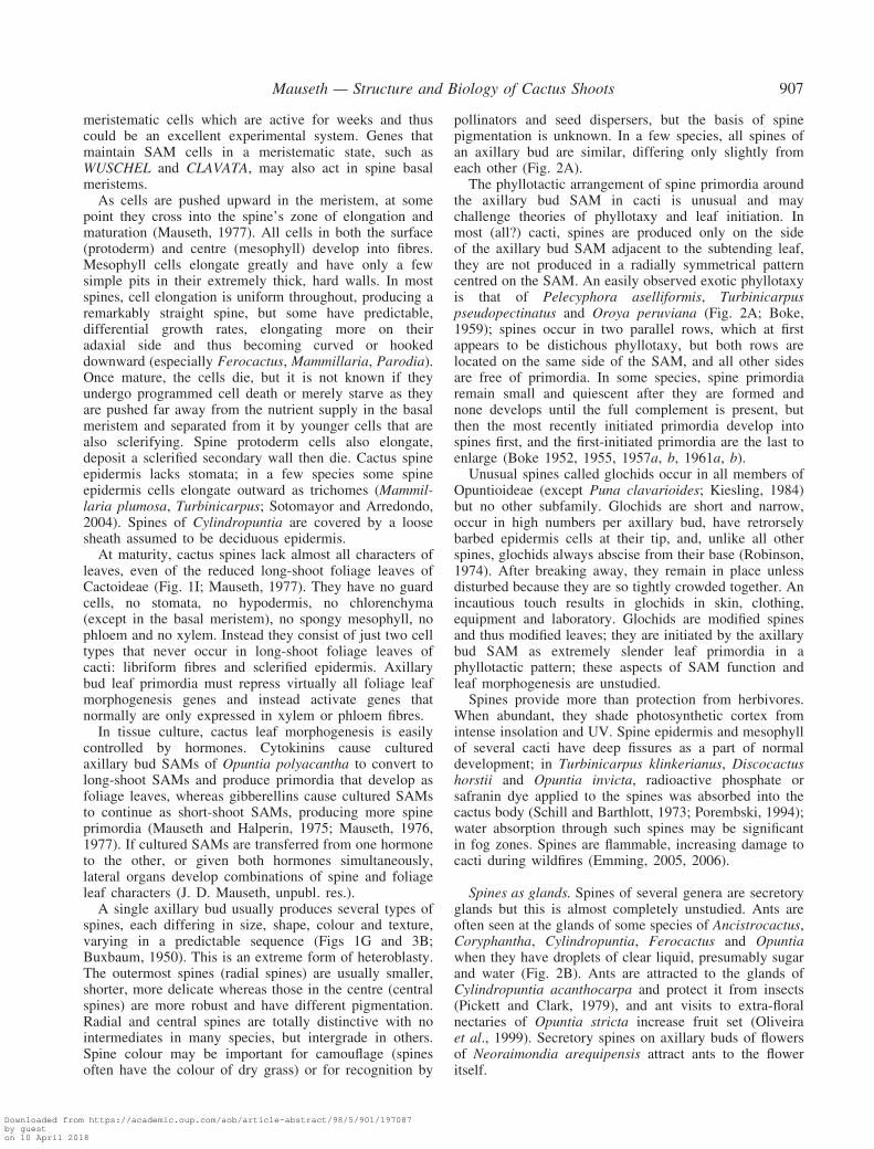
meristematic cells which are active for weeks and thuscould be an excellent experimental system. Genes thatmaintain SAM cells in a meristematic state, such asWUSCHEL and CLAVATA, may also act in spine basalmeristems.
As cells are pushed upward in the meristem, at somepoint they cross into the spine’s zone of elongation andmaturation (Mauseth, 1977). All cells in both the surface(protoderm) and centre (mesophyll) develop into fibres.Mesophyll cells elongate greatly and have only a fewsimple pits in their extremely thick, hard walls. In mostspines, cell elongation is uniform throughout, producing aremarkably straight spine, but some have predictable,differential growth rates, elongating more on theiradaxial side and thus becoming curved or hookeddownward (especially Ferocactus, Mammillaria, Parodia).Once mature, the cells die, but it is not known if theyundergo programmed cell death or merely starve as theyare pushed far away from the nutrient supply in the basalmeristem and separated from it by younger cells that arealso sclerifying. Spine protoderm cells also elongate,deposit a sclerified secondary wall then die. Cactus spineepidermis lacks stomata; in a few species some spineepidermis cells elongate outward as trichomes (Mammil-laria plumosa, Turbinicarpus; Sotomayor and Arredondo,2004). Spines of Cylindropuntia are covered by a loosesheath assumed to be deciduous epidermis.
At maturity, cactus spines lack almost all characters ofleaves, even of the reduced long-shoot foliage leaves ofCactoideae (Fig. 1I; Mauseth, 1977). They have no guardcells, no stomata, no hypodermis, no chlorenchyma(except in the basal meristem), no spongy mesophyll, nophloem and no xylem. Instead they consist of just two celltypes that never occur in long-shoot foliage leaves ofcacti: libriform fibres and sclerified epidermis. Axillarybud leaf primordia must repress virtually all foliage leafmorphogenesis genes and instead activate genes thatnormally are only expressed in xylem or phloem fibres.
In tissue culture, cactus leaf morphogenesis is easilycontrolled by hormones. Cytokinins cause culturedaxillary bud SAMs of Opuntia polyacantha to convert tolong-shoot SAMs and produce primordia that develop asfoliage leaves, whereas gibberellins cause cultured SAMsto continue as short-shoot SAMs, producing more spineprimordia (Mauseth and Halperin, 1975; Mauseth, 1976,1977). If cultured SAMs are transferred from one hormoneto the other, or given both hormones simultaneously,lateral organs develop combinations of spine and foliageleaf characters (J. D. Mauseth, unpubl. res.).
A single axillary bud usually produces several types ofspines, each differing in size, shape, colour and texture,varying in a predictable sequence (Figs 1G and 3B;Buxbaum, 1950). This is an extreme form of heteroblasty.The outermost spines (radial spines) are usually smaller,shorter, more delicate whereas those in the centre (centralspines) are more robust and have different pigmentation.Radial and central spines are totally distinctive with nointermediates in many species, but intergrade in others.Spine colour may be important for camouflage (spinesoften have the colour of dry grass) or for recognition by
pollinators and seed dispersers, but the basis of spinepigmentation is unknown. In a few species, all spines ofan axillary bud are similar, differing only slightly fromeach other (Fig. 2A).
The phyllotactic arrangement of spine primordia aroundthe axillary bud SAM in cacti is unusual and maychallenge theories of phyllotaxy and leaf initiation. Inmost (all?) cacti, spines are produced only on the sideof the axillary bud SAM adjacent to the subtending leaf,they are not produced in a radially symmetrical patterncentred on the SAM. An easily observed exotic phyllotaxyis that of Pelecyphora aselliformis, Turbinicarpuspseudopectinatus and Oroya peruviana (Fig. 2A; Boke,1959); spines occur in two parallel rows, which at firstappears to be distichous phyllotaxy, but both rows arelocated on the same side of the SAM, and all other sidesare free of primordia. In some species, spine primordiaremain small and quiescent after they are formed andnone develops until the full complement is present, butthen the most recently initiated primordia develop intospines first, and the first-initiated primordia are the last toenlarge (Boke 1952, 1955, 1957a, b, 1961a, b).
Unusual spines called glochids occur in all members ofOpuntioideae (except Puna clavarioides; Kiesling, 1984)but no other subfamily. Glochids are short and narrow,occur in high numbers per axillary bud, have retrorselybarbed epidermis cells at their tip, and, unlike all otherspines, glochids always abscise from their base (Robinson,1974). After breaking away, they remain in place unlessdisturbed because they are so tightly crowded together. Anincautious touch results in glochids in skin, clothing,equipment and laboratory. Glochids are modified spinesand thus modified leaves; they are initiated by the axillarybud SAM as extremely slender leaf primordia in aphyllotactic pattern; these aspects of SAM function andleaf morphogenesis are unstudied.
Spines provide more than protection from herbivores.When abundant, they shade photosynthetic cortex fromintense insolation and UV. Spine epidermis and mesophyllof several cacti have deep fissures as a part of normaldevelopment; in Turbinicarpus klinkerianus, Discocactushorstii and Opuntia invicta, radioactive phosphate orsafranin dye applied to the spines was absorbed into thecactus body (Schill and Barthlott, 1973; Porembski, 1994);water absorption through such spines may be significantin fog zones. Spines are flammable, increasing damage tocacti during wildfires (Emming, 2005, 2006).
Spines as glands. Spines of several genera are secretoryglands but this is almost completely unstudied. Ants areoften seen at the glands of some species of Ancistrocactus,Coryphantha, Cylindropuntia, Ferocactus and Opuntiawhen they have droplets of clear liquid, presumably sugarand water (Fig. 2B). Ants are attracted to the glands ofCylindropuntia acanthocarpa and protect it from insects(Pickett and Clark, 1979), and ant visits to extra-floralnectaries of Opuntia stricta increase fruit set (Oliveiraet al., 1999). Secretory spines on axillary buds of flowersof Neoraimondia arequipensis attract ants to the floweritself.
Mauseth — Structure and Biology of Cactus Shoots 907
Downloaded from https://academic.oup.com/aob/article-abstract/98/5/901/197087by gueston 10 April 2018

Glandular spines of Ancistrocactus scheeri are short,broad, and taper abruptly to a sharp, narrow spine-like tip;their mesophyll cells are short, blunt, living fibres withthin walls and large intercellular spaces (Mauseth, 1982).Sugars and water secreted by these fibres exude from thetop of the glandular spine. After some unknown period,each gland collapses. In Ancistrocactus and Coryphantha,numerous glandular spines are formed in each axillary budover a period of at least several months, perhaps >1 year;only one or two glands are active at any time, but anyparticular axillary bud will be producing secretory productfor a protracted time (Dicht and Luthy, 2005).
Secretory spines in Calymmanthium substerile producea thick white material. This has been seen only in culti-vated plants protected from rain. It has not been studied.
Axillary buds
In cactus literature, the region in a long-shoot leaf axilis called an ‘areole’, not simply an axillary bud. This termis useful because the bud’s spines persist even if theaxillary bud SAM goes on to produce a flower and fruit.Flowering in most angiosperms causes bud scaleabscission, so after the fruit is shed, the region is littlemore than a set of scars, but in cacti the entire set ofspines is still present. Furthermore, some cacti producespines for a prolonged period, longer than most axillarybuds produce bud scales, so these growing structures aremore appropriately considered short-shoots rather thanmerely buds. ‘Areole’ refers to the region at all stages ofits development.
Cactus axillary buds become active immediately andproduce spine primordiawhile still within a fewmicrometresof the long-shoot SAM, still within its apical depression(Boke, 1944, 1952, 1980; Mauseth et al., 2002). Spineprimordia themselves develop immediately, such that manyspines project upward, protecting the shoot apex fromherbivores (Fig. 1D).Young axillary buds are carried upwardand outward with growth of the cortex, and leaf/bud traceselongate as well. Typically, cortex immediately interior to anaxillary bud stops expanding slightly earlier than doessurrounding cortex, thus the bud becomes located in its ownwell-like depression (only a few millimetres deep and wide;Figs 1D andG and 2A). This depression is lined by epidermisand hypodermis, both beingmore delicate and having thinnercell walls than epidermis and hypodermis cells locatedbetween areole depressions. In Blossfeldia liliputana,stomata are not present anywhere except in the areoledepressions (Barthlott and Porembski, 1996; Mauseth,2006a), and in Maihuenia poeppigii (Fig. 1B; Mauseth,1999a) areole depressions are the only areas in whichepidermis does not immediately convert to cork cambium,so in this species, too, areole depressions are the only regionsof the stem that have stomata (M. poeppigii has persistentmacroscopic foliage leaves, B. liliputana does not).
The axillary bud SAM produces an abundance ofuniseriate, multicellular trichomes along with spineprimordia. In most species, it appears as if every singleareole epidermis cell becomes either part of a spineprimordium or a trichome; there appear to be no ordinary
epidermis cells within the areole. Trichomes in all speciesdie immediately, thus the SAM is protected by an almostimpenetrable mass of dead trichomes and spines.
After producing spine primordia and trichomes (andglochids in Opuntioideae), the axillary bud SAM remainscapable of further growth, either as a floral bud (Fig. 2G),a vegetative branch (a long-shoot; Figs 1C and 3D), or asa short-shoot. In species that bloom with flowers on newgrowth near the shoot tip (many species), the axillary budSAM develops as a floral bud as soon as spine primordiumproduction is completed. If axillary bud SAMs becomedormant for 1 or more years after forming spines, theplant blooms with flowers located farther from the shootapex. In many species of Hatiora, Rhipsalis, Schlumber-gera and Opuntioideae, young axillary buds immediatelygrow out as branches (Buxbaum, 1950), but, in most cacti,branching only occurs from axillary buds that are severalto many years old and which are thus located in regionswith enough strength to support the weight of branches.Many plants branch only from axillary buds located at thebase of the trunk: their SAMs remain dormant fordecades, yet develop as normal branches. Many giantcolumnar cacti and barrel-shaped cacti have few or nobranches while growing normally; of their thousands ofaxillary buds (about 10 000 axillary buds in single shootsof Trichocereus pasacana; J. D. Mauseth, unpubl. res.),most do nothing other than produce spines and flowers.However, if these shoots are cut off near their base, one orseveral axillary buds become active and grow out asbranches: they were suppressed by extreme apicaldominance.
Axillary buds in some cacti are capable of more thanproducing only one flower and later one branch. Buds ofLepismium cruciforme, Myrtillocactus, Pachycereus gate-sii, P. marginatus, P. schottii and Rhipsalis russellii bearseveral flowers or fruits simultaneously (Barthlott andTaylor, 1995; Arias et al., 2003), those of Neoraimondia(including Neocardenasia) produce several flowers peryear for many years. Each time a flower is produced theaxillary bud becomes slightly longer and the reason forcalling it a short-shoot becomes more obvious. Withextreme age (how old?), Neoraimondia short-shootsbecome up to 85mm long, and may even branch; theyhave pith, secondary xylem and phloem, cortex and bark(Fig. 2C; Rauh, 1957; Mauseth and Kiesling, 1997;Kiesling and Mauseth, 2000).
Other unusual aspects of the growth pattern ofNeoraimondia arequipensis are worth mentioning here.Their long-shoots are massively succulent, very broad andheavy (40 cm in diameter) and grow to 7m tall. At thatpoint, a long-shoot stops growing and one of its basal-most areoles grows out as a lateral shoot right at groundlevel, its eventual weight supported by the soil. Lateralshoots apparently grow rapidly because their apical-most15 or 30 cm of epidermis has the clean, fresh look ofbeing <1 year old; within a few years, this branch reachesits full length and stops, then another basal areole repeatsthe process. A typical plant has five to ten giant branchesthat have stopped elongating and one single branch that isgrowing: apparently the plant channels most resources to
908 Mauseth — Structure and Biology of Cactus Shoots
Downloaded from https://academic.oup.com/aob/article-abstract/98/5/901/197087by gueston 10 April 2018

one branch at a time. But the non-growing branches arenot moribund: they are photosynthesizing and theiraxillary buds all flower perennially (Mauseth et al.,2002). Neoraimondia biology has many intriguing aspectsbut these giant, frost-sensitive plants are not easy tocultivate.
Axillary buds of many Opuntioideae, a few Cactoideaeand several Pereskia occasionally and sporadicallyproduce a new spine from time to time over manyyears. These too are short-shoots but always remain only afew millimetres long; their anatomy has not been studied.
Unusual branching of axillary buds. In cacti and mostother stem-succulents, cortex below and surrounding anaxillary bud grows outward in the form of a cone (tubercle)or ridge (rib; Figs 1D and E and 3B and C; see Ribs andtubercles below). Growth of ribs and tubercles has notbeen studied, but they appear to have a basal growth zonelocated proximal to the axillary bud, between it and thestem. Consequently their growth causes the axillary bud tobe carried outward along with the tip of the rib or tubercle,so axillary buds and all associated spines, flower parts orbranches are located at the apex of a rib or tubercle.
In contrast, in a small subgroup of Cactoideae (e.g.Coryphantha, Mammillaria), the growth zone is locateddirectly below the axillary bud SAM, which consequentlyis stretched as the tubercle grows (Boke, 1952, 1953,1955, 1958, 1961a, b; Dicht and Luthy, 2005). InMammillaria, the bud SAM always divides dichotomouslyand one of the two new SAMs is carried outward alongwith the tubercle tip while the other remains stationary, atthe base of the tubercle. Both new meristems becomeradially symmetrical but have different fates: the distalSAM at the tubercle tip produces only spine primordia, itnever flowers or grows as a lateral branch, whereas theproximal one does produce flowers and lateral branchesbut almost never spines (Fig. 2D). Flowers or newbranches of mammillarias emerge from between thecrowded bases of the tubercles, not from the tubercle tipsand not adjacent to the spines as is typical of most cacti.Remarkably, if tubercle tips are cultured with high levelsof cytokinin, the spine-producing SAM can be induced toform a branch with microscopic foliage leaves on long-shoots (J. D. Mauseth, unpubl. res.).
In Coryphantha and Ancistrocactus, growth of thetubercle below the axillary bud causes the SAM toelongate but not divide dichotomously. Perhaps it acts asif forming a crest because it produces leaf primordia alongits entire length. These leaf primordia develop intoextrafloral nectaries (glandular spines; Fig. 2B; see Spinesas glands above). In addition, radial growth of the tuberclecortex upward below the elongate SAM is inhibited, so thenectaries are located in a groove running along thetubercle’s adaxial side (Boke, 1961b).
Phase change, heteroblasty and the transition fromjuvenile to adult
Seedlings of most angiosperms produce leaves andstems that differ at least slightly from those produced
when the plant is older. This is called ‘heteroblasty,’ butadditional characters differ between seedlings and olderplants, and the term ‘phase change’ is more inclusive(Howell, 1998). Phase change is occasionally associatedwith conversion from the juvenile state (incapable offlowering) to the adult state (able to flower). In classicexamples such as Citrus and Hedera, the juvenile/adulttransition occurs simultaneously with phase change, butin many species, phase change is completed before thejuvenile/adult transition occurs: the plant grows with itsmature phase morphology for one to several years beforebecoming old enough to flower.
All cacti undergo phase change. Compared with olderplants, seedlings have narrower primary stems with fewercortex and pith cells; more delicate epidermis andhypodermis; shorter ribs or tubercles (and species withribbed adults may have tuberculate seedlings); shorter,more delicate spines. Most produce wide-band tracheids(WBTs; see Wide-band tracheids below; Fig. 3E, F and H)in their primary and secondary xylem (Loza-Cornejo et al.,2003; Mauseth, 2004c). As the seedling ages, eachsuccessive bit of growth becomes more robust, itscharacters progressively more similar to those of anolder plant. Species that will grow to have slender shootsstop producing WBT wood and switch to making fibrouswood instead (see Dimorphic wood below). These changesare not accompanied by a juvenile/adult transition becausealmost no cactus can bloom before it is 1 year old (inmany cases, several years or decades old), so most cactigrow with their mature morphology for years even thoughthey are still juvenile.
The juvenile/adult transition is accompanied by noobvious morphological changes in most cacti, but in othersthere are stunning changes in anatomy, morphology andphysiology. The differences between juvenile andadult phases are much more extensive and dramatic thanthose of any other group of plants. Once old enough toflower, these cacti produce an adult body called acephalium.
Terminal cephalia. Melocactus and Discocactus (do notconfuse with Disocactus) have terminal cephalia. Youngplants grow as juveniles with unbranched globose to shortcylindrical shoots with prominent ribs and areoles, eachwith a small number of stout spines (Fig. 1E; Mauseth,1989). Most of the green shoot surface is unobscured,visible and photosynthetic because ribs are large, areolessmall and spines are few. The juvenile phase lasts severalto many years, varying with species and growingconditions, and the biochemical trigger to become adultis unknown: plants of M. matanzanus (commerciallyavailable) grown with fertilizer, water and full sunlightbecome adults while only 3 years old, during which timethey have produced about 160 leaves and areoles. As aplant converts from juvenile to adult, almost all aspects ofits growth change. The adult shoot—the cephalium—isproduced by the same SAM that produced the juvenileshoot (juvenile shoot and cephalium are the two ends ofone shoot) (Niklas and Mauseth, 1981; Mauseth, 1989).Phyllotaxy becomes very high, and the adult SAM
Mauseth — Structure and Biology of Cactus Shoots 909
Downloaded from https://academic.oup.com/aob/article-abstract/98/5/901/197087by gueston 10 April 2018

produces small, closely spaced tubercles rather thanprominent ribs as it did while it was juvenile. Spinenumber per areole increases greatly and cephalium spinesare short and slender. Trichomes are produced in abun-dance. Because areoles are so closely spaced and thedensity of spines and trichomes is so high, the surface ofthe adult shoot is completely hidden under an impenet-rable, solid mass (about 1�0 cm thick) of spines and deadtrichomes (Mauseth et al., 2002). No light penetrates thismass, photosynthesis is impossible, and there are nostomata, no guard cells, no ordinary epidermis cells. Theadult SAM becomes smaller, produces a narrower cortexfree of chloroplasts but with cortical bundles and closelyspaced leaf/bud traces. Cephalium pith is also narrow,so the entire adult region is much narrower than thejuvenile. The transition is abrupt with little or nointermediate tissues. Adult shoot secondary xylem consistsof WBTs and vessels, and that of the juvenile shootconsists of an inner region of fibrous wood surrounded byan outer, more recently produced layer of WBT wood;apparently the juvenile/adult transition also affects thevascular cambium such that once the SAM begins pro-ducing adult morphology at the shoot’s apex, the cambiumbegins producing wood with adult morphology throughoutthe shoot.
Axillary buds in the cephalium produce flowers. Beinglocated on a tiny tubercle below a thick layer of spinesand trichomes, each flower bud is remarkably wellprotected from predation. During anthesis flower budselongate and petals curve outward just above the spines.Ovules and nectaries, still located at the base of the massof spines, are accessible to pollinators through a petal-lined flower tube. Flowers close after just 1 d, the perianthwithers and remains in place, protecting the ovary. Whenready, the mature fruit swells, pushing itself up above themass of spines, becoming visible to seed dispersers(Cortes Figueira et al., 1994).
A cephalium in this position is a terminal cephaliumbecause it is at the apex of the shoot, not because itterminates the plant’s growth. Instead, the plant continuesits growth for many years, but as with any other speciesthat undergoes a juvenile/adult transition, all furthergrowth is with the adult organization (the cephalium isnot an inflorescence, is not ephemeral). The cephaliumbecomes longer every year, every year there are moreflowers and fruits, but every year the juvenile portionmerely becomes older—and it is the only photosynthetictissue the plant has. Because the shoot is produced byone single SAM and does not branch, no new photo-synthetic cortex can be added, so the ratio of photosyn-thetic tissue to heterotrophic tissue decreases every year.Melocacti easily become 20 or 30 years old in cultivation,continuing to rely on the same, old chlorenchyma cellsthey produced when they were juveniles. Under normalconditions, melocacti never branch, but if a mature plantis decapitated, an axillary bud of either the cephalium orthe juvenile body will grow out as a lateral branchwith juvenile characters. At some point this switches toadult growth, a new cephalium. All 33 species ofMelocactus have this morphology; no known species
retains intermediate stages in cephalium evolution (Taylor,1991).
Discocacti resemble melocacti, but their cephalia growmore slowly and even old plants that have bloomed foryears have only extremely short cephalia. Discocactigrafted onto hardy rootstocks are easy to cultivate.
Plants of Backebergia militaris (Pachycereus militaris)are giant columnar cacti, up to 5m tall (Fig. 2E). Theygrow as juveniles with broad stems and prominent ribsuntil at least 3–4m tall, then they add several ribs andsoon switch to producing tubercles (Cattabriga, 2004;Mauseth et al., 2005). In this short transition region,spines are shorter, narrower, more brittle and translucent.Production of trichomes increases. The adult body is onlyslightly narrower than the juvenile, and the thick layer oflong, densely packed spines causes the cephalium toappear broader than the juvenile body. Unlike Melocactus,the adult body of B. militaris does have a few cells thatbecome ordinary epidermis cells, and stomata are present;the outer cortex is weakly chlorophyllous, but certainlylittle light penetrates the spines. Just as in Melocactus andDiscocactus, each year the cephalium becomes longer,whereas the juvenile body remains the same length.
Backebergia, however, periodically abscises its ceph-alia, which releases one of the uppermost axillary buds onthe juvenile portion from apical dominance. The budgrows out as a new lateral branch with juvenilemorphology and fresh chlorophyllous tissue. Once thebranch becomes 2–3m long, it too converts to adultgrowth and becomes topped with a terminal cephalium.This process occurs repeatedly, resulting in giant, high-ly branched plants. Backebergia militaris cannot toleratefrost but small plants grow readily in a greenhouse. Adultportions of Pachycereus schottii have more and longerspines than do juvenile portions.
Terminal, temporary cephalia occur in Arrojadoa,Cephalocereus (Neodawsonia) apicicephalium andStephanocereus leucostele (Table 2). After the shoot hasbloomed with a set of flowers emerging from a cephaliumencircling the shoot tip, its SAM returns to vegetativegrowth and then makes a segment (many centimetreslong) of green stem incapable of flowering. In thefollowing year, it makes another terminal, temporarycephalium. The shoot alternates between non-floweringzones and ring-shaped flowering zones, which remainrecognizable for years, long after all flowers and fruitshave matured and abscised; internal anatomy is notknown.
Stephanocereus luetzelburgii, a poorly known species,grows as a broad column until about 20 cm tall, then itswitches to growing as a much narrower column, perhapsaccompanied by a juvenile/adult transition (Taylor andZappi, 2004).
Lateral cephalia. Lateral cephalia are regions with adultcharacters located on one side of the shoot, not at its apex(Fig. 1F). They have been studied in Cephalocereus(Vasquez Sanchez et al., 2005) and Espostoa (includingVatricania; Buxbaum, 1952, 1959; Rauh, 1957; Mauseth,1999b; Mauseth et al., 2002). A seedling grows as a
910 Mauseth — Structure and Biology of Cactus Shoots
Downloaded from https://academic.oup.com/aob/article-abstract/98/5/901/197087by gueston 10 April 2018

juvenile green column with prominent ribs and stout,sparse spines on all sides for several years. When oldenough to undergo the juvenile/adult transition, develop-ment of only some ribs on one side is altered, all tissuesbeing added to the other ribs on the rest of the bodycontinue to develop with juvenile characters; as the shootcontinues to grow from one single SAM, some leafprimordia and their associated node and internode tissuesdevelop with adult morphology, the rest develop withjuvenile characters. Adult characters are similar to thosein Melocactus: cortex is thin and non-chlorophyllous;short, small tubercles with long, slender spines areproduced instead of large ribs and stout spines; andthere are also abundant trichomes and bark. Only areolesin the cephalium produce flowers; other areoles at thesame level (thus with the same age) but in juvenileregions do not. Differential growth of cortex and ribs/tubercles disrupts phyllotaxy but the SAM is not affectedand continues to grow for years, simultaneously producingreproductive adult tissues and chlorophyllous juveniletissues. Lateral cephalia occur in numerous genera(Table 2).
In Pilosocereus, areoles that produce flowerssimultaneously produce copious amounts of longtrichomes, giving the shoot the appearance of having acephalium. However, internal portions of the shoot arenot affected, and once the trichomes break off afterseveral years, that portion of the shoot looks like anyother; such regions are pseudocephalia. Cephalocleis-tocactus produces exceptionally long spines on justone side, giving the impression of a weakly formedlateral cephalium, but flowering is not restricted to those
areoles. All areoles are adult, so the role of the cephalium-like region is unknown.
Other types of phase change. The juvenile/adulttransition of Browningia candelaris is more or less theopposite of producing a terminal cephalium. Juvenileplants grow as vertical, unbranched determinate columnswith prominent ribs and abundant long spines. Once theshoot reaches about 2m tall, it stops growing and five toten apical areoles grow out as lateral branches. These areslightly narrower than the juvenile shoot (the trunk), havemany low ribs with short weak spines that could almost beoverlooked. These branches constitute the adult body andare the only part that bears flowers (Mauseth et al., 2002).
Several species, especially Lepismium (Pfeiffera) ian-thothele appear to be neotenous: their adult bodiesstrongly resemble the seedling phase of other lepismiums(Barthlott and Taylor, 1995).
Dwarfism, gigantism
Evolutionary dwarfism of shoots appears common in-cacti. The ancestors of cacti were probably woody, non-succulent trees or large shrubs; this body form occursin Pereskia (Pereskioideae), Maihuenia patagonica (Mai-huenioideae), Pereskiopsis (Opuntioideae) and Cactoideae(Leptocereus, Calymmanthium, Acanthocereus and manyothers). However, many clades now have genera orspecies whose plants consist of dwarf shoots <10 cm tall,often <3 cm (Kiesling, 1995; Table 2). These have WBTwood (see Wide-band tracheids below), a type of woodcharacteristic of seedlings, so dwarfism may be linked toneoteny.
TABLE 2. Genera with at least some species with unusual features; especially noteworthy species are indicated (for illustrationsand more examples, see Anderson, 2001; Mauseth et al., 2002; Hunt, 2006)
Broad stems Carnegiea gigantea, Cephalocereus, Coryphantha (C. elephantidens), Echinocactus (E. grusonii, which iseasy to obtain and cultivate), Echinopsis (E. [Soehrensia] bruchii), Ferocactus, Gymnocalycium (G. saglionis),Neobuxbaumia (N. macrocephala, N. tetetzo), Oreocereus (O. celsianus), Pachycereus (gigantic plants),Trichocereus (T. pasacana, T. terscheckii)
Cephalia Terminal cephalia: Backebergia (Pachycereus) militaris, Discocactus, MelocactusTerminal but temporary cephalia: Arrojadoa, Cephalocereus (Neodawsonia) apicicephalium, StephanocereusLateral cephalia: Coleocephalocereus (Buiningia), Espostoa (Pseudoespostoa, Thrixanthocereus, Vatricania),Espostoopsis (Austrocephalocereus, Gerocephalus), Facheiroa, Micranthocereus (Siccobaccatus)Pseudocephalium: Cereus mortensenii, Pilosocereus
Climbing byadventitious roots
Hylocereus, Selenicereus, Weberocereus
Determinate shoots Hatiora, Opuntioideae (all species), Rhipsalis, SchlumbergeraDwarf shoots Ariocarpus agavoides, A. scaphirostris, Astrophytum asterias, Blossfeldia liliputana, Copiapoa hypogaea,
C. laui, C. tenuissima, Echinopsis chamaecereus (Chamaecereus silvestrii), Epithelantha, Eriosyce esmeraldana,E. krausii, Eriosyce occulta, Escobaria duncanii, E. minima, Frailea, Maihuenia poeppigii (Maihuenioideae),Maihueniopsis (M. clavarioides: height above soil level = 0.0 cm), Mammillaria luethyi, M. saboae, Mila,Parodia nothominuscula, P. subterranea, P. tenuicylindrica, Pediocactus (tiny plants but difficult to cultivate),Pterocactus (Opuntioideae), Rebutia, Sclerocactus (difficult), Turbinicarpus
Leaf production rapid Aporocactus flagelliformis, Cleistocactus, Espostoa, Hylocereus, Pilosocereus, Selenicereus (vigorous grower)Leaf production slow Ariocarpus, Lophophora, Pediocactus, SclerocactusExtremely few branches Ariocarpus, Astrophytum, Blossfeldia, Carnegiea, Cephalocereus, Ferocactus, Mammillaria, Melocactus,
Neobuxbaumia, Oreocereus celsianus, Pachycereus, Trichocereus pasacana, T. terscheckii. Some species ofFerocactus, Mammillaria and Pachycereus are highly branched
Prostrate columnar Cereus kronleinii, Echinopsis hahniana, Haageocereus decumbens, H. icensis, H. tenuis, Harrisia pomanensis,Praecereus saxicola, Stenocereus eruca
Ribs only two Disocactus, Epiphyllum, Hatiora (Rhipsalidopsis, Epiphyllopsis, Pseudozygocactus), Lepismium, Pseudorhipsalis,Rhipsalis (R. elliptica, R. pachyptera, R. russellii), Schlumbergera (‘Christmas cacti’, commonly calledZygocactus), Selenicereus anthonyanus
Mauseth — Structure and Biology of Cactus Shoots 911
Downloaded from https://academic.oup.com/aob/article-abstract/98/5/901/197087by gueston 10 April 2018

In contrast, many species of Pachycereus (P. fulviceps,P. weberi), Trichocereus (T. atacamensis, T. pasacana,T. terscheckii), Cephalocereus senilis and Neobuxbaumiatetetzo are gigantic. Within Opuntioideae, O. echios ofthe Galapagos Islands must be the result of tremendousevolutionary increase in body size from a smaller ancestor.Griffith (2004a, b) has pointed out that all subfamilies ofCactaceae except Pereskioideae also contain small, dwarfgeophytic plants, thus it is theoretically possible that verysmall body size is basal in the family.
Floral shoots
Most cactus flowers are inside out, with perianthlocated physically above stamens, both located abovecarpels, all buried deeply within a shoot (Fig. 2F; Boke,1963, 1964, 1966, 1968; Leuenberger, 1986). When acactus axillary bud produces a flower, it first initiatesseveral to many leaf primordia, nodes and internodes, thenswitches to producing the primordia of petals (there areoften no distinctively sepal-like structures), stamens andcarpels. All primordia are present in an ordinary acropetalsequence while the bud is microscopic (Ross, 1982).When cell enlargement occurs, it stops earlier in thecentre of the bud than in peripheral regions, so the ovaryis elevated little, stamens are elevated a bit more andpetals are elevated most. All floral organ primordiabecome located on the inside of a conical depression in theend of the elongating floral shoot (Fig. 2F). At the rim-likeapex of the depression are the first-formed, most proximalperianth primordia and the last-formed, most distal leafprimordia; on the outside of the floral shoot areprogressively older leaves and areoles in ordinary phyl-lotactic sequence (Fig. 2F and G). The object we see whenviewing a cactus ‘flower’ from the side is really just along-shoot (not flower) surmounted by petals. The trueflower (except for the petals) is completely hidden insidethe long-shoot. Floral shoots are >10 cm long in manycacti and reach 30 cm in Epiphyllum crenatum andE. oxypetalum (Anderson, 2001): ovary and ovules are30 cm below the uppermost leaves. After pollination andfertilization, the true fruit develops inside the base of thelong-shoot, which itself develops as a false fruit; just asin an apple fruit, the boundary between inner true fruit andouter false fruit is not readily apparent. Only the regionimmediately exterior to the ovary converts to false fruit,all the distal long-shoot tissue is abscised along with thestyle, stamens and perianth (most cacti have dozens ofstamens and petals, an important consideration for theABC model of floral morphogenesis).
Each node of the floral branch often has a scale-likeleaf and an axillary cluster of spines. In Cylindropuntiafulgida (‘chain fruit cholla’), C. leptocaulis, Pereskiasacharosa and a few others, the axillary buds on the‘flower’ produce floral shoots which later become ‘fruits’whose axillary buds repeat the process. Axillary buds onthe false fruits of C. leptocaulis even produce non-floralbranches. Calymmanthium substerile goes one step farther:the apical rim with the last leaves and the first petals doesnot grow radially as the floral shoot elongates, so it
remains just a tiny hole. During anthesis, the flower’sexpansion actually rips the floral shoot open such thatpetals, stamens and style elongate through ruptured, dyinglong-shoot tissues (Mauseth et al., 2002).
The long-shoot nature of the exterior of a cactus‘flower’ is important because in many genera its nodesbear large, thin photosynthetic leaves (Fig. 1H). They arereferred to as ‘scales’ or ‘bracts’ but they develop fromleaf primordia, have a lamina (23mm long, 14mm widein Browningia candelaris; Mauseth et al., 2002) withextensive leaf venation, axillary buds, and some have anabscission zone. Thus almost all ‘leafless’ cacti (subfamilyCactoideae) have not only microscopic long-shoot foliageleaves (see Leaves above) but also large, relativelyordinary leaves as well (they differ from petals, which arepigmented and lack axillary buds and spines). Many cactiare adapted to mesic habitats in which virtually allassociated plants have photosynthetic leaves, and rain-forest epiphytic cacti occur in very moist habitats. Yetnone of these uses floral leaf genes to produce large,photosynthetic leaves on their vegetative body. As mostcacti evolved to be exclusively stem-photosynthetic, theygave up the beneficial capacity that drought-deciduousplants have; modern cacti (other than pereskias, maihue-nias and some Opuntioideae) cannot have an extensivephotosynthetic surface area (large leaves) during rainyseasons and then abscise that extra surface area duringdrought. Many Euphorbia and Pachypodium combinestem-succulence with stem-photosynthesis and drought-deciduous leaves; it seems as if cacti should have thegenetic capacity to do this also, but instead remain‘leafless’ except when flowering.
TISSUES OF THE CACTUS SHOOT:PRIMARY BODY
Shoot apical meristems
Evolutionary modification of one aspect of plant biologyoften affects other aspects. Such interactions are extensivein cacti, and the co-evolution involving SAMs, increasedcortex succulence and decreased branching is especiallyinteresting. All cacti with relatively narrow stems(diameter <1�0 cm) have SAMs between 90 and 300 mm,a rather ordinary size for seed plants (Boke, 1941;Gifford, 1954; Mauseth, 1978d, 2004d). However, allcacti with greatly enlarged stems (due to having a verythick cortex, which is possible due to having corticalbundles; see Cortex below) have exceptionally largeSAMs, up to 2565mm (>2�5mm) diameter in Echinocactusplatyacanthus (also E. grusonii, which is commerciallyavailable and easy to cultivate). The only other plantswith such large SAMs are cycads, which also havebroad-diameter primary bodies and high phyllotaxy(Foster, 1940). The exceptionally broad cortex makescactus shoots exceptionally heavy per unit length; e.g. a1�0-m-long section of Trichocereus pasacana shootweighs about 32 000 g (J. D. Mauseth, unpubl. res.)while an equal length of Arabidopsis thaliana wouldweigh <0�5 g. Almost certainly, this increased weight
912 Mauseth — Structure and Biology of Cactus Shoots
Downloaded from https://academic.oup.com/aob/article-abstract/98/5/901/197087by gueston 10 April 2018

caused selection of mutations that decrease branching, andmany cacti with broad stems have few or no branches(Cody, 2002; Table 2).
Related to reduced branching is a reduced number ofSAMs that produce the shoot system. In non-succulenttrees such as Pinus or Acer, each plant is highly branched,each has thousands of twigs and thus thousands of SAMs.Even if each SAM is of ordinary size with only a fewhundred cells, the shoot is being produced by hundreds ofthousands of apical meristem cells. In contrast, sparselybranched cacti have only a few SAMs, and unbranchedcacti have just one single SAM that produces the entireshoot body (reminder: the bulk of cactus shoots consists ofprimary tissues derived directly from a SAM, notsecondary tissues produced by cambia). If unbranchedcacti had a SAM of ordinary size with just a few hundredmeristem cells, each cell would have to undergo tens ofthousands of rounds of cell division, and the number ofcopy-error mutations created with each replication wouldaccumulate to unacceptable levels (Klekowski, 1988)before a cactus shoot had reached maturity. But becausesparsely branched cacti have gigantic SAMs, each withthousands of cells, the number of rounds of cell divisionrequired of each meristem cell is reduced, as is the risk ofintroducing copy-error mutations.
The evolution of SAMs up to 2500mm in diameter fromancestors whose SAMs were much smaller must haverequired extensive modification of genes that control shootapex morphogenesis. SAM genes in Arabidopsis thalianaand other model plants probably play the same rolesin cacti but must have evolved to interact over muchgreater distances and greater volumes of meristematiccells (Mauseth, 2004d).
Rate of leaf production and length of plastochron varytremendously in the family, although studies are needed.SAMs in Ariocarpus, Lophophora, Pediocactus andSclerocactus may produce only one to five leaf primordiaper year (Table 2), but plants of Cleistocactus, Espostoaand several other genera probably have the highest leafproduction rates and the shortest plastochrons in the entireplant kingdom. Shoots of C. strausii have up to 30 ribs,each with leaves and axillary buds (spines clusters)located every 3mm; a shoot may grow 300mm per year,which is 100 leaves per rib, and 3000 leaves on all 30 ribs.The growing season is about 9 months or 270 d, thus eachSAM produces about 11 leaves per day with a plastochronof 2�2 h (J. D. Mauseth, unpubl. res.).
Cactus SAMs are located in depressions at the shoot tip,they are not the most apical point physically (Fig. 1D).Newly formed cortex cells grow upward slightly soonerthan do newly formed pith cells, so cortex actuallyprotrudes beyond the SAM. The apical depression may beas much as 3�0 cm deep and 20 cm wide in large globosecacti such as Echinocactus or Echinopsis, so newlyformed epidermis and leaf primordia are carried upwardand outward by growth of subapical tissues. Following arow of leaf primordia in its phyllotactic spiral from oldestto youngest, you would follow them up the outside of theshoot, across the ring-shaped top of the shoot and thendown the inside of the apical depression.
Indeterminate SAMs and monopodial growth. Theorderly nature of ribs and phyllotactic spirals of tuberclesindicates that shoot growth is monopodial and indeterm-inate in most Cactoideae. Cactus SAMs become dormantin winter or dry seasons but never form terminal buds and,in almost all cases, the first-formed nodes and internodesof one year grow to be as wide as those of the previousyear, so columnar cacti tend to have uniform, straight ribsand globose cacti have uniform spirals of tubercles.Seasonal growth increments along a shoot’s length canoccasionally be identified due to constrictions of the ribsor markings in their cuticle (Otis and Buskirk, 1986). InBackebergia militaris immature portions of ribs in thedormant shoot apex form a bit of bark, which preventsthem from expanding fully in the following growingseason: the shoot’s longitudinal growth increments aremarked by constrictions (Fig. 2D; Mauseth et al., 2005).Constricted monopodial shoots are especially pronouncedin Armatocereus, whose shoots consist of verticallyaligned segments, strongly resembling the jointed bodiesof an Opuntia or Cylindropuntia (but these latter havedeterminate shoots and sympodial growth). Demographicstudies may be possible with Armatocereus; each segmentdemarcated by a constriction indicates a single growthepisode. In A. procerus, they may be correlated toepisodic El Nino rains rather than annual growth cycles(Mauseth et al., 2002).
Determinate SAMs and sympodial growth. In contrast tothe evolution of giant SAMs in many Cactoideae, SAMsin Opuntioideae evolved to be determinate, functioningbriefly before being replaced by a branch derived froman axillary bud (Fig. 1C). This is almost universalin Opuntioideae; only Pereskiopsis, Brasiliopuntia, Con-solea and Tacinga have any indeterminate shoots. Plantsof Pereskiopsis are highly branched shrubs with manyindeterminate shoots (Arias Montes, 1996); those ofBrasiliopuntia and Consolea have a single indeterminate,radially symmetrical trunk but all branches are determin-ate, laterally flattened cladodes. Another species, Tacingafunalis, has radially symmetrical, indeterminate shoots.All other opuntioid genera grow with determinate SAMsonly (Mauseth, 2005). The jointed cylindrical stems ofchollas (Cylindropuntia) are each determinate shoots; theflat ‘pads’ or ‘ears’ of prickly pears (genus Opuntia orsubgenus Platyopuntia) are determinate cladodes. Lessfamiliar opuntioid genera such as Maihueniopsis,Pterocactus and Tephrocactus consist of sympodial setsof globose determinate shoots (Kiesling, 1982, 1984; Huntand Taylor, 2002; Mauseth et al., 2002; Griffith, 2005).Typically, the SAM has finished producing all internodes,nodes, leaf primordia and axillary buds while it is still<1�0 cm long. Apparently their SAMs convert to masses oflarge parenchyma cells at maturity. In contrast, aseedling’s epicotyl SAM persists longer, although it toois ultimately determinate; this needs study (Table 3).
Determinate SAMs are associated with unusualbranching patterns and shoot polymorphism in severalrainforest epiphytes in Cactoideae (Barthlott and Taylor,1995). In Hatiora salicornioides, each determinate shoot
Mauseth — Structure and Biology of Cactus Shoots 913
Downloaded from https://academic.oup.com/aob/article-abstract/98/5/901/197087by gueston 10 April 2018

has one narrow, long basal internode (1mm by 3–4mm)followed by four to six broad, short internodes (3mm ·1mm), followed by five to eight internodes that form aconcave disc at the shoot apex; shoots are shaped likeinverted beer or wine bottles (common name: ‘drunkard’sdream’). Its SAM disorganizes into a plate of parenchyma,and three to five of the axillary buds in the topmost flatdisc become active simultaneously, forming a whorl ofshoots identical to the one to which they are attached. Thispattern repeats indefinitely. In Rhipsalis mesembryan-themoides, an axillary bud near the base of a shoot growsout as a long (20 cm) determinate shoot; almost all itsaxillary buds grow out as short (1�5 cm) determinateshoots which do not branch under ordinary circumstancesbut which bear flowers. Once the long shoot stopsgrowing, one of its basal-most branches grows out asanother long shoot. In Rhipsalis burchellii, a basal axillarybud grows out as a long (60 cm) determinate shoot. Onceit stops growing, several apical-most axillary buds growout as a whorl of shorter determinate shoots, and whenthey stop, several apical-most axillary buds on each ofthem grow out as a whorl of even shorter determinateshoots. This repeats until the last determinate shoots growto be only about 6�0 cm long, then the pattern repeatsas one of the basal-most axillary buds on the originallong shoot grows out as another very long determinateshoot.
Dichotomous branching of SAMs. Dichotomous branch-ing occurs in at least two species of Mammillaria (M.perbella and M. parkinsonii; Boke, 1976) and one Echi-nocereus (E. reichenbachii; Boke and Ross, 1978) andsporadically in several other genera. For several years,plants grow as unbranched short columnar shoots withradial symmetry and a set of intersecting phyllotacticspirals of tubercles. At some point in time, the shoot apexbecomes oval rather than round and phyllotaxy becomesabnormal. The shoot apex becomes even more elongateand gradually the phyllotaxy resolves itself into twoseparate sets of phyllotactic spirals, each set centred onthe ends of the oval-shaped apex: the apex has dividedinto two separate SAMs, each producing ordinary radiallysymmetrical shoots. After several years, both apices ofM. perbella divide dichotomously again, this timeperpendicular to the previous division.
During dichotomous branching the SAM temporarilyswitches to bilateral symmetry, and, if viewed in medianlongitudinal section, it is extremely broad due to a lateralexpansion of the central cell zone and pith-rib meristem.The peripheral zone appears unaffected (Boke, 1976;Boke and Ross, 1978). Cells in the centre of thebroadened SAM begin dividing regularly, giving rise toa layered pattern typical of a peripheral zone, and leafprimordia are formed in the centre of the broad apex. Atthis point, the SAM has divided into two separatemeristems. If the SAM is viewed in a median longitudinalsection perpendicular to that described above, it appearsnormal throughout the process.
Perhaps related to dichotomous branching is formationof crested shoots. A SAM becomes extremely broadin one plane as described above, but instead of dividing, itcontinues to broaden (Boke and Ross, 1978). SAMs asmuch as 1m wide are known, and despite havingdimensions on the order of 50mm tall · 200 mm thick ·1000 000mm wide, they produce leaf primordia, nodes,internodes and axillary buds. Phyllotaxy is irregularin most crests. Crested cacti are often propagated bycuttings; many can be obtained for research. Colour-basedchimeras are also now available commercially.
Cortex
Cortical bundles. A key innovation in the evolution ofmany cacti (in particular Cactoideae) must have been theacquisition of cortical bundles, a network of collateralbundles that vascularizes the cortex and permitted it toevolve to a thickness not found in any other plant of anykind (Fig. 2H). Although several cactus clades havenarrow shoots with a relatively thin cortex, mostCactoideae have cortexes that range from extremelybroad to extraordinary compared with shoots of all non-cactus plants; the cortex is 300mm thick in Echinocactusplatyacanthus (compared with 0�048mm in Arabidopsisthaliana), and a range of 10–70mm is common in Cactoi-deae (mean thickness is 19�9mm; Mauseth, 2000).
Cortical bundles apparently were not an early stepin cactus evolution. They are completely absent fromsubfamilies Pereskioideae, Maihuenioideae and Opuntioi-deae; they are present in all Cactoideae except Blossfeldialiliputana (Boke, 1980; Mauseth and Sajeva, 1992;Mauseth and Landrum, 1997; Mauseth, 1999a, b;Terrazas and Arias, 2003; Mauseth, 2005, 2006a). DNAcladograms suggest B. liliputana is the earliest-divergentmember of Cactoideae (Nyffeler, 2002; Crozier, 2004,2005), so presence of cortical bundles is a synapomorphyfor the rest of Cactoideae.
Cortical bundles are critically important for theevolution of a broad cortex because, even if a stem hasa thick, wax-covered cuticle, it gradually loses water todry desert air, so epidermis, hypodermis and outer regionsof cortex must be kept hydrated by some means. If thecortex is unvascularized (as it is in almost all vascularplants; Howard, 1979), then water must move from thecentral vascular cylinder to epidermis by diffusion, whichis slow over distances of more than a few millimetres
TABLE 3. Resources for research on cacti (these lists do notinclude all possible sources or journals)
Sources ofplant material
Huntington Botanical Garden (San Marino, CA),Desert Botanical Garden (Phoenix, AZ),Le Jardin Exotique (Monaco), StadtischeSukkulentensammlung (Zurich), plus numerouscommercial nurseries
Reviewedjournals
Bradleya, Haseltonia, Desert Plants
Natural historyjournals
British Cactus and Succulent Journal, Cactusand Succulent Journal (USA), Kakteen undandere Sukkulenten (Germany), Quepo (Peru),Succulentes (France, Monaco)
914 Mauseth — Structure and Biology of Cactus Shoots
Downloaded from https://academic.oup.com/aob/article-abstract/98/5/901/197087by gueston 10 April 2018

(Barcikowski and Nobel, 1984), thus limiting any increasein cortex thickness. But with the evolution of corticalbundles, water can be transported rapidly in bulk anddistributed throughout the outermost regions of the stem,keeping chlorenchyma, hypodermis and epidermishydrated no matter how distant they are from the xylemin the central ring of bundles. Similarly, as cortex evolvedto be thicker, the chlorenchyma and the sugars it producesbecame located farther from central cylinder phloem, butcortical bundles allow mass flow of phloem sap across thethickest cortex. In contrast, other stem-photosyntheticsucculents, such as euphorbias and stapelias, lack corticalbundles and never have a truly thick, truly voluminouscortex similar to that common in Cactoideae (Mauseth,2004a, b). Although they are excellent examples ofevolutionary convergence with cacti, their shoots are notas wide, they never have giant globose or columnarprimary bodies; the euphorbia that are broad achieve theirwidth by accumulation of wood.
The molecular genetic basis of cortical bundlemorphogenesis is not known, but these bundles greatlyresemble leaf venation (Fig. 2H; Mauseth and Sajeva,1992). Cortical bundles are collateral, slender with just afew narrow conducting cells, they have a similar spacingbetween veins, they form a network (three dimensionalwhereas leaf venation is two dimensional), and in manyspecies they end in a cluster of short, broad terminaltracheids. Also like leaf veins, cortical bundles neverextend to the epidermis or hypodermis but instead liejust at the base of the photosynthetic tissues, the palisademesophyll of leaves, the palisade cortex in cacti (seebelow in this section). Cortical bundles may have evolvedby means of mutations that allowed cortex cells toectopically activate genes normally expressed onlyin leaves.
Unlike leaf veins, cortical bundles must remainfunctional for decades, as long as the cortex isphotosynthetic and the epidermis is permitting gasexchange (cactus epidermis lives for years; see Epidermisbelow). A vascular cambium arises in each cortical bundleand produces abundant secondary phloem and, in mostcases, at least a bit of secondary xylem (Mauseth andSajeva, 1992). As is typical of all vascular plants, sievetube members and companion cells collapse after theystop conducting, and old cortical bundles in the trunks ofold plants have large masses of collapsed phloem.Typically, little secondary xylem is produced in corticalbundles, and it usually consists of just narrow vesselelements. In a few species, cortical bundles are especiallycomplex, having phloem fibre caps (reviewed in TerrazasSalgado and Mauseth, 2002; Terrazas and Arias, 2003).
At least some species of subfamily Opuntioideae haveleaf/bud traces that ramify somewhat, sparsely vascular-izing a restricted region of cortex between the stele andthe leaf and its axillary bud (Gibson, 1976; Mauseth,2005).
Cortex ground tissues. In all stem-photosynthetic, stem-succulent cacti (i.e. all except Pereskia, Maihuenia, anda very few Cactoideae), stem cortex consists of three
regions: (1) an outermost chlorophyllous, photosyntheticpalisade cortex; (2) a lightly- or non-chlorophyllous regionin the centre of the ribs or tubercles (rib centre cortex);and (3) a non-chlorophyllous inner cortex located betweenthe stem’s ring of vascular bundles and the bases of theribs or tubercles. Cells of the outermost, photosyntheticcortex are arranged in rows (palisades) perpendicular tothe stem surface (Sajeva and Mauseth, 1991). Palisadecells have little cell–cell contact, so there are extensiveintercellular spaces and free surface area, permitting rapiddiffusion and uptake of carbon dioxide. Palisade cortex isalways much thicker than that of leaf palisade mesophyll;the range is at least 540mm (five cells per palisade),in various Haageocereus species, up to 4845mm (26 cells)in Discocactus alteolens (J. D. Mauseth, unpubl. res.);systematic studies correlating thickness with phylogenyand ecology are needed. Cactus cortical bundles do notpenetrate deeply into palisade cortex despite its thicknessbut instead end within its base, leaving as much as1000mm of palisade chlorenchyma unvascularized.
Palisade cortex develops by orderly cell divisions withpericlinal walls such that all cells within each palisadeappear to be derived from just one or two progenitor cells.In cacti with tubercles (see Ribs and tubercles below),palisade cortex is developed more or less equally all theway around the tubercle; adaxial, abaxial and lateral sidesappear similar. Mucilage cells and druses are present in thepalisade cortex of many species.
Cortex in the centre of ribs or tubercles is rathernondescript. It usually consists of larger, more roundedcells not aligned in rows. Chloroplasts may be sparselypresent. This region stores water adjacent to thechlorenchyma; its cells swell and widen the ribs ortubercles as the shoot absorbs water, and they shrink aswater is gradually lost during drought. In cacti withslender stems bearing just three or four tall, narrow ribs(Acanthocereus, Calymmanthium, Dendrocereus) or justtwo ribs (which makes the shoot a leaflike cladode;Fig. 3D and Table 2), most of the shoot’s water-storagecapacity occurs in the centre cortex of the ribs. In cactiwith broad stems bearing many low ribs, the inner cortexbetween the stele and the rib bases is the morevoluminous water-storage region (Mauseth, 2000). Bothleaf traces and cortical bundles traverse the rib/tuberclecentre cortex.
Cortex between the stem’s central vascular cylinder andthe bases of the ribs or tubercles is inner cortex, and it isusually the main site of water storage. Many Cactoideaefacilitate transfer of water stored in the inner cortex to thechlorenchymatous palisade cortex by constructing theirrespective walls differently (Mauseth, 1995). In someregions of inner cortex, called collapsible cortex, cellshave thin, undulate or plicate walls that fold easily as thecell gives up water, thus allowing the entire cell to shrinkreadily without plasmolysing (Fig. 2I). In contrast, wallsof chlorenchyma cells are thicker and straighter, withlittle tendency to buckle (Sajeva and Mauseth, 1991).Theoretically as a plant loses water during drought, water-storage cells give up water easily, chlorenchyma cells aremore resistant and thus water would be transferred to
Mauseth — Structure and Biology of Cactus Shoots 915
Downloaded from https://academic.oup.com/aob/article-abstract/98/5/901/197087by gueston 10 April 2018

them; although cacti probably rarely have enough waterto keep their shoots fully hydrated, not all tissues sufferdehydration equally. Instead, palisade chlorenchymaappears to remain turgid even as inner cortex cells shrink.Collapsible cortex cells are lacking in Opuntioideae but dooccur in some non-cactus stem-succulents (Mauseth,2004a, b, 2005).
Many long, slender shoots in Cactoideae have reactioncortex, a tissue that elevates the tips of prostrate shoots(J. D. Mauseth, unpubl. res.). A few species of Cactoideaehave shoots that always grow horizontally but their shoottips are upturned, not pointed horizontally (Fig. 3A;Mauseth et al., 2002). A region of inner cortex about 10–20 cm proximal to the shoot tip and adjacent to stelephloem on the convex side of the shoot is especiallyturgid (reaction cortex is hard, like the flesh of a firmapple) and generates the force necessary to elevate theshoot tip. The outermost inner cortex on the same side ofthe shoot and all the cortex on all other sides is softer andless turgid. As the tip continues to grow, progressivelymore distal regions act as reaction cortex and moreproximal ones stop. The high turgidity that constitutes thereaction cortex affects cells only transitorily and migratesdistally as rapidly as the shoot tip elongates, generating aparticular bending force. Reaction cortex never enablesthe shoot to grow upright, it only elevates the tip.
Secretory structures in cortex. Several secretory struc-tures are common but not universally present in cactuscortex. Depending on the species, crystalliferous cells withcalcium oxalate druses range from absent to abundant,small to large, and distributed uniformly throughout thecortex to being more prominent in certain areas. Crystalsand is very rare and only Jasminocereus thouarsii isknown to have tiny crystals in its walls (Mauseth, 1996).Mucilage cells follow the same pattern as druses, varyingfrom absent to abundant, distributed uniformly orin particular regions. Mucilage cells may contain drusesor crystal sand or neither. Mucilage is secreted acrossthe plasmalemma then trapped by the wall in many cases;as mucilage accumulates, the protoplast shrinks andultimately undergoes programmed cell death (Mauseth,1980b; Trachtenberg and Fahn, 1981; Trachtenberg andMayer, 1982). In a few species, mucilage passes throughthe wall and accumulates in intercellular spaces; adjacentcells remain alive and without any obvious mucilagewithin themselves (Mauseth, 2005); this has not beenstudied in detail. All species of Consolea, Nopalea,Opuntia and Tunilla (all are Opuntioideae with flattenedcladodes) have wide (660mm · 3600mm) canals filledwith mucilage cells floating freely in extracellular mucil-age, unattached by middle lamellas; the canals are linedwith a multilayered epithelium. Mucilage-cell canalsoccur just exterior to the primary phloem, grow wider asinner epithelium cells detach and float into the lumen(Mauseth, 1980c, 2005). Large mucilage cell canals alsolie at the base of the ribs in Uebelmannia gummifera(Nyffeler, 1997, 1998). Many species of Mammillariahave epithelium-lined canals that ooze a milky white orsemi-milky, cloudy liquid when the plant is cut or
damaged (Boke, 1960; Mauseth, 1978b, c; Wittler andMauseth, 1984a, b; Luthy, 1995). The secretory product isthe result of modifications of many organelles as well asthe primary cell wall, and cells degenerate completely atmaturity, forming a long, branched, tubular lumen.Surrounding epithelium cells convert to secretory cells,lyse and thus add to the secretory product and widen thelumen.
Ribs and tubercles
Having a folded surface, rather than a smooth one,affects a shoot’s strength, flexibility, ability to swellwithout tearing and its surface-to-volume ratio. The shootsurface is folded longitudinally into ribs, or bothlongitudinally and transversely into tubercles, in almostall stem-succulent plants, cacti and non-cacti alike(Figs 1D, E and G, 2A, B and D and 3B and C;Porembski et al., 1991; Mauseth, 2000, 2004a, b).Tubercles occur in sets of intersecting phyllotactic spiralsbut ribs run vertically and unite nodes that are notmembers of the same phyllotactic series.
Unlike animals, plants generate epidermis cells only atapical meristems and in lateral organ primordia (leaf,flower parts), never in regions of mature nodes andinternodes. If a shoot with a smooth cylindrical orspherical surface were to absorb too much water, itssurface would tend to increase, but due both to its inabilityto generate new cells and the inextensibility of matureepidermis cells, the shoot’s surface would be torn open.But by having a plicate surface, as water is absorbed andshoot volume increases, rib bases merely spread apart,increasing the rib’s volume without requiring an increasein surface area. As rib bases widen, the inner cortex canalso expand: shoots change volume but not surface areaso there is no damage to epidermis or hypodermis(Mauseth, 2000).
Tubercles are associated with short, broad shoots,whereas ribs typify long or columnar shoots. Whereas ribsallow the inner cortex to expand and shrink radiallywithout damaging the shoot surface, tubercles allow theentire shoot to shorten or lengthen as water contentchanges. But shoots can shrink and lengthen only if theycompletely lack fibres in both xylem and phloem, thustubercles occur only on shoots sufficiently short and broadto be supported by turgor pressure rather than fibrouswood (see Wide-band tracheid wood below). Ribs ratherthan tubercles may be more functional on long orcolumnar shoots by providing mechanical support. Largecacti have a tough hypodermis (see Hypodermis below),and ribs project the hypodermis outward as a continuouspleated sheet farther from the centre of the shoot, giving itgreater leverage in resisting shoot flexion (Niklas et al.,2000, 2003). A tuberculate surface is so flexible it couldnot strengthen a long, slender shoot and indeed suchshoots in cacti always have ribs, never tubercles. Rathersurprisingly, many short, globose cacti have ribs ratherthan tubercles and thus cannot shrink longitudinally.
Any degree of plication increases a shoot’s S : V ratioover that of a smooth cylindrical or spherical form, but
916 Mauseth — Structure and Biology of Cactus Shoots
Downloaded from https://academic.oup.com/aob/article-abstract/98/5/901/197087by gueston 10 April 2018

beyond that, the relationship is complex (Nobel, 1988;Mauseth, 2000). Rib height affects both rib volume andsurface (taller ribs have more volume and more surfacethan do shorter ribs with the same base width). But if twoshoots differ only in the number of ribs (both have equalrib height), the one with the greater number of ribs musthave proportionally narrower rib bases, so tissue volumeof all ribs is equal in the two shoots (but the one withmore ribs has greater surface). As rib height and numberincrease, self-shading increases as well. Many rainforestepiphytic cacti have only two ribs (Fig. 3D; distichousphyllotaxy; Table 2) giving the shoots a leaflikeappearance with a very high S : V ratio. In contrast, upto 120 narrow ribs occur in Echinofossulocactus (Mauseth,2004d).
The number of ribs or phyllotactic spirals present isrelated to the diameter of the SAM (Mauseth, 1978a, d,1979, 2004d). SAMs typically enlarge after seedgermination or the initial outgrowth of a branch, and astheir basal diameter increases, they have more roomavailable for leaf primordium initiation. Seedlings oftenhave few phyllotactic spirals or few ribs but add more asthe stem enlarges; a cactus seedling has a narrow diameterand low phyllotaxy because it is being produced by anarrow SAM with only a few layers of cells in itsperipheral zone and pith-rib meristem, but that same SAMproduces a much wider stem when the plant is severalmonths or years older because the meristem itself hasgrown wider and added more layers of cells to itsperipheral zone and pith-rib meristem. The numbers ofphyllotactic spirals of tubercles in globose stems areusually Fibonacci numbers, and seedlings may progressfrom 3 + 5 to 5 + 8 to 8 + 13 and so on as theSAM enlarges. SAM size stabilizes at some point andphyllotaxy then remains constant and is typically easy tosee and count in non-seedling plants. In young plants withenlarging SAMs, irregularities in phyllotaxy occur withthe changes.
In cacti with ribs, rib number is not at all restricted tothe Fibonacci series and can increase by as few asone or two ribs at a time. A seedling may have four ribsthen increase to five then six and so on. In genera withlow rib numbers (Epiphyllum, two; Hylocereus, three;Acanthocereus and Dendrocereus, four), the numberstabilizes quickly while the plant or lateral branch isstill young, but genera with many ribs (Browningia,Pachycereus, up to 20 ribs; Carnegiea, Cephalocereus andTrichocereus, up to 30) might add more ribs after they aremany years old and many metres tall. Phyllotactic spiralsare often not at all easy to identify in columnar cacti.Species with ribbed adults often have tuberculateseedlings.
Some species with low number of ribs switch bet-ween producing ribs and tubercles. In full sunlight,Acanthocereus has long, four-ribbed stout shoots withlarge formidable spines, and it grows much like a thicketof blackberries: shoots lean against neighbouring brushand low branches of trees. If a shoot of Acanthocereusbends enough for its tip to touch soil (especially ifshaded), it typically produces several centimetres of
terete (not ribbed) stem with spirals of low tubercles, verysmall weak spines and adventitious roots. At some point(after becoming well-rooted?) the branch curves upwardand resumes growth as a stout, four-ribbed shoot again.Similar sun/shade forms occur in Harrisia and Monvillea(J. D. Mauseth, unpubl. res.). Selenicereus wittii hasamazing two-ribbed shoots that press themselves tightlyagainst trees at high water line in Amazonian inundationforests; the exposed side of the two ribs has a thick cuticleand deep chlorophyllous palisade cortex, the side thatadheres to the tree has adventitious roots, thin cuticle andis weakly chlorophyllous (Barthlott et al., 1997). Shootsof Epiphyllum always grow out initially as terete, smoothshoots with neither tubercles nor ribs, but after they reacha specified length, their SAM switches from spiralphyllotaxy to distichous, and all further growth is as atwo-ribbed leaf-like cladode: individual shoots are tereteat one end, flat at the other (Fig. 3D).
The cortex of Pereskioideae, Maihuenioideae and mostOpuntioideae is much simpler than that in Cactoideae.Pereskias and maihuenias have a thin cortex <3mm thickand with a more or less uniform consistency; it nevergrows out as ribs or tubercles, it never has a palisadecortex, and it has so few intercellular spaces thatphotosynthetic absorption of carbon dioxide would bedifficult (Fig. 1A; Boke, 1954; Sajeva and Mauseth,1991). Most Opuntioideae have a thick, chlorophyllouspalisade cortex overlying a less or non-chlorophyllousinner cortex (Mauseth, 2005). Those with laterallyflattened cladodes (Fig. 1C) lack surface plications butOpuntioideae with radially symmetrical stems often havelow, flat tubercle-like projections (those with short orglobose shoots) or elongate rib-like ridges that are notunited vertically into continuous ribs (those with moreelongate shoot segments). No member of Opuntioideae isknown to have cortical bundles but some have slightlyramified leaf traces (Gibson, 1976; Mauseth, 2005).Mucilage, druses and crystal sand are common.
Epidermis
In most cactus species, the epidermis of stems persistsfor decades as a living, transparent tissue capable offacilitating gas exchange. Whereas stem epidermis in mostseed plants is ephemeral [lasting only as long as an herblives (about 6 weeks for Arabidopsis thaliana, a fewmonths for Zea) or dying when bark is formed on aperennial plant], cactus epidermis is present and alive aslong as green colour is visible in the underlying cortex.Furthermore, when cacti do form bark at the base of oldtrunks, the cork cambium arises by renewed cell divisionin epidermis cells; despite having been exposed to UVradiation, heat and dry air for decades, epidermis cells andnuclei are sufficiently healthy to come out of cell cyclearrest, begin dividing and producing cork cells andsclereids (see Secondary body below; Mauseth, 1996;Mauseth et al., 1998). Furthermore, whereas stomata arerare or absent in stem epidermis of Pereskia (Eggli, 1984),they are abundant in stem epidermis of most other cacti(Sajeva and Mauseth, 1991). The evolution of a persistent
Mauseth — Structure and Biology of Cactus Shoots 917
Downloaded from https://academic.oup.com/aob/article-abstract/98/5/901/197087by gueston 10 April 2018

stem epidermis with a high density of stomata—necessaryfor shoot-based photosynthesis—must have preceded theevolution of the ‘leafless’ condition.
Other features of cactus stem epidermis have beenreviewed previously (Gibson and Horak, 1978; TerrazasSalgado and Mauseth, 2002; Loza-Cornejo and Terrazas,2003), but several points are important. In some species,distribution of stomata is restricted and precisely con-trolled; in Maihuenia (plants with small persistent leaves)and Blossfeldia (dwarf, globose ‘leafless’ cacti), stemstomata occur predominantly in the depressions in whichaxillary buds are located (Barthlott and Porembski, 1996;Mauseth, 1999a, 2006a), and in Peniocereus (slender,ribbed shoots) stomata are restricted to the depressionsbetween ribs (Loza-Cornejo and Terrazas, 2003); in theseshoots, many cortex chlorenchyma cells are not close toany stoma. Epidermis cell walls tend to be thin, notthickened and not lignified. Cuticle thickness varies buthas not been studied systematically for correlations withhabitat; it is often thinner (1�0–10mm) than one wouldexpect of a xerophyte, and the same was found for non-cactus stem-succulents in Euphorbia, Stapelia and similardrought-adapted plants (Mauseth, 2004a, b; Terrazas et al.,2005); it may be that thick cuticles are more characteristicof non-succulent xerophytes (Fahn and Cutler, 1992).Studies are needed to determine if the cuticle and waxesof long-lived epidermis cells are eroded and becomethinner with age or if epidermis cells synthesize morethroughout their long life. Similarly, the ‘self-cleaning’features found in some other taxa (Neinhuis and Barthlott,1997) would be beneficial in perennial epidermis of cacti;research is needed. Shoot epidermis protects internaltissues from UV-B radiation in alpine plants (Korner,2003), and Darling (1989) reported UV-opacityin epidermis/hypodermis of Carnegiea, but, consideringthe long functional life spans of cactus shoot epidermis,hypodermis and photosynthetic cortex, more studies areneeded.
Long, multicellular uniseriate trichomes are abundantat every node of every cactus (no known exceptions), butinternode epidermis almost universally lacks any sortof outgrowth (Fig. 1D and G). Almost every epidermiscell within an axillary bud develops into a trichome whilethe bud is young (see Axillary buds above), but allinternode epidermis cells (those that lie between axillarybuds, constituting the bulk of the long-shoot surface)instead develop into flat or cube-shaped or slightlybulging cells. The exceptions are shoots of Astrophytum(easily recognizable because they do have tufts of short,white hairs all over their bodies) and various micros-copically small projections in Ariocarpus fissuratus,Monvillea spegazzinii, Opuntia tomentosa and Penio-cereus (Mauseth et al., 1998; Mauseth, 2005). Multipleepidermis (produced by periclinal divisions in epidermiscells) is common, but often occurs only in small patchessurrounded by ordinary unistratose epidermis.
Hypodermis
A hypodermis of several layers of cells with extremelythick, hard walls is present in most species (Gibson and
Horak, 1978; Mauseth et al., 1985, 1998; Mauseth andRoss, 1988; Mauseth, 1996, 1999b; Mauseth and Kiesling,1997; Terrazas Salgado and Mauseth, 2002; Loza-Cornejoand Terrazas, 2003; Terrazas and Arias, 2003). However,most studies have been of species with medium to verylarge shoots; a recent study of dwarf cacti with small, softbodies found that either there was no hypodermis or it wascomposed of just a few layers of cells with thin walls(Mauseth, 2006a). A large number of cactus species haveadults whose disc-shaped or globose shoots are smallenough (about 4 cm in height or 6 cm diameter; Table 2)that lack of mechanically strong hypodermis tissues mayactually be common. Similarly, the slender shoots ofMaihuenia and Pereskia lack a hypodermis (Mauseth andLandrum, 1997; Mauseth, 1999a). A tough, elastic surfacelayer is thought to resist the expansion of a turgid cortexand thus provide rigidity and strength; it may be thatin cacti this is important to support a broad cortex, not theshoot as a whole. A tough hypodermis also providesprotection against predators, including parasitic plants(Mauseth et al., 1984, 1985, 2006).
Hypodermis cells, when present, often have suchextremely thick walls that almost no lumen remains; itcan be impossible to even identify the number of layers ofcells present in a mature hypodermis. However, hypo-dermis cells remain alive even when very old, such asin the trunks of giant columnar cacti (Mauseth, 1996,1999b; Mauseth and Kiesling, 1997). Chemistry of thickhypodermis walls has not been studied; in many speciesthey do not stain with safranin (thus they are probably notlignified), whereas in others they stain intensely. Typicallyhypodermis walls are so brittle they fracture duringmicrotoming, even after use of various softening agents.Gibson and Horak (1978) reported that no cactushypodermis has ever been found with lignified walls atmaturity, but they gave no documentation; this tissueneeds further study. Remarkably, attack by the parasiticdicot Ligaria cuneifolia (Loranthaceae) causes thick-walled, mature hypodermis cells of Corryocactus brevi-stylus to resume cell division and act as a cork cambium(J. D. Mauseth, unpubl. res.). Crystals, typically druses butalso prisms, or silica bodies are present in the hypodermisof many but not all species; druses are especiallycommon in hypodermis of Opuntioideae (Mauseth,2005), but are not universally present as has beenclaimed. Tannins are often present. Various features ofcactus hypodermis have been reviewed elsewhere(Terrazas and Loza-Cornejo, 2002; Terrazas Salgado andMauseth, 2002; Loza-Cornejo and Terrazas, 2003).
Pith
Pith ranges from narrow to moderately broad (1–2mm diameter in slender stems of rainforest epiphytes(Epiphyllum, Rhipsalis; Fig. 3D) to 145mm in Denmoza;unpubl. res.) but is typically not voluminous relative tothe cortex of the same stem (Mauseth, 2000).Medullary bundles are present in many Cactoideae, butare absent from all other subfamilies (Mauseth, 1993b).Like cortical bundles, medullary bundles often have
918 Mauseth — Structure and Biology of Cactus Shoots
Downloaded from https://academic.oup.com/aob/article-abstract/98/5/901/197087by gueston 10 April 2018

considerable secondary phloem and only a trace ofsecondary xylem; they almost always lack fibres. Pithmatrix consists predominantly of parenchyma but mayhave mucilage cells, druses or laticifers. Surprisingly, theperimedullary zone (outermost pith adjacent to pro-toxylem and medullary rays) of some Opuntioideaeconsists of WBT (Mauseth, 2006b), a cell type associatedwith xylem rather than ground tissue such as pith or cortex(see Wide-band tracheid wood below).
Pith and innermost regions of medullary rays in trunksof Pereskia lychnidiflora undergo cell division and expand(dilatate), reaching a diameter of 5 cm even after thetrunks have become woody (Bailey, 1963b).
Primary vascular tissues
All cactus stems, even the most flattened cladodesin Opuntioideae, have a single ring of collateral vascularbundles (a eustele, typical of all non-monocot seedplants). These primary vascular tissues have few or nounusual features in Cactaceae (Bailey, 1961; Gibson,1976; Mauseth, 2004c). Tall, slender shoots that willform fibrous wood (all Pereskia, most non-cladodeOpuntioideae, most nonspherical Cactoideae) have a capof fibres (usually nucleate and septate) immediatelyexterior to the primary phloem. Species that will havenonfibrous wood in their adult bodies lack phloem fibrecaps. Primary phloem consists of a small number of sievetube member/companion cell pairs and either sparse orabundant nonconducting phloem parenchyma. Vascularcambium is formed while the vascular bundle is youngand primary phloem is quickly replaced by secondaryphloem (Bailey and Srivastava, 1962).
Primary xylem consists of narrow tracheary elementsand associated matrix. Protoxylem has a matrix ofparenchyma; metaxylem has a matrix of either paren-chyma (plants with globose stems) or fibres (usually plantswith long, slender stems). In almost every species,primary xylem contains a mix of both vessel elementsand WBTs (see next paragraph). WBTs have not beenfound in stem primary xylem of Pereskia, Leptocereus,Dendrocereus, Epiphyllum and Rhipsalis and severalothers (Mauseth, 2004c).
Wide-band tracheids. WBTs are short, broad tracheidswhose secondary wall occurs only as a set of rings(annular pattern) or one or two helixes (Fig. 3E and F;helical pattern; Muller-Stoll and Suss, 1970; Mausethet al., 1995; Mauseth, 2004c) (WBTs have also beencalled ‘vascular tracheids,’ which is now used for adifferent type of tracheary element; Carlquist, 1988).‘Wide-band’ indicates that secondary wall depositioncontinues until the annular or helical walls protrudedeeply into the tracheid lumen, in many cases almostoccluding it. WBTs never have scalariform, reticulate orpitted walls; they never have perforations. Their annularor helical secondary walls are the patterns typicallyassociated with protoxylem, which differentiates while anorgan is still elongating and thus its tracheary elementsmust be extensible. The other wall patterns (reticulate,scalariform and pitted) are stronger and less extensible
and are more typical of metaxylem and secondary xylem,which differentiate after organ elongation has stopped.
WBTs have a combination of features, some ordinary,some unexpected. An annular or helical secondary wall isnot unusual but being so wide that it almost preventsconduction is unusual. Having this wall pattern in proto-xylem is not unusual, but WBTs occur instead of fibresin metaxylem and secondary xylem of many cacti,resulting in an unusually weak wood. WBTs are neverthe sole tracheary element in cactus xylem but instead areaccompanied by vessels: having xylem lacking fibres anddominated by tracheids rather than vessels is unusualin angiosperms.
Perhaps the key benefit of having WBTs is that they arereversibly extensible: as a cactus loses water duringdrought, the annular or helical walls permit WBTs toshrink, thus reducing their volume. As long as lumenvolume shrinks to match that of the remaining waterin the tracheid, cavitation will not occur. During aprolonged drought, WBTs might shrink greatly in length(small, disc-shaped or globose shoots, with tuberclesinstead of ribs, often shrink until their apices are at orbelow ground level) but will probably remain uncavitatedand thus capable of conduction once drought ends. Such amechanism is functional only if the shoot has no othermechanical tissues that resist shrinkage; stems with WBTsin their primary and secondary xylem typically lack fibresin both xylem and phloem (see also Wide-band tracheidwood and Dimorphic wood below).
TISSUES OF THE CACTUS SHOOT:SECONDARY BODY
All cacti are woody plants, all develop a vascularcambium that produces both secondary xylem (wood)and secondary phloem, and all produce bark from a corkcambium. Although many cacti have soft parenchymatousbodies, none is truly an herb (a plant that lacks a vascularcambium). Many dwarf cacti whose globose shoots nevergrow larger than 2 or 3 cm in diameter (Table 2) produceonly a small amount of delicate wood that decomposesquickly after the shoot dies, but they do have wood.
Secondary xylem (wood)
Diversification of wood in Cactaceae has beenextraordinarily extensive. All modifications are easy tounderstand, all are variations on the standard organizationof wood and the vascular cambium typical of ordinarywoody plants; despite occasional statements to thecontrary, no cactus has anomalous secondary growth ofany kind. In fact, despite the fact that many cacti arehighly derived, the pereskias are the largest trees withordinary vascular cambium and ordinary wood in theCaryophyllales.
Several important wood features are universal in Cacta-ceae. No cactus produces heartwood: even in samplescollected from the base of pereskias and old columnarcacti, pith cells are alive, as are primary xylemparenchyma, all ray cells and all xylary fibres (cactus
Mauseth — Structure and Biology of Cactus Shoots 919
Downloaded from https://academic.oup.com/aob/article-abstract/98/5/901/197087by gueston 10 April 2018

wood fibres do not undergo programmed cell death;Mauseth et al., 1984, 1998; Mauseth and Ross, 1988;Mauseth and Plemons, 1995; Mauseth and Plemons-Rodriguez, 1997, 1998; Mauseth, 1993a, 1996, 1999a, b,Mauseth and Kiesling, 1997; Mauseth and Landrum,1997). No cactus has tyloses. Growth rings are detectablein only a few genera that have woods lacking fibres(Fig. 3E; Mauseth et al., 1998; Mauseth, 2006a, b). Allwoods consist of a ray system produced by short rayinitials and an axial system produced by long fusiforminitials (for a possible exception, see WBT wood withtracheids in the rays below); primary rays extend frompith to vascular cambium and secondary rays are initiatedwithin the axial system by conversion of fusiform initialsto ray initials. Vascular cambium was studied by Baileyand Srivastava (1962).
Woods with a fibrous matrix. Short globose cacti areso broad relative to their height that turgid cortex and pithare their main skeletal support, but all tall cacti or oneswith slender shoots rely on strong wood just as any non-succulent woody plant does (Bailey, 1963a–c, 1964;Gibson, 1973, 1977; Gibson and Horak, 1978; Mausethand Ross, 1988; Mauseth, 1996, 1999b, 2006b; Mausethand Landrum, 1997; Terrazas, 2000; Soffiati andAngyalossy, 2003; Terrazas and Arias, 2003; Terrazaset al., 2005). Tall, slender cactus shoots all have a hardfibrous wood similar to that found in the majority of dicottrees. Most cells produced by fusiform initials mature intoxylary fibres with thick, lignified walls that give the woodgreat strength. Xylary fibres in cacti are living, nucleateand usually septate. Wall thickness and hardness varyfrom taxon to taxon, but these fibres are extraordinarilyhard in Cylindropuntia, the pendant epiphytes in Epiphyl-lum and Rhipsalis and the scandent, sprawling stems ofAcanthocereus, Selenicereus and Hylocereus.
Vessels and paratracheal parenchyma are embeddedwithin the fibre matrix. Vessel diameter varies over asmall range within each shoot, which improves conductivesafety (Stevenson and Mauseth, 2001, 2004; Mauseth andStevenson, 2004), and vessel width is influenced by wateravailability during development (Arnold and Mauseth,1999). Vessels are rather narrow in fibrous woods of cacti(Gibson and Horak, 1978; Mauseth and Plemons-Rodriguez, 1997), and occur singly or in small clustersof two to about ten; wood is diffuse-porous. There are nogrowth rings in any cactus with fibrous wood. Axialparenchyma is exclusively scanty paratracheal, consistingof just a few slender parenchyma cells in direct contactwith one side of a vessel (not completely encircling avessel or cluster of vessels). Apotracheal parenchyma (amass of axial parenchyma cells, none of which actuallycontact a vessel) does not occur.
Rays in fibrous wood are multiseriate and consistmostly of parenchyma cells. Perforated ray cells occur in atleast 16 species in seven genera of Pachycereeae (largeNorth American cacti; Terrazas, 2000). Ray cells mayhave just thin, primary walls or thickened, lignified, pittedsecondary walls (but not so thick as to resemble sclereids).Ray sclerification correlates with wood hardness: species
with very hard wood with extremely thick-walled xylaryfibres often have sclerified ray cells, but species withsofter, weaker woods with thin-walled xylary fibres oftenhave non-sclerified ray cells. Individual rays are oftensclerified in some regions, parenchymatous in others.
Fibrous wood like this has little capacity to store waterwithin itself and is not particularly adapted to xerichabitats (Carlquist, 1988). When vessels are under tensionand close to cavitation, little water is available in eitherscanty paratracheal parenchyma or narrow, partiallylignified rays; water stored in cortex or pith is abundantbut not immediately accessible to vessels. However,fibrous wood is present in all slender, long shoots thatrequire mechanical stiffness (both upright erect cacti andpendant species), and such cacti occur in many habitats,ranging from the severely xeric coastal deserts of Chileand Peru to rainforests (Mauseth et al., 2002).
A common modification is an increase in the ray systemas a percentage of total wood volume. This has occurredin several ways, first by increasing the height and width ofmasses of ray initials which thus produce large rays(Fig. 3G). But in quite a few cacti, even many with hard,fibrous wood, primary xylem rays dilatate; after ray cellsare several years old, they undergo cell division andenlarge, widening rays and pushing axial masses of fibresand vessels farther apart. Pith usually dilatates as well, butexpansion is rather limited if the wood’s fibre matrix isstrong (Bailey, 1963b, c). Alternatively, the percentage ofwood composed of rays is increased by keeping raysnarrow but increasing their number, which requires thataxial masses of fibres and vessels become narrower. Thishas a double benefit of increasing water-storage capacitywithin wood itself and also placing every vessel closer toray parenchyma. However, such wood is heavy and weak;it occurs in species whose shoots are small even whenmature (Mauseth and Plemons-Rodriguez, 1997, 1998).
Wide-band tracheid wood. Virtually all cacti with broadglobose or disc-shaped shoots (Table 2) have an axialsystem in which vessels occur in a matrix of WBTs andparenchyma but which lacks xylary fibres (Fig. 3E and F;Gibson, 1977; Mauseth, 1989, 2006a, b; Mauseth et al.,1998; Terrazas Salgado and Mauseth, 2002; Terrazas andArias, 2003). Rays consist entirely of parenchyma cellswith no wall thickening or lignification. Wide-bandtracheid wood can contract during drought because none ofits component cells has strong lignified walls runninglongitudinally, and phloem too lacks both secondarysclereids and fibre caps. In addition, vessel elements havepseudohelical pitting in which pits are so broad that eachalmost completely wraps around the vessel; cells appear tohave helical secondary walls (Mauseth and Fujii, 1994).The pseudohelical wall is weak in the direction of thecell’s longitudinal axis, so these vessel elements, just likeWBTs, can be pulled into a longer shape when livingparenchyma cells around them absorb water and elongate,and they can contract as parenchyma cells around themshrink during drought. As long as WBTs and vesselelements shrink such that their lumen volume continues to
920 Mauseth — Structure and Biology of Cactus Shoots
Downloaded from https://academic.oup.com/aob/article-abstract/98/5/901/197087by gueston 10 April 2018

match the volume of water remaining within them, theywill not cavitate.
Wide-band tracheid wood can be either ring-porous ordiffuse porous. When viewed in transverse section, ring-porous WBT wood appears to have annual growth rings,although typically the shoots are older than a count ofrings would indicate (Fig. 3E; controlled studies areneeded). The innermost portion of each ring is a narrowband only several cells thick radially, consisting predom-inantly or exclusively of vessels and parenchyma. In somespecies there may be a few WBTs present as well. Theoutermost, much thicker portion of each ring resembleslatewood and consists predominantly of WBTs with asmall amount of axial parenchyma and, in some species,a few vessels. WBT wood in Tribe Cacteae (NorthAmerican globose cacti) has little axial parenchyma;consequently the WBTs have a somewhat hexagonalshape and form a closely packed, orderly mass. In TribeNotocacteae (South American globose cacti), the regular-ity of latewood WBTs is disrupted by more abundant axialparenchyma and vessels, so these WBTs tend to appearmore circular in cross-section and their arrangement is notas orderly.
Shoots having WBT wood range from large, heavy,giant globose stems to tiny dwarf spheres. In the smallestdwarf cacti (in both Opuntioideae and Cactoideae),medullary rays (parenchyma located between eachvascular bundle) are wide and interfascicular cambiamay not form within them, so the vascular cambiumconsists of just fascicular cambium in narrow vascularbundles (Mauseth, 2006a). This may contain onlyfusiform initials and thus produce only axial elements(WBTs, vessel elements and axial parenchyma), and onlya tiny amount of wood is produced (<500mm radiallyeven in mature plants). It is easy to mistakenly assumethat these are not woody plants. But in other species, thefascicular cambium contains both fusiform and ray initialsso the shoots have narrow secondary rays and broadmedullary rays. In shoots that are somewhat larger, a weakinterfascicular cambium may form across the medullaryrays, producing parenchymatous xylem and phloem rays.In the largest, heaviest shoots with WBT wood, thevascular cambium forms as a complete ring (fascicularand interfascicular, as is typical in most woody plants) andis very active, producing a considerable amount ofsecondary xylem (but <5 cm thick).
In many cacti, wood at the junctions of a shoot and itsbranches must be especially hard and fibrous otherwisethe branches would twist or break off. But in manyOpuntioideae, structural failure of junctions betweendeterminate shoots is essential for vegetative reproduction(Fig. 1C). Wood at these junctions has fewer fibres andmore parenchyma than it does in the rest of the shoot(Nobel and Meyer, 1991; Bobich and Nobel, 2001;Bobich, 2005).
WBT wood with tracheids in the rays. In Opuntioideae,wood has tremendous diversity (Gibson, 1977, 1978a, b;Mauseth, 2006b). Some species have ordinary fibrouswood, others have WBT wood, but there are two
variations in Opuntioideae in which WBTs are locatedin the rays themselves. In one type, the distinctionbetween ray system and axial system is not clear becauseWBTs occur in both; axial regions are defined by thepresence of vessels (and by sieve tube membersin corresponding regions of secondary phloem), and raysare areas with no vessels (and corresponding regions ofsecondary phloem lack sieve tube members). These samespecies also have WBTs in their perimedullary zones (seePith above). Ray WBTs can be distinctly larger than axialWBTs in the same shoot. In the second variationin Opuntioideae, WBTs occur only in rays, not in theaxial system; rays are very wide, consisting of WBTs andparenchyma, the fusiform initial-derived axial systemsconsist of just one or two rows of vessels and a few axialparenchyma cells.
Research is needed on vascular cambia of species withWBT wood. In most cacti, just as in non-succulent dicotsand conifers, differences between rays and axial systemare obvious, but this is not so true in cactus WBT wood.Cells derived from fusiform initials—WBTs, vesselelements and axial parenchyma—are very short, whereascells derived from ray initials—ray parenchyma—aresomewhat elongated. WBTs derived from ray initialsin Opuntioideae obscure this even further. In very broadrays, marginal ray cells next to the axial system are fullyas long as axial cells and only the cells in the centre of theray are definitely shorter; there is a gradual intergradationof length between fusiform initials and ray initials in thesespecies. Finally, in many Notocacteae, what appear to beuniseriate or biseriate rays will often contain one or twoWBTs somewhere, further complicating the concept of rayand axial cells in derived cacti.
Parenchymatous wood. Very rarely, the axial systemconsists of just vessels with pseudohelical secondary wallsin a matrix of parenchyma; there are neither fibres norWBTs. Rays also consist of just parenchyma, so this woodis a mass of parenchyma with vessels running through it.Each vessel is in contact with at least one paratrachealparenchyma cell. Parenchymatous wood is obviouslyextremely weak and has never been found as the only typeof wood in a shoot; instead, it occurs in some cactus rootsand as the second phase of a dimorphic wood (see nextparagraph; Mauseth and Stone-Palmquist, 2001; Stone-Palmquist and Mauseth, 2002).
Dimorphic and monomorphic wood. Shoots that producetwo distinctly different types of wood have dimorphicwood, those with a single type have monomorphic wood(Mauseth and Plemons, 1995; Mauseth and Plemons-Rodriguez, 1997, 1998; Mauseth and Stone-Palmquist,2001; Stone-Palmquist and Mauseth, 2002). Most plants(all non-cactus woody plants?) produce just a single typeof wood in any given shoot. In contrast, the epicotyl ofmany cactus seedlings grows into a shoot that producestwo distinctly different types of wood: newly formedvascular cambium in the seedling’s shoot produces WBTwood for several weeks or months but then undergoes aphase change and switches to producing fibrous wood(Fig. 3H; Loza-Cornejo et al., 2003; Mauseth, 2004c).
Mauseth — Structure and Biology of Cactus Shoots 921
Downloaded from https://academic.oup.com/aob/article-abstract/98/5/901/197087by gueston 10 April 2018

Timing of the phase change is related to the shape of theadult shoot: phase change occurs early in slender seedlingsthat quickly need the mechanical support of a strongwood. It occurs when shoots have grown to a height ofonly 100–165mm in Cephalocereus senilis, Cleistocactusfieldianus, Stenocereus thurberi, and is even earlierin others, occurring when shoots are only 30mm or lesstall in Neobuxbaumia polylopha, Rauhocereus riosanien-sis, Weberbauerocereus albus (all grow to have shootsmany metres tall). If an older shoot is examinedin transverse sections, wood produced right at its basewill have an innermost WBT wood and all the rest will befibrous wood; if the section is taken higher in the shoot(200mm or higher in these examples), all wood will befibrous wood. In species whose adult shoots are alwaysglobose or disc-shaped, turgor in the cortex and pith isalways adequate for skeletal support so fibrous wood isunnecessary; they have monomorphic WBT wood, theynever undergo the phase change to producing fibrouswood in their shoots (their roots, which must be strongenough to prevent them from rolling, do have fibrouswood; Stone-Palmquist and Mauseth, 2002).
Between these two extremes is late phase change,in which the first phase lasts for years. Young plantsin these species are globose but ultimately grow tallenough to require the strength of fibrous wood, then phasechange occurs. A transverse section near the base of theseshoots reveals a thick accumulation of WBT wood andmuch less fibrous wood, although all subsequent growth isfibrous wood. Because the WBT phase is so protracted,even transverse sections 1m or more above the root/shootjunction will have first-phase WBT wood (Mauseth andPlemons, 1995). Transition may be abrupt with purelyWBT wood followed by purely fibrous wood, but it is notuncommon to find small groups of fibres scattered amongthe outermost WBTs, those produced a few weeks beforethe shoot definitely switches to making fibrous wood.Once a layer of rigid fibrous wood is deposited, the WBTwood can no longer avoid cavitation by shortening.
The WBT ! fibrous wood dimorphism is possible onlybecause the first wood of almost all cactus epicotyls isWBT wood (first-formed wood of branches has not beenstudied). Seedlings of non-cactus woody plants produce anordinary wood with a matrix of tracheids in conifers andbasal angiosperms, of fibres and vessels in eudicots. Butwhen WBT wood evolved as the first-formed wood ofcactus seedlings, it was problematic because this type ofwood is not strong enough to support slender or large,columnar shoots and thus a phase change to fibrous woodwas necessary, resulting in wood dimorphism. It is notknown what selective pressure results in WBT woodin seedlings, but it is absent from Pereskia and a fewCactoideae (Leptocereus, Neoabbottia, and the epiphytesEpiphyllum and Rhipsalis; Mauseth, 2004c). Conseq-uently, these species do not have wood dimorphism; theyproduce only fibrous wood all their lives, just as mostnon-cactus woody plants do.
Two other types of wood dimorphism (fibrous ! WBT)and (fibrous ! parenchymatous) occur in somecacti (Samaipaticereus corroanus, Werckleocereus
[Weberocereus] glaber; Mauseth and Plemons, 1995).These shoots produce a strong self-supporting wood whileyoung and growing vertically, but at some point theybecome top-heavy and topple into the branches ofsurrounding trees. Their weight is then supported by thetree, not by their own wood, and these cactus shootsswitch to producing WBT wood or parenchymatous wood.
A fibrous ! WBT dimorphism appears to occur as partof a juvenile-to-adult transition in Melocactus intortus (seePhase change, heteroblasty and the transition from juvenileto adult phase above). The very innermost, basal wood isseedling WBT wood as would be expected, but then theshoot switches to producing fibrous wood during the restof its juvenile growth. Once its SAM undergoes thejuvenile/adult transition and begins producing the adultbody (the cephalium), the vascular cambium throughoutthe entire plant also switches and begins producing WBTwood. Transverse sections through the cephalium revealonly monomorphic WBT wood, sections through the topof the juvenile portion reveal fibrous ! WBT dimorphism,and at the very base is a WBT ! fibrous ! WBTtrimorphic wood (Mauseth, 1989).
Secondary phloem
Secondary phloem seems to have few noteworthyfeatures. It consists of both phloem rays and an axialsystem with sieve tube members and companion cells(Fig. 3I; Bailey, 1961; Srivastava and Bailey, 1962;Mauseth and Ross, 1988; Mauseth, 1989, 1996, 1999a, b;Mauseth and Kiesling, 1997; Terrazas et al., 2005).Phloem rays consist exclusively of parenchyma with thinwalls. In some species, sieve tube members andcompanion cells constitute most of the volume of thephloem axial system, there is only sparse non-conductingparenchyma present. Consequently, when sieve tubemembers and companion cells collapse, almost all thephloem collapses. Such phloem can accumulate for yearswithout becoming voluminous. In other species, non-conducting parenchyma is more abundant such that evenafter sieve tube members collapse, the phloem retainssignificant volume and, as it accumulates it may becomeseveral millimetres thick. In those species that havefibrous wood, non-conducting phloem parenchyma cellsusually become secondary sclereids after surroundingsieve tube members collapse (Mauseth, 1996; Terrazaset al., 2005). In species with non-fibrous woods,secondary sclereids are not formed (Mauseth, 2005,2006a). In most cacti, there is so little accumulation ofsecondary xylem that secondary phloem, cortex, hypo-dermis and epidermis are not pushed far outward. There islittle circumferential stretching, consequently there islittle or no dilatation growth.
Bark
With few exceptions, cork cambium of cacti arises fromepidermis cells (Mauseth and Ross, 1988; Mauseth, 1989,1996, 1999a, b; Mauseth and Kiesling, 1997; TerrazasSalgado and Mauseth, 2002). Unlike most other seed
922 Mauseth — Structure and Biology of Cactus Shoots
Downloaded from https://academic.oup.com/aob/article-abstract/98/5/901/197087by gueston 10 April 2018

plants, cacti form only this one cork cambium, it is notreplaced by a new one formed later, deeper within thecortex or secondary phloem. Because the one and onlycork cambium is superficial, cactus bark consists simplyof alternating bands of cork cells and sclereids (bothproduced by the cork cambium); it never containshypodermis, cortex, primary or secondary phloem as istypical of bark in many seed plants. The water-storingcortex is never shed. Lenticels appear to be absent.Phelloderm occurs in 12species of Stenocereus (Terrazaset al., 2005).
Cork cambium arises in cortex in short-shoots ofNeoraimondia. These shoots produce flowers perennially,eventually becoming several centimetres long (seeAxillary buds above). All their protoderm cells developinto spine primordia, flower primordia or trichomes,leaving no ordinary epidermis cells available to act ascork cambium (Mauseth and Kiesling, 1997).
ACKNOWLEDGEMENTS
I thank Gloria Montenegro, Teresa Terrazas, RobertoKiesling and Carlos Ostolaza for years of collaboration.For generous financial support, I thank the Lozano LongInstitute of Latin American Studies at the University ofTexas and the Cactus and Succulent Society of America.
LITERATURE CITEDAnderson EF. 2001. The cactus family. Portland, OR: Timber Press.Arias S, Terrazas T, Cameron K. 2003. Phylogenetic analysis of
Pachycereus (Cactaceae, Pachycereeae) based on chloroplast andnuclear DNA sequences. Systematic Botany 28: 547–557.
Arias Montes AS. 1996. Revision taxonomica del genero PereskiopsisBritton et Rose (Cactaceae). Mexico: Universidad NacionalAutonoma de Mexico.
Arnold DH, Mauseth JD. 1999. Effects of environmental factors ondevelopment of wood. American Journal of Botany 86: 367–371.
Backeberg C. 1958. Die Cactaceae. Handbuch der Kakteenkunde. I.Einleitung und Beschreibung der Peireskioideae und Opuntioideae.Jena: VEB Gustav Fisher Verlag.
Backeberg C. 1959a. Die Cactaceae. Handbuch der Kakteenkunde. II.Cereoideae (Hylocereeae—Cereeae [Austrocereinae]). Jena: VEBGustav Fisher Verlag.
Backeberg C. 1959b. Die Cactaceae. Handbuch der Kakteenkunde. III.Cereoideae (Austrocactinae). Jena: VEB Gustav Fisher Verlag.
Backeberg C. 1960. Die Cactaceae. Handbuch der Kakteenkunde. IV.Cereoideae (Boreocereeae). Jena: VEB Gustav Fisher Verlag.
Backeberg C. 1961. Die Cactaceae. Handbuch der Kakteenkunde. V.Cereoideae (Boreocactinae). Jena: VEB Gustav Fisher Verlag.
Backeberg C. 1962. Die Cactaceae. Handbuch der Kakteenkunde. VI.Nachtrage und Index. Jena: VEB Gustav Fisher Verlag.
Bailey IW. 1960. Comparative anatomy of the leaf-bearing Cactaceae. 1.Foliar vasculature of Pereskia, Pereskiopsis and Quiabentia. Journalof the Arnold Arboretum 41: 341–356 (with seven plates).
Bailey IW. 1961. Comparative anatomy of the leaf-bearing Cactaceae. II.Structure and distribution of sclerenchyma in the phloem ofPereskia, Pereskiopsis and Quiabentia. Journal of the ArnoldArboretum 42: 144–156.
Bailey IW. 1963a. Comparative anatomy of the leaf-bearing Cactaceae.VIII. The xylem of pereskias from southern Mexico and CentralAmerica. Journal of the Arnold Arboretum 44: 211–216 (with fiveplates).
Bailey IW. 1963b. Comparative anatomy of the leaf-bearing Cactaceae.IX. The xylem of Pereskia grandifolia and Pereskia bleo. Journalof the Arnold Arboretum 44: 222–226 (with five plates).
Bailey IW. 1963c. Comparative anatomy of the leaf-bearing Cactaceae. X.The xylem of Pereskia colombiana, Pereskia guamacho, Pereskia
cubensis, and Pereskia portulacifolia. Journal of the ArnoldArboretum 44: 390–396 (with five plates).
Bailey IW. 1964. Comparative anatomy of the leaf-bearing Cactaceae. XI.The xylem of Pereskiopsis and Quiabentia. Journal of the ArnoldArboretum 45: 140–149 (with eight plates).
Bailey IW, Srivastava LM. 1962. Comparative anatomy of the leaf-bearing Cactaceae. IV. The fusiform initials of the cambium and theform and structure of their derivatives. Journal of the ArnoldArboretum 43: 187–197 (with five plates).
Barcikowski W, Nobel PS. 1984. Water relations of cacti duringdesiccation: distribution of water in tissues. Botanical Gazette 145:110–115.
Barthlott W, Hunt DR. 1993. Cactaceae. In: Kubitzki K, Rohwer JG,Bittrich V, eds. The families and genera of vascular plants. Vol. 2.Berlin: Springer-Verlag, 161–197.
Barthlott W, Porembski S. 1996. Ecology and morphology of Blossfeldialiliputana (Cactaceae): a poikilohydric and almost astomate succu-lent. Botanica Acta 109: 161–166.
Barthlott W, Taylor NP. 1995. Notes towards a monograph ofRhipsalideae (Cactaceae). Bradleya 13: 43–79.
Barthlott W, Porembski S, Kluge M, Hopke J, Schmidt L. 1997.Selenicereus wittii (Cactaceae): an epiphyte adapted to AmazonianIgapo inundation forests. Plant Systematics and Evolution 206:175–185.
Bobich EG. 2005. Vegetative reproduction, population structure, andmorphology of Cylindropuntia fulgida var. mamillata in a desertgrassland. International Journal of Plant Sciences 166: 97–104.
Bobich EG, Nobel PS. 2001. Biomechanics and anatomy of cladodejunctions for two Opuntia (Cactaceae) species and their hybrid.American Journal of Botany 88: 391–400.
Boke NH. 1941. Zonation in the shoot apices of Trichocereusspachianus and Opuntia cylindrica. American Journal of Botany28: 656–664.
Boke NH. 1944. Histogenesis of the leaf and areole in Opuntia cylindrica.American Journal of Botany 31: 299–316.
Boke NH. 1951. Histogenesis of the vegetative shoot in Echinocereus.American Journal of Botany 38: 23–38.
Boke NH. 1952. Leaf and areole development in Coryphantha. AmericanJournal of Botany 39: 134–145.
Boke NH. 1953. Tubercle development in Mammillaria heyderi.American Journal of Botany 40: 239–247.
Boke NH. 1954. Organogenesis of the vegetative shoot in Pereskia.American Journal of Botany 41: 619–637.
Boke NH. 1955. Dimorphic areoles of Epithelantha. American Journalof Botany 42: 725–733.
Boke NH. 1957a. Comparative histogenesis of the areoles in Homaloce-phala and Echinocactus. American Journal of Botany 44:368–380.
Boke NH. 1957b. Structure and development of the shoot in Toumeya.American Journal of Botany 44: 888–896.
Boke NH. 1958. Areole histogenesis in Mammillaria lasiacantha.American Journal of Botany 45: 473–479.
Boke NH. 1959. Endomorphic and ectomorphic characters in Pelecyphoraand Encephalocarpus. American Journal of Botany 46: 197–209.
Boke NH. 1960. Anatomy and development in Solisia. American Journalof Botany 47: 59–65.
Boke NH. 1961a. Structure and development of the shoot in Dolichothele.American Journal of Botany 48: 316–321.
Boke NH. 1961b. Areole dimorphism in Coryphantha. American Journalof Botany 48: 593–603.
Boke NH. 1963. Anatomy and development of the flower and fruit ofPereskia pititache. American Journal of Botany 50: 843–858.
Boke NH. 1964. The cactus gynoecium: a new interpretation. AmericanJournal of Botany 51: 598–610.
Boke NH. 1966. Ontogeny and structure of the flower and fruit ofPereskia aculeata. American Journal of Botany 53: 534–542.
Boke NH. 1967. The spiniferous areole of Mammillaria herrerae.Phytomorphology 17: 141–147.
Boke NH. 1968. Structure and development of the flower andfruit of Pereskia diaz-romeroana. American Journal of Botany 55:1254–1260.
Boke NH. 1976. Dichotomous branching in Mammillaria (Cactaceae).American Journal of Botany 63: 1380–1384.
Mauseth — Structure and Biology of Cactus Shoots 923
Downloaded from https://academic.oup.com/aob/article-abstract/98/5/901/197087by gueston 10 April 2018

Boke NH. 1980. Developmental morphology and anatomy in Cactaceae.BioScience 30: 605–610.
Boke NH, Ross RG. 1978. Fasciation and dichotomous branchingin Echinocereus (Cactaceae). American Journal of Botany 65:522–530.
Buxbaum F. 1950. Morphology of cacti. Pasadena, CA: Abbey GardenPress.
Buxbaum F. 1952. Morphologie des ‘Spaltcephaliums’ von Espostoasericata. Osterreichische Botanische Zeitschrift 99: 89–99.
Buxbaum F. 1959. Die behaartblutigen Cephalientrager Sudamerikas.Osterreichische Botanische Zeitschrift 106: 138–158.
Carlquist S. 1988. Comparative wood anatomy. Berlin: Springer-Verlag.Cattabriga A. 2004. Backebergia militaris. Cactus & Co. 8: 131–162.Cody ML. 2002. Growth form variation in columnar cacti (Cactaceae:
Pachycereeae) within and between North American habitats. In:Fleming TH, Valiente-Banuet A, eds. Columnar cacti and theirmutualists. Tucson, AZ: University of Arizona Press, 164–188.
Cortes Figueira JE, Vasconcellos-Neto J, Garcia MA, Teixeira deSouza AL. 1994. Saurocory in Melocactus violaceus (Cactaceae).Biotropica 26: 295–301.
Crozier BS. 2004. Subfamilies of Cactaceae Juss., includingBlossfeldioideae Subfam. nov. Phytologia 86: 52–64.
Crozier BS. 2005. Systematics of Cactaceae Juss.: phylogeny, cpDNAevolution and classification, with emphasis on the genus MammillariaHaw. PhD Dissertation. University of Texas, Austin, TX.
Darling MS. 1989. Epidermis and hypodermis of the saguaro cactus(Cereus giganteus): anatomy and spectral properties. AmericanJournal of Botany 76: 1698–1706.
Dicht RF, Luthy AD. 2005. Coryphantha, cacti of Mexico and SouthernUSA. Berlin: Springer-Verlag.
Edwards EJ, Nyffeler R, Donoghue MJ. 2005. Basal cactus phylogeny:implications of Pereskia (Cactaceae) paraphyly for the transition tothe cactus life form. American Journal of Botany 92: 1177–1188.
Eggli U. 1984. Stomatal types of Cactaceae. Plant Systematics andEvolution 146: 197–214.
Emming J. 2005. Nevadagascar? The threat that invasive weeds andwildfires pose to our North American Desert Biomes. Part 1. TheMojave Desert and Joshua Tree woodlands. Cactus and SucculentJournal (USA) 77: 302–312.
Emming J. 2006. The threat that invasive weeds and wildfires pose to ourNorth American Desert Biomes. Part 2. The Sonoran saguaros.Cactus and Succulent Journal (USA) 78: 15–21.
Fahn A, Cutler DF. 1992. Xerophytes. Handbuch der Pflanzenanatomie.Band 13, teil 3. Berlin: Borntrager.
Fleming AJ. 2005. The control of leaf development. New Phytologist 166:9–20.
Foster AS. 1940. Further studies on zonal structure and growth of theshoot apex of Cycas revoluta Thunb. American Journal of Botany27: 487–501.
Foster AS, Gifford EM.1974. Comparative morphology of vascularplants. San Francisco, CA: WH Freeman.
Gibson AC. 1973. Comparative anatomy of secondary xylem in Cactoi-deae (Cactaceae). Biotropica 5: 29–65.
Gibson AC. 1976. Vascular organization in shoots of Cactaceae. 1.Development and morphology of primary vasculature in Pereskioi-deae and Opuntioideae. American Journal of Botany 63: 414–426.
Gibson AC. 1977. Wood anatomy of opuntias with cylindrical to globularstems. Botanical Gazette 138: 334–351.
Gibson AC. 1978a. Structure of Pterocactus tuberosus, a cactus geophyte.Cactus and Succulent Journal (USA) 50: 41–43.
Gibson AC. 1978b. Wood anatomy of platyopuntias. Aliso 9: 279–307.Gibson AC, Horak KE. 1978. Systematic anatomy and phylogeny of
Mexican columnar cacti. Annals of the Missouri Botanical Garden65: 999–1057.
Gibson AC, Nobel PS. 1986. The cactus primer. Cambridge, MA:Harvard University Press.
Gifford Jr EM. 1954. The shoot apex in angiosperms. The BotanicalReview 20: 477–529.
Griffith MP. 2004a. What did the first cactus look like? An attempt toreconcile the morphological and molecular evidence. Taxon 53:493–499.
Griffith MP. 2004b. Early cactus evolution: the postmodern view.Haseltonia 10: 3–11.
Griffith MP. 2005. Cactus evolution and systematics: studies in theOpuntioideae (Cactaceae). PhD Dissertation, Claremont GraduateUniversity Claremont, NH.
Griffith MP, Porter JM. 2003. Back to the basics: a simple method ofDNA extraction for mucilaginous cacti. Bradleya 21: 126–128.
Howard RA. 1979. The stem-node-leaf continuum of the Dicotyledoneae.In: Metcalfe CR, Chalk L, eds. Anatomy of the dicotyledons. Vol. 1.London: Clarendon Press, 76–87.
Howell SH. 1998. Molecular genetics of plant development. Cambridge:Cambridge University Press.
Hunt D. 2006. The new cactus lexicon. Milborne Port: dh books.Hunt D, Taylor N. 2002. Studies in the Opuntioideae (Cactaceae).
Sherborne: D Hunt.Kiesling R. 1982. Puna, un genero nuevo de Opuntioideae (Cactaceae).
Hickenia 1: 291–294.Kiesling R. 1984. Estudios en Cactaceae de Argentina: Maihueniopsis,
Tephrocactus y generos afines (Opuntioideae). Darwiniana 25:171–215.
Kiesling R. 1995. Argentine notocacti of the genus Parodia. Cactus andSucculent Journal (USA) 67: 14–22.
Kiesling R, Mauseth JD. 2000. History and taxonomy of Neoraimondiaherzogiana (Cactaceae). Haseltonia 7: 47–52.
Klekowski EJ. 1988. Mutation, developmental selection and plantevolution. New York, NY: Columbia University Press.
Korner C. 2003. Alpine plant life: functional plant ecology ofhigh mountain ecosystems. Berlin: Springer-Verlag.
Leuenberger BE. 1986. Pereskia (Cactaceae). Memoirs of the New YorkBotanical Garden 41: 1–141.
Leuenberger BE. 1997. Maihuenia—monograph of a Patagonian genusof Cactaceae. Botanische Jahrbucher fur Systematik, Pflan-zengeschichte und Pflanzengeographie 119: 1–92.
Loza-Cornejo S, Terrazas T. 2003. Epidermal and hypodermalcharacteristics in North American Cactoideae (Cactaceae). Journalof Plant Research 116: 27–35.
Loza-Cornejo S, Terrazas T, Lopez-Mata L, Trejo C. 2003.Caracterısticas morfo-anatomicas y metabolismo fotosintetico en-plantulas de Stenocereus queretaroensis (Cactaceae): su significadoadaptativo. Interciencia 28: 83–89.
Luthy JM. 1995. Taxonomische Untersuchung der Gattung MammillariaHaw. (Cactaceae). Berlin: Verlag Arbeitskreis fur Mammillarien-freunde (AfM) e. V.
Mauseth JD. 1976. Cytokinin- and gibberellic acid-induced effects on thestructure and metabolism of shoot apical meristems in Opuntiapolyacantha (Cactaceae). American Journal of Botany 63:1295–1301.
Mauseth JD. 1977. Cytokinin- and gibberellic acid-induced effects on thedetermination and morphogenesis of leaf primordia in Opuntiapolyacantha (Cactaceae). American Journal of Botany 64: 337–346.
Mauseth JD. 1978a. An investigation of the morphogenetic mechanismswhich control the development of zonation in seedling shoot apicalmeristems. American Journal of Botany 65: 158–167.
Mauseth JD. 1978b. The structure and development of an unusual type ofarticulated laticifer in Mammillaria (Cactaceae). American Journalof Botany 65: 415–520.
Mauseth JD. 1978c. Further studies of the unusual type of lati-ciferous canals in Mammillaria (Cactaceae): structure and develop-ment of the semi-milky type. American Journal of Botany 65:1098–1102.
Mauseth JD. 1978d. An investigation of the phylogenetic and ontogeneticvariability of shoot apical meristems in the Cactaceae. AmericanJournal of Botany 65: 326–333.
Mauseth JD. 1979. Cytokinin-elicited formation of the pith-rib meristemand other effects of growth regulators on the morphogenesis ofEchinocereus (Cactaceae) seedling shoot apical meristems.American Journal of Botany 66: 446–451.
Mauseth JD. 1980a. A morphometric study of the ultrastructure ofEchinocereus engelmannii (Cactaceae). I. Shoot apical meristems atgermination. American Journal of Botany 67: 173–181.
Mauseth JD. 1980b. A stereological morphometric study of theultrastructure of mucilage cells in Opuntia polyacantha (Cactaceae).Botanical Gazette 141: 373–378.
Mauseth JD. 1980c. Release of whole cells of Nopalea (Cactaceae) intosecretory canals. Botanical Gazette 141: 15–18.
924 Mauseth — Structure and Biology of Cactus Shoots
Downloaded from https://academic.oup.com/aob/article-abstract/98/5/901/197087by gueston 10 April 2018

Mauseth JD. 1982. Development and ultrastructure of extrafloralnectaries in Ancistrocactus scheeri (Cactaceae). Botanical Gazette143: 273–277.
Mauseth JD. 1984. Effect of growth rate, morphogenic activity, andphylogeny on shoot apical ultrastructure in Opuntia polyacantha(Cactaceae). American Journal of Botany 71: 1283–1292.
Mauseth JD. 1989. Comparative structure–function studies within asingle strongly dimorphic species, Melocactus intortus (Cactaceae).Bradleya 7: 1–12.
Mauseth JD. 1993a. Water-storing and cavitation-preventing adaptationsin wood of cacti. Annals of Botany 72: 81–89.
Mauseth JD. 1993b. Medullary bundles and the evolution of cacti.American Journal of Botany 80: 928–932.
Mauseth JD. 1995. Collapsible water-storage cells in cacti. Bulletin of theTorrey Botanical Club 122: 145–151.
Mauseth JD. 1996. Comparative anatomy of tribes Cereeae andBrowningieae (Cactaceae). Bradleya 14: 66–81.
Mauseth JD. 1999a. Anatomical adaptations to xeric conditionsin Maihuenia (Cactaceae), a relictual, leaf-bearing cactus. Journalof Plant Research 112: 307–315.
Mauseth JD. 1999b. Comparative anatomy of Espostoa,Pseudoespostoa, Thrixanthocereus, and Vatricania (Cactaceae).Bradleya 17: 33–43.
Mauseth JD. 2000. Theoretical aspects of surface-to-volume ratios andwater-storage capacities of succulent shoots. American Journal ofBotany 88: 1107–1115.
Mauseth JD. 2004a. Cacti and other succulents: stem anatomy of ‘othersucculents’ has little in common with that of cacti. Bradleya 22:131–140.
Mauseth JD. 2004b. The structure of photosynthetic, succulent stemsin plants other than cacti. International Journal of Plant Sciences165: 1–9.
Mauseth JD. 2004c. Wide-band tracheids are present in almost all speciesof Cactaceae. Journal of Plant Research 117: 69–76.
Mauseth JD. 2004d. Giant shoot apical meristems in cacti have ordinaryleaf primordia but altered phyllotaxy and shoot diameter. Annals ofBotany 94: 145–153.
Mauseth JD. 2005. Anatomical features, other than wood, in subfamilyOpuntioideae (Cactaceae). Haseltonia 11: 2–14.
Mauseth JD. 2006a. Blossfeldia lacks cortical bundles and persistentepidermis; is it basal within Cactoideae? Bradleya 24: 73–82.
Mauseth JD. 2006b. Wood in the cactus subfamily Opuntioideae hasextremely diverse structure. Bradleya 24: 93–106.
Mauseth JD, Fujii T. 1994. Resin-casing: a method for investigatingapoplastic spaces. American Journal of Botany 81: 104–110.
Mauseth JD, Halperin W. 1975. Hormonal control of organogenesisin Opuntia polyacantha (Cactaceae). American Journal of Botany62: 869–877.
Mauseth JD, Kiesling R. 1997. Comparative anatomy of Neoraimondiaroseiflora and Neocardenasia herzogiana (Cactaceae). Haseltonia 5:37–50.
Mauseth JD, Landrum JV. 1997. Relictual vegetative anatomicalcharacters in Cactaceae: the genus Pereskia. Journal of PlantResearch 110: 55–64.
Mauseth JD, Plemons BJ. 1995. Developmentally variable, polymorphicwoods in cacti. American Journal of Botany 82: 1199–1205.
Mauseth JD, Plemons-Rodriguez BJ. 1997. Presence of paratrachealwater storage tissue does not alter vessel characters in cactus wood.American Journal of Botany 84: 815–822.
Mauseth JD, Plemons-Rodriguez BJ. 1998. Evolution of extremexeromorphic characters in wood: a study of nine evolutionary linesin Cactaceae. American Journal of Botany 85: 209–218.
Mauseth JD, Ross R. 1988. Systematic anatomy of the primitive cereoidcactus Leptocereus quadricostatus. Bradleya 6: 49–64.
Mauseth JD, Sajeva M. 1992. Cortical bundles in the persistent,photosynthetic stems of cacti. Annals of Botany 70: 317–324.
Mauseth JD, Stevenson JF. 2004. Theoretical considerations of vesseldiameter and conductive safety in populations of vessels. Interna-tional Journal of Plant Sciences 165: 359–368.
Mauseth JD, Stone-Palmquist ME. 2001. Root wood differs stronglyfrom shoot wood within individual plants of many Cactaceae.International Journal of Plant Sciences 162: 767–776.
Mauseth JD, Montenegro G, Walckowiak AM. 1984. Studies of theholoparasite Tristerix aphyllus (Loranthaceae) infecting Tricho-cereus chilensis (Cactaceae). Canadian Journal of Botany 62:847–857.
Mauseth JD, Montenegro G, Walckowiak AM. 1985. Host infection andflower formation by the parasite Tristerix aphyllus (Loranthaceae).Canadian Journal of Botany 63: 567–581.
Mauseth JD, Uozumi Y, Plemons BJ, Landrum JV. 1995. Structuraland systematic study of an unusual tracheid type in cacti. Journalof Plant Research 108: 517–526.
Mauseth JD, Terrazas T, Loza-Cornejo S. 1998. Anatomy of relictualmembers of subfamily Cactoideae, IOS Group 1a (Cactaceae).Bradleya 16: 31–43.
Mauseth JD, Kiesling R, Ostolaza C. 2002. A cactus odyssey: journeysin the wilds of Bolivia, Peru, and Argentina. Portland, OR: TimberPress.
Mauseth JD, Terrazas T, Vazquez-Sanchez M, Arias S. 2005. Fieldobservations on Backebergia and other cacti from Balsas Basin,Mexico. Cactus and Succulent Journal (USA) 77: 132–143.
Mauseth, JD, Benigno S, Caceres Huamani F, Ostozala C. 2006. Amistletoe that attacks cacti: Ligaria cuneifolia infects Corryocactusbrevistylus. Cactus and Succulent Journal (USA) 78: 88–91.
Muller-Stoll WR, Suss H. 1970. Uber tupfelfreie Tracheiden mit Spiral-und Ringverdickungen bei Trichocereus schickendantzii Britt. etRose (Cactaceae). Berichte der Deutschen Botanischen Gesellschaft83: 237–244.
Neinhuis C, Barthlott W. 1997. Characterization and distribution ofwater-repellent, self-cleaning plant surfaces. Annals of Botany 79:667–677.
Niklas KJ, Mauseth JD. 1981. Relationships among shoot apicalmeristem ontogenic features in Trichocereus pachanoi and Melo-cactus matanzanus (Cactaceae). American Journal of Botany 68:100–105.
Niklas KJ, Molina-Freaner F, Tinoco-Ojanguern C, Paolillo DJ. 2000.Wood biomechanics and anatomy of Pachycereus pringlei.American Journal of Botany 87: 469–481.
Niklas KJ, Molina-Freaner F, Tinoco-Ojanguern C, Hogan CJ,Paolillo DJ. 2003. On the mechanical properties of the rare endemiccactus Stenocereus eruca and the related species S. gummosus.American Journal of Botany 90: 663–674.
Nobel PS. 1988. Environmental biology of agaves and cacti. Cambridge:Cambridge University Press.
Nobel PS, Meyer RW. 1991. Biomechanics of cladodes and cladode–cladode junctions for Opuntia ficus-indica (Cactaceae). AmericanJournal of Botany 78: 1252–1259.
Nyffeler R. 1997. Stem anatomy of Uebelmannia (Cactaceae)—withspecial reference to Uebelmannia gummifera. Botanica Acta 110:489–495.
Nyffeler R. 1998. The genus Uebelmannia (Cactaceae). BotanischeJahrbucher fur Pflanzensystematik 120: 145–163.
Nyffeler R. 2002. Phylogenetic relations in the cactus family (Cactaceae)based on evidence from TRNK/MATK and TRNL-TRNF sequences.American Journal of Botany 89: 312–326.
Oliveira PS, Rico-Gray V, Dıaz-Castelazo C, Castillo-Guevara C.1999. Interaction between ants, extrafloral nectaries and insectherbivores in Neotropical coastal sand dunes: herbivore deterrenceby visiting ants increases fruit set in Opuntia stricta (Cactaceae).Functional Ecology 13: 623–631.
Otis GW, Buskirk R. 1986. Annual growth bands in a tropical dry forestcactus, Lemaireocereus aragonii. Cactus and Succulent Journal(USA) 58: 25–29.
Pickett CH, Clark WD. 1979. The function of extrafloral nectariesin Opuntia acanthocarpa (Cactaceae). American Journal of Botany66: 618–625.
Porembski S. 1994. Opuntia invicta, eine nebelabsorbierende Cactaceaeaus Baja California (Mexico). Beirtage zur Biologie der Pflanzen 68:73–79.
Porembski S, Martens-Aly B, Barthlott W. 1991. Surface/volume-ratiosof plants with special consideration of succulents. Beitrage zurBiologie der Pflanzen 66: 189–209.
Rauh W. 1957. Uber cephaloide Blutenregionen bei Kakteen,mit besonderer Berucksichtigung der Blutenkurztriebe von
Mauseth — Structure and Biology of Cactus Shoots 925
Downloaded from https://academic.oup.com/aob/article-abstract/98/5/901/197087by gueston 10 April 2018

Neoraimon dia Br. et R. Beitrage zur Biologie der Pflanzen 34:129–146 (with eight plates).
Rauh W. 1979. Kakteen an ihren Standorten. Berlin: Paul Parey Verlag.Robinson H. 1974. Scanning electron microscope studies of the spines
and glochids of the Opuntioideae (Cactaceae). American Journal ofBotany 61: 278–283.
Ross R. 1982. Initiation of stamens, carpels, and receptacle in theCactaceae. American Journal of Botany 69: 369–379.
Sajeva M, Mauseth JD. 1991. Leaflike structure in the photosynthetic,succulent stems of cacti. Annals of Botany 68: 405–411.
Schill R, Barthlott W. 1973. Kakteendornen als wasserabsorbierendeOrgane. Die Naturwissenschaften 60: 202–203.
Soffiati P, Angyalossy V. 2003. Stem anatomy of Cipocereus (Cactaceae).Bradleya 21: 39–48.
Sotomayor M, Arredondo A. 2004. Turbinicarpus; spines and seedlingdevelopment. Cactus & Co. 8: 102–114.
Srivastava LM, Bailey IW. 1962. Comparative anatomy of theleaf-bearing Cactaceae. V. The secondary phloem. Journal of theArnold Arboretum 43: 234–272 (with six plates).
Stevenson JF, Mauseth JD. 2001. Cavitation and embolism repair in thedimorphic wood of Rhipsalis lumbricoides (Cactaceae). Haseltonia8: 51–57.
Stevenson JF, Mauseth JD. 2004. Effects of environment on vesselcharacters in cactus wood. International Journal of Plant Sciences165: 347–357.
Stone-Palmquist M, Mauseth JD. 2002. The structure of enlarged storageroots in cacti. International Journal of Plant Sciences 163: 89–98.
Taylor NP. 1991. The genus Melocactus (Cactaceae) in Central and SouthAmerica. Bradleya 9: 1–80.
Taylor N, Zappi D. 2004. Cacti of Eastern Brazil. Kew: The RoyalBotanic Gardens.
Terrazas T. 2000. Occurrence of perforated ray cells in genera ofPachycereeae (Cactaceae). Journal of the International Associationof Wood Anatomists 21: 457–462.
Terrazas T, Arias S. 2003. Comparative stem anatomy in the subfamilyCactoideae. The Botanical Review 68: 444–473.
Terrazas T, Loza-Cornejo S. 2002. Phylogenetic relationships ofPachycereeae: a cladistic analysis based on anatomical–morpholo-gical data. In: Fleming TH, Valiente-Banuet A, eds. Columnar cactiand their mutualists. Tucson, AZ: University of Arizona Press,66–85.
Terrazas T, Loza-Cornejo S, Arreola-Nava HJ. 2005. Anatomıacaulinar de las especies del genero Stenocereus (Cactaceae). ActaBotanica Venezuelica 28: 321–336.
Terrazas Salgado T, Mauseth JD. 2002. Shoot anatomy and morpho-logy. In: Nobel PS, ed. Cacti: biology and uses. Berkeley, CA:University of California Press, 23–40.
Trachtenberg A, Fahn A. 1981. The mucilage cells of Opuntia ficus-indica (L.) Mill.—development, ultrastructure, and mucilage secre-tion. Botanical Gazette 142: 206–213.
Trachtenberg A, Mayer AM. 1982. Quantitative autoradiography ofmucilage secretion in Opuntia ficus-indica (L.) Mill. Biology of theCell 44: 69–76.
Vazquez Sanchez M, Terrazas T, Arias S. 2005. Morfologıa y anatomıadel cefalio de Cephalocereus senilis (Cactaceae). Anales del JardınBotanico de Madrid 62: 1–9.
Wittler GH, Mauseth JD. 1984a. The ultrastructure of developing latexducts in Mammillaria heyderi (Cactaceae). American Journal ofBotany 71: 100–110.
Wittler GH, Mauseth JD. 1984b. Schizogeny and ultrastructure ofdeveloping latex ducts in Mammillaria guerreronis (Cactaceae).American Journal of Botany 71: 1128–1138.
926 Mauseth — Structure and Biology of Cactus Shoots
Downloaded from https://academic.oup.com/aob/article-abstract/98/5/901/197087by gueston 10 April 2018