Embed Size (px)
Citation preview

RESEARCH ARTICLE Open Access
Structured reporting has the potential toreduce reporting times of dual-energy x-rayabsorptiometry examsSu Hwan Kim1†, Lara M. Sobez1†, Judith E. Spiro1, Adrian Curta1, Felix Ceelen2, Eric Kampmann3, Martin Goepfert1,Raphael Bodensohn4, Felix G. Meinel5, Wieland H. Sommer1, Nora N. Sommer1† and Franziska Galiè1*†
Abstract
Background: In recent years, structured reporting has been shown to be beneficial with regard to reportcompleteness and clinical decision-making as compared to free-text reports (FTR). However, the impact ofstructured reporting on reporting efficiency has not been thoroughly evaluted yet. The aim of this study was tocompare reporting times and report quality of structured reports (SR) to conventional free-text reports of dual-energy x-ray absorptiometry exams (DXA).
Methods: FTRs and SRs of DXA were retrospectively generated by 2 radiology residents and 2 final-year medicalstudents. Time was measured from the first view of the exam until the report was saved. A random sample of DXAreports was selected and sent to 2 referring physicians for further evaluation of report quality.
Results: A total of 104 DXA reports (both FTRs and SRs) were generated and 48 randomly selected reports wereevaluated by referring physicians. Reporting times were shorter for SRs in both radiology residents and medicalstudents with median reporting times of 2.7 min (residents: 2.7, medical students: 2.7) for SRs and 6.1 min (residents:5.0, medical students: 7.5) for FTRs. Information extraction was perceived to be significantly easier from SRs vs FTRs(P < 0.001). SRs were rated to answer the clinical question significantly better than FTRs (P < 0.007). Overall reportquality was rated significantly higher for SRs compared to FTRs (P < 0.001) with 96% of SRs vs 79% of FTRsreceiving high or very high-quality ratings. All readers except for one resident preferred structured reporting overfree-text reporting and both referring clinicians preferred SRs over FTRs for DXA.
Conclusions: Template-based structured reporting of DXA might lead to shorter reporting times and increasedreport quality.
Keywords: X-rays, Clinical decision-making, Decision trees, Quality improvement, Bone density
© The Author(s). 2020 Open Access This article is licensed under a Creative Commons Attribution 4.0 International License,which permits use, sharing, adaptation, distribution and reproduction in any medium or format, as long as you giveappropriate credit to the original author(s) and the source, provide a link to the Creative Commons licence, and indicate ifchanges were made. The images or other third party material in this article are included in the article's Creative Commonslicence, unless indicated otherwise in a credit line to the material. If material is not included in the article's Creative Commonslicence and your intended use is not permitted by statutory regulation or exceeds the permitted use, you will need to obtainpermission directly from the copyright holder. To view a copy of this licence, visit http://creativecommons.org/licenses/by/4.0/.The Creative Commons Public Domain Dedication waiver (http://creativecommons.org/publicdomain/zero/1.0/) applies to thedata made available in this article, unless otherwise stated in a credit line to the data.
* Correspondence: [email protected]†Su Hwan Kim and Lara M. Sobez shared first authors (contributed equally).†Nora N. Sommer and Franziska Galiè shared last authors (contributed equally).1Department of Radiology, University Hospital, LMU Munich, Munich,GermanyFull list of author information is available at the end of the article
Kim et al. BMC Musculoskeletal Disorders (2020) 21:248 https://doi.org/10.1186/s12891-020-03200-w

IntroductionIn previous years, there have been efforts by severalinternational radiological societies to improve the qualityof radiological reports through structured reporting [1–5]. Several studies have shown that structured reports(SR) tend to be more complete and may contribute tobetter clinical decision-making compared to conven-tional free-text reports (FTR) [6–13]. Structured report-ing may be especially useful in highly standardizedexams, e.g. cranial MRI scans in multiple sclerosis pa-tients [14] or videofluoroscopic exams [15]. Structuredreporting has also been shown to be beneficial in exami-nations with complex criteria which need to be providedfor referring physicians (e.g. in oncologic imaging forstaging of rectal cancer [2], hepatocellular carcinoma[13], pancreatic carcinoma [3] or diffuse large B-celllymphoma [16]). However, there is also an ongoing de-bate on potential disadvantages of structured reporting,such as the risk of oversimplification [17], distraction byadditional software [18], and the challenge of integratingstructured reporting tools into the clinical workflow[19].Another aspect of the debate is the matter of time with
suspected prolonged reporting times for SRs, especiallyin the transition phase from current free-text reportingpractices to structured reporting [1, 20]. One concern isthat productivity might be reduced because of more timespent looking at the template rather than the study [21].To date, there are only a few studies that evaluated theimpact of structured reporting on reporting times, onefor leg-length discrepancy measurements [22], one inthe area of emergency radiology [23] and another oneon mammography and ultrasound in breast cancerpatients [24] – all of them showing the potential of im-proved reporting times when using structured reporting.Nevertheless, as a change in workflow from the currentpractice of creating FTRs using speech recognition sys-tems to creating SRs is likely to take some time, a goodstudy design is necessary to evaluate the potentiallytime-saving effect of structured reporting. It maytherefore be advisable to focus on a very standardizedexam with limited complexity such as the dual x-rayabsorptiometry exam (DXA). Although there is a con-tinuous discussion on whether alternative types of bonedensitometry measurements, such as quantitative com-puter tomography (QCT), might be superior to DXA[25, 26], DXA is still viewed as the international goldstandard for bone mineral density measurements bymany authors [27–30].The International Society for Clinical Densitometry
(ISCD) has published a best practice guideline for DXAreporting [31] and provided an adult DXA sample reportthat follows these official positions. A recent study
revealed that major errors in DXA reporting are verycommon and their occurrence can be reduced drasticallywhen a reporting template in accordance with these ISCDsuggestions is implemented [32]. In addition, the latest2019 ISCD Official Positions specify detailed requirementsof baseline and follow-up DXA reports each [33].However, it has yet to be shown if structured reporting
of DXA exams does not only improve report accuracybut is also time-saving.Therefore, we aimed at comparing reporting times of
SRs compared to FTRs for DXA while at the same timeevaluating completeness of information, facilitation ofinformation extraction and overall quality.
Materials and methodsPatient selection and study designRetrospectively, 26 DXA scans with acquisition dates be-tween April 1st and June 30th, 2016 were randomly se-lected from the 100 most recent scans in ourinstitutional radiology information system. Images of thefemur and lumbar spine were acquired during the samesession, using Lunar Prodigy (GE Healthcare, Chicago,USA).For most patients, the referring physicians requested
the DXA for clinical surveillance of osteoporosis and insome cases to confirm osteoporosis. Exams were in-cluded in the study if both the femur and the lumbarvertebral column had been measured on the same dayand at least one previous scan acquired at our hospitalwas available for comparison. Our patient sample con-sisted of 3 men (mean age: 76 years, min. 67, max. 90)and 23 women (mean age: 70 years, min. 35, max. 86).The study was approved by the institutional reviewboard. Informed consent was waived by the review boardas data were analyzed anonymously as part of our de-partment`s internal quality management program.
Sample size calculationsSample size calculations were performed based on ex-pected differences of reporting times for DXA exams ofconventional FTRs compared to SRs. Since there havebeen no prior studies on this issue, mean reporting timesof FTRs for DXA by the residents that had been generatedduring the training phase were applied as a rough esti-mate. Based on the average reporting time of 318 s ± 75SD, a reduction of 20% in reporting time for a SR wasconsidered as a relevant effect size. Assuming those differ-ences between both reporting types, a sample size of N =44 (22 per group) would be required to achieve a power of80% at a level of significance α = 0.05. To account for thepossibility of overestimating the reduction in reportingtime for SRs, an extra of 8 reports were added, leading toa final sample size of N = 52 (26 per report type). This
Kim et al. BMC Musculoskeletal Disorders (2020) 21:248 Page 2 of 10
sample size was used for both residents and medical stu-dents, adding up to 104 reports in total.
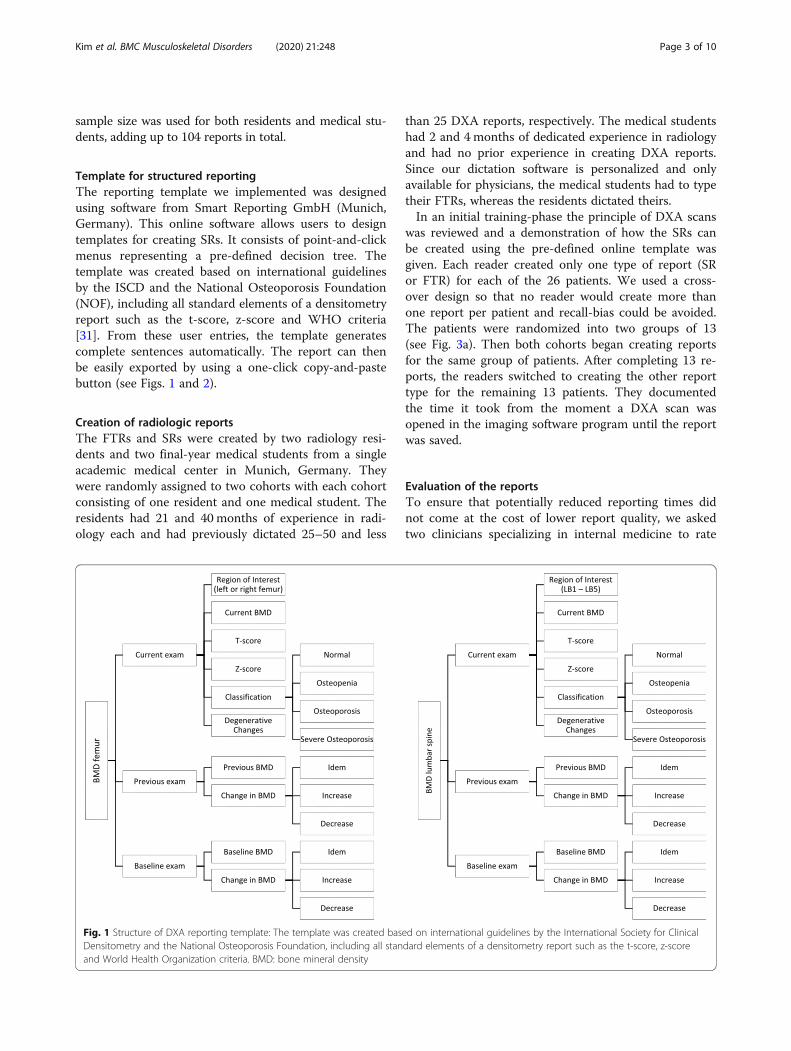
Template for structured reportingThe reporting template we implemented was designedusing software from Smart Reporting GmbH (Munich,Germany). This online software allows users to designtemplates for creating SRs. It consists of point-and-clickmenus representing a pre-defined decision tree. Thetemplate was created based on international guidelinesby the ISCD and the National Osteoporosis Foundation(NOF), including all standard elements of a densitometryreport such as the t-score, z-score and WHO criteria[31]. From these user entries, the template generatescomplete sentences automatically. The report can thenbe easily exported by using a one-click copy-and-pastebutton (see Figs. 1 and 2).
Creation of radiologic reportsThe FTRs and SRs were created by two radiology resi-dents and two final-year medical students from a singleacademic medical center in Munich, Germany. Theywere randomly assigned to two cohorts with each cohortconsisting of one resident and one medical student. Theresidents had 21 and 40 months of experience in radi-ology each and had previously dictated 25–50 and less
than 25 DXA reports, respectively. The medical studentshad 2 and 4months of dedicated experience in radiologyand had no prior experience in creating DXA reports.Since our dictation software is personalized and onlyavailable for physicians, the medical students had to typetheir FTRs, whereas the residents dictated theirs.In an initial training-phase the principle of DXA scans
was reviewed and a demonstration of how the SRs canbe created using the pre-defined online template wasgiven. Each reader created only one type of report (SRor FTR) for each of the 26 patients. We used a cross-over design so that no reader would create more thanone report per patient and recall-bias could be avoided.The patients were randomized into two groups of 13(see Fig. 3a). Then both cohorts began creating reportsfor the same group of patients. After completing 13 re-ports, the readers switched to creating the other reporttype for the remaining 13 patients. They documentedthe time it took from the moment a DXA scan wasopened in the imaging software program until the reportwas saved.
Evaluation of the reportsTo ensure that potentially reduced reporting times didnot come at the cost of lower report quality, we askedtwo clinicians specializing in internal medicine to rate
Fig. 1 Structure of DXA reporting template: The template was created based on international guidelines by the International Society for ClinicalDensitometry and the National Osteoporosis Foundation, including all standard elements of a densitometry report such as the t-score, z-scoreand World Health Organization criteria. BMD: bone mineral density
Kim et al. BMC Musculoskeletal Disorders (2020) 21:248 Page 3 of 10

the reports (2 and 8 years of experience with patientswho require DXA exams).From each of the two patient groups, six patients were
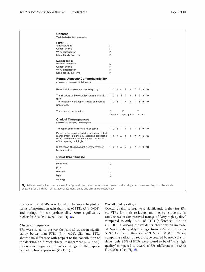
randomly selected, resulting in a total of 12 patients, whosereports were evaluated by the clinicians. There were fourreports for each of the 12 patients, adding up to 48 reportsin total (see Fig. 3b). These anonymized reports containedthe clinical question, a unique report ID, age and gender ofthe patient. SRs and FTRs were uniformly formatted. Theevaluating physicians were blinded to report type and tothe level of the reader’s experience who had created the re-port. A standardized questionnaire for DXA report evalu-ation was provided based on an extensive literature review[7, 10, 17, 24, 34–37] to ascertain the most important fea-tures of a good radiology report. Among others, content,comprehensibility and clinical relevance were rated on a10-point Likert scale (see Fig. 4).
Reader’s surveyAfter completing the reports, the four readers answeredan anonymous online survey containing Likert scale andopen-ended questions regarding the template and theiropinions on structured reporting vs. free-text reporting(see Table in Additional file 3).
Statistical analysisData are reported as medians with interquartile range(IQR) and minimum and maximum for ratings on a 10-point Likert scale and as frequencies and percentages forcategorical items. The reporting times for each report
type were compared separately for residents and medicalstudents (Mann-Whitney-U-test) as voice-recognitionsoftware was unavailable to the medical students. Tocompare reporting times between residents and medicalstudents for the template-based SRs the Wilcoxonsigned rank test for paired data was used since one resi-dent and one medical student always created a SR forthe same patient. The report ratings by referring physi-cians were compared using the Wilcoxon signed ranktest (Likert scale items and overall rating). The level ofsignificance was defined at α < 0.05. SPSS Version 20was used for all statistical calculations.
ResultsReporting timesBoth the residents and the medical students requiredless time for SRs than for FTRs (see Table 1 and Tablein Additional file 1). The median reporting time of resi-dents amounted to 4.96 min for FTRs, compared to 2.71min for SRs, which corresponds to a reduction of 45.4%(P < 0.001). The effect was even more distinct for themedical students who required 64.4% less time for SRsthan for FTRs (median for FTRs: 7.53 min; median forSRs: 2.68 min; P < 0.001).The difference in reporting time between SRs and
FTRs was particularly pronounced for patients withmore than one previous comparison. In these follow-upexams, residents had a 53.0% shorter reporting time forSRs than for FTRs (median for FTRs: 6.07 min; medianfor SRs: 2.85 min; P < 0.001) while the reduction
Fig. 2 Screenshot of DXA reporting template: When pre-defined options are selected or values are entered into text fields, the final report text isgenerated automatically [retrieved from Smart Radiology software, “X-ray bone density” template, on 18 Aug 2018]
Kim et al. BMC Musculoskeletal Disorders (2020) 21:248 Page 4 of 10

for medical students amounted to 66.6% (median forFTRs: 9.93 min; median for SRs: 3.32 min; P < 0.001).Comparison of reporting times of SRs created by resi-
dents vs. medical students showed no significant differ-ence, either for all reports (P = 0.159) or for the two sub-groups (for single exams: P = 0.969; for follow-up exams:P = 0.060). Reporting times of FTRs created by residentsvs. medical students were not compared since the resi-dents used a free-speech dictation software while themedical students typed their reports.
Report evaluationBoth FTRs and SRs were evaluated by referring physi-cians in terms of content, structure, comprehensibilityand impact on clinical decision-making (see Table inAdditional file 2):
Content and appropriateness of report lengthAll eight pre-defined key features were included in thecontent of all FTRs and SRs. With respect to the reportextent, all SRs were considered appropriate while only79.2% of FTRs received the same rating. Among theremaining FTRs, 14.6% were rated as “too long” and 6.3%as “too short”. The percentage of FTRs found to have anappropriate extent was equal in both residents and med-ical students (79.2% in both). Yet, whereas all remainingFTRs by medical students were viewed as “too long”(20.9%), the ratings of the remaining FTRs by residentswere mixed (“too short”: 12.5%, “too long”: 8.3%).
Structure and comprehensibilitySRs were rated to be significantly superior to FTRs inthe extraction of relevant information (P < 0.001). Also,
Fig. 3 Study design: A DXA scans of 26 patients were randomized and examined by two medical students and two radiology residents; eachreader created either a SR or FTR for each scan B 48 out of 104 reports were evaluated by two clinicians specializing in internal medicine; MS:medical student, RR: radiology resident, FTR: free text report, SR: structured report
Kim et al. BMC Musculoskeletal Disorders (2020) 21:248 Page 5 of 10
the structure of SRs was found to be more helpful interms of information gain than that of FTRs (P < 0.001),and ratings for comprehensibility were significantlyhigher for SRs (P < 0.001) (see Fig. 5).
Clinical consequencesSRs were rated to answer the clinical question signifi-cantly better than FTRs (P < 0.01). SRs and FTRsshowed no difference with respect to the contribution tothe decision on further clinical management (P = 0.707).SRs received significantly higher ratings for the expres-sion of a clear impression (P < 0.01).
Overall quality ratingsOverall quality ratings were significantly higher for SRsvs. FTRs for both residents and medical students. Intotal, 64.6% of SRs received ratings of “very high quality”compared to only 16.7% of FTRs (difference: + 47.9%;P < 0.0001). Among the residents, there was an increaseof “very high quality” ratings from 25% for FTRs to58.3% for SRs (difference: + 33.3%; P = 0.0010). Whencomparing ratings by report type created by medical stu-dents, only 8.3% of FTRs were found to be of “very highquality” compared to 70.8% of SRs (difference: + 62.5%;P < 0.0001) (see Fig. 6).
Fig. 4 Report evaluation questionnaire: This figure shows the report evaluation questionnaire using checkboxes and 10-point Likert scalequestions for the three main categories (content, clarity and clinical consequences)
Kim et al. BMC Musculoskeletal Disorders (2020) 21:248 Page 6 of 10

Preference of report typeOne resident and both medical students stated struc-tured reporting as their preferred reporting type forDXA examinations. Completeness, shorter reportingtimes and structure were viewed as the main strengthsof SRs. Only the more experienced resident (40 monthsexperience in radiology) preferred free-text reporting to
structured reporting, explaining that he enjoyed free-speech dictation more than clicking and entering num-bers, and that he did not like to use additional programs.Both evaluating clinicians preferred SRs to FTRs. Thestructure of SRs was perceived to reduce error rates andfacilitate a quick extraction of relevant information.
DiscussionTemplate-based structured reports can significantlyshorten reporting times and improve report qualityOur findings that SRs exhibit better completeness and re-sult in higher satisfaction of referring physicians are con-sistent with and extend those from prior reports [6–13,15, 16]. However, one common concern has been thatstructured reporting might be more time-consuming andcomplex than free-text reporting and thus impede prod-uctivity [18, 38].Here we provide evidence that SRs may not only lead
to better completeness and higher satisfaction of refer-ring physicians but also save time, at least for highlystandardized examination types such as DXA. Notably,the more experienced the subject was, the smaller thedifference between SRs and FTRs became in terms ofreporting time. This observation could be explained bythe fact that the medical students typed their FTRs un-like the residents who used the free-speech dictationsoftware. As typing takes more time than free-speechdictation our study might overestimate the difference be-tween the reporting times for medical students and resi-dents. Another explanation could be that residents weremore efficient in generating FTRs as they had a moreadvanced level of experience.One major advantage of structured reporting is that
the user is less prone to make careless mistakes. In thisstudy, for instance, the template not only indicated thereference ranges of the t-score in the decision tree (seeFig. 2), but also performed automatic calculations of thechange of bone mineral density over time (in %).
Table 1 Reporting times in minutes of radiology residents andfinal-year medical students for DXA exams
Median IQR Min. Max. P
OVERALL AVERAGE (n = 104)
Residents (n = 52)
FTR 4.96 3.98–6.15 2.77 7.45
SR 2.71 2.15–2.90 1.93 5.25 < 0.001
Medical students (n = 52)
FTR 7.53 5.96–10.65 4.45 11.90
SR 2.68 2.33–3.52 1.78 7.38 < 0.001
FIRST DXA EXAM (n = 48)
Residents (n = 24)
FTR 3.83 3.14–4.70 2.77 5.25
SR 2.18 2.00–2.75 1.93 2.85 < 0.001
Medical students (n = 24)
FTR 6.13 5.37–6.85 4.45 7.63
SR 2.33 1.99–2.38 1.78 4.38 < 0.001
FOLLOW-UP DXA EXAM (n = 56)
Residents (n = 28)
FTR 6.07 5.20–6.70 4.47 7.45
SR 2.85 2.58–3.14 2.17 3.14 < 0.001
Medical students (n = 28)
FTR 9.93 8.55–11.27 5.88 11.90 < 0.001
SR 3.32 2.92–3.71 2.28 7.38
Median reporting time, interquartile range (IQR), Minimum (Min.), Maximum(Max.), P-values for comparisons between free-text reports (FTRs) andstructured reports (SRs) using Mann-Whitney U test
Fig. 5 Comprehensibility: Ratings for comprehensibility weresignificantly higher for SR than for FTR
Fig. 6 Overall report quality: SR received significantly higher ratingsfor overall report quality than FTR
Kim et al. BMC Musculoskeletal Disorders (2020) 21:248 Page 7 of 10

Specific DXA report contents that may be missed orincorrectly reported in FTRs include the assessment ofBMD changes across non-cross-calibrated machines,fracture risk and vertebral fracture assessment.All in all, these findings have major medical and eco-
nomic implications. In the light of the current demo-graphic transition, the workload in radiology is rapidlyincreasing and structured reporting could make a rele-vant contribution to improving the efficiency of radio-logic workflow. This is particularly true for DXA examsas the prevalence of osteoporosis is increasing in manycountries [39–42]. More importantly, structured report-ing of DXA exams can make a relevant contribution toimproving patient outcome by providing the clinicianwith more comprehensible reports with higher quality.
Structured reporting as an educational tool for residentsand medical studentsThe template used in this study reveals that structuredreporting can further be a powerful educational tool.Using info boxes within the user interface of the soft-ware, relevant background information and exemplaryimages and reports were displayed. In general, the infoboxes might be particularly beneficial to illustrate ana-tomical images, classifications and up-to-date guidelines.This feature is highly useful for training inexperiencedstudents and residents in a self-guided way. This theoryis supported by our finding that the medical studentspreferred learning DXA reporting with a template andwere able to generate very good SRs quickly. However,the medical students also reported that structuredreporting might lead to a superficial evaluation due tojust clicking through the template, which is a commonconcern about structured reporting [43]. In the presentstudy, all four readers could easily utilize the softwareafter a short initial training which indicates that only aminimum level of adaptation is required to switch fromfree-text reporting to structured reporting.
LimitationsDespite the benefits of structured reporting over free-text reporting highlighted in this study, several limita-tions need to be acknowledged.First, due to the retrospective nature of our study, our
subjects created the reports in a study setting and not inactual clinical practice. Thus, findings will need to bevalidated using the template during routine clinicalreporting.Second, in clinical practice there is a broad spectrum
of reports (FTR as well as SR) being created. For ex-ample, a survey of 265 radiologists in the United Statesfound that only 51% used structured reporting for atleast half of their reports [44]. Another survey in Italyfound that 56% of radiologists never used structured
reporting [17]. When it comes to bone density measure-ments, many centers are still creating FTRs while othershave adopted templates like the ISCD’s which is essen-tially a form containing headings and sentences withblanks for the individual BMD, Z and T-Scores, amongothers [45]. Furthermore, fully structured online tem-plates with more flexibility, like the one used in thisstudy have been developed. These templates generatesentences automatically in standardized language depend-ing on user entries. Other centers are even attempting togenerate their DXA reports fully automatically, althoughthey currently still require revision by radiologists [46].Given these different approaches to structured reportingof bone density measurements with varying degrees ofautomation, it might also be beneficial to further evaluateand compare these different types of structured reporting.Third, the number of subjects who created reports and
their experience were limited. A prospective study in-cluding a more varied sample of reporting individuals islikely to provide further important insights into the po-tential of a widespread use of this template. One inter-esting hypothesis that could be tested is whether thetime difference between FTRs and SRs decreases furtherwith increasing experience, although our study indicatesthat the time required for SRs varies less with experiencecompared to FTRs.Additionally, one may argue that the evaluation of re-
port quality is rather subjective and is largely influencedby the evaluating clinician, potentially limiting thegeneralizability of our findings. Evaluations of the reportquality by a larger, more diverse group of clinicians maybe beneficial. However, due to the highly standardizednature of DXA exams, we believe that the quality ratingsare likely to be consistent even among many referringphysicians.Finally, the observations of the present study cannot
be generalized to other radiology examinations and theirreports. Reporting times might be less likely to be im-proved by structured reporting in less standardized,highly variable exams, since a much more complex tem-plate structure would be required. But at the same time,reports of highly complex exams might especially benefitfrom the guidance of a template, since SRs were shownto exhibit higher completeness and allow better extrac-tion of information [6–13]. Importantly, the extent towhich structured reporting can improve reporting effi-ciency and quality strongly depends on the technical fea-tures of the utilized software. For instance, an automaticinsertion of technical details into the radiology reportwas found to significantly improve report accuracy [47].In a similar manner, features such as an automatic inser-tion of references to previous reports or an automaticidentification of certain types of information could cre-ate added value, even for less standardized exam.
Kim et al. BMC Musculoskeletal Disorders (2020) 21:248 Page 8 of 10

Further evaluation of different types of structuredreporting templates in prospective (multicenter) studieswith readers at various levels of experience and a largernumber of evaluating physicians is likely to provide abroader impression.
ConclusionIn highly standardized exams such as radiographic bonedensity measurements, template-based structured report-ing might lead to shorter reporting times. At the sametime, structured reporting may improve report quality andserve as an effective educational tool for medical studentsand radiology residents during their training.
Supplementary informationSupplementary information accompanies this paper at https://doi.org/10.1186/s12891-020-03200-w.
Additional file 1. Reporting times for DXA exams.
Additional file 2. Report evaluation by referring clinicians.
Additional file 3. Reader’s survey.
AbbreviationsDXA: Dual X-ray absorptiometry; FTR: Free-text report; SR: Structured report;ISCD: The International Society for Clinical Densitometry
AcknowledgementsNone.
Authors’ contributionsSK: primary author, data analysis and interpretation. LS: primary author, datacollection, analysis and interpretation. JE, AC, FC, EK, MG, RB, FM: participatedin study, revision of scientific content. WS, NS, FG: conceived ideas / studydesign, revisions of scientific content. The author(s) read and approved thefinal manuscript.
FundingThe authors received no specific funding for this work.
Availability of data and materialsThe datasets used and/or analysed during the current study are availablefrom the corresponding author on reasonable request.
Ethics approval and consent to participateNot applicable.
Consent for publicationNot applicable.
Competing interestsThe following authors of this manuscript declare relationships with SmartReporting GmbH (online software company for structured reportingtemplates):- Su Hwan Kim: created and reviewed structured templates.- Lara Sobez: created structured templates.- Franziska Galiè: created and reviewed structured templates, backgroundresearch.- Nora Sommer: created and reviewed structured templates, backgroundresearch.- Wieland Sommer: co-founder.None of the other authors declare any conflict of interest relevant to thecontent of this study.
Author details1Department of Radiology, University Hospital, LMU Munich, Munich,Germany. 2Munich Transplant Center, University Hospital, LMU Munich,Munich, Germany. 3Department of Internal Medicine III, University Hospital,LMU Munich, Munich, Germany. 4Department of Radiation Oncology,University Hospital, LMU Munich, Munich, Germany. 5Department ofDiagnostic and Interventional Radiology, Paediatric Radiology andNeuroradiology, University Medical Center Rostock, Rostock, Germany.
Received: 3 February 2020 Accepted: 10 March 2020
References1. Gunderman RB, McNeive LR. Is structured reporting the answer? Radiology.
2014;273(1):7–9.2. Radiology ESo. Good practice for radiological reporting. Guidelines from the
European Society of Radiology (ESR). Insights Imaging. 2011;2(2):93–6.3. Morgan TA, Helibrun ME, Kahn CE. Reporting initiative of the Radiological
Society of North America: progress and new directions. Radiology. 2014;273(3):642–5.
4. Dunnick NR, Langlotz CP. The radiology report of the future: a summary ofthe 2007 intersociety conference. J Am Coll Radiol. 2008;5(5):626–9.
5. KSAR SGfRC. Essential items for structured reporting of rectal Cancer MRI:2016 consensus recommendation from the Korean Society of AbdominalRadiology. Korean J Radiol. 2017;18(1):132–51.
6. Norenberg D, Sommer WH, Thasler W, D'Haese J, Rentsch M, Kolben T,Schreyer A, Rist C, Reiser M, Armbruster M. Structured Reporting ofRectalMagnetic Resonance Imaging in Suspected Primary Rectal Cancer:Potential Benefits for Surgical Planning and InterdisciplinaryCommunication. Investigative radiology. 2017;52(4):232–39.
7. Gassenmaier S, Armbruster M, Haasters F, Helfen T, Henzler T, Alibek S,Pforringer D, Sommer WH, Sommer NN. Structured reporting of MRI of theshoulder - improvement of report quality? Eur Radiol. 2017;27(10):4110–9.
8. Brook OR, Brook A, Vollmer CM, Kent TS, Sanchez N, Pedrosa I. Structuredreporting of multiphasic CT for pancreatic cancer: potential effect onstaging and surgical planning. Radiology. 2015;274(2):464–72.
9. Sahni VA, Silveira PC, Sainani NI, Khorasani R. Impact of a structured reporttemplate on the quality of MRI reports for rectal Cancer staging. AJR Am JRoentgenol. 2015;205(3):584–8.
10. Schwartz LH, Panicek DM, Berk AR, Li Y, Hricak H. Improving communicationof diagnostic radiology findings through structured reporting. Radiology.2011;260(1):174–81.
11. Sabel BO, Plum JL, Kneidinger N, Leuschner G, Koletzko L, Raziorrouh B,Schinner R, Kunz WG, Schoeppe F, Thierfelder KM, et al. Structured reportingof CT examinations in acute pulmonary embolism. J Cardiovasc ComputTomogr. 2017;11(3):188–95.
12. Wildman-Tobriner B, Allen BC, Bashir MR, Camp M, Miller C, Fiorillo LE,Cubre A, Javadi S, Bibbey AD, Ehieli WL, et al. Structured reporting of CTenterography for inflammatory bowel disease: effect on key featurereporting, accuracy across training levels, and subjective assessment ofdisease by referring physicians. Abdom Radiol (NY). 2017;42(9):2243–50.
13. Flusberg M, Ganeles J, Ekinci T, Goldberg-Stein S, Paroder V, Kobi M,Chernyak V. Impact of a structured report template on the quality of CT andMRI reports for hepatocellular carcinoma diagnosis. J Am Coll Radiol. 2017;14(9):1206–11.
14. Dickerson E, Davenport MS, Syed F, Stuve O, Cohen JA, Rinker JR, GoldmanMD, Segal BM, Foerster BR, Michigan Radiology Quality C. Effect of templatereporting of brain MRIs for multiple sclerosis on report thoroughness andneurologist-rated quality: results of a prospective quality improvementproject. J Am Coll Radiol. 2017;14(3):371–9 e371.
15. Schoeppe F, Sommer WH, Haack M, Havel M, Rheinwald M, WechtenbruchJ, Fischer MR, Meinel FG, Sabel BO, Sommer NN. Structured reports ofvideofluoroscopic swallowing studies have the potential to improve overallreport quality compared to free text reports. Eur Radiol. 2018;28(1):308–15.
16. Schoeppe F, Sommer WH, Norenberg D, Verbeek M, Bogner C, WestphalenCB, Dreyling M, Rummeny EJ, Fingerle AA. Structured reporting adds clinicalvalue in primary CT staging of diffuse large B-cell lymphoma. Eur Radiol.2018;28(9):3702–9.
17. Faggioni L, Coppola F, Ferrari R, Neri E, Regge D. Usage of structuredreporting in radiological practice: results from an Italian online survey. EurRadiol. 2017;27(5):1934–43.
Kim et al. BMC Musculoskeletal Disorders (2020) 21:248 Page 9 of 10

18. Weiss DL, Langlotz CP. Structured reporting: patient care enhancement orproductivity nightmare? Radiology. 2008;249(3):739–47.
19. Hangiandreou NJ, Stekel SF, Tradup DJ. Comprehensive clinicalimplementation of DICOM structured reporting across a radiologyultrasound practice: lessons learned. J Am Coll Radiol. 2017;14(2):298–300.
20. Reiner BI. Optimizing technology development and adoption in medicalimaging using the principles of innovation diffusion, part I: theoretical,historical, and contemporary considerations. J Digit Imaging. 2011;24(5):750–3.
21. Ganeshan D, Duong PT, Probyn L, Lenchik L, McArthur TA, Retrouvey M,Ghobadi EH, Desouches SL, Pastel D, Francis IR. Structured reporting inradiology. Acad Radiol. 2018;25(1):66–73.
22. Towbin AJ, Hawkins CM. Use of a web-based calculator and a structuredreport generator to improve efficiency, accuracy, and consistency ofradiology reporting. J Digit Imaging. 2017;30(5):584–8.
23. Hanna TN, Shekhani H, Maddu K, Zhang C, Chen Z, Johnson JO. Structuredreport compliance: effect on audio dictation time, report length, and totalradiologist study time. Emerg Radiol. 2016;23(5):449–53.
24. Segrelles JD, Medina R, Blanquer I, Marti-Bonmati L. Increasing the efficiency onproducing radiology reports for breast Cancer diagnosis by means ofstructured reports. A comparative study. Methods Inf Med. 2017;56(3):248–60.
25. Li N, Li XM, Xu L, Sun WJ, Cheng XG, Tian W. Comparison of QCT and DXA:osteoporosis detection rates in postmenopausal women. Int J Endocrinol.2013;2013:895474.
26. Khoo BC, Brown K, Cann C, Zhu K, Henzell S, Low V, Gustafsson S, Price RI,Prince RL. Comparison of QCT-derived and DXA-derived areal bone mineraldensity and T scores. Osteoporos Int. 2009;20(9):1539–45.
27. Lukaszewicz A, Uricchio J, Gerasymchuk G. The art of the radiology report:practical and stylistic guidelines for perfecting the conveyance of imagingfindings. Can Assoc Radiol J. 2016;67(4):318–21.
28. Morgan SL, Prater GL. Quality in dual-energy X-ray absorptiometry scans.Bone. 2017;104:13–28.
29. Punda M, Grazio S. Bone densitometry--the gold standard for diagnosis ofosteoporosis. Reumatizam. 2014;61(2):70–4.
30. Dimai HP. Use of dual-energy X-ray absorptiometry (DXA) for diagnosis andfracture risk assessment; WHO-criteria, T- and Z-score, and referencedatabases. Bone. 2017;104:39–43.
31. Lewiecki EM, Binkley N, Morgan SL, Shuhart CR, Camargos BM, Carey JJ,Gordon CM, Jankowski LG, Lee JK, Leslie WD, et al. Best practices for dual-energy X-ray absorptiometry measurement and reporting: International Societyfor Clinical Densitometry Guidance. J Clin Densitom. 2016;19(2):127–40.
32. Krueger D, Shives E, Siglinsky E, Libber J, Buehring B, Hansen KE, Binkley N.DXA Errors Are Common and Reduced by Use of a Reporting Template.Journal of Clinical Densitometry. 2019;22(1):115–24.
33. 2019 ISCD Official Positions – Adult [https://www.iscd.org/official-positions/2019-iscd-official-positions-adult/]. Accessed 6 Mar 2020.
34. Wallis A, McCoubrie P. The radiology report--are we getting the messageacross? Clin Radiol. 2011;66(11):1015–22.
35. Rothman M. Malpractice issues in radiology: radiology reports. AJR Am JRoentgenol. 1998;170(4):1108–9.
36. Yang C, Kasales CJ, Ouyang T, Peterson CM, Sarwani NI, Tappouni R, BrunoM. A succinct rating scale for radiology report quality. SAGE Open Med.2014;2:2050312114563101.
37. Wallis A, Edey A, Prothero D, McCoubrie P. The Bristol radiology reportassessment tool (BRRAT): developing a workplace-based assessment tool forradiology reporting skills. Clin Radiol. 2013;68(11):1146–54.
38. Bosmans JM, Peremans L, Menni M, De Schepper AM, Duyck PO, Parizel PM.Structured reporting: if, why, when, how-and at what expense? Results of afocus group meeting of radiology professionals from eight countries.Insights Imaging. 2012;3(3):295–302.
39. Wright NC, Looker AC, Saag KG, Curtis JR, Delzell ES, Randall S, Dawson-Hughes B. The recent prevalence of osteoporosis and low bone mass in theUnited States based on bone mineral density at the femoral neck or lumbarspine. J Bone Miner Res. 2014;29(11):2520–6.
40. Lotters FJ, van den Bergh JP, de Vries F, Rutten-van Molken MP. Current andfuture incidence and costs of osteoporosis-related fractures in theNetherlands: combining claims data with BMD measurements. Calcif TissueInt. 2016;98(3):235–43.
41. Khadilkar AV, Mandlik RM. Epidemiology and treatment of osteoporosis inwomen: an Indian perspective. Int J Women's Health. 2015;7:841–50.
42. Chen P, Li Z, Hu Y. Prevalence of osteoporosis in China: a meta-analysis andsystematic review. BMC Public Health. 2016;16(1):1039.
43. Srinivasa Babu A, Brooks ML. The malpractice liability of radiology reports:minimizing the risk. Radiographics. 2015;35(2):547–54.
44. Powell DK, Silberzweig JE. State of structured reporting in radiology, asurvey. Acad Radiol. 2015;22(2):226–33.
45. US Adult DXA Sample Report [https://iscd.app.box.com/v/US-Adult-DXA-Sample-Report]. Accessed 30 Oct 2018.
46. Tsai IT, Tsai MY, Wu MT, Chen CK. Development of an automated bonemineral density software application: facilitation radiologic reporting andimprovement of accuracy. J Digit Imaging. 2016;29(3):380–7.
47. Abujudeh HH, Govindan S, Narin O, Johnson JO, Thrall JH, Rosenthal DI.Automatically inserted technical details improve radiology report accuracy. JAm Coll Radiol. 2011;8(9):635–7.
Publisher’s NoteSpringer Nature remains neutral with regard to jurisdictional claims inpublished maps and institutional affiliations.
Kim et al. BMC Musculoskeletal Disorders (2020) 21:248 Page 10 of 10