Embed Size (px)
Citation preview

Journal of Environmental Biology �October, 2007�
Structure of immune organ in edible catfish, Mystus gulio
B. Deivasigamani**[email protected]
CAS in Marine Biology, Annamalai University, Parangipettai-608 502, India
(Received: September 28, 2005 ; Revised received: March 17, 2006 ; Accepted: April 28, 2006)
Abstract: The light microscopic study describes the anatomy and histomorphology of head-kidney in bagrid catfish, Mystus gulio. Showing numerous
lymphocytes, interrenal cells, reticular cells, postcardinal vein, blood sinuses and melanomacrophage centers.
Key words: Head-kidney, Lymphocytes, Interrenal gland, Melanomacrophage, Mystus gulio
PDF file of full length paper is available with author
Introduction
Among vertebrates, fishes are the earliest group to possess
a well defined immune system with lymphomyeloid tissues consisting
of mixed lymphoid and myeloid elements (Sailendri and
Muthukkaruppan, 1975b; Grace and Manning, 1980; Pitchappan,
1980; Rafin and Wignstand, 1981; Pica and Corte, 1987). The
main lymphoid organs of fish are thymus, head-kidney, spleen and
blood tissue (Corbel, 1975; Ellis, 1977; Zabotkina, 2005;
Deivasigamani, 2006). The head-kidney (HK) is a unique, important
lymphoid organ in fish (Sailendri and Muthukkaruppan, 1975b;
Ellis, 1980). It contains more lymphocytes than spleen and has
been shown to be actively involved in antibody production (Sailendri
and Muthukkaruppan, 1975a; Zapata, 1979; Ellis et al., 1989;
Romano et al., 2002). The role of head-kidney as a major site of
production of erythroid, lymphoid and myeloid cells has been
established (Smith et al., 1970; Zapata, 1979; Romano et al., 2002).
The capacity of the head-kidney to trap antigens and produce
antibodies has been demonstrated in a few species (Ellis et al.,
1989; Zapata and Cooper, 1990).The present study describes the
anatomy and histomorphology of head-kidney in bagrid catfish,
Mystus gulio at the light microscopic level.
Materials and Methods
Live specimens of Mystus gulio (Hamilton) of the family
bagridae were collected from Ogyampet estuary near Chennai. A
total of 60 adult fish of both sexes, weighing 40-45 g (standard
length 11-15 cm), were used in the present study. A stock of fish was
maintained in the laboratory in a large tank. After acclimatization for
at least three days, the fish were transferred to smaller aquarium
tanks with continuous aeration. They were fed with bits of earthworms
and tubifex.
The fish were immobilized by immersion in ice-cold water
for about a minute and then dissected and their immune organ of
head-kidney were removed to fixed in Bouin’s fluid for about 24 hr,
processed, embedded in paraffin wax and cut into 6 µm thick sections.
The sections were stained by the hematoxylin and eosin. The
imprint was prepared on a clean microslide from cut surface of
freshly dissected spleen. They were air-dried and stained by the
Leishman stain.
Calibration of microscope was made using stage and ocular
micrometers. Measurements of the dimensions of the different types
of cells and those of MMCs (melanomacrophage centres) were
made under high-power objective magnification (45 X).
Results and Discussion
In Mystus gulio the kidney (Fig.1) is a paired but fused
retroperitoneal organ that extends as a thick sheet of tissue dorsal
in the body cavity in close contact with the osseous vertebral column.
The kidney consists of two well-separated segments, an anterior
head-kidney (HK) and a posterior trunk-kidney (TK). The trunk-
kidney begins as a thin layer near the posterior end of the peritoneal
cavity, soon widens and bifurcates into two thick lobes with a marked
concavity, made by the pressing air-bladder, at the anterior end.
The posterior cardinal veins emerge from the dorsal rim of the
concavity close to the spinal column, run parallel, beneath the air
bladder and enter into the corresponding half of the HK. The HK’s
are also fused and appear as a bilobed structure that lies
perpendicular to the dorsal body wall and lining the anterior blunt
surface of the air bladder. Thus, it is very close to the heart and the
pharyngeal chamber. Its association with the trunk-kidney is obscure
though the two are well connected by the posterior cardinal veins.
The difference between the HK and the TK is that the former is
composed of haemolymphoid and endocrine (interrenal, homologue
of adrenal) tissues whereas the later is purely a renal tissue. The
HK in adult fish weighs about 0.5-0.9 g and measures about 5-10
mm in length and 3-5 mm in width. The lobes of the HK are almost
of the same dimensions.
A thin connective tissue capsule covers the HK (Fig. 2-4).
The capsule does not seem to extend into the pulp of the HK as
trabeculae. However, the HK pulp is supported by a closely knitted
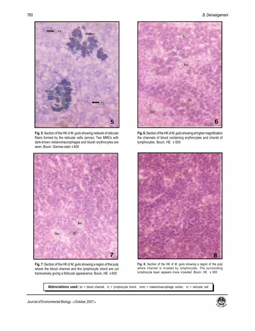
network of reticular cells and their fibers (Fig. 5). The entire pulpconsists of anastamosing channels of blood by chords of lymphocytes
(Fig. 3-7). In cross sections, these channels cells bounded and
Journal of Environmental Biology October 2007, 28(4) 757-764 (2007)
©Triveni Enterprises, Lucknow (India) For personal use only
Free paper downloaded from: www. jeb.co.in Commercial distribution of this copy is illegal

Journal of Environmental Biology �October, 2007�
758 B. Deivasigamani
Fig. 1: Dissection of M. gulio showing the location of the head-
kidney (arrow) in relation to trunk-kidney and air-bladder. The
concavity (arrow head) at the anterior surface of the trunk-kidney
is made visible by the stretching of the body wall x 1.25
Fig. 2: Section of the HK of M. gulio showing the organization
of the pulp into channels of blood vessels, chords of lymphocytes
and large and small venous sinuses. The entire pulp is covered
by a thin capsule. Bouin, HE x 60
Fig. 3: Higher magnification of part of the section showing the
connective tissue capsule (arrow), subcapsular sinus, channels of
blood chords of lymphocytes (arrow head), and venous sinuses.
Difference in the organization of the pulp between the central and
peripheral areas, evident. Bouin, HE x 130
Fig. 4: Section of the HK of M. gulio showing the difference in the
pulp organization between the peripheral and central areas. Bouin,
HE x 130
Abbreviations used: ab = air–bladder, tk = trunk–kidney, vs = venous sinus, ss = sub capsular-sinus, cc = channel-chord

Journal of Environmental Biology �October, 2007�
759Immune organ in catfish, Mystus gulio
chords appear as follicles, each consisting of a central blood vessel
surrounded by a thick layer of closely packed, dark-staining
lymphocytes (Fig. 7). The channels of blood cells do not seem to
have any connective tissue boundary separating them from the
surrounding layers of lymphocytes. The content of these channels
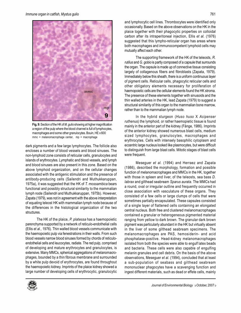
are generally erythrocytes (Fig. 6 and 7). However, invasion of
these channels by lymphocytes, macrophages and other
granulocytes from the surrounding layers is not uncommon (Fig. 8
and 9). All these channels converge towards and open into venous
sinuses of varying sizes throughout the HK parenchyma (Fig. 1-3
and 10). The subcapsular sinuses are generally larger than those
in the middle. The organization of the pulp into channels and chords
is more pronounced in the middle than in the periphery where the
lymphocytes are clumped as smaller groups surrounded by thin
channels of blood cells (Fig. 3, 4). Generally, the channel-chord
organization becomes obliterated closer to the larger sinuses.
In addition to the plexus of channels and chords of cells,
there are numerous discrete MMCs of varying sizes (40.6 µm x
20.13 µm to 130.90 µm x 109.10 µm) and irregular shapes
distributed throughout the HK, though their frequency is slightly higher
in the center (Fig. 10) than in the periphery. Each MMC is an
aggregation of at least two types of cells (Fig. 11-14): (i)
melanomacrophages in various shades ranging from light yellowish
brown to dark blackish brown and (ii) erythrocytes with pycnotic
nuclei. In sections stained with Giemsa stain, the distinction between
the two cell types is more apparent (Fig. 5), the erythrocytes appearing
bluish and the macrophages, dark brownish.
In the early stages of its formation, an MMC includes
macrophages in different degrees of pigmentation and erythrocytes
(Fig. 11). At this stage, presence of a thin connective tissue strand
separating it from the adjacent area is well evident. Further, a few
cells-thick layer of lymphocytes surrounds the center. During the
subsequent stage, the macrophages become increasingly darker
with increased deposition of pigments in their cytoplasm, the
connective tissue boundary is not discernible, and the lymphocyte
layer grows to several cells-thick (Fig. 12). In advanced stage,
most of the macrophages in the MMC appear dark brown to black
brown and the surrounding lymphocyte layer has completely
disappeared, and, instead, the center is encircled by a number of
erythrocytes with pycnotic nuclei (Fig. 13). An MMC in a well-
advanced stage has almost all its macrophages fully laden with
blackish brown pigments, and is surrounded by thicker layer of
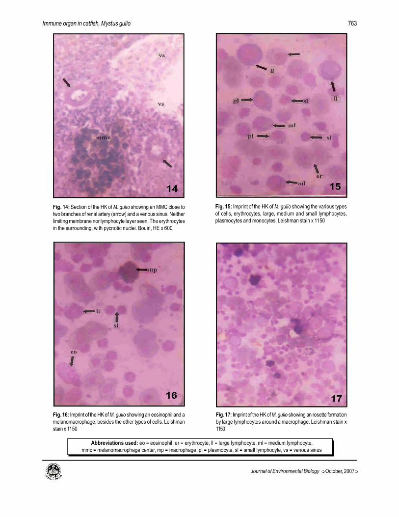
erythrocytes with pycnotic nuclei (Fig. 14).
The types of cells observed in the head-kidney viz., reticular
cells, erythrocytes, lymphocytes of various sizes, monocytes,
eosinophils, plasmocytes and macrophages could be identified in
the HK histological (Fig. 5) and smear (Fig. 15 and 16) preparations.
In addition, a large number of developing cells of RBC’s and
granulocytes could be seen in the imprints. Melanomacrophages
could also be seen in the smear preparation. The dimensions and
other characteristics of these cells are almost the same as in the
spleen. Structures resembling rosette formation by large lymphocytes
around macrophages are very rarely identified (Fig. 17).
In most of the teleosts, the kidney lies along the ventral side
of the vertebral column separated from the body cavity by tough
connective tissue septum (Ellis et al., 1976; Zapata, 1979). It consists
of two segments, the head-kidney representing the pronephros
and trunk-kidney, the functional excretory organ in the adults. The
two parts are ontogenetically and, in the adult, anatomically distinct.
The anatomical distinction is very much pronounced in the case of
the catfish, M. gulio, as observed in the present study and M.
vittatus (Gurumani, 1983), the head-kidney being well separated
from the trunk-kidney by the air-bladder, and the connection between
the two being the posterior cardinal veins. As a result of this
separation, the bilobed head-kidney comes to lie on the anterior
surface of the air-bladder, very close and dorsal to the heart. The
right and the left posterior cardinal veins traverse the respective
halves of the head-kidney before joining the common cardinal vein.
Though several workers have studied the histomorphology
of the head-kidney of teleosts and indicated its lymphoid nature,
most of their works pertain to the endocrine interrenal and chromaffin
tissues, homologues of the adrenal cortical and medullary tissues,
respectively, of mammals, that lie around the posterior cardinal
veins within the head-kidney. Even among those works that deal
with immunological or non-immune defense aspects of the HK, only
a few have paid attention to the detailed histomorphology of the
organ. Thus the literature on this aspect, when compared to that of
the spleen, is very meagre.
The HK of M. gulio is covered by a rather thin connective
tissue capsule that does not extend into the pulp to form a trabecular
system as in the spleen. Therefore, ascribing any blood storage
and release function to the HK is rather difficult. However, a closely
knitted network of reticular cells and their fibers provide the support
for the pulp. The pulp itself consists of channels of blood cells,
representing red pulp, bounded by chords of lymphoid tissue, mostly
lymphocytes, representing white pulp. The blood sinuses are
prominent, especially the subcapsular ones. In addition, MMCs of
varying shapes, sizes and in different stages of development are
seen in the pulp.
The HK of the cichlid teleost T. mossambica is encapsulated
by thin strands of collagen fibers and includes lymphatic and blood
vessels and sinuses (Sailendri and Muthukkaruppan, 1975b). A
broad subcapsular or marginal sinus lies directly beneath the capsule
that enters at different points into the pulp as trabeculae carrying
blood vessels. The HK of this species is mainly a lymphoreticular
organ and consists of two different zones, a deeply stained lymphoid
zone and a non-lymphoid zone. The lymphoid zone is represented
by a number of lymphoid follicles, each of which consists of tightly
packed small lymphocytes surrounding large vacuolated cells with
Journal of Environmental Biology �October, 2007�
760 B. Deivasigamani
Fig. 5: Section of the HK of M. gulio showing network of reticular
fibers formed by the reticular cells (arrow). Two MMCs with
dark-brown melanomacrophages and bluish erythrocytes are
seen. Bouin, Giemsa stain x 600
Fig. 6: Section of the HK of M. gulio showing at higher magnification
the channels of blood containing erythrocytes and chords of
lymphocytes. Bouin, HE x 600
Fig. 7: Section of the HK of M. gulio showing a region of the pulp
where the blood channel and the lymphocyte chord are cut
transversely giving a follicular appearance. Bouin, HE x 600
Fig. 8: Section of the HK of M. gulio showing a region of the pulp
where channel is invaded by lymphocytes. The surrounding
lymphocyte layer appears more crowded. Bouin, HE x 600
Abbreviations used: bc = blood channel, lc = lymphocyte chord, mmc = melanomacrophage center, rc = reticular cell
Journal of Environmental Biology �October, 2007�
761Immune organ in catfish, Mystus gulio
dark pigments and a few large lymphocytes. The follicle also
encloses a number of blood vessels and blood sinuses. The
non-lymphoid zone consists of reticular cells, granulocytes and
islands of erythrocytes. Lymphatic and blood vessels, and lymph
and blood sinuses are also present in this zone. Based on the
above lymphoid organization, and on the cellular changes
associated with the antigenic stimulation and the presence of
antibody-producing cells (Sailendri and Muthukkaruppan,
1975a), it was suggested that the HK of T. mossambica bears
functional and possibly structural similarity to the mammalian
lymph node (Sailendri and Muthukkaruppan, 1975b). However,
Zapata (1979), was not in agreement with the above interpretation
of equating teleost HK with mammalian lymph node because of
the differences in the histological organization of the two
structures.
The HK of the plaice, P. platessa has a haemopoietic
parenchyma supported by a network of reticulo-endothelial cells
(Ellis et al., 1976). Thin walled blood vessels communicate with
the haemopoietic pulp via fenestrations in their walls. From such
blood vessels narrow blood sinuses formed by chords of reticulo-
endothelial cells and leucocytes, radiate. The red pulp, comprised
of developing and mature erythrocytes and granulocytes, is
extensive. Many MMCs, spherical aggregations of melanomacro-
phages, bounded by a thin fibrous membrane and surrounded
by a white pulp devoid of erythrocytes, are found throughout
the haemopoietic kidney. Imprints of the plaice kidney showed a
large number of developing cells of erythrocytic, granulocytic
and lymphocytic cell lines. Thrombocytes were identified only
occasionally. Based on the above observations on the HK in the
plaice together with their phagocytic properties on colloidal
carbon after its intraperitoneal injection, Ellis et al. (1976)
suggested that this lympho-reticular organ has areas where
both macrophages and immunocompetent lymphoid cells may
mutually affect each other.
The supporting framework of the HK of the teleosts, R.
rutilus and G. gobio is partly composed of a capsule that surrounds
the organ. The capsule is made up of connective tissue consisting
largely of collagenous fibers and fibroblasts (Zapata, 1979).
Immediately below this sheath, there is a uniform continuous layer
of pigment cells. Reticular cells, phagocytic reticular cells and
other obligatory elements necessary for proliferation of
haemopoietic cells are the cellular elements found the HK stroma.
The presence of these elements together with sinusoids and the
thin walled arteries in the HK, lead Zapata (1979) to suggest a
structural similarity of this organ to the mammalian bone marrow,
rather than to the mammalian lymph node.
In the hybrid sturgeon (Huso huso X Acipenser
ruthenus) the lymphoid, or rather haemopietic tissue is found
mainly in the anterior part of the kidney (Fänge, 1986). Imprints
of the anterior kidney showed numerous blast cells, medium
sized lymphocytes, granulocytes, macrophages and
erythrocytes. Cells with intensely basophilic cytoplasm and
eccentric large nucleus looked like plasmocytes, but were difficult
to distinguish from large blast cells. Mitotic stages of blast cells
were frequent.
Meseguer et al. (1994) and Herraez and Zapata
(1986), described the morphology, formation and possible
function of melanomacrophages and MMCs in the HK, together
with those in spleen and liver, of the teleosts, sea bass D.
labrax and gilthead seabream Sparus aurata. The MMCs had
a round, oval or irregular outline and frequently occurred in
close association with vasculature of these organs. They
consisted of a few cells or large clumps of cells that were
sometimes partially encapsulated. These capsules consisted
of a single layer of flattened cells containing an elongated
central nucleus. Both free and clustered melanomacrophages
contained a granular or heterogeneous pigmented material
ranging from yellow to dark brown. The granular dark brown
pigment was particularly abundant in the HK but virtually absent
in the liver of some gilthead seabream specimens. The
melanomacrophages are PAS, hemosiderin- and acid
phosphatase-positive. Head-kidney melanomacrophages
isolated from both the species were able to engulf latex beads
and bacteria. These cells were also capable of engulfing
melanin granules and cell debris. On the basis of the above
observations, Meseguer et al. (1994), concluded that at least
a sub-populat ion of seabass and gi l thead seabream
mononuclear phagocytes have a scavenging function and
ingest different materials, such as dead or effete cells, mainly
Fig. 9: Section of the HK of M. gulio showing at higher magnification
a region of the pulp where the blood channel is full of lymphocytes,
macrophages and some other granulocytes. Bouin, HE x 600mmc = melanomacrophage center, mp = macrophage

Journal of Environmental Biology �October, 2007�
762 B. Deivasigamani
Fig. 13: Section of the HK of M. gulio showing an MMC in an
advanced stage. Many of the macrophages laden with dark-
brown pigments. Limiting membrane not visible. Lymphocyte
layer disappeared. Bouin, HE x 600
Fig. 11: Section of the HK of M. gulio showing an MMC close to
a branch of posterior cardinal vein, in its early stage of formation.
The macrophages inside the center show varying degrees of
pigment deposition. A thin limiting membrane (arrow) and a layer
of closely packed lymphocytes around the center visible. Bouin,
HE x 600, pcv = posterior cardinal vein
Fig. 12: Section of the HK of M. gulio showing two MMCs with
many yellow brownish and a few black brownish macrophages.
The limiting membrane is not visible. The surrounding lymphocyte
layer is thicker. Bouin, HE x 600
Fig. 10: Section of the HK of M. gulio showing the distribution of
many MMCs (arrow) in the center. Bouin, HE x 130
Journal of Environmental Biology �October, 2007�
763Immune organ in catfish, Mystus gulio
Fig. 14: Section of the HK of M. gulio showing an MMC close to
two branches of renal artery (arrow) and a venous sinus. Neither
limiting membrane nor lymphocyte layer seen. The erythrocytes
in the surrounding, with pycnotic nuclei. Bouin, HE x 600
Fig. 15: Imprint of the HK of M. gulio showing the various types
of cells, erythrocytes, large, medium and small lymphocytes,
plasmocytes and monocytes. Leishman stain x 1150
Fig. 16: Imprint of the HK of M. gulio showing an eosinophil and a
melanomacrophage, besides the other types of cells. Leishman
stain x 1150
Fig. 17: Imprint of the HK of M. gulio showing an rosette formation
by large lymphocytes around a macrophage. Leishman stain x
1150
Abbreviations used: eo = eosinophil, er = erythrocyte, ll = large lymphocyte, ml = medium lymphocyte,
mmc = melanomacrophage center, mp = macrophage, pl = plasmocyte, sl = small lymphocyte, vs = venous sinus

Journal of Environmental Biology �October, 2007�
764 B. Deivasigamani
erythrocytes, cell debris and melanin from melanocytes. The
melanomacrophages migrate as free cells, aggregate and
accumulate to form the MMCs, the largest being surrounded
by fibroblast-like reticular cells. During this process, a part of
the engulfed material is partially digested by the lysosomic
hydrolase and processed for reut i l izat ion. Thus, the
melanomacrophages in the HK together with those in the spleen
of these teleosts are mainly involved in iron and haemoglobin
metabolism and tissue catabolism.
The HK of M. gulio showed well organized sinuses and
channels of red pulp, chords of white pulp and numerous MMCs
in different stages of formation. In addition, immature cells of
erythrocytes and other granulocytes, and rosette forming cells
could be identified in HK imprint preparations. Based on these
observations and in the light of the information available in the
literature, it may be suggested that the HK of M. gulio has
haemopoietic, immune and phagocytic functions.
Acknowledgments
The author is grateful to his research guide Dr. N. Gurumani,
HoD Department of Microbiology, Pachaiyappa’s College, Chennai,
for providing the facilities to carry out the work and Dr. T.
Balasubramanian, Director of CAS in Marine Biology, Annamalai
University for his support and encouragement.
References
Corbel, M.J.: The immune response in fish: A review. J. Fish Biol., 7, 539-
563 (1975).
Deivasigamani, B.: Lymphoid organ of head-kidney in air breathing fish,
Channa punctatus. J. Aquatic Biol., 21, 163-166 (2006).
Ellis, A.E.: The leucocytes of fish: A review. J. Fish Biol., 11, 453-491
(1977).
Ellis, A.E.: Antigen trapping in the spleen and kidney of the plaice, Pleuronectes
platessa L. J. Fish Dis., 3, 413-426 (1980).
Ellis, A.E., A.L.S. Munroe and R.J.Roberts: Defence mechanisms in fish. J.
Fish Biol., 8, 67-78 (1976).
Ellis, A.E., R.J. Roberts and P. Tytler: The anatomy and physiology of
teleosts. In: Fish pathology (Ed.: R.J. Roberts). Bailliere Tindall,
London, UK. pp. 13-55 (1989).
Fange, R.: Physiology of haemopoiesis. In: Fish physiology: Recent advance
(Eds.: S. Nilsson and S. Holmgren). London. Groom Helm. pp. 1-23 (1986).
Grace, M.F. and M.J.Marning: Histogenesis of the lymphoid organs in rainbow
trout, Salmo gairdneri. Dev. Comp. Immunol., 4, 255-264 (1980).
Gurumani, N.: Histomorphology of the adrenal tissue of the fresh water
catfish, Mystus vittatus (Bloch). J. Reprod. Biol. Comp. Endocrinol.,
3, 30-35 (1983).
Herraez, M.P. and A.G. Zapata: Structure and function of the maleno-
macrophage centers of the gold fish, Carassius auratus. Vet. Immunol.
Immunopathol., 12, 117-126 (1986).
Meseguer, J., A.L. Ruiz and M.A. Esteban: Melano–macrophages of the
seawater teleosts, sea bass (Dicentrarchus labrax) and gilthead
seabream (Sparus aurata): Morphology, formation and possible
function. Cell Tissue Res., 277, 1-10 (1994).
Pica, A. and F.D. Corte: Haemopoiesis, lymphomieloid tissues, spleen and
thymus of Torpedoes in normal conditions and after treatment with
cobamamide and folic acid. Arch. Ital. Anat. Embriol., 92, 249-261 (1987).
Pitchappan, R.M.: Review on the phylogeny of splenic structure and function.
Dev. Comp. Immunol., 4, 395-416 (1980).
Rafin, S. and K.G. Wingstrand: Structure of intestine, pancreas and spleen of
the Australian Lungfish, Neoceratodus forsteri (Krefft). Zoologica
Scripta., 10, 223-239 (1981).
Romano, N., S.Ceccariglia, L.Mastrolia and M.Mazzini: Cytology of
lymphomyeloid head kidney of Antarctic fishes Trematomus bernacchii
(Nototheniidae) and Chionodraco hamatus (Channicthyidae). Tissue
and Cell, 34, 2 (2002).
Sailendri, K. and V.R.Muthukkaruppan: The immune response of the teleost,
Tilapia mossambica to soluble and cellular antigens. J. Exp. Zool.,
191, 371-383 (1975a).
Sailendri, K. and V.R. Muthukkaruppan: Morphology of lymphoid organs in a
cichild teleost, Tilapia mossambica (Peters). J. Morphol., 147, 109-
122 (1975b).
Smith, M., M. Potter and B. Merchant: Plasmacytopoiesis in the pronephros
of the teleost, Lepomis macrochirus. J. Immunol., 99, 876-882 (1970).
Zabotkina, E.A.: Comparative morphological characteristics of spleen of the
perch Perca fluviatilis L. from lakes with different water pH level. J.
Envol. Bio. Physio., 41, 112-118 (2005).
Zapata, A.: Ultrastructural study of the teleost fish kidney. Dev. Comp.
Immunol., 3, 55-65 (1979).
Zapata, A. and E.L.Cooper: The immune system: Comparative
Histophysiology. John Wiley Sons, Chichester, U.K. (1990).