Embed Size (px)
Citation preview

Structure of Coatomer Cage Proteinsand the Relationship among COPI,COPII, and Clathrin Vesicle CoatsChangwook Lee1,2 and Jonathan Goldberg1,2,*1Howard Hughes Medical Institute2Structural Biology Program
Memorial Sloan-Kettering Cancer Center, 1275 York Avenue, New York, NY 10065, USA
*Correspondence: [email protected] 10.1016/j.cell.2010.05.030
SUMMARY
COPI-coated vesicles form at the Golgi apparatusfrom two cytosolic components, ARF G protein andcoatomer, a heptameric complex that can poly-merize into a cage to deform the membrane intoa bud. Although coatomer shares a common evolu-tionary origin with COPII and clathrin vesicle coatproteins, the architectural relationship among thethree cages is unclear. Strikingly, the ab0-COP coreof coatomer crystallizes as a triskelion in which threecopies of a b0-COP b-propeller domain convergethrough their axial ends. We infer that the trimerconstitutes the vertex of the COPI cage. Our modelproposes that the COPI cage is intermediate indesign between COPII and clathrin: COPI shareswith clathrin an arrangement of three curved a-sole-noid legs radiating from a common center, and COPIshares with COPII highly similar vertex interactionsinvolving the axial ends of b-propeller domains.
INTRODUCTION
Vesicle transport pathways operate through rounds of vesicle
budding and fusion reactions, and the underlying reaction mech-
anisms are conserved from yeast to humans. Budding occurs
when cytoplasmic coat protein (COP) complexes assemble on
a membrane surface, where they capture cargo proteins and
polymerize into spherical cages to deform the membrane into
a bud. Eukaryotic cells contain a series of COP complexes:
COPI, which buds vesicles from the Golgi apparatus, COPII,
which operates at the endoplasmic reticulum, and clathrin/adap-
tin, which is involved in budding from the plasma membrane,
trans-Golgi network, and endosomal compartments (Bonifacino
and Glick, 2004).
COPI-coated vesicles form on the Golgi apparatus by the
stepwise recruitment of two cytosolic components: an ARF-
family G protein and coatomer, a 550 kDa cytoplasmic complex
of seven COPs—a-, b-, b0-, g-, d-, 3-, and z-COP (Waters et al.,
1991; Serafini et al., 1991). Budding is initiated by the exchange
of GDP for GTP on ARF catalyzed by a Golgi-localized guanine-
nucleotide exchange factor (GEF) of the Sec7 family (Peyroche
et al., 1996; Chardin et al., 1996). ARF-GTP binds to the
membrane by embedding an N-terminal a helix in the bilayer
and in turn recruits coatomer through a direct, GTP-dependent
interaction (Antonny et al., 1997; Zhao et al., 1997). Coatomer
complexes, attached to the membrane surface through ARF-
GTP, can bind cargo molecules and then self-assemble to
form spherical cages that yield COPI-coated vesicles (Bremser
et al., 1999; Nickel et al., 1998; Orcl et al., 1993; Spang et al.,
1998).
The architecture and functional organization of clathrin and
COPII vesicle coats have been studied extensively by elec-
tron microscopy (EM) and X-ray crystallography (reviewed by
McMahon and Mills [2004]; see Stagg et al. [2007] for a structural
comparison). The clathrin cage is built from triskelion assembly
units—trimers of clathrin heavy chain—that are centered on
the vertices of the cage, and the long a-solenoid legs curve
toward and interdigitate with neighboring legs as they extend
to the adjacent vertices (Fotin et al., 2004). The N-terminal
b-propeller domain of the clathrin heavy chain projects inwards
from the cage to interact with the adaptor protein (AP) complex,
a tetramer of two large and two smaller proteins that is respon-
sible for cargo binding and membrane apposition (Collins
et al., 2002; Heldwein et al., 2004; ter Haar et al., 2000; and
reviewed by Owen et al. [2004]). Like clathrin, the COPII cage
is formed from a-solenoid and b-propeller protein domains,
reflecting their common evolutionary origin, but the cage archi-
tecture is quite distinct. The COPII cage assembly unit—two
copies of Sec13/31—is a 28 nm long rod comprising a central
a-solenoid dimer capped by two b-propeller domains at each
end. The rod forms the edge of a cuboctahedron cage, and
four rods converge to form the vertex with no interdigitation of
assembly units (Stagg et al., 2006, 2008; Fath et al., 2007).
Far less is known about the architecture of the COPI coat, as
neither EM reconstructions of the cage nor crystal structures of
coatomer complexes have been reported. The cargo-binding
and cage-forming portions of coatomer remain together in
cytosol and bind en bloc to Golgi membranes during COPI
coat assembly, unlike the stepwise accretion of clathrin and
COPII coats (Hara-Kuge et al., 1994). The functional complexes
within heptameric coatomer have been identified through
Cell 142, 123–132, July 9, 2010 ª2010 Elsevier Inc. 123
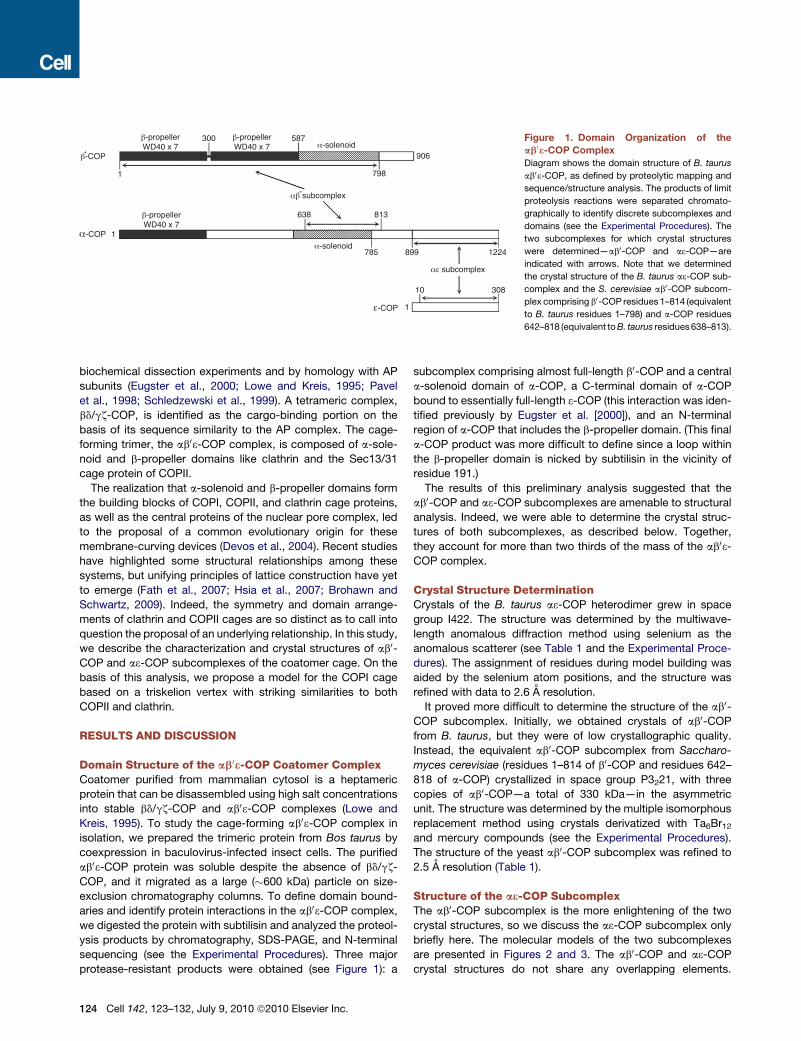
Figure 1. Domain Organization of the
ab03-COP Complex
Diagram shows the domain structure of B. taurus
ab03-COP, as defined by proteolytic mapping and
sequence/structure analysis. The products of limit
proteolysis reactions were separated chromato-
graphically to identify discrete subcomplexes and
domains (see the Experimental Procedures). The
two subcomplexes for which crystal structures
were determined—ab0-COP and a3-COP—are
indicated with arrows. Note that we determined
the crystal structure of the B. taurus a3-COP sub-
complex and the S. cerevisiae ab0-COP subcom-
plex comprising b0-COP residues 1–814 (equivalent
to B. taurus residues 1–798) and a-COP residues
642–818 (equivalent to B. taurus residues 638–813).
biochemical dissection experiments and by homology with AP
subunits (Eugster et al., 2000; Lowe and Kreis, 1995; Pavel
et al., 1998; Schledzewski et al., 1999). A tetrameric complex,
bd/gz-COP, is identified as the cargo-binding portion on the
basis of its sequence similarity to the AP complex. The cage-
forming trimer, the ab03-COP complex, is composed of a-sole-
noid and b-propeller domains like clathrin and the Sec13/31
cage protein of COPII.
The realization that a-solenoid and b-propeller domains form
the building blocks of COPI, COPII, and clathrin cage proteins,
as well as the central proteins of the nuclear pore complex, led
to the proposal of a common evolutionary origin for these
membrane-curving devices (Devos et al., 2004). Recent studies
have highlighted some structural relationships among these
systems, but unifying principles of lattice construction have yet
to emerge (Fath et al., 2007; Hsia et al., 2007; Brohawn and
Schwartz, 2009). Indeed, the symmetry and domain arrange-
ments of clathrin and COPII cages are so distinct as to call into
question the proposal of an underlying relationship. In this study,
we describe the characterization and crystal structures of ab0-
COP and a3-COP subcomplexes of the coatomer cage. On the
basis of this analysis, we propose a model for the COPI cage
based on a triskelion vertex with striking similarities to both
COPII and clathrin.
RESULTS AND DISCUSSION
Domain Structure of the ab03-COP Coatomer ComplexCoatomer purified from mammalian cytosol is a heptameric
protein that can be disassembled using high salt concentrations
into stable bd/gz-COP and ab03-COP complexes (Lowe and
Kreis, 1995). To study the cage-forming ab03-COP complex in
isolation, we prepared the trimeric protein from Bos taurus by
coexpression in baculovirus-infected insect cells. The purified
ab03-COP protein was soluble despite the absence of bd/gz-
COP, and it migrated as a large (�600 kDa) particle on size-
exclusion chromatography columns. To define domain bound-
aries and identify protein interactions in the ab03-COP complex,
we digested the protein with subtilisin and analyzed the proteol-
ysis products by chromatography, SDS-PAGE, and N-terminal
sequencing (see the Experimental Procedures). Three major
protease-resistant products were obtained (see Figure 1): a
124 Cell 142, 123–132, July 9, 2010 ª2010 Elsevier Inc.
subcomplex comprising almost full-length b0-COP and a central
a-solenoid domain of a-COP, a C-terminal domain of a-COP
bound to essentially full-length 3-COP (this interaction was iden-
tified previously by Eugster et al. [2000]), and an N-terminal
region of a-COP that includes the b-propeller domain. (This final
a-COP product was more difficult to define since a loop within
the b-propeller domain is nicked by subtilisin in the vicinity of
residue 191.)
The results of this preliminary analysis suggested that the
ab0-COP and a3-COP subcomplexes are amenable to structural
analysis. Indeed, we were able to determine the crystal struc-
tures of both subcomplexes, as described below. Together,
they account for more than two thirds of the mass of the ab03-
COP complex.
Crystal Structure DeterminationCrystals of the B. taurus a3-COP heterodimer grew in space
group I422. The structure was determined by the multiwave-
length anomalous diffraction method using selenium as the
anomalous scatterer (see Table 1 and the Experimental Proce-
dures). The assignment of residues during model building was
aided by the selenium atom positions, and the structure was
refined with data to 2.6 A resolution.
It proved more difficult to determine the structure of the ab0-
COP subcomplex. Initially, we obtained crystals of ab0-COP
from B. taurus, but they were of low crystallographic quality.
Instead, the equivalent ab0-COP subcomplex from Saccharo-
myces cerevisiae (residues 1–814 of b0-COP and residues 642–
818 of a-COP) crystallized in space group P3221, with three
copies of ab0-COP—a total of 330 kDa—in the asymmetric
unit. The structure was determined by the multiple isomorphous
replacement method using crystals derivatized with Ta6Br12
and mercury compounds (see the Experimental Procedures).
The structure of the yeast ab0-COP subcomplex was refined to
2.5 A resolution (Table 1).
Structure of the a3-COP SubcomplexThe ab0-COP subcomplex is the more enlightening of the two
crystal structures, so we discuss the a3-COP subcomplex only
briefly here. The molecular models of the two subcomplexes
are presented in Figures 2 and 3. The ab0-COP and a3-COP
crystal structures do not share any overlapping elements.

Table 1. Data Collection and Refinement Statistics
ab03 Subcomplex ab0 ab0 ab0 ab0 ab0 a3 a3
Data Set Native1 Native2 TA6Br121 TA6Br122 Thimerosal MAD Native
Space group P3221 P3221 P3221 P3221 P3221 I422 I422
Cell parameters
a, b, c (A)
152.6,
152.6,
294.5
153.3,
153.3,
296.0
153.3,
153.3,
296.1
151.8,
151.8,
294.1
152.6,
152.6,
295.6
176.9,
176.9,
141.5
176.6,
176.6,
141.2
Data Processing Peak Inflection Remote
Wavelength (A) 0.9790 1.0090 1.2424 1.2424 1.0009 0.9792 0.9794 0.9642 0.9794
Resolution (A) 25–2.5 35–3.1 35–3.5 35–3.4 35–3.2 35–3.0 35–3.0 35–3.0 50–2.6
Rmerge (%)a 7.7 (39.9)b 9.1 (37.9)b 11.9 (39.2) 9.0 (32.3) 8.7 (34.8) 7.7 (36.0) 6.9 (45.7) 8.6 (50.3) 5.0 (38.3)
I/s 19.6 (2.8) 20.7 (4.3) 8.8 (2.0) 9.5 (2.4) 14.8 (3.0) 13.8 (2.3) 13.1 (1.9) 12.1 (1.9) 28.8 (3.3)
Completeness (%) 96.3 (92.3) 96.5 (97.2) 92.6 (92.3) 95.3 (97.2) 93.3 (95.2) 89.3 (94.1) 90.0 (94.1) 85.7 (92.6) 99.3 (98.8)
Redundancy 3.2 (3.0) 5.3 (5.3) 1.9 (1.9) 1.6 (1.6) 2.4 (2.4) 2.1 (2.1) 2.1 (2.1) 2.0 (2.0) 3.9 (3.8)
Phasing power 1.2 1.3 1.0
Refinement Statistics
Data range (A) 25–2.5 30–2.6
Reflections 125081 32909
Nonhydrogen atoms 23463 4874
Water molecules 270 34
Rms D bonds (A)c 0.007 0.007
Rms D angles (�)c 1.36 1.38
R factor (%)d 22.1 22.2
Rfree (%)d,e 27.2 26.3a Rmerge = 100 3
Ph
Pi j Ii(h)� <I(h)> j /
Ph <I(h)>, where Ii(h) is the ith measurement and <I(h)> is the weighted mean of all measurement of I(h) for Miller
indices h.b The highest-resolution shell is shown in parenthesis.c Root-mean-square deviation (rms D) from target geometries.d R factor = 100 3
PjFP – FP(calc)j/
PFP.
e Rfree was calculated with 5% of the data.
Rather, the two subcomplexes are connected by a highly acidic
�80 residue linker region of a-COP (residues 818–900) that may
be unstructured according to our proteolysis results (Figure 1).
This suggests that the 65 kDa a3-COP subcomplex is flexibly
linked to a larger core region of ab03-COP (Figures 3C and 3D).
This is reminiscent of the C-terminal a-solenoid domain of
Sec31 in the COPII cage, which is flexibly linked to the assembly
A B
unit core and probably projects from the cage in toward the
membrane vesicle (Fath et al., 2007).
The sequence of 3-COP contains tetratricopeptide repeats
(TPR), and the a3-COP crystal structure reveals that 3-COP
forms a characteristic TPR-protein superhelix (Das et al., 1998),
which coils like a snake around one end of a-COP, to form tight
dimer contacts (Figure 3D). Starting from its N terminus, the
Figure 2. Structural Analysis of the ab0-COP
Subcomplex
(A) Experimental electron density map (calculated
with data to 3.1 A resolution and contoured at
1.5 s) of the crystal asymmetric unit of the ab0-
COP subcomplex. This map was calculated with
the MIRAS phases after density modification
including three-fold noncrystallographic sym-
metry (NCS) averaging. The map is viewed along
the three-fold NCS axis.
(B) Surface representation of the ab0-COP
triskelion. The figure is oriented as in (A), with the
three b0-COP subunits colored in three shades of
orange and with the a-COP subunits colored three
shades of green. The label ‘‘b-propeller domains’’
denotes the two b-propeller domains on each
copy of b0-COP.
Cell 142, 123–132, July 9, 2010 ª2010 Elsevier Inc. 125

A
C
B
D
Figure 3. Architecture of the ab0-COP and
a3-COP Subcomplexes
(A) Ribbon diagram of the ab0-COP triskelion,
viewed in the same orientation as Figure 2. Three
copies of the ab0-COP heterodimer associate in
the crystal asymmetric unit. We infer that this
arrangement corresponds to the vertex of the
COPI cage. The crystal structure was determined
by MIRAS phasing and refined to 2.5 A resolution
(Table 1). The complex comprises residues 1–814
of b0-COP and residues 642–818 of a-COP
(colored as in Figure 2B). b strands are drawn as
arrows and a helices as cylinders.
(B) This view of the ab0-COP triskelion is rotated
90� about a horizontal axis relative to (A).
(C) Close-up view of one copy of the ab0-COP sub-
complex, in the same orientation as the uppermost
copy in (A). Starting from its N terminus, the b0-COP
subunit (orange) has two b-propeller domains
followed by an a-solenoid domain comprising 16
a helices. In the structure, the a-COP subunit
(green) has 12 a helices; these are numbered
from helix a4 to highlight the similarity to the sole-
noid domain of b0-COP (see main text for details).
(D) Ribbon diagram of the a3-COP subcomplex
drawn as a stereo pair. This structure was deter-
mined by MAD phasing and refined to 2.6 A resolu-
tion (Table 1). In the picture a-COP is colored green
and 3-COP is red.
See also Figure S1, Figure S3, and Figure S4.
TPR domain of 3-COP coils in a right-handed fashion along one
end of the 80-A-long rod-shaped a-COP molecule. The TPR
domain ends at residue 280, and the C-terminal 20 residues of
3-COP form a final a helix that makes additional contacts with
a-COP, so that 3-COP envelops about one-third of the a-COP
rod in total. The a-COP C-terminal domain has a mixed tertiary
structure of five discrete elements: an N-terminal mostly helical
region (residues 915–965), a b-hairpin ‘‘finger’’ (residues 967–
983) that is encircled by 3-COP, a three-helix bundle (residues
1003–1074) followed by a short a-solenoid region (residues
1078–1151), and finally a nest of three orthogonally oriented
b-hairpins (residues 1165–1210) toward the C terminus of the
molecule. Overall, the 65 kDa a3-COP subcomplex is a compact
rod, 115 A long and�35 A diameter. Its possible location relative
to other elements in the COPI lattice is discussed below.
Architectural Overview of the ab0-COP SubcomplexThe ab0-COP subcomplex forms a curved structure composed
of the 90 kDa b0-COP molecule and 20 kDa a-COP a-solenoid
domain (Figures 1 and 2). b0-COP has the domain arrangement
b-propeller-b-propeller-a-solenoid, and three copies of the ab0-
COP subcomplex converge through the N-terminal b-propeller
domains to form a triskelion (Figures 2 and 3). The a-solenoid
domain of a-COP binds in an antiparallel manner to the a-sole-
noid of b0-COP to extend the legs of the triskelion (Figures 3A
and 3C), so that each of the legs is approximately 175 A along
the curved path and 120 A measured radially (both distances
are measured from the triskelion center). The ab0-COP triskelion
126 Cell 142, 123–132, July 9, 2010 ª2010 Elsevier Inc.
does not form around a crystallographic three-fold axis; rather,
the three copies of ab0-COP come together in the asymmetric
unit of the crystal. Indeed, the interfacial contacts at the center
of the triskelion are not exactly three-fold related, and the long
a-solenoid legs deviate incrementally from the three-fold relation
the farther they extend from the triskelion center, as might be
expected for noncrystallographic symmetry.
Finally, the molecular model we have built for b0-COP most
likely includes the entirety of the structured portion of the
molecule. A C-terminal element (75–100 residues in yeast and
mammalian sequences; see Figure 1) of the b0-COP polypeptide
was omitted from the crystallographic study, but we conclude
that this region is probably unstructured since it is highly acidic
and its sequence is not conserved (indeed, this C-terminal
element is absent in b0-COP from Schizosaccharomyces pombe).
Structure and Domain Organization of the ab0-COPSubcomplexThe arrangement of protein domains in the b0-COP subunit is
remarkably similar to that observed in the Sec13/31 complex of
the COPII cage [which forms one half of the (Sec13/31)2 assembly
unit (Fath et al., 2007)]. Sec13/31 adopts a b-propeller-b-
propeller-a-solenoid arrangement in which the small Sec13
subunit forms the second b-propeller domain and is thus sand-
wiched between the N-terminal b-propeller and C-terminal
a-solenoid domains of Sec31. The b-propeller domains of
b0-COP, like Sec13/31, both comprise seven blades (Figure S1
available online and Figure 4B). The N-terminal b-propeller

A
B C
Figure 4. Contact Surfaces of the ab0-COP
Triskelion
(A) The picture on the left shows the ab0-COP
trimer viewed along the three-fold symmetry
axis. The close-up view on the right shows the resi-
dues of b0-COP that contribute to the triskelion
contact surfaces. The three copies of residue
Ser114 are drawn as CPK spheres and their loca-
tions indicated by asterisks; this residue is
mutated to tyrosine in the sec27-95 mutant (Eug-
ster et al., 2004). Oxygen and nitrogen atoms are
colored red and blue, respectively.
(B) In this picture, the top-left copy of ab0-COP
from (A) has been rotated 90� about a vertical
axis. The view is along the axes of the b-propeller
domains of b0-COP. The seven blades of the
N-terminal b-propeller domain are labeled 1–7,
and the four b strands of blade 1 are labeled A–D
(this nomenclature is used in the sequence align-
ment Figure S1).
(C) Surface representation of the ab0-COP mole-
cule in (B). Residues on the N-terminal b-propeller
involved in interfacial contacts at the triskelion
center are colored red. Two yeast mutants that
were isolated in genetic screens, sec27-1 and
sec27-95, harbor mutations in key regions of the
b0-COP molecule: the sec27-1 mutation G688D
is located beneath the protein surface near the
a-solenoid/a-solenoid interface with a-COP, and
the sec27-95 mutation S114Y is located just
beneath surface residues that form contacts at
the triskelion center.
(residues 1–301) is characterized by a regular and compact struc-
ture involving short connecting loops between the b strands at
both axial ends of the b-propeller. The effect is to create relatively
flat axial ends, which seems to be important for interactions at the
triskelion center (Figures 3 and 4). The C-terminal b-propeller
(residues 304–600) interacts with the N-terminal b-propeller
through a relatively small interaction area, whereby loops contrib-
uted from blades 1, 2, and 7 of the C-terminal b-propeller interact
with residues on loops 5, 6, and 7 of the N-terminal b-propeller
(see Figure S1 for the numbering scheme). The small interaction
area suggests that there might be some flexibility at this site con-
necting the two b-propellers. Sites of potential flexibility have
been identified in the COPII (Sec13/31)2 assembly unit, and these
are proposed to be important for lattice adaptability and the
formation of different size cages (Lederkremer et al., 2001; Fath
et al., 2007; Stagg et al., 2008).
In the COPII system, cage architecture is determined by the
geometry of the protein-protein contacts at the dyad vertex
center and by the spatial relationships of the b-propeller and
a-solenoid domains of the assembly unit (Fath et al., 2007; Stagg
et al., 2007, 2008). In particular, the axes of the b-propeller
domains of Sec13 and Sec31 are inclined at a 50� angle, and
the domains are displaced �15 A from each other. The
b-propeller domains of b0-COP are juxtaposed in a slightly
different configuration. The axes of the two b-propellers are
almost parallel (as can be seen most clearly in Figure 4C, which
is viewed along the b-propeller axes and reveals the ‘‘pore’’ of
each domain), and the axes are displaced �25 A from each
other. However, the prevailing observation in this context is
that the polarity of the b-propeller domains is conserved in
COPI and COPII (the arrows in Figure 5 are meant to convey
this relationship). Finally, a characteristic feature of the ab0-
COP subcomplex is the �90� angle between the axes of the
a-solenoid and the C-terminal b-propeller domain, which helps
to create curvature and yields the triskelion form of the trimer
(Figures 2 and 3). The similarity of the curved aspect to clathrin
heavy chain suggests that this is almost certainly an important
feature of COPI cage design (Fotin et al., 2004). The COPII
assembly unit is different in this regard, as the axis of the
Sec31 a-solenoid domain is roughly parallel with the Sec13
b-propeller axis (Figure 5), the result of which is that the
(Sec13/31)2 assembly unit is a relatively straight rod (Figure S2,
central panel).
The a-solenoid domain of b0-COP is �90 A long and is
composed of sixteen a helices (Figure 3C and Figure S1). The
a-solenoid is a relatively straight rod, and the curvature present
in the a-solenoid region arises not from b0-COP but from the 40�
angled interaction with the a-COP a-solenoid domain (Figures
3A and 3C). The antiparallel interaction between the a-solenoids
of a-COP and b0-COP is highly reminiscent of the homodimer
interaction involving the a-solenoid region of Sec31 near the
center of the COPII assembly unit (Fath et al., 2007). Indeed,
on closer inspection, the a-COP and b0-COP a-solenoids are
seen to interact around an approximate two-fold symmetry
axis; the axis runs between the centers of helix 11 of each
a-solenoid domain and is roughly in the plane of the triskelion.
Cell 142, 123–132, July 9, 2010 ª2010 Elsevier Inc. 127

Figure 5. Relationship among COPI, COPII,
and Clathrin Cages
Schematic diagram compares the vertex geometry
of COPI, COPII, and clathrin cages. The inner
b-propeller domains that form vertex contacts are
drawn as orange cylinders, the outer b-propeller
domains as yellow cylinders, and the a-solenoid
domains as linked green hexagons. (The hexagons
are purely schematic; their size and number have
no meaning.) See also Figure S2.
To highlight this relationship, we numbered the a-COP a helices
to coincide with b0-COP (Figure S3 shows sequence and struc-
tural alignments of the a-COP and b0-COP a-solenoid domains).
This relationship, together with their similar domain composi-
tions, hints at a common evolutionary origin for the a-COP and
b0-COP proteins (Figure 1).
The functional relevance of the interaction we observe
between a-COP and b0-COP is supported by the G688D muta-
tion in b0-COP, which is present in the original sec27-1 yeast
mutant (Duden et al., 1994; Eugster et al., 2004). The phenotype
is a defect in Golgi-to-ER transport (of dilysine cargo) and, strik-
ingly, a destabilization of a-COP in the cell (Eugster et al., 2004).
The G688D mutation maps to a helix 7 of b0-COP close to the
interface with a-COP (Figures 3C and 4D). The mutation may
cause local instability of the a-solenoid structure in this region
of b0-COP, and this would weaken the interactions with a-COP
in the vicinity of a helices 13–15 (Figure S4).
In summary, through this structural analysis of the ab0-COP
subcomplex, we can recognize differences between the
assembly unit cores of COPI and COPII in terms of the juxtapo-
sition of domains in the propeller-propeller-solenoid array. But
the overriding conclusion is that the ab0-COP subcomplex and
the (Sec13/31)2 assembly unit of COPII are fundamentally
related. The common features—the propeller-propeller-solenoid
arrangement, the polarity of the b-propeller domains and their
flat axial ends, and the antiparallel interactions of a-solenoid
rods—imply an evolutionarily conserved function for the
propeller-propeller-solenoid array. And since these structural
features are key to COPII vertex architecture, we propose that
the ab0-COP triskelion constitutes the vertex of the COPI cage
(Figure 5).
Model for the COPI Vertex and Common ArchitecturalPrinciples of COPI, COPII, and Clathrin VerticesThe COPI triskelion is intermediate in design between COPII and
clathrin: the domain organization and vertex contacts are strik-
ingly similar to COPII, but the triskelion form—curved legs radi-
ating from a three-fold center—closely resembles the clathrin
assembly unit (Figure 5).
In the schematic diagram (Figure 5), we have oriented the
COPI and clathrin triskelions so that the a-solenoid legs adopt
a clockwise curve. This corresponds to a view of the outer face
of the triskelion in the clathrin cage (Fotin et al., 2004). The COPII
vertex is likewise a view of its outer face (Fath et al., 2007). In the
128 Cell 142, 123–132, July 9, 2010 ª2010 Elsevier Inc.
absence of an EM image of the cage, we cannot make this
assignment for the COPI triskelion, but the difference (whether
clockwise or anticlockwise) is a trivial one with respect to cage
design, and the resemblance to clathrin in symmetry and form
is evident.
The resemblance of the COPI vertex interactions to COPII is
even more striking (Figure 5). The COPI vertex has the simpler
arrangement: three ab0-COP molecules are situated around
a three-fold rotation axis, so all three N-terminal b-propellers of
b0-COP interact in the same way (here we ignore deviations
from three-fold symmetry in the crystal asymmetric unit). In the
COPII vertex, four copies of the assembly unit are situated
around a two-fold rotation axis, so all four cannot interact in
the same way. A proximal pair of Sec31 b-propellers interacts
in a somewhat different manner to the distal pair (Fath et al.,
2007). Nevertheless, the two types of Sec31 interactions and
the b0-COP interaction are all variations on a geometric theme
of b-propeller domains interacting via their flat axial ends
(Figure 5).
In the absence of an EM image of the COPI cage, we do not
have direct evidence that the ab0-COP triskelion constitutes
the vertex. When we tested the ab0-COP subcomplex for trimer
formation in vitro, we detected only monomeric ab0-COP in
gel filtration experiments using protein concentrations up to
30 mg/ml protein (data not shown). The same negative result
was obtained in previous studies on the COPII vertex: a core
Sec13/31 construct of two b-propellers remains monomeric
(dimer and tetramer formation is undetectable) using as much
as 30 mg/ml protein (Fath et al., 2007). This was despite the
fact that the role of Sec31 at the COPII cage vertex has been
assigned definitively based on the concordance of the crystal
structure and the EM density map (Fath et al., 2007; Stagg
et al., 2007, 2008). The vertex interactions in COPI and COPII
cages—both of which lack the a-solenoid interdigitation of the
clathrin cage—seem to be exceptionally weak. This fits with
the view that, in a protein polyhedron, very weak interactions
between assembly units can yield a very stable cage, and even
modest-strength interactions may severely compromise the
cage disassembly reaction (Zlotnick, 1994).
Indirect evidence for the role of the ab0-COP triskelion as the
vertex comes from the sec27-95 temperature-sensitive mutant
of b0-COP, which harbors the mutation S114Y (Eugster et al.,
2004; Prinz et al., 2000). Residue Ser114 is located on the
N-terminal b-propeller of b0-COP, close to the triskelion contact

A
C
B
D
Figure 6. Model for the Architecture of the
COPI Lattice
(A) Known and unknown elements of the ab03-COP
complex. The ab0-COP and a3-COP subcom-
plexes (circled with dotted lines) whose structures
we have determined account for more than two-
thirds of the total mass of the ab03-COP complex.
The remaining portion is the N-terminal region of
a-COP whose sequence indicates an N-terminal
b-propeller domain followed by an �300-residue
region of unknown structure (possibly a second
b-propeller domain). The speculative element of
the diagram is the dimer contact (indicated by
the question mark) that brings together two copies
of ab03-COP. We have drawn this as an a-COP-a-
COP interaction as one possibility; alternatively,
b0-COP might mediate this interaction. Either
way, we propose that two copies of ab03-COP
would converge to form the assembly unit of COPI.
(B) A model for the COPI vertex, and correspond-
ing ribbon diagrams of the ab0-COP and a3-COP
crystal structures.
(C) Based on a three-fold rotation axis at the
vertex, the COPI cage symmetry most likely is
related to clathrin cage symmetry. Thus the COPI
lattice model is composed of hexagons and
pentagons.
(D) One possible arrangement of hexagonal and
pentagonal units forming an icosahedral COPI
cage. This type of structure is formed by clathrin
in vitro (Fotin et al., 2004).
surface (Figures 4A and 4C). The mutation of Ser114 to tyrosine
will affect key residues at the triskelion interface, in particular
Pro97, Asp98, and Tyr99, and possibly also Phe77 and Asp117
(described below). The sec27-95 mutant is defective in retro-
grade Golgi-to-ER transport (of the dilysine cargo molecule
Emp47p), but, unlike the sec27-1 mutant, a-COP is not destabi-
lized in mutant cells grown at the permissive temperature
(Eugster et al., 2004). sec27-1 and sec27-95 are the only b0-COP
mutants to have been characterized to date, and the two muta-
tions map to strategically important regions of the b0-COP mole-
cule (Figure 4C). This concordance of structure and function
lends support to our model for the coatomer vertex, but a direct
test for a triskelion COPI vertex geometry probably will require an
electron cryomicroscopy analysis of reconstituted cages.
At the triskelion center, the b0-COP subunits associate through
pairwise interactions involving a small, circumscribed area of
the axial end of one N-terminal b-propeller and the side of the
adjacent b-propeller (Figures 4 and 5). (Two of the propeller-
propeller interfaces form very similar interactions, whereas the
third interface has rotated apart as a result of crystal packing
distortions; hence, this description applies to the two similar
interfaces.) The interface involves residues from loops 2B-2C,
2D-3A, 3B-3C, and 3D-4A on the axial end of the N-terminal
b-propeller with residues from loops 4B-4C and 4D-5A of the
side of the adjacent b-propeller (Figure 4 and Figure S1). Key
contacts involve the side chains of residues Phe77, Asp98,
Tyr99, Phe142, Arg163, and Glu184 (S. cerevisiae b0-COP
numbering). The COPI interface cannot be compared in molec-
ular detail with the corresponding COPII interfaces, since the
COPII vertex is based on a model fit into relatively low-resolution
EM maps. However, we note that the geometry of the b0-COP-b0-
COP interaction seems most similar to the proximal-distal
b-propeller contacts (defined as cII and cIII contacts) at the
COPII vertex (Fath et al., 2007; Stagg et al., 2008). Finally, the
contact interfaces at both the COPI and COPII vertices seem
to involve small surface areas, consistent with the weak interac-
tions required to facilitate cage disassembly.
Implications for the Architecture of the COPI CageIn our schematic representation (Figure 6), we illustrate a
possible arrangement of protein components in the COPI cage
formed by ab03-COP. The known elements of the cage design
are the atomic structures of the ab0-COP and a3-COP subcom-
plexes (which account for �75% of the structured polypeptide
in ab03-COP) and the symmetry and form of the triskelion, which
we infer is the vertex of the cage.
The unknown element of the design is the connection that joins
adjacent triskelia. In the simplest model, two copies of ab03-COP
connect to form a (ab03-COP)2 dimer as the assembly unit of the
COPI cage. For simplicity, in Figure 6A we have drawn this
connection between the N-terminal regions of two copies of
a-COP (this is entirely speculative). In this arrangement, the
COPI cage assembles in a very similar manner to COPII. Thus,
the assembly unit (Figure 6A) is a dimeric molecule—rod-shaped
(Sec13/31)2 or S-shaped (ab03-COP)2—with terminal b-propeller
domains that interact to drive self-assembly (Figure 6B). In the
resultant lattice, Figure 6C, the edge is formed by the assembly
unit (ab03-COP)2, but the symmetry is governed by the three-fold
Cell 142, 123–132, July 9, 2010 ª2010 Elsevier Inc. 129

center of the triskelion to yield a clathrin-like array of hexagonal
and pentagonal shapes. Finally, the compulsion to maximize the
number of stable bonds between assembly units drives the
formation of the spherical cage, with the same symmetry as a cla-
thrin cage (Figure 6D).
According to our model, the architectural core of the COPI
cage comprises the propeller-propeller-solenoid array of b0-
COP, the central a-solenoid domain of a-COP, and an additional
N-terminal region (possibly the entirety) of a-COP to form the
connection at the center of the assembly unit (Figures 1 and 6).
We have as yet been unable to express soluble portions of the
a-COP N terminus, but future biochemical and structural studies
should address the role of this region in cage assembly. We infer
that the a3-COP subcomplex is not part of the architectural core
of the cage as it is flexibly linked to the ab0-COP subcomplex via
a highly acidic �80 residue linker of a-COP (Figures 3C and 3D).
Moreover, the 3-COP protein is not essential for yeast growth,
although its absence does compromise the stability of a-COP
(Duden et al., 1998), consistent with the intimate, coiled interac-
tion observed in the crystal structure of the a3-COP subcomplex
(Figure 3D). In the case of the COPII cage, the C-terminal a-sole-
noid domain of Sec31 is flexibly linked to the assembly unit core
via an �340 residue proline-rich linker; importantly, a 50 residue
peptide at the center of this linker interacts with the cargo-
binding Sec23/24,Sar1 complex (Bi et al., 2007). It will be inter-
esting to test the role of the a3-COP subcomplex and acidic
linker in the COPI coat, and specifically whether these regions
project toward the membrane to interact with the cargo-binding
bd/gz-COP complex.
In conclusion, this analysis of the ab03-COP complex estab-
lishes architectural principles that are common to the three major
classes of vesicular cages, and a simple transformation of
design—from COPII to COPI to clathrin—is revealed (Figure 5).
The findings should provide a foundation for molecular-level
studies of the dynamic processes of COPI coat assembly and
disassembly.
EXPERIMENTAL PROCEDURES
Protein Production
The ab03-COP complex from B. taurus was prepared by coexpression of the
three full-length proteins in insect cells infected with engineered baculoviruses
(relative molecular mass of a-COP 135K, b0-COP 100K, 3-COP 34K). Insect
cells were harvested 48 hr after infection and lysed by sonication, and protein
was purified by Ni2+-IMAC chromatography, with an N-terminal His6 tag
included on the a-COP subunit. The His6 tag was removed with TEV protease
(Invitrogen), and the protein was purified further by ion-exchange (Mono Q) and
size-exclusion chromatography on a Superdex 200 column.
For crystallographic studies, the S. cerevisiae ab0-COP subcomplex
(comprising residues 642–818 of a-COP and residues 1–814 of b0-COP, as indi-
cated by the domain analysis) was coexpressed in insect cells and purified as
before. For the a3-COP subcomplex, B. taurus genes encoding residues
899–1224 of a-COP and residues 10–308 of 3-COP were both cloned into
pET28b (Novagen); the COP subunits were expressed separately in E. coli
BL21(DE3) cells after induction at 20�C with 0.4 mM IPTG. In both cases, cells
were harvested 15 hr after induction and lysed by sonication, and protein was
purified by Ni2+-IMAC chromatography. Histidine tags were removed with
thrombin protease (Sigma). At this stage, the a-COP and 3-COP protein
solutions were combined and the subcomplex was purified further by ion-
exchange (Mono Q) and size-exclusion chromatography (Superdex 200
column). Selenomethionine-substituted protein was made by expression of
130 Cell 142, 123–132, July 9, 2010 ª2010 Elsevier Inc.
the a-COP and 3-COP proteins in B834(DE3) E. coli cells (Novagen) with
M9 minimal media plus selenomethionine. Prior to crystallization experiments,
both the ab0-COP and a3-COP subcomplexes were concentrated to 20 mg/ml
and flash frozen in liquid nitrogen for storage.
Domain Analysis
Limited proteolysis experiments were carried out to define domain boundaries
and to identify subcomplexes within the trimeric ab03-COP complex. The full-
length complex from B. taurus was digested with subtilisin across a range of
protein concentrations and incubation times. The reaction was terminated
with 1 mM PMSF, and limit products were separated by ion-exchange
(Mono Q) and gel-filtration chromatography and then analyzed by SDS-
PAGE and N-terminal sequencing. This characterization identified a core
ab0-COP subcomplex and an a3-COP subcomplex (Figure 1).
Protein Crystallization and Structure Determination of ab0-COP
The yeast ab0-COP complex was crystallized at 22�C by the hanging-drop
method by the addition of 1 ml of a 20 mg/ml protein solution to 1 ml well solution
comprising 0.8 M K2HPO4 and 0.8 M NaH2PO4, 100 mM Bis-Tris propane
(pH 8.5), and 1% (w/v) PEG 4000. The crystals are trigonal, space group
P3221 (a = b = 153.2 A, c = 295.1 A) and contain three copies of ab0-COP in
the asymmetric unit. For diffraction studies, crystals were transferred to a
cryoprotection solution comprising well solution plus 30% glycerol and flash
frozen in liquid nitrogen.
The crystal structure of the ab0-COP complex was determined by multiple
isomorphous replacement with anomalous scattering (MIRAS). Heavy atom-
derivatized crystals were prepared via a 24 hr soak with 1 mM thimerosal
and an 18 hr soak with Ta6Br12 (kindly provided by Dimitar Nikolov). Native
and derivative X-ray data were collected from frozen crystals at beamline
X25 of the National Synchrotron Light Source (NSLS). Data were processed
with the program HKL2000 (Otwinowski and Minor, 1997), and MIR phasing
analysis was done with the program SOLVE (Terwilliger and Berendzen,
1999) with data between 20 and 3.5 A resolution (data set Native2 in Table 1).
SOLVE identified five Ta6Br12 molecules (for both Ta6Br12 derivatives, which
gave similar phase information) and ten mercury positions (for the thimerosal
derivative) and, after refinement, reported a mean figure of merit (f.o.m.) of
0.37 (f.o.m. of 0.22 for the highest-resolution bin). The initial electron density
map was improved by density modification with three-fold noncrystallographic
symmetry averaging with data between 20 and 3.1 A resolution. The resulting
electron density map was of high quality. Successive rounds of positional
refinement with the program CNS (Brunger et al., 1998) and model building
with O (Jones et al., 1991) reduced the R factor to a final value of 22.1%
(Rfree = 27.2%) for native data between 25 and 2.5 A resolution (data set
Native1 in Table 1). The final model comprises 23,463 protein atoms and
270 water molecules, with just two Ramachandran violations (residues 491
and 679 in one copy of b0-COP). The model contains three copies of b0-COP
(residues 1–814) and a-COP (residues 642–818) in the asymmetric unit. Of
these, the following residues were not modeled as a result of weak electron
density: b0-COP residues 1, 813, and 814 in the first copy, b0-COP residues
1, 492, and 493 in the second copy, and b0-COP residues 491–493 and 814
and a-COP residues 817 and 818 in the third copy. The X-ray data and refine-
ment statistics are summarized in Table 1.
Crystallization and MAD Structure Determination of a3-COP
For crystallization of the B. taurus a3-COP complex, 1 ml protein solution (in
20 mM Tris-HCl, 150 mM NaCl, 5 mM DTT [pH 7.5]) was equilibrated with
1 ml well solution comprising 11% PEG-4000, 100 mM sodium citrate
(pH 5.5) and 5 mM DTT. The crystals, which appeared after 3 days, contain
one a3-COP complex in the asymmetric unit (space group I422, a = b =
176.9 A, c = 141.5 A, 71% solvent). For X-ray diffraction experiments, crystals
were transferred to well solution containing an additional 30% glycerol and
then flash frozen in liquid nitrogen.
Multiwavelength anomalous diffraction (MAD) data at three wavelengths
were collected at beamline 24ID of the Advanced Photon Source (APS) and
processed as before (Table 1). Native data to 2.6 A resolution were collected
from a single frozen crystal at beamline X25 of the NSLS. The MAD data anal-
ysis was done with the program SOLVE (Terwilliger and Berendzen, 1999) with

data between 35 and 3.0 A resolution. SOLVE found 15 of the 16 selenium sites
and refined these to give a mean f.o.m. = 0.46 (0.18 for the highest-resolution
bin). Electron density modification with RESOLVE (Terwilliger and Berendzen,
1999) yielded an initial electron density map of excellent quality. Model
building, aided by the position of the selenium atoms, was combined with posi-
tional refinement with CNS (Brunger et al., 1998) to reduce the refinement R
factor to 22.2% (Rfree = 26.3%) for native data between 30 and 2.6 A resolution
(Table 1). The final model comprises 4874 protein atoms and 34 water mole-
cules. There is one outlier in a Ramachandran plot of the final model
(Phe906 of a-COP). The following residues have been omitted because of
weak electron density: a-COP residues 899–904 and 3-COP residues 10–16
and 308.
Sequence Analysis
For the preparation of Figure S1, b0-COP sequences from 13 organisms were
aligned: H. sapiens (P35606), Mus musculus (O55029), Rattus norvegicus
(EDL77461), B. taurus (CAA51285), Xenopus laevis (NP_001080221), Caeno-
rhabditis elegans (NP_501671), Arabidopsis thaliana (NP_001154480),
Danio rerio (AAI62672), Drosophila melanogaster (NP_524836), Candida glab-
rata (XP_447187), Cryptococcus neoformans (XP_777447), S. cerevisiae
(EDN61984), and Schizosaccharomyces pombe (XP_001713153).
ACCESSION NUMBERS
Atomic coordinates and structure factors for the ab0-COP and a3-COP struc-
tures have been deposited in the Protein Data Bank under PDB codes 3MKQ
and 3MKR, respectively.
SUPPLEMENTAL INFORMATION
Supplemental Information includes four figures and can be found with this
article online at doi:10.1016/j.cell.2010.05.030.
ACKNOWLEDGMENTS
We thank staff at beamline X25 of the NSLS and beamline 24ID of the APS for
use of and assistance with synchrotron facilities. We thank Felix Wieland and
Marianna Breitman for providing full-length cDNA and baculoviruses for
expression of COPI subunits. This work was supported by a grant from the
Howard Hughes Medical Institute.
Received: February 5, 2010
Revised: April 7, 2010
Accepted: April 22, 2010
Published online: June 24, 2010
REFERENCES
Antonny, B., Beraud-Dufour, S., Chardin, P., and Chabre, M. (1997).
N-terminal hydrophobic residues of the G-protein ADP-ribosylation factor-1
insert into membrane phospholipids upon GDP to GTP exchange. Bio-
chemistry 36, 4675–4684.
Bi, X., Mancias, J.D., and Goldberg, J. (2007). Insights into COPII coat nucle-
ation from the structure of Sec23.Sar1 complexed with the active fragment of
Sec31. Dev. Cell 13, 635–645.
Bonifacino, J.S., and Glick, B.S. (2004). The mechanisms of vesicle budding
and fusion. Cell 116, 153–166.
Bremser, M., Nickel, W., Schweikert, M., Ravazzola, M., Amherdt, M., Hughes,
C.A., Sollner, T.H., Rothman, J.E., and Wieland, F.T. (1999). Coupling of coat
assembly and vesicle budding to packaging of putative cargo receptors.
Cell 96, 495–506.
Brohawn, S., and Schwartz, T.U. (2009). Molecular architecture of the Nup84-
Nup145C-Sec13 edge element in the nuclear pore lattice. Nat. Struct. Biol. 16,
1173–1178.
Brunger, A.T., Adams, P.D., Clore, G.M., DeLano, W.L., Gros, P., Grosse-
Kunstleve, R.W., Jiang, J.S., Kuszewski, J., Nilges, M., Pannu, N.S., et al.
(1998). Crystallography & NMR system: a new software suite for macromolec-
ular structure determination. Acta Crystallogr. D Biol. Crystallogr. 54, 905–921.
Chardin, P., Paris, S., Antonny, B., Robineau, S., Beraud-Dufour, S., Jackson,
C.L., and Chabre, M. (1996). A human exchange factor for ARF contains Sec7-
and pleckstrin-homology domains. Nature 384, 481–484.
Collins, B.M., McCoy, A.J., Kent, H.M., Evans, P.R., and Owen, D.J. (2002).
Molecular architecture and functional model of the endocytic AP2 complex.
Cell 109, 523–535.
Das, A.K., Cohen, P.W., and Barford, D. (1998). The structure of the tetratrico-
peptide repeats of protein phosphatase 5: implications for TPR-mediated
protein-protein interactions. EMBO J. 17, 1192–1199.
Devos, D., Dokudovskaya, S., Alber, F., Williams, R., Chait, B.T., Sali, A., and
Rout, M.P. (2004). Components of coated vesicles and nuclear pore
complexes share a common molecular architecture. PLoS Biol. 2, 2085–2093.
Duden, R., Hosobuchi, M., Hamamoto, S., Winey, M., Byers, B., and Schek-
man, R. (1994). Yeast beta- and beta0-coat proteins (COP). Two coatomer
subunits essential for endoplasmic reticulum-to-Golgi protein traffic. J. Biol.
Chem. 269, 24486–24495.
Duden, R., Kajikawa, L., Wuestehube, L., and Schekman, R. (1998). 3-COP is
a structural component of coatomer that functions to stabilize a-COP. EMBO
J. 17, 985–995.
Eugster, A., Frigerio, G., Dale, M., and Duden, R. (2000). COP I domains
required for coatomer integrity, and novel interactions with ARF and ARF-
GAP. EMBO J. 19, 3905–3917.
Eugster, A., Frigerio, G., Dale, M., and Duden, R. (2004). The a- and b0-COP
WD40 domains mediate cargo-selective interactions with distinct di-lysine
motifs. Mol. Biol. Cell 15, 1011–1023.
Fath, S., Mancias, J.D., Bi, X., and Goldberg, J. (2007). Structure and organi-
zation of coat proteins in the COPII cage. Cell 129, 1325–1336.
Fotin, A., Cheng, Y., Sliz, P., Grigorieff, N., Harrison, S.C., Kirchhausen, T., and
Walz, T. (2004). Molecular model for a complete clathrin lattice from electron
cryomicroscopy. Nature 432, 573–579.
Hara-Kuge, S., Kuge, O., Orci, L., Amherdt, M., Ravazzola, M., Wieland, F.T.,
and Rothman, J.E. (1994). En bloc incorporation of coatomer subunits during
the assembly of COP-coated vesicles. J. Cell Biol. 124, 883–892.
Heldwein, E.E., Macia, E., Wang, J., Yin, H.L., Kirchhausen, T., and Harrison,
S.C. (2004). Crystal structure of the clathrin adaptor protein 1 core. Proc.
Natl. Acad. Sci. USA 101, 14108–14113.
Hsia, K.-C., Stavropoulos, P., Blobel, G., and Hoelz, A. (2007). Architecture of
a coat for the nuclear pore membrane. Cell 131, 1313–1326.
Jones, T.A., Zou, J.Y., Cowan, S.W., and Kjeldgaard, M. (1991). Improved
methods for building protein models in electron density maps and the location
of errors in these models. Acta Crystallogr. A 47, 110–119.
Lederkremer, G.Z., Cheng, Y., Petre, B.M., Vogan, E., Springer, S., Schekman,
R., Walz, T., and Kirchhausen, T. (2001). Structure of the Sec23p/24p
and Sec13p/31p complexes of COPII. Proc. Natl. Acad. Sci. USA 98,
10704–10709.
Lowe, M., and Kreis, T.E. (1995). In vitro assembly and disassembly of coat-
omer. J. Biol. Chem. 270, 31364–31371.
McMahon, H.T., and Mills, I.G. (2004). COP and clathrin-coated vesicle
budding: different pathways, common approaches. Curr. Opin. Cell Biol. 16,
379–391.
Nickel, W., Malsam, J., Gorgas, K., Ravazzola, M., Jenne, N., Helms, J.B., and
Wieland, F.T. (1998). Uptake by COPI-coated vesicles of both anterograde
and retrograde cargo is inhibited by GTPgammaS in vitro. J. Cell Sci. 111,
3081–3090.
Orcl, L., Palmer, D.J., Amherdt, M., and Rothman, J.E. (1993). Coated vesicle
assembly in the Golgi requires only coatomer and ARF proteins from the
cytosol. Nature 364, 732–734.
Cell 142, 123–132, July 9, 2010 ª2010 Elsevier Inc. 131

Otwinowski, W., and Minor, W. (1997). Processing of X-ray diffraction data
collected in oscillation mode. Methods Enzymol. 276, 307–326.
Owen, D.J., Collins, B.M., and Evans, P.R. (2004). Adaptors for clathrin coats:
structure and function. Annu. Rev. Cell Dev. Biol. 20, 153–191.
Pavel, J., Harter, C., and Wieland, F.T. (1998). Reversible dissociation of coat-
omer: functional characterization of a beta/delta-coat protein subcomplex.
Proc. Natl. Acad. Sci. USA 95, 2140–2145.
Peyroche, A., Paris, S., and Jackson, C.L. (1996). Nucleotide exchange on ARF
mediated by yeast Gea1 protein. Nature 384, 479–481.
Prinz, W.A., Grzyb, L., Veenhuis, M., Kahana, J.A., Silver, P.A., and Rapoport,
T.A. (2000). Mutants affecting the structure of the cortical endoplasmic retic-
ulum in Saccharomyces cerevisiae. J. Cell Biol. 150, 461–474.
Schledzewski, K., Brinkmann, H., and Mendel, R.R. (1999). Phylogenetic
analysis of components of the eukaryotic vesicle transport system reveals
a common origin of adaptor protein complexes 1, 2, and 3 and the F subcom-
plex of the coatomer COPI. J. Mol. Evol. 48, 770–778.
Serafini, T., Orci, L., Amherdt, M., Brunner, M., Kahn, R.A., and Rothman, J.E.
(1991). ADP-ribosylation factor is a subunit of the coat of Golgi-derived COP-
coated vesicles: a novel role for a GTP-binding protein. Cell 67, 239–253.
Spang, A., Matsuoka, K., Hamamoto, S., Schekman, R., and Orci, L. (1998).
Coatomer, Arf1p, and nucleotide are required to bud coat protein complex
I-coated vesicles from large synthetic liposomes. Proc. Natl. Acad. Sci. USA
95, 11199–11204.
132 Cell 142, 123–132, July 9, 2010 ª2010 Elsevier Inc.
Stagg, S.M., Gurkan, C., Fowler, D.M., LaPointe, P., Foss, T.R., Potter, C.S.,
Carragher, B., and Balch, W.E. (2006). Structure of the Sec13/31 COPII coat
cage. Nature 439, 234–238.
Stagg, S.M., LaPointe, P., and Balch, W.E. (2007). Structural design of cage
and coat scaffolds that direct membrane traffic. Curr. Opin. Struct. Biol. 17,
221–228.
Stagg, S.M., LaPointe, P., Razvi, A., Gurkan, C., Potter, C.S., Carragher, B.,
and Balch, W.E. (2008). Structural basis for cargo regulation of COPII coat
assembly. Cell 134, 474–484.
ter Haar, E., Harrison, S.C., and Kirchhausen, T. (2000). Peptide-in-groove
interactions link target proteins to the b-propeller of clathrin. Proc. Natl.
Acad. Sci. USA 97, 1096–1100.
Terwilliger, T.C., and Berendzen, J. (1999). Automated MAD and MIR structure
solution. Acta Crystallogr. D Biol. Crystallogr. 55, 849–861.
Waters, M.G., Serafini, T., and Rothman, J.E. (1991). ‘Coatomer’: a cytosolic
protein complex containing subunits of non-clathrin-coated Golgi transport
vesicles. Nature 349, 248–251.
Zhao, L., Helms, J.B., Brugger, B., Harter, C., Martoglio, B., Graf, R., Brunner,
J., and Wieland, F.T. (1997). Direct and GTP-dependent interaction of ADP
ribosylation factor 1 with coatomer subunit beta. Proc. Natl. Acad. Sci. USA
94, 4418–4423.
Zlotnick, A. (1994). To build a virus capsid. An equilibrium model of the self
assembly of polyhedral protein complexes. J. Mol. Biol. 241, 59–67.