Embed Size (px)
Citation preview

Structure, Function, and Genetics of Lipoprotein(a) Konrad Schmidt1,2, Asma Noureen2, Florian Kronenberg2, Gerd Utermann1,*
1Division of Human Genetics and 2Division of Genetic Epidemiology, Medical University of Innsbruck, Innsbruck, Austria *Corresponding Author: Gerd Utermann, MD Division of Human Genetics Department of Medical Genetics, Molecular and Clinical Pharmacology Innsbruck Medical University Schöpfstr. 41, A-6020 Innsbruck, AUSTRIA Phone: (+43)-512-9003-70546 Fax: (+43)-512-9003-73510 E-mail: [email protected]
1
by guest, on March 1, 2019
ww
w.jlr.org
Dow
nloaded from
Abstract
Lipoprotein(a) [Lp(a)] has attracted interest of researchers and physicians due to its intriguing
properties including an intragenic multiallelic copy number variation in the LPA gene and the strong
association with coronary heart disease. This review summarizes present knowledge of the structure,
function, and genetics of Lp(a) with emphasis on the molecular and population genetics of the
Lp(a)/LPA trait as well as aspects of genetic epidemiology. It highlights the role of genetics in
establishing Lp(a) as a risk factor for coronary heart disease but also discusses uncertainties,
controversies, and lack of knowledge on several aspects of the genetic Lp(a) trait, not least its
function.
Introduction to the lipoprotein(a) trait
Human Lipoprotein(a) (Lp(a)) is a macromolecular complex in plasma that was first described in 1963
by the Norwegian physician Kåre Berg (1). Ever since its discovery, this enigmatic particle has
intrigued basic researchers and clinicians due to its unknown physiological function and its
association with atherosclerotic diseases, in particular coronary heart disease (CHD) (reviewed in (2)).
Lp(a) is composed of one molecule of a low density lipoprotein (LDL)-particle containing
apolipoprotein B-100 and one molecule of a large, highly polymorphic glycoprotein named
apolipoprotein(a) (apo(a)) (3-6). A characteristic feature of apo(a) is the presence of loop-like
structures called kringles (7;8). Kringle domains are triple loop structures stabilized by three internal
disulfide bonds and are also present in other coagulation factors, such as plasminogen, prothrombin,
urokinase and tissue-type plasminogen activators (9-12). In contrast to plasminogen, the linker
domain between kringles is glycosylated in apo(a). Apo (a) is synthesized by the liver (13). The two
components of Lp(a) are covalently linked together by a disulfide bond between apoB-100 of the LDL
moiety and one of the kringle domains in apo(a) (4;5;14-16). The assembly of Lp(a) is believed to
occur at the hepatocyte cell membrane surface (17), but other scenarios have also been proposed
(reviewed in (18;19)).
Lp(a) was originally described as a dichotomous (Lp+, Lp-) genetic trait (20), but soon it became
evident that it is quantitative rather than qualitative in nature (21-23). Lp(a) plasma concentrations
are highly heritable (24-28). The major locus controlling the Lp(a) concentrations is the LPA gene
(MIM 152200; ENSG00000198670) on the reverse strand of chromosome 6q27 (29-31) which
2
by guest, on March 1, 2019
ww
w.jlr.org
Dow
nloaded from

encodes the apolipoprotein(a) component of Lp(a) (25;26;32-34). Close LPA orthologues are found in
all apes and in old-world monkeys.
Intra- and inter-population differences in Lp(a) concentrations
Plasma concentrations of Lp(a) show remarkable variation between individuals that exceeds those of
other plasma lipoprotein components by far. Such variation in Lp(a) levels exists not only among
individuals within a population but also between different human populations (35-37) and has been
observed in non-human primates, too (38;39). Human Lp(a) concentrations range from <0.1mg/dL to
more than 200mg/dL, thus exhibiting up to three orders of magnitude difference among individuals.
Between populations, up to threefold difference in their mean values are observed. On average,
Africans have two- to threefold higher Lp(a) plasma concentrations than Europeans and most Asian
populations. No explanation has yet been found as to why this trait shows such extensive variation,
but some of the underlying genetic variation has been elucidated.
Structure and evolution of the human LPA gene
The LPA gene is closely related to plasminogen (PLG) from which it has evolved by duplications,
deletions, gene conversions as well as point mutations (40). PLG is characterized by five different
paralogous kringle domains (Kringles I to V), each present as single copies. The two genes diverged
during primate evolution about 40 million years ago (41). Human LPA shares a high sequence
homology (78% to 100%) to human PLG in both the un-translated and coding regions (41). In the
human lineage, an expansion and differentiation of the Kringle IV domain in LPA resulted in ten
different types of KIV domains, all specific in their amino acid composition, while Kringles I to III were
lost by deletion. In macaques and baboons, Kringle V was also lost (42;43). Further expansion of one
of the KIV domains (Kringle IV type 2, KIV-2) resulted in the multiallelic (1 to >40 copies) intragenic
copy number variation (CNV) known as the KIV-2 CNV (Figure 1 and Figure 2A). The other Kringle IV
encoding domains (KIV-1 and KIV-3 to KIV-10) are present only as single copies (44). All kringle copies
are transcribed and translated, hence the KIV-2 CNV leads to a size polymorphism of the encoded
apo(a) (45). Isoform sizes ranging from 300kDa to 800kDa have been determined by SDS-PAGE (6)
but these values are not accurate because of e.g. the anomalous mobility of glycosylated proteins in
SDS-PAGE. Each KIV unit consists of two exons that are separated by a long intron, which varies in
length, depending on the KIV type it is found on. The short introns, which separate the KIV copies
between the second exon of one KIV copy and the first exon of the next KIV copy, are far higher
conserved in size. The Kringle V (KV) and the protease domain (PD) are composed of two and six
3
by guest, on March 1, 2019
ww
w.jlr.org
Dow
nloaded from

exons, respectively (Figure 2).
The cluster of PLG and LPA on chromosome 6 also harbors the LPA-like gene LPAL2 (also known as
APOARGC; Ensembl:ENSG00000213071), which appears to be partially transcribed in the liver but is
subjected to nonsense mediated decay (46).
Structure and assembly of the Lp(a) particle
ApoB and apo(a) are present in Lp(a) in a molar ratio of 1:1 and apo(a) can be separated from the
LDL-like moiety only by reductive cleavage (4;5). Heterozygotes for two differently sized apo(a)
isoforms have two distinct particles in plasma (47). The LDL moiety of Lp(a) is spherical and similar in
lipid composition to LDL. The assembly of the Lp(a) particle occurs in two steps. The first is non-
covalent docking of the KIV-5 to KIV-8 domains to the amino terminus of apolipoprotein B-100
(reviewed in (19)). In the second step, the covalent binding of apo(a) to apoB occurs through the
formation of a disulfide bond between the only unpaired cysteine in apo(a) in KIV-9 (Cys1568, in the
old nomenclature Cys4057) (16;48)*) with Cys4326 in apoB (49;50).
Images from atomic force microscopy suggest that apo(a) is attached to LDL at two sites with its N-
and C-terminal domains (51). This proposed structure is, however, difficult to reconcile with the well-
established assembly process. Small angel X-ray scattering suggests that apo (a) is placed above the
surface and wrapped around the LDL moiety. It underwent a conformational change from a compact
to an extended form upon binding to a lysine analog. A study using hydrodynamic techniques and
electron microscopy concluded, however, that the bulk of apo(a) is extended away from the
lipoprotein surface (52). This model is attractive because it allows for ready interactions of the
floating N-terminal “tail“ of apo(a) with potential ligands.
Function of Lp(a)
To date, the physiological function of Lp(a) remains mysterious, even more so given the huge inter-
individual variation of Lp(a) levels and considering the traits high heritability. A splice site variant first
described by Ogorelkova et al. in Austrians (53) and later in Finns with a minor allele frequency (MAF)
* In accordance with HGVS guidelines, for positioning of bases and amino acids we use the transcript LPA-001 ENST00000316300.9 of ENSG00000198670, the LPA gene, as it is accessible as the current human reference sequence. Due to the variable number of KIV-2 copies, the actual position of bases in individual LPA alleles will vary. The first cloned apo(a) cDNA (41) contained 37 KIV and thus 29 KIV-2 copies in contrast to the 6 KIV-2 repeats represented in ENSG00000198670. Accordingly, older terminology for bases and amino acids often differs from the new one. To facilitate identification in the literature, we additionally state the older designations in some cases.
4
by guest, on March 1, 2019
ww
w.jlr.org
Dow
nloaded from
of about 5% was recently brought to prominence as a frequent loss of function (LoF) variant in a
Finnish population study (54). Notably, the large number of identified homozygotes for the variant
had no clinical signs or recognizable deficits in that Northern European population. Evaluation of
clinical data from 227 homozygotes or compound heterozygotes for two splice variants in LPA
resulted in no indication for an increased mortality or morbidity. This led the authors to conclude
that Lp(a) is of no functional importance (54). However, Lp(a) may well have functional properties
that are not (anymore) needed in the environment of Finland or Europe in general. On the other
hand, several of the previously reported or hypothesized functions of apo(a)/Lp(a) seem unlikely
considering this genetic evidence. Nevertheless, these functions will be summarized in the following.
Several functions have been proposed for apo(a)/Lp(a) in vitro which might as well explain its
pathogenic potential. The structural components of the Lp(a) particle have led to the suggestion that
it may serve as a link between the cholesterol transport and the fibrinolytic system and may
modulate blood clotting and fibrinolytic processes (55). Some studies have shown that Lp(a) and
apo(a) indeed have an effect on many steps involved in coagulation and fibrinolysis cascades under
in vitro conditions (56;57). While apo(a) itself lacks fibrinolytic activity, it is reported to prevent the
conversion of plasminogen to plasmin by inhibiting activators such as streptokinase, urokinase and
tissue type plasminogen activator (t-PA) (reviewed in (19)). However, genetic studies in several ten
thousands of individuals found that neither Lp(a) concentrations nor genetic variants associated with
high Lp(a) concentrations were connected with the risk of venous thrombosis or venous
thromboembolism (58;59). The role of Lp(a) in coagulation is reviewed elsewhere in this Thematic
Review series (57).
Another hypothesis is that Lp(a) is involved in wound healing and tissue repair (60). Lp(a), by
interacting through apo(a), is recognized by different macromolecules and receptors present at the
surface of macrophages, endothelial cells, fibroblasts and platelets (61-63). Binding of Lp(a) to
endothelial cells and smooth muscle cells is enhanced manifold by the protein defensin (64), which is
released by neutrophils. In vitro studies have shown an interaction between Lp(a) and components
of the vascular wall and extra cellular matrix including fibrin, fibronectin, glycosaminoglycans,
proteoglycans (65;66), and DANCE (FIBULIN 5; FBLN5; developmental arteries and neural crest
epidermal growth factor (EGF)-like protein) (67). Lysine binding sites present in the kringle domains
(KIV-6 to KIV-10) of apo(a) (19) partially mediate these interactions. Based on immunochemical
studies (68), it is suggested that delivery of cholesterol to the sites of injury and wound healing may
occur through binding of Lp(a) to fibrin (60). From structural homology of its domains, apo(a) might
5
by guest, on March 1, 2019
ww
w.jlr.org
Dow
nloaded from
also possess growth factor-like properties as different growth factors, including the hepatocyte
growth factor (HGF), which has evolved from an ancestral kringle containing serine protease (69).
Several interleukin-6 (IL-6) responsive elements have been identified in the promoter region of LPA
(70), and some studies have suggested that apo(a)/Lp(a) could act as an acute phase protein (71;72)).
However, also opposite effects have been reported (73;74). These inconsistent findings might be
explained by opposing effects of different cytokines on apo(a) expression (75). The increased
expression of apo(a) induced by IL-6 found in monkey hepatocyte cultures (75) was recently
supported by studies demonstrating higher Lp(a) in individuals with increased IL-6 levels and the
Lp(a)-lowering effect achieved by blockade of the IL-6 receptor through the monoclonal antibody
tocilizumab (76). Furthermore, utilizing reporter gene assays, this study confirmed a specific effect of
IL-6 on apo(a) expression localized to the IL-6 responsive element at c.-46 to c.-40 in LPA.
Newer studies suggest that Lp(a) functions as a preferential carrier of oxidized phospholipids (OxPLs)
and is a “sink” for OxPLs (77). It was suggested that the atherogenic potential of Lp(a) may be
partially due to the observed correlation of Lp(a) levels with these pro-inflammatory OxPLs (78;79). A
lysine binding site present in the KIV-10 domain of apo(a) has been suggested to mediate the
interaction between OxPLs and apo(a)/Lp(a) (43).
Whether the protease domain in LPA is completely inactive is still not resolved. The cleavage site
generating active plasmin has mutated during the evolution that led to human LPA. Furthermore, the
protease domain of LPA acquired a 27 amino acid long deletion. This predicts that it cannot be
activated to plasmin. However, the amino acids forming the catalytic triad in plasmin are conserved
in human apo(a), and some studies have reported that apo(a) has retained a proteolytic activity
(80;81).
Until recently, the absence or very low concentrations of Lp(a) in plasma had not been recorded to
be associated with any deficiency syndrome. Recent studies, however, reported the association
between very low Lp(a) concentrations with an increased risk of type 2 diabetes (82-85).
Metabolism of Lp(a)
LPA is mainly transcribed in the liver (13). Significant amounts of apo(a) mRNA were consistently
detected in hepatocytes from humans, baboons, and macaques (41;42;86;87). Minor amounts of
apo(a) mRNA have also been discovered in testes, brain, lung, and adrenal and pituitary glands from
humans and monkeys (41;42). Illegitimate expression of apo(a) mRNA in lymphocytes has been used
6
by guest, on March 1, 2019
ww
w.jlr.org
Dow
nloaded from
in human genetic studies to circumvent the problem posed by the tissue expression pattern (53).
However, it has been elegantly demonstrated that apo(a) on Lp(a) derives from the liver only, as
apo(a) isoform sizes change to the donors’ genotype after liver transplantations (13).
The site of Lp(a) assembly from apo(a) and LDL(-like) particles remains controversial (reviewed in
(18;19)). Different experimental approaches have resulted in different scenarios. No apo(a)/apoB
complexes could be demonstrated in the endoplasmic reticulum or Golgi apparatus from HepG2 cells
transfected with human LPA constructs even when the exit of proteins from the Golgi was blocked,
but such complexes were found in the cell media, suggesting an extracellular assembly (88) or
assembly at the hepatocyte surface (17). In contrast, from kinetic turnover data it has been
concluded that assembly occurs intracellularly (89). These contradictory findings have not yet been
resolved.
The site and pathway of Lp(a) catabolism have not been identified and still remain a mystery. The
role of the LDL receptor (LDLR) for the removal of Lp(a) from plasma remains unclear, although
binding of Lp(a) to LDLR (90;91) and other members of the LDL receptor family (92) have been
demonstrated. It has also been shown in vitro that Lp(a)-LDL complexes are recognized by the LDLR
(91). Turnover studies have shown that the biological half-life of Lp(a) and LDL are similar (93) but
one study concluded that the fractional catabolic rate (FCR) of Lp(a) is 30% lower than that of LDL
(90). Three independent studies demonstrated that differences in Lp(a) plasma concentrations are
not due to differences in catabolism but rather by different production rates (93-95). Fractional
catabolic rates for Lp(a) with short and long isoforms were not significantly different (95).
Conclusions on the role of the LDLR from in vivo turnover studies were controversial. Krempler et al.
(90) concluded that LDL and Lp(a) are cleared by the same mechanism. However, turnover studies in
homozygous FH patients by Rader and colleagues (95) convincingly demonstrated that the LDLR is
not physiologically important. Studies in mice suggested that the clearance of Lp(a) is mediated by
apo(a) and not the LDL moiety of Lp(a) (96). However, since mice do not have a gene for apo(a) and
no Lp(a) in plasma and hence presumably no coevolved removal system, these studies have to be
viewed with some caution. In view of the turnover data in FH patients, the elevated Lp(a)
concentrations observed in patients with familial hypercholesterolemia due to defective LDLR (97-
102) and PCSK9 gain of function mutations (103) need an explanation. Though some studies
(104;105) did not confirm the earlier observations, but these have been supported by large more
recent studies which also confirmed that Lp(a) is an independent risk factor for CVD in FH patients
(106;107). To explain the discrepant results from FH family studies it has been suggested that the
7
by guest, on March 1, 2019
ww
w.jlr.org
Dow
nloaded from
effect on Lp(a) levels depends on the type of mutation in the LDLR (108) or that other mechanisms
are causing high Lp(a) concentrations in FH families (102;104). A further conundrum is that PCSK9
inhibitors, which act through the LDLR pathway, can lower Lp(a) levels by 20% to 30% (109) whereas
HMG-CoA inhibitors do not (110-112). Hence which, if any, of the reported receptors acts as a
binding site in vivo and removes Lp(a) from plasma is presently unclear.
The finding that Lp(a) levels are strongly influenced by chronic kidney disease (113;114) and
observations such as the presence of apo(a) fragments in urine (115;116) as well as an arteriovenous
difference in Lp(a) concentrations in the renal circulation (117) suggest that the kidneys may play a
major role in Lp(a) catabolism.
Occurrence of LPA/Lp(a) in other species
Apart from humans, presence of a polymorphic protein having immunochemical properties and
molecular mass similar to human apo(a) and/or a gene homologous to human LPA has been
demonstrated in catarrhines (i.e. Old World monkeys and apes) (39-42;118;119). Today the
corresponding genes have been partly sequenced and are accessible in online databases (western
common chimpanzee: ENSPTRG00000018770; western lowland gorilla: ENSGGOG00000016065;
Sumatran orangutan: ENSPPYG00000029841; olive baboon: ENSPANG00000014232; rhesus
macaque: ENSMMUG00000016201). Apart from the above mentioned species, an apo(a)-like protein
has also been detected in the European and African hedgehog (120;121). Unlike primate apo(a), it is
composed of multiple, diversified tandem repeats of domains which are homologous to plasminogen
kringle III, but lacks the protease domain and other kringles (121). Phylogenetic analysis and
sequence comparisons of primate and hedgehog apo(a)/LPA indicate that both genes evolved
independently which is an example of convergent evolution at the molecular level (40). Reports on
the occurrence of Lp(a) in a new world monkey species (122) and in guinea pigs (123) and derived
hypothesis have not been confirmed and are considered to be based on immunochemical artifacts
(39;121).
Genetics of Lp(a)
Heritability of the Lp(a) trait
The heritability (h2) of the quantitative Lp(a) trait obtained from twin, family, and sib-pair studies is
exceptionally high (70% to ≥90%) in all populations studied so far from Europe, Asia, and Africa (24-
28;32;124-127). This genetic control is mainly exerted through the LPA locus. LPA alleles are
8
by guest, on March 1, 2019
ww
w.jlr.org
Dow
nloaded from
expressed co-dominantly. Gene loci on chromosome 6q other than LPA explaining an additional small
fraction of the variation in Lp(a) concentrations have been postulated (128) but have not been
confirmed. Further loci identified to be associated with Lp(a) levels in some other linkage studies
include regions on chromosomes 13q22-31, 11p14-15 and 1q23 (129;130) but these have not been
confirmed, either.
Genetic polymorphisms within LPA
The KIV-2 copy number variation (KIV-2 CNV)
Copy number variation (CNV) is defined as changes of DNA segments typically more than 1 kb in size
and present at a variable number in comparison with a reference genome (131). Copy number
variants can involve deletions, insertions, duplications and higher copy numbers, the latter termed as
multi-allelic CNVs (131). By this definition, the KIV-2 CNV is a multi-allelic CNV harbored by the LPA
locus. It was one of the first multiallelic CNVs which has been described and extensively studied
before the term was coined. The number of KIV-2 copies varies from 1 to >40 and the KIV-2 CNV
exhibits >95% size heterozygosity in most populations (Figure 1). Each KIV-2 copy has a size of ~5.5
kb and consists of two exons, (Figure 2). The second exons of all the KIV-2 copies are 100% identical
in their nucleotide sequence. The first exons are also identical in their amino acid sequence among
each other, but can differ by three synonymous SNPs (41;132) and have been classified accordingly
as types A, B, and C. It is noteworthy that the KIV-2 exon 1 type B is completely identical to the first
exon of KIV-3, and KIV-2 exons 2 share complete sequence identity with the second exon of KIV-1
(Figure 2B), which probably has resulted in an erroneous allocation of SNPs in public databases (see
below). The human reference sequence of LPA (ENSG00000198670; Chromosome 6: 160,531,483-
160,664,259 reverse strand. GRCh38:CM000668.2) includes 6 KIV-2 copies, one of which is of type B,
all others are of type A. The apo(a) mRNA cloned by McLean et al. in 1987, however, included 28 KIV-
2 repeats, 4 among them of type B (41). Often the assignment of amino acids in apo(a) is still based
on this longer molecule, and a nomenclature counting kringles from 1 to 37, with kringles 2 to 29
being the KIV-2 copies, is found in older publications on Lp(a)/apo(a), which has also resulted in some
confusion.
The high internal homology in the KIV-2 CNV also extends to its introns (133). The underlying reason
for this high degree of homology is still unclear. It has been suggested that this nearly perfect
sequence identity of the KIV-2 units implies a recent evolution (41;133;134) or frequent expansions
and contractions of the locus (41). Another tempting hypothesis is that purifying selection in the
9
by guest, on March 1, 2019
ww
w.jlr.org
Dow
nloaded from
sense that deleterious mutations are removed from the gene pool as a type of natural selection, is
acting on the KIV-2 domains, but a high degree of sequence similarity can also result from frequent
gene conversion (133) which can homogenize the sequences of the duplicated regions (135).
The frequency distributions of different sized LPA alleles are heterogeneous across different ethnic
groups. In Asians the frequency distribution of KIV-2 CNV sizes is shifted towards longer alleles as
compared to Africans and Europeans, with the latter two showing rather similar size distributions
(35-37) (Figure 3). . A further discussion of apo(a) in different ethnic groups can be found in this
Thematic Series (136).
Different techniques have been employed to determine the size of the KIV-2 CNV, which includes
pulsed-field gel electrophoresis (PFGE)/Southern blotting of genomic DNA (25;137), quantitative PCR
(qPCR) (138),Western blotting of plasma using apo(a) specific antibodies, and fiber-fluorescence in
situ hybridization (fiber-FISH) (139) (Figure 1). While PFGE allows the determination of the number of
KIV-2 copies in both alleles of an individual, qPCR can only give an estimate of the sum of KIV-2
copies on both alleles of the genomic sample. Fiber-FISH is the most precise technique available to
determine the size of the KIV-2 CNV, whereby the number of KIV-2 copies on individual alleles can be
counted under the fluorescence microscope. However, this method is not feasible for handling larger
sample sizes, but served to define standards used in large population studies (138;139). Both PFGE
and fiber-FISH demand a high quantity of well-preserved, non-fragmented DNA and hence are
limited by conditions of sample collection and technical demand. Western blotting has been widely
used to determine apo(a) isoform size and associated Lp(a) concentrations (see below for the latter)
and therefore provides phenotypic information. Compared to PFGE this method has advantages and
drawbacks. Expressed alleles i.e. alleles producing apo(a) are recognized and the amount of Lp(a)
contributed by each allele can be determined in heterozygotes with two expressed alleles and
exhibiting double band phenotypes. In these individuals also the CNV genotype can be directly
deduced from the phenotype. The frequencies of double band phenotypes reported in the literature
vary however considerably and range from 30% to 90% (see (136) in this Thematic Review series).
The reason for this is twofold. First there are technical limitations. The number of double band
phenotypes will depend on the sensitivity of the blotting system and the resolution of the gel system.
Both will vary between studies. Second it will depend on the genetic architecture of the Lp(a)/apo(a)
trait in the population under study. As outlined below populations differ in allele-associated Lp(a)
concentrations. Hence in populations were long alleles are associated with higher concentrations e.g.
Africans, more double band phenotypes are expected to be detected. Given an optimal detection
10
by guest, on March 1, 2019
ww
w.jlr.org
Dow
nloaded from
system, that does not exist, the “true” number of double band phenotypes to be expected in a
population will depend on the number of loss of function alleles (null alleles) in that population, and
that is presently unknown. Further it is presently difficult to recognize truncated isoforms or splice
variants at the protein level, if no prior knowledge from molecular analysis exists. PFGE on the other
hand allows for determination of the CNV genotype but provides no phenotypic information. The
application of both methods simultaneously provides the most comprehensive information.
Phenotypic significance of the KIV-2 CNV
Copy number variation is implicated as a major driving force during evolution, especially within the
human and great ape lineage (140-143). Additional copies of genes provide the redundancy that
allows some copies to acquire new or modified functions and diverse expression patterns, while the
original function of the gene is maintained by other copies (144-146). However, copy number
variation of a gene can also have pathological consequences, and changes in the copy number of
genes have been related to autism, epilepsy, schizophrenia, Alzheimer’s and Parkinson’s disease,
congenital anomalies, intellectual disability and various other common and rare diseases (147).
While no function of the KIV-2 CNV has been established so far, Kringle domains in general are
known to be independent structural (148;149) and functional domains (150-152) and their main
function lies in the interaction with other proteins (153). Kringle domains of different proteins have
the same triple loop structure but are otherwise diversified and consequently bind different proteins
and ligands (48;63;150;151). So far, the identified ligands for the KIV-2 domain include the
extracellular matrix protein DANCE/FIBULIN 5 (67) and beta2-glycoprotein I (154). However, the in
vivo relevance and the functional implications of these interactions remain unclear.
Whether the marked differences in the KIV-2 CNV allele size distributions that are observed across
populations (36;37) are related to any functional effect of LPA is currently unclear. As the KIV-2 CNV
is in the coding region, it might not be neutral to selection. Hence it is tempting to speculate that
natural selection has left its mark on the KIV-2 allele size distributions in populations. A strong
argument for a significant function and possible selective advantage of the CNV is its presence
already in non-human primates. However, where it has been studied, e.g. chimpanzees (39),
baboons (38), or rhesus monkeys (155), the average size of apo(a) isoforms is shorter than in human
populations. On the other hand, differences in allele frequencies might as well have resulted from
neutral effects such as genetic drift alone. However, such questions cannot be addressed in the
absence of data on the mutation rate and mode of evolution of this CNV. Currently, very little is
11
by guest, on March 1, 2019
ww
w.jlr.org
Dow
nloaded from

known about the evolutionary dynamics of the KIV-2 CNV.
It has been observed in different studies that certain SNP haplotypes are associated with a narrow
range of KIV-2 sizes (156;157), possibly indicating that size changes in the KIV-2 CNV are mostly of
comparatively restricted magnitude per mutation event. On the other hand, a size change of the KIV-
2 CNV in one segregating allele described in a family study by Lackner et al. was rather large with a
loss of nine KIV-2 units and was not accompanied with an exchange of markers up- and downstream
of the CNV (158). These observations indicate that sister chromatid exchange and complex gene
conversion events are involved in changing the number of KIV-2 repeats.
LPA/ apo(a) size polymorphism and inverse correlation with Lp(a) levels.
Though of unknown physiological or evolutionary function, the KIV-2 CNV of human LPA is of clinical
interest due to the causal relationship between the number of KIV-2 repeats and Lp(a) levels, which
in turn are a risk factor for CHD at elevated concentrations (159-162). Short KIV-2 CNV alleles have
been shown to be associated with an increased risk for CHD in some populations (163-167).
An inverse correlation of CNV length with Lp(a) levels has been demonstrated in almost all analyzed
populations (reviewed in reference (2)) (Figure 4). Underlying the inverse correlation are differences
in the processing of apo(a) isoforms during transit through the secretory pathway from the
hepatocytes. Their retention time in the endoplasmic reticulum and the degree of pre-secretory
degradation correlate with isoform size, hence shorter apo(a) isoforms are secreted more efficiently
than longer ones (88;168;169). It should be emphasized that these experiments have established
that the association of Lp(a) plasma levels with KIV-2 CNV size reflects a general causal relationship.
In line with these results from cell culture experiments, turnover studies in humans have
demonstrated that inter-individual differences in Lp(a) levels reflect size-dependent synthesis rates
of apo(a) isoforms rather than the rate of Lp(a) catabolism (93;95).
Population studies have shown that the inverse correlation is far from linear and varies in strength
depending on the population. In general, it is weaker in populations of African descent than in Asians
or Europeans (24;35;36;170;171), explaining 61% to 69% of the variance in Lp(a) levels in populations
of European descent but only 19% to 44% in African populations (24;35;36;170;172-174).
Furthermore, differences in KIV-2 allele frequency distributions alone do not explain the vast
differences observed in Lp(a) levels across populations. In general, a large variation in Lp(a) levels is
observed for isoforms of the same size (94;175). Hence, Lp(a) concentrations are not determined
12
by guest, on March 1, 2019
ww
w.jlr.org
Dow
nloaded from

entirely by the KIV-2 CNV.
The Lp(a) concentrations associated with particular LPA alleles can be assessed experimentally when
apo(a) isoform size is determined by sodium dodecyl sulfate (SDS) agarose gel electrophoresis
followed by immunoblotting (Western Blotting). Here, a (semi-) quantitative analysis of the blot by
densitometry allows one to allocate the relative proportion of the total plasma concentration in an
individual to a specific LPA allele, thus determining allele-specific Lp(a) concentrations (176) (Figure
1). However, without prior knowledge of the actual KIV-2 CNV size of both alleles, neither can non-
expressed alleles be identified nor can samples with a single band in the Western Blots be evaluated,
as they could either be size homozygotes or carriers of one null allele. Consequently, a combination
of genotyping of the KIV-2 CNV and phenotyping for apo(a) expression allows the most reliable
assignment of allele (or isoform) associated Lp(a) concentrations.
Sequence variation in LPA affecting Lp(a) levels
As mentioned before, LPA is the major gene controlling the Lp(a) trait and explains 70% to 90% of the
variance in Lp(a) levels (24;26). However, the KIV-2 CNV alone explains only a considerably smaller
fraction (19%-77%) of the variation in Lp(a) concentrations depending on the population and
methods used (35;36;173;177). Many studies aimed at identifying sequence variation in the LPA gene
other than the KIV-2 size polymorphism have been conducted in an effort to explain the fraction of
variation in Lp(a) plasma concentrations not explained by the KIV-2 CNV. Sequence variations
associated with Lp(a) levels have been identified in different regions of LPA including the promoter.
However, it should be kept in mind that association does not mean causality. Many reported variants
only tag the underlying causal genetic variation (e.g. KIV-2 CNV) in LPA affecting the Lp(a) levels. A
causal effect on Lp(a) levels has only been experimentally shown for a few variants, and for some
other this appears likely due to their type of mutation. In this review, we concentrate on variants for
which a causal mechanism has either been described or appears plausible, and those which have
been described extensively in the literature. Several of these SNPs explain, either alone or in
combination, parts of the variation in Lp(a) levels between populations (53;178;179). As will be
discussed, differences in the nature of the association of SNPs and KIV-2 CNV sizes between
populations might contribute to this. Other SNPs might just discriminate the ethnic ancestry of LPA
alleles and thus explain a large part of population differences in Lp(a) concentrations, but without
suggesting or identifying the underlying causal variants (180).
13
by guest, on March 1, 2019
ww
w.jlr.org
Dow
nloaded from
5’Pentanucleotide repeat polymorphism (5’PNRP)
A repeat polymorphism of the pentanucleotide sequence TTTTA (5'PNRP) is found in the promoter
region at -1373 upstream of the transcription start site of LPA (70). Alleles with four to twelve
repeats have been reported. However, alleles with eight repeats are the most frequent in all
populations (156;175;181). This polymorphism has been repeatedly shown to be associated with
Lp(a) levels (175;181-183) and explains - in a statistical sense - 3% to 14% of the variability in Lp(a)
levels in Europeans (181). Higher Lp(a) levels have been reported for alleles harboring lower number
of pentanucleotide repeats in Europeans, but this association was not found in Africans (181). Also in
Europeans, alleles with ten or eleven repeats were found to be in significant linkage disequilibrium
(LD) with short KIV-2 CNV alleles (175) and were associated with lower Lp(a) levels in Europeans
(175;181), which contrasted with the general inverse correlation between the KIV-2 CNV size and
Lp(a) levels. However, in vitro analysis of the promoter activity for different 5’PNRP sizes have
suggested that this polymorphism is not causally associated with Lp(a) but rather in LD with unknown
causal sequence variation in LPA as no differences in the promoter activities were observed for
constructs carrying different number of PNRP repeats (184;185).
SNPs in the 5’ region of LPA
In addition to the 5'PNRP, the effect of single nucleotide polymorphisms (SNPs) in the 5’flanking
region of LPA on Lp(a) concentrations has been investigated. In particular the two SNPs rs1853021
(c.-49T>C, traditionally +93 C/T) and rs1800769 (c.-21G>A; traditionally +121 G/A)) were identified
(186;187) which subsequently have been found to be associated with Lp(a) levels. For rs1853021, a C
to T substitution introduces an alternative start codon (ATG). In vitro, this leads to a 60% lower
translational activity for the rs1853021T allele as compared to the wild type rs1853021C and results
in lower Lp(a) levels (183;186-188). In Sub-Saharan Africans, such an effect on Lp(a) levels has been
reported, with variant carriers exhibiting 50% to 60% lower Lp(a) levels than expected (189). In
contrast, no such association was found in Europeans, which might be explained by LD with long KIV-
2/low Lp(a) alleles, thus masking any possible effect (189). For rs1800769, higher Lp(a) levels in vivo
(156;178;183;190) and a higher transcriptional activity in vitro (183) have been reported for the
variant allele. In African Americans, this promoter variant was strongly associated with intermediate
KIV-2 CNV sizes, while for European Americans it was more frequent on longer alleles, but in both
populations an increase of CNV-size-adjusted Lp(a) levels was observed for the variant allele, which
consequently could explain in part the higher Lp(a) concentrations observed in Africans (178). In
Mexican Americans, however, the effect of the variant was masked by its LD with longer KIV-2 CNV 14
by guest, on March 1, 2019
ww
w.jlr.org
Dow
nloaded from
alleles (191). Though the first study was conducted in dialysis patients, rs1800769 again illustrates
the relevance of both ancestry and adjustment for KIV-2 CNV size in studies on Lp(a) concentrations.
In Europeans, variations in an enhancer region within a DNase I-hypersensitive site 20kb upstream of
LPA (192) have been shown to affect the activity of reporter-gene constructs either positively
(rs9347440; 2.5fold increase) or negatively (rs7758766 and rs7760010, 0.7 and 0.6 fold activity,
respectively) (193). Again, LD with KIV-2 CNV size was observed, but effects remained detectable
after correction for allele size, with rs9347440 being associated with 70% higher and rs7760010 with
40% lower Lp(a) concentrations than expected (193).
SNPs in other regions of LPA
Further studies have investigated SNPs in other coding and non-coding regions of the LPA locus. SNPs
in the coding sequences of KIV-6 to KIV-10, with population-specific distributions in Africans and
Europeans have been studied by Ogorelkova et al. (179). Non-synonymous substitutions in KIV-6
(rs200561706; p.Ser1193Phe) and in KIV-8 (p.Gly1393Arg, without rs number so far) were found to
be associated with significantly lower Lp(a) levels in Africans (179). In contrast, a non-synonymous
substitution (p.Arg1508Trp) in KIV-9 (rs140720828), which has a frequency of 8% in Khoi San from
South Africa, resulted in significantly increased Lp(a) levels. Also residing in KIV-8, rs41272110
(p.Thr1399Pro; or Thr3888Pro) has been found to be associated with lower than expected Lp(a)
levels in Europeans by several studies (178;190) though with inconsistent result in the study by
Ogorelkova et al. (179). All these results were reported for Lp(a) levels corrected for KIV-2 CNV size.
The coding region of the KIV-10 harbors a non-synonymous SNP (rs1801693) which results in a
change of methionine to threonine (p.Met1679Thr) (194). No functional significance of this SNP has
been reported (195) but it has been shown to be in LD with the KIV-2 CNV (195) as well as SNPs in
the 5’ flanking region of LPA (156) and, since it was described early, it has been investigated
extensively in studies on the genetic architecture of LPA.
Two other SNPs in LPA that have been extensively investigated include rs3798220, a non-
synonymous (p.Ile1891Met, also often referred to as Ile4399Met) SNP located in the protease-like
domain of LPA, and rs10455872, which is located in the long intron of the KIV-7 domain. These two
SNPs are associated with high Lp(a) concentrations and together explain about 36% of variation in
Lp(a) levels in Europeans. Both are associated with short KIV-2 CNV alleles (157;196;197) which
suggest that the association with high Lp(a) is due to LD with the CNV size. However, while the causal
15
by guest, on March 1, 2019
ww
w.jlr.org
Dow
nloaded from
effect of the CNV size is not mediated by expression levels, for the rs10455872 variant allele higher
mRNA levels have been reported (198).
Non-expressed apo(a) alleles (“null alleles”) i.e. where no isoform is detected by immunoblotting for
the corresponding LPA allele (36) (Figure 1), are found in all populations studied so far but seem to
be more frequent in Europeans than in other populations. Null alleles are distributed over the whole
size range of the KIV-2 CNV (36). For some null alleles, the genetic basis has been revealed, and
functional studies have been performed. This includes a +1 donor slice site mutation (rs41272114) in
the intron of KIV-8, which reaches frequencies of up to 5% in individual populations from Europe and
South Asia, and even up to 18% in South Americans (53) (data from the 1000 Genomes Project
(1000G) (199). The substitution (c.4289+1G>A) results in a truncated apo(a) isoform (53). This variant
has recently been rediscovered in a large study from Finland in a search for low frequency loss-of-
function (LoF) variants by exome sequencing (54). Other variants associated with null alleles were
discovered in the KIV-2 CNV (132;200) (see below).
Sequence variation within the KIV-2 CNV
Comparatively little is known about sequence variation within the KIV-2 CNV. Up to now, this region
of the LPA gene has been left out in many studies aimed at screening the LPA domains for sequence
variation, supposedly due to technical limitations posed by the high degree of intra-domain
homology, which also still provides challenges for the large scale resequencing projects like the 1000
Genomes Project (199).
Considering the sheer length of the sequence composing the KIV-2 CNV, it might be expected that
mutations accumulate in this region. From the ATG start codon to the 3’UTR and excluding the KIV-2
CNV, LPA encloses 99.7 kb. This correlates to the sequence length of a KIV-2 CNV with approximately
18 KIV-2 repeats. Hence, LPA alleles with at least 18 KIV-2 are almost 50% composed of KIV-2 CNV
sequence. Likewise, all the non-repetitive coding sequence in LPA sums up to 4070bp, which
approximately equals the coding sequences of only 12 KIV-2 repeats. As most LPA alleles are
harboring more than 21 KIV repeats (35;36), the KIV-2 CNV encodes the bulk of amino acids found in
most apo(a) isoforms. It is plausible to expect that sequence variation within the KIV-2 repeats might
explain more of the differences in the Lp(a) levels observed with the same sized apo(a) isoforms, and
an example was given with the detection of a null-allele caused by a variant in the KIV-2 CNV (132).
Also, variants within the KIV-2 copies may be functionally important, e.g. change the binding to
ligands. It is noteworthy that in the chimpanzee reference sequence from Western common
16
by guest, on March 1, 2019
ww
w.jlr.org
Dow
nloaded from

chimpanzees (Pan troglodytes verus), which comprises four KIV-2 orthologous copies
(ENSPTRG00000018770; CHIMP2.1.4:CM000320.2), these copies considerably differ in their amino
acid composition from each other, and only two are identical.
Variants in the CNV may be of different types and patterns as depicted in Figure 5, which illustrates
the complex nature of variation in the CNV. An example is a DraIII restriction site polymorphism that
is found in the introns of some copies of KIV-2 but not in others (184). Depending on the distribution
of the KIV-2 copies harboring the DraIII site, different patterns have been observed. Population-
specific differences in the proportion of the KIV-2 copies containing this polymorphism exist. The
frequency of the alleles with at least one KIV-2 copy having the DraIII site was shown to be 25% in
Europeans, while the frequency of such KIV-2 copies in Chinese was as high as 50% (184). In Chinese,
the patterns of the KIV-2 copies harboring this polymorphism show frequency distributions that are
different from those found in Africans and Europeans. In Europeans, the distribution patterns of the
KIV-2 copies with the DraIII site were found to be in LD with the TTTTA repeat polymorphism in the
5’-flanking region of LPA and variants located 3’ of the KIV-2 CNV, in the intron between KIV-6 and
KIV-7. Moreover, the DraIII pattern was found to be in LD with the number of KIV-2 repeats and
some patterns were restricted to a narrow range of CNV sizes. In Europeans, one particular pattern
of this polymorphism was found to be associated with very low Lp(a) levels (184). The exact sites
constituting the polymorphism have not yet been defined.
Two early studies have reported a near complete sequence conservation in the KIV-2 coding
sequences (133;134). However, these studies were restricted to samples of European descent only.
On the other hand, a study by Parson et al. (132) identified a functionally important sequence
variation in a European individual with an isoform much shorter than predicted from the KIV-2 CNV
size determined by PFGE. This SNP was detected at the 60th nucleotide in the first exon of KIV-2, and
introduces a stop codon resulting in a truncated protein which lacks the kringle domains needed to
form an Lp(a) particle. Hence mutations that result in impaired complex formation represent another
mechanism resulting in “null alleles” for Lp(a). This nonsense mutation has a frequency of 2% in
Europeans (132).
Some SNPs within the KIV-2 domain have recently been reported by the 1000 Genomes Project
(1000G) and other large re-sequencing projects. However, for the samples covered in 1000G, the
relevant phenotypic data about Lp(a) concentrations and the KIV-2 isoform sizes are missing, and
consequently it is impossible to analyze the effect of any such variation on Lp(a) levels and its
association with the KIV-2 CNV size. Currently, the widely used Next Generation Sequencing NGS 17
by guest, on March 1, 2019
ww
w.jlr.org
Dow
nloaded from

technologies used in the 1000G project do not provide long enough sequence reads for precise
alignment in case of the repetitive KIV-2 CNV. With short sequence reads, the assignment of any
variation to the KIV-2 domain is not reliable due to a high degree of homology between the KIV-2 and
the flanking non-repetitive kringle domains (KIV-1 and KIV-3) in particular (Figure 3). Moreover, the
variants reported by 1000G are often allocated to specific KIV-2 copies. This assignment is
questionable given the multi-allelic nature of the KIV-2 CNV and the fact that the human LPA
reference sequence is composed of 6 KIV-2 copies only while the number of possible KIV-2 repeats
ranges from 1 to >40, and in most populations the median is in the range of approximately 16 to 24
repeats (35;36) (Schmidt K, Kraft HG, Noureen A, Utermann G, et al. unpublished data).
In a recent study, the coding exons and directly flanking intronic regions of the KIV-2 CNV were
sequenced from single alleles of individuals from six populations, including Africans, Europeans, and
Asians (200). Several synonymous and non-synonymous variants as well as two previously
unreported splice site variants were identified. Most of these variants were detected in African
alleles. Among the variants found, an African specific acceptor splice site variant (KIV-2 exon 2 -6T>G,
with six dbSNP entries, one for each KIV-2 copy: rs759106280, rs767154989, rs752434799,
rs762062960, rs765664550, rs750828303) associated with short KIV-2 CNV size was present in all
four analyzed African populations at high population frequencies (10% to 40%). This variant appears
to be associated with lower Lp(a) levels than expected considering the KIV-2 CNV size of the alleles
carrying it. In contrast to the frequent African specific acceptor splice site variant, a rare donor splice
site variant (KIV-2 exon 1 +1G>A, rs750988762, rs758511296, rs780283322, rs747169744,
rs768983109, rs781627054) was found that was shared among Africans and Europeans and
associated with non-detectable Lp(a). In Asians, only the synonymous variants that define the KIV-2
type B and type C were found at very high frequencies (70%) and observed with a broad range of
different KIV-2 CNV sizes. These variants were rare in Europeans and Africans. A strong bottleneck
suggested to have occurred during the migration of modern humans to East Asia might explain the
enrichment of these variants - and the lack of others - in Asians. An intriguing observation was that
the variants that were frequent in the population were shared among the KIV-2 copies of the alleles
carrying them. The acceptor splice site variant, as well as type B/C-defining variants, were observed
at high intra-allelic frequencies (relative proportion of the variant vs. wild type carrying KIV-2 copies),
i.e. they are shared among KIV-2 copies of an allele. Even more intriguing was the observation that
similar intra-allelic frequencies were observed for the same variants carried on different sized KIV-2
alleles (200).
18
by guest, on March 1, 2019
ww
w.jlr.org
Dow
nloaded from
The findings suggest that the LPA locus harbors more sequence variation in the KIV-2 region,
explaining an additional fraction of variation in Lp(a) levels across ethnicities. They cast doubt on
previous claims that most of the SNPs accounting for inter-ethnic differences in Lp(a) levels (i.e.
African and European Americans) have been identified (178;180).
In this context it may be helpful to remember that the term „explains“ frequently used in a statistical
sense, should not be confused with a molecular causal explanation. A rigorous proof of causality that
SNPs in LPA affect plasma Lp(a) levels has only been provided in a few cases.
Population-specific differences exist in the association of SNPs with the KIV-2 CNV size, and the
associated Lp(a) levels could be influenced by the differences in the LD patterns across populations.
The SNP markers associated with Lp(a) levels or predicting the risk for CHD in one population may
not be applicable to other populations. One example is rs3798220, where the variant allele is
observed with vastly different KIV-2 CNV sizes in Europeans on the one hand and in non-Europeans
on the other hand (157;201;202). On the other hand, population-specific SNPs in LD with a narrow
range of KIV-2 CNV sizes can be causally associated with Lp(a) levels. One example could be the
frequent African specific acceptor splice variant site detected in the screening of the KIV-2 region,
which is associated with short KIV-2 alleles and comparatively low Lp(a) (Schmidt et al., publication in
preparation). This variant might provide some explanation for the missing association of the short
apo(a) size with CHD in individuals of African descent.
It remains to be determined whether CNV size changes can also explain the observation that the
same variants are found on multiple KIV-2 copies on the same allele. Here, inter-locus gene
conversion might also play an essential role. The same applies for the observation that similar intra-
allelic frequencies of specific KIV-2 variants are kept even between alleles of different KIV-2 CNV
sizes. More extensive SNP haplotype data within the KIV-2 CNV may provide further insights into the
mechanism of KIV-2 CNV size changes and their role in the spreading of variants across the KIV-2
copies. Recent technical breakthroughs like single strand sequencing will allow one in the future to
generate data to answer these questions.
Together, the genetic analysis of the LPA gene has shown that Lp(a) concentrations are determined
by a complex interplay of the KIV-2 CNV with SNPs in all domains of the gene including the CNV, and
that other factors play only a minor part in the population at large (Figure 6).
19
by guest, on March 1, 2019
ww
w.jlr.org
Dow
nloaded from
Genetics establishes Lp(a) as a risk factor for cardiovascular disease - the Mendelian randomization approach
Numerous studies have repeatedly shown an association between elevated Lp(a) plasma levels and
atherosclerotic cardiovascular diseases (CVD), in particular coronary heart disease (CHD)
(159;162;164) and stroke (164). Retrospective case-control studies reported a significant association
between high Lp(a) concentrations and CHD (203-206). However, such studies were disputed due to
possible selection bias and also because they left the question unresolved whether high Lp(a)
concentration were the cause or a result of the processes leading to CHD. The later scenario is also
known as reverse causation. Prospective studies on the other hand initially challenged the view that
Lp(a) is an independent risk factor. As we know now, the largest of these early studies (207) was
biased (208). The method for Lp(a) quantification used in this study underestimates the
concentration of Lp(a) for particles containing short apo(a) isoforms. These are exactly those
contributing most to the elevated Lp(a) concentrations in patients. Even though further prospective
studies and a large meta-analysis of prospective studies supported Lp(a) as a risk factor, final proof
was lacking (162;209). Only Mendelian randomization studies finally allowed the ability to rule out
reversed causation as the reason for increased Lp(a) levels in CHD and established a causal role of
Lp(a) levels in CHD (97;138;157;163;165;210;211). First studies of this kind had already been
performed in parallel with prospective studies, but their impact was not fully recognized at the time.
In the meanwhile, this study type has been extended including instrumental variable regression
analysis (138;212) and is widely used to support causality of biomarkers for various outcomes, which
might allow the transition of a risk marker to a risk factor (213). On the other hand, this approach, if
sufficiently powered studies are available, can be used to exclude causality as this was recently done
for C-reactive protein (CRP) and CHD (214;215).
KIV-2 size and CHD risk
The idea of the type of study termed “Mendelian randomization” was first published by Katan,
following discussions at a European Lipoprotein Club meeting (216) and was first put into practice to
answer the question whether or not Lp(a) is an independent risk factor for CHD/MI (163;210). The
principal idea of the Mendelian randomization approach (216) applied to Lp(a) and its role in CHD is
as follows. Given that high Lp(a) levels are associated with CHD in a causal manner, then any genetic
variant in the LPA gene that affect Lp(a) levels must also be associated with CHD to the extent
predicted by their influence on Lp(a) levels (Figure 7). KIV-2 CNV size, which has a strong causal
association with Lp(a) levels, has been used to test this hypothesis and the association of the KIV-2 20
by guest, on March 1, 2019
ww
w.jlr.org
Dow
nloaded from

CNV with CHD was proven to not reflect reversed causation (97;138;157;163;165;210;211). A
systematic review by Erquo et al. of 40 studies involving 58,000 participants confirmed that short
apo(a) size is indeed associated with CHD (164). This meta-analysis demonstrated that individuals
with short isoforms are at approximately two fold higher risk for CHD and stroke than individuals
with longer apo (a) isoforms. Mendelian randomization studies using apo(a) isoforms were confirmed
and complemented by studies using KIV-2 copy numbers determined by PFGE (165) and more
recently by a large study in which copy numbers were determined by qPCR (138) (Figures 8A and
8B).
SNP as markers for CHD risk
Recent genome-wide association studies (GWAS) and candidate gene approaches using SNPs both
have led to a further understanding of the association between genetic variation at the LPA locus
with Lp(a) levels and CHD. Clarke et al. used a custom genotyping chip containing 48,742 SNPs in
2,100 candidate genes to search for genetic associations with CHD in almost 8000 CHD cases and
8000 controls of European descent (157). The results revealed that several SNPs in the LPA region
were associated with Lp(a) levels and CHD. The strongest association was observed for rs10455872
(an intronic SNP) and rs3798220 (a non-synonymous SNP in the protease domain) (157). A
subsequent meta-analysis of these findings reported that CHD risk in the carriers of the minor alleles
of rs10455872 and rs3798220 was increased by 42% and 57% respectively (217).
Another GWAS in Europeans (218) revealed that a haplotype composed of four SNPs, two in LPA
(rs7767084 and rs10755578) and two in the neighboring genes LPAL2 (rs3127599) and SLC22A3
(rs2048327), is strongly associated with CHD. The rare haplotype CCTC showed an 82% higher risk
while the more common haplotype CTTG increased the risk for CHD by 20% when compared with the
most frequent TCTC haplotype. However, the association of these two haplotypes with CHD reflects
the association of rs3798220 or rs10455872 with CHD. The haplotypes CCTC and CTTG showed an
association with CHD only in carriers of the variant alleles of rs3798220 and rs10455872, respectively
(219;220). Moreover, the association of these two variants with the risk for CHD disappeared upon
stratification for Lp(a) levels (157). Hence all reported SNP associations so far can be explained by LD
of the SNPs with short CNV alleles and the associated high Lp(a) levels. It is notable that the minor
rs3798220 allele allows tagging of only a subgroup of high risk short KIV CNV alleles in Europeans
(202). In a reversed approach it has recently been shown that the splice site variant rs41272114
which results in low Lp(a) protects against CHD (54;221).
21
by guest, on March 1, 2019
ww
w.jlr.org
Dow
nloaded from

Association of LPA variants with CHD and ethnicity
The association of KIV-2 CNV size and SNPs in LPA with Lp(a) levels is not identical in individuals of
different ethnic backgrounds, which has to be considered in Mendelian Randomization studies. A
Mendelian Randomization study in Asian Indians that failed to find an association (173) was taken as
evidence for controversial outcomes of early Mendelian Randomization studies
(97;138;157;163;165;210;211). The reason for the different outcome of the study in Asian Indians is
simple. One condition for a Mendelian Randomization study was only insufficiently fulfilled. The
association between short KIV-2 CNV alleles and high Lp(a) concentrations with CHD is strong in
Europeans and white North Americans and very strong in East Asians (Chinese and Japanese) (164),
but is very weak in Asian Indians (173). Hence any association if present is more difficult to detect in
Asian Indians and requires much larger sample sizes. The same problem exists when studying African
populations. While one study in African Americans reported an association between short apo(a)
isoforms and high Lp(a) in men only (222), other studies found no association between Lp(a) or
apo(a) and CHD in this ethnic group (223;224). Genetic studies in African Americans can be
complicated by the fact that they represent an admixed population, with substantial contribution of
European and Asian ancestry to their gene pool (reviewed in (225)). Furthermore, the African
American population is composed of descendants from various genetically diverse African
populations (reviewed in (226)). Studies from African populations in Africa on the role of LPA in CHD
are still lacking.
As outlined, GWAS and candidate gene approaches have identified SNPs within LPA associated with
CHD (157;196;197;217;218). The question whether the results of such association studies in one
population (i.e. Europeans) are valid for other ethnic groups has recently been addressed for the
non-synonymous SNP rs3798220 (201). This SNP has been promoted by commercial laboratories for
CHD risk screening as it tags high risk LPA alleles in Europeans, claiming that it was a cost efficient
way to assess the risk potential of LPA and provide guidance on the benefit of aspirin therapy for
prevention of cardiovascular disease (227). The variant allele of r33798220 is found at low
frequencies in African Americans and Europeans while at much higher frequencies in Asian
populations and with highest frequencies in Admixed Americans (Mexicans, Columbians, Puerto
Ricans, and Peruvians), but it is absent in autochthonous Africans (199;201). Almost all the studies
that have investigated the association between rs3798220 and CHD were based on individuals of
European descent. The only GWA study on genetic risk factors for CHD in East Asians (Japanese),
which included rs3798220, did not find any association between the variant and coronary artery
22
by guest, on March 1, 2019
ww
w.jlr.org
Dow
nloaded from
disease (CAD) (228). No association between the rs3798220 variant and high Lp(a) levels were
reported in a small sample of Americans of East Asian and Hispanic descent (229) and Chinese CAD
patients (230). In a large study conducted in various Asian populations, the variant allele neither
associated with short KIV-2 CNV alleles nor with high Lp(a) concentrations in East and Southeast
Asians (201). This finding suggests that rs379220 itself is not causal and its association with high Lp(a)
and consequently increased CHD risk in Europeans could most likely be explained by the LD with
short isoform size that comes with very high Lp(a) levels in Europeans. However, the LD pattern of
rs3798220 with other SNPs in LPA appears to be shared between continental groups, thus
highlighting the importance of also assessing KIV-2 CNV sizes and considering the genealogy of
individuals when conducting SNP-based association studies at the LPA locus. Furthermore, rs3798220
is very rare in Asian Indians and it was shown that this SNP cannot explain Lp(a) attributed risk for
CAD in that population (173;201).
Impact of gene loci independent from the LPA locus on Lp(a) levels.
Lp(a) levels do not appear to be much influenced by non-genetic factors under physiological
conditions in healthy individuals. However, some genetic conditions have strong effects on Lp(a)
concentrations. Lp(a) is increased in familial hypercholesterolemia (FH) (99) and familial defective
apolipoprotein B-100 (231). The increase in FH is dose-dependent i.e. highest in FH homozygotes
(98). In lipoprotein lipase deficiency (232) and abetalipoproteinaemia Lp(a) (233) levels are
decreased. A recent GWAS in African Americans identified the APOE gene locus to be associated with
Lp(a) concentrations (234). This finding will require confirmation in different populations and
functional elucidation. Other genes that have an influence on Lp(a) concentrations were not reported
up to now since e.g. GWAS searching for Lp(a) modulating genes have to be sufficiently powered to
detect other “minor” genes besides the “giant” LPA locus (213).
New phenotypic associations of Lp(a)
Lp(a) and the corresponding gene locus have been investigated in the context of various diseases
with sometimes surprising results. Only recently and in the meanwhile confirmed by several studies
including Mendelian randomization approaches, Lp(a) concentrations were found to be associated
with the presence of aortic-valve calcification, incident aortic stenosis, aortic-valve replacement and
progression of aortic stenosis (235-238). Furthermore, very recently an association between Lp(a)
concentrations and heart failure in more than 98,000 individuals from the general population has
been described (239). When participants with a myocardial infarction or aortic valve stenosis in that
23
by guest, on March 1, 2019
ww
w.jlr.org
Dow
nloaded from

study were excluded in the analysis, the risk estimates were attenuated and mediation analysis
revealed that 63% of heart failure risk was mediated via myocardial infarction or aortic valve stenosis
(239).
Mendelian randomization studies helped to shed some new light on the possible link of Lp(a) to the
fibrinolytic system with blood clotting and fibrinolysis for which numerous mostly in vitro studies had
been published in the past (58). Analyzing 41,000 individuals, neither Lp(a) tertiles nor the sum of K-
IV repeats were found to be associated with the risk of venous thrombosis in general population
studies (58). This finding has been confirmed by a further study investigated the two LPA variants
rs10455872 and rs3798220 as surrogate markers for high Lp(a) levels (59). Both studies, however,
found an association with atherosclerotic forms of CVD (58;59). It is currently not clear whether the
situation is different in children: a meta-analysis of eight studies including almost 600 children with
venous thromboembolism and a suitable number of controls found elevated Lp(a) levels to be
associated with a more than fourfold increased risk for the first onset venous thromboembolism but
not recurrent cases (240). Large genetic studies in children are lacking.
Recent epidemiological studies (82-85) and a Mendelian randomization study surprisingly reported
an association between very low Lp(a) concentrations and long apo(a) isoforms with an increased
risk of type 2 diabetes (T2DM) (83) mostly in Caucasian and Chinese subjects. Some studies used the
SNP rs10455872 to exclude a causal association with T2DM. This SNP is usually associated with high
Lp(a) concentrations. These studies did not find an association between the non-carrier status of this
SNP and T2DM (83;84). However, as we discussed recently (241), the non-carrier status of
rs10455872 includes about 86% of the population with a very wide range in Lp(a) concentrations and
with the inclusion of long and short apo(a) isoforms (83). It is therefore not a genetic marker of very
low Lp(a) concentrations and therefore not an appropriate tool to support or exclude a causal role of
Lp(a) T2DM. Any functional mechanism underlying the association between low Lp(a) concentrations
and T2DM is unclear, and no higher frequency of diabetes was reported for the Lp(a)-deficient
individuals in the Finish study by Lim et al. (54).
Conclusions
This review underscores the intriguing facets of the highly atherogenic Lp(a) and its genetic
determination. The multi-allelic copy number variation in the LPA gene was the first example for the
24
by guest, on March 1, 2019
ww
w.jlr.org
Dow
nloaded from

powerful Mendelian randomization studies, a method that became quite popular during the last
decade. It provided a very strong support for the causality of this lipoprotein for coronary heart
disease. There are still large gaps in our understanding of the Lp(a)/LPA trait and open avenues to
future research. Some new and surprising associations and sometimes contradictory results show
that “there’s life in the old dog yet” (112).
25
by guest, on March 1, 2019
ww
w.jlr.org
Dow
nloaded from

References
1. Berg, K. 1963. A new serum type system in man - the Lp system. Acta Path Microbiol Scand
59: 369-382.
2. Kronenberg, F. and Utermann, G. 2013. Lipoprotein(a) - resurrected by genetics. J. Intern. Med. 273: 6-30.
3. Ehnholm, C., Garoff, H., Renkonen, O., and Simons, K. 1972. Protein and carbohydrate composition of Lp(a)lipoprotein from human plasma. Biochemistry 11: 3229-3232.
4. Gaubatz, J. W., Heideman, C., Gotto, A. M., Jr., Morrisett, J. D., and Dahlen, G. H. 1983. Human plasma lipoprotein(a): structural properties. J. Biol. Chem. 258: 4582-4589.
5. Utermann, G. and Weber, W. 1983. Protein composition of Lp(a) lipoprotein from human plasma. FEBS Lett. 154: 357-361.
6. Utermann, G. 1989. The mysteries of lipoprotein(a). Science 246: 904-910.
7. Eaton, D. L., Fless, G. M., Kohr, W. J., McLean, J. W., Xu, Q.-T., Miller, C. G., Lawn, R. M., and Scanu, A. M. 1987. Partial amino acid sequence of apolipoprotein[a] shows that it is homologous to plasminogen. Proc. Natl. Acad. Sci. U. S. A 84: 3224-3228.
8. Kratzin, H., Armstrong, V. W., Niehaus, M., Hilschmann, N., and Seidel, D. 1987. Structural relationship of an apolipoprotein(a) phenotype (570 kDa) to plasminogen: homologous kringle domains are linked by carbohydraterich regions. Biol. Chem. Hoppe Seyler 368: 1533-1544.
9. Patthy, L. 1985. Evolution of the proteases of blood coagulation and fibrinolysis by assembly from modules. Cell 41: 657-663.
10. Walz, D. A., Hewett-Emmett, D., and Seegers, W. H. 1977. Amino acid sequence of human prothrombin fragments 1 and 2. Proc. Natl. Acad. Sci. U. S. A 74: 1969-1972.
11. Gunzler, W. A., Steffens, G. J., Otting, F., Kim, S. M., Frankus, E., and Flohe, L. 1982. The primary structure of high molecular mass urokinase from human urine. The complete amino acid sequence of the A chain. Hoppe Seylers. Z. Physiol Chem. 363: 1155-1165.
12. Pennica, D., Holmes, W. E., Kohr, W. J., Harkins, R. N., Vehar, G. A., Ward, C. A., Bennett, W. F., Yelverton, E., Seeburg, P. H., Heyneker, H. L., Goeddel, D. V., and Collen, D. 1983. Cloning and expression of human tissue-type plasminogen activator cDNA in E. coli. Nature 301: 214-221.
13. Kraft, H. G., Menzel, H. J., Hoppichler, F., Vogel, W., and Utermann, G. 1989. Changes of genetic apolipoprotein phenotypes caused by liver transplantation. Implications for apolipoprotein synthesis. J. Clin. Invest. 83: 137-142.
14. Sommer, A., Gorges, R., Kostner, G. M., Paltauf, F., and Hermetter, A. 1991. Sulfhydryl-selective fluorescence labeling of lipoprotein(a) reveals evidence for one single disulfide linkage between apoproteins(a) and B-100. Biochemistry 30: 11245-11249.
15. Guevara, J., Jr., Spurlino, J., Jan, A. Y., Yang, C., Tulinsky, A., Venkataram Prasad, B. V., Gaubatz, J. W., and Morrisett, J. D. 1993. Proposed mechanisms for binding of apo[a] kringle type 9 to apo B-100 in human lipoprotein[a]. Biophys. J. 64: 686-700.
16. Brunner, C., Kraft, H. G., Utermann, G., and Müller, H. J. 1993. Cys(4057) of apolipoprotein(a) is essential for lipoprotein(a) assembly. Proc. Natl. Acad. Sci. U. S. A 90: 11643-11647.
17. White, A. L. and Lanford, R. E. 1994. Cell surface assembly of lipoprotein(a) in primary cultures of baboon hepatocytes. J. Biol. Chem. 269: 28716-28723.
26
by guest, on March 1, 2019
ww
w.jlr.org
Dow
nloaded from
18. Dieplinger, H. and Utermann, G. 1999. The seventh myth of lipoprotein(a): where and how is it assembled? Curr. Opin. Lipidol. 10: 275-283.
19. Koschinsky, M. L. and Marcovina, S. M. 2004. Structure-function relationships in apolipoprotein(a): insights into lipoprotein(a) assembly and pathogenicity. Curr. Opin. Lipidol. 15: 167-174.
20. Berg, K. and Mohr, J. 1963. Genetics of the LP system. Acta Genet. Stat. Med. 13: 349-360.
21. Ehnholm, C., Garoff, H., Simons, K., and Aro, H. 1971. Purification and quantitation of the human plasma lipoprotein carrying the Lp(a) antigen. Biochim. Biophys. Acta 236: 431-439.
22. Albers, J. J. and Hazzard, W. R. 1974. Immunochemical quantification of human plasma Lp(a) lipoprotein. Lipids 9: 15-26.
23. Schultz, J. S., Schreffler, D. C., Sing, C. F., and Harvie, N. R. 1974. The genetics of the Lp antigen. I. Its quantitation and distribution in a sample population. Ann. Hum. Genet. 38: 39-46.
24. Schmidt, K., Kraft, H. G., Parson, W., and Utermann, G. 2006. Genetics of the Lp(a)/apo(a) system in an autochthonous Black African population from the Gabon. Eur. J. Hum. Genet. 14: 190-201.
25. Kraft, H. G., Köchl, S., Menzel, H. J., Sandholzer, C., and Utermann, G. 1992. The apolipoprotein(a) gene: a transcribed hypervariable locus controlling plasma lipoprotein(a) concentration. Hum. Genet. 90: 220-230.
26. Boerwinkle, E., Leffert, C. C., Lin, J., Lackner, C., Chiesa, G., and Hobbs, H. H. 1992. Apolipoprotein(a) gene accounts for greater than 90% of the variation in plasma lipoprotein(a) concentrations. J. Clin. Invest. 90: 52-60.
27. Austin, M. A., Sandholzer, C., Selby, J. V., Newman, B., Krauss, R. M., and Utermann, G. 1992. Lipoprotein(a) in women twins: heritability and relationship to apolipoprotein(a) phenotypes. Am. J. Hum. Genet. 51: 829-840.
28. Rao, F., Schork, A. J., Maihofer, A. X., Nievergelt, C. M., Marcovina, S. M., Miller, E. R., Witztum, J. L., O'Connor, D. T., and Tsimikas, S. 2015. Heritability of Biomarkers of Oxidized Lipoproteins: Twin Pair Study. Arterioscler. Thromb. Vasc. Biol. 35: 1704-1711.
29. Frank, S. L., Klisak, I., Sparkes, R. S., Mohandas, T., Tomlinson, J. E., McLean, J. W., Lawn, R. M., and Lusis, A. J. 1988. The apolipoprotein (a) gene resides on human chromosome 6q26-27 in close proximity to the homologous gene for plasminogen. Hum. Genet. 79: 352-356.
30. Drayna, D. T., Hegele, R. A., Hass, P. E., Emi, M., Wu, L. L., Eaton, D. L., Lawn, R. M., Williams, R. R., White, R. L., and Lalouel, J. M. 1988. Genetic linkage between lipoprotein(a) phenotype and a DNA polymorphism in the plasminogen gene. Genomics 3: 230-236.
31. Lindahl, G., Gersdorf, E., Menzel, H. J., Duba, C., Cleve, H., Humphries, S., and Utermann, G. 1989. The gene for the Lp(a)-specific glycoprotein is closely linked to the gene for plasminogen on chromosome 6. Hum. Genet. 81: 149-152.
32. Scholz, M., Kraft, H. G., Lingenhel, A., Delport, R., Vorster, E. H., Bickeböller, H., and Utermann, G. 1999. Genetic control of lipoprotein(a) concentrations is different in Africans and Caucasians. Eur. J. Hum. Genet. 7: 169-178.
33. Demeester, C. A., Bu, X., Gray, R. J., Lusis, A. J., and Rotter, J. I. 1995. Genetic variation in lipoprotein (a) levels in families enriched for coronary artery disease is determined almost entirely by the apolipoprotein (a) gene locus. Am. J. Hum. Genet. 56: 287-293.
27
by guest, on March 1, 2019
ww
w.jlr.org
Dow
nloaded from
34. Mooser, V., Scheer, D., Marcovina, S. M., Wang, J. P., Guerra, R., Cohen, J., and Hobbs, H. H. 1997. The Apo(a) gene is the major determinant of variation in plasma Lp(a) levels in African Americans. Am. J. Hum. Genet. 61: 402-417.
35. Sandholzer, C., Hallman, D. M., Saha, N., Sigurdsson, G., Lackner, C., Császár, A., Boerwinkle, E., and Utermann, G. 1991. Effects of the apolipoprotein(a) size polymorphism on the lipoprotein(a) concentration in 7 ethnic groups. Hum. Genet. 86: 607-614.
36. Kraft, H. G., Lingenhel, A., Pang, R. W. C., Delport, R., Trommsdorff, M., Vermaak, H., Janus, E. D., and Utermann, G. 1996. Frequency distributions of apolipoprotein(a) kringle IV repeat alleles and their effects on lipoprotein(a) levels in Caucasian, Asian, and African populations: The distribution of null alleles is non-random. Eur. J. Hum. Genet. 4: 74-87.
37. Gaw, A., Boerwinkle, E., Cohen, J. C., and Hobbs, H. H. 1994. Comparative analysis of the apo(a) gene, apo(a) glycoprotein, and plasma concentrations of Lp(a) in three ethnic groups: evidence for no common "Null" allele at the apo(a) locus. J. Clin. Invest. 93: 2526-2534.
38. Williams-Blangero, S. and Rainwater, D. L. 1991. Variation in Lp(a) levels and apo(a) isoform frequencies in five baboon subspecies. Hum. Biol. 63: 65-76.
39. Doucet, C., Huby, T., Chapman, J., and Thillet, J. 1994. Lipoprotein[a] in the Chimpanzee - Relationship of Apo[a] Phenotype to Elevated Plasma Lp[a] Levels. J. Lipid Res. 35: 263-270.
40. Lawn, R. M., Schwartz, K., and Patthy, L. 1997. Convergent evolution of apolipoprotein(a) in primates and hedgehog. Proc. Natl. Acad. Sci. U. S. A 94: 11992-11997.
41. McLean, J. W., Tomlinson, J. E., Kuang, W.-J., Eaton, D. L., Chen, E. Y., Fless, G. M., Scanu, A. M., and Lawn, R. M. 1987. cDNA sequence of human apolipoprotein(a) is homologous to plasminogen. Nature 330: 132-137.
42. Tomlinson, J. E., McLean, J. W., and Lawn, R. M. 1989. Rhesus monkey apolipoprotein(a). Sequence, evolution, and sites of synthesis. J. Biol. Chem. 264: 5957-5965.
43. Leibundgut, G., Scipione, C., Yin, H., Schneider, M., Boffa, M. B., Green, S., Yang, X., Dennis, E., Witztum, J. L., Koschinsky, M. L., and Tsimikas, S. 2013. Determinants of binding of oxidized phospholipids on apolipoprotein (a) and lipoprotein (a). J. Lipid Res. 54: 2815-2830.
44. Haibach, C., Kraft, H. G., Köchl, S., Abe, A., and Utermann, G. 1998. The number of kringle IV repeats 3-10 is invariable in the human apo(a) gene. Gene 208: 253-258.
45. Utermann, G., Menzel, H. J., Kraft, H. G., Duba, H. C., Kemmler, H. G., and Seitz, C. 1987. Lp(a) glycoprotein phenotypes: inheritance and relation to Lp(a)-lipoprotein concentrations in plasma. J. Clin. Invest. 80: 458-465.
46. Byrne, C. D., Schwartz, K., Meer, K., Cheng, J. F., and Lawn, R. M. 1994. The Human Apolipoprotein(a)/Plasminogen Gene Cluster Contains a Novel Homologue Transcribed in Liver. Arterioscler. Thromb. 14: 534-541.
47. Kraft, H. G., Sandholzer, C., Menzel, H. J., and Utermann, G. 1992. Apolipoprotein(a) alleles determine lipoprotein(a) particle density and concentration in plasma. Arterioscler. Thromb. 12: 302-306.
48. Koschinsky, M. L., Côté, G. P., Gabel, B., and Van der Hoek, Y. Y. 1993. Identification of the cysteine residue in apolipoprotein(a) that mediates extracellular coupling with apolipoprotein B-100. J. Biol. Chem. 268: 19819-19825.
49. McCormick, S. P. A., No, J. K., Taylor, S., Flynn, L. M., Hammer, R. E., and Young, S. G. 1995. Mutagenesis of the human apolipoprotein B gene in a yeast artificial chromosome reveals the site of attachment for apolipoprotein(a). Proc. Natl. Acad. Sci. U. S. A 92: 10147-10151.
28
by guest, on March 1, 2019
ww
w.jlr.org
Dow
nloaded from
50. Callow, M. J. and Rubin, E. M. 1995. Site-specific mutagenesis demonstrates that cysteine 4326 of apolipoprotein B is required for covalent linkage with apolipoprotein(a) in vivo. J. Biol. Chem. 270: 23914-23917.
51. Xu, S. H. 1998. Apolipoprotein(a) binds to low-density lipoprotein at two distant sites in lipoprotein(a). Biochemistry 37: 9284-9294.
52. Phillips, M. L., Lembertas, A. V., Schumaker, V. N., Lawn, R. M., Shire, S. J., and Zioncheck, T. F. 1993. Physical properties of recombinant apolipoprotein(a) and its association with LDL to form an Lp(a)-like complex. Biochemistry 32: 3722-3728.
53. Ogorelkova, M., Gruber, A., and Utermann, G. 1999. Molecular basis of congenital Lp(a) deficiency: a frequent apo(a) 'null' mutation in Caucasians. Hum. Mol. Genet. 8: 2087-2096.
54. Lim, E. T., Wurtz, P., Havulinna, A. S., Palta, P., Tukiainen, T., Rehnstrom, K., Esko, T., Magi, R., Inouye, M., Lappalainen, T., Chan, Y., Salem, R. M., Lek, M., Flannick, J., Sim, X., Manning, A., Ladenvall, C., Bumpstead, S., Hamalainen, E., Aalto, K., Maksimow, M., Salmi, M., Blankenberg, S., Ardissino, D., Shah, S., Horne, B., McPherson, R., Hovingh, G. K., Reilly, M. P., Watkins, H., Goel, A., Farrall, M., Girelli, D., Reiner, A. P., Stitziel, N. O., Kathiresan, S., Gabriel, S., Barrett, J. C., Lehtimaki, T., Laakso, M., Groop, L., Kaprio, J., Perola, M., McCarthy, M. I., Boehnke, M., Altshuler, D. M., Lindgren, C. M., Hirschhorn, J. N., Metspalu, A., Freimer, N. B., Zeller, T., Jalkanen, S., Koskinen, S., Raitakari, O., Durbin, R., MacArthur, D. G., Salomaa, V., Ripatti, S., Daly, M. J., and Palotie, A. 2014. Distribution and Medical Impact of Loss-of-Function Variants in the Finnish Founder Population. PLoS. Genet. 10: e1004494.
55. Miles, L. A. and Plow, E. F. 1990. Lp(a): An interloper into the fibrinolytic system. Thromb. Haemost. 63: 331-335.
56. Koschinsky, M. L. 2006. Novel insights into Lp(a) physiology and pathogenicity: more questions than answers? Cardiovasc. Hematol. Disord. Drug Targets. 6: 267-278.
57. Boffa, M. B. and Koschinsky, M. L. 2015. Lipoprotein(a): truly a direct prothrombotic factor in cardiovascular disease? J. Lipid Res. (in press).
58. Kamstrup, P. R., Tybjaerg-Hansen, A., and Nordestgaard, B. G. 2012. Genetic Evidence That Lipoprotein(a) Associates With Atherosclerotic Stenosis Rather Than Venous Thrombosis. Arterioscler. Thromb. Vasc. Biol. 32: 1732-1741.
59. Helgadottir, A., Gretarsdottir, S., Thorleifsson, G., Holm, H., Patel, R. S., Gudnason, T., Jones, G. T., Van Rij, A. M., Eapen, D. J., Baas, A. F., Tregouet, D. A., Morange, P. E., Emmerich, J., Lindblad, B., Gottsater, A., Kiemeny, L. A., Lindholt, J. S., Sakalihasan, N., Ferrell, R. E., Carey, D. J., Elmore, J. R., Tsao, P. S., Grarup, N., Jorgensen, T., Witte, D. R., Hansen, T., Pedersen, O., Pola, R., Gaetani, E., Magnadottir, H. B., Wijmenga, C., Tromp, G., Ronkainen, A., Ruigrok, Y. M., Blankensteijn, J. D., Mueller, T., Wells, P. S., Corral, J., Soria, J. M., Souto, J. C., Peden, J. F., Jalilzadeh, S., Mayosi, B. M., Keavney, B., Strawbridge, R. J., Sabater-Lleal, M., Gertow, K., Baldassarre, D., Nyyssonen, K., Rauramaa, R., Smit, A. J., Mannarino, E., Giral, P., Tremoli, E., de, F. U., Humphries, S. E., Hamsten, A., Haraldsdottir, V., Olafsson, I., Magnusson, M. K., Samani, N. J., Levey, A. I., Markus, H. S., Kostulas, K., Dichgans, M., Berger, K., Kuhlenbaumer, G., Ringelstein, E. B., Stoll, M., Seedorf, U., Rothwell, P. M., Powell, J. T., Kuivaniemi, H., Onundarson, P. T., Valdimarsson, E., Matthiasson, S. E., Gudbjartsson, D. F., Thorgeirsson, G., Quyyumi, A. A., Watkins, H., Farrall, M., Thorsteinsdottir, U., and Stefansson, K. 2012. Apolipoprotein(a) Genetic Sequence Variants Associated With Systemic Atherosclerosis and Coronary Atherosclerotic Burden But Not With Venous Thromboembolism. J. Am. Coll. Cardiol. 60: 722-729.
60. Brown, M. S. and Goldstein, J. L. 1987. Plasma lipoproteins: teaching old dogmas new tricks. Nature 330: 113-114.
29
by guest, on March 1, 2019
ww
w.jlr.org
Dow
nloaded from

61. Pillarisetti, S., Paka, L., Obunike, J. C., Berglund, L., and Goldberg, I. J. 1997. Subendothelial retention of lipoprotein (a) - Evidence that reduced heparan sulfate promotes lipoprotein binding to subendothelial matrix. J. Clin. Invest. 100: 867-874.
62. Hughes, S. D., Lou, X. J., Ighani, S., Verstuyft, J., Grainger, D. J., Lawn, R. M., and Rubin, E. M. 1997. Lipoprotein(a) vascular accumulation in mice - In vivo analysis of the role of lysine binding sites using recombinant adenovirus. J. Clin. Invest. 100: 1493-1500.
63. Keesler, G. A., Gabel, B. R., Devlin, C. M., Koschinsky, M. L., and Tabas, I. 1996. The binding activity of the macrophage lipoprotein(a) apolipoprotein(a) receptor is induced by cholesterol via a post-translational mechanism and recognizes distinct kringle domains on apolipoprotein(a). J. Biol. Chem. 271: 32096-32104.
64. Higazi, A. A. R., Lavi, E., Bdeir, K., Ulrich, A. M., Jamieson, D. G., Rader, D. J., Usher, D. C., Kane, W., Ganz, T., and Cines, D. B. 1997. Defensin stimulates the binding of lipoprotein (a) to human vascular endothelial and smooth muscle cells. Blood 89: 4290-4298.
65. Kostner, G. M. and Bihari-Varga, M. 1990. Is the atherogenicity of Lp(a) caused by its reactivity with proteoglycans? Eur. Heart J. 11 Suppl E: 184-189.
66. Van der Hoek, Y. Y., Sangrar, W., Côté, G. P., Kastelein, J. J. P., and Koschinsky, M. L. 1994. Binding of recombinant apolipoprotein(a) to extracellular matrix proteins. Arterioscler. Thromb. 14: 1792-1798.
67. Kapetanopoulos, A., Fresser, F., Millonig, G., Shaul, Y., Baier, G., and Utermann, G. 2002. Direct interaction of the extracellular matrix protein DANCE with apolipoprotein(a) mediated by the kringle IV-type 2 domain. Mol. Genet. Genomics 267: 440-446.
68. Walton, K. W., Hitchens, J., Magnani, H. N., and Khan, M. 1974. A study of methods of identification and estimation of Lp(a) lipoprotein and of its significance in health, hyperlipidaemia and atherosclerosis. Atherosclerosis 20: 323-346.
69. Donate, L. E., Gherardi, E., Srinivasan, N., Sowdhamini, R., Aparicio, S., and Blundell, T. L. 1994. Molecular evolution and domain structure of plasminogen-related growth factors (HGF/SF and HGF1/MSP). Protein Sci. 3: 2378-2394.
70. Wade, D. P., Clarke, J. G., Lindahl, G. E., Liu, A. C., Zysow, B. R., Meer, K., Schwartz, K., and Lawn, R. M. 1993. 5' control regions of the apolipoprotein(a) gene and members of the related plasminogen gene family. Proc. Natl. Acad. Sci. U. S. A 90: 1369-1373.
71. Noma, A., Abe, A., Maeda, S., Seishima, M., Makino, K., Yano, Y., and Shimokawa, K. 1994. Lp(A) - An Acute-Phase Reactant. Chem. Phys. Lipids 67-8: 411-417.
72. Lippi, G., Braga, V., Adami, S., and Guidi, G. 1998. Modification of serum apolipoprotein A-I, apolipoprotein B and lipoprotein(a) levels after bisphosphonates-induced acute phase response. Clin. Chim. Acta 271: 79-87.
73. Chimienti, G., Aquilino, F., Rotelli, M. T., Russo, F., Lupo, L., and Pepe, G. 2006. Lipoprotein(a), lipids and proinflammatory cytokines in patients undergoing major abdominal surgery. Br. J. Surg. 93: 347-353.
74. Mooser, V., Berger, M. M., Tappy, L., Cayeux, C., Marcovina, S. M., Darioli, R., Nicod, P., and Chiolero, R. 2000. Major reduction in plasma Lp(a) levels during sepsis and burns. Arterioscler. Thromb. Vasc. Biol. 20: 1137-1142.
75. Ramharack, R., Barkalow, D., and Spahr, M. A. 1998. Dominant negative effect of TGF-b1 and TNF-a on basal and IL-6-induced lipoprotein(a) and apolipoprotein(a) mRNA expression in primary monkey hepatocyte cultures. Arterioscler. Thromb. Vasc. Biol. 18: 984-990.
76. Müller, N., Schulte, D. M., Türk, K., Freitag-Wolf, S., Hampe, J., Zeuner, R., Schröder, J. O., Gouni-Berthold, I., Berthold, H. K., Krone, W., Rose-John, S., Schreiber, S., and Laudes, M.
30
by guest, on March 1, 2019
ww
w.jlr.org
Dow
nloaded from

2015. IL-6 blockade by monoclonal antibodies inhibits apolipoprotein (a) expression and lipoprotein (a) synthesis in humans. J. Lipid Res. 56: 1034-1042.
77. Bergmark, C., Dewan, A., Orsoni, A., Merki, E., Miller, E. R., Shin, M. J., Binder, C. J., Horkko, S., Krauss, R. M., Chapman, M. J., Witztum, J. L., and Tsimikas, S. 2008. A novel function of lipoprotein [a] as a preferential carrier of oxidized phospholipids in human plasma. J Lipid Res. 49: 2230-2239.
78. Tsimikas, S., Brilakis, E. S., Miller, E. R., McConnell, J. P., Lennon, R. J., Kornman, K. S., Witztum, J. L., and Berger, P. B. 2005. Oxidized phospholipids, Lp(a) lipoprotein, and coronary artery disease. N. Engl. J. Med. 353: 46-57.
79. Kiechl, S., Willeit, J., Mayr, M., Viehweider, B., Oberhollenzer, F., Kronenberg, F., Wiedermann, C. J., Oberthaler, S., Xu, Q., Witztum, J. L., and Tsimikas, S. 2007. Oxidized Phospholipids, Lipoprotein(a), Lipoprotein-Associated Phospholipase A2 Activity and 10-Year Cardiovascular Outcomes: Prospective Results from the Bruneck Study. Arterioscler. Thromb. Vasc. Biol. 27: 1788-1795.
80. Salonen, E. M., Jauhiainen, M., Zardi, L., Vaheri, A., and Ehnholm, C. 1989. Lipoprotein(a) binds to fibronectin and has serine proteinase activity capable of cleaving it. EMBO J 8: 4035-4040.
81. Jauhiainen, M., Metso, J., Koskinen, P., and Ehnholm, C. 1991. Characterization of the enzyme activity of human plasma lipoprotein (a) using synthetic peptide substrates. Biochem. J. 274: 491-496.
82. Mora, S., Kamstrup, P. R., Rifai, N., Nordestgaard, B. G., Buring, J. E., and Ridker, P. M. 2010. Lipoprotein(a) and risk of type 2 diabetes. Clin. Chem. 56: 1252-1260.
83. Kamstrup, P. R. and Nordestgaard, B. G. 2013. Lipoprotein(a) concentrations, isoform size, and risk of type 2 diabetes: a Mendelian randomisation study. Lancet Diabetes Endocrinol. 1: 220-227.
84. Ye, Z., Haycock, P. C., Gurdasani, D., Pomilla, C., Boekholdt, S. M., Tsimikas, S., Khaw, K. T., Wareham, N. J., Sandhu, M. S., and Forouhi, N. G. 2014. The association between circulating lipoprotein(a) and type 2 diabetes: is it causal? Diabetes 63: 332-342.
85. Ding, L., Song, A., Dai, M., Xu, M., Sun, W., Xu, B., Sun, J., Wang, T., Xu, Y., Lu, J., Wang, W., Bi, Y., and Ning, G. 2015. Serum lipoprotein (a) concentrations are inversely associated with T2D, prediabetes, and insulin resistance in a middle-aged and elderly Chinese population. J. Lipid Res. 56: 920-926.
86. Azrolan, N., Gavish, D., and Breslow, J. L. 1991. Plasma lipoprotein(a) concentration is controlled by apolipoprotein(a) (apo(a)) protein size and the abundance of hepatic apo(a) mRNA in a cynomolgus monkey model. J. Biol. Chem. 266: 13866-13872.
87. Koschinsky, M. L., Beisiegel, U., Henne-Bruns, D., Eaton, D. L., and Lawn, R. M. 1990. Apolipoprotein(a) size heterogeneity is related to variable number of repeat sequences in its mRNA. Biochemistry 29: 640-644.
88. Brunner, C., Lobentanz, E. M., Pethö-Schramm, A., Ernst, A., Kang, C., Dieplinger, H., Müller, H. J., and Utermann, G. 1996. The number of identical kringle IV repeats in apolipoprotein(a) affects its processing and secretion by HepG2 cells. J. Biol. Chem. 271: 32403-32410.
89. Su, W., Campos, H., Judge, H., Walsh, B. W., and Sacks, F. M. 1998. Metabolism of Apo(a) and ApoB100 of lipoprotein(a) in women: effect of postmenopausal estrogen replacement. J. Clin. Endocrinol. Metab 83: 3267-3276.
31
by guest, on March 1, 2019
ww
w.jlr.org
Dow
nloaded from

90. Krempler, F., Kostner, G. M., Roscher, A., Haslauer, F., Bolzano, K., and Sandhofer, F. 1983. Studies on the role of specific cell surface receptors in the removal of lipoprotein(a) in man. J. Clin. Invest. 71: 1431-1441.
91. Hofer, G., Steyrer, E., Kostner, G. M., and Hermetter, A. 1997. LDL-mediated interaction of Lp[a] with HepG2 cells: a novel fluorescence microscopy approach. J. Lipid Res. 38: 2411-2421.
92. Niemeier, A., Willnow, T., Dieplinger, H., Jacobsen, C., Meyer, N., Hilpert, J., and Beisiegel, U. 1999. Identification of Megalin/gp330 as a receptor for lipoprotein(a) in vitro. Arterioscler. Thromb. Vasc. Biol. 19: 552-561.
93. Krempler, F., Kostner, G. M., Bolzano, K., and Sandhofer, F. 1980. Turnover of lipoprotein(a) in man. J. Clin. Invest. 65: 1483-1490.
94. Perombelon, Y. F. N., Soutar, A. K., and Knight, B. L. 1994. Variation in Lipoprotein(A) Concentration Associated with Different Apolipoprotein(A) Alleles. J. Clin. Invest. 93: 1481-1492.
95. Rader, D. J., Cain, W., Ikewaki, K., Talley, G., Zech, L. A., Usher, D., and Brewer, H. B. 1994. The inverse association of plasma lipoprotein(a) concentrations with apolipoprotein(a) isoform size is not due to differences in Lp(a) catabolism but to differences in production rate. J. Clin. Invest. 93: 2758-2763.
96. Cain, W. J., Millar, J. S., Himebauch, A. S., Tietge, U. J., Maugeais, C., Usher, D., and Rader, D. J. 2005. Lipoprotein [a] is cleared from the plasma primarily by the liver in a process mediated by apolipoprotein [a]. J. Lipid Res. 46: 2681-2691.
97. Seed, M., Hoppichler, F., Reaveley, D., McCarthy, S., Thompson, G. R., Boerwinkle, E., and Utermann, G. 1990. Relation of serum lipoprotein(a) concentration and apolipoprotein(a) phenotype to coronary heart disease in patients with familial hypercholesterolemia. N. Engl. J. Med. 322: 1494-1499.
98. Kraft, H. G., Lingenhel, A., Raal, F. J., Hohenegger, M., and Utermann, G. 2000. Lipoprotein(a) in homozygous familial hypercholesterolemia. Arterioscler. Thromb. Vasc. Biol. 20: 522-528.
99. Utermann, G., Hoppichler, F., Dieplinger, H., Seed, M., Thompson, G., and Boerwinkle, E. 1989. Defects in the low density lipoprotein receptor gene affect lipoprotein (a) levels: multiplicative interaction of two gene loci associated with premature atherosclerosis. Proc. Natl. Acad. Sci. U. S. A 86: 4171-4174.
100. Wiklund, O., Angelin, B., Olofsson, S.-O., Eriksson, M., Fager, G., Berglund, L., and Bondjers, G. 1990. Apolipoprotein(a) and ischaemic heart disease in familial hypercholesterolaemia. Lancet 335: 1360-1363.
101. MBewu, A. D., Bhatnagar, D., Durrington, P. N., Hunt, L., Ishola, M., Arrol, S., Mackness, M., Lockley, P., and Miller, J. P. 1991. Serum lipoprotein(a) in patients heterozygous familial hypercholesterolaemia, their relatives, and unrelated control populations. Arterioscler. Thromb. 11: 940-946.
102. Friedlander, Y. and Leitersdorf, E. 1995. Segregation analysis of plasma lipoprotein(a) levels in pedigrees with molecularly defined familial hypercholesterolemia. Genet. Epidemiol. 12: 129-143.
103. Tada, H., Kawashiri, M. A., Yoshida, T., Teramoto, R., Nohara, A., Konno, T., Inazu, A., Mabuchi, H., Yamagishi, M., and Hayashi, K. 2016. Lipoprotein(a) in Familial Hypercholesterolemia With Proprotein Convertase Subtilisin/Kexin Type 9 (PCSK9) Gain-of-Function Mutations. Circ. J. 80: 512-518.
32
by guest, on March 1, 2019
ww
w.jlr.org
Dow
nloaded from

104. Soutar, A. K., McCarthy, S. N., Seed, M., and Knight, B. L. 1991. Relationship between apolipoprotein(a) phenotype, lipoprotein(a) concentration in plasma, and low density lipoprotein receptor function in a large kindred with familial hypercholesterolemia due to the pro664 -> leu mutation in the LDL receptor gene. J. Clin. Invest. 88: 483-492.
105. Ghiselli, G., Gaddi, A., Barozzi, G., Ciarrocchi, A., and Descovich, G. 1992. Plasma lipoprotein(a) concentration in familial hypercholesterolemic patients without coronary artery disease. Metabolism 41: 833-838.
106. Jansen, A. C., van Aalst-Cohen, E. S., Tanck, M. W., Trip, M. D., Lansberg, P. J., Liem, A. H., van Lennep, H. W., Sijbrands, E. J., and Kastelein, J. J. 2004. The contribution of classical risk factors to cardiovascular disease in familial hypercholesterolaemia: data in 2400 patients. J. Intern. Med. 256: 482-490.
107. Miller, P. E., Martin, S. S., Toth, P. P., Santos, R. D., Blaha, M. J., Nasir, K., Virani, S. S., Post, W. S., Blumenthal, R. S., and Jones, S. R. 2015. Screening and advanced lipid phenotyping in familial hypercholesterolemia: The Very Large Database of Lipids Study-17 (VLDL-17). J. Clin. Lipidol. 9: 676-683.
108. Leitersdorf, E., Friedlander, Y., Bard, J.-M., Fruchart, J.-C., Eisenberg, S., and Stein, Y. 1991. Diverse effect of ethnicity on plasma lipoprotein[a] levels in heterozygote patients with familial hypercholesterolemia. J. Lipid Res. 32: 1513-1519.
109. Raal, F. J., Giugliano, R. P., Sabatine, M. S., Koren, M. J., Langslet, G., Bays, H., Blom, D., Eriksson, M., Dent, R., Wasserman, S. M., Huang, F., Xue, A., Albizem, M., Scott, R., and Stein, E. A. 2014. Reduction in lipoprotein(a) with PCSK9 monoclonal antibody evolocumab (AMG 145): a pooled analysis of more than 1,300 patients in 4 phase II trials. J. Am. Coll. Cardiol. 63: 1278-1288.
110. Kostner, G. M., Gavish, D., Leopold, B., Bolzano, K., Weintraub, M. S., and Breslow, J. L. 1989. HMG CoA reductase inhibitors lower LDL cholesterol without reducing Lp(a) levels. Circulation 80: 1313-1319.
111. Khera, A. V., Everett, B. M., Caulfield, M. P., Hantash, F. M., Wohlgemuth, J., Ridker, P. M., and Mora, S. 2014. Lipoprotein(a) concentrations, Rosuvastatin therapy, and residual vascular risk: an analysis from the JUPITER Trial (Justification for the Use of Statins in Prevention: An Intervention Trial Evaluating Rosuvastatin). Circulation 129: 635-642.
112. Kronenberg, F. 2014. Lipoprotein(a): there's life in the old dog yet. Circulation 129: 619-621.
113. Kronenberg, F., Utermann, G., and Dieplinger, H. 1996. Lipoprotein(a) in renal disease. Am. J. Kidney Dis. 27: 1-25.
114. Kronenberg, F. 2014. Causes and consequences of lipoprotein(a) abnormalities in kidney disease. Clin. Exp. Nephrol. 18: 234-237.
115. Kostner, K. M., Maurer, G., Huber, K., Stefenelli, T., Dieplinger, H., Steyrer, E., and Kostner, G. M. 1996. Urinary excretion of apo(a) fragments. Role in apo(a) catabolism. Arterioscler. Thromb. Vasc. Biol. 16: 905-911.
116. Mooser, V., Marcovina, S. M., White, A. L., and Hobbs, H. H. 1996. Kringle-containing fragments of apolipoprotein(a) circulate in human plasma and are excreted into the urine. J. Clin. Invest. 98: 2414-2424.
117. Kronenberg, F., Trenkwalder, E., Lingenhel, A., Friedrich, G., Lhotta, K., Schober, M., Moes, N., König, P., Utermann, G., and Dieplinger, H. 1997. Renovascular arteriovenous differences in Lp(a) plasma concentrations suggest removal of Lp(a) from the renal circulation. J. Lipid Res. 38: 1755-1763.
33
by guest, on March 1, 2019
ww
w.jlr.org
Dow
nloaded from

118. Hixson, J. E., Britten, M. L., Manis, G. S., and Rainwater, D. L. 1989. Apolipoprotein(a) (Apo(a)) glycoprotein isoforms result from size differences in Apo(a) mRNA in baboons. J. Biol. Chem. 264: 6013-6016.
119. Rainwater, D. L. and Manis, G. S. 1988. Immunochemical characterization and quantitation of lipoprotein(a) in baboons. Development of an assay depending on two antigenetically distinct proteins. Atherosclerosis 73: 23-31.
120. Laplaud, P. M., Beaubatie, L., Rall, S. C., Jr., Luc, G., and Saboureau, M. 1988. Lipoprotein[a] is the major apoB-containing lipoprotein in the plasma of a hibernator, the hedgehog (Erinaceus europaeus). J. Lipid Res. 29: 1157-1170.
121. Lawn, R. M., Boonmark, N. W., Schwartz, K., Lindahl, G. E., Wade, D. P., Byrne, C. D., Fong, K. J., Meer, K., and Patthy, L. 1995. The recurring evolution of lipoprotein(a) - Insights from cloning of hedgehog apolipoprotein(a). J. Biol. Chem. 270: 24004-24009.
122. Guo, H. C., Michel, J. B., Blouquit, Y., and Chapman, M. J. 1991. Lipoprotein(a) and apolipoprotein(a) in a New World monkey, the common marmoset (Callithrix jacchus). Association of variable plasma lipoprotein(a) levels with a single apolipoprotein(a) isoform. Arterioscler. Thromb. 11: 1030-1041.
123. Rath, M. and Pauling, L. 1990. Immunological evidence for the accumulation of lipoprotein(a) in the atherosclerotic lesion of the hypoascorbemic guinea pig. Proc. Natl. Acad. Sci. U. S. A 87: 9388-9390.
124. Chen, T. J., Ji, C. Y., and Hu, Y. H. 2009. Genetic and environmental influences on serum lipids and the effects of puberty: a Chinese twin study. Acta Paediatr. 98: 1029-1036.
125. Ober, C., Abney, M., and McPeek, M. S. 2001. The genetic dissection of complex traits in a founder population. Am. J. Hum. Genet. 69: 1068-1079.
126. Rao, V. S., Khadrinarasimhaih, N. B., Kanjilal, S., Mukerjee, M., and Kakkar, V. V. 2009. Genetic contribution to variation in atherothrombotic phenotypes in the Asian Indian population. The Indian Atherosclerosis Research Study. Thromb. Haemost. 102: 379-388.
127. Hong, Y. L., Dahlén, G. H., Pedersen, N., Heller, D. A., McClearn, G. E., and De Faire, U. 1995. Potential environmental effects on adult lipoprotein(a) levels: Results from Swedish twins. Atherosclerosis 117: 295-304.
128. Ober, C., Nord, A. S., Thompson, E. E., Pan, L., Tan, Z., Cusanovich, D., Sun, Y., Nicolae, R., Edelstein, C., Schneider, D. H., Billstrand, C., Pfaffinger, D., Phillips, N., Anderson, R. L., Philips, B., Rajagopalan, R., Hatsukami, T. S., Rieder, M. J., Heagerty, P. J., Nickerson, D. A., Abney, M., Marcovina, S., Jarvik, G. P., Scanu, A. M., and Nicolae, D. L. 2009. Genome-wide association study of plasma lipoprotein(a) levels identifies multiple genes on chromosome 6q. J. Lipid Res. 50: 798-806.
129. Broeckel, U., Hengstenberg, C., Mayer, B., Holmer, S., Martin, L. J., Comuzzie, A. G., Blangero, J., Nurnberg, P., Reis, A., Riegger, G. A., Jacob, H. J., and Schunkert, H. 2002. A comprehensive linkage analysis for myocardial infarction and its related risk factors. Nat. Genet. 30: 210-214.
130. Barlera, S., Specchia, C., Farrall, M., Chiodini, B. D., Franzosi, M. G., Rust, S., Green, F., Nicolis, E. B., Peden, J., Assmann, G., Collins, R., Hamsten, A., Tognoni, G., and Watkins, H. 2007. Multiple QTL influence the serum Lp(a) concentration: a genome-wide linkage screen in the PROCARDIS study. Eur. J. Hum. Genet. 15: 221-227.
131. Feuk, L., Carson, A. R., and Scherer, S. W. 2006. Structural variation in the human genome. Nat. Rev. Genet. 7: 85-97.
132. Parson, W., Kraft, H. G., Niederstatter, H., Lingenhel, A. W., Kochl, S., Fresser, F., and Utermann, G. 2004. A common nonsense mutation in the repetitive Kringle IV-2 domain of
34
by guest, on March 1, 2019
ww
w.jlr.org
Dow
nloaded from
human apolipoprotein(a) results in a truncated protein and low plasma Lp(a). Hum. Mutat. 24: 474-480.
133. Rosby, O., Alestrom, P., and Berg, K. 1999. Sequence conservation in kringle IV-type 2 repeats of the LPA gene. Atherosclerosis 148: 353-364.
134. Rosby, O., Alestrom, P., and Berg, K. 1997. High-degree sequence conservation in LPA kringle IV-type 2 exons and introns. Clin. Genet. 52: 293-302.
135. Chen, J. M., Cooper, D. N., Chuzhanova, N., Ferec, C., and Patrinos, G. P. 2007. Gene conversion: mechanisms, evolution and human disease. Nat. Rev. Genet. 8: 762-775.
136. Enkhmaa, B., Anuurad, E., and Berglund, L. 2015. Lipoprotein(a): Impact by ethnicity, environmental and medical conditions. J. Lipid Res. (in press).
137. Lackner, C., Boerwinkle, E., Leffert, C. C., Rahmig, T., and Hobbs, H. H. 1991. Molecular basis of apolipoprotein (a) isoform size heterogeneity as revealed by pulsed-field gel electrophoresis. J. Clin. Invest. 87: 2153-2161.
138. Kamstrup, P. R., Tybjaerg-Hansen, A., Steffensen, R., and Nordestgaard, B. G. 2009. Genetically elevated lipoprotein(a) and increased risk of myocardial infarction. JAMA 301: 2331-2339.
139. Erdel, M., Hubalek, M., Lingenhel, A., Kofler, K., Duba, H. C., and Utermann, G. 1999. Counting the repetitive kringle-IV repeats in the gene encoding human apolipoprotein(a) by fibre-FISH [letter]. Nat. Genet. 21: 357-358.
140. Stankiewicz, P., Shaw, C. J., Withers, M., Inoue, K., and Lupski, J. R. 2004. Serial segmental duplications during primate evolution result in complex human genome architecture. Genome Res. 14: 2209-2220.
141. Nahon, J. L. 2003. Birth of 'human-specific' genes during primate evolution. Genetica 118: 193-208.
142. Bailey, J. A. and Eichler, E. E. 2006. Primate segmental duplications: crucibles of evolution, diversity and disease. Nat. Rev. Genet. 7: 552-564.
143. Dumas, L., Kim, Y. H., Karimpour-Fard, A., Cox, M., Hopkins, J., Pollack, J. R., and Sikela, J. M. 2007. Gene copy number variation spanning 60 million years of human and primate evolution. Genome Res. 17: 1266-1277.
144. Inoue, K., Khajavi, M., Ohyama, T., Hirabayashi, S., Wilson, J., Reggin, J. D., Mancias, P., Butler, I. J., Wilkinson, M. F., Wegner, M., and Lupski, J. R. 2004. Molecular mechanism for distinct neurological phenotypes conveyed by allelic truncating mutations. Nat. Genet. 36: 361-369.
145. Gonzalez, E., Kulkarni, H., Bolivar, H., Mangano, A., Sanchez, R., Catano, G., Nibbs, R. J., Freedman, B. I., Quinones, M. P., Bamshad, M. J., Murthy, K. K., Rovin, B. H., Bradley, W., Clark, R. A., Anderson, S. A., O'connell, R. J., Agan, B. K., Ahuja, S. S., Bologna, R., Sen, L., Dolan, M. J., and Ahuja, S. K. 2005. The influence of CCL3L1 gene-containing segmental duplications on HIV-1/AIDS susceptibility. Science 307: 1434-1440.
146. Perry, G. H., Dominy, N. J., Claw, K. G., Lee, A. S., Fiegler, H., Redon, R., Werner, J., Villanea, F. A., Mountain, J. L., Misra, R., Carter, N. P., Lee, C., and Stone, A. C. 2007. Diet and the evolution of human amylase gene copy number variation. Nat. Genet. 39: 1256-1260.
147. Stankiewicz, P. and Lupski, J. R. 2010. Structural variation in the human genome and its role in disease. Annu. Rev. Med. 61: 437-455.
148. Castellino, F. J., Ploplis, V. A., Powell, J. R., and Strickland, D. K. 1981. The existence of independent domain structures in human Lys77-plasminogen. J. Biol. Chem. 256: 4778-4782.
35
by guest, on March 1, 2019
ww
w.jlr.org
Dow
nloaded from

149. Ploplis, V. A., Strickland, D. K., and Castellino, F. J. 1981. Calorimetric evaluation of the existence of separate domains in bovine prothrombin. Biochemistry 20: 15-21.
150. Esmon, C. T. and Jackson, C. M. 1974. The conversion of prothrombin to thrombin. IV. The function of the fragment 2 region during activation in the presence of factor V. J. Biol. Chem. 249: 7791-7797.
151. Lerch, P. G., Rickli, E. E., Lergier, W., and Gillessen, D. 1980. Localization of individual lysine-binding regions in human plasminogen and investigations on their complex-forming properties. Eur. J. Biochem. 107: 7-13.
152. Varadi, A. and Patthy, L. 1981. Kringle 5 of human plasminogen carries a benzamidine-binding site. Biochem. Biophys. Res. Commun. 103: 97-102.
153. Patthy, L., Trexler, M., Vali, Z., Banyai, L., and Varadi, A. 1984. Kringles: modules specialized for protein binding. Homology of the gelatin-binding region of fibronectin with the kringle structures of proteases. FEBS Lett. 171: 131-136.
154. Kochl, S., Fresser, F., Lobentanz, E., Baier, G., and Utermann, G. 1997. Novel interaction of apolipoprotein(a) with beta-2 glycoprotein I mediated by the kringle IV domain. Blood 90: 1482-1489.
155. Enkhmaa, B., Abbuthalha, A., Anuurad, E., Zhang, W., Tarantal, A. F., and Berglund, L. 2015. Rhesus monkey (Macaca mulatta) lipoprotein(a) and apolipoprotein(a): high frequency of small size apolipoprotein(a) isoforms. J. Med. Primatol. 44: 117-124.
156. Puckey, L. H., Lawn, R. M., and Knight, B. L. 1997. Polymorphisms in the apolipoprotein(a) gene and their relationship to allele size and plasma lipoprotein(a) concentration. Hum. Mol. Genet. 6: 1099-1107.
157. Clarke, R., Peden, J. F., Hopewell, J. C., Kyriakou, T., Goel, A., Heath, S. C., Parish, S., Barlera, S., Franzosi, M. G., Rust, S., Bennett, D., Silveira, A., Malarstig, A., Green, F. R., Lathrop, M., Gigante, B., Leander, K., de, F. U., Seedorf, U., Hamsten, A., Collins, R., Watkins, H., and Farrall, M. 2009. Genetic variants associated with Lp(a) lipoprotein level and coronary disease. N. Engl. J. Med. 361: 2518-2528.
158. Lackner, C., Cohen, J. C., and Hobbs, H. H. 1993. Molecular definition of the extreme size polymorphism in apolipoprotein(a). Hum. Mol. Genet. 2: 933-940.
159. Kamstrup, P. R., Benn, M., Tybaerg-Hansen, A., and Nordestgaard, B. G. 2008. Extreme lipoprotein(a) levels and risk of myocardial infarction in the general population: the Copenhagen City Heart Study. Circulation 117: 176-184.
160. Berglund, L. and Anuurad, E. 2008. Role of lipoprotein(a) in cardiovascular disease current and future perspectives. J. Am. Coll. Cardiol. 52: 132-134.
161. Bennet, A., Di, A. E., Erqou, S., Eiriksdottir, G., Sigurdsson, G., Woodward, M., Rumley, A., Lowe, G. D., Danesh, J., and Gudnason, V. 2008. Lipoprotein(a) levels and risk of future coronary heart disease: large-scale prospective data. Arch. Intern. Med. 168: 598-608.
162. Danesh, J., Collins, R., and Peto, R. 2000. Lipoprotein(a) and coronary heart disease : meta-analysis of prospective studies. Circulation 102: 1082-1085.
163. Sandholzer, C., Saha, N., Kark, J. D., Rees, A., Jaross, W., Dieplinger, H., Hoppichler, F., Boerwinkle, E., and Utermann, G. 1992. Apo(a) isoforms predict risk for coronary heart disease: a study in six populations. Arterioscler. Thromb. 12: 1214-1226.
164. Erqou, S., Thompson, A., Di, A. E., Saleheen, D., Kaptoge, S., Marcovina, S., and Danesh, J. 2010. Apolipoprotein(a) isoforms and the risk of vascular disease: systematic review of 40 studies involving 58,000 participants. J. Am. Coll. Cardiol. 55: 2160-2167.
36
by guest, on March 1, 2019
ww
w.jlr.org
Dow
nloaded from

165. Kraft, H. G., Lingenhel, A., Köchl, S., Hoppichler, F., Kronenberg, F., Abe, A., Mühlberger, V., Schönitzer, D., and Utermann, G. 1996. Apolipoprotein(a) Kringle IV repeat number predicts risk for coronary heart disease. Arterioscler. Thromb. Vasc. Biol. 16: 713-719.
166. Wild, S. H., Fortmann, S. P., and Marcovina, S. M. 1997. A prospective case-control study of lipoprotein(a) levels and apo(a) size and risk of coronary heart disease in Stanford Five-City Project participants. Arterioscler. Thromb. Vasc. Biol. 17: 239-245.
167. Kronenberg, F., Kronenberg, M. F., Kiechl, S., Trenkwalder, E., Santer, P., Oberhollenzer, F., Egger, G., Utermann, G., and Willeit, J. 1999. Role of lipoprotein(a) and apolipoprotein(a) phenotype in atherogenesis: prospective results from the Bruneck Study. Circulation 100: 1154-1160.
168. White, A. L., Guerra, B., and Lanford, R. E. 1997. Influence of allelic variation on apolipoprotein(a) folding in the endoplasmic reticulum. J. Biol. Chem. 272: 5048-5055.
169. Lobentanz, E.-M., Krasznai, K., Gruber, A., Brunner, C., Müller, H.-J., Sattler, J., Kraft, H.-G., Utermann, G., and Dieplinger, H. 1998. Intracellular metabolism of human apolipoprotein(a) in stably transfected Hep G2 cells. Biochemistry 37: 5417-5425.
170. Marcovina, S. M., Albers, J. J., Jacobs, D. R., Jr., Perkins, L. L., Lewis, C. E., Howard, B. V., and Savage, P. 1993. Lipoprotein[a] concentrations and apolipoprotein[a] phenotypes in Caucasians and African Americans: The CARDIA Study. Arterioscler. Thromb. 13: 1037-1045.
171. Ali, S., Bunker, C. H., Aston, C. E., Ukoli, F. A., and Kamboh, M. I. 1998. Apolipoprotein A kringle 4 polymorphism and serum lipoprotein (a) concentrations in African blacks. Hum. Biol. 70: 477-490.
172. Helmhold, M., Bigge, J., Muche, R., Mainoo, J., Thiery, J., Seidel, D., and Armstrong, V. W. 1991. Contribution of the apo[a] phenotype to plasma Lp[a] concentrations shows considerable ethnic variation. J. Lipid Res. 32: 1919-1928.
173. Geethanjali, F. S., Luthra, K., Lingenhel, A., Kanagasaba-Pathy, A. S., Jacob, J., Srivastava, L. M., Vasisht, S., Kraft, H. G., and Utermann, G. 2003. Analysis of the apo(a) size polymorphism in Asian Indian populations: association with Lp(a) concentration and coronary heart disease. Atherosclerosis 169: 121-130.
174. Marcovina, S. M., Albers, J. J., Wijsman, E., Zhang, Z. H., Chapman, N. H., and Kennedy, H. 1996. Differences in Lp[a] concentrations and apo[a] polymorphs between black and white Americans. J. Lipid Res. 37: 2569-2585.
175. Mooser, V., Mancini, F. P., Bopp, S., Pethö-Schramm, A., Guerra, R., Boerwinkle, E., Müller, H.-J., and Hobbs, H. H. 1995. Sequence polymorphisms in the apo(a) gene associated with specific levels of Lp(a) in plasma. Hum. Mol. Genet. 4: 173-181.
176. Kronenberg, F., Kuen, E., Ritz, E., Junker, R., König, P., Kraatz, G., Lhotta, K., Mann, J. F. E., Müller, G. A., Neyer, U., Riegel, W., Riegler, P., Schwenger, V., and Von Eckardstein, A. 2000. Lipoprotein(a) serum concentrations and apolipoprotein(a) phenotypes in mild and moderate renal failure. J. Am. Soc. Nephrol. 11: 105-115.
177. Holmer, S. R., Hengstenberg, C., Kraft, H. G., Mayer, B., Poll, M., Kurzinger, S., Fischer, M., Lowel, H., Klein, G., Riegger, G. A., and Schunkert, H. 2003. Association of polymorphisms of the apolipoprotein(a) gene with lipoprotein(a) levels and myocardial infarction. Circulation 107: 696-701.
178. Chretien, J. P., Coresh, J., Berthier-Schaad, Y., Kao, W. H., Fink, N. E., Klag, M. J., Marcovina, S. M., Giaculli, F., and Smith, M. W. 2006. Three single-nucleotide polymorphisms in LPA account for most of the increase in lipoprotein(a) level elevation in African Americans compared with European Americans. J. Med. Genet. 43: 917-923.
37
by guest, on March 1, 2019
ww
w.jlr.org
Dow
nloaded from
179. Ogorelkova, M., Kraft, H. G., Ehnholm, C., and Utermann, G. 2001. Single nucleotide polymorphisms in exons of the apo(a) kringles IV types 6 to 10 domain affect Lp(a) plasma concentrations and have different patterns in Africans and Caucasians. Hum. Mol. Genet. 10: 815-824.
180. Deo, R. C., Wilson, J. G., Xing, C., Lawson, K., Kao, W. H., Reich, D., Tandon, A., Akylbekova, E., Patterson, N., Mosley, T. H., Jr., Boerwinkle, E., and Taylor, H. A., Jr. 2011. Single-nucleotide polymorphisms in LPA explain most of the ancestry-specific variation in Lp(a) levels in African Americans. PLoS. ONE. 6: e14581.
181. Trommsdorff, M., Köchl, S., Lingenhel, A., Kronenberg, F., Delport, R., Vermaak, H., Lemming, L., Klausen, I. C., Faergeman, O., Utermann, G., and Kraft, H.-G. 1995. A pentanucleotide repeat polymorphism in the 5' control region of the apolipoprotein(a) gene is associated with lipoprotein(a) plasma concentrations in Caucasians. J. Clin. Invest. 96: 150-157.
182. Amemiya, H., Arinami, T., Kikuchi, S., Yamakawa-Kobayashi, K., Li, L., Fujiwara, H., Hiroe, M., Marumo, F., and Hamaguchi, H. 1996. Apolipoprotein(a) size and pentanucleotide repeat polymorphisms are associated with the degree of atherosclerosis in coronary heart disease. Atherosclerosis 123: 181-191.
183. Suzuki, K., Kuriyama, M., Saito, T., and Ichinose, A. 1997. Plasma lipoprotein(a) levels and expression of the apolipoprotein(a) gene are dependent on the nucleotide polymorphisms in its 5'-flanking region. J. Clin. Invest. 99: 1361-1366.
184. Mancini, F. P., Mooser, V., Guerra, R., and Hobbs, H. H. 1995. Sequence microheterogeneity in apolipoprotein(a) gene repeats and the relationship to plasma Lp(a) levels. Hum. Mol. Genet. 4: 1535-1542.
185. Bopp, S., Kochl, S., Acquati, F., Magnaghi, P., Petho-Schramm, A., Kraft, H. G., Utermann, G., Muller, H. J., and Taramelli, R. 1995. Ten allelic apolipoprotein[a] 5' flanking fragments exhibit comparable promoter activities in HepG2 cells. J. Lipid Res. 36: 1721-1728.
186. Ichinose, A. 1995. Characterization of the apolipoprotein(a) gene. Biochem. Biophys. Res. Commun. 209: 365-371.
187. Ichinose, A. and Kuriyama, M. 1995. Detection of polymorphisms in the 5'-flanking region of the gene for apolipoprotein(a). Biochem. Biophys. Res. Commun. 209: 372-378.
188. Zysow, B. R., Lindahl, G. E., Wade, D. P., Knight, B. L., and Lawn, R. M. 1995. C/T polymorphism in the 5' untranslated region of the apolipoprotein(a) gene introduces an upstream ATG and reduces in vitro translation. Arterioscler. Thromb. 15: 58-64.
189. Kraft, H. G., Windegger, M., Menzel, H. J., and Utermann, G. 1998. Significant impact of the +93 C/T polymorphism in the apolipoprotein(a) gene on Lp(a) concentrations in Africans but not in Caucasians: confounding effect of linkage disequilibrium. Hum. Mol. Genet. 7: 257-264.
190. Prins, J., Leus, F. R., Bouma, B. N., and van Rijn, H. J. 1999. The identification of polymorphisms in the coding region of the apolipoprotein (a) gene--association with earlier identified polymorphic sites and influence on the lipoprotein (a) concentration. Thromb. Haemost. 82: 1709-1717.
191. Rainwater, D. L., Kammerer, C. M., VandeBerg, J. L., and Hixson, J. E. 1997. Characterization of the genetic elements controlling lipoprotein(a) concentrations in Mexican Americans. Evidence for at least three controlling elements linked to LPA, the locus encoding apolipoprotein(a). Atherosclerosis 128: 223-233.
38
by guest, on March 1, 2019
ww
w.jlr.org
Dow
nloaded from
192. Wade, D. P., Puckey, L. H., Knight, B. L., Acquati, F., Mihalich, A., and Taramelli, R. 1997. Characterization of multiple enhancer regions upstream of the apolipoprotein(a) gene. J. Biol. Chem. 272: 30387-30399.
193. Puckey, L. H. and Knight, B. L. 2003. Sequence and functional changes in a putative enhancer region upstream of the apolipoprotein(a) gene. Atherosclerosis 166: 119-127.
194. Van der Hoek, Y. Y., Wittekoek, M. E., Beisiegel, U., Kastelein, J. J. P., and Koschinsky, M. L. 1993. The apolipoprotein(a) kringle IV repeats which differ from the major repeat kringle are present in variably-sized isoforms. Hum. Mol. Genet. 2: 361-366.
195. Kraft, H. G., Haibach, C., Lingenhel, A., Brunner, C., Trommsdorff, M., Kronenberg, F., Müller, H. J., and Utermann, G. 1995. Sequence polymorphism in kringle IV 37 in linkage disequilibrium with the apolipoprotein (a) size polymorphism. Hum. Genet. 95: 275-282.
196. Luke, M. M., Kane, J. P., Liu, D. M., Rowland, C. M., Shiffman, D., Cassano, J., Catanese, J. J., Pullinger, C. R., Leong, D. U., Arellano, A. R., Tong, C. H., Movsesyan, I., Naya-Vigne, J., Noordhof, C., Feric, N. T., Malloy, M. J., Topol, E. J., Koschinsky, M. L., Devlin, J. J., and Ellis, S. G. 2007. A polymorphism in the protease-like domain of apolipoprotein(a) is associated with severe coronary artery disease. Arterioscler. Thromb. Vasc. Biol. 27: 2030-2036.
197. Lanktree, M. B., Anand, S. S., Yusuf, S., and Hegele, R. A. 2010. Comprehensive analysis of genomic variation in the LPA locus and its relationship to plasma lipoprotein(a) in South Asians, Chinese, and European Caucasians. Circ. Cardiovasc. Genet. 3: 39-46.
198. Lu, W., Cheng, Y. C., Chen, K., Wang, H., Gerhard, G. S., Still, C. D., Chu, X., Yang, R., Parihar, A., O'Connell, J. R., Pollin, T. I., Angles-Cano, E., Quon, M. J., Mitchell, B. D., Shuldiner, A. R., and Fu, M. 2015. Evidence for several independent genetic variants affecting lipoprotein (a) cholesterol levels. Hum. Mol. Genet. 24: 2390-2400.
199. Auton, A., Brooks, L. D., Durbin, R. M., Garrison, E. P., Kang, H. M., Korbel, J. O., Marchini, J. L., McCarthy, S., McVean, G. A., and Abecasis, G. R. 2015. A global reference for human genetic variation. Nature 526: 68-74.
200. Noureen, A., Fresser, F., Utermann, G., and Schmidt, K. 2015. Sequence Variation within the KIV-2 Copy Number Polymorphism of the Human LPA Gene in African, Asian, and European Populations. PLoS One 10: e0121582.
201. Khalifa, M., Noureen, A., Ertelthalner, K., Bandegi, A. R., Delport, R., Firdaus, W. J., Geethanjali, F. S., Luthra, K., Makemaharn, O., Pang, R. W., Salem, A. H., Sasaki, J., Schiefenhoevel, W., Lingenhel, A., Kronenberg, F., Utermann, G., and Schmidt, K. 2015. Lack of association of rs3798220 with small apolipoprotein(a) isoforms and high lipoprotein(a) levels in East and Southeast Asians. Atherosclerosis 242: 521-528.
202. Kronenberg, F. 2014. Genetic determination of lipoprotein(a) and its association with cardiovascular disease. Convenient does not always mean better. J. Intern. Med. 276: 243-247.
203. Dahlen, G. H., Guyton, J. R., Attar, M., Farmer, J. A., Kautz, J. A., and Gotto, A. M., Jr. 1986. Association of levels of lipoprotein Lp(a), plasma lipids, and other lipoproteins with coronary artery disease documented by angiography. Circulation 74: 758-765.
204. Kostner, G. M., Avogaro, P., Cazzolato, G., Marth, E., Bittolo-Bon, G., and Qunici, G. B. 1981. Lipoprotein Lp(a) and the risk for myocardial infarction. Atherosclerosis 38: 51-61.
205. Orth-Gomer, K., Mittleman, M. A., Schenck-Gustafsson, K., Wamala, S. P., Eriksson, M., Belkic, K., Kirkeeide, R., Svane, B., and Ryden, L. 1997. Lipoprotein(a) as a determinant of coronary heart disease in young women. Circulation 95: 329-334.
39
by guest, on March 1, 2019
ww
w.jlr.org
Dow
nloaded from

206. Rhoads, G. G., Dahlen, G., Berg, K., Morton, N. E., and Dannenberg, A. L. 1986. Lp(a) lipoprotein as a risk factor for myocardial infarction. JAMA 256: 2540-2544.
207. Ridker, P. M., Hennekens, C. H., and Stampfer, M. J. 1993. A prospective study of lipoprotein(a) and the risk of myocardial infarction. JAMA 270: 2195-2199.
208. Rifai, N., Ma, J., Sacks, F. M., Ridker, P. M., Hernandez, W. J., Stampfer, M. J., and Marcovina, S. M. 2004. Apolipoprotein(a) size and lipoprotein(a) concentration and future risk of angina pectoris with evidence of severe coronary atherosclerosis in men: The Physicians' Health Study. Clin. Chem. 50: 1364-1371.
209. Erqou, S., Kaptoge, S., Perry, P. L., Di, A. E., Thompson, A., White, I. R., Marcovina, S. M., Collins, R., Thompson, S. G., and Danesh, J. 2009. Lipoprotein(a) concentration and the risk of coronary heart disease, stroke, and nonvascular mortality. JAMA 302: 412-423.
210. Sandholzer, C., Boerwinkle, E., Saha, N., Tong, M. C., and Utermann, G. 1992. Apolipoprotein(a) phenotypes, Lp(a) concentration and plasma lipid levels in relation to coronary heart disease in a Chinese population: Evidence for the role of the apo(a) gene in coronary heart disease. J. Clin. Invest. 89: 1040-1046.
211. Kronenberg, F., Neyer, U., Lhotta, K., Trenkwalder, E., Auinger, M., Pribasnig, A., Meisl, T., König, P., and Dieplinger, H. 1999. The low molecular weight apo(a) phenotype is an independent predictor for coronary artery disease in hemodialysis patients: a prospective follow-up. J. Am. Soc. Nephrol. 10: 1027-1036.
212. Laschkolnig, A., Kollerits, B., Lamina, C., Meisinger, C., Rantner, B., Stadler, M., Peters, A., Koenig, W., Stöckl, A., Dähnhardt, D., Böger, C. A., Krämer, B. K., Fraedrich, G., Strauch, K., and Kronenberg, F. 2014. Lipoprotein(a) concentrations, apolipoprotein(a) phenotypes and peripheral arterial disease in three independent cohorts. Cardiovasc. Res. 103: 28-36.
213. Kronenberg, F. 2016. Human genetics and the causal role of lipoprotein(a) for various diseases. Cardiovasc. Drugs Ther. 30: 87-100.
214. Zacho, J., Tybjaerg-Hansen, A., Jensen, J. S., Grande, P., Sillesen, H., and Nordestgaard, B. G. 2008. Genetically elevated C-reactive protein and ischemic vascular disease. N. Engl. J Med. 359: 1897-1908.
215. Wensley, F., Gao, P., Burgess, S., Kaptoge, S., Di, A. E., Shah, T., Engert, J. C., Clarke, R., Davey-Smith, G., Nordestgaard, B. G., Saleheen, D., Samani, N. J., Sandhu, M., Anand, S., Pepys, M. B., Smeeth, L., Whittaker, J., Casas, J. P., Thompson, S. G., Hingorani, A. D., and Danesh, J. 2011. Association between C reactive protein and coronary heart disease: mendelian randomisation analysis based on individual participant data. BMJ 342: d548.
216. Katan, M. B. 1986. Apolipoprotein E isoforms, serum cholesterol, and cancer. Lancet 1: 507-508.
217. Li, Y., Luke, M. M., Shiffman, D., and Devlin, J. J. 2011. Genetic variants in the apolipoprotein(a) gene and coronary heart disease. Circ. Cardiovasc. Genet. 4: 565-573.
218. Trégouët, D. A., Konig, I. R., Erdmann, J., Munteanu, A., Braund, P. S., Hall, A. S., Grosshennig, A., Linsel-Nitschke, P., Perret, C., DeSuremain, M., Meitinger, T., Wright, B. J., Preuss, M., Balmforth, A. J., Ball, S. G., Meisinger, C., Germain, C., Evans, A., Arveiler, D., Luc, G., Ruidavets, J. B., Morrison, C., van der Harst, P., Schreiber, S., Neureuther, K., Schafer, A., Bugert, P., El Mokhtari, N. E., Schrezenmeir, J., Stark, K., Rubin, D., Wichmann, H. E., Hengstenberg, C., Ouwehand, W., Ziegler, A., Tiret, L., Thompson, J. R., Cambien, F., Schunkert, H., and Samani, N. J. 2009. Genome-wide haplotype association study identifies the SLC22A3-LPAL2-LPA gene cluster as a risk locus for coronary artery disease. Nat. Genet. 41: 283-285.
40
by guest, on March 1, 2019
ww
w.jlr.org
Dow
nloaded from

219. Shiffman, D., Louie, J. Z., Rowland, C. M., Malloy, M. J., Kane, J. P., and Devlin, J. J. 2010. Single variants can explain the association between coronary heart disease and haplotypes in the apolipoprotein(a) locus. Atherosclerosis 212: 193-196.
220. Koch, W., Mueller, J. C., Schrempf, M., Wolferstetter, H., Kirchhofer, J., Schomig, A., and Kastrati, A. 2013. Two rare variants explain association with acute myocardial infarction in an extended genomic region including the apolipoprotein(A) gene. Ann. Hum. Genet. 77: 47-55.
221. Kyriakou, T., Seedorf, U., Goel, A., Hopewell, J. C., Clarke, R., Watkins, H., and Farrall, M. 2014. A Common LPA Null Allele Associates With Lower Lipoprotein(a) Levels and Coronary Artery Disease Risk. Arterioscler. Thromb. Vasc. Biol. 34: 2095-2099.
222. Paultre, F., Pearson, T. A., Weil, H. F., Tuck, C. H., Myerson, M., Rubin, J., Francis, C. K., Marx, H. F., Philbin, E. F., Reed, R. G., and Berglund, L. 2000. High levels of Lp(a) with a small apo(a) isoform are associated with coronary artery disease in African American and White men. Arterioscler. Thromb. Vasc. Biol. 20: 2619-2624.
223. Moliterno, D. J., Jokinen, E. V., Miserez, A. R., Lange, R. A., Willard, J. E., Boerwinkle, E., Hillis, L. D., and Hobbs, H. H. 1995. No association between plasma lipoprotein(a) concentrations and the presence or absence of coronary atherosclerosis in African-Americans. Arterioscler. Thromb. Vasc. Biol. 15: 850-855.
224. Guerra, R., Yu, Z., Marcovina, S., Peshock, R., Cohen, J. C., and Hobbs, H. H. 2005. Lipoprotein(a) and Apolipoprotein(a) Isoforms. No Association With Coronary Artery Calcification in The Dallas Heart Study. Circulation 111: 1471-1479.
225. Mersha, T. B. and Abebe, T. 2015. Self-reported race/ethnicity in the age of genomic research: its potential impact on understanding health disparities. Hum. Genomics 9: 1.
226. Reed, F. A. and Tishkoff, S. A. 2006. African human diversity, origins and migrations. Curr. Opin. Genet. Dev. 16: 597-605.
227. Shiffman, D., Slawsky, K., Fusfeld, L., Devlin, J. J., and Goss, T. F. 2012. Cost-effectiveness model of use of genetic testing as an aid in assessing the likely benefit of aspirin therapy for primary prevention of cardiovascular disease. Clin. Ther. 34: 1387-1394.
228. Takeuchi, F., Yokota, M., Yamamoto, K., Nakashima, E., Katsuya, T., Asano, H., Isono, M., Nabika, T., Sugiyama, T., Fujioka, A., Awata, N., Ohnaka, K., Nakatochi, M., Kitajima, H., Rakugi, H., Nakamura, J., Ohkubo, T., Imai, Y., Shimamoto, K., Yamori, Y., Yamaguchi, S., Kobayashi, S., Takayanagi, R., Ogihara, T., and Kato, N. 2012. Genome-wide association study of coronary artery disease in the Japanese. Eur. J. Hum. Genet. 20: 333-340.
229. Rowland, C. M., Pullinger, C. R., Luke, M. M., Shiffman, D., Green, L., Movsesyan, I., Devlin, J. J., Malloy, M. J., Kane, J. P., and Undas, A. 2014. Lipoprotein (a), LPA Ile4399Met, and fibrin clot properties. Thromb. Res. 133: 863-867.
230. Li, Z. G., Li, G., Zhou, Y. L., Chen, Z. J., Yang, J. Q., Zhang, Y., Sun, S., and Zhong, S. L. 2013. Lack of association between lipoprotein(a) genetic variants and subsequent cardiovascular events in Chinese Han patients with coronary artery disease after percutaneous coronary intervention. Lipids Health Dis. 12: 127.
231. Van der Hoek, Y. Y., Lingenhel, A., Kraft, H. G., Defesche, J. C., Kastelein, J. J. P., and Utermann, G. 1997. Sib-pair analysis detects elevated Lp(a) levels and large variation of Lp(a) concentration in subjects with familiar defective ApoB. J. Clin. Invest. 99: 2269-2273.
232. Sandholzer, C., Feussner, G., Brunzell, J., and Utermann, G. 1992. Distribution of apolipoprotein(a) in the plasma from patients with lipoprotein lipase deficiency with type III hyperlipoproteinemia: no evidence for a triglyceride-rich precursor of lipoprotein(a). J. Clin. Invest. 90: 1958-1965.
41
by guest, on March 1, 2019
ww
w.jlr.org
Dow
nloaded from
233. Menzel, H.-J., Dieplinger, H., Lackner, C., Hoppichler, F., Lloyd, J. K., Muller, D. R., Labeur, C., Talmud, P. J., and Utermann, G. 1990. Abetalipoproteinemia with an apoB-100-lipoprotein(a) glycoprotein complex in plasma: indication for an assembly defect. J. Biol. Chem. 265: 981-986.
234. Li, J., Lange, L. A., Sabourin, J., Duan, Q., Valdar, W., Willis, M. S., Li, Y., Wilson, J. G., and Lange, E. M. 2015. Genome- and exome-wide association study of serum lipoprotein (a) in the Jackson Heart Study. J. Hum. Genet. 60: 755-761.
235. Thanassoulis, G., Campbell, C. Y., Owens, D. S., Smith, J. G., Smith, A. V., Peloso, G. M., Kerr, K. F., Pechlivanis, S., Budoff, M. J., Harris, T. B., Malhotra, R., O'Brien, K. D., Kamstrup, P. R., Nordestgaard, B. G., Tybjaerg-Hansen, A., Allison, M. A., Aspelund, T., Criqui, M. H., Heckbert, S. R., Hwang, S. J., Liu, Y., Sjogren, M., van der Pals, J., Kalsch, H., Muhleisen, T. W., Nothen, M. M., Cupples, L. A., Caslake, M., Di, A. E., Danesh, J., Rotter, J. I., Sigurdsson, S., Wong, Q., Erbel, R., Kathiresan, S., Melander, O., Gudnason, V., O'Donnell, C. J., and Post, W. S. 2013. Genetic associations with valvular calcification and aortic stenosis. N. Engl. J. Med. 368: 503-512.
236. Kamstrup, P. R., Tybjaerg-Hansen, A., and Nordestgaard, B. G. 2014. Elevated lipoprotein(a) and risk of aortic valve stenosis in the general population. J. Am. Coll. Cardiol. 63: 470-477.
237. Arsenault, B. J., Boekholdt, S. M., Dube, M. P., Rheaume, E., Wareham, N. J., Khaw, K. T., Sandhu, M. S., and Tardif, J. C. 2014. Lipoprotein(a) levels, genotype, and incident aortic valve stenosis: a prospective mendelian randomization study and replication in a case-control cohort. Circ. Cardiovasc. Genet. 7: 304-310.
238. Capoulade, R., Chan, K. L., Yeang, C., Mathieu, P., Bosse, Y., Dumesnil, J. G., Tam, J. W., Teo, K. K., Mahmut, A., Yang, X., Witztum, J. L., Arsenault, B. J., Despres, J. P., Pibarot, P., and Tsimikas, S. 2015. Oxidized Phospholipids, Lipoprotein(a), and Progression of Calcific Aortic Valve Stenosis. J. Am. Coll. Cardiol. 66: 1236-1246.
239. Kamstrup, P. R. and Nordestgaard, B. G. 2016. Elevated Lipoprotein(a) Levels, LPA Risk Genotypes, and Increased Risk of Heart Failure in the General Population. JACC. Heart Fail. 4: 78-87.
240. Young, G., Albisetti, M., Bonduel, M., Brandao, L., Chan, A., Friedrichs, F., Goldenberg, N. A., Grabowski, E., Heller, C., Journeycake, J., Kenet, G., Krumpel, A., Kurnik, K., Lubetsky, A., Male, C., Manco-Johnson, M., Mathew, P., Monagle, P., van, O. H., Simioni, P., Svirin, P., Tormene, D., and Nowak-Gottl, U. 2008. Impact of inherited thrombophilia on venous thromboembolism in children: a systematic review and meta-analysis of observational studies. Circulation 118: 1373-1382.
241. Lamina, C. and Kronenberg, F. 2013. The mysterious lipoprotein(a) is still good for a surprise. Lancet Diabetes & Endocrinology 1: 170-172.
42
by guest, on March 1, 2019
ww
w.jlr.org
Dow
nloaded from

Legends to Figures
Figure 1: Panel A: Schematic illustration of the structural homology between plasminogen and
apolipoprotein(a). Plasminogen contains five different kringle structures (KI–KV) and a protease
domain. Apo(a) is missing KI to KIII but has a variable number of KIV copies. The minimum number is
10 (KIV type 1 to 10 including one copy of KIV type 2). An individual can have more than 40 KIV type 2
copies which is the variable part of apo(a). Adapted and reprinted with permission of (2).
Panel B: Illustration of a two-color fibre-FISH image of the LPA KIV type 2 domain from a 20-repeat
allele using 4-kb (red) and 1.2-kb (green) intron probes, which enable the KIV type-2 repeat number
to be counted (for details see (139)).
Panel C: Analysis of 4 samples from one family by PFGE/Southern blotting and Western blotting. This
analysis demonstrates a null allele by simultaneous analysis of DNA by PFGE/Southern blotting (left)
and plasma by Western blotting (right) from the same individuals. The arrow marks the absence of a
signal corresponding to the allele with 15 KIV-2 repeats on the Western blot of the individual marked
as K5. Figure adapted and reprinted with permission from Noureen et al. (200).
Figure 2: Panel A: Exon-intron structure of different domains of the human LPA gene. Each KIV
domain (KIV-2 in red, KIV-1 & KIV-3 to KIV-10 shown in black) and KV (green) consists of two exons
while the protease domain (purple) has six exons. The location of the KpnI cutting sites and the non-
synonymous SNP rs3798220 is shown (137;157). Modified from Khalifa et al. (201).
Panel B: Exon-intron structure of the KIV-2 domain and directly flanking KIV-1 and KIV-3 domains.
The KIV-2 CNV is shown according to the LPA reference sequence (ENSG00000198670; GRCh38),
which contains 6 KIV-2 copies. Each KIV-2 copy has a size of 5.5kb and consists of two exons separated
by a long intron of 4kb. A short intron of 1.2kb separates the two KIV-2 copies. Exon 2 (182bp) of each
KIV-2 copy is identical (red). Exon 1(160bp) is of three different types: A (shown in blue), B (shown in
green) and C (which is not depicted here). A, B and C types of exon 1 are differentiated by
synonymous mutations. The third KIV-2 copy in the LPA reference sequence (ENSG00000198670;
GRCh38) contains exon 1 of type B and is shown here accordingly. Note that the type B exon 1 of KIV-
2 is 100% identical in its nucleotide sequence to KIV-3 exon 1 (both shown in green), and KIV-2 exon 2
has 100% identical nucleotide sequence as KIV-1 exon 2 (shown in red). This figure is not drawn to the
scale.
Figure 3: Frequency distribution of KIV CNV alleles in three continental groups. Short repeats are
more frequent in Africans and long repeats in Asians. Data are from Scholz et al. (32), Schmidt et al.
43
by guest, on March 1, 2019
ww
w.jlr.org
Dow
nloaded from
(24), Kraft et al. (36), and unpublished.
Figure 4: Inverse correlation of KIV-2 repeat length with Lp(a) plasma concentration. Apo(a) allele-
associated Lp(a) concentrations were determined in individuals from an African population
(Gabonese Bantu) by measuring total Lp(a) concentrations by ELISA and assigning the appropriate
fraction to each of the 194 alleles by densitometric evaluation of immunoblots. Allele-associated
levels are plotted against the number of KIV repeats. The Figure demonstrates large differences in
Lp(a) levels for KIV alleles of the same size and presence of short alleles with low Lp(a)
concentrations. Data are from Schmidt et al. (24).
Figure 5: Possible distributions of sequence variations in the KIV-2 CNV. Different scenarios for the
distributions of sequence variants (shown as filled circles) in KIV-2 copies of different sizes are
illustrated. Panel A shows low and high intra-allelic frequencies of a variant on short alleles, panel B
for longer alleles. In panel C the same number of KIV-2 copies harbors the variant on a short and a
longer allele, i.e. the intra-allelic frequency of the variant is higher on the short allele. Thus detection
of the variant is more likely if present on the short allele. In panel D both alleles have the same intra-
allelic frequency (20 %), though the number of copies carrying the variant is different. Hence the
probability of detection is the same. The order of variant carrying KIV-2 copies within the allele might
vary, as shown in panel E. These scenarios cannot be distinguished by present methods. Different
variants can be allocated in cis (panel F, shown for a genotype with one short and one longer allele)
or in trans (panel G). While scenarios F and G cannot be distinguished in analyses based on diploid
samples, this is possible if analysis is based on separated alleles. Figure reprinted with permission
from Noureen et al. (200).
Figure 6: Scheme illustrating the determination of Lp(a) plasma levels by LPA gene variation and
other factors. Major determinants are the KIV-2 CNV, which codes for isoform size in plasma. Since
roughly 70-90% of all subjects express two apo(a) isoforms in plasma a mixture of Lp(a) particles can
be found in plasma. On average small apo(a) alleles result in high Lp(a) concentrations and large
apo(a) alleles result in low Lp(a) concentrations as shown as isoform-specific Lp(a) concentration in
this example. Therefore the total measured Lp(a) concentration in this individual is a mixture of
particles consisting to a major extent of Lp(a) of small size. Besides the KIV-2 CNV, other genetic
variants within the LPA gene region as well as non-genetic factors have an influence on Lp(a)
concentrations.
44
by guest, on March 1, 2019
ww
w.jlr.org
Dow
nloaded from
Figure 7: Mendelian randomization approach to demonstrate a causal association between Lp(a)
concentration and CHD. Because a low number of KIV copies (11–22 copies) is associated with high
Lp(a) levels and high Lp(a) levels are associated with CHD, it follows that a low number of KIV copies
will be associated with CHD if the association between Lp(a) and CHD is causal. As the latter is indeed
the case, reverse causation (i.e that CHD is secondarily causing an increase in Lp(a) levels) can be
excluded. Figure reprinted with permission of (2).
Figure 8: KIV-2 CNV and risk for CHD. Panel A: Apo(a) isoforms and risk for myocardial infarction.
The percentage of short apo(a) isoforms (≤22 KIV-2 repeats) in 1570 controls and 1013 patients with
myocardial infarction from 6 ethnic groups is shown. Note that short isoforms differ in frequency
between populations, but are more frequent in patients in each ethnic group. Data are taken from
Sandholzer et al. (163), Figure adapted and reprinted with permission from (213). Panel B: Risk of
myocardial infarction by quartiles of genomic KIV-2 repeats in the Copenhagen City Heart Study. KIV-2
copies are determined by qPCR and reflect therefore the sum of KIV-2 repeats of both apo(a) alleles.
The data are multivariable adjusted and show a stepwise increase in risk with decrease of KIV-2 copy
number. Figure is created based on the data from Kamstrup et al. (138).
45
by guest, on March 1, 2019
ww
w.jlr.org
Dow
nloaded from
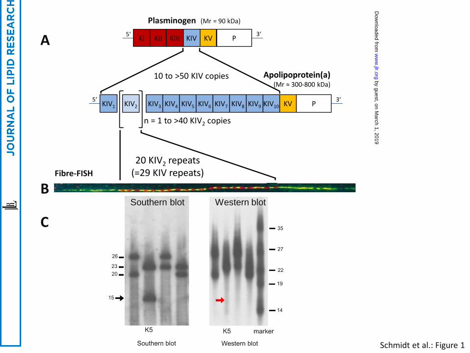
Schmidt et al.: Figure 1
Plasminogen (Mr = 90 kDa)
KIV2 KIV1 KV P
n = 1 to >40 KIV2 copies
KIV3 KIV4 KIV5 KIV6 KIV7 KIV8 KIV9 KIV10 3‘ 5‘
KV P KI KII KIII KIV 3‘ 5‘
20 KIV2 repeats (=29 KIV repeats)
Southern blot Western blot
Apolipoprotein(a) (Mr ≈ 300-800 kDa)
10 to >50 KIV copies
Fibre-FISH
A
B
C
by guest, on March 1, 2019
ww
w.jlr.org
Dow
nloaded from

A
B
Schmidt et al.: Figure 2
by guest, on March 1, 2019
ww
w.jlr.org
Dow
nloaded from
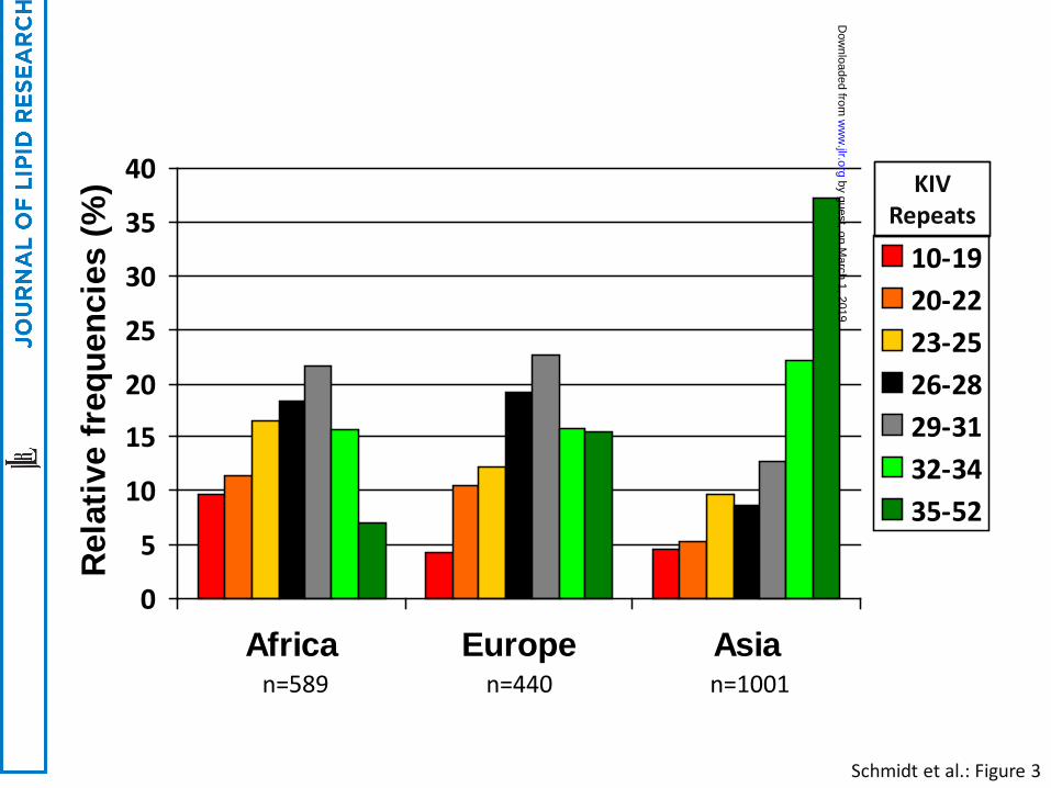
Schmidt et al.: Figure 3
0
5
10
15
20
25
30
35
40
Africa Europe Asia
Rel
ativ
e fr
eque
ncie
s (%
)
10-1920-2223-2526-2829-3132-3435-52
KIV Repeats
n=589 n=440 n=1001
by guest, on March 1, 2019
ww
w.jlr.org
Dow
nloaded from

Schmidt et al.: Figure 4
by guest, on March 1, 2019
ww
w.jlr.org
Dow
nloaded from

Schmidt et al.: Figure 5
by guest, on March 1, 2019
ww
w.jlr.org
Dow
nloaded from
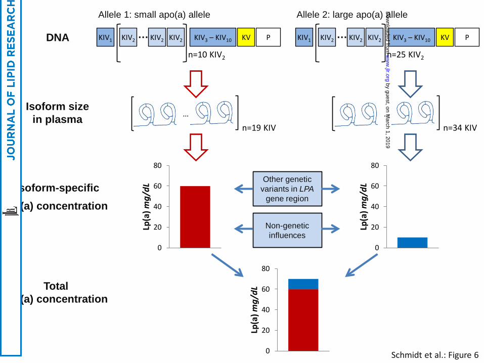
Schmidt et al.: Figure 6
DNA KIV2 KIV1 KIV2 KIV2 KIV3 – KIV10 KV P …
n=10 KIV2
…
n=19 KIV
…
n=34 KIV
Isoform size in plasma
Allele 1: small apo(a) allele Allele 2: large apo(a) allele
0
20
40
60
80
Lp(a
) mg/
dL
0
20
40
60
80
Lp(a
) mg/
dL
0
20
40
60
80
Lp(a
) mg/
dL
Isoform-specific Lp(a) concentration
Total Lp(a) concentration
Other genetic variants in LPA
gene region
Non-genetic influences
KIV2 KIV1 KIV2 KIV2 KIV3 – KIV10 KV P …
n=25 KIV2
by guest, on March 1, 2019
ww
w.jlr.org
Dow
nloaded from

Small apo(a) alleles
(=10-22 KIV copies)
Strong association (explains
about 50% of Lp(a) levels)
Strong association
High Lp(a) CVD Association
No reverse causation
Schmidt et al.: Figure 7
by guest, on March 1, 2019
ww
w.jlr.org
Dow
nloaded from

0
10
20
30
40
50
60
Tyrolean Welsh German Israeli Chinese Indian
Controls CHD
% of controls / patients with small apo(a) isoforms
Populations
Combined OR = 1.78 p<0.001
0.81.01.21.41.61.82.0
Q1: 6-30 Q2: 31-35 Q3: 36-40 Q4: 41-99
OR
(95%
CI)
Quartiles and Sum of KIV-2 Repeats
B
A
Schmidt et al.: Figure 8
by guest, on March 1, 2019
ww
w.jlr.org
Dow
nloaded from