Embed Size (px)
Citation preview
Structure and self-assemblyof viruses
Reidun TwarockDepartments of Mathematics and Biology
University of York
KITP, June 2006
Overview:
• Part I: Virus structure- Caspar-Klug Models - Viral Tiling Theory: virus architecture via generalised lattices- Tubular malformations and crosslinking structures
• Part II: Assembly models- Prototype assembly models based on VTT - The role of RNA for assembly of MS2.
• Part III: Implications for genome organisation- Models for the structure of RNA and DNA in viral capsids
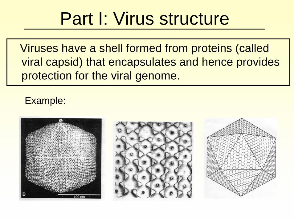
Part I: Virus structureViruses have a shell formed from proteins (called viral capsid) that encapsulates and hence provides protection for the viral genome.
Example:
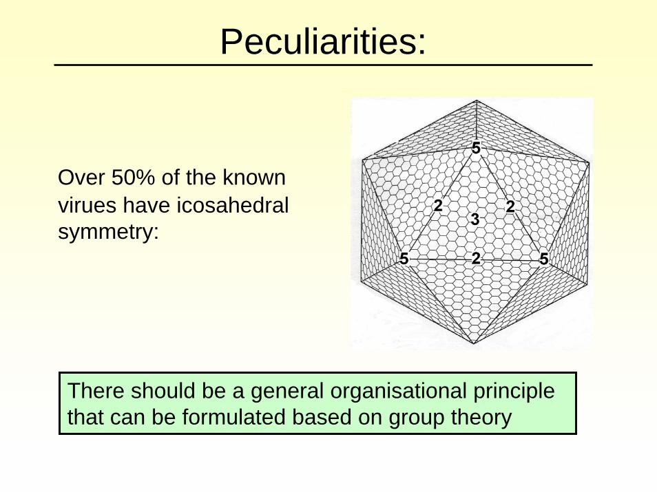
Peculiarities:
Over 50% of the known virues have icosahedralsymmetry:
There should be a general organisational principle that can be formulated based on group theory
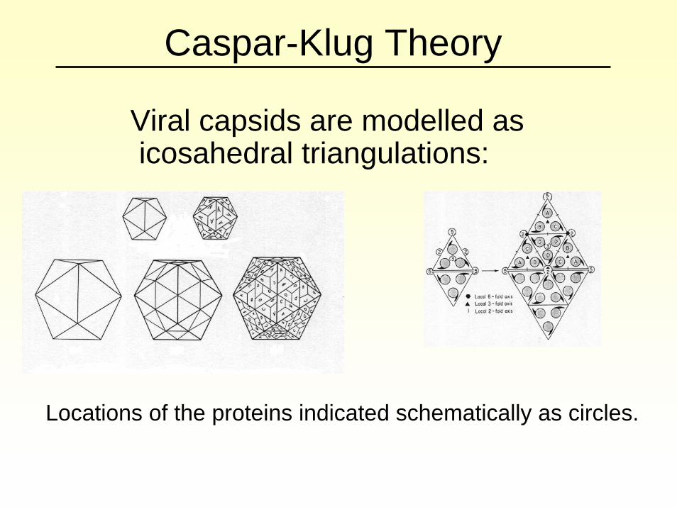
Caspar-Klug Theory
Viral capsids are modelled as icosahedral triangulations:
Locations of the proteins indicated schematically as circles.
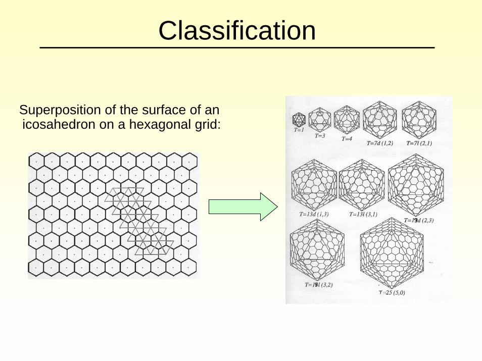
Classification
Superposition of the surface of an icosahedron on a hexagonal grid:
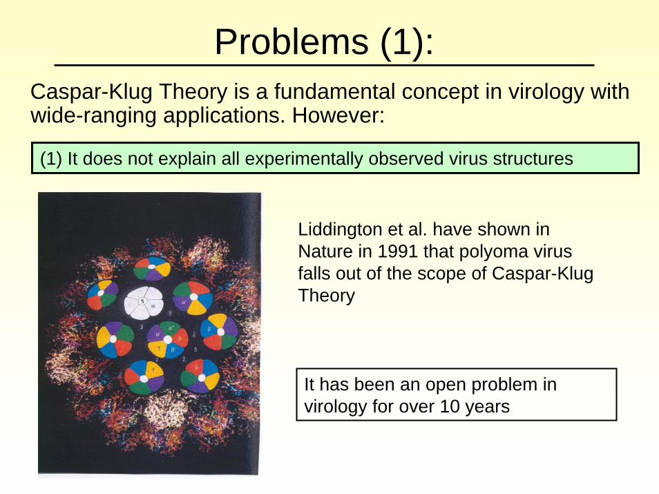
Problems (1):Caspar-Klug Theory is a fundamental concept in virology with wide-ranging applications. However:
(1) It does not explain all experimentally observed virus structures
Liddington et al. have shown in Nature in 1991 that polyoma virus falls out of the scope of Caspar-KlugTheory
It has been an open problem in virology for over 10 years
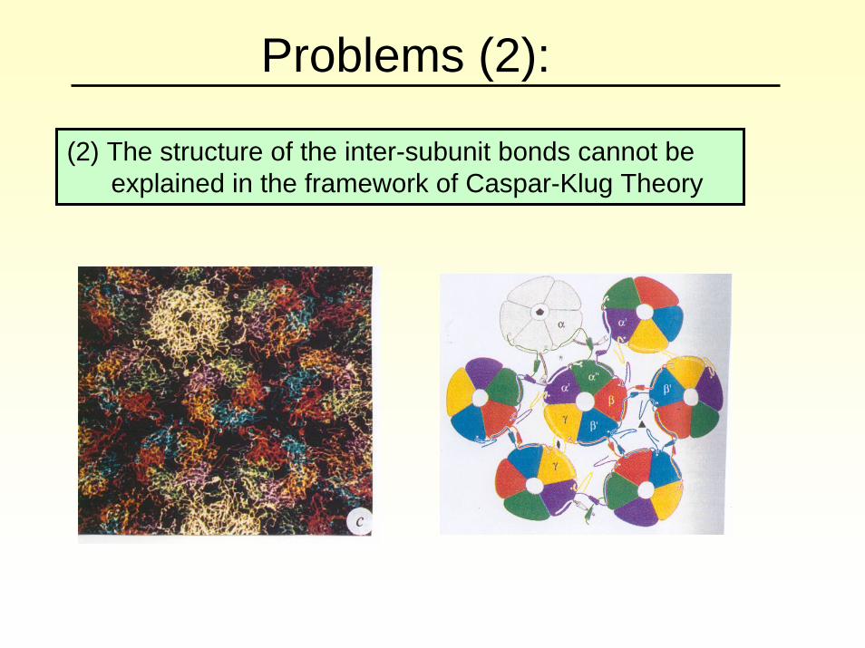
(2) The structure of the inter-subunit bonds cannot be explained in the framework of Caspar-Klug Theory
Problems (2):
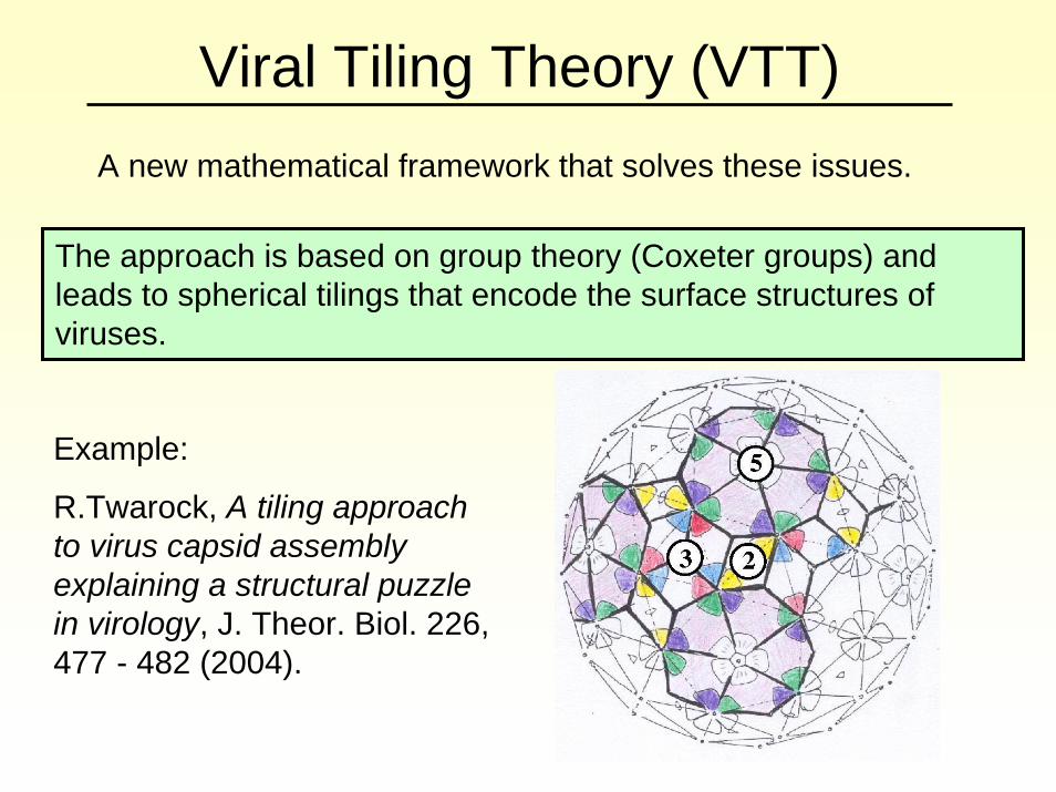
Viral Tiling Theory (VTT)A new mathematical framework that solves these issues.
The approach is based on group theory (Coxeter groups) and leads to spherical tilings that encode the surface structures of viruses.
Example:
R.Twarock, A tiling approach to virus capsid assembly explaining a structural puzzle in virology, J. Theor. Biol. 226, 477 - 482 (2004).
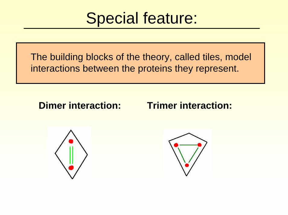
Special feature:
The building blocks of the theory, called tiles, model interactions between the proteins they represent.
Dimer interaction: Trimer interaction:
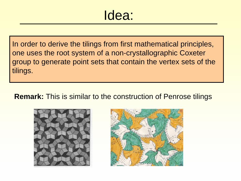
Idea:
In order to derive the tilings from first mathematical principles, one uses the root system of a non-crystallographic Coxetergroup to generate point sets that contain the vertex sets of thetilings.
Remark: This is similar to the construction of Penrose tilings
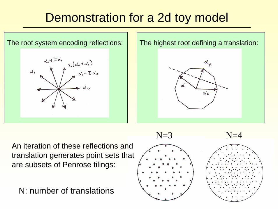
Demonstration for a 2d toy model
The root system encoding reflections: The highest root defining a translation:
An iteration of these reflections and translation generates point sets that are subsets of Penrose tilings:
N=3
N: number of translations
N=4
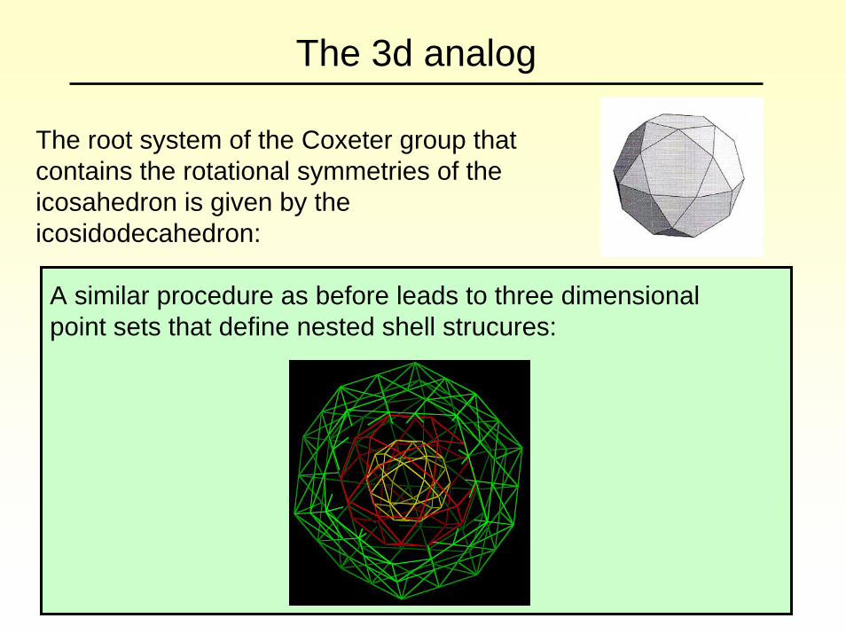
The 3d analog
The root system of the Coxeter group that contains the rotational symmetries of the icosahedron is given by the icosidodecahedron:
A similar procedure as before leads to three dimensional point sets that define nested shell strucures:

ClassificationThe surface structures of viruses can be obtained via thismethod and have been classified. [T.Keef and R.Twarock, A novel series of polyhedra as blueprints for viral capsids in the family of Papovaviridae, q-bio.BM/0512047].
There are three different types of particles with all-pentamer capsids Cryo-em micrograph:
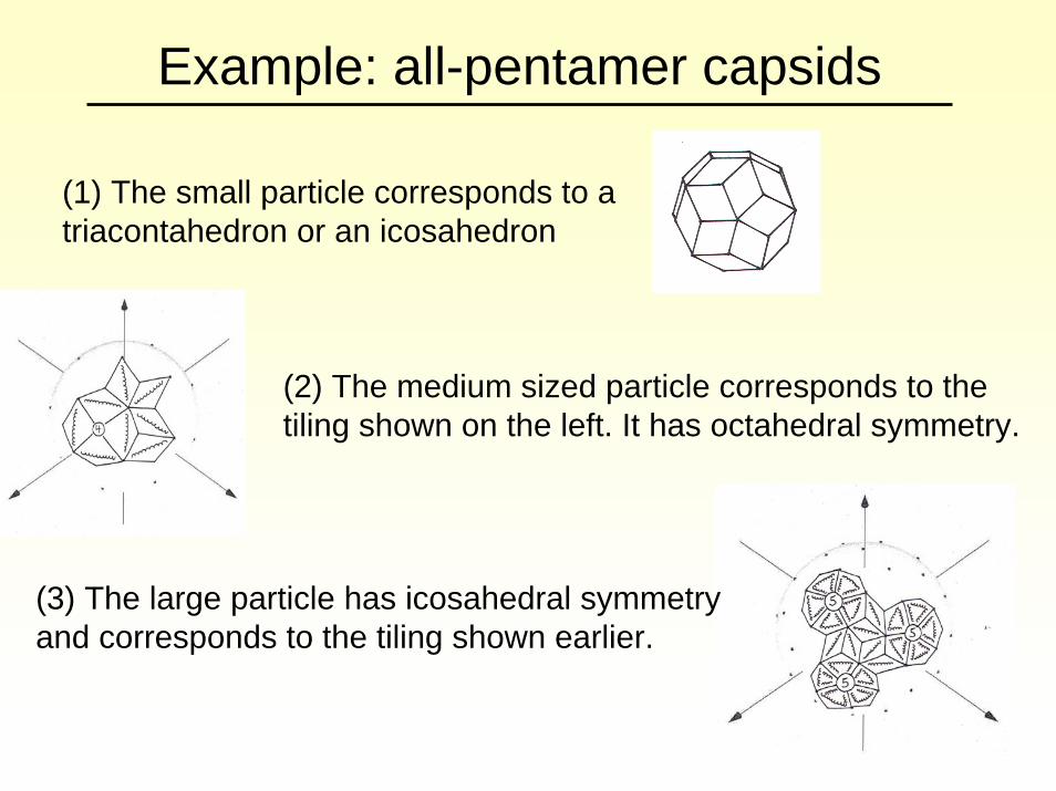
Example: all-pentamer capsids
(1) The small particle corresponds to a triacontahedron or an icosahedron
(2) The medium sized particle corresponds to the tiling shown on the left. It has octahedral symmetry.
(3) The large particle has icosahedral symmetry and corresponds to the tiling shown earlier.
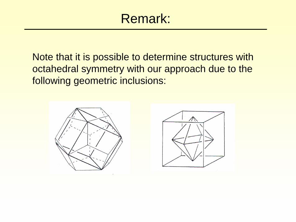
Remark:
Note that it is possible to determine structures with octahedral symmetry with our approach due to the following geometric inclusions:
Comparison with experiments
(2) Since tiles encode inter-subunit bonds, the bonding structure in the capsids is predicted by the tilings.
(1) The relative radii of the three particles are predicted by our theory.
Our predictions in (1) and (2) agree well with experimental observations.
For experimental data on SV40, see e.g. Kanesashi et al.
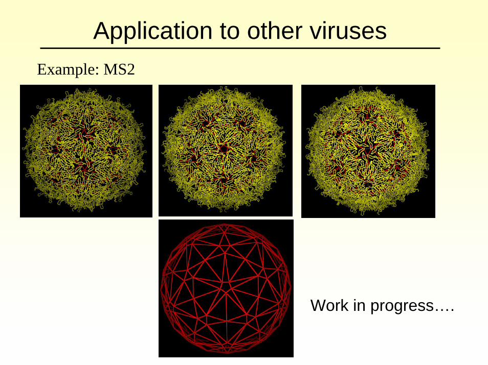
Application to other virusesExample: MS2
Work in progress….
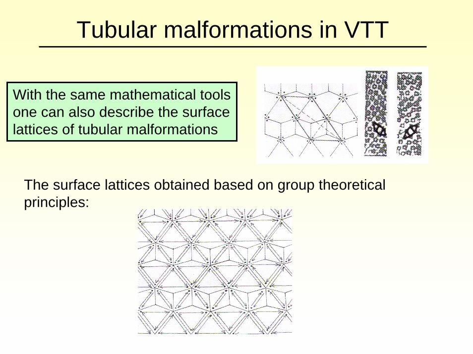
Tubular malformations in VTT
With the same mathematical tools one can also describe the surface lattices of tubular malformations
The surface lattices obtained based on group theoretical principles:
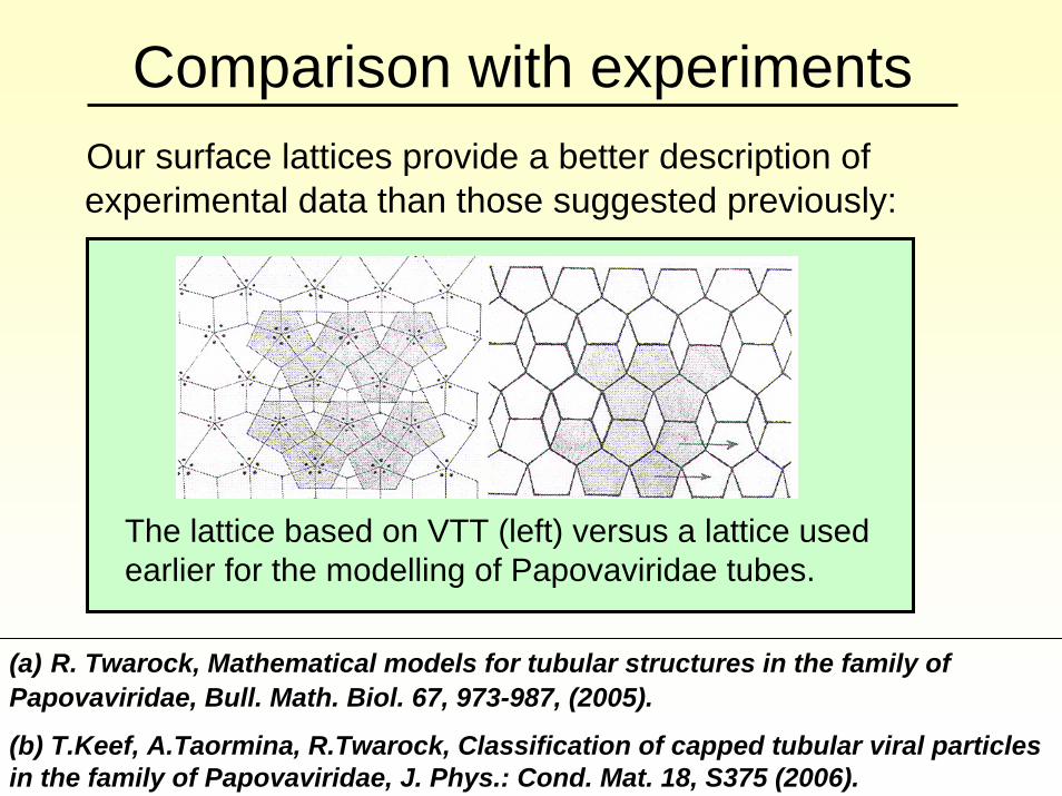
Comparison with experimentsOur surface lattices provide a better description of experimental data than those suggested previously:
(a) R. Twarock, Mathematical models for tubular structures in the family of Papovaviridae, Bull. Math. Biol. 67, 973-987, (2005).
(b) T.Keef, A.Taormina, R.Twarock, Classification of capped tubular viral particles in the family of Papovaviridae, J. Phys.: Cond. Mat. 18, S375 (2006).
The lattice based on VTT (left) versus a lattice used earlier for the modelling of Papovaviridae tubes.
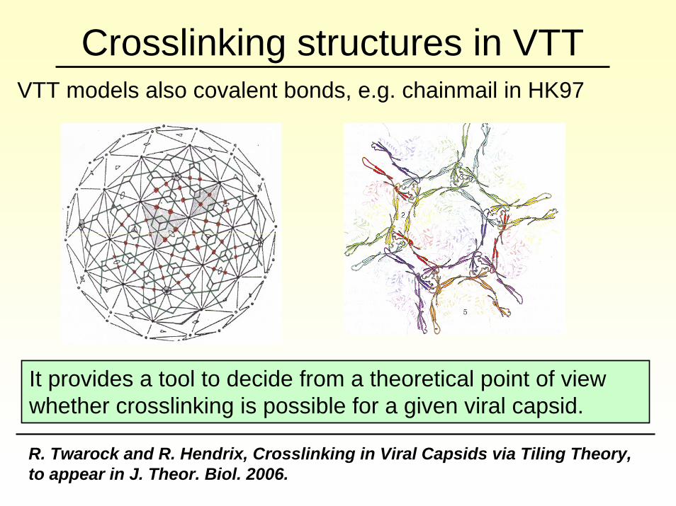
Crosslinking structures in VTTVTT models also covalent bonds, e.g. chainmail in HK97
It provides a tool to decide from a theoretical point of view whether crosslinking is possible for a given viral capsid.
R. Twarock and R. Hendrix, Crosslinking in Viral Capsids via Tiling Theory, to appear in J. Theor. Biol. 2006.
Part II: Assembly models
(a) T.Keef, A.Taormina, R.Twarock, Assembly Models for Papovaviridabased on Tiling Theory, Phys. Biol. 2, 175-188, 2005.
(b) (b) T.Keef, C. Micheletti, R.Twarock, Master equation approach to the assembly of viral capsids, to appear in Theor. Biol., June 2006.
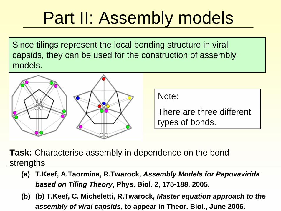
Since tilings represent the local bonding structure in viral capsids, they can be used for the construction of assembly models.
Note:
There are three different types of bonds.
Task: Characterise assembly in dependence on the bond strengths
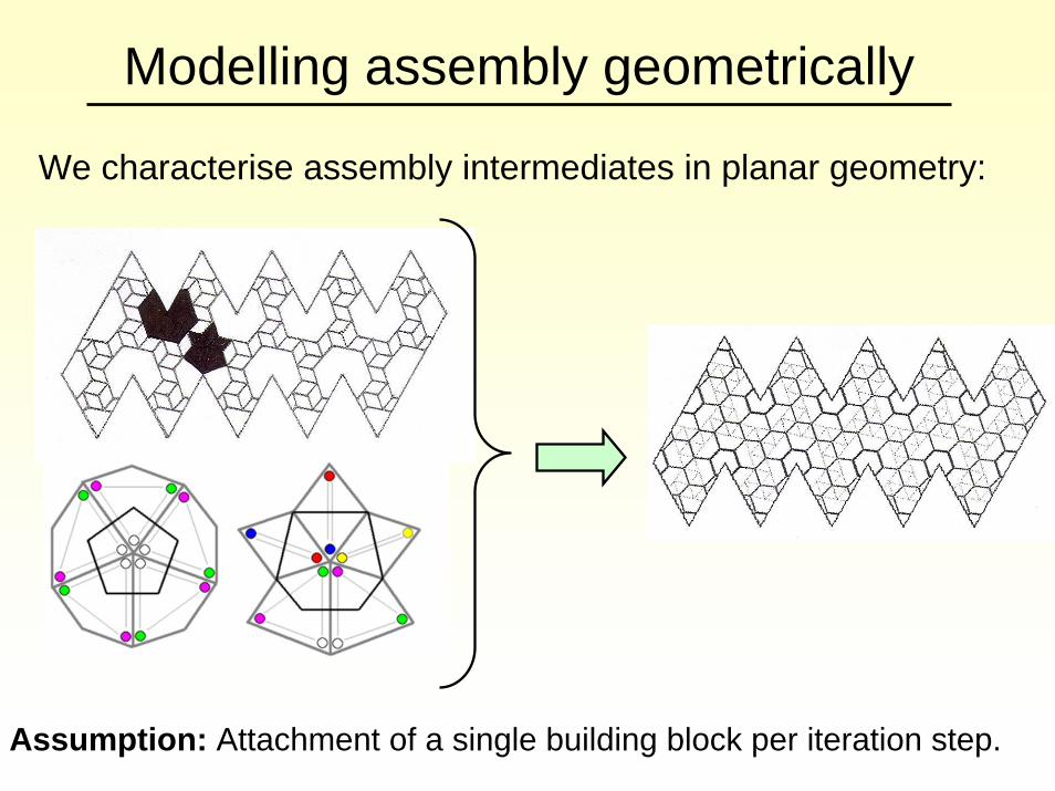
Modelling assembly geometrically
We characterise assembly intermediates in planar geometry:
Assumption: Attachment of a single building block per iteration step.
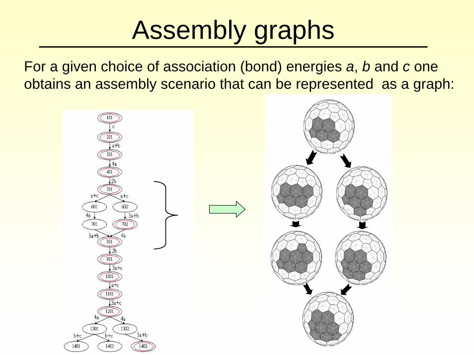
Assembly graphsFor a given choice of association (bond) energies a, b and c one obtains an assembly scenario that can be represented as a graph:
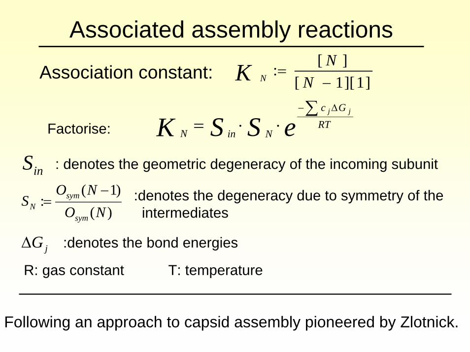
Associated assembly reactions
Association constant:
]1][1[][:
−=
NNK N
Factorise: eSSK RTGc
NinN
jj∑⋅⋅=
Δ−
inS
)()1(
:NO
NOS
sym
symN
−=
jGΔ
: denotes the geometric degeneracy of the incoming subunit
:denotes the degeneracy due to symmetry of the intermediates
:denotes the bond energies
R: gas constant T: temperature
Following an approach to capsid assembly pioneered by Zlotnick.
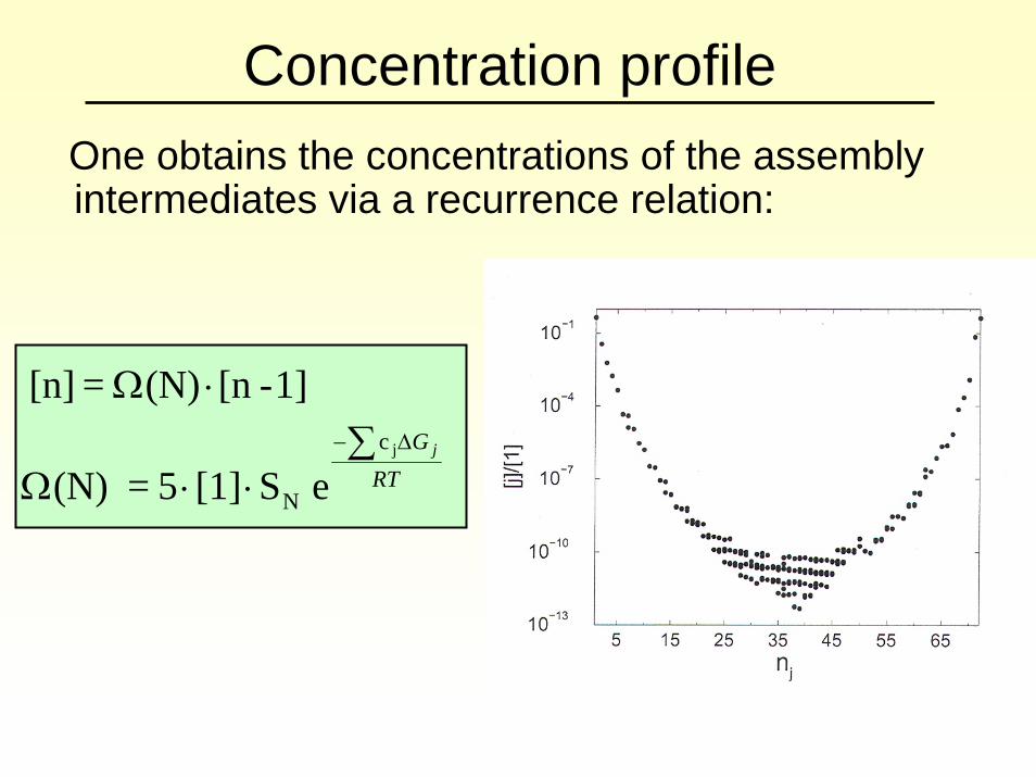
Concentration profileOne obtains the concentrations of the assembly intermediates via a recurrence relation:
RTG j∑
⋅⋅Ω
⋅ΩΔ− jc
N e S[1] 5 = (N)
1]-[n(N) = [n]
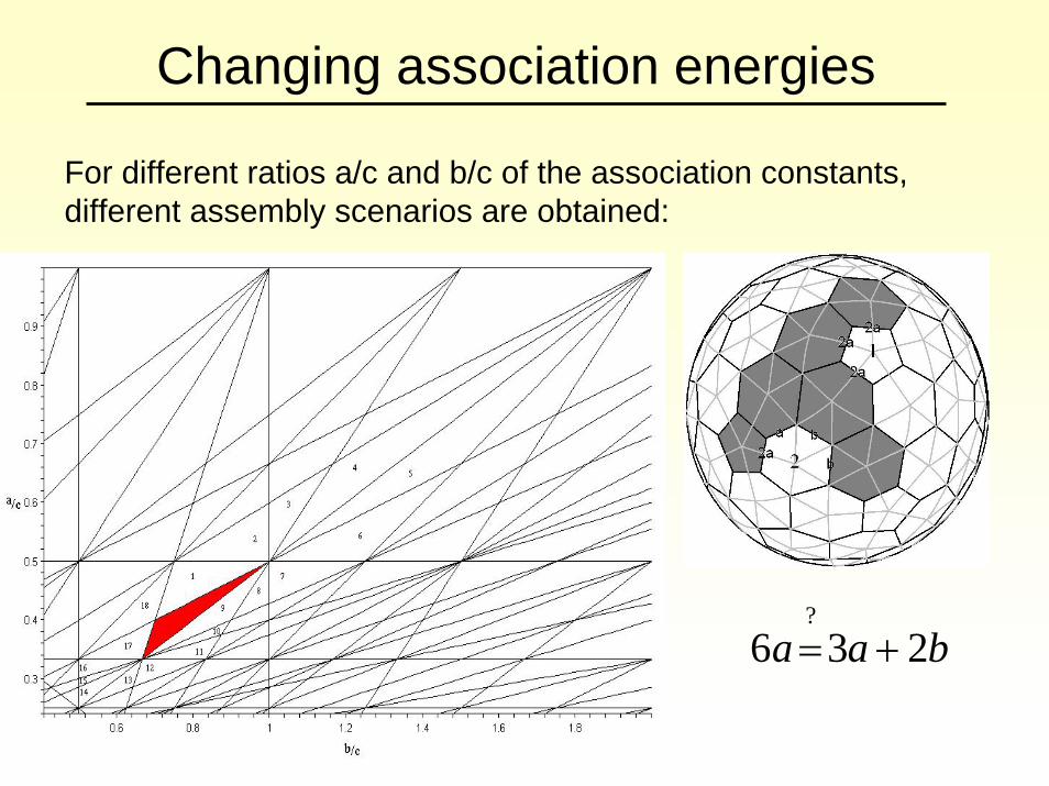
Changing association energies
For different ratios a/c and b/c of the association constants, different assembly scenarios are obtained:
baa 236?
+=
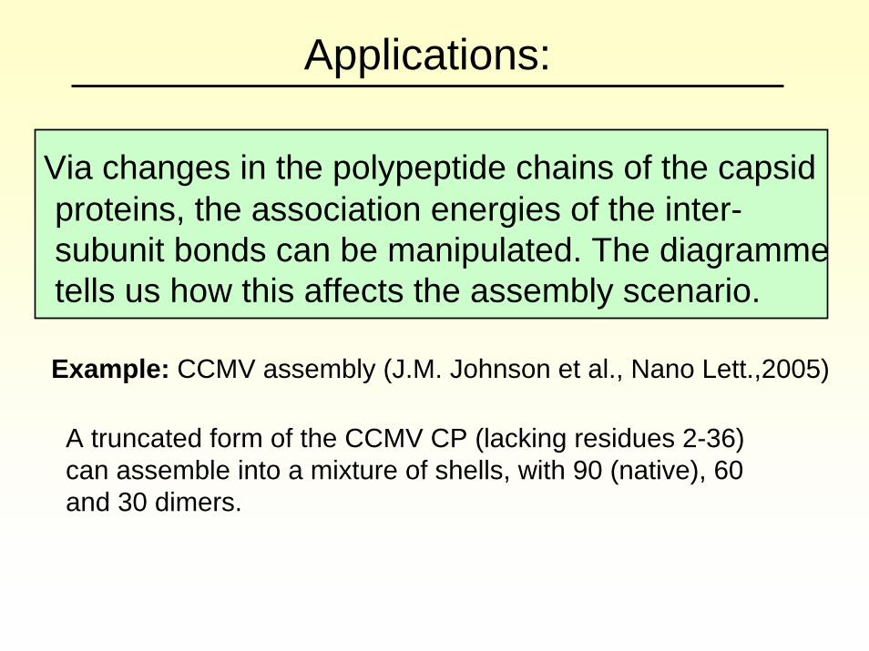
Applications:
Via changes in the polypeptide chains of the capsidproteins, the association energies of the inter-subunit bonds can be manipulated. The diagrammetells us how this affects the assembly scenario.
Example: CCMV assembly (J.M. Johnson et al., Nano Lett.,2005)
A truncated form of the CCMV CP (lacking residues 2-36)can assemble into a mixture of shells, with 90 (native), 60 and 30 dimers.

Computation of the dominant pathway
The relative probabilities of the assembly intermediates are computed via a master-equation approach. One hence obtains the dominant pathway of assembly.
A geometric analysis of the assembly intermediates on this pathway shows a characteristic pattern:
A formation of bonds with association energies 2a, a and bis pertinent to the selection of the dominant pathway.

Expanding the ensemble of intermediatesAllowing for the 20% energetically most favourable intermediates, one obtains slight changes in the dominant pathway:
A change at the start of the dominant pathway.
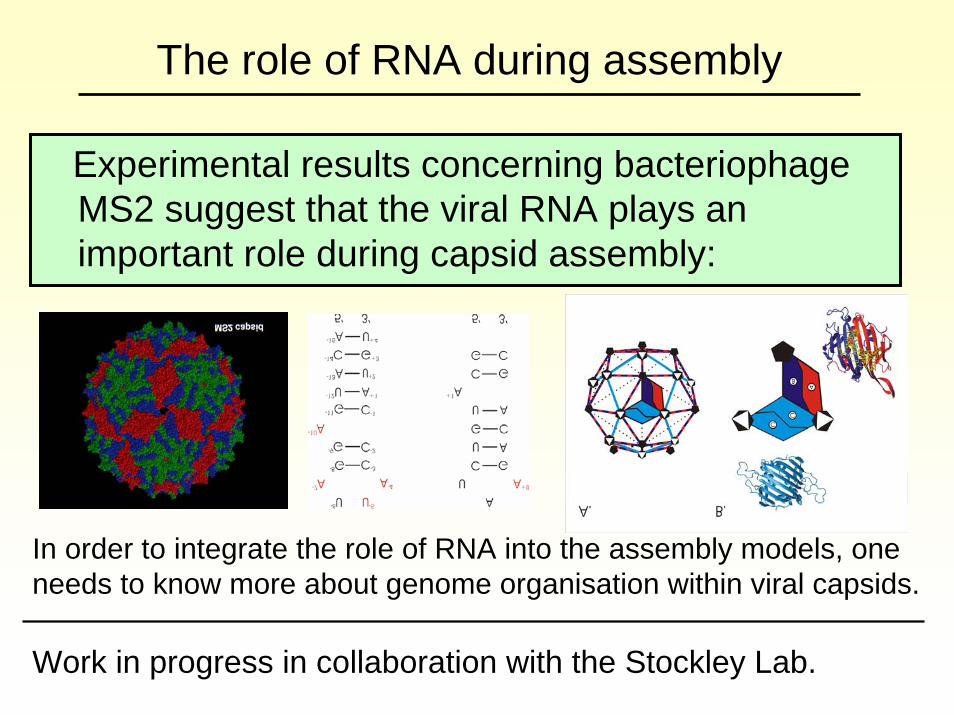
The role of RNA during assembly
Experimental results concerning bacteriophageMS2 suggest that the viral RNA plays an important role during capsid assembly:
In order to integrate the role of RNA into the assembly models, one needs to know more about genome organisation within viral capsids.
Work in progress in collaboration with the Stockley Lab.
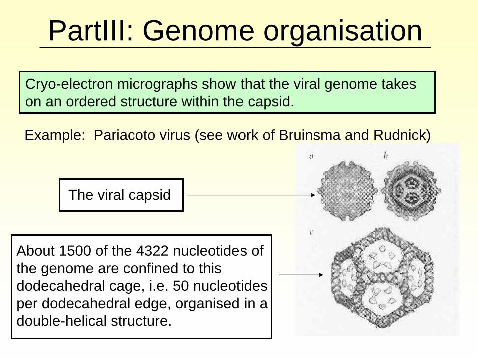
PartIII: Genome organisationCryo-electron micrographs show that the viral genome takes on an ordered structure within the capsid.
Example: Pariacoto virus (see work of Bruinsma and Rudnick)
The viral capsid
About 1500 of the 4322 nucleotides of the genome are confined to this dodecahedral cage, i.e. 50 nucleotides per dodecahedral edge, organised in a double-helical structure.
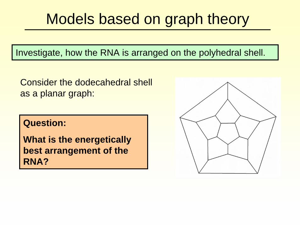
Models based on graph theory
Investigate, how the RNA is arranged on the polyhedral shell.
Consider the dodecahedral shell as a planar graph:
Question:
What is the energetically best arrangement of the RNA?
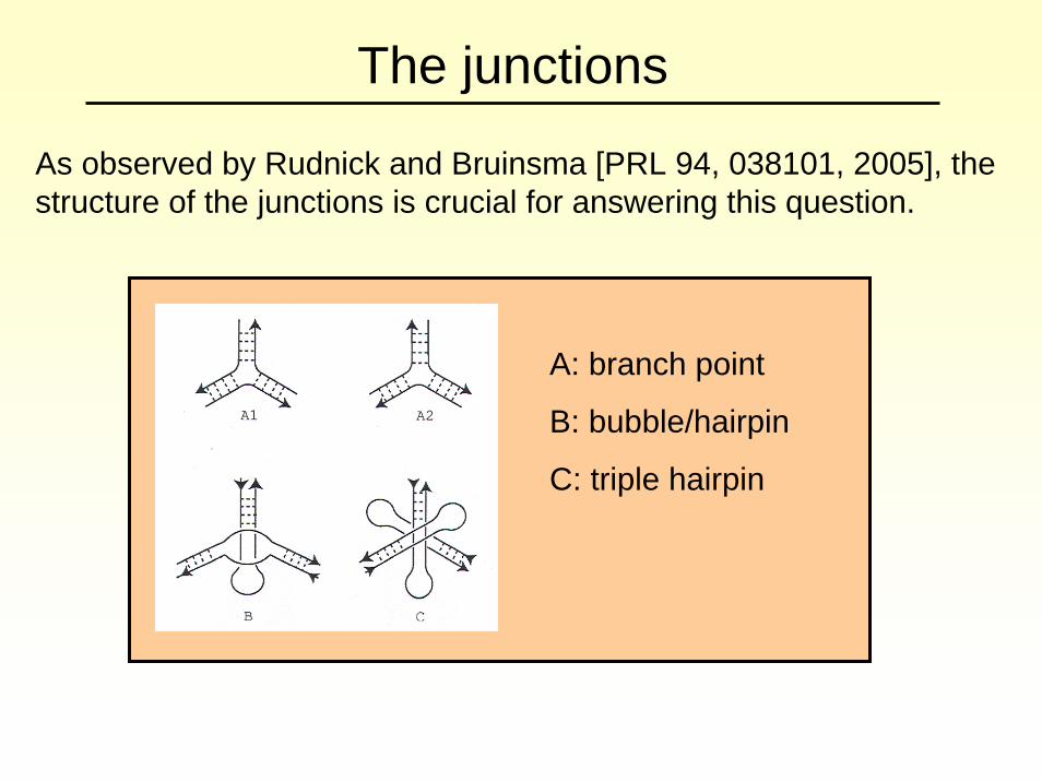
The junctions
As observed by Rudnick and Bruinsma [PRL 94, 038101, 2005], the structure of the junctions is crucial for answering this question.
A: branch point
B: bubble/hairpin
C: triple hairpin
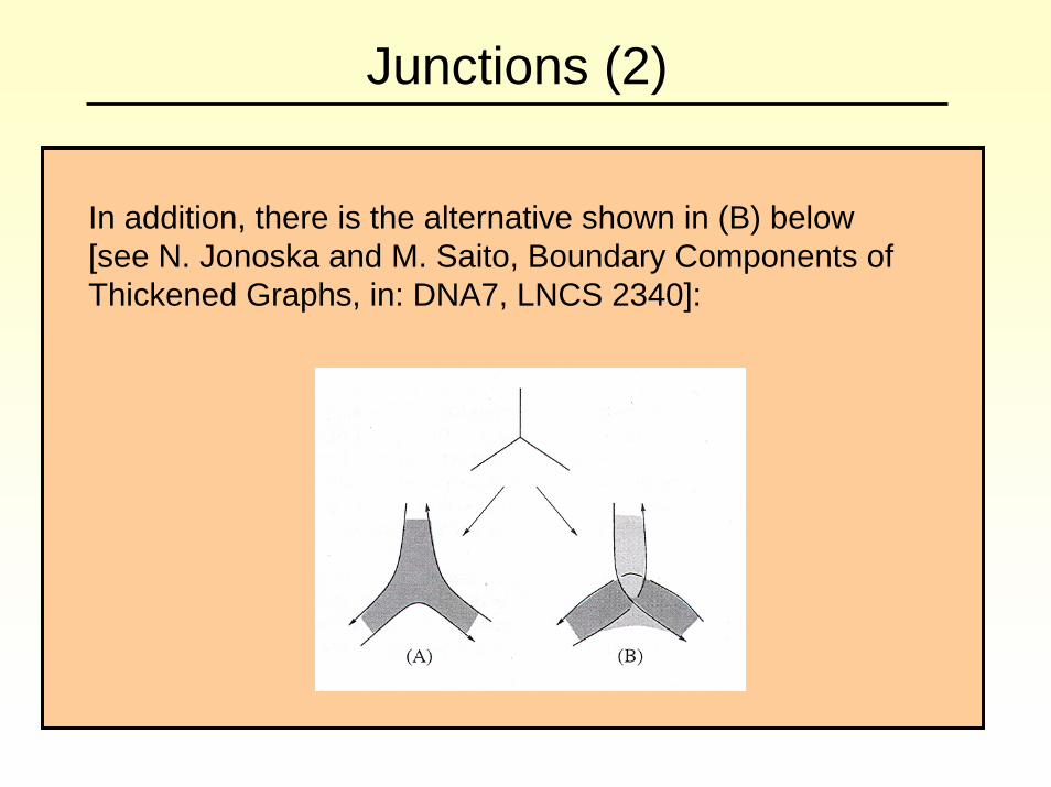
Junctions (2)
In addition, there is the alternative shown in (B) below [see N. Jonoska and M. Saito, Boundary Components of Thickened Graphs, in: DNA7, LNCS 2340]:
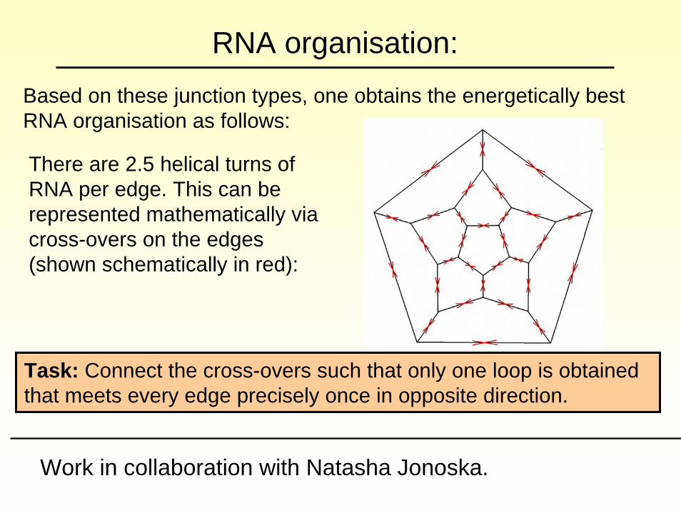
RNA organisation:Based on these junction types, one obtains the energetically best RNA organisation as follows:
There are 2.5 helical turns of RNA per edge. This can be represented mathematically via cross-overs on the edges (shown schematically in red):
Task: Connect the cross-overs such that only one loop is obtained that meets every edge precisely once in opposite direction.
Work in collaboration with Natasha Jonoska.
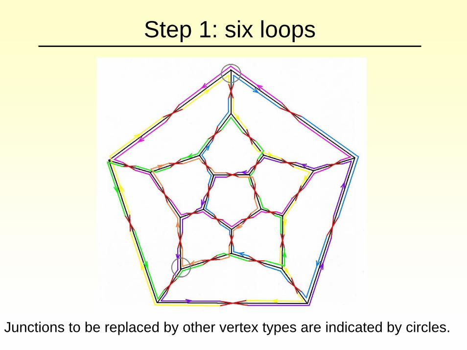
Step 1: six loops
Junctions to be replaced by other vertex types are indicated by circles.
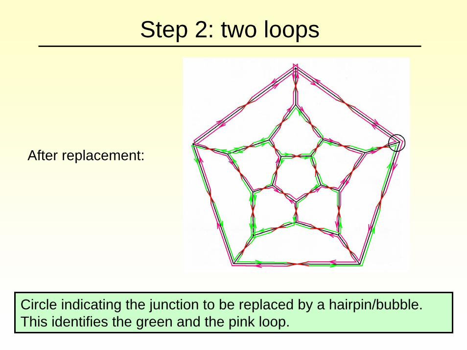
Step 2: two loops
After replacement:
Circle indicating the junction to be replaced by a hairpin/bubble. This identifies the green and the pink loop.

Without extra helical turns:
Summary and outlookWe have developed new mathematical tools for the description of virus structure and assembly.
They are based on group theory and tiling theory, and have been used in order to model
• the structure of viral capsids in terms tilings that encode the locations of the capsid proteins and the bonds between them
• the structure of tubular malformations
• the assembly process
• the structure of the viral genome within the capsids
Outlook
Financial support by an EPSRC Advanced Research Fellowship and EPSRC grant GR/T26979/01 are gratefully acknowledged.
A 3d representation of proteins via encasing forms.A 3d representation of proteins via encasing forms.•• The role of RNA during assembly of RNA viruses.The role of RNA during assembly of RNA viruses.•• Assembly via agglomeration of intermediates. Assembly via agglomeration of intermediates. •• The dependence on experimental conditions (The dependence on experimental conditions (egeg pH value)pH value)•• Simultaneous assembly of different species. Simultaneous assembly of different species.
Our group is currently working on assembly models that include:
Applications include
•• the use of the use of capsidscapsids for drug delivery.for drug delivery.•• interference with interference with capsidcapsid assembly for antiassembly for anti--viral drug design. viral drug design.