Embed Size (px)
Citation preview

Structure and Function of Macromolecules - 1
As we stated in our carbon introduction, the majority of the molecules found in livingorganisms are based on carbon, (along with nitrogen, oxygen and hydrogen in thefunctional groups). Their specific chemical properties are, to a large extent,determined by the functional groups attached to the carbon backbones.
Many of our molecules are large, and are assembled from smaller molecules that areeither identical to each other, or similar to each other. These large molecules arecalled macromolecules or polymers. The "building blocks" of these polymers arecalled monomers or subunits, and have a common structure. Because our polymersare large molecules, and based on carbon, we can get a great diversity of them fromjust a few small monomers, by varying number, sequence and bonding arrangements.
Our biological macromolecules are grouped into four categories: proteins, nucleicacids, lipids and carbohydrates. We shall discuss structure and functions of eachgroup.
Most of our biological molecules are assembled or broken down using the same typeof chemical reaction, one which involves adding or removing water molecules.Macromolecules are formed from their subunits by removing molecules of water (ahydrogen (-H) from one subunit and the hydroxyl (-OH) from the second) to join thesubunits together. This is called a dehydration synthesis, or condensation.When larger molecules are broken down, such as in digestion, water molecules areadded in to break the macromolecules into their subunits, a process calledhydrolysis. The enzymes that facilitate digestion are called hydrolytic enzymes.
Let's now look with some detail at the major compounds of living organisms. We shalllook at Proteins, Lipids and Carbohydrates, and briefly the fourth: Nucleic Acids. Wewill deal with the nucleic acids in depth during our unit on molecular genetics.
Structure and Function of Macromolecules - 2
Amino Acids and ProteinsProteins are very large molecules composed of combinations of about 20 differentamino acids. The precise physical shape of a protein is very important for its function.A single cell may have 10,000 or more different proteins. This diversity of proteins isessential for the functioning of each cell in a living organism. It has been estimatedthat there may be over 100,000 different kinds of proteins. As much as half of thenon-water component of a typical cell can be protein.
Functions of ProteinEnzymes
• Globular proteins that facilitate chemical reactions.Defense Proteins
• Antibodies• Protein toxins
Transport Proteins• Plasma membrane proteins carry substances through membranes or form
channels or pumps for passage• Oxygen carrier in circulation (hemoglobin)• Mineral protein carriers (iron, zinc)
Structural/Support Proteins (Fibrous proteins)• Connective tissue in animals (collagen – the most abundant vertebrate protein)• Webs, cocoon s and other arthropod structures• Hair, nails horns, etc. (keratin)• Fibrins used in blood clotting
Contractile Proteins – locomotion and movement• Muscle• Cilia and flagella,• Microtubules, microfilaments and intermediate filaments
Regulatory Proteins• Hormones• Gene Regulators• Osmotic regulation
Receptor Proteins• Membrane surface receptor proteins• Signal transduction proteins
Recognition Proteins• Glycoproteins (carbohydrate-protein hybrids) for identification of "self".
Storage Proteins (specialized)• Examples are casein in milk, ferritin for iron storage, calmodulin for calcium and
albumin in eggsEnergy transfer molecules
• Cytochromes
Structure and Function of Macromolecules - 3
Protein StructureProtein structure is critical for its function. Each protein has a unique shape orconformation. However, all proteins are composed exclusively of subunits of aminoacids, which join together in long chains called polypeptides that fold or coil into theunique shape of the functional protein.
To discuss the formation of a protein we need to first discuss the structure of aminoacids.
Amino acids• Amino acids contain Carbon, Hydrogen, Oxygen, Nitrogen, and sometimes Sulfur• Amino acids have two function groups
NH2 Amino functional groupCOOH Carboxyl functional group
• Both functional groups attach to a specific asymmetric carbon (one in which bondsto four different atoms or molecular fragments) called the alpha (α) carbon, of thecarbon chain. The third bonding site of the alpha carbon is typically Hydrogen.The alpha carbon will have at its fourth bonding site a side chain, or R group thatgives the amino acid its unique structure and properties.
• There are 20+ different amino acids in protein. All have a common structure [seetext for structures of the different amino acids] except for the R group.
HO—C=0 = carboxyl (acid) functional group; H—N—H = amino functional group | | H
Characteristics of amino acids• Some amino acids have R groups that are polar that contain oxygen or
sometimes just –H. Polar amino acids are hydrophilic.• Some amino acids have R groups that are nonpolar, typically with -CH2 or -CH3
R groups. nonpolar amino acids are hydrophobic.• Amino acids that ionize have R groups that are acidic (generally with a (-)
charge) or basic.• Amino acids with hydrocarbon ring R groups are often aromatic.• Amino acids, when ionized also have the amino functional group positively
charged and the carboxyl functional group negatively charged.

Structure and Function of Macromolecules - 4
The unique properties of the different amino acid R groups will affect the structure ofthe protein formed so that the number, kind, and bonding sequence of amino acids ina protein is critical. For example:
• Cysteine contains sulfur in the R group, so cysteines can form disulfide bonds(disulfide bridges) linking amino acids in the chain together.
• Proline causes kinks in the amino acid.• Methionine is the first amino acid in a protein.• Amino acids are joined together by a dehydration synthesis of amino/carboxyl
groups forming a peptide bond.
How do amino acids join to make a protein?1. A protein starts as a chain of amino acids, called a polypeptide2. Amino acids are joined by the peptide bond, via dehydration synthesis t o
form the polypeptide that occurs between the carboxyl functional group of oneamino acid and the amino functional group of the second.
3. The polypeptide chain is referred to as the primary structure of the protein.4. The specific amino acids in the polypeptide chain will determine its ultimate
conformation, or shape, and hence, its function. Even one amino acidsubstitution in the bonding sequence of a polypeptide can dramatically alter thefinal protein's shape and ability to function.
Peptide Bond
How do polypeptides vary?1. Number of amino acids in the chain: 50—1000 or so2. Which kind of amino acids are in the chain (of the 20 types)3. How many of each kind of amino acid4. The bonding order or sequence of amino acids
Note: The first protein sequenced was insulin, a tiny protein, which was accomplishedabout 50 years ago by Frederick Sanger's group in Cambridge. Today we use supercomputers to sequence proteins, but even for the computer, it's a challenge.
Structure and Function of Macromolecules - 5
A Closer Look at Protein shape and structureThe polypeptide chain is just the beginning of a protein. Functional proteins undergofurther processing to realize a final functional shape or conformation. Some proteinsare composed of more than one polypeptide. The surface structure of the protein iscritical for its function, such as with hemoglobin where exterior facing R groups mustbe polar to hold the heme (iron containing) group that binds oxygen molecules. Infact, virtually all proteins have their nonpolar amino acids oriented in the interior ofthe protein, leaving polar and charged amino acids to face their aqueous environmentof the cell.
The function of many proteins depends on a specific region of the protein that bindsto another molecule. Antibodies, critical to the immune system, function by binding tospecific regions of the antigen molecules, to deactivate them. An enzyme binds tothe substrate (the reactants) at a specific active site on the enzyme.
How do proteins acquire their unique functional shapes?The peptide bonds that form between amino acids result in the primary structure of aprotein, the polypeptide. The polypeptide undergoes a number of modificationsbefore assuming its functional shape.
Secondary StructureAs peptide bonds are formed, aligning the amino acids, hydrogen atoms of aminofunctional groups are attracted to the double bonded oxygen atom at the peptidebond and form hydrogen bonds.
This bonding coils the polypeptide into the secondary structure of the protein,most commonly the alpha (αααα)))) helix, discovered by Linus Pauling. The α-helix coils atevery 4t h amino acid.
Some regions of the polypeptide have portions that lie parallel to each other (still heldby hydrogen bonds) instead of in the alpha helix, in which the amino acids' hydrogenbonds form a pleated structure. Fibrous proteins have significant pleated structures,called the beta (ββββ ) pleated sheet.
MotifsThere are common patterns associated with the secondary structure of proteins.These patterns are called motifs. The βαβ motif, for example, causes a fold in aprotein. Motifs will be discussed in gene regulation, where transcription factors bindto the DNA at specific "binding motifs". One such motif is the"α turn α" or "helix turn helix" motif. A second is the β α β α β motif.

Structure and Function of Macromolecules - 6
Tertiary StructureFollowing the secondary shape, openings for bonding along the side chains (the Rgroups) of amino acids causes more folding or twisting to obtain a final, three-dimensional functional protein, called the tertiary structure.
• Hydrophobic regions typically form in the tertiary structure among groups ofamino acids with non-polar side chains forcing those amino acids to the interior ofthe protein with a very precise and tight fit. van der Waals interactions occurbetween these amino acids. Any change in a nonpolar amino acid will affect thisprecise fit and disrupt the shape of the final protein.
• Disulfide bonds (which are strong covalent bonds) between nearby cysteinemolecules are important to the tertiary structure as well, as are hydrogen bondsand some ionic bonds between charged R-groups. The final conformation formost proteins is a globular shape.
DomainsThe instructions for protein structure are coded in DNA molecules. Within a gene (aregion of DNA that codes for polypeptide instructions) are areas called exons. (Wewill discuss exons molecular genetics unit.) To our point here, however, is that eachexon-coded section of a protein folds independently into a unit called the domain.The domains are connected by the rest of the polypeptide. Functionally, domains mayperform different functions for a given protein. For example, one domain of anenzyme might be the attachment site for a co-factor and a second domain mayfunction as the active sire of the enzyme.
Quaternary Protein Structure If two or more polypeptide chains join in aggregates, they form a quaternarystructure, such as in the protein molecule, hemoglobin. Often quaternary proteins arecomplexed with a different molecule, often a mineral. Hemoglobin contains iron, forexample.
Collagen, a common protein found in connective tissue, has a collagen helix,produced when three polypeptides coil around each other. Consequently collagenfibers are very strong.
Getting the Protein "Folded": ChaparoninsAlthough we may present the sequence of protein structure (primary, secondary andtertiary as an automatic process, it is not spontaneous. Special proteins, calledchaparonins, are essential for protein folding. It is now believed that chaparoninsfunction to help proteins that are not folding correctly to unfold and refold correctly.Many work best at higher temperatures when the bonds needed for secondary andtertiary shape are unstable. Science does not yet know how chaparonins really work.

Structure and Function of Macromolecules - 7
One area of study is looking at the role of defective chaparonins in some diseases,such as cystic fibrosis and Alzheimer's.
Protein StabilityAs we have seen, the physical structure, or conformation, of a protein is maintainedby weak bonds. Many of these bonds are hydrogen bonds formed from the polarityof the amino acids and their “R” groups. If these weak bonds are broken, the proteinstructure is destroyed and the molecule can no longer function. This process is calleddenaturation.
Things which can denature protein:1. Heat (as low as 110 F, many @ 130 F)2. Heavy metals (e.g., silver, mercury)3. pH changes4. Salts5. Alcohols
Ethyl alcohol least toxic6. Many proteins will denature if placed in a non-polar substance.7. Other chemicals
Enzymes are seriously affected by denaturation – but other proteins of the body canalso be denatured. Although in most cases, a denatured protein loses its functionpermanently, in some cases, re-naturation can occur if the substance that promotesthe denaturation is removed from the protein. This is more true of chemicaldenaturants and particularly in experimental environments.

Structure and Function of Macromolecules - 8
Nucleotides and Nucleic AcidsNucleic acids are our information carrying compounds -- our genetic molecules. Aswith many of our other compounds, the nucleic acids are composed of subunits callednucleotides. Nucleotides also have independent functions.
Functions of Nucleotides• Components of nucleic acids (which are long chains of nucleotides)• Energy carrier molecules (ATP)
• Energy transport coenzymes (NAD+, NADP+, FAD+)• Chemical intracellular messengers
(e.g., Cyclic AMP, a cyclic nucleotide that carries messages from the cellmembrane to molecules within the cell, to stimulate essential reactions)
ATP Energy Transfer Nucleotides NAD
Functions of Nucleic AcidsStorage of genetic information (DNA)Transmit genetic information from generation to generation (DNA)Transmit genetic information for cell use (RNA)DNA self-replication
Most of the information on nucleotides and nucleic acids will be discussed when wediscuss genetics and energy relationships of cells. For now we shall just present thebasic structure of the nucleotides and nucleic acids.
Nucleotide Structure1. 5–carbon sugar component
RiboseDeoxyribose

Structure and Function of Macromolecules - 9
2. Phosphate groupAttached to the sugar's 5' carbon with a phosphodiester bond
3. Nitrogen Base component attached to the sugar's 1'carbon.There are two types of nitrogen bases:• Single six-sided ring
pyrimidinesCytosineThymineUracil
• Double ring purines(six- and five-sided)
AdenineGuanine
Arrangement of a Nucleotide:
Nucleic acids (polynucleotides) are formed when covalent phosphodiester linkagesform between one nucleotide's sugar's 3' carbon and the phosphate of the nextnucleotide to form long chains. In DNA, a double chain is formed when 2 nitrogenbases hydrogen bond between the sugar-phosphate backbones. RNA molecules aresingle chains.

Structure and Function of Macromolecules - 1 0
Genes (specific regions of DNA molecules) contain the hereditary information of anorganism. The linear sequence of nitrogen bases of the nucleotides determines theamino acid sequence for proteins in the cells and tissues. As with all of biology, theprocesses of evolution are validated in DNA information. Organisms more closelyrelated evolutionarily, have more similar DNA. When the sequence of the beta chainof human hemoglobin is compared to other animals, other primates have virtually nodifferences, and the number of differences increases when less closely related animalsare checked.
Structure and Function of Macromolecules - 1 1
LipidsMany of our common substances are lipids, which include fats, oils (triglycerides),phospholipids, steroids (or sterols), prostaglandins, waxes and terpenes. Lipidsgenerally are not polymers, although some are reasonably large molecules. Lipids aregrouped together because they are (mostly) hydrophobic and not soluble in water.Most lipids have a large proportion of C-H bonds that are nonpolar.
Lipid Functions• Fuel reserve molecules (Lipids are energy rich, having 9 calories/gram.) Humans
store fat reserves in adipose tissue. Adipose cells have a remarkable ability toswell and shrink depending on the amount of fat reserves they contain.
• Structure of cell membranes (the function of phospholipids)• Protective surface coatings and insulation• Many hormones (regulatory chemicals)
Lipid Characteristics• Most lipids are strictly nonpolar and hydrophobic, so they dissolve in nonpolar
substances, but not in water. Many lipids in water tend to take on a conformationthat will expose any polar portion to the water and protect the nonpolar regions.This is critical to phospholipids and cell membrane structure.
• Most lipids feel "greasy"• Lipids contain large regions of just carbon and hydrogen, as carbon-carbon bonds
and carbon-hydrogen bonds and small amounts of oxygen relative to the carbon-hydrogen atoms.
• The most common lipids are the triglycerides (fats and oils)
Fats and Oils (the Triglycerides or Triacylglycerols)The terms fats and oils are terms of convention
Fats are "hard" or solid at room temperatureOils are liquids at room temperature
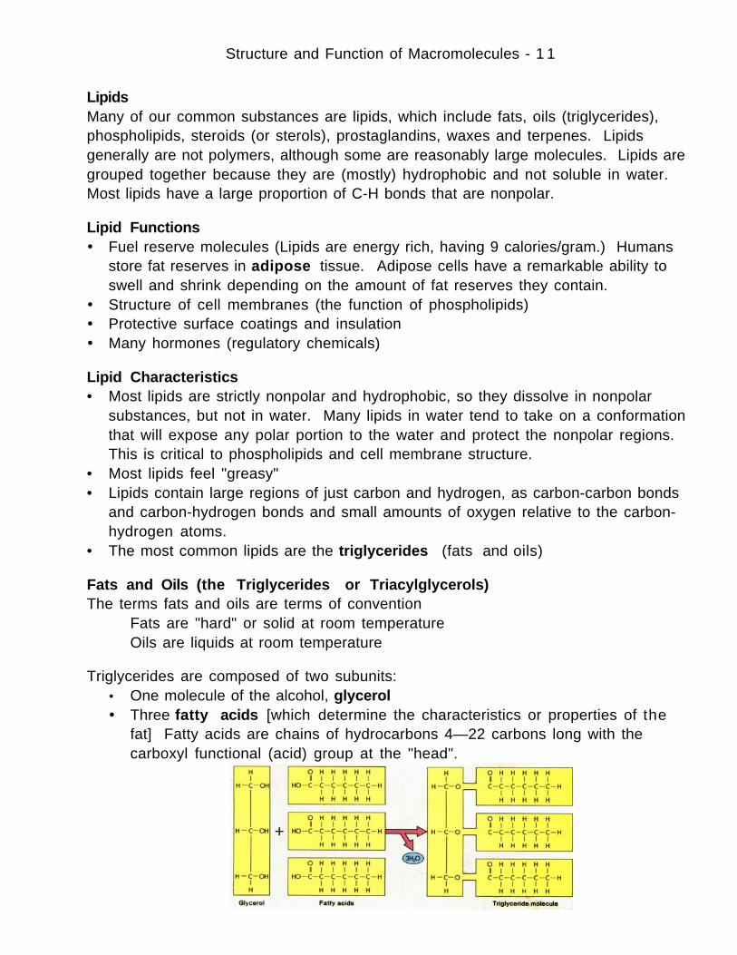
Triglycerides are composed of two subunits:• One molecule of the alcohol, glycerol• Three fatty acids [which determine the characteristics or properties of the
fat] Fatty acids are chains of hydrocarbons 4—22 carbons long with thecarboxyl functional (acid) group at the "head".

Structure and Function of Macromolecules - 1 2
The fatty acid hydrocarbon tails are strictly non-polar, so that triglycerides arehydrophobic molecules.
Each carbon within the chain has 2 spots for bonds with hydrogen
• If each carbon has 2 hydrogens the fatty acid is saturated
H H H H H H H O=C–C–C–C–C–C–C–C–H HO H H H H H H H
• If two carbon atoms are double bonded, so that there is less hydrogen in the fattyacid, it is monounsaturated
H H H H H H H H H O=C–C–C–C=C–C–C-C-C-C-C–C–H HO H H H H H H H H H H H
• If more than 2 carbon atoms are unsaturated, the fatty acid is polyunsaturated
H H H H H H H O=C–C–C–C=C–C–C-C-C-C=C–C–H HO H H H H H H H H H H H
• A trans-fatty acid might look like this:
H H H H H H H H H H O=C–C–C–C=C–C–C-C-C-C-C–C–H HO H H H H H H H H H H
Ester bonds attach the glycerol (by a dehydration reaction) to each of the 3 fattyacids, by removing the H from the glycerol's hydroxyl function group and the hydroxylfunctional group from the carboxyl head of each fatty acid.
O H H H || | | |
glycerol H-C-O---C-C-C-C fatty acid tail | | | | H H-H-H
Ester Bond

Structure and Function of Macromolecules - 1 3
Ways that fatty acids are different:1. Length of chain in fatty acid
• Usually an even number of 4 – 26 carbons long (most are 14 - 18)Short chains are more solubleShort chains are more easily broken downShort chains oxidize more easily (process by which fats become "rancid")
2. Degree of saturationSaturatedMonounsaturatedPolyunsaturated
• Most plant fats tend to be unsaturated, but fats from tropical plants tend tobe very saturated
• Fish oils tend to be unsaturated (from cold water and salt water fish). Otheranimal fats tend to be saturated
3. Liquid vs solid• Short chains and unsaturated chains are liquid at room temperature
(molecules are smaller and less dense (the double bonds distort themolecules so they don't fit close together)
• Saturated chains are solid (denser) because chains fit together better
Synthetic FatsOlestra is a synthetic fat, marketed under the trade name of Olean. It mimics thetexture and properties of triglycerides, is fat soluble, but not digestible or absorbedinto the body, so all Olestra consumed passes through the digestive tract. Hence, itis considered to be calorie-free. Olestra is a sucrose polyester, composed of fattyacids attached to sucrose rather than glycerol. Six to eight fatty acids are attachedto the sucrose molecule so the lipase digestive enzymes can't function to hydrolyzethe ester bonds.
Olestra is synthesized from cottonseed or soybean oil, heated in a base-catalyzedreaction with methanol to detach the fatty acids as methyl esters. The glycerolsettles out and is drawn off, and the fatty acid methyl esters are distilled. Sucroseand another base catalyst are then added to the fatty acid methyl esters, withemulsifiers. Under high temperature, sucrose polyesters form and methanol isremoved. Further processing removes leftover fatty acid esters and emulsifiers. Theprocessing is completed with bleaching and deodorizing. (This information about the structureand processing of Olestra was taken from C&E News 4 /21 /97 . )
Simplesse is a fat substitute that mimics the texture of fat in the oral cavity. It issynthesized from egg and milk proteins. The shape of the simplesse molecule isspherical, resembling miniature marbles, so the product has the slick texture of fat.Simplesse is not heat stable, and cannot substitute for fats in frying or baking.

Structure and Function of Macromolecules - 1 4
WaxesWaxes are similar to fats, although the alcohol component contains more carbon, andmore fatty acids are bonded. Waxes are solids.
Phospholipids• Structural molecules – major component of all membranes of cells• Phospholipids are composed of a glycerol molecule with two fatty acids attached
by ester bonds and a polar phosphate-containing compound attached to the thirdcarbon.
• The value of phospholipid structure is that the phosphate region of the molecule ischarged, ideal for the cell membrane structure, and the fatty acid portions arestrictly non-polar. Molecules that have these properties are said to beamphipathic.
• Hydrophilic portion in the phosphate region (which is negatively charged)• Hydrophobic portion in the fatty acid tails• Often phospholipids in solution will form micelles, droplets in which the tails point
inward and the hydrophilic heads to the outer circumference. Cell membranes arestructured from a phospholipid bilayer -- with the heads pointed to the externaland the internal environments.
• The most common phospholipid is lecithin

Structure and Function of Macromolecules - 1 5
Sterols (Steroids)Steroids are composed of hydrocarbon chains with four interconnected rings.Different steroids have different functional groups and are used for variety ofpurposes:
Vitamins A & DHormones (adrenal cortex & sex hormones)Cholesterol
Precursor to most steroid hormones and vitamin DNecessary for structure of nerve system cellsComponent of animal cell membranes – not found in plants apart fromtrace amountsCholesterol is made in the liver from digested fatty acids
Although rarely found in plants, certain plant steroids, such as the soy flavinoids, aresimilar in structure to the estrogen hormones of animals.

Structure and Function of Macromolecules - 1 6
TerpenesTerpenes are found in plants, and include some important pigments such as thecarotenoid pigments that are responsible for the orange, red and yellow colors ofmany plants. There are over 22,000 different terpenes in plants. Many of thearomatic oils found in plants are terpenes. Taxol, an extract from yew, is used totreat ovarian cancer, and digitalin is a cardiac medicine. Two plant hormones,abscissic acid and the gibberllins, are also terpenes, as are two of the electrontransfer molecules (ubiquinone and plastoquinone). Economically, rubber is animportant terpene. Terpenes are lipid soluble and hydrophobic. Terpenes arecomposed of isoprene units (C5H8).
(β-carotene)
EicosanoidsEicosanoids are modified fatty acids that are important chemical messengers invertebrates. They include prostaglandins, thromboxanes and leukotrienes. They aresynthesized from the omega-3 and omega-6 long-chain essential fatty acids. They areimportant in immune responses and in regulating body functions such as bloodpressure, blood-clotting and immune system responses. Eicosanoids often haveantagonistic response. One will cause vasodilatation and a second will causevasoconstriction of blood vessels, for example.

Structure and Function of Macromolecules - 1 7
CarbohydratesThe word carbohydrate is one of convention, derived from carbon and water, thecomponent elements of the carbohydrate monomers (subunits). Carbohydratesinclude the simple sugars (properly called monosaccharides and disaccharides) and thelarge polymers or polysaccharides. There are a few oligosaccharides as well.
Carbohydrate Functions• Basic energy source (fuel) for virtually all living organisms• Structural molecules, especially of plants, most fungi and arthropods (e.g.,
cellulose, chitin)• Fuel reserve molecules (e.g., starch, glycogen)
All carbohydrates are composed of one or more monosaccharides. The simplesugars are formed from one or two monosaccharides, and the complexcarbohydrates (polymers) are formed from long chains of monosaccharides, formedby dehydration synthesis reactions.
Structure of the monosaccharideChemically, monosaccharides contain Carbon, Hydrogen and OxygenThe ratio of atoms in a monosaccharide is: (CH2O)
e.g. Cn(H2O)nC6H12O6C3H6O3
The functional groups of monosaccharides are:–OH Hydroxyl=O Carbonyl
A monosaccharide will have one carbonyl functional group, and the remaining oxygenatoms will all be in hydroxyl functional groups. Isomers are common.
The common monosaccharides of living organisms are:C6H12O6 (glucose, galactose, fructose)
Some 5 carbon (ribose, ribulose, xylose)Some 3 carbon (glyceraldehyde)

Structure and Function of Macromolecules - 1 8
Note: Although we show monosaccharides and other carbohydrates in the chainstructure, the equilibrium for carbohydrates in living organisms favors a ringshape. Your text has some good illustrations of the ring forms of somecommon sugars. In glucose, for example, the ring is formed when the number1 carbon joins to the number 5 carbon.
Formation of Disaccharides and polysaccharidesDisaccharidesDisaccharides are 2 monosaccharides joined by a dehydration synthesis, or
condensation, which is the removal of a water molecule. The "H" is taken fromone monosaccharide and the "OH" from the second. The two molecules are thenjoined by a C—O—C bond, called a glycosidic bond.
Examples of common disaccharides are sucrose, lactose, and maltose.• Lactose is the sugar found in milk, consisting of a glucose bonded to a
galactose.• Maltose is most commonly a breakdown product of starch, and consists of two
glucose monomers.• Sucrose is a common disaccharide of plants, and is formed from glucose and
fructose. (Sucrose is also the only "sugar" which legally must be called "sugar".A food product can be called "sugar-free" if it contains no sucrose, even thoughit may contain large amounts of other mono- or disaccharides.)

Structure and Function of Macromolecules - 1 9
PolysaccharidesPolysaccharides are formed by joining several monosaccharides, each to the next by adehydration synthesis, forming glycosidic bonds. The position of the bonds isimportant to the structure and function of the polysaccharides.
The common polysaccharides are:Starch (alpha 1–4 glucose linkages) (boat) (hydroxyl groups on the top side)
Glycogen• Both starch and glycogen are polysaccharides of glucose. Starch is a very
long coiled, unbranched (amylose) or branching chain (amylopectin), withabout 1000 glucose molecules in any branch. Glycogen branchesfrequently (about every 10 or so glucose units) and is more easily brokendown.
• Starch and glycogen are important fuel storage molecules. Starch is themost important storage molecule in plants and is stored in amyloplasts,one of the plant organelles. Cells that are specialized for nutrient storagein plants are full of amyloplasts. (A botanist who specializes in suchthings, can identify amyloplasts from different types of plants.Amyloplasts are also called starch grains (but not by biology students).

Structure and Function of Macromolecules - 2 0
Cellulose (beta 1–4 linkage) (chair) (alternating top/bottom hydroxyl groups)• Long chains of glucose forming linear, flat (pleated) molecules• Cellulose is for most living organisms, non-digestible. Few organisms
have the enzyme needed to break down cellulose. Cellulose andrelated compounds form most of what we call fiber.
• It is estimated that cellulose if the most abundant organic compoundon earth.
• The beta 1-4 linkage means that -OH groups of one cellulose moleculecan hydrogen bond to adjacent cellulose molecules forming themicrofibrils we see in cell walls.
Chitin• Long modified glucose chains, in which a nitrogen-containing functional
group replaces one of the hydroxyl groups on each glucose subunit.• Chitin forms the exoskeleton of many invertebrate animals (mostly
arthropods) and certain groups of fungi whose cell walls are composedof chitin. Many exoskeletons also have calcium carbonate crystalsimpregnated into the chitin.
OligosaccharidesOligosaccharides are composed of a few monosaccharides bonded together. Legumescontain some oligosacchaides, most of which can not be digested by humans.Bacteria in our intestines can digest these sugars, and their respiratory by-productsoften cause intestinal "discomfort" and social embarrassment. In spite of this"reputation", legumes are nutrient dense food items and should be an important partof one's diet.
PS: Although your text speculates about the sweetness and non-digestibility of the L-stereoisomers o four common sugars, they are not a part of our current vast array of sweeteners on the market.