Embed Size (px)
Citation preview
Structure 14, 737–748, April 2006 ª2006 Elsevier Ltd All rights reserved DOI 10.1016/j.str.2006.01.009
The Structure of a Ketoreductase Determinesthe Organization of the b-Carbon ProcessingEnzymes of Modular Polyketide Synthases
Adrian T. Keatinge-Clay1,* and Robert M. Stroud1
1Department of Biochemistry and BiophysicsUniversity of California, San Francisco600 16th StreetSan Francisco, California 94107
Summary
The structure of the ketoreductase (KR) from the firstmodule of the erythromycin synthase with NADPH
bound was solved to 1.79 A resolution. The 51 kDa do-main has two subdomains, each similar to a short-
chain dehydrogenase/reductase (SDR) monomer.One subdomain has a truncated Rossmann fold and
serves a purely structural role stabilizing the othersubdomain, which catalyzes the reduction of the
b-carbonyl of a polyketide and possibly the epimeriza-tion of an a-substituent. The structure enabled us to
define the domain boundaries of KR, the dehydratase(DH), and the enoylreductase (ER). It also constrains
the three-dimensional organization of these domainswithin a module, revealing that KR does not make di-
meric contacts across the 2-fold axis of the module.The quaternary structure elucidates how substrates
are shuttled between the active sites of polyketidesynthases (PKSs), as well as related fatty acid syn-
thases (FASs), and suggests how domains can beswapped to make hybrid synthases that produce
novel polyketides.
Introduction
Modular PKSs are multienzyme factories, expressedprincipally in actinomycetes, that synthesize a vast arrayof complex molecules, including many important phar-maceuticals (O’Hagan, 1991; Staunton and Wilkinson,1997; Staunton and Weissman, 2001). The soil bacteriaStreptomyces erythraea uses a modular PKS to fusesimple carbon building blocks into the antibiotic erythro-mycin. In assembly line fashion, each of the six modulesof the erythromycin synthase condenses a 3-carbonmethylmalonyl extender unit onto a polyketide chain ini-tiated by a propionyl primer unit, yielding the 21-carbonprecursor of erythromycin, 6-deoxyerythronolide B (6-dEB) (Figure 1) (Donadio and Katz, 1992). Within a mod-ule, an extender unit is selected by an acyltransferase(AT), shuttled by an acyl carrier protein (ACP) via anw18 A long phosphopantetheinyl arm, and condensedto the growing polyketide chain by a ketosynthase (KS).
Three b-carbon processing enzymes can act on theb-keto group formed after each condensation: KR usesNADPH to stereospecifically reduce it to a hydroxylgroup, DH removes the hydroxyl group to create a dou-ble bond, and ER uses NADPH to reduce the doublebond. In this way, a methylene functionality is producedby a complete module, which contains each of these en-zyme activities. To create a more functionalized ketide
*Correspondence: [email protected]
unit, enzymes acting on the b-carbon are either inactiveor absent from the module that controls its addition tothe growing polyketide chain.
While some successes in engineering modular PKSsto produce novel polyketides have been reported, themajority of engineered synthases are nonfunctional. Amajor obstacle is the lack of structural information. Clearboundaries between domains have not been described,and little is known about the interfaces between do-mains. For example, in order to introduce new extenderunits into a polyketide chain, chimeric modules havebeen constructed in which the AT has been replacedwith an AT of different substrate specificity. However,the incorporated AT usually shows diminished specific-ity and catalytic efficiency, most likely due to structuralperturbation of that domain (Ruan et al., 1997).
The structure of an ER fragment has been reported(Gogos et al., 2003). However, all other structural infor-mation on modular PKSs comes from outside the bound-aries of the module—from the structures of docking do-mains that connect the module-containing proteins(Broadhurst et al., 2003) and of thioesterases (TEs) lo-cated after the final module (Tsai et al., 2001, 2002).The most accepted model for the higher-order structureof PKSs is a dimeric, head-to-head, tail-to-tail organiza-tion (Staunton et al., 1996). It is supported by the dimericstructures of TE and the docking domains, as well as bylimited proteolysis experiments of proteins from theerythromycin synthase (also called the 6-dEB synthaseor DEBS), which revealed that fragments containingneighboring KS and AT are dimeric (Staunton and Weiss-man, 2001). In the head-to-head, tail-to-tail model, it isproposed that KS and AT form the center of the module,while some or all of DH, ER, KR, and ACP do not makecontact across the 2-fold axis of the module. The do-mains always occur in the same order: KS, AT, DH, a largeinterdomain linker (Bedford et al., 1996), ER, KR, andACP. Animalian FASs, which are believed to be structur-ally homologous to complete PKS modules, also containthese domains. Reconstructions of the human FAS fromelectron micrographs reveal a head-to-head, tail-to-taildimer (Asturias et al., 2005).
Ideally, to determine the arrangement of domainswithin a module, a complete module would be crystal-lized and its structure solved. However, the connectionsbetween domains of a complete module (w440 kDa)might be flexible and impede crystallization. We decidedto dissect a module into stable domains and crystallizethem, focusing on a modular fragment that would be in-formative about domain boundaries and their organiza-tion within a module. The large interdomain linker andthe ‘‘KR’’ region always appear together and are presentin greater than 95% of the modules. In a full module, theyborder DH, ER, and ACP. Hybrid synthases containinga KR replacement have only been successful when thelarge interdomain linker was included, yet its functionwas unclear (Bedford et al., 1996; McDaniel et al.,1997). In limited proteolysis experiments of DEBS1,a stable fragment that contains the large interdomainlinker and the ‘‘KR’’ region from the first module was

Structure738Structure738
Figure 1. The Erythromycin Synthase Do-
main Structure
(A) Domains with predicted or known struc-
ture are colored, while regions that have no
hypothesized structure are white. The struc-
ture of the T2 trypsinolysis fragment pre-
sented here allowed for the adjustment of do-
main boundaries, as indicated by the arrow.
LDD, loading didomain.
(B) The synthase produces 6-deoxyerythro-
nolide B (6-dEB), a precursor of erythromycin.
identified and labeled ‘‘T2’’ (Figure 1) (Aparicio et al.,1994). Recently, several fragments from the erythromy-cin and tylactone synthases with boundaries based onT2 were isolated and demonstrated to retain KR activity(Siskos et al., 2005).
We determined the structure of the T2 fragment. It ismonomeric but composed of two subdomains, each re-sembling an SDR monomer (Oppermann et al., 2003).The large interdomain linker forms a structural subdo-main, while the ‘‘KR’’ region and 70 residues C-terminalto it form a catalytic subdomain. The locations of the N-and C-termini, as well as a loop into which ER is insertedwhen present, enabled us to position the b-carbon pro-cessing enzymes and ACP relative to each other. Theisolated KR domain is an active reductase and may dou-ble as an epimerase. Two residues hypothesized to beimportant in controlling the stereochemistry of ketore-duction are not essential for catalysis. The structuraland catalytic details presented are applicable to thehighly related animalian FAS.
Results
Initial Characterization and CrystallographyThe region encoding T2 was cloned from a plasmid har-boring a synthetic version of eryAI (the DEBS1 gene)with codons optimized for Escherichia coli (Kodumalet al., 2004). It was expressed in E. coli BL21(DE3), puri-fied, and shown to be monomeric by both analytical ul-tracentrifugation and gel filtration (Figure S1; see theSupplemental Data available with this article online).Crystals of the apoprotein grew but did not diffract be-yond 6 A. After rescreening in the presence of 5 mMNADPH, a condition was identified in which two crystalforms were obtained, both diffracting beyond 2 A. Thestructure was solved by using selenomethionine-la-beled protein by single-wavelength anomalous disper-sion (Table 1). Crystals could be grown or soaked withdiketide substrate and product analogs, but no corre-sponding density was observed.
Overall Structure
The large interdomain linker and the ‘‘KR’’ region bothadopt folds similar to those observed in members of
the SDR family of enzymes (Jornvall et al., 1995; Opper-mann et al., 2003) and associate to form one stable do-main (Figure 2). However, only the ‘‘KR’’ region bindsNADPH and has the conserved catalytic residues ob-served in other SDR enzymes. The structure revealsthat the w100 residues C-terminal to the catalytic tyro-sine are part of the catalytic portion. This extends theC-terminal boundary of KR w70 residues from its formerplacement. The ‘‘KR’’ region and this C-terminal regionwill be referred to as the catalytic subdomain of KR.The subdomain formed by the large interdomain linkerappears to have a purely structural role and will be re-ferred to as the structural subdomain of KR. Thus, theKR domain is comprised of a structural subdomainand a catalytic subdomain.
When the structure was compared to other enzymesin the Protein Data Bank, it was found to be most struc-turally homologous to the dehydrogenase portion of ratperoxisomal multifunctional enzyme 2 (MFE-2) (PDBcode: 1GZ6), which helps oxidize fatty acids in a b-oxi-dation pathway (Holm and Sander, 1996; Haapalainenet al., 2003). This enzyme is dimeric, and each monomerpossesses a C-terminal b-strand that spans both mono-mers. These two b-strands apparently strengthen thedimer by bridging the b-sheets of the monomers, ina manner similar to how b1 and b8 bridge the structuraland catalytic subdomains of KR (Figure 2B).
The structural subdomain of KR begins with the bridg-ing b-strand b1, which contains a (H/L/M/F/Y)XXXW se-quence motif. This sequence serves as an importantlandmark by which to identify the start of KR since thereis very little sequence conservation in the structural sub-domain. The few conserved residues in the structuralhalf clearly serve a structural role (primarily on a3, a4, b1,b7, and b8, such as T1572, G1594, E1602, and R1649).Compared to the catalytic subdomain, the structuralsubdomain is missing aC and bC. The second bridgingb-strand, b8, exits the structural subdomain towarda loop that begins the Rossmann fold of the catalyticsubdomain. The fold finishes with a series of short heli-ces ending 12 residues from ACP.
A long groove runs from the top of NADPH to aH(Figure 2C). The adenine ring of NADPH and its ribosehave similar temperature factors to that of the protein,

The Structure of a Modular PKS Ketoreductase739
Table 1. Crystallization Data and Refinement Statistics
Seleno-Protein Native Native
Data Collection
Space group P1 P1 P1
Cell dimensions
a, b, c (A) 44.0, 47.3, 63.3 43.2, 46.3, 61.5 42.0, 42.9, 61.0
a, b, g (º) 94.6, 89.5, 97.9 94.7, 90.2, 98.9 90.0, 103.8, 100.7
Resolution (A) 50–2.30 50–1.81 50–1.79
Rmerge 0.069 (0.254) 0.058 (0.302) 0.061 (0.320)
I/s(I) 10.8 (4.4) 8.1 (2.0) 8.1 (2.0)
Completeness (%) 96.4 (91.4) 93.7 (80.9) 94.2 (84.7)
Redundancy 1.5 (1.3) 1.8 (1.6) 1.8 (1.6)
Refinement
Resolution (A) 50–2.30 50–1.81 50–1.79
Number of reflections 24,037 40,140 36,130
Rwork/Rfree 0.235/0.263 0.227/0.257
Number of atoms
Protein 3,458 3,382
NADPH 48 48
Water 180 236
B factors
Protein 34 34
NADPH 50 56
Water 38 44
Rms deviations
Bond lengths (A) 0.007 0.007
Bond angles (º) 1.2 1.2
although the nicotinamide half is invisible in the electrondensity maps. The adenine ring stacks against R1698and hydrogen bonds to D1726 and V1727. The adenineribose phosphate hydrogen bonds to S1699 and formsa salt bridge with R1698. As in many SDR enzyme struc-tures, part of the substrate binding site is disordered(Price et al., 2004). In one crystal form, aFG is invisible,and, in the other, its temperature factors are elevated.
The catalytic tyrosine, Y1813, and serine, S1800, arepositioned similar to other SDR enzymes (Figure 3)(Oppermann et al., 2003). However, P1815 breaks aFso that Y1813 is not in the helix as in other SDR enzymes.Also, compared to other SDR structures, the positionsof the conserved lysine, K1776, and asparagine, N1817,are swapped. Even so, the K1776 amine forms thesame interaction observed in other SDR enzymes, hy-drogen bonding with the backbone carbonyl 2 residuesN-terminal of the catalytic serine. The N1817 side chainhydrogen bonds to the Y1813 backbone carbonyl.
Control in Catalysis
Recent enzymology of isolated KRs demonstrated thatincubation of the first KR of the erythromycin synthasewith an unreduced diketide mixture (the a-substituentslowly epimerizes in solution) resulted in the exclusiveconversion to the expected reduced diketide (Siskoset al., 2005). To determine if our construct was active,it was incubated with a similar diketide mixture (Fig-ure 4A). The polyketide chain used is one carbon shorter,representative of the diketide presented to this KR whenthe loading didomain (LDD) is primed by an acetyl grouprather than a propionyl group (Kao et al., 1994). Most ofthe substrate was reduced to the expected product, andit had the same mass and retention time as an authenticsample (G. Liu, personal communication). However,
w3% of the product possessed an alternate stereo-chemistry.
At least four of the chiral centers of 6-dEB are con-trolled by the KRs of the erythromycin synthase. The firstKR catalyzes a reduction that results in the hydroxylgroup having an ‘‘R’’ stereochemistry, while the second,fifth, and sixth KRs catalyze the ‘‘S’’ reaction (quotationmarks are used because lowest priority is given to the g-carbon of the growing polyketide, a deviation from theRS system). From a sequence alignment of KRs that cat-alyze opposite reactions, an aspartate has been identi-fied that is conserved in KRs that catalyze the ‘‘R’’ re-duction (D1758 in the first KR) (Reid et al., 2003). Thestructure of the first KR suggests that F1801 mightalso help specify the ‘‘R’’ reduction.
To determine the importance of each of these resi-dues, the single mutant D1758A and the double mutantD1758A/F1801G were assayed in the context of a modelsynthase (Figure 4B). The engineered synthase com-posed of the loading didomain and module 1 on one pro-tein and module 2 and TE on another can synthesize a tri-ketide lactone (TKL) in vivo (Menzella et al., 2005). Boththe D1758A and D1758A/F1801G mutants are capable ofproducing the ‘‘natural’’ TKL, albeit at reduced titerscompared to the unmutated synthase (w40% andw10%). The identity of the ‘‘natural’’ TKL was confirmedthrough nuclear magnetic resonance spectroscopy; itsspectrum matched a previous report (Kao et al., 1994).The mutants did not produce any novel TKLs, as judgedby liquid chromatography/mass spectrometry.
Oligomerization States
Some or all of the b-carbon processing enzymes are hy-pothesized not to make contact through the 2-fold axisof the module in the head-to-head, tail-to-tail model(Staunton et al., 1996). The KR structure reported here

Structure740
Figure 2. KR Alignment and Structure
(A) The aligned KRs are from modules containing the same set of domains. The first two catalyze reductions resulting in an ‘‘R’’ stereochemistry;
the latter two catalyze reductions resulting in an ‘‘S’’ stereochemistry. Secondary structure elements are indicated above the sequence as coils
and arrows for helices and strands, respectively. Elements are colored light blue and dark blue for the structural and catalytic halves of KR, re-
spectively. Catalytic residues are marked by asterisks, and residues that may confer stereospecificity are marked by circles. Sequences: 1st KR
of the erythromycin synthase, the 14th KR of the amphotericin synthase, the 2nd KR of the erythromycin synthase, and the 6th KR of the epothilone
synthase.
(B) A stereodiagram displays the secondary structural elements of KR. NADPH and the catalytic tyrosine are represented as sticks.
(C) A stereodiagram of the active site illustrates the groove where NADPH and the polyketide bind. The left side of the groove is occluded by
F1801.
shows that the KR domain is monomeric; however, theER fragment structure shows that the ER domain is di-meric (Gogos et al., 2003). To confirm that the b-carbonprocessing enzymes are dimeric, the region encodingDH, ER, and KR from the fourth module of the erythromy-
cin synthase (DH+ER+KR) was cloned and expressed.Purified DH+ER+KR migrated at w233 kDa by gel filtra-tion, which corresponds well with the expected dimermass of 222 kDa. By itself, KR migrates at w57 kDa, con-sistent with it being a monomer (Figure S1).

The Structure of a Modular PKS Ketoreductase741
Figure 3. Chemistry of Reduction
(A) A stereodiagram of the active site shows
the catalytic Y1813 and S1800, other key res-
idues, and the NADPH nicotinamide (mod-
eled, since no density was observed).
(B) Reduction catalyzed by the first KR. A di-
ketide binds in the active site preloaded with
NADPH. The b-carbonyl, bound by S1800 and
Y1813, is attacked by the NADPH hydride
from below. The oxygen accepts a proton
from Y1813, resulting in a b-hydroxyl group
of ‘‘R’’ stereochemistry.
Figure 4. Functional Assays
(A) The isolated KR domain catalyzes the reduction of a diketide mix-
ture, as shown by LC/MS ion count traces. The small peak between
1+2 and 3 (indicated by an asterisk) has the same mass as 3 and is
probably a stereoisomer.
(B) Representative LC/MS ion count traces of culture extract from
E. coli K207-3 expressing engineered triketide lactone synthases.
The top trace shows compounds produced by cells containing the
unmutated synthase. The bottom trace shows the background when
IPTG is not added and the synthase is not expressed. Synthases with
mutations to residues in the first KR domain (indicated by an asterisk)
hypothesized to help control the stereochemistry of reduction still
produce the natural TKL, albeit at lower titers compared to the un-
mutated synthase (w40% for D1758A; w10% for D1758A/F1801G).
Domain BoundariesA sequence analysis of a module containing an ER sug-gests that the structural and catalytic halves of KR formindividual domains separated by an ER domain. The KRstructure reported here reveals that the structural andcatalytic subdomains are intimately associated. Bymapping our structure onto the predicted secondarystructure of the fourth module of the erythromycin syn-thase, which contains all of the b-carbon processing en-zymes, the boundaries of KR and the neighboring do-mains could be more clearly defined (Figure 5A).
The boundaries of DH have not been adjusted sincethe erythromycin synthase was sequenced and anno-tated (Donadio and Katz, 1992). Using the former DHboundaries, w120 residues lie between DH and b1 ofthe structural half of KR. This region is predicted by theprogram 3D-PSSM to contain four consecutive long b-strands, indicative of the hotdog fold in hydratases anddehydratases (Kelley et al., 2000). However, the catalyticresidues of DH lie within the former boundaries of DH,which is also predicted to contain four consecutivelong b-strands. When the sequence containing both pu-tative hotdog folds was put into 3D-PSSM for fold recog-nition, thioesterase II was returned as a match. This en-zyme contains the recently discovered double hotdogfold (Li et al., 2000). Alignments between modules withand without DH also suggest that DH is comprised ofboth regions.
ER is a reductase in the medium-chain dehydroge-nase/reductase (MDR) family, as shown through se-quence homology and partial crystal structure, and itis best structurally represented by quinone oxidoreduc-tase (Gogos et al., 2003; Thorn et al., 1995; Edwardset al., 1996). The KR structure reveals that when ER ispresent, it is positioned between the two KR subdo-mains. Less than 15 residues separate the last b-strandof the structural subdomain of KR from the first b-strandof ER, and only 10 residues separate the last b-strand of

Structure742
Figure 5. Domain Boundaries and Organization
(A) Complete modules from diverse synthases are aligned to display domain boundaries. The secondary structure, depicted by coils and arrows
above the aligned sequences, was predicted by the program 3D-PSSM, and the secondary structure labels indicate where elements are
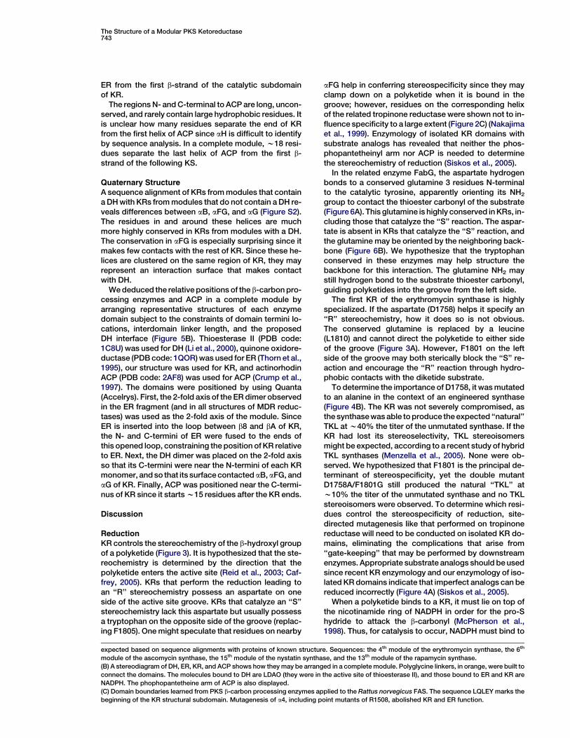
The Structure of a Modular PKS Ketoreductase743
ER from the first b-strand of the catalytic subdomainof KR.
The regions N- and C-terminal to ACP are long, uncon-served, and rarely contain large hydrophobic residues. Itis unclear how many residues separate the end of KRfrom the first helix of ACP since aH is difficult to identifyby sequence analysis. In a complete module, w18 resi-dues separate the last helix of ACP from the first b-strand of the following KS.
Quaternary StructureA sequence alignment of KRs from modules that containa DH with KRs from modules that do not contain a DH re-veals differences between aB, aFG, and aG (Figure S2).The residues in and around these helices are muchmore highly conserved in KRs from modules with a DH.The conservation in aFG is especially surprising since itmakes few contacts with the rest of KR. Since these he-lices are clustered on the same region of KR, they mayrepresent an interaction surface that makes contactwith DH.
We deduced the relative positions of the b-carbon pro-cessing enzymes and ACP in a complete module byarranging representative structures of each enzymedomain subject to the constraints of domain termini lo-cations, interdomain linker length, and the proposedDH interface (Figure 5B). Thioesterase II (PDB code:1C8U) was used for DH (Li et al., 2000), quinone oxidore-ductase (PDB code: 1QOR) was used for ER (Thorn et al.,1995), our structure was used for KR, and actinorhodinACP (PDB code: 2AF8) was used for ACP (Crump et al.,1997). The domains were positioned by using Quanta(Accelrys). First, the 2-fold axis of the ER dimer observedin the ER fragment (and in all structures of MDR reduc-tases) was used as the 2-fold axis of the module. SinceER is inserted into the loop between b8 and bA of KR,the N- and C-termini of ER were fused to the ends ofthis opened loop, constraining the position of KR relativeto ER. Next, the DH dimer was placed on the 2-fold axisso that its C-termini were near the N-termini of each KRmonomer, and so that its surface contacted aB, aFG, andaG of KR. Finally, ACP was positioned near the C-termi-nus of KR since it starts w15 residues after the KR ends.
Discussion
ReductionKR controls the stereochemistry of the b-hydroxyl groupof a polyketide (Figure 3). It is hypothesized that the ste-reochemistry is determined by the direction that thepolyketide enters the active site (Reid et al., 2003; Caf-frey, 2005). KRs that perform the reduction leading toan ‘‘R’’ stereochemistry possess an aspartate on oneside of the active site groove. KRs that catalyze an ‘‘S’’stereochemistry lack this aspartate but usually possessa tryptophan on the opposite side of the groove (replac-ing F1805). One might speculate that residues on nearby
aFG help in conferring stereospecificity since they mayclamp down on a polyketide when it is bound in thegroove; however, residues on the corresponding helixof the related tropinone reductase were shown not to in-fluence specificity to a large extent (Figure 2C) (Nakajimaet al., 1999). Enzymology of isolated KR domains withsubstrate analogs has revealed that neither the phos-phopantetheinyl arm nor ACP is needed to determinethe stereochemistry of reduction (Siskos et al., 2005).
In the related enzyme FabG, the aspartate hydrogenbonds to a conserved glutamine 3 residues N-terminalto the catalytic tyrosine, apparently orienting its NH2
group to contact the thioester carbonyl of the substrate(Figure 6A). This glutamine is highly conserved in KRs, in-cluding those that catalyze the ‘‘S’’ reaction. The aspar-tate is absent in KRs that catalyze the ‘‘S’’ reaction, andthe glutamine may be oriented by the neighboring back-bone (Figure 6B). We hypothesize that the tryptophanconserved in these enzymes may help structure thebackbone for this interaction. The glutamine NH2 maystill hydrogen bond to the substrate thioester carbonyl,guiding polyketides into the groove from the left side.
The first KR of the erythromycin synthase is highlyspecialized. If the aspartate (D1758) helps it specify an‘‘R’’ stereochemistry, how it does so is not obvious.The conserved glutamine is replaced by a leucine(L1810) and cannot direct the polyketide to either sideof the groove (Figure 3A). However, F1801 on the leftside of the groove may both sterically block the ‘‘S’’ re-action and encourage the ‘‘R’’ reaction through hydro-phobic contacts with the diketide substrate.
To determine the importance of D1758, it was mutatedto an alanine in the context of an engineered synthase(Figure 4B). The KR was not severely compromised, asthe synthase was able to produce the expected ‘‘natural’’TKL at w40% the titer of the unmutated synthase. If theKR had lost its stereoselectivity, TKL stereoisomersmight be expected, according to a recent study of hybridTKL synthases (Menzella et al., 2005). None were ob-served. We hypothesized that F1801 is the principal de-terminant of stereospecificity, yet the double mutantD1758A/F1801G still produced the natural ‘‘TKL’’ atw10% the titer of the unmutated synthase and no TKLstereoisomers were observed. To determine which resi-dues control the stereospecificity of reduction, site-directed mutagenesis like that performed on tropinonereductase will need to be conducted on isolated KR do-mains, eliminating the complications that arise from‘‘gate-keeping’’ that may be performed by downstreamenzymes. Appropriate substrate analogs should be usedsince recent KR enzymology and our enzymology of iso-lated KR domains indicate that imperfect analogs can bereduced incorrectly (Figure 4A) (Siskos et al., 2005).
When a polyketide binds to a KR, it must lie on top ofthe nicotinamide ring of NADPH in order for the pro-Shydride to attack the b-carbonyl (McPherson et al.,1998). Thus, for catalysis to occur, NADPH must bind to
expected based on sequence alignments with proteins of known structure. Sequences: the 4th module of the erythromycin synthase, the 6th
module of the ascomycin synthase, the 15th module of the nystatin synthase, and the 13th module of the rapamycin synthase.
(B) A stereodiagram of DH, ER, KR, and ACP shows how they may be arranged in a complete module. Polyglycine linkers, in orange, were built to
connect the domains. The molecules bound to DH are LDAO (they were in the active site of thioesterase II), and those bound to ER and KR are
NADPH. The phophopantetheine arm of ACP is also displayed.
(C) Domain boundaries learned from PKS b-carbon processing enzymes applied to the Rattus norvegicus FAS. The sequence LQLEY marks the
beginning of the KR structural subdomain. Mutagenesis of a4, including point mutants of R1508, abolished KR and ER function.

Structure744
Figure 6. Control of Stereochemistry
(A) In the related fatty acid synthase enzyme,
FabG (PDB code: 2C07), the aspartate orients
the glutamine.
(B) Stereochemistry may be controlled in part
by the NH2 group of a conserved glutamine
that can donate a hydrogen bond to the sub-
strate thioester carbonyl. In enzymes that
catalyze an ‘‘R’’ reduction, a conserved as-
partate may orient the glutamine NH2 so
that a polyketide enters from the right side
of the groove. In enzymes catalyzing the ‘‘S’’
reaction, a tryptophan on the left side of the
groove may help orient the glutamine NH2
so that the polyketide enters from that side.
the protein before the polyketide. Well-diffracting crys-tals of KR could not be grown in the absence of NADPH,suggesting that the cofactor orders the enzyme for ca-talysis as it does in FabG (Price et al., 2004).
Epimerization
The first and third modules of the erythromycin synthasecatalyze epimerizations of the a-methyl group, althoughthe enzyme that performs this reaction has not been de-termined (Weissman et al., 1997; Caffrey, 2005). Thereare several reasons to believe that KR doubles as anepimerase even though no SDR enzyme is known to cat-alyze a similar reaction: (1) No module lacking a KR hasbeen observed to catalyze an epimerization. (2) A mod-ule that contains a reductase-incompetent KR, but noother b-carbon processing enzyme, can catalyze epime-rization. (3) Furthermore, the tyrosine that is catalytic inreductase-competent KRs is still conserved in theseKRs. The epimerase activity must be independent ofNADPH since the nucleotide binding motif of the KRfrom the third module of the erythromycin synthase isabsent.
Alignments of KRs from epimerization-catalyzingmodules that do not contain other b-carbon processingenzymes implicate the active site tyrosine in the abstrac-tion of the acidic hydrogen on the polyketide a-carbon(Figure 7). After the subsequent enolization, the polyke-tide may exit the active site and tautomerize to one oftwo keto forms, one being the epimerized polyketide.The nonproductive tautomerization yields the originalsubstrate that can be accepted by KR until it is epimer-ized. The catalytic tyrosines of other solved SDR struc-tures reside within aF; however, Y1813 from the KR ofthe first module of the erythromycin synthase lies out-side this helix and may have sufficient mobility to accessthe acidic polyketide hydrogen. Y1813 is apparently lib-erated by the helix-breaking P1815. Its average temper-ature factor is twice that of N1817, one turn into aF (34 A3
versus 17 A3) (Figure 7B). In the third KR of the erythro-mycin synthase, the asparagine corresponding toN1817 is replaced by a serine. To compensate for themissing asparagine side chain, aF may need to partiallyunravel, again liberating the tyrosine for the epimeriza-tion reaction. In nonepimerizing KRs, a more orderedaF probably prevents the tyrosine from accessing theacidic polyketide hydrogen (Figure 7C). Intriguingly,DHs may also catalyze epimerization, since, in some
modules known to catalyze epimerization (such as thefourth module of the ascomycin synthase or the sixthmodule of the rapamycin synthase), the KR tyrosine isabsent but a supposedly inactive DH is present. This re-action is easier to understand than KR-catalyzed epime-rization, as the first step catalyzed by a DH during dehy-dration is the abstraction of an a-carbon hydrogen.
Domain OrganizationOutside of the module, the docking domains and TE aredimeric (Tsai et al., 2001, 2002; Broadhurst et al., 2003).However, it was unclear how many domains within themodule would also be dimeric. If every enzyme withinthe module made contacts across the 2-fold axis, ACPwould have to diffuse farther than the peptide linkerson each side would permit. The deduced quaternarystructure of the b-carbon processing enzymes indicatesa surprising amount of contact across the 2-fold axis.However, the structural half of KR enables the KR do-main to be monomeric and loop out from the 2-foldaxis, allowing DH and ER to come closer to each other.KS probably makes contact across the 2-fold axis sinceall known type II KSs (type II enzymes are functional asseparate polypeptides; several of these structureshave been solved) are dimeric and its active site lies atthe dimer interface. However, AT may loop out sinceall known type II ATs are monomeric. There may be pro-tein-protein contacts all along the 2-fold axis. A largenumber of inactive enzymes are present within modules,possibly indicating that they make important contacts.
The active sites of DH, ER, and KR are all accessible toACP. Bound by peptide linkers on both ends, ACP candiffuse between each enzyme in the module as well asthe next KS or TE, akin to the lipoyl domains and biotinyldomains of other multienzyme proteins (Perham, 2000).The linkers also prevent a polyketide from interactingwith enzymes farther away in the synthase. However,a polyketide bound to the ACP of the third module ofthe epothilone synthase may have sufficient freedomto access DH of the fourth module (Tang et al., 2000).The w18 A phosphopantetheinyl arm contributes rela-tively little translational freedom to the polyketide com-pared to the peptide linkers on both ends of ACP.
Most SDR enzymes are either dimeric or tetrameric,and the principle interface is formed by helices aE andaF (Jornvall et al., 1995). The structural subdomain ofKR contributes these important stabilizing interactions

The Structure of a Modular PKS Ketoreductase745
Figure 7. Epimerization
(A) Hypothesized mechanism of epimerization. The diketide formed by the first KS enters the active site of the first KR. The mobile Y1813 acts as
a base, acquiring the acidic hydrogen of the diketide to form an enolate. The enolate oxygen accepts the proton back from Y1813, and the enol-
ized diketide is released from the KR. An uncatalyzed tautomerization back to the keto form results in a mixture of the original diketide and the
epimerized diketide. The original diketide can be accepted until it is epimerized.
(B) A stereodiagram of the KR active site displays the 2Fo 2 Fc electron density maps contoured at 1.5s. P1815 breaks the helix that contains the
catalytic tyrosine in related SDR enzymes, allowing it greater mobility. The temperature factors for Y1813 and neighboring residues are compar-
atively high.
(C) The sequence surrounding the catalytic tyrosine from each KR of the erythromycin synthase. The first and third modules catalyze epimeri-
zation. Residues that may allow the tyrosine sufficient freedom to catalyze epimerization are underlined.
to the catalytic subdomain through the interaction of a3and a4 with aE and aF. In fungal MFE-2, two consecutivedehydrogenase domains are present—one that oxidizesshort chains and one that oxidizes long chains. The frag-ment of Candida tropicalis MFE-2 containing these de-hydrogenases was crystallized and found to be mono-meric, indicating that the two dehydrogenase domainsassociate (Ylianttila et al., 2004). Perhaps KR evolvedfrom such an enzyme, losing the catalytic, but not struc-tural properties, of one domain.
If modular synthases evolved through the fusion ofgenes encoding type II enzymes, it is plausible that anancestral type II ER inserted into the loop between thetwo subdomains of KR. The ER fold begins after b8 ofthe KR structural subdomain and ends before bA of thecatalytic subdomain. The N- and C-termini of relatedMDR enzymes are near one another; thus, the ancestralinsertion may have been relatively uncomplicated. Thesegment joining ER to the KR catalytic subdomain ismore highly conserved in sequence and length than thesegment joining it to the KR structural subdomain, pos-sibly indicating that it helps fix the relative positions ofthe KR and ER domains. Similar domain insertions may
have also occurred with AT and the rare methyltransfer-ase domain (Tang et al., 2000; Cheng et al., 2003).
PKS Modules and FASs
It is hypothesized that modular PKSs evolved from FASssince a complete module contains the same domains inthe same order. If so, they might share a similar three-dimensional organization. The animalian FAS has beenshown to possess some conformational flexibility, butit is fairly rigid on the whole. Reconstructions of the hu-man FAS from electron micrographs have been made at16 A resolution, and crystals have even been grown. Thelatest reconstructions show the head-to-head, tail-to-tail organization of the FAS domains through a long 2-fold axis (Asturias et al., 2005).
A w520 residue region termed the ‘‘core’’ (residuesw1010–1530) precedes ER in the well-studied Rattusnorvegicus FAS. It can be much shorter: the Drosophilamelanogaster FAS core is w380 residues. Mutagenesisperformed on the C-terminal portion of the rat FAS coreinactivated KR and ER (Witkowski et al., 2004). From theKR structure presented here, it is now clear that the

Structure746
mutations were of a4 in the structural subdomain of KR,a helix that provides many stabilizing contacts to thecatalytic subdomain (Figure 5C). Without these interac-tions, the structural and catalytic subdomains of KRcould dissociate, conceivably destabilizing ER in theprocess since it is positioned between them. The se-quence identifying the b-strand b1, which begins thestructural subdomain, is more subtle than in PKSs, ap-pearing as 2 large hydrophobic residues separated by3 variable residues; it is LQLEY in R. norvegicus. It isprobable that the sequence N-terminal to the structuralhalf of KR is from the second hotdog fold of DH. Thus,the core probably consists of the C-terminal half of DHand the N-terminal half of KR.
The KR structure suggests catalytic similarities be-tween PKS and FAS enzymes even at the stereochemi-cal level. The KRs from complete modules possess theaspartate, but not the tryptophan, in the active sitegroove (Figure 6B). This pattern is indicative of the ‘‘R’’reduction, suggesting that DH only accepts a b-hydroxylgroup of this stereochemistry. FAS KRs also possessthe aspartate, but not tryptophan, and are known to cat-alyze the ‘‘R’’ stereochemistry (Smith et al., 2003). TheFAS DH accepts the resulting ‘‘R’’ product. The gluta-mine hypothesized to guide substrates into the KR ac-tive site groove is also conserved in FAS KRs. Finally,the mysterious swapping of the asparagine and lysinepositions, compared to other SDR enzymes, also occursin FAS KRs.
The KR structure may also hint at the origin of the FASKR. On several levels, the KR domain shows similaritiesto an enzyme from a b-oxidation pathway, the dehydro-genase of MFE-2. This enzyme catalyzes virtually thesame reaction as KR. Its substrate is a (3R)-hydroxyacylchain (the product of a neighboring hydratase that pos-sesses the double hotdog fold hypothesized for DH).Thus, the hydroxyl group of its reactant is of the samestereochemistry as the hydroxyl group of the productfrom a KR in a complete module or FAS. The dehydro-genase of MFE-2 is unique in b-oxidation pathways inthat it can oxidize branched fatty acids (acyl chainswith an a-substituent, commonplace in polyketides).These catalytic similarities and the structural homologyof these enzymes suggest an evolutionary link.
The precise boundaries between the b-carbonprocessing domains will be valuable in the design ofhybrid synthases since they detail how domains canbe swapped more cleanly. In time, structural and engi-neering studies will elucidate how substrate specificityand stereochemistry are controlled by these enzymes,enabling the synthesis of designer polyketides.
Experimental Procedures
Cloning
The DNA encoding T2, from S1444 to R1925, was PCRed from syn-
thetic eryAI with primers 50-GGAGATATACATATGTCTACCGAGGT
TGATGAAGTC-30 and 50-GTGGTGCTCGAGTCATCAGCGTGGCTC
AGCAGCGGCCTGC-30, cut with NdeI and XhoI, and ligated into
pET28b. The DNA encoding DH+ER+KR, from H2362 to S3409, was
PCRed from synthetic eryAII with primers 50-GGAGATATACATATG
CACCGCCCAGCAGATGTTAGC-30 and 50-GTGGTGCTCGAGTCAT
CACGATTCGCTACGACCGGCTAAG-30, cut with NdeI and XhoI,
and ligated into pET28b (Kodumal et al., 2004).
Expression and Purification
BL21(DE3) cells transformed with either the plasmid encoding KR or
DH+ER+KR were grown in LB with 20 mg/l kanamycin at 37ºC until
they reached 0.4 OD600. They were induced with 1 mM IPTG and
grown for 40 hr at 18ºC. Cells were spun down and resuspended
in 40 mM HEPES (pH 7.0), 0.5 M NaCl, 5 mM b-ME, and 1 mM
PMSF, sonicated, and spun down at 30,000 3 g for 30 min. The su-
pernatant was poured over a nickel-NTA column, washed with 25
mM imidazole (in lysis buffer), and the protein was eluted with 250
mM imidazole. The protein was exchanged into gel filtration buffer
(10 mM HEPES, 150 mM NaCl, 1 mM DTT [pH 7.0]), and thrombin
was added to cleave off the histidine tag at 21ºC for 1 hr. Thrombin
was removed by a benzamidine sepharose column, and the protein
mixture was concentrated and run through a Sephadex 200 gel filtra-
tion column. Selenomethionine-incorporated KR was expressed in
BL21(DE3) cells in M9 (4 g/l glucose, 6 g/l Na2HPO4, 3 g/l KH2PO4,
1 g/l NH4Cl, 0.5 g/l NaCl, 1 mM MgSO4, 0.1 mM CaCl2) minimal media
with 20 mg/l kanamycin. After growing to 0.4 OD600 at 37ºC, they
were supplied with lysine, phenylalanine, and threonine, each at
100 mg/l; isoleucine, leucine, and valine, each at 50 mg/l; and seleno-
methionine at 60 mg/l. After 15 min, they were induced with 1 mM
IPTG and grown for 40 hr at 18ºC. The purification was the same
as for the native protein.
Gel Filtration
A Sephadex 200 gel filtration column was equilibrated with gel filtra-
tion buffer and injected with 0.5 ml samples. The KR and DH+ER+KR
fragments were compared to known standards. Dextran blue 2000
and tyrosine were used to determine Vo and Vt, respectively. Kav =
(Ve 2 Vo)/(Vt 2 Vo), where Ve is the volume at which the protein elutes.
Crystallization and Data Collection
The T2 fragment was concentrated to 3 mg/ml in gel filtration buffer
and crystallized by hanging drop vapor diffusion in 35% PEG3350,
0.2 M guanidinium hydrochloride, 0.1 M HEPES (pH 7.0). Crystals
grew to full size (200 3 100 3 50 mm) in 1 week. Two crystal forms
were present in the drop and often grew together in twinned crystals,
and surgery was often needed to separate them. Crystals were fro-
zen in mother liquor in the nitrogen stream. Data were collected at
the ALS Beamline 8.3.1.
Data Processing and Refinement
ELVES processed the anomalous dispersion data (Holton and Alber,
2004). The selenium sites were located, and phases were generated
by using CNS (Brunger et al., 1998). A model was built with Quanta
for 60% of the protein before it was used to generate phases. The
rest of the model was created through multiple rounds of building
and refining in CNS.
Isolated KR Domain Assay
Analagous to a recent study, reactions were incubated in 100 ml in
400 mM NaH2PO4 (pH 7.5) for 16 hr at 22ºC, by using 10 mM NADPH,
3 mM unreduced diketide analog (G. Liu, personal communication),
and 1 mM isolated KR domain (Siskos et al., 2005). The reaction was
extracted with 1 ml ethyl acetate. The extract was separated by
a 0%–30% acetonitrile/water (0.1% TFA) gradient on a C18 re-
verse-phase column and analyzed by the ion count trace from a con-
nected mass spectrometer. The products were compared to au-
thentic standards (G. Liu, personal communication).
Triketide Lactone Synthase Assay
The loading didomain and module 1 were cloned between the NdeI
and EcoRI sites of pET21, module 2 and TE were cloned between
the NdeI and EcoRI sites of pET28, and both plasmids were trans-
formed into E. coli K207-3 (Menzella et al., 2005). The D1758A mutant
was engineered by using the Quickchange protocol (Stratagene)
with primers 50-GGCGGCAACCTTGGATGCCGGCACCGTCGATAC
TC-30 and 50-GAGTATCGACGGTGCCGGCATCCAAGGTTGCCG
CC-30. The D1758A/F1801G double mutant was engineered by using
the Quickchange protocol on the D1758A plasmid with primers 50-CG
TGCTGTTTTCCAGTGGTGCGTCGGCCTTTGGTG-30 and 50-CACCA
AAGGCCGACGCACCACTGGAAAACAGCACG-30. A starter culture
was grown overnight in LB at 37ºC, was used to inoculate 1 l M9 min-
imal media supplied with 1 mM b-alanine, 50 mg/l carbenecillin, and

The Structure of a Modular PKS Ketoreductase747
20 mg/l kanamycin, and was grown at 37ºC until the OD600 reached
0.5. The temperature was decreased to 20ºC, and cells were induced
with 1 mM IPTG; media were supplemented with 50 mM glutamate
(pH 7.0), 50 mM succinate (pH 7.0), and 50 mM propionate (pH 7.0).
After 3 days, cells were spun down, and the supernatant was acidi-
fied to pH 1.5 with phosphoric acid and extracted twice with methy-
lene chloride. Methylene choride was evaporated, and the extract
was separated by a 20%–30% acetonitrile/water (0.1% TFA) gradient
on a C18 reverse-phase column. Fractions were analyzed by mass
spectrometry and nuclear magnetic resonance spectroscopy.
Supplemental Data
Supplemental Data including Figures S1 and S2 are available at
http://www.structure.org/cgi/content/full/14/4/737/DC1/.
Acknowledgments
We thank Daniel Santi for supplying the synthetic polyketide syn-
thase genes, Ralph Reid for his help in analyzing KR enzymology
and domain boundaries, Hugo Menzella and Sunil Chandran for
help with the triketide lactone assay, Gary Liu for providing the dike-
tide analogs, and Janet Finer-Moore for help in crystallography and
interpreting experimental data. Research was supported by the Na-
tional Cancer Institute National Institutes of Health (NIH) grant
CA63081 and Training Grant #5 T32 CA090270.
Received: November 1, 2005
Revised: January 11, 2006
Accepted: January 17, 2006
Published online: March 15, 2006
References
Aparicio, J.F., Caffrey, P., Marsden, A.F., Staunton, J., and Leadlay,
P.F. (1994). Limited proteolysis and active-site studies of the first
multienzyme component of the erythromycin-producing polyketide
synthase. J. Biol. Chem. 269, 8524–8528.
Asturias, F.J., Chadick, J.Z., Cheung, I.K., Stark, H., Witkowski, A.,
Joshi, A.K., and Smith, S. (2005). Structure and molecular organiza-
tion of mammalian fatty acid synthase. Nat. Struct. Mol. Biol. 12,
225–232.
Bedford, D., Jacobsen, J.R., Luo, G., Cane, D.E., and Khosla, C.
(1996). A functional chimeric modular polyketide synthase gener-
ated via domain replacement. Chem. Biol. 3, 827–831.
Broadhurst, R.W., Nietlispach, D., Wheatcroft, M.P., Leadlay, P.F.,
and Weissman, K.J. (2003). The structure of docking domains in
modular polyketide synthases. Chem. Biol. 10, 723–731.
Brunger, A.T., Adams, P.D., Clore, G.M., DeLano, W.L., Gros, P.,
Gross-Kunstleve, R.W., Jiang, J.S., Kuszewski, J., Nilges, M.,
Pannu, N.S., et al. (1998). Crystallography & NMR system: a new
software suite for macromolecular structure determination. Acta
Crystallogr. D Biol. Crystallogr. 54, 905–921.
Caffrey, P. (2005). The stereochemistry of ketoreduction. Chem.
Biol. 12, 1060–1062.
Cheng, Y.Q., Tang, G.L., and Shen, B. (2003). Type I polyketide syn-
thase requiring a discrete acyltransferase for polyketide biosynthe-
sis. Proc. Natl. Acad. Sci. USA 100, 3149–3154.
Crump, M.P., Crosby, J., Dempsey, C.E., Parkinson, J.A., Murray,
M., Hopwood, D.A., and Simpson, T.J. (1997). Solution structure of
the actinorhodin polyketide synthase acyl carrier protein from Strep-
tomyces coelicolor A3(2). Biochemistry 36, 6000–6008.
Donadio, S., and Katz, L. (1992). Organization of the enzymatic do-
mains in the multifunctional polyketide synthase involved in erythro-
mycin formation in Saccharopolyspora erythraea. Gene 111, 51–60.
Edwards, K.J., Barton, J.D., Rossjohn, J., Thorn, J.M., Taylor, G.L.,
and Ollis, D.L. (1996). Structural and sequence comparisons of qui-
none oxidoreductase, zeta-crystallin, and glucose and alcohol de-
hydrogenases. Arch. Biochem. Biophys. 328, 173–183.
Gogos, A., Mu, H., and Shapiro, L. (2003). Putative enoyl reductase
domain of a polyketide synthase. 1PQW (see http://www.rcsb.
org/pdb/navbarsearch.do?newSearch=yes&isAuthorSearch=no
&radioset=All&inputQuickSearch=1pqw&image.x=42&image.y=5).
Haapalainen, A.M., Koski, M.K., Qin, Y.M., Hiltunen, J.K., and Glum-
off, T. (2003). Binary structure of the two-domain (3R)-hydroxyacyl-
CoA dehydrogenase from rat peroxisomal multifunctional enzyme
type 2 at 2.38 A resolution. Structure (Camb) 11, 87–97.
Holm, L., and Sander, C. (1996). Mapping the protein universe. Sci-
ence 273, 595–603.
Holton, J., and Alber, T. (2004). Automated protein crystal structure
determination using ELVES. Proc. Natl. Acad. Sci. USA 101, 1537–
1542.
Jornvall, H., Persson, B., Krook, M., Atrian, S., Gonzalez-Duarte, R.,
Jeffrey, J., and Ghosh, D. (1995). Short-chain dehydrogenases/re-
ductases (SDR). Biochemistry 34, 6003–6013.
Kao, C.M., Luo, G., Katz, L., Cane, D.E., and Khosla, C. (1994). Engi-
neered biosynthesis of a triketide lactone from an incomplete mod-
ular polyketide synthase. J. Am. Chem. Soc. 116, 11612–11613.
Kelley, L.A., MacCallum, R.M., and Sternberg, M.T. (2000). Enhanced
genome annotation using structural profiles in the program 3D-
PSSM. J. Mol. Biol. 299, 499–520.
Kodumal, S.J., Patel, K.G., Reid, R., Menzella, H.G., Welch, M., and
Santi, D.V. (2004). Total synthesis of long DNA sequences: synthesis
of a contiguous 32-kb polyketide synthase gene cluster. Proc. Natl.
Acad. Sci. USA 101, 15573–15578.
Li, J., Derewenda, U., Dauter, Z., Smith, S., and Derewenda, Z.S.
(2000). Crystal structure of the Escherichia coli thioesterase II, a ho-
molog of the human Nef binding enzyme. Nat. Struct. Biol. 7, 555–
559.
McDaniel, R., Kao, C.M., Hwang, S.J., and Khosla, C. (1997). Engi-
neered intermodular and intramodular polyketide synthase fusions.
Chem. Biol. 4, 667–674.
McPherson, M., Khosla, C., and Cane, D.E. (1998). Erythromycin bio-
synthesis: the b-ketoreductase domains catalyze the stereospecific
transfer of the 4-pro-S hydride of NADPH. J. Am. Chem. Soc. 120,
3267–3268.
Menzella, H.G., Reid, R., Carney, J.R., Chandran, S.S., Reisinger,
S.J., Patel, K.G., Hopwood, D.A., and Santi, D.V. (2005). Combinato-
rial polyketide biosynthesis by de novo design and rearrangement of
modular polyketide synthase genes. Nat. Biotechnol. 23, 1171–1176.
Nakajima, K., Kato, H., Oda, J., Yamada, Y., and Hashimoto, T.
(1999). Site-directed mutagenesis of putative substrate-binding res-
idues reveals a mechanism controlling the different stereospecific-
ities of two tropinone reductases. J. Biol. Chem. 274, 16563–16568.
O’Hagan, D.O. (1991). The Polyketide Metabolites (Chichester, UK:
Ellis Horwood).
Oppermann, U., Filling, C., Hult, M., Shafqat, N., Wu, X., Lindh, M.,
Shafqat, J., Nordling, E., Kalberg, Y., Persson, B., et al. (2003).
Short-chain dehydrogenases/reductases (SDR): the 2002 update.
Chem. Biol. Interact. 143–144, 247–253.
Perham, R.N. (2000). Swinging arms and swinging domains in multi-
functional enzymes: catalytic machines for multistep reactions.
Annu. Rev. Biochem. 69, 961–1004.
Price, A.C., Zhang, Y.M., Rock, C.O., and White, S.W. (2004). Cofac-
tor-induced conformational rearrangements establish a catalytically
competent active site and a proton relay conduit in FabG. Structure
(Camb) 12, 417–428.
Reid, R., Piagentini, M., Rodriguez, E., Ashley, G., Viswanathan, N.,
Carney, J., Santi, D.V., Hutchinson, C.R., and McDaniel, R. (2003).
A model of structure and catalysis for ketoreductase domains in
modular polyketide synthases. Biochemistry 42, 72–79.
Ruan, X., Pereda, A., Stassi, D., Zeidner, D., Summers, R.G., Jack-
son, M., Shivakumar, A., Kakavas, S., Staver, M.J., Donadio, S.,
et al. (1997). Acyltransferase domain substitutions in erythromycin
polyketide synthase yield novel erythromycin derivatives. J. Bacter-
iol. 179, 6416–6425.
Siskos, A.P., Baerga-Ortiz, A., Bali, S., Stein, V., Mamdani, H., Spite-
ller, D., Popovic, B., Spencer, J.B., Staunton, J., Weissman, K.J.,
et al. (2005). Molecular basis of Celmer’s rules: stereochemistry of
catalysis by isolated ketoreductase domains from modular polyke-
tide synthases. Chem. Biol. 12, 1145–1153.
Structure748
Smith, S., Witkowski, A., and Joshi, A.K. (2003). Structural and func-
tional organization of the animal fatty acid synthase. Prog. Lipid Res.
42, 289–317.
Staunton, J., and Weissman, K.J. (2001). Polyketide biosynthesis:
a millennium review. Nat. Prod. Rep. 18, 380–416.
Staunton, J., and Wilkinson, B. (1997). Biosynthesis of erythromycin
and rapamycin. Chem. Rev. 97, 2611–2630.
Staunton, J., Caffrey, P., Aparicio, J.F., Roberts, G.A., Bethell, S.S.,
and Leadlay, P.F. (1996). Evidence for a double-helical structure for
modular polyketide synthases. Nat. Struct. Biol. 3, 188–192.
Tang, L., Shah, S., Chung, L., Shah, S., Chung, L., Carney, J., Katz, L.,
Khosla, C., and Julien, B. (2000). Cloning and heterologous expres-
sion of the epothilone gene cluster. Science 287, 640–642.
Thorn, J.M., Barton, J.D., Dixon, N.E., Ollis, D.L., and Edwards, K.J.
(1995). Crystal structure of Escherichia coli QOR quinone oxidore-
ductase complexed with NADPH. J. Mol. Biol. 249, 785–799.
Tsai, S.C., Miercke, L.J., Krucinski, J., Gokhale, R., Chen, J.C., Fos-
ter, P.G., Cane, D.E., Khosla, C., and Stroud, R.M. (2001). Crystal
structure of the macrocycle-forming thioesterase domain of the
erythromycin polyketide synthase: versatility from a unique sub-
strate channel. Proc. Natl. Acad. Sci. USA 98, 14808–14813.
Tsai, S.C., Lu, H., Reid, D.E., Khosla, C., and Stroud, R.M. (2002). In-
sights into channel architecture and substrate specificity from crys-
tal structures of two macrocycle-forming thioesterases of modular
polyketide synthases. Biochemistry 41, 12598–12606.
Weissman, K.J., Timoney, M., Bycroft, M., Grice, P., Hanefeld, U.,
Staunton, J., and Leadlay, P.F. (1997). The molecular basis of Celm-
ers rules: the stereochemistry of the condensation step in chain ex-
tension on the erythromycin polyketide synthase. Biochemistry 36,
13849–13855.
Witkowski, A., Joshi, A.K., and Smith, S. (2004). Characterization of
the b-carbon processing reactions of the mammalian cytosolic fatty
acid synthase: role of the central core. Biochemistry 43, 10458–
10466.
Ylianttila, M.S., Qin, Y.M., Hiltunen, J.K., Glumoff, T., et al. (2004).
Site-directed mutagenesis to enable and improve crystallizability
of Candida tropicalis (3R)-hydroxyacyl-CoA dehydrogenase. Bio-
chem. Biophys. Res. Commun. 324, 25–30.
Accession Numbers
The atomic coordinates have been deposited in the Protein Data
Bank with accession codes 2FR0 and 2FR1 for the two crystal forms
of KR and 2FRO for the model.
Note Added in Proof
The structure of the porcine FAS was solved at 4.5 A resolution dur-
ing the proof stage of this paper (Maier, T., Jenni, S., and Ban, N. Ar-
chitecture of Mammalian Fatty Acid Synthase at 4.5 A Resolution.
Science 311:1258–1262). It reveals that the FAS is organized as
a head-to-head homodimer and establishes that the overall quater-
nary structure of the b-carbon processing enzymes is as we propose
here. It also shows that DH has a double hotdog fold and that KR
shares an interface with it. Ban and co-workers were unable to fit
the density adjacent to the catalytic subdomain of KR and suggest
that it might be from ACP or TE. Our structural data show that it is
from the structural subdomain of KR.