Embed Size (px)
Citation preview
Structural Basis of Carbohydrate Recognition byCalreticulin*□S
Received for publication, July 27, 2010, and in revised form, September 6, 2010 Published, JBC Papers in Press, September 29, 2010, DOI 10.1074/jbc.M110.168294
Guennadi Kozlov‡, Cosmin L. Pocanschi§1, Angelika Rosenauer‡, Sara Bastos-Aristizabal‡, Alexei Gorelik‡,David B. Williams§, and Kalle Gehring‡2
From the ‡Department of Biochemistry, Groupe de Recherche Axe sur la Structure des Proteines, McGill University, Montreal,Quebec H3G 0B1, Canada and the §Departments of Biochemistry and Immunology, University of Toronto, Toronto,Ontario M5S 1A8, Canada
The calnexin cycle is a process by which glycosylated proteinsare subjected to folding cycles in the endoplasmic reticulumlumen via binding to the membrane protein calnexin (CNX) orto its soluble homolog calreticulin (CRT). CNX and CRT specif-ically recognize monoglucosylated Glc1Man9GlcNAc2 glycans,but the structural determinants underlying this specificity areunknown. Here, we report a 1.95-A crystal structure of the CRTlectin domain in complex with the tetrasaccharide �-Glc-(133)-�-Man-(132)-�-Man-(132)-Man. The tetrasaccharidebinds to a long channel onCRT formedby a concave�-sheet. Allfour sugar moieties are engaged in the protein binding via anextensive network of hydrogen bonds and hydrophobic con-tacts. The structure explains the requirement for glucose at thenonreducing end of the carbohydrate; the oxygen O2 of glucoseperfectly fits to a pocket formed by CRT side chains while form-ing direct hydrogen bonds with the carbonyl of Gly124 and theside chain of Lys111. The structure also explains a requirementfor the Cys105–Cys137 disulfide bond in CRT/CNX for efficientcarbohydrate binding. The Cys105–Cys137 disulfide bond isinvolved in intimate contacts with the third and fourth sugarmoieties of the Glc1Man3 tetrasaccharide. Finally, the structurerationalizes previous mutagenesis of CRT and lays a structuralgroundwork for future studies of the role of CNX/CRT indiverse biological pathways.
Protein glycosylation plays an important role in maturationand quality control of proteins in the endoplasmic reticulum(ER)3 (1–3). Nascent polypeptide chains of the secretory path-way are translocated into the lumen of the ER where theyimmediately become N-glycosylated with Glc3Man9GlcNAc2
on the side chain of asparagine residues in Asn-X-Ser/Thrmotifs (see Scheme 1). The processing of the glycan starts withremoval of the outermost glucose by the action of glucosidase I.Glucosidase II then trims the next glucose, upon which themonoglucosylated proteins selectively bind to the membraneER protein calnexin (CNX) or its soluble homolog calreticulin(CRT) to enter the calnexin cycle. In the calnexin cycle, thenascent protein bound to CNX or CRT interacts with chaper-ones to promote its folding. The glucose and adjacent threemannose residues are critically important for carbohydrate rec-ognition by CNX and CRT (Scheme 1) (4–6). Removal of theterminal glucose from correctly folded protein substratesreleases them from binding to CNX or CRT and allows theirexit from the ER to their final destination. Non-native proteins,which lack the terminal glucose residue, can be reglucosylatedby the enzyme UDP-glucose-glycoprotein glucosyltransferaseto re-enter the calnexin cycle. Multiple rounds of deglucosyla-tion-reglucosylation cycles can occur until a glycoproteinreaches its native fold or, if terminally misfolded, it is targetedfor degradation via the ER-associated degradation pathway (7).The previous crystal structure of CNX revealed two main
structural components, a globular lectin domain and anextended arm-like domain called the P-domain (8). The lectindomain shows a fold similar to leguminous lectins and largelyconsists of a �-sandwich formed by two curved �-sheets. It alsocontains a single high affinity calcium-binding site that plays animportant role in stabilizing the protein (9) but does not partic-ipate in carbohydrate recognition (8). The proline-rich P-do-main interrupts the lectin domain between residues Pro270 andPhe415. The P-domain consists of four copies of a repeat motif(termed type 1) and four copies of second repeat motif (type 2)in a “11112222” configuration to form a long hook-like arm thatinteracts with the thiol oxidoreductase ERp57 (10, 11). TheCRT P-domain shows a similar modular arrangement but con-sists of only three repeats of each type (10, 12, 13). CRT alsopossesses a highly negative C terminus, which binds calciumions with millimolar affinity, but the C terminus does not con-tribute to glycan binding (14, 15).A portion of the carbohydrate-binding site was identified in
CNX by soaking the crystals with glucose, but the study did notaddress the question of specificity toward monoglucosylatedsubstrates and how remaining mannose moieties of the glycanrecognize CNX (8). Subsequentmutagenesis studies confirmedthe importance of CNX residues for carbohydrate binding (16).Parallel studies of CRT identified the residues that are essential
* This work was supported by Canadian Institutes of Health Research GrantsMOP-81277 (to K. G.) and MOP-53310 (to D. B. W.).
The atomic coordinates and structure factors (codes 3O0V, 3O0W, and 3O0X)have been deposited in the Protein Data Bank, Research Collaboratory forStructural Bioinformatics, Rutgers University, New Brunswick, NJ(http://www.rcsb.org/).
□S The on-line version of this article (available at http://www.jbc.org) containssupplemental Figs. 1– 6.
1 Present address: Centre for Research in Neurodegenerative Diseases, Uni-versity of Toronto, Toronto, Ontario M5S 3H2, Canada.
2 To whom correspondence should be addressed: Dept. of Biochemistry,McGill University, 3649 Promenade Sir William Osler, Montreal, QuebecH3G 0B1, Canada. Tel.: 514-398-7287; Fax: 514-398-2983; E-mail: [email protected].
3 The abbreviations used are: ER, endoplasmic reticulum; CNX, calnexin; CRT,calreticulin.
THE JOURNAL OF BIOLOGICAL CHEMISTRY VOL. 285, NO. 49, pp. 38612–38620, December 3, 2010© 2010 by The American Society for Biochemistry and Molecular Biology, Inc. Printed in the U.S.A.
38612 JOURNAL OF BIOLOGICAL CHEMISTRY VOLUME 285 • NUMBER 49 • DECEMBER 3, 2010
by guest on June 4, 2018http://w
ww
.jbc.org/D
ownloaded from
for oligosaccharide interactions but also hinted at possible dif-ferences between carbohydrate recognition by CNX and CRT(17–19). The CNX structure did not explain the earlier obser-vation that treatment with dithiothreitol impairs carbohydratebinding by CRT (6).Here, we determined the high resolution crystal structure of
a fragment of CRT corresponding to lectin domain in complexwith a tetrasaccharide fragment from the glucosylated arm ofthe Glc1Man9GlcNAc2 glycan. The structure explains CRTspecificity for monoglucosylated protein substrates and ratio-nalizes mutagenesis studies of the protein family.
MATERIALS AND METHODS
Protein Expression, Preparation, and Purification—TheC163S mutant of the mouse CRT lectin domain (residues18–206 and 301–368 linked by Gly-Ser-Gly-Ser-Gly) wascloned into pET29a (Amersham Biosciences-Pharmacia) andexpressed inEscherichia coliBL21(DE3) in rich (LB)medium asa fusion protein with bothN-terminal and C-terminal His-tags.Residues are numbered according to the unprocessed, imma-ture protein sequence. For labeling for NMR experiments, cellswere grown in M9 minimal medium with 15N-NH4Cl and[U-13C]glucose. For production of a selenomethionine-labeledprotein, the expression plasmidwas transformed into theE. colimethionine auxotroph strain DL41(DE3), and the protein wasproduced using LeMaster medium (20). Cells were harvestedand broken in TSC buffer (50 mM Tris, 300 mM NaCl, 3 mM
CaCl2, pH 8.0). The fusion proteinwas purified by affinity chro-matography on Ni2�-charged Sepharose resin, and the N-ter-minal tag was removed by cleavage with thrombin, leaving theGly-Ser-Met N-terminal extension and Leu-Glu-His6 C-termi-nal extension. The cleaved protein was additionally purifiedusing size-exclusion chromatography using HPLC buffer (20
mM Tris, 100 mM NaCl, 3 mM CaCl2, pH 7.5). Selenomethi-onine-labeled protein was purified in a similar manner. Thetetrasaccharide,�-D-glucopyranose-(133)-�-D-mannopyranose-(132)-�-D-mannopyranose-(132)-D-mannose (Glc1Man3),was purchased from the Alberta Research Council (Edmonton,Canada) and used without further purification.Crystallization—Initial crystallization conditions were iden-
tified utilizing hanging drop vapor diffusion using Classics IIscreen (Qiagen). The best crystals for unliganded CRT wereobtained by equilibrating a 1.0-�l drop of a protein (6mg/ml) inbuffer (20mMTris, 100mMNaCl, 3mMCaCl2, pH7.5) and thenmixed with 1.0 �l of reservoir solution containing 29% (w/v)PEGmonomethyl ether 2000, 0.2 M KSCN, 10 mM taurine, and0.1 M Tris (pH 8.5) and suspended over 1 ml of reservoir solu-tion. Crystals grew in 3–7 days at 22 °C. For cryoprotection, thereservoir composition with the addition of 7% glycerol wasused. For data collection, crystalswere picked up in a nylon loopand flash cooled in an N2 cold stream (Oxford Cryosystem).The crystals contain one molecule in the asymmetric unit (Z �4), corresponding to Vm � 2.08 Å3 Da�1 and a solvent contentof 40.8% (21).The best crystals for tetrasaccharide-bound CRT were
obtained by equilibrating a 1.0-�l drop of a protein (6 mg/ml)/tetrasaccharidemixture at 1:2molar ratio in buffer (20mMTris,100 mMNaCl, 3 mM CaCl2, pH 7.5), mixed with 1.0 �l of reser-voir solution containing 25.5% (w/v) PEG monomethyl ether2000, 0.15 MKSCN, and 0.1 M Tris (pH 8.0) and suspended over1 ml of reservoir solution. Crystals grew in 3–7 days at 22 °C.For cryoprotection, the reservoir composition with the addi-tion of 7% glycerol was used. For data collection, crystals werepicked up in a nylon loop and flash cooled in a N2 cold stream(Oxford Cryosystem). The crystals contain one molecule in theasymmetric unit (Z � 4), corresponding to Vm � 1.96 Å3 Da�1
and a solvent content of 37.4% (21).Structure Solution and Refinement—The single-wavelength
anomalous diffraction ion data sets from selenomethionine-labeled crystals of tetrasaccharide-boundCRTand anative dataset from a crystal of unliganded CRT were collected at a wave-length of 0.9769 Å on an Area Detector Systems Corp. Quan-tum-210 CCD detector at beamline A1 at the Cornell HighEnergy Synchrotron Source (Table 1). Data processing andscaling were performed with HKL2000 (22). The startingphases for the orthorhombic crystal of tetrasaccharide-boundCRT were obtained using molecular replacement with CNXstructure (Protein Data Bank code 1JHN) followed by directrefinement against experimentally derived selenium sites usingPHASER (23). The resultingmapwas subjected to densitymod-ification with the program ARP/wARP (24) that allowed forautomated model building of �90% of the residues.The partial model obtained from ARP/wARP was extended
manually with the help of the program Coot (25) and wasimproved by several cycles of refinement, using the programREFMAC (26). Of 273 residues of the construct, the finalmodeldoes not include GSM of the cloning linker at the N terminus,P204PK206, GSGSG of the linker replacing the P-domain andE363EQRLK368LEHHHHHH C-terminal residues. In addition,one Glc1Man3 molecule, one calcium ion and 177 water mole-cules were included in the model. The final model had good
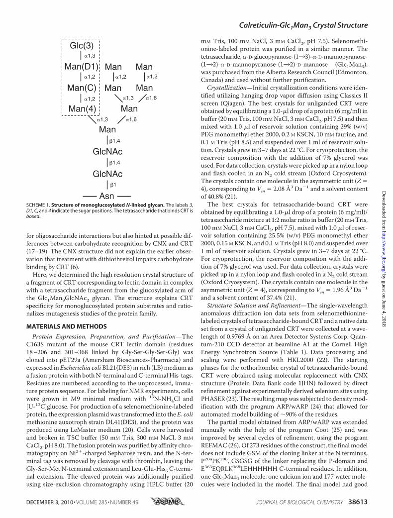
SCHEME 1. Structure of monoglucosylated N-linked glycan. The labels 3,D1, C, and 4 indicate the sugar positions. The tetrasaccharide that binds CRT isboxed.
Calreticulin-Glc1Man3 Crystal Structure
DECEMBER 3, 2010 • VOLUME 285 • NUMBER 49 JOURNAL OF BIOLOGICAL CHEMISTRY 38613
by guest on June 4, 2018http://w
ww
.jbc.org/D
ownloaded from
stereochemistry with no outliers in the Ramachandran plotcomputed using PROCHECK (27).For the unliganded CRT and monoclinic crystals of tet-
rasaccharide-bound CRT, the structures were obtained bymolecular replacement with the orthorhombic tetrasaccha-ride-bound CRT structure using PHASER (23) and improvedby several cycles of refinement, using the program REFMAC(26) and model refitting followed by the translation-libration-screw (TLS) refinement (28). For the unliganded CRT, the finalmodel does not include L203PPK206, GSGSG of the linker andE364QRLK368LEHHHHHH C-terminal residues. One calciumion and 56 water molecules were included in the model. Thefinal model has good stereochemistry with no outliers in theRamachandran plot computed using PROCHECK (27).Isothermal Titration Calorimetry—Experiments were car-
ried out on a MicroCal iTC200 titration calorimeter (GEHealthcare) using the VPViewer software for instrument con-trol and data acquisition. The buffer used for isothermal titra-tion calorimetry experiments contained 20 mM Tris, 100 mM
NaCl, 3 mM CaCl2, pH 7.5. During a titration experiment, asample of the CRT lectin domain was kept at 293 K in a stirred(1000 rpm) reaction cell of 0.2 ml. Nineteen injections, each of2-�l volume and 4-s duration with a 150-s interval betweeninjections, were carried out using a 39.4-�l syringe filled withGlc1Man3 solution. Titration experiments were performedwith 30 �M protein solution in the cell and 300 �M carbohy-drate solution in the syringe. The calorimetric data wereprocessed using the software package ORIGIN (version 7) todetermine theGibbs free energy of binding, molar binding stoi-chiometry (N), molar binding entropy (�S), and molar bindingenthalpy (�H).
RESULTS
Crystallization and Structure Determination of CRT LectinDomain—Previous attempts to crystallize CRTwere likely hin-dered by the intrinsic mobility of the arm-like P-domain andunstructured C terminus (supplemental Fig. 1). To overcomethis, we deleted the P-domain (residues 207–300) of mouseCRT and replaced it with a short linker. Additionally, Cys163was mutated to serine to avoid intermolecular disulfide bondformation. Although we were able to obtain purified protein,extensive screening did not yield any crystals.We hypothesizedthat the presence of an unstructured C-terminal tail addition-ally hindered crystallization. To define the boundaries of thefolded domain, we subjected full-length and P-domain-deletedmouse CRT to limited proteolysis using trypsin, chymotrypsin,proteinase K, and V8 protease. Characterization by mass spec-trometry of fragments from trypsin digestion suggested the Cterminus could be removed by cleavage at Lys368 to produce astable fragment and NMR spectroscopy confirmed that it wassoluble and well folded (supplemental Figs. 2 and 3). Based onthese results, we recloned the lectin domain of mouse CRT toinclude residues 18–206 and 301–368. Crystallization trialsproduced long needle-like crystals that could be improvedusing additives including a Glc1Man3 tetrasaccharide. Large,well diffracting crystals were obtained in the primitive orthor-hombic (space group P212121) andmonoclinic (P21) formswiththe best diffraction extending to beyond 2.0 Å.We obtained unbiased, experimental phases for the orthor-
hombic crystals using selenomethionine-labeled protein andsingle wavelength anomalous diffraction (Table 1). Subse-quently, this structure was used for molecular replacement tosolve the structure of the monoclinic crystal form. The asym-
TABLE 1Data collection and refinement statistics
CRT CRT/Glc1Man3 CRT/Glc1Man3Data collectionSpace group P212121 P212121 P21Cell dimensionsa, b, c (Å) 43.11, 75.32, 79.59 42.73, 43.77, 133.34 74.62, 43.23, 84.63�, �, � 90.00°, 90.00°, 90.00° 90.00°, 90.00°, 90.00° 90.00°, 96.05°, 90.00°
Resolution (Å)a 50-2.30 (2.34-2.30) 50-1.95 (1.98-1.95) 50-2.00 (2.03-2.00)Rsym 0.071 (0.280) 0.062 (0.142) 0.069 (0.248)I/�I 25.9 (6.3) 23.2 (5.9) 19.8 (5.4)Completeness (%) 99.8 (100.0) 96.5 (80.2) 100.0 (99.9)Redundancy 8.1 (7.2) 4.5 (2.3) 4.5 (3.8)
RefinementResolution (Å) 54.7-2.30 66.7-1.95 84.2-2.01No. of reflections 11,377 17,355 34,462Rwork/Rfree 0.214/0.274 0.197/0.251 0.191/0.240No. of atomsProtein 2018 1994 4033Glc1Man3 0 45 90Calcium ions 1 1 2Water 56 177 374
B-factorsProtein 25.9 22.2 22.7Glc1Man3 24.5 27.1Calcium ions 27.5 16.4 21.0Water 29.2 28.0 35.8
r.m.s.d.Bond lengths (Å) 0.007 0.009 0.009Bond angles 0.94° 1.27° 1.22°
Ramachandran statistics (%)Most favored regions 87.8 90.9 88.8Additional allowed regions 12.2 9.1 11.2
a Highest resolution shell is shown in parentheses.
Calreticulin-Glc1Man3 Crystal Structure
38614 JOURNAL OF BIOLOGICAL CHEMISTRY VOLUME 285 • NUMBER 49 • DECEMBER 3, 2010
by guest on June 4, 2018http://w
ww
.jbc.org/D
ownloaded from
metric units of orthorhombic andmonoclinic crystal structurescontain one and two copies of the CRT-Glc1Man3 complex,respectively. Despite different crystal contacts in both crystalforms, all three copies are nearly identical suggesting that thestructures are not influenced by crystal packing.Structure of CRT Lectin Domain—The structure gives an
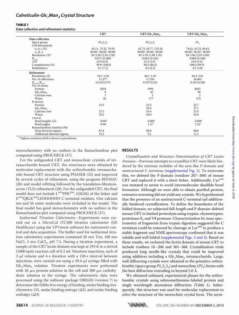
accurate definition of the CRT lectin domain boundaries run-ning from Asp18 to Phe202 and from Pro301 to Glu363. No den-sity was observed for Leu203–Lys207 and the following linkerGSGSG due to disorder in the crystal. Also, the C-terminalsequence Glu364–Lys368 with the C-terminal His-tag were dis-ordered. The structure of CRT lectin domain displays a jelly rollfold largely formed by a sandwich of two large �-sheets (Fig.1A). The hydrophobic interactions between the seven-strandedconcave �-sheet and six-stranded convex �-sheet are crucialfor structural integrity of the domain. There is also a small�-sheet covering the surface where the concave and convexsheets create space between them. The structure also showstwo short �-helices (Ala32–Arg36 and Leu196–Asp199) and along �-helix (Glu336–Asp362) that runs along and beyond theconvex �-sheet. There are also several protruding loops inthe domain. Thus, a flap-like�6–�7 loop is covering a part of theconcave �-sheet while packing against the �1-�2 loop that rises
against the �4 strand. Another longloop between strands �2 and �3 isstabilizing theC-terminal half of thelong helix �3. The Cys105–Cys137disulfide bridges the beginning ofstrand �6 with the end of strand �7.
The structure clearly defines acalcium-binding site in theCRT lec-tin domain. The calcium ion is coor-dinated by the side chain of Asp328,backbone carbonyls of Gln26, Lys62,and Lys64, and by two water mole-cules (Fig. 1B).Structural Basis of Carbohydrate
Recognition—The tetrasaccharideGlc1Man3 corresponds to the branchof the Glc1Man9GlcNAc2 glycanthat binds to the lectin domain ofCRT/CNX. By isothermal titrationcalorimetry, we confirmed that theisolated, recombinant domain re-tained all of the lectin functions.The affinity of 0.7 �M for Glc1Man3is very close to the reported value forintact CRT (supplemental Fig. 4)(29).The electron densitymap showed
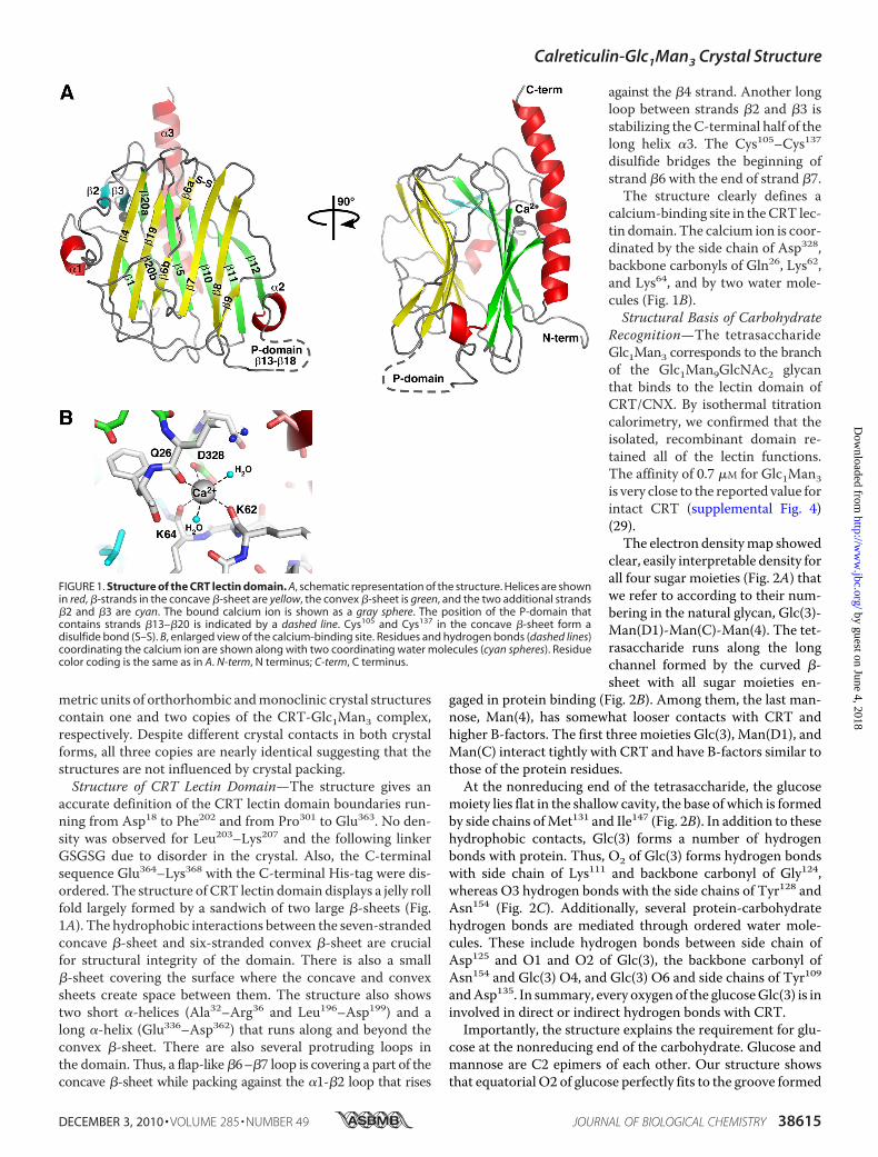
clear, easily interpretable density forall four sugar moieties (Fig. 2A) thatwe refer to according to their num-bering in the natural glycan, Glc(3)-Man(D1)-Man(C)-Man(4). The tet-rasaccharide runs along the longchannel formed by the curved �-sheet with all sugar moieties en-
gaged in protein binding (Fig. 2B). Among them, the last man-nose, Man(4), has somewhat looser contacts with CRT andhigher B-factors. The first three moieties Glc(3), Man(D1), andMan(C) interact tightly with CRT and have B-factors similar tothose of the protein residues.At the nonreducing end of the tetrasaccharide, the glucose
moiety lies flat in the shallow cavity, the base of which is formedby side chains ofMet131 and Ile147 (Fig. 2B). In addition to thesehydrophobic contacts, Glc(3) forms a number of hydrogenbonds with protein. Thus, O2 of Glc(3) forms hydrogen bondswith side chain of Lys111 and backbone carbonyl of Gly124,whereas O3 hydrogen bonds with the side chains of Tyr128 andAsn154 (Fig. 2C). Additionally, several protein-carbohydratehydrogen bonds are mediated through ordered water mole-cules. These include hydrogen bonds between side chain ofAsp125 and O1 and O2 of Glc(3), the backbone carbonyl ofAsn154 and Glc(3) O4, and Glc(3) O6 and side chains of Tyr109andAsp135. In summary, every oxygen of the glucoseGlc(3) is ininvolved in direct or indirect hydrogen bonds with CRT.Importantly, the structure explains the requirement for glu-
cose at the nonreducing end of the carbohydrate. Glucose andmannose are C2 epimers of each other. Our structure showsthat equatorial O2 of glucose perfectly fits to the groove formed
FIGURE 1. Structure of the CRT lectin domain. A, schematic representation of the structure. Helices are shownin red, �-strands in the concave �-sheet are yellow, the convex �-sheet is green, and the two additional strands�2 and �3 are cyan. The bound calcium ion is shown as a gray sphere. The position of the P-domain thatcontains strands �13–�20 is indicated by a dashed line. Cys105 and Cys137 in the concave �-sheet form adisulfide bond (S–S). B, enlarged view of the calcium-binding site. Residues and hydrogen bonds (dashed lines)coordinating the calcium ion are shown along with two coordinating water molecules (cyan spheres). Residuecolor coding is the same as in A. N-term, N terminus; C-term, C terminus.
Calreticulin-Glc1Man3 Crystal Structure
DECEMBER 3, 2010 • VOLUME 285 • NUMBER 49 JOURNAL OF BIOLOGICAL CHEMISTRY 38615
by guest on June 4, 2018http://w
ww
.jbc.org/D
ownloaded from
by CRT side chains (Fig. 2D). Mannose in this position wouldcause a steric clash with the sulfur atom of Met131 and the lossof hydrogen bonds with carbonyl of Gly124 and the side chain ofLys111. Previous mutagenesis studies showed that even the sin-gle K111A mutation impairs CRT-carbohydrate interactions(17, 18).The structure also explains the specificity for monoglucosy-
lated glycans (Glc1Man9GlcNAc2) over the precursor with twoglucose residues (Glc2Man9GlcNAc2). Although the carbohy-drate-binding site can accommodate a glucose residue in thesecond position, the sugar linkages are different. Binding of thetetrasaccharide �-Glc-(133)-�-Glc-(133)-�-Man-(132)-Man would result in the loss of hydrogen bonds and unfavor-able interactions as the lastmannose residue intersects the pro-tein surface.In contrast to the extensive protein contacts by the first
sugar,Man(D1) andMan(C)mainly use their O4–O6 edges forinteractions with CRT (Fig. 2C). In particular, O4 of Man(D1)occupies a crucial position engaging in three direct hydrogenbonds with side chain of Tyr109 and both the side chain andbackbone carbonyl of Asp317. The side chain of Asp317 alsomakes a direct hydrogen bond with O6 of Man(D1). AlthoughO2 of Man(D1) is directed away from the �-sheet, it forms awater-mediated hydrogen bond with the side chain of Asp125located in the carbohydrate-interacting loop. Asp135 of CRT is
crucial for Man(C) binding, as itsside chain forms direct hydrogenbonds with O4 and O6 of Man(C).Two water molecules assist theother Man(C)-CRT hydrogen bonds:between O3 and the side chains ofTyr109 and Asp135, and between O6and the side chain of Tyr109 andbackbone amide of Trp319.
Previous studies showed thattreatment with dithiothreitol abro-gates carbohydrate binding by CRT(6). We similarly see no binding inthe presence of the non-thiol-re-ducing agent TCEP (tris(2-carboxy-ethyl)phosphine) (supplemental Fig.4B). These cysteines are also essen-tial to the chaperone function ofCRT (30). The CRT/Glc1Man3 struc-ture provides a basis to explain theseobservations as the Cys105–Cys137disulfide bond is involved in con-tacts with the Man(C) and Man(4)moieties of the Glc1Man3 tetrasac-charide. Namely, the C5–C6 bondof Man(C) and C1–O1 bond ofMan(4) partially wrap around theCRT disulfide bond (Fig. 2C, right).Reduction of this disulfide bondwould clearly disrupt binding ofthe last two mannose moieties.Both Man(C) and Man(4) are alsoengaged in hydrophobic interac-
tions with the side chain of Trp319.The C1–O1 bond of Man(4) is directed away from the pro-
tein. Therefore, it is likely that the following mannose residueMan(3) of Glc1Man9GlcNAc2 glycan does not make any signif-icant interactions with CRT, and the full essence of CRT-car-bohydrate recognition is captured in our structure. In agree-ment with this conclusion, Glc1Man3 was shown to competeeffectively with Glc1Man9 for binding to CNX (6).Identical tetrasaccharide conformations were observed in
the two crystal forms. This clearly demonstrates that the con-formation is not affected by crystal packing. There is a singlecrystal contact between the C6–O6 bond of Man(4) at the farend of tetrasaccharide and the side chain of Glu345 of anotherCRT molecule in the P212121 crystal form, whereas the carbo-hydrate does not contact any symmetry-related molecules inthe P21 crystal form. Additionally, we note that the bound cal-cium ion is positioned far from the carbohydrate-binding siteand is not involved in glycan recognition.We overlaid the tetrasaccharide-bound and unliganded CRT
lectin domain structures to assess the conformational changesin CRT upon carbohydrate binding, The overlay results in anroot mean square deviation of 0.2 Å over 229 C� atoms demon-strating that the two structures are nearly identical (Fig. 3A).The only significant change occurs in the flap-like loop contain-ing Gly124 and Asp125, residues involved in glucose binding.
FIGURE 2. Structural basis of Glc1Man3 recognition by CRT. A, omit map calculated in the absence of tet-rasaccharide shows well defined electron density (blue) for all four sugar moieties. The tetrasaccharide binds ina cavity on the concave �-sheet. B, surface representation of CRT shows the side chains of Phe74, Met131, His145,Ile147, Trp319, and the Cys105–Cys137 disulfide bridge form the walls of the cavity in contact with the glycan(magenta). C, oxygens in the tetrasaccharide form a network of hydrogen bonds (dotted lines) with orderedwater molecules (cyan spheres) and CRT. Residues that disrupt CRT binding when mutated are shown in gray(17–19). D, the equatorial oxygen (O2) of glucose makes hydrogen bonds with the side chain of Lys111 andbackbone carbonyl of Gly124. Mannose has an axial O2, which clashes sterically with the underlying side chainof Met131 to prevent binding in that position.
Calreticulin-Glc1Man3 Crystal Structure
38616 JOURNAL OF BIOLOGICAL CHEMISTRY VOLUME 285 • NUMBER 49 • DECEMBER 3, 2010
by guest on June 4, 2018http://w
ww
.jbc.org/D
ownloaded from

The loop conformation in the unliganded state is stabilized byside chains of Lys111 and Asp317 that make hydrogen bondswith the backbone carbonyl of Asp125 and amides of Gly124 andAsp125, respectively (Fig. 3B). When oligosaccharide bindsCRT, side chains of Lys111 and Asp317 engage in direct hydro-gen bonding with sugar moieties. The released Gly124 andAsp125 rotate allowing carbonyl of Gly124 to form a hydrogenbond with Glc(3), whereas the side chain of Asp125 interactswith carbohydrates via an ordered water molecule (Fig. 3C).Structure Correlates well with Mutagenesis Results—Due to
the interest in recognition of glycosylated proteins by CRT/CNX, vast mutagenesis data have been obtained with CRT tocharacterize its binding to oligosaccharides. Mutagenesis ofCRT by different groups showed that Tyr109, Lys111, Tyr128,Asp135, and Asp317 are critical for carbohydrate binding (17–
19). The crystal structure shows that the side chains of all ofthese residues form direct hydrogen bonds with carbohydratemoieties (Fig. 2C). The structure is also in perfect agreementwith the results showing reduced carbohydrate binding for theW319I andW319A CRTmutants (19). The bulky side chain ofthis residue is involved in hydrophobic contacts with the reduc-ing end mannose residue in our structure (Fig. 2B). Similarly,substitutions of Met131 reduced but did not prevent binding(17, 18). On the other hand, mutations of Asp125 did not affectoligosaccharide binding (17), as the contacts of this residuewith sugar are mediated through a water molecule. The struc-ture further explains the observation that D160G and D160Amutations do not affect carbohydrate binding (18, 19), as thisresidue is 13 Å away from the bound ligand. The structure alsoreveals that the 75% decrease in carbohydrate affinity in theR73L CRTmutant (17) results frommid-range conformationalchanges, as this residue is�10Å away from the bound tetrasac-charide and is unable to participate directly in binding.ComparisonwithCNXandOther ERLectins—Sequence sim-
ilarity between homologous regions of CRT and CNX led toassumptions that the structures of these proteins are very sim-ilar and resulted in use of homology models of CRT based onthe CNX structure. Consequently, these models were used tointerpret results from single point and deletion mutagenesisthat proved to be inaccurate in some cases. As an example, theGlu217 of CNX, which participates in ligand binding, was incor-rectly assigned as homologous to Asp160 of CRT, which is farfrom the carbohydrate-binding site (17, 18). On the other hand,while theCRTconstruct used for glycan-independent substratebinding studies was missing several residues of the long C-ter-minal helix (31), the thermal denaturation curve for that con-struct is similar to that of the lectin domain studied here (sup-plemental Fig. 5).The CRT structure allows for an accurate structural align-
ment of CRT and CNX. The structural overlay of unligandedCRT and CNX shows 33% sequence identity between their lec-tin domains and a root mean square deviation of 1.7 Å over 195CA atoms. The structural similarities are strongest in the�-sheet regions, whereas the loops often adopt divergent con-formations (Fig. 4A). Conspicuous differences between the twostructures occur in the helical regions. Thus, the short �1 and�2 CRT helices are absent in the CNX structure. Similarly, theC-terminal helix is much shorter (10 versus 25 residues) in theCNXstructure. The reason for this divergence is unclear, as thisregion is well conserved between the two proteins (Fig. 4B).Although this may reflect a genuine difference between CNXand CRT, it is possible that the native CNX C-terminal helix islonger than observed previously.Despite differences elsewhere, the oligosaccharide-binding
surface is nearly identical in both proteins (Fig. 4C). The resi-dues that are critical for carbohydrate binding are very wellconserved and adopt very similar conformations in both CRTand CNX. Glucose soaking of CNX crystals showed that glu-cose contacts Met189 of CNX (equivalent to Met131 of mouseCRT) and makes hydrogen bonds with Tyr165 (Tyr109), Lys167
(Lys111), Tyr186 (Tyr128), Glu217 (Asn154), and possibly Glu426
(Asp317) (8). This is slightly shifted from the position observed
FIGURE 3. CRT undergoes limited conformational changes upon carbohy-drate binding. A, overlay of unliganded (yellow) and Glc1Man3-bound (green)CRT lectin domain structures shows differences in the large loop betweenstrands �6 and �7. B, the conformation of the loop in unliganded CRT isstabilized by hydrogen bonds between the side chain of Asp317 and amides ofGly124 and Asp125 and between the side chain of Lys111 and carbonyl ofAsp125. C, in the complex, sugar residues Glc(3) and Man(D1) engage loopresidues Gly124 and Asp125 through a 60 ° rotation in the � backbone angle ofGly124. This rotation enables the side chain of Asp125 to participate in carbo-hydrate binding via an ordered water molecule (cyan sphere). The interactionsof Asp317 with Gly124 are replaced by hydrogen bonds of Asp317 with Man(D1).
Calreticulin-Glc1Man3 Crystal Structure
DECEMBER 3, 2010 • VOLUME 285 • NUMBER 49 JOURNAL OF BIOLOGICAL CHEMISTRY 38617
by guest on June 4, 2018http://w
ww
.jbc.org/D
ownloaded from

in our crystal structure and involves residues (Tyr109 andAsp317 of CRT) that form hydrogen bonds with the followingmannose in the CRT-tetrasaccharide complex. The reason forthis discrepancy could simply result from lowbinding affinity ofglucose to CNX coupled with the low resolution of crystallo-graphic data set leading to some positional shift of the glucosemoiety in the CNX structure. Or, the isolated glucose moietycould bind at a slightly different location when it is not part ofan oligosaccharide chain. Nonetheless, the superposition of thecarbohydrate binding residues is striking and suggests thatCNX and CRT bind carbohydrates in an identical fashion.The calcium ion is coordinated in the CNX structure by side
chains of Asp437 (Asp328 of CRT) and Asp118 (Asp63), carbonylof Ser75 (Gln26) and, possibly, by carbonyl of Lys119 (Lys64). Themissing coordinating groups were not observed possibly due toinsufficient resolution. In CRT, an equivalent calcium ion iscoordinated by both oxygen atoms of Asp328 side chain, car-bonyls of Gln26, Lys62, and Lys64 and by two water molecules.A structural similarity search using the Dali database (32)
showed that the CRT lectin domain is most similar to CNX(Z-score, 27.5) as expected. In addition, the CRT structure issimilar to Emp47p (Z-score, 15.5), ERGIC-53 (Z-score, 14.3),and VIP36 (Z-score, 13.7). VIP36 and ERGIC-53 are transportlectin proteins that are involved in trafficking of glycosylated
proteins out of the ER, whereasEmp46/47p is a yeast homolog ofERGIC-53. Interestingly, these pro-teins have specificity toward thedeglucosylatedD1 armof highman-nose glycans (33–35), the same armrecognized by CRT/CNX in itsmonoglucosylated state. The crystalstructures of VIP36 in complexwith �-Man-(132)-Man and �-Man-(132)-�-Man-(133)-�-Man-(134)-GlcNAc have been deter-mined (36). Despite a significantoverlap in oligosaccharide specific-ity and the use of a similar structuralscaffold between CRT/CNX andVIP36, they use differing surfaces tobind carbohydrates (supplementalFig. 6). This is an example of how asimilar fold is adapted for bindingsomewhat differing ligands.
DISCUSSION
The high resolution crystal struc-ture of the CRT lectin domain incomplex with the Glc1Man3 tet-rasaccharide illuminates the molec-ular basis of monoglucosylatedGlc1Man9GlcNAc2 glycan functionin the calnexin cycle. The structureexplains the requirement for a sin-gle glucose at the nonreducing endof the carbohydrate and allows foran accurate structural alignment
between CRT and CNX. The striking similarity in the sugar-binding sites suggests that CNX and CRT interact with mono-glucosylated substrates in identical fashion.To gain insight into the structure of full-length CRT with a
bound glycoprotein, we overlaid the CRT lectin domain with amodel of the P-domain derived from the structure of CNX (Fig.5). As the CRT P-domain is shorter and has only three repeatmodules, we removed the third module of the CNX P-domain.The CNX P-domain was chosen as a model because the crystalstructure includes information about the relative orientation ofthe P-and lectin domains that is absent from theNMRstructureof the isolated CRT P-domain (12, 13). The tip of the CRTP-domain contains a binding site for ERp57, an oxidoreductaseinvolved in disulfide bond formation in glycoproteins. Themodel gives an idea of how the bound glycoprotein might bepositioned relative to the lectin domain, the P-domain andERp57. CRT also contains an �55-residue-long C-terminalextension that is rich (�60%) in glutamate and aspartate resi-dues. Because of its abundant negative charges, this C-terminaldomain is unlikely to be structured in solution, but it becomesmore ordered upon binding calcium (37).To exit the CNX/CRT cycle, monoglucosylated glycopro-
teins are processed by glucosidase II to remove the terminalglucose residue. The accessibility of the glycan for processing
FIGURE 4. Structural comparison of the CRT and CNX lectin domains. A, overlay of the CRT (yellow) and CNX(Protein Data Bank code 1JHN; cyan) structures. The termini and boundaries of the P-domain of CRT areindicated. B, structure-based sequence alignment of mouse CRT and dog CNX lectin domains. Secondarystructure of CRT is shown above the alignment with �-strands and �-helices labeled. Residues that make directhydrogen bonds with Glc1Man3 are highlighted in cyan, and those making van der Waals contacts are high-lighted in gray. The position of the internal P-domain comprising strands �13 to �18 of CRT is indicated.C, enlarged view of the carbohydrate-binding site shows nearly identical positioning of key residues in CRT andCNX. Residue numbers refer to CRT. N-term, N terminus; C-term, C terminus.
Calreticulin-Glc1Man3 Crystal Structure
38618 JOURNAL OF BIOLOGICAL CHEMISTRY VOLUME 285 • NUMBER 49 • DECEMBER 3, 2010
by guest on June 4, 2018http://w
ww
.jbc.org/D
ownloaded from
while bound to CNX/CRT has been the subject of some debate(38, 39). Our structure shows that the bond betweenGlc(3) andMan(D1) targeted by glucosidase II is not easily accessible. Itseems likely that the glycosylated substrate has to dissociatefrom CRT for deglucosylation to occur.Earlier reports also suggested that CRT undergoes con-
formational changes upon carbohydrate binding (40). Com-parison of the tetrasaccharide-bound and unliganded CRTlectin domain structures demonstrates that the only significantchange occurs in the flap-like loop that is involved in glucosebinding. On the other hand, loss of the bound calcium ion islikely to strongly destabilize the CRT lectin domain. Numerousstudies have shown calcium-dependent conformationalchanges in CRT (41–43). Although our carbohydrate-boundCRT structure confirms that the calcium-binding site is too farto affect interactions with glycans, the tightly bound calciumion is important for the structural integrity of the lectin domain.With a Kd of �2 �M (14), loss of this high affinity calcium ion isunlikely to occur within the ER, but it may occur for CRT out-side of the ER (44).CNX and CRT exhibit overlapping but distinct patterns of
interaction with folding glycoproteins in vivo (45–47). Giventhat the lectin sites of these chaperones are essentially identical,the basis for observed differences in glycoprotein binding spec-ificity must reside in other properties. Previous studies haveshown that that the distinct luminal versus membrane-boundtopologies of CRT and CNX contribute to selection of clientglycoproteins (45, 48, 49). It is also likely that the reported abil-ity of CRT and CNX to bind directly to peptides (31, 50, 51) or
to polypeptide segments of non-native protein conformers (9,40) contributes to substrate selection. Consequently, the iden-tification of such peptide binding sites on these chaperones is ofconsiderable interest. However, despite the fact that the lectindomains of both CNX (50) and CRT4 are capable of bindinghydrophobic peptideswithmicromolarKd, our efforts to obtainco-crystals of theCRT lectin domainwith such peptides have sofar been unsuccessful. Likewise, we have been unable to co-crystallize the lectin domain of CRT with ATP. Both CNX andCRThave been reported to bindATP (41, 42), and the presenceof this nucleotide has been shown to enhance their abilities tobind non-native polypeptides and to suppress their aggregationin vitro (9, 40). Given the conformational changes that havebeen reported to accompany nucleotide or peptide binding (9,41, 42, 50), it may be that such conformations are less amenableto crystallization.CRT has also been reported to bind zinc, and four histidines
within the lectin domain have been implicated in binding (40,43, 52–56). Examination of these histidines reveals that someare buried but that His42 is exposed and adjacent to other resi-dues, Asp118, Asp121, His123, and Asp125, that could potentiallybind zinc.In conclusion, we have determined the structure of the CRT
lectin domain in complex with its physiological ligand. Thestructure provides the framework for the design and interpre-tation of mutants that affect the multiple physiological func-tions of CRT.
Acknowledgments—Data acquisition at theMacromolecularDiffrac-tion (MacCHESS) facility at the Cornell High Energy SynchrotronSource was supported by National Science Foundation Award DMR0225180 and National Institutes of Health Award RR-01646.
REFERENCES1. Helenius, A., and Aebi, M. (2004) Annu. Rev. Biochem. 73, 1019–10492. Caramelo, J. J., and Parodi, A. J. (2008) J. Biol. Chem. 283, 10221–102253. Lederkremer, G. Z. (2009) Curr. Opin. Struct. Biol. 19, 515–5234. Ware, F. E., Vassilakos, A., Peterson, P. A., Jackson,M. R., Lehrman,M. A.,
and Williams, D. B. (1995) J. Biol. Chem. 270, 4697–47045. Spiro, R. G., Zhu, Q., Bhoyroo, V., and Soling, H. D. (1996) J. Biol. Chem.
271, 11588–115946. Vassilakos, A., Michalak, M., Lehrman, M. A., and Williams, D. B. (1998)
Biochemistry 37, 3480–34907. Solda, T., Galli, C., Kaufman, R. J., and Molinari, M. (2007)Mol. Cell. 27,
238–2498. Schrag, J. D., Bergeron, J. J., Li, Y., Borisova, S., Hahn, M., Thomas, D. Y.,
and Cygler, M. (2001)Mol. Cell 8, 633–6449. Brockmeier, A., andWilliams, D. B. (2006)Biochemistry 45, 12906–1291610. Frickel, E. M., Riek, R., Jelesarov, I., Helenius, A., Wuthrich, K., and Ell-
gaard, L. (2002) Proc. Natl. Acad. Sci. U.S.A. 99, 1954–195911. Kozlov, G., Maattanen, P., Schrag, J. D., Pollock, S., Cygler, M., Nagar, B.,
Thomas, D. Y., and Gehring, K. (2006) Structure 14, 1331–133912. Ellgaard, L., Riek, R., Herrmann, T., Guntert, P., Braun, D., Helenius, A.,
and Wuthrich, K. (2001) Proc. Natl. Acad. Sci. U.S.A. 98, 3133–313813. Ellgaard, L., Bettendorff, P., Braun, D., Herrmann, T., Fiorito, F., Jelesarov,
I., Guntert, P., Helenius, A., and Wuthrich, K. (2002) J. Mol. Biol. 322,773–784
14. Baksh, S., and Michalak, M. (1991) J. Biol. Chem. 266, 21458–2146515. Peterson, J. R., and Helenius, A. (1999) J. Cell Sci. 112, 2775–2784
4 C. L. Pocanschi, unpublished observations.
FIGURE 5. Structural model of full-length CRT. The lectin domain is shownin green, and the bound carbohydrate is shown in magenta. The approximateorientation of the P-domain (dark blue) is shown based on the CNX structurewith one repeat unit removed to match the shorter CRT P-domain. The boundcarbohydrate is shown as part of the N-linked glycan linked to an asparagineresidue of an unfolded protein. The C terminus of CRT contains a Glu-, Asp-rich sequence, which binds calcium, and a KDEL ER retention signal. The res-idues that define the boundaries of crystallized CRT fragment and the portionof the P-domain that binds the thiol oxidoreductase, ERp57, are labeled.
Calreticulin-Glc1Man3 Crystal Structure
DECEMBER 3, 2010 • VOLUME 285 • NUMBER 49 JOURNAL OF BIOLOGICAL CHEMISTRY 38619
by guest on June 4, 2018http://w
ww
.jbc.org/D
ownloaded from

16. Leach, M. R., and Williams, D. B. (2004) J. Biol. Chem. 279, 9072–907917. Kapoor, M., Ellgaard, L., Gopalakrishnapai, J., Schirra, C., Gemma, E.,
Oscarson, S., Helenius, A., and Surolia, A. (2004) Biochemistry 43, 97–10618. Thomson, S. P., and Williams, D. B. (2005) Cell Stress Chaperones 10,
242–25119. Gopalakrishnapai, J., Gupta, G., Karthikeyan, T., Sinha, S., Kandiah, E.,
Gemma, E., Oscarson, S., and Surolia, A. (2006) Biochem. Biophys. Res.Commun. 351, 14–20
20. Hendrickson,W. A., Horton, J. R., and LeMaster, D.M. (1990) EMBO J. 9,1665–1672
21. Matthews, B. W. (1968) J. Mol. Biol. 33, 491–49722. Otwinowski, Z., andMinor, W. (1997) inMethods in Enzymology (Carter,
C. W., and Sweet, R. M., eds) pp. 307–326, Vol. 276, Part A, AcademicPress, New York
23. McCoy, A. J., Grosse-Kunstleve, R. W., Adams, P. D., Winn, M. D., Sto-roni, L. C., and Read, R. J. (2007) J. Appl. Crystallogr. 40, 658–674
24. Perrakis, A., Sixma, T. K., Wilson, K. S., and Lamzin, V. S. (1997) ActaCrystallogr. D Biol. Crystallogr. 53, 448–455
25. Emsley, P., and Cowtan, K. (2004)Acta Crystallogr. D Biol. Crystallogr. 60,2126–2132
26. Murshudov, G. N., Vagin, A. A., Lebedev, A., Wilson, K. S., and Dodson,E. J. (1999) Acta Crystallogr. D Biol. Crystallogr. 55, 247–255
27. Laskowski, R. A., MacArthur, M. W., Moss, D. S., and Thornton, J. M.(1993) J. Appl. Crystallogr. 26, 283–291
28. Winn, M. D., Murshudov, G. N., and Papiz, M. Z. (2003) Methods Enzy-mol. 374, 300–321
29. Kapoor, M., Srinivas, H., Kandiah, E., Gemma, E., Ellgaard, L., Oscarson,S., Helenius, A., and Surolia, A. (2003) J. Biol. Chem. 278, 6194–6200
30. Martin, V., Groenendyk, J., Steiner, S. S., Guo, L., Dabrowska, M., Parker,J. M., Muller-Esterl, W., Opas, M., and Michalak, M. (2006) J. Biol. Chem.281, 2338–2346
31. Rizvi, S. M., Mancino, L., Thammavongsa, V., Cantley, R. L., and Ragha-van, M. (2004)Mol. Cell 15, 913–923
32. Holm, L., and Sander, C. (1995) Trends Biochem. Sci. 20, 478–48033. Hara-Kuge, S., Ohkura, T., Seko, A., and Yamashita, K. (1999) Glycobiol-
ogy 9, 833–83934. Kamiya, Y., Yamaguchi, Y., Takahashi, N., Arata, Y., Kasai, K., Ihara, Y.,
Matsuo, I., Ito, Y., Yamamoto, K., and Kato, K. (2005) J. Biol. Chem. 280,37178–37182
35. Kamiya, Y., Kamiya, D., Yamamoto, K., Nyfeler, B., Hauri, H. P., and Kato,K. (2008) J. Biol. Chem. 283, 1857–1861
36. Satoh, T., Cowieson, N. P., Hakamata, W., Ideo, H., Fukushima, K., Kuri-hara, M., Kato, R., Yamashita, K., and Wakatsuki, S. (2007) J. Biol. Chem.282, 28246–28255
37. Villamil Giraldo, A. M., LopezMedus, M., Gonzalez Lebrero, M., Pagano,
R. S., Labriola, C. A., Landolfo, L., Delfino, J. M., Parodi, A. J., and Cara-melo, J. J. (2010) J. Biol. Chem. 285, 4544–4553
38. Rodan, A. R., Simons, J. F., Trombetta, E. S., and Helenius, A. (1996)EMBO J. 15, 6921–6930
39. Zapun, A., Petrescu, S. M., Rudd, P. M., Dwek, R. A., Thomas, D. Y., andBergeron, J. J. (1997) Cell 88, 29–38
40. Saito, Y., Ihara, Y., Leach, M. R., Cohen-Doyle, M. F., and Williams, D. B.(1999) EMBO J. 18, 6718–6729
41. Ou, W. J., Bergeron, J. J., Li, Y., Kang, C. Y., and Thomas, D. Y. (1995)J. Biol. Chem. 270, 18051–18059
42. Corbett, E. F., Michalak, K. M., Oikawa, K., Johnson, S., Campbell, I. D.,Eggleton, P., Kay, C., and Michalak, M. (2000) J. Biol. Chem. 275,27177–27185
43. Li, Z., Stafford, W. F., and Bouvier, M. (2001) Biochemistry 40,11193–11201
44. Gold, L. I., Eggleton, P., Sweetwyne,M. T., VanDuyn, L. B., Greives,M. R.,Naylor, S. M., Michalak,M., andMurphy-Ullrich, J. E. (2010) FASEB J. 24,665–683
45. Danilczyk, U. G., Cohen-Doyle, M. F., and Williams, D. B. (2000) J. Biol.Chem. 275, 13089–13097
46. Peterson, J. R., Ora, A., Van, P. N., and Helenius, A. (1995)Mol. Biol. Cell6, 1173–1184
47. Pieren,M.,Galli, C., Denzel, A., andMolinari,M. (2005) J. Biol. Chem.280,28265–28271
48. Wada, I., Imai, S., Kai, M., Sakane, F., and Kanoh, H. (1995) J. Biol. Chem.270, 20298–20304
49. Hebert, D. N., Zhang, J. X., Chen,W., Foellmer, B., andHelenius, A. (1997)J. Cell Biol. 139, 613–623
50. Brockmeier, A., Brockmeier, U., and Williams, D. B. (2009) J. Biol. Chem.284, 3433–3444
51. Sandhu, N., Duus, K., Jørgensen, C. S., Hansen, P. R., Bruun, S. W., Peder-sen, L. Ø., Højrup, P., and Houen, G. (2007) Biochim. Biophys. Acta 1774,701–713
52. Leach, M. R., Cohen-Doyle, M. F., Thomas, D. Y., and Williams, D. B.(2002) J. Biol. Chem. 277, 29686–29697
53. Baksh, S., Spamer, C., Heilmann, C., and Michalak, M. (1995) FEBS Lett.376, 53–57
54. Guo, L., Groenendyk, J., Papp, S., Dabrowska, M., Knoblach, B., Kay, C.,Parker, J. M., Opas, M., and Michalak, M. (2003) J. Biol. Chem. 278,50645–50653
55. Khanna, N. C., Tokuda,M., andWaisman, D.M. (1986) J. Biol. Chem. 261,8883–8887
56. Tan, Y., Chen, M., Li, Z., Mabuchi, K., and Bouvier, M. (2006) Biochim.Biophys. Acta 1760, 745–753
Calreticulin-Glc1Man3 Crystal Structure
38620 JOURNAL OF BIOLOGICAL CHEMISTRY VOLUME 285 • NUMBER 49 • DECEMBER 3, 2010
by guest on June 4, 2018http://w
ww
.jbc.org/D
ownloaded from

Alexei Gorelik, David B. Williams and Kalle GehringGuennadi Kozlov, Cosmin L. Pocanschi, Angelika Rosenauer, Sara Bastos-Aristizabal,
Structural Basis of Carbohydrate Recognition by Calreticulin
doi: 10.1074/jbc.M110.168294 originally published online September 29, 20102010, 285:38612-38620.J. Biol. Chem.
10.1074/jbc.M110.168294Access the most updated version of this article at doi:
Alerts:
When a correction for this article is posted•
When this article is cited•
to choose from all of JBC's e-mail alertsClick here
Supplemental material:
http://www.jbc.org/content/suppl/2010/09/29/M110.168294.DC1
http://www.jbc.org/content/285/49/38612.full.html#ref-list-1
This article cites 56 references, 27 of which can be accessed free at
by guest on June 4, 2018http://w
ww
.jbc.org/D
ownloaded from