Embed Size (px)
Citation preview

Behavioral/Systems/Cognitive
Structural Basis for Map Formation in the ThalamocorticalPathway of the Barrelless Mouse
Fulvia Gheorghita, Rudolf Kraftsik, Roger Dubois, and Egbert WelkerDepartement de Biologie Cellulaire et de Morphologie, Faculte de Medecine, Universite de Lausanne, CH-1005 Lausanne, Switzerland
Barrelless mice (BRL) homozygous for the BRL mutation that disrupts the gene coding for adenylyl cyclase type I on chromosome 11 lackspatial segregation of layer IV cortical cells and of the thalamocortical axons (TCAs) into barrel domains. Despite these morphologicalperturbations, a functional topographic map has been demonstrated. We reconstructed individual biocytin-injected TCAs from thala-mus to barrel cortex in NOR (normal) and BRL mice to analyze to what extent the TCA arborization pattern and bouton distribution couldexplain the topographic representation of the whisker follicles. In BRL, the geometry of TCA is modified within layer IV as well as ininfragranular layers. However, in both strains, the spatial distribution of TCA in layer IV reflects the spatial relationship of their cellbodies in the ventrobasal nucleus of the thalamus. The morphometric analysis revealed that TCAs of both strains have the same length,branch number, and number of axonal boutons in layer IV. However, in barrelless, the boutons are distributed within a larger tangentialextent. Analysis of the distribution of boutons from neighboring thalamic neurons demonstrated the existence in layer IV of domains ofhigh bouton density that in both strains equal the size and shape of individual barrels. We propose that the domains of high boutondensity are at the basis of the whisker map in barrelless mice.
Key words: mice; barrel cortex; thalamocortical; adenylyl cyclase I; cortical map; biocytin
IntroductionIn direct sensory pathways of the mammalian brain, axons pro-jecting from thalamus to the primary sensory cortex have a ste-reotypic morphology. In the thalamus, they send collaterals to thereticular nucleus and subsequently ascend through the internalcapsule to terminate in cortical layers VI and IV (Jones, 1985,1986). Subcortically, the topological organization of thalamocor-tical axons (TCAs) undergoes distinct transformations (Adams etal., 1997). In cortex, the spatial segregation of TCAs underlies thefunctional parcellation (Blasdel and Lund, 1983; Humphrey etal., 1985; Jensen and Killackey, 1987a; Antonini et al., 1998,1999). During development, sensory activity is an important fac-tor in determining the spatial distribution of TCAs as was dem-onstrated after visual deprivation in cats (Antonini and Stryker,1993) and whisker follicle denervation in rats (Jensen and Kil-lackey, 1987b). Detailed studies of thalamic projections in thewhisker-to-barrel pathway revealed that modifying neuronaltransmission in cortex influences the spatial configuration ofTCA during development. Blocking the activity through theNMDA receptor in the conditional NR1 knock-out affects TCAmorphology but does not modify the cytoarchitectural differen-
tiation into barrels (Iwasato et al., 2000; Lee et al., 2005). Barrelformation is perturbed in several mouse lines: the knock-out forthe metabotropic glutamate receptor 5 (mGluR5) receptor (Han-nan et al., 2001), the monoamine oxidase A (MAOA)-deficientmouse (Cases et al., 1996), the GAP-43 (growth-associated pro-tein 43) (Maier et al., 1999) and in the mouse mutant barrelless,which lacks functional adenylyl cyclase I (Welker et al., 1996;Abdel-Majid et al., 1998; Lu et al., 2003). Whereas a morpholog-ical analysis of the TCA axons is not available for the mGluR5knock-out, in the other mouse lines lack of barrel formation isattributable to a disrupted clustering of TCA within barrel do-mains (Welker et al., 1996; Rebsam et al., 2002; McIlvain et al.,2003). Despite this altered arborization pattern of TCAs, a func-tional topographic map was demonstrated in SI of both MAOAand barrelless mice (Welker et al., 1996; Yang et al., 2001).
The aims of the current study are the following: (1) to charac-terize the arborization pattern of TCA in wild-type and barrellessmice to identify the level at which the mutation affected axonalmorphology, and (2) to analyze whether the tangential distribu-tion of the TCA boutons in barrelless mice can explain the estab-lishment of a functional map of the sensory periphery. We recon-structed individual biocytin-labeled TCAs from thalamus tocortex and registered the spatial distribution of the axonal bou-tons that correspond to TCA synapses (White et al., 2004). Wedemonstrated that TCAs in barrelless are identical to those inwild type with respect to total axonal length and number of ax-onal boutons, but that the mutation affects the tangential extentof the axonal plexuses in layers IV and VI, confirming our previ-ous report (Welker et al., 1996). Superposition of the distributionof axonal boutons from TCAs with cell bodies that are adjacent inthe thalamus showed that within the cortex tangential domains
Received March 24, 2006; revised July 27, 2006; accepted Aug. 14, 2006.This work was supported by the Swiss National Science Foundation (310000-108246). We thank Laurent Tettoni
for support throughout this study; Graham Knott for helpful comments on this manuscript; Nathalie Borrajo-Trappand Nathalie Mueller for histology; and Gilles Bronchti, Vincent Castagne, Christel Genoud, Charles Quairiaux, FabienPichon, Aouatef Abaza, and Jan-Harry Cabungcal for helpful discussions.
Correspondence should be addressed to Egbert Welker, Departement de Biologie Cellulaire et de Morphologie,Faculte de Medecine, Universite de Lausanne, Rue du Bugnon 9, CH-1005 Lausanne, Switzerland. E-mail:[email protected].
DOI:10.1523/JNEUROSCI.1263-06.2006Copyright © 2006 Society for Neuroscience 0270-6474/06/2610057-11$15.00/0
The Journal of Neuroscience, September 27, 2006 • 26(39):10057–10067 • 10057
are created with high density of axonalboutons that equal the size of individualbarrels.
Materials and MethodsAnimalsThe barrelless mice (Welker et al., 1996) used inthis study are from a breeding line that wasstarted after the discovery of a mutation thathad occurred in the normal (NOR) strain (i.e.,mice bred for a standard pattern of mystacialvibrissas) (Van der Loos et al., 1984). Barrellessmice are homozygous for the BRL mutationthat disrupted the gene coding for adenylyl cy-clase type I on chromosome 11 (Welker et al.,1996; Abdel-Majid et al., 1998). In total, sixNOR and 11 barrelless animals were used in thisstudy. Mice of both strains were exposed to thesame experimental procedures (see below) thatwere approved by the Office Veterinaire Can-tonal (Lausanne, Switzerland), in accordancewith Swiss laws.
Tracing and reconstruction ofthalamocortical axonsAdult females (age, 6 –10 weeks) were anesthe-tized with Nembutal (60 mg of sodium pento-barbital/kg of body weight, i.p.) and placed in astereotaxic frame. Glass pipettes filled with 2%biocytin in 1 M potassium acetate were loweredinto the ventrobasal nucleus of the thalamus(VB) of the right hemisphere. Stimulation-dependent neuronal activity was recordedthrough the pipette to identify the representa-tion of the mystacial whisker follicles. Subse-quently, the tracer was iontophoretically in-jected using a 150 –200 nA positive current for10 –15 min (1 s on, 1 s off) according to theprotocol of Pinault (1996). In most cases, theinjection was made in the representation of fol-licle C2. After 24 h, mice were reanesthetizedwith a lethal dose of Nembutal and were fixedvia transcardial perfusion of 4% paraformaldehyde in phosphate buffer(0.1 M; pH 7.2). Brains were removed and postfixed for 2 h in samefixative after which they were placed in a 30% solution of sucrose over-night. Brains were cut coronally on a freezing microtome at 50 �m, andthe sections were processed for the revelation of biocytin using the avi-din– biotin reaction kit from Vectra stain. Sections were Nissl stainedwith methylene blue. For additional details on the histological procedure,see Dolleman-Van der Weel et al. (1994). Cortical areas were assignedusing cytoarchitectonic criteria as described by Caviness (1975).
The number of labeled thalamic neurons was counted. A total of 35NOR and 17 BRL axons were hand-drawn using an ordinary light micro-scope fitted with a drawing tube. These drawings were used to study theaxonal trajectory and the principal axonal bifurcations in the somatosen-sory cortex. From these sets of axons, eight NOR axons (from four mice)and nine BRL axons (from three mice) were randomly assigned for three-dimensional reconstruction using a Zeiss (Oberkochen, Germany) mi-croscope equipped with a video camera (930XP 3CCD Color Video;Sony, Tokyo, Japan), a microcomputer-controlled stage, and a recon-struction program, Neurolucida (MicroBrightField, Williston, VT). Fig-ure 1 gives the positions of the injection sites of these reconstructedaxons.
Per mouse, two to six axons were reconstructed simultaneously fromVB to the cortex over 10 –25 sections. In the cortex, the entire axon wasreconstructed, and the branch points (nodes) and endings were plotted.Axonal branches represent the axonal segments between two consecutivenodes. The branch was “incomplete” if, for any reason, its reconstructionwas not possible. Branches with a length �4 �m were not traced but their
position was identified. The position of the axonal boutons was alsoplotted. A correction (2.66�) was applied for the shrinkage in the z-axisbecause of the histological procedure.
The NEUROLUCIDA data file was transferred to a UNIX station(Silicon Graphics, Mountain View, CA) for additional analysis usingMAXSIM software (Tettoni et al., 1996).
MeasurementsMAXSIM software enabled measurements of the entire axonal intracor-tical arborization and its components: the trunks and the tangential plex-uses. Trunks were defined as parts of the axon that traversed the corticallayers and gave off branches in the two target layers: the upper layer VIand layer IV. The tangential plexus of layers I–IV are all the branchescoming off by the axonal trunks in layers IV, III, II, and I. The tangentialplexus of layers V–VI is formed by the branches that course tangentiallyat the white matter (WM)/cortical border and at the VI/V interface. Thefollowing morphological parameters were determined.
The “neurometric” parameters. To characterize the axonal tree and thedistribution of boutons, we measured the axonal length, number of ax-onal branches, and the position of each axonal bouton. Measurements ofthese parameters were made first on the entire intracortical arborizationby selecting the axon at its cortical entrance (Fig. 2 A, arrow). Using theStrahler centripetal ordering (Fig. 2C), the program further allows us totopologically characterize the axon. Two compartments were distin-guished: a “transmission” compartment formed by the Strahler’s orders0 and 1, and a “conduction” compartment formed by the sum of theorders 2, 3, and 4 (Fig. 2 B, C).
To obtain the parameters of the layer IV tangential plexus, the en-
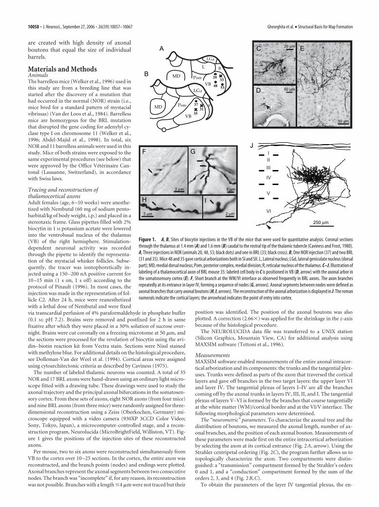
Figure 1. A, B, Sites of biocytin injections in the VB of the mice that were used for quantitative analysis. Coronal sectionsthrough the thalamus at 1.4 mm (A) and 1.6 mm (B) caudal to the rostral tip of the thalamic tubercle (Caviness and Frost, 1980).A, Three injections in NOR (animals 20, 48, 53; black dots) and one in BRL (33; black cross). B, One NOR injection (37) and two BRL(31 and 35). Mice 48 and 35 gave cortical arborizations both in SI and SII. L, Lateral nucleus; LGd, lateral geniculate nucleus (dorsalpart); MD, medial dorsal nucleus; Pom, posterior complex, medial division; R, reticular nucleus of the thalamus. C–J, Illustration oflabeling of a thalamocortical axon of BRL mouse 35: labeled cell body in C is positioned in VB (D, arrow) with the axonal arbor inthe somatosensory cortex (E). F, Short branch at the WM/VI interface as observed frequently in BRL axons. The axon branchesrepeatedly at its entrance in layer IV, forming a sequence of nodes (G, arrows). Axonal segments between nodes were defined asaxonal branches that carry axonal boutons (H, I, arrows). The reconstruction of the axonal arborization is displayed in J. The romannumerals indicate the cortical layers; the arrowhead indicates the point of entry into cortex.
10058 • J. Neurosci., September 27, 2006 • 26(39):10057–10067 Gheorghita et al. • Structural Basis for Map Formation

trance of the trunk in this layer was identified (Fig. 2 E, green crosses). Forthe V–VI plexus, the tangential oriented infragranular branches (Fig. 2 E,blue) were selected at the emergence of their trunk. The points at whichthe axonal trunks entered layer IV were further used to determine the“entrance surface” of the axon in layer IV (see below).
The “surface” parameters. These parameters are two-dimensional andrepresent the areal extent of the axon projection in a plane tangential tothe pial surface overlying the representation of the large mystacial whis-ker follicles (Woolsey and Van der Loos, 1970). In this plane, we deter-mined the entrance surface of the axon in layer IV (Fig. 2 F) and analyzedthe distribution of axonal boutons of the tangential plexuses in layer IVand layer VI using a grid consisting of 20 � 20 �m squares. In eachsquare, the density of boutons was calculated. The total surface area is thesum of squares in which one or more boutons were present. For addi-tional analysis, we used five density ranges: (0,1], (1,2], (2,4], (4,8], and(8,64].
StatisticsStatistical analyses were performed using the SAS software package (SASInstitute, Cary, NC) and according to SAS/STAT user’s guide, version 6.The distribution of parameters was tested for normality, and rank trans-formations were used if necessary. The differences were considered to bestatistically significant at p � 0.05.
The morphometric parameters were compared using a multivariateANOVA (SAS/STAT user’s guide, version 6; GLM procedure). A factoranalysis (FACTOR procedure) was used to obtain a global image of the
interdependencies between the parameters. Only factors with an eigen-value �1 were considered to well characterize the variability inside atangential plexus. A regression model was used (GLM and REG proce-dures) to study the hypothesis of homogeneity of slopes between strainsfor each layer and between layers inside each strain.
A � 2 test (FREQ procedure) was used to tests the differences betweenthe frequency distribution of the number of terminals reached after agiven number of nodes in NOR and BRL.
ResultsNeuronal labeling and subcortical trajectorySubcortical axonal trajectory is common in NOR and BRLBiocytin injections were made in the VB of the thalamus wherethe large mystacial whisker follicles are represented (Fig.1A,B,D). For both NOR and BRL, injections situated more dor-sally resulted in labeled cells whose axons gave arborization in SI,whereas those situated more ventrally, gave arborizations in SIand SII. The labeled axons were seen to arise directly from thesoma or from a dendrite close to its emergence from the soma.
The study of the hand-drawn TCAs from NOR (n � 35) and
Figure 3. Display of five thalamocortical axons in NOR and in BRL to illustrate the trajectoryfrom VB to the somatosensory cortex and maintenance of thalamic neighborhood relationshipin cortical termination in both strains. The NOR axons are represented in the left column; the BRLaxons are represented in the right column. In the top part, axons are displayed in a coronalplane, turned such that the radial orientation of the axonal arbor is vertical; in the bottom part,axons are displayed in a sagittal plane (insets to the left, indicate the orientation of the recon-structed axons). Boxes, Spatial relationship of the labeled neurons in the VB, as displayed in acoronal plane using the same color coding as for the axons. Filled arrowheads indicate entry inthe cortex; open arrowheads indicate the point where the axons leave the internal capsule (IC)to enter subcortical WM; line segments, the border between layers IV and V; all axons gave offcollaterals into the reticular thalamic nucleus (RTN). Note that despite the high divergence ofthe cortical termination in BRL, the cellular neighborhood in the thalamus was respected at thecortical level. In these reconstructions, the boutons are not displayed. The scale bar at thebottom pertains to NOR and BRL (coronal and sagittal).
Figure 2. Axonal subdivisions used for the morphometric analysis as illustrated for a BRLaxon. A, The intracortical arborization rendered yellow from the point where the axon enters thecortex (arrow). B, Transmission compartment (red) and conduction compartment (black). C,Topological Strahler ordering is centripetal, coding each terminal branch 0 and proceedingtoward the root of the branch. When two branches of the same order meet, the order number ofthe proximal branch is incremented by 1; otherwise, the largest order is used. The transmissioncompartment is formed by the Strahler’s orders 0 and 1 and contains most of the tangentialplexuses rich in boutons. The conduction compartment is the sum of Strahler’s orders 2, 3, and4, and corresponds to the axonal trunks. D, E, Green crosses in E define the axonal plexus in layerIV and above (D, green). The branches running tangentially in the infragranular layers wereselected at their emergence from the radial trunks and formed together the tangential plexus inlayers V–VI (D, blue). Some terminal branches of the V–VI plexus reached layer IV withoutgiving arborizations in this layer (D, arrowhead). F, The axonal trunk was turned in a tangentialposition; the marked points 1– 4 were unified in a convex polygon to calculate the area of entryinto layer IV. The insets in E and F indicate the orientation of the reconstructed axons.
Gheorghita et al. • Structural Basis for Map Formation J. Neurosci., September 27, 2006 • 26(39):10057–10067 • 10059
BRL (n � 17) showed that the subcorticaltrajectory is very similar in both strains(Fig. 3). Inside VB, the TCA change direc-tion several times at variable distancesfrom the cell body before arriving at theVB border. TCAs cross the thalamic retic-ular nucleus (RTN) parallel to each other.In this nucleus, the axons give off collater-als (1– 4 per axon; mean number, 1.9) thatform a dense plexus oriented perpendicu-lar to the trajectory of the labeled axons.Leaving the RTN, TCAs enter the internalcapsule in which axons follow divergingtrajectories through the striatum. Figure 3illustrates this divergence both in a coronaland sagittal plane. It shows that axonsfrom neurons that are close neighbors inVB do not maintain this proximity withintheir trajectory to the somatosensory cor-tex. On reaching the subcortical WM,some TCAs run for long distances at theborder with layer VI; other axons enter thecortex and travel tangentially for some dis-tance in layer VI. The cortical entrance andthe eventual axonal branching at theWM/VI interface are, in all cases, withinthe boundaries of the somatosensorycortex.
Using the set of computer-reconstructedaxons, we found that the subcortical trajec-tory of TCA of NOR axons has a mean lengthof 3.4 � 0.4 mm and is not different fromthat of BRL axons (3.7 � 0.4 mm).
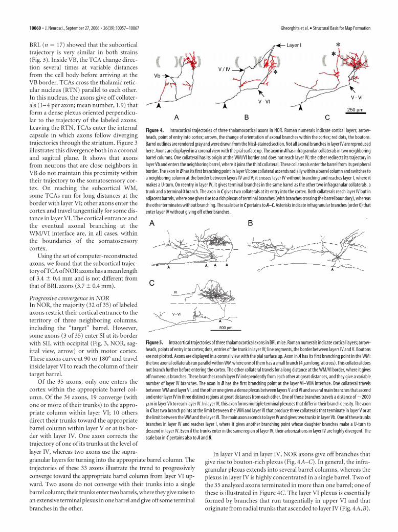
Progressive convergence in NORIn NOR, the majority (32 of 35) of labeledaxons restrict their cortical entrance to theterritory of three neighboring columns,including the “target” barrel. However,some axons (3 of 35) enter SI at its borderwith SII, with occipital (Fig. 3, NOR, sag-ittal view, arrow) or with motor cortex.These axons curve at 90 or 180° and travelinside layer VI to reach the column of theirtarget barrel.
Of the 35 axons, only one enters thecortex within the appropriate barrel col-umn. Of the 34 axons, 19 converge (withone or more of their trunks) to the appro-priate column within layer VI; 10 othersdirect their trunks toward the appropriatebarrel column within layer V or at its bor-der with layer IV. One axon corrects thetrajectory of one of its trunks at the level oflayer IV, whereas two axons use the supra-granular layers for turning into the appropriate barrel column. Thetrajectories of these 33 axons illustrate the trend to progressivelyconverge toward the appropriate barrel column from layer VI up-ward. Two axons do not converge with their trunks into a singlebarrel column; their trunks enter two barrels, where they give raise toan extensive terminal plexus in one barrel and give off some terminalbranches in the other.
In layer VI and in layer IV, NOR axons give off branches thatgive rise to bouton-rich plexus (Fig. 4A–C). In general, the infra-granular plexus extends into several barrel columns, whereas theplexus in layer IV is highly concentrated in a single barrel. Two ofthe 35 analyzed axons terminated in more than one barrel; one ofthese is illustrated in Figure 4C. The layer VI plexus is essentiallyformed by branches that run tangentially in upper VI and thatoriginate from radial trunks that ascended to layer IV (Fig. 4A,B).
Figure 4. Intracortical trajectories of three thalamocortical axons in NOR. Roman numerals indicate cortical layers; arrow-heads, point of entry into cortex; arrows, the change of orientation of axonal branches within the cortex; red dots, the boutons.Barrel outlines are rendered gray and were drawn from the Nissl-stained section. Not all axonal branches in layer IV are reproducedhere. Axons are displayed in a coronal view with the pial surface up. The axon in A has infragranular collaterals in two neighboringbarrel columns. One collateral has its origin at the WM/VI border and does not reach layer IV; the other redirects its trajectory inlayer Vb and enters the neighboring barrel, where it joins the third collateral. These collaterals enter the barrel from its peripheralborder. The axon in B has its first branching point in layer VI: one collateral ascends radially within a barrel column and switches toa neighboring column at the border between layers IV and V; it crosses layer IV without branching and reaches layer I, where itmakes a U-turn. On reentry in layer IV, it gives terminal branches in the same barrel as the other two infragranular collaterals, atrunk and a terminal 0 branch. The axon in C gives two collaterals at its entry into the cortex. Both collaterals reach layer IV but inadjacent barrels, where one gives rise to a rich plexus of terminal branches (with branches crossing the barrel boundary), whereasthe other terminates without branching. The scale bar in C pertains to A–C. Asterisks indicate infragranular branches (order 0) thatenter layer IV without giving off other branches.
Figure 5. Intracortical trajectories of three thalamocortical axons in BRL mice. Roman numerals indicate cortical layers; arrow-heads, points of entry into cortex; dots, entries of the trunk in layer IV; line segments, the border between layers IV and V. Boutonsare not plotted. Axons are displayed in a coronal view with the pial surface up. Axon in A has its first branching point in the WM:the two axonal collaterals run parallel within WM where one of them has a small branch (4 �m long; at cross). This collateral doesnot branch further before entering the cortex. The other collateral travels for a long distance at the WM/VI border, where it givesoff numerous branches. These branches reach layer IV independently from each other at great distances, and they give a variablenumber of layer IV branches. The axon in B has the first branching point at the layer VI–WM interface. One collateral travelsbetween WM and layer VI, and the other one gives a dense plexus between layers V and VI and several main branches that ascendand enter layer IV in three distinct regions at great distances from each other. One of these branches travels a distance of �2000�m in layer Vb to reach layer IV. In layer IV, this axon forms multiple terminal plexuses that differ in their branch density. The axonin C has two branch points at the limit between the WM and layer VI that produce three collaterals that terminate in layer V or atthe limit between the WM and the layer VI. The main axon ascends to layer IV and gives two trunks in layer Vb. One of these trunksbranches in layer IV and reaches layer I, where it gives another branching point whose daughter branches make a U-turn todescend in layer IV. Even if the trunks enter in the same region of layer IV, their arborizations in layer IV are highly divergent. Thescale bar in C pertains also to A and B.
10060 • J. Neurosci., September 27, 2006 • 26(39):10057–10067 Gheorghita et al. • Structural Basis for Map Formation
In two cases, we identified branches that arise from nodes at theWM/VI border climb in upper VI where they contribute to theinfragranular plexus (Fig. 4B).
Progressive divergence in BRLAt the cortical level, BRL axons are highly divergent (Fig. 3) anddo not enter SI within a three barrel column area as describedabove for NOR. Of the 17 hand-drawn BRL axons, 12 enter SIwith several, highly divergent, branches (Fig. 5A) that climb up tolayer IV. In addition to these long, bouton-bearing branches, wealso notice a large number of very short axonal branches (�4�m) that are formed by the axon at the level of the border be-tween the white matter and layer VI (Fig. 1F). However, fouraxons enter SI with a single trunk, but bifurcate in the infragranu-lar layers and give off branches that run �1.5 mm tangentiallyinside layer Vb, before entering layer IV (Fig. 5B). One axonapproaches layer IV with a single axonal trunk that bifurcates justbefore entering (Fig. 5C).
The number of branches by which a TCA enters layer IV inbarrelless is twice that of NOR [BRL, 5.4 � 2.2 (SD); NOR, 3.3 �1.4 (SD); p � 0.005]. In addition, the surface of entrance of thesebranches is 31 times larger than in NOR (Table 1, p � 0.001).Several of these branches are given off at the border betweenwhite matter and layer VI or during the intracortical trajectory ofthe axon. The result of this branching pattern is a progressivedivergence of the axonal arborization while reaching layer IV,which is in contrast with the increase convergence by which TCAapproaches layer IV in NOR.
As in NOR, TCA in barrelless give rise of axonal bouton-richplexuses in layers IV and VI. However, inside the layer IV plexusin barrelless, axonal branches seem not to encounter a spatialrestriction, which adds another factor to the total divergence ofthe axonal arbor.
Despite the divergence, in their cortical termination, BRL ax-ons conserve their topographic relationship as found in the thal-
amus (Fig. 3, BRL). An example is depicted in Figure 6. In thisBRL mouse, the biocytin injection was performed more ventrallyin the thalamus and TCA made branches in both SI and SII. Thebiocytin-labeled cell bodies were close together in VB. One axon,instead of reaching SI by traveling at the WM/VI interface, en-tered in the cortex at the SI/SII border and gave branches in layerVI of the SII. It subsequently traveled in layer III for �1500 �m toreach the same region in SI as its VB neighbor.
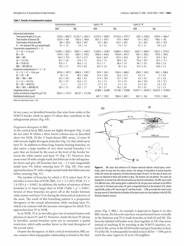
Table 1. Results of morphometric analysis
Intracortical arborization Layers I–IV Layers V–VI
NOR BRL NOR BRL NOR BRL
Intracortical arborizationTotal axonal length (L) in �m 17,022 � 8057.7 21,555.7 � 5632.1 12,274.5 � 5858.7 14,152.8 � 4777.7 2921 � 2190.2 3744.9 � 3036.7Total number of boutons (B) 1233.9 � 836 1336.3 � 590.6 1057.7 � 679.1 1112 � 464.9 175 � 207.2 198.3 � 192Total number of branches (BR) 209 � 102.3 196.4 � 40.7 167 � 80.3 154.1 � 38.2 33 � 28.5 28.5 � 19.9D � No. boutons/100 �m axonal length 6.7 � 1.9 5.9 � 1.4 8 � 2.2 7.9 � 1.3 4.7 � 2.1 3.9 � 2.5
Transmission compartment (0 � 1)LT � L0 � L1 in �m 13,940.3 � 6632.1 16581.7 � 5442.5 11,260.6 � 5328.9 12,809.6 � 4216.5 2624.7 � 2064.1 3362.3 � 2759.3B0 � B1 1195.9 � 812 1289.1 � 570.5 1026.4 � 656.6 1076.8 � 451.9 169.1 � 205.2 193.9 � 190.3BR0 � BR1 165.4 � 77.9 155.7 � 31.7 136.4 � 62.9 127.6 � 29.4 29.1 � 25.1 25.9 � 16.1L0 � L1/L (%) 81.7 � 3.8 75.6 � 7.3 91.9 � 1.5 90.9 � 2.9 91.6 � 10.7 92.5 � 11.1B0 � B1/B (%) 97.0 � 1 96.6 � 1.2 97.3 � 1 96.9 � 1.4 96.7 � 3.8 94.6 � 7.4BR0 � BR1/BR (%) 79.5 � 1.7 79.3 � 0.9 82.5 � 2.9 83 � 3.2 91.5 � 9.3 92.8 � 7.5
Conduction compartment (2 � 3 � 4)LC � L2 � L3 � L4 in �m 3019.6 � 1471.1 4988.4 � 1101.7 1014 � 542.1 1342.6 � 680.8 298.8 � 320.3 398.1 � 580.4B2 � B3 � B4 38 � 25 48.3 � 26.8 31.4 � 23.6 35.4 � 21.5 5.9 � 6 5 � 6.5BR2 � BR3 � BR4 43.3 � 24.1 40.6 � 9.3 31.4 � 18.4 25.7 � 10.7 3.9 � 4.4 2.7 � 2.8L2 � L3 � L4/L (%) 18.1 � 3.9 24.4 � 7.3 8.1 � 1.5 9.1 � 2.9 8.6 � 10.5 7.8 � 12.1B2 � B3 � B4/B (%) 3 � 1 3.5 � 1.1 2.7 � 1.0 3.1 � 1.4 3.3 � 3.8 2.4 � 2.9BR2 � BR3 � BR4/BR (%) 20.4 � 1.5 20.5 � 1.1 18 � 3.1 16.2 � 2.8 8.5 � 9.3 7.2 � 7.5
Tangential surfaces (�m2) 63,880 � 26,840 173,760 � 55,000 54,057.1 � 28,447.3 78,342.9 � 75,960.5Surface of entrance in layer IV (�m2) SE 2834.5 � 3172.1 88,127 � 111,554Mediolateral extent (�m) 441.7 � 129.5 1360.4 � 609.1 382.1 � 224 1119.5 � 548.3
Values are expressed as mean � SD.
Figure 6. BRL axons that arborize in SII. Roman numerals indicate cortical layers; arrow-heads, point of entry into the cortex; arrows, the change of orientation of axonal brancheswithin the cortex; line segments, the limit between layers IV and V. For the sake of clarity, oneaxon was depicted in black and another in gray. The boutons are not plotted. The axons aredisplayed in a coronal view with the pial surface up (inset for orientation). The BRL axons reachSI in different ways. After having given a collateral in SII, the gray axon continues in the WM tocurve into SI. The black axon enters SII, gives a tangential branch at the interface V/VI, climbssuperficially, makes a 90° turn in layer IV, and then travels �1500 �m inside the cortex to jointhe gray axon in SI. Note that the cell bodies of the above axons are close together in the VB. RTN,Reticular thalamic nucleus.
Gheorghita et al. • Structural Basis for Map Formation J. Neurosci., September 27, 2006 • 26(39):10057–10067 • 10061

Quantitative analysis of individual axonsAnalysis of the intracortical arborizationsTable 1 lists the quantitative parameters used to analyze the dif-ferences between NOR and BRL axons. On average, the intracor-tical arborizations of TCA in NOR mice have a length (L) of 17.0mm (SD, 8.1 mm), carry 1234 (SD, 836) boutons (B), and areformed by 209 (SD, 196) branches (BR). In the granular andsupragranular layers, these axons spread over a mean tangentialsurface (S4) of 0.083 mm 2 (SD, 0.026), the intracortical arboriza-tion of BRL axons have a tendency to be longer (21.5 � 5.6 mm)than those in NOR, but possesses the same mean number ofboutons (1336 � 590) and mean number of branches (196 � 41).The difference in length is mainly attributable to a 40% increaseof the conduction compartment in BRL. The tangential surfaceS4 in BRL is 0.17 mm 2 (SD, 0.055 mm 2), twice that in NOR ( p �0.002).
The differences between NOR and BRL axons were statisti-cally analyzed using a one-way multivariate analysis and a factoranalysis on the combination of the following five parameters: B
represents the total number of boutons of the entire intracorticalarbor (Fig. 2A, the yellow portion of the axon); LT representslength of the transmission compartment of the cortical arboriza-tion (all the branches of order 0 and 1 found mostly in the layersI–IV and V–VI) (Fig. 2B, the red portion of the axon); LC is thelength of the conduction compartment of the intracortical ar-borization (the axonal trunks plus all the branches of order 2, 3,and 4 inside the tangential plexuses) (Fig. 2B, blue); SE is theentrance surface of the axonal trunks in layer IV (Fig. 2F); S4 isthe tangential surface of the axon inside layers I–IV.
LC and SE parameters characterize the part of the axon bywhich it reaches layer IV. The parameters V, S4, and LT charac-terize the tangential plexuses where most of the boutons and the0 and 1 order branches are found.
A one-way multivariate analysis on the combination of thefive parameters LT, LC, B, S4, and SE demonstrated an important“strain effect” ( p � 0.009). To analyze the interdependencies ofthe five parameters, a factor analysis was performed on NOR andBRL axons as a single sample. It demonstrated that in both strainsindividual TCAs are characterized by (only) two factors. Figure7A indicates how the five parameters correlate to these two fac-tors in NOR and in BRL. For NOR axons, a single factor (“factor1”) was sufficient to explain 72% of the variability with an eigen-value of 3.63. This factor comprises four of the parameters, exceptSE that was found to be part of second factor (“factor 2”) respon-sible for only 18% of the variability and which had no statisticalsignificance (eigenvalue �1). In BRL axons, factor 1 covered only56% of the variability and the analysis identified a significantcontribution of factor 2 comprising 27% of the variability with aneigenvalue of 1.38. For these axons, factor 1 highly correlatedwith B, LT, and S4, and factor 2 correlated with LC and SE. Thismultivariate analysis therefore indicates that the strain differencebetween TCA of NOR and BRL is expressed by two parameters,LC and SE, whereas other parameters seem to have been unaf-
Figure 7. A, Table showing the correlations of factor 1 and factor 2 with the five parametersLT, LC, B, S4, and SE. Significant correlations are in gray; the eigenvalues and the proportion ofthe variability explained by each factor are given at the bottom. Factors were determined forNOR and BRL samples separately. LT, Length of the transmission compartment of the wholeintracortical arborization; LC, length of the conduction compartment of the whole intracorticalarborization; B, number of boutons of the whole intracortical arborization; S4, total tangentialextent of the axons in layers I–IV; SE, surface of entrance of the axonal trunks in layer IV. B,Graphical display of the value of factors 1 and 2 for each individual axon. Line segments indicatethe level at which factor 2 segregates the axons from NOR mice (open dots) and the BRL axons(black dots). Statistical analysis proved this segregation to be significant ( p � 0.001).
Figure 8. Frequency distribution of the number of terminal branches reached by a hypothet-ical action potential after traversing a given number of nodes within the intracortical arboriza-tion of NOR and BRL axons. The two distributions are of similar shape, but the BRL distributionis significantly shifted to the left (�2 test, p � 0.001). As a consequence, in BRL axons, actionpotentials will reach the terminal axonal branches by traversing a smaller number ofbifurcations.
10062 • J. Neurosci., September 27, 2006 • 26(39):10057–10067 Gheorghita et al. • Structural Basis for Map Formation

fected by the mutation. Figure 7B is a graphical representation ofthe distribution of these two factors and demonstrates how factor2 segregates the group of BRL axons from those of NOR.
Because LC represents the length of the conduction compart-ment, we analyzed whether the strain difference for this parame-ter also affects the geometrical complexity of the thalamocorticalaxon. For this, we determined for each terminal branch (order 0)the number of nodes (axonal bifurcations) that an action poten-tial has to pass before reaching it from its entrance in the cortex.Subsequently, terminal branches were classed according to thenumber of nodes, and Figure 8 displays their frequency distribu-tion. Statistical analysis identified that this distribution is signif-icantly different between the two strains: terminal branches inBRL axons are reached by passing fewer nodes than in NOR (� 2
test, p � 0.001). This shows that thalamocortical axons in BRLhave a longer conduction compartment (LC) but have a simplerbranching pattern than those in NOR.
We further analyzed the strain difference in the geometry ofthe TCAs by testing the correlation between the branch numberand the size of their tangential spread. As shown in Figure 9A, inboth strains a significant linear correlation between the two pa-rameters is found. However, the correlation is significantly dif-ferent between the two strains ( p � 0.005): compared with NOR,the same variation in branch number produces in BRL a signifi-cantly larger increase in tangential spread in layer IV.
The multivariate statistical analyses described above identifiesthat the BRL mutation affects the parameters characterizing theconduction compartment of the thalamocortical axon as well asthe geometry of the axonal arborization. Therefore, some of theresults of the univariate analysis are worth mentioning (Table 1).
The total length of the conduction compartment in BRL was1.6 times longer as in NOR (Table 1, p � 0.02), and its surface ofentrance was 31-fold greater (Table 1, p � 0.001). No statisticaldifferences were found for the transmission compartment (Table1). Significant strain differences were detected for the extent ofthe tangential surface (S4), which was twice as large in BRL as inNOR ( p � 0.002). Also, the mean mediolateral extent of theaxons, as determined in the coronal plane, showed a significantdifference by being three times larger in BRL than in NOR (BRL,1.3 mm; NOR, 0.4 mm; p � 0.002).
Also, the correlation function between the tangential surfaceof the axonal plexus and the axonal length is significantly differ-ent between the strains (Fig. 9B,C). This correlation function is,
in both strains, significantly different be-tween the plexus in layers I–IV and that inlayer V–VI: for NOR, p � 0.01; and forBRL, p � 0.008.
Distribution of bouton densities areas inlayers I–IVAs mentioned above, by using the sameaxonal length, number of boutons, and ax-onal branches, BRL axons made surfacestwice as large as those of the NOR axons.Therefore, the density of boutons per unitof tangential surface was greater for NORaxons than for BRL axons ( p value formean density analysis). In this final part ofthe analysis, we studied the distribution ofaxonal boutons within the plexuses inlayer IV and in layer VI. For this, we calcu-lated the density of boutons per 20 � 20�m area of cortical surface and analyzedthe distribution within a series of density
ranges. Figure 10 displays the density distribution for three NORand three BRL axons as well as the relative distribution of thedensity classes per strain. In NOR, the TCA distributes axonalboutons in a restricted and compact part of layer IV, with a largearea of high bouton density range situated in the center of theaxonal plexus. In BRL, the total cortical area receiving TCA bou-tons in layer IV is larger, but the area covered by high boutondensity is distributed throughout the plexus, not forming a com-pact area.
Performing a one-way univariate statistical ANOVA on the ab-solute values of the density areas inside layer IV confirmed the straindifference and showed that, in BRL, the surface with the lowestbouton density (0,1] was more than twice as large as in NOR ( p �0.001) and formed 33% of the total surface in NOR and 43% inBRL (Fig. 10G–I). Surfaces of intermediate density (1,2], (2,4],and (4,8] were approximately twice as large in BRL than in NOR( p � 0.001; p � 0.006; p � 0.01), and each of them represented14 –20% of the total tangential surface in both NOR and BRL. Asmaller part of the total tangential extent of the axons was occu-pied by surfaces with a high bouton density (8,64] (20% in NORvs 6% in BRL). This analysis revealed a disproportionate distri-bution of axonal boutons in the layer IV tangential plexus of BRLaxons that resulted in a twofold larger area of low and mediumdensities as compared with NOR. The high densities (8,64] forma compartment that in absolute value was of similar size in bothstrains.
Performing the same statistical analysis on the density areasformed by the boutons of the plexus in layers V–VI showed nosignificant differences between strains (Fig. 10 J–L).
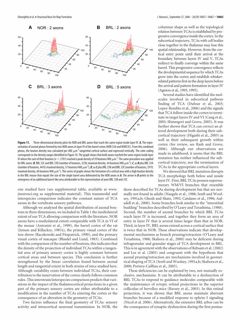
Realizing that cortical compartmentalization is not attribut-able to a single TCA, but several hundreds, we analyzed the dis-tribution of boutons formed by a pair of reconstructed axons thathave their cell bodies close to each other in VB. Figure 11A de-picts two NOR axons that reached the same cortical barrel andthat together formed a maximum density peak of 39 boutons/400�m 2. In BRL, the two axons tended to form their high-densitysurfaces in the same cortical region (Fig. 11B) and reached amaximal density that was similar with those formed by NORaxons. In BRL, this area of high density is surrounded by a largearea of low density. Adding a third BRL axon to this analysisresulted in a even more pronounced delineation of a high-densityarea (Fig. 11C) that is only slightly larger in its tangential dimen-
Figure 9. Correlations between the parameters characterizing NOR and BRL axons. A, The tangential extent of NOR and BRLaxons inside layer IV is plotted as a function of their total number of intracortical branches (i.e., axonal segments betweenconsecutive nodes). Data on NOR axons are indicated by dots; BRL axons, by crosses. In both strains, the two parameters aresignificantly correlated ( p � 0.009 for both NOR and BRL), but for the same number of branches BRL axons span a significantlylarger cortical surface than NOR axons (strain effect, p � 0.001). For the same increase in the number of branches, BRL axonsincreased their tangential surfaces more than NOR axons (analysis on heterogeneity of the slopes of the regression lines of NOR andBRL, p � 0.005). The tangential extent of NOR (B) and BRL (C) axons inside layer IV or VI is plotted as a function of the total axonallength in these layers. For the same increase of total axonal length, NOR axons increased their tangential surface in the infragranu-lar layers more than inside layer IV (layer and length effect or comparison between NOR I–IV and NOR V–VI slopes, p � 0.01). Thisslope difference also exists in BRL ( p � 0.008).
Gheorghita et al. • Structural Basis for Map Formation J. Neurosci., September 27, 2006 • 26(39):10057–10067 • 10063

sion than the barrel-shaped area in NOR.The addition of the bouton distribution oftwo additional TCAs shows the emergenceof a second area of high density (Fig. 11D).From these cumulative histograms, weconclude that in barrelless the high-density domains are at the basis of a topo-graphic representation of the whiskerfollicles.
DiscussionTo elucidate the possible structural basisunderlying the barrelless phenotype, wereconstructed TCAs in NOR and BRLmice. Comparing the population of hand-drawn axons with the morphometricaldata from the three-dimensional (3D)-reconstructed axons confirms that theconclusions drawn from the quantitativeanalysis are representative for the totalsample of labeled TCAs. These conclu-sions are the following: (1) The subcorticaltrajectory of BRL axons is the same as ofNOR axons. TCAs in both strains have thesame total axonal length, bear the samenumber of branches and axonal boutons.(2) BRL axons possess branches at the in-terface between white matter and layer VI,that were not observed in NOR. (3) Thenumber of branches by which TCA axonsin BRL reach layer IV is increased in com-parison to NOR; also, the areal surfacecovered by these entering branches islarger in BRL than in NOR. (4) The com-parison of the geometry between the twopopulations of TCA revealed that an ac-tion potential in BRL axons has to cross asmaller number of axonal bifurcations be-fore reaching the terminal branches. (5)Within layer IV, BRL axons distribute ax-onal boutons over a larger cortical area,forming a terminal plexus in which thebouton density is lower when comparedwith the compact, high bouton dense ter-minal plexus invariably formed by NORaxons. However, NOR and BRL axonshave the same high-density area of axonalboutons, and in both strains, axons withthe cell bodies close together in the thala-mus made their high-density areas in thesame cortical region.
A number of the morphometric pa-rameters (total length, number ofbranches, and axonal boutons) are indis-tinguishable between the two strains stud-ied here and, interestingly, have an almostidentical value for TCAs terminating in theprimary visual cortex of the mouse (Anto-nini et al., 1999). TCA axons in the pri-mary visual cortex of the tree shrew (Rac-zkowski and Fitzpatrick, 1990) andmacaque (Blasdel and Lund, 1983) bearcomparable numbers of boutons as the ax-
Figure 10. Two-dimensional density plots illustrating tangential distribution of the boutons of NOR and BRL axons within layer IV andVI. For individual axons, the density of boutons was determined separately for the plexus in layer IV and layer VI by calculating the numberofboutonsper400�m 2 areaoftangentialcorticalsurface.Distributionsaredisplayedonagridwithalinedistanceof200�200�m.Thecolor bar gives the coding used for the five density ranges applied. AIV–CIV, Bouton distribution in layer IV of three NOR axons; DIV–FIV,distribution in layer IV of three BRL axons; AVI–CVI, distribution of boutons in layer VI of the NOR axons displayed above; DVI–FVI,distribution in layer VI of the three BRL axons. The pie charts (G, H, J, K ) represent the mean percentages of each of the five density rangesas calculated for NOR and BRL plexuses separately. The mean absolute values of the surface areas in 100 �m 2 are given in parentheses.These latter values are displayed in the bar graphs (I, L) allowing comparison between strains; the asterisks indicate the significantdifferences (one-way univariate analysis; p � 0.05). Error bars indicate SD.
10064 • J. Neurosci., September 27, 2006 • 26(39):10057–10067 Gheorghita et al. • Structural Basis for Map Formation
ons studied here (see supplemental table, available at www.jneurosci.org as supplemental material). This transmodal andinterspecies comparison indicates the constant nature of TCAaxons in the vertebrate sensory pathways.
Although we analyzed the spatial distribution of axonal bou-tons in three dimensions, we included in Table 1 the mediolateralextent of our TCA allowing comparison with the literature. NORaxons have a mediolateral extent comparable with TCA in VI ofthe mouse (Antonini et al., 1999), the barrel cortex of the rat(Jensen and Killackey, 1987a), the primary visual cortex of thetree shrew (Raczkowski and Fitzpatrick, 1990), and the primaryvisual cortex of macaque (Blasdel and Lund, 1983). Combinedwith the comparison of the number of boutons, this indicates thatthe density of the projection of individual TCAs within a tangen-tial area of primary sensory cortex is highly constant betweencortical areas and between species. This conclusion is furtherstrengthened by the linear correlation found between axonallength and tangential extent in both strains of mice studied here.Although variability exists between individual TCAs, their con-tribution to the innervation of the cortex clearly follows commonrules. This interstrain/interspecies comparison predicts that vari-ations in the impact of the thalamocortical projections in a givenpart of the primary sensory cortex are either attributable to amodification in the number of TCAs entering the cortex or theconsequence of an alteration in the geometry of TCAs.
Two factors influence the final geometry of TCAs: sensoryactivity and intracortical neuronal transmission. In NOR, the
columnar shape as well as the topologicalrelation between TCAs is established by pro-gressive convergence inside the cortex. In thesubcortical trajectory, TCAs with cell bodiesclose together in the thalamus may lose thisspatial relationship. However, from the cor-tical entry point until their arrival at theboundary between layers IV and V, TCAsredirect to finally converge within the samebarrel. This progressive convergence reflectsthe developmental sequence by which TCAsgrow into the cortex and establish whisker-related patterns first in the deep layers beforethe arrival and pattern formation in layer IV(Agmon et al., 1993, 1995).
Several studies have identified the mol-ecules involved in subcortical pathwayfinding of TCA (Dufour et al., 2003;Lopez-Bendito et al., 2006) and the signalsthat TCA follow inside the cortex to termi-nate in target layers IV and VI (Cang et al.,2005; Shimogori and Grove, 2005). It wasfurther shown that TCA can correct an al-tered development both during their sub-cortical trajectory (Higashi et al., 2005) aswell as their subsequent growth withincortex (for review, see Rash and Grove,2006). Although our observations aremade in adulthood, it seems that the BRLmutation has neither influenced the sub-cortical trajectory, nor the termination ofTCAs in the appropriate cortical layers.
We showed that BRL mutation disruptsTCA morphology both below and insidelayer IV. First, BRL TCAs possess supernu-merary WM/VI branches that resemble
those described for TCAs during development but that are nor-mally not found in adults (Naegele et al., 1988; Senft and Wool-sey, 1991a,b; Ghosh and Shatz, 1992; Catalano et al., 1996; Aul-adell et al., 2000). Some branches look similar to the “interstitialbudding” branches described by O’Leary and Terashima, (1988).Second, the number of axonal branches by which BRL TCAsreach layer IV is increased, and together they form an area ofentry in layer IV that is several times larger than that in NOR.Third, in layer IV, BRL axons extend across a cortical surface thatis twice that in NOR. These observations indicate that develop-mental mechanisms as branch pruning/retraction (O’Leary andTerashima, 1988; Skaliora et al., 2000) may be deficient duringinfragranular and granular stages of TCA development in BRL.This is in agreement with the observations of Rebsam et al. (2002)and Lee et al. (2005) and congruent with the hypothesis thataxonal pruning/retraction are mechanisms involved in geomet-rical shaping of TCA (Senft and Woolsey, 1991a,b; Skaliora et al.,2000; Portera-Cailliau et al., 2005).
These deficiencies can be explained by two, not mutually ex-clusive, mechanisms. It can be attributable to a dysfunction ofBRL TCAs to respond to guidance molecules comparable withthe maintenance of ectopic retinal projections in the superiorcolliculus of barrelless mice (Ravary et al., 2003). In this retinalprojection, it was shown that BRL axons maintain aberrantbranches because of a modified response to ephrin-5 signaling(Nicol et al. 2006). Alternatively, the extensive BRL arbor can bethe consequence of synaptic dysfunction during the first postna-
Figure 11. Three-dimensional density plots for NOR and BRL axons that reach the same region inside layer IV. A, The repre-sentation of axonal plexus formed by two NOR axons in layer IV of the barrel cortex (NOR 53A and NOR53C). From this combinedplexus, the bouton density was calculated per 400 �m 2 tangential cortical surface and expressed vertically. The color codingcorresponds to the density ranges identified in Figure 10. The graph shows that both axons reached the same region inside layerIV where the sum of their boutons (n � 2701) reached a peak density of 39 boutons/400 �m 2. The same procedure was appliedfor BRL axons: B, BRL 33C and BRL 33D (number of boutons, 3238; maximal density, 44 boutons/400 �m 2); C, as B plus BRL 33A(number of boutons, 4410; maximal density, 57 boutons/400 �m 2); D, as C plus BRL 33B and BRL 33E (number of boutons, 5919;maximal density, 60 boutons/400 �m 2). This series of graphs shows the formation of a cortical area with a high bouton densityin the BRL mouse that equals the size of the single barrel area delineated by the NOR axons in A. The arrow in D points to theemergence of an additional barrel-like area attributable to the representation of axon BRL 33B and 33E.
Gheorghita et al. • Structural Basis for Map Formation J. Neurosci., September 27, 2006 • 26(39):10057–10067 • 10065

tal week when the induction of long-term potentiation wasshown to be impaired in the BRL mutant (Lu et al., 2003), ahypothesis that further underlines the role of activity-dependentsynaptic plasticity in shaping TCAs (Erzurumlu and Kind, 2001).
In layer IV, a TCA in NOR creates a compact terminal plexuswith the size of a barrel, 20% of which is characterized by an areaof high density of thalamocortical (TC) boutons. In BRL, thelayer IV plexus has an area comparable with up to 2.5 barrels andonly 6% of this area is characterized by a high density of TCboutons. The high degree of divergence of TCA termination inlayer IV is the basis for the lack of segregation of whisker input toindividual layer IV neurons in BRL strain (Welker et al., 1996).The total areal extent of the high-density domains in BRL andNOR are similar, indicating that this aspect of the TCA plexustends to be resistant to the genetic variation. A similar result hasbeen described by Antonini and Stryker (1993), who demon-strated that decreasing sensory activation in the visual pathwaydid not alter the high-density area of geniculocortical axonseither.
However, the current study reveals that, in BRL, TCAs ofneighboring thalamic neurons form a compact area of high-density innervation that has the tangential size of a single barrel.We propose that this high-density area underlies the topologicparcellation of SI in barrelless mice shown previously (Welker etal., 1996). This implies that not all parts of the layer IV plexus ofan individual TCA contribute in equal manner to the establish-ment of a functional map in SI. This notion is in harmony withthe Gaussian distribution of TCA boutons in SI of the monkeycortex (Garraghty and Sur, 1990), where the peak of the curvecoincides with the corresponding part of the sensory periphery.Enlargement of the functional representation during adult plas-ticity may therefore be the consequence of a proportional in-crease in the activation of the part of the TCA termination thathas a low bouton density (Garraghty et al., 1989; Garraghty andSur, 1990; Rausell and Jones, 1995; Jones and Pons, 1998).
In conclusion, we propose that developmental mechanismsthat shape thalamocortical axons before their arrival in corticallayer IV are deficient in the BRL mouse, but that thalamic neigh-borhood relationships are maintained in the cortical projection,in which the spatial distribution of TC boutons underlie the es-tablishment of a functional map of the mystacial whisker follicles.
ReferencesAbdel-Majid RM, Leong WL, Schalkwyk LC, Smallman DS, Wong ST, Storm
DR, Fine A, Dobson MJ, Guernsey DL, Neumann PE (1998) Loss ofadenylyl cyclase I activity disrupts patterning of mouse somatosensorycortex. Nat Genet 19:289 –291.
Adams NC, Lozsadi DA, Guillery RW (1997) Complexities in the thalamo-cortical and corticothalamic pathways. Eur J Neurosci 9:204 –209.
Agmon A, Yang LT, O’Dowd DK, Jones EG (1993) Organized growth ofthalamocortical axons from the deep tier of terminations into layer IV ofdeveloping mouse barrel cortex. J Neurosci 13:5365–5382.
Agmon A, Yang LT, Jones EG, O’Dowd DK (1995) Topological precision inthe thalamic projection to neonatal mouse barrel cortex. J Neurosci15:549 –561.
Antonini A, Stryker MP (1993) Development of individual geniculocorticalarbors in cat striate cortex and effects of binocular impulse blockade.J Neurosci 13:3549 –3573.
Antonini A, Gillespie DC, Crair MC, Stryker MP (1998) Morphology ofsingle geniculocortical afferents and functional recovery of the visual cor-tex after reverse monocular deprivation in the kitten. J Neurosci18:9896 –9909.
Antonini A, Fagiolini M, Stryker MP (1999) Anatomical correlates of func-tional plasticity in mouse visual cortex. J Neurosci 19:4388 – 4406.
Auladell C, Perez-Sust P, Super H, Soriano E (2000) The early development
of thalamocortical and corticothalamic projections in the mouse. AnatEmbryol (Berl) 201:169 –179.
Blasdel GG, Lund JS (1983) Termination of afferent axons in macaque stri-ate cortex. J Neurosci 3:1389 –1413.
Cang J, Renteria RC, Kaneko M, Liu X, Copenhagen DR, Stryker MP (2005)Development of precise maps in visual cortex requires patterned sponta-neous activity in the retina. Neuron 48:797– 809.
Cases O, Vitalis T, Seif I, De Maeyer E, Sotelo C, Gaspar P (1996) Lack ofbarrels in the somatosensory cortex of monoamine oxidase A-deficientmice: role of a serotonin excess during the critical period. Neuron16:297–307.
Catalano SM, Robertson RT, Killackey HP (1996) Individual axon mor-phology and thalamocortical topography in developing rat somatosen-sory cortex. J Comp Neurol 367:36 –53.
Caviness Jr VS (1975) Architectonic map of neocortex of the normal mouse.J Comp Neurol 164:247–263.
Caviness Jr VS, Frost DO (1980) Tangential organization of thalamic pro-jections to the neocortex in the mouse. J Comp Neurol 194:335–367.
Dolleman-Van der Weel MJ, Wouterlood FG, Witter MP (1994) Multipleanterograde tracing, combining Phaseolus vulgaris leucoagglutinin withrhodamine- and biotin-conjugated dextran amine. J Neurosci Methods51:9 –21.
Dufour A, Seibt J, Passante L, Depaepe V, Ciossek T, Frisen J, Kullander K,Flanagan JG, Polleux F, Vanderhaeghen P (2003) Area specificity andtopography of thalamocortical projections are controlled by ephrin/Ephgenes. Neuron 39:453– 465.
Erzurumlu RS, Kind PC (2001) Neural activity: sculptor of “barrels” in theneocortex. Trends Neurosci 24:589 –595.
Garraghty PE, Sur M (1990) Morphology of single intracellularly stainedaxons terminating in area 3b of macaque monkeys. J Comp Neurol294:583–593.
Garraghty PE, Pons TP, Sur M, Kaas JH (1989) The arbors of axons termi-nating in middle cortical layers of somatosensory area 3b in owl monkeys.Somatosens Mot Res 6:401– 411.
Ghosh A, Shatz CJ (1992) Pathfinding and target selection by developinggeniculocortical axons. J Neurosci 12:39 –55.
Hannan AJ, Blakemore C, Katsnelson A, Vitalis T, Huber KM, Bear M, RoderJ, Kim D, Shin HS, Kind PC (2001) PLC-beta 1, activated via mGluRs,mediates activity-dependent differentiation in cerebral cortex. Nat Neu-rosci 4:282–288.
Higashi S, Hioki K, Kurotani T, Kasim N, Molnar Z (2005) Functionalthalamocortical synapse reorganization from subplate to layer IV duringpostnatal development in the reeler-like mutant rat (shaking rat Ka-wasaki). J Neurosci 25:1395–1406.
Humphrey AL, Sur M, Uhlrich DJ, Sherman SM (1985) Projection patternsof individual X- and Y-cell axons from the lateral geniculate nucleus tocortical area 17 in the cat. J Comp Neurol 233:159 –189.
Iwasato T, Datwani A, Wolf AM, Nishiyama H, Taguchi Y, Tonegawa S,Knopfel T, Erzurumlu RS, Itohara S (2000) Cortex-restricted disruptionof NMDAR1 impairs neuronal patterns in the barrel cortex. Nature406:726 –731.
Jensen KF, Killackey HP (1987a) Terminal arbors of axons projecting to thesomatosensory cortex of the adult rat. I. The normal morphology of spe-cific thalamocortical afferents. J Neurosci 7:3529 –3543.
Jensen KF, Killackey HP (1987b) Terminal arbors of axons projecting to thesomatosensory cortex of the adult rat. II. The altered morphology ofthalamocortical afferents following neonatal infraorbital nerve cut. J Neu-rosci 7:3544 –3553.
Jones EG (1985) Principle of thalamic organization. In: The thalamus, pp100 –113. New York: Plenum.
Jones EG (1986) Connectivity of the primate sensory-motor cortex. In: Ce-rebral cortex, Vol 5, Sensory-motor areas and aspects of cortical connec-tivity (Jones EG, Peters Alan, eds), pp 127–147. New York: Plenum.
Jones EG, Pons TP (1998) Thalamic and brainstem contributions to large-scale plasticity of primate somatosensory cortex. Science 282:1121–1125.
Lee LJ, Iwasato T, Itohara S, Erzurumlu RS (2005) Exuberant thalamocor-tical axon arborization in cortex-specific NMDAR1 knockout mice.J Comp Neurol 485:280 –292.
Lopez-Bendito G, Cautinat A, Sanchez JA, Bielle F, Flames N, Garratt AN,Talmage DA, Role LW, Charnay P, Marin O, Garel S (2006) Tangentialneuronal migration controls axon guidance: a role for neuregulin-1 inthalamocortical axon navigation. Cell 125:127–142.
10066 • J. Neurosci., September 27, 2006 • 26(39):10057–10067 Gheorghita et al. • Structural Basis for Map Formation
Lu HC, She WC, Plas DT, Neumann PE, Janz R, Crair MC (2003) Adenylylcyclase I regulates AMPA receptor trafficking during mouse cortical “bar-rel” map development. Nat Neurosci 6:939 –947.
Maier DL, Mani S, Donovan SL, Soppet D, Tessarollo L, McCasland JS, MeiriKF (1999) Disrupted cortical map and absence of cortical barrels ingrowth-associated protein (GAP)-43 knockout mice. Proc Natl Acad SciUSA 96:9397–9402.
McIlvain VA, Robertson DR, Maimone MM, McCasland JS (2003) Abnor-mal thalamocortical pathfinding and terminal arbors lead to enlargedbarrels in neonatal GAP-43 heterozygous mice. J Comp Neurol462:252–264.
Naegele JR, Jhaveri S, Schneider GE (1988) Sharpening of topographicalprojections and maturation of geniculocortical axon arbors in the ham-ster. J Comp Neurol 277:593– 607.
Nicol X, Muzerelle A, Rio JP, Metin C, Gaspar P (2006) Requirement ofadenylate cyclase 1 for the ephrin-A5-dependent retraction of exuberantretinal axons. J Neurosci 26:862– 872.
O’Leary DD, Terashima T (1988) Cortical axons branch to multiple subcor-tical targets by interstitial axon budding: implications for target recogni-tion and “waiting periods.” Neuron 1:901–910.
Pinault D (1996) A novel single-cell staining procedure performed in vivounder electrophysiological control: morpho-functional features of juxta-cellularly labeled thalamic cells and other central neurons with biocytin orNeurobiotin. J Neurosci Methods 65:113–136.
Portera-Cailliau C, Weimer RM, De Paola V, Caroni P, Svoboda K (2005)Diverse modes of axon elaboration in the developing neocortex. PLoSBiol 3:e272.
Raczkowski D, Fitzpatrick D (1990) Terminal arbors of individual, physio-logically identified geniculocortical axons in the tree shrew’s striate cor-tex. J Comp Neurol 302:500 –514.
Rash BG, Grove EA (2006) Area and layer patterning in the developing ce-rebral cortex. Curr Opin Neurobiol 16:25–34.
Rausell E, Jones EG (1995) Extent of intracortical arborization of thalamo-cortical axons as a determinant of representational plasticity in monkeysomatic sensory cortex. J Neurosci 15:4270 – 4288.
Ravary A, Muzerelle A, Herve D, Pascoli V, Ba-Charvet KN, Girault JA,
Welker E, Gaspar P (2003) Adenylate cyclase 1 as a key actor in therefinement of retinal projection maps. J Neurosci 23:2228 –2238.
Rebsam A, Seif I, Gaspar P (2002) Refinement of thalamocortical arbors andemergence of barrel domains in the primary somatosensory cortex: astudy of normal and monoamine oxidase a knock-out mice. J Neurosci22:8541– 8552.
Senft SL, Woolsey TA (1991a) Growth of thalamic afferents into mousebarrel cortex. Cereb Cortex 1:308 –335.
Senft SL, Woolsey TA (1991b) Computer-aided analyses of thalamocorticalafferent ingrowth. Cereb Cortex 1:336 –347.
Shimogori T, Grove EA (2005) Fibroblast growth factor 8 regulates neocor-tical guidance of area-specific thalamic innervation. J Neurosci25:6550 – 6560.
Skaliora I, Adams R, Blakemore C (2000) Morphology and growth patternsof developing thalamocortical axons. J Neurosci 20:3650 –3662.
Tettoni L, Lehmann P, Houzel JC, Innocenti GM (1996) Maxsim, softwarefor the analysis of multiple axonal arbors and their simulated activation.J Neurosci Methods 67:1–9.
Van der Loos H, Dorfl J, Welker E (1984) Variation in pattern of mystacialvibrissae in mice. A quantitative study of ICR stock and several inbredstrains. J Hered 75:326 –336.
Welker E, Armstrong-James M, Bronchti G, Ourednik W, Gheorghita-Baechler F, Dubois R, Guernsey DL, Van der Loos H, Neumann PE(1996) Altered sensory processing in the somatosensory cortex of themouse mutant barrelless. Science 271:1864 –1867.
White EL, Weinfeld E, Lev DL (2004) Quantitative analysis of synaptic dis-tribution along thalamocortical axons in adult mouse barrels. J CompNeurol 479:56 – 69.
Woolsey TA, Van der Loos H (1970) The structural organization of layer IVin the somatosensory region (SI) of mouse cerebral cortex. The descrip-tion of a cortical field composed of discrete cytoarchitectonic units. BrainRes 17:205–242.
Yang Z, Seif I, Armstrong-James M (2001) Differences in somatosensoryprocessing in S1 barrel cortex between normal and monoamine oxidase Aknockout (Tg8) adult mice. Cereb Cortex 11:26 –36.
Gheorghita et al. • Structural Basis for Map Formation J. Neurosci., September 27, 2006 • 26(39):10057–10067 • 10067


![Disrupted Cities [Stephen Graham]](https://img.dokumen.tips/doc/110x75/55cf97f4550346d03394a6f7/disrupted-cities-stephen-graham.jpg)