Embed Size (px)
Citation preview

Structural Studies of Detergent-Solubilized and Vesicle-Reconstituted Low-DensityLipoprotein (LDL) Receptor†
Kumkum Saxena‡ and G. Graham Shipley*
Departments of Biophysics and Biochemistry, Center for AdVanced Biomedical Research,Boston UniVersity School of Medicine, Boston, Massachusetts 02118
ReceiVed June 30, 1997; ReVised Manuscript ReceiVed October 20, 1997X
ABSTRACT: The low-density lipoprotein (LDL) receptor plays a key role in maintaining circulating andcellular cholesterol homeostasis. The LDL receptor is a transmembrane glycoprotein whose biochemicaland genetic properties have been extensively studied notably by Brown, Goldstein and colleagues [Brown,M. S., & Goldstein, J. L., (1986)Science 232, 34-47]. However, few if any structural studies of theLDL receptor have been reported, and details of its secondary and tertiary structure are lacking. In anattempt to determine the low-resolution structure of the LDL receptor, we have purified the receptor frombovine adrenal cortices using modifications of the method of Schneider et al. [Schneider, W. J., Goldstein,J. L., & Brown, M. S. (1985)Methods in Enzymol. 109, 405-417]. Using circular dichroism, the secondarystructure of the detergent-solubilized bovine LDL receptor at 25°C was shown to be 19%R-helix, 42%â-sheet, and 39% random coil. Interestingly, the detergent-solubilized receptor appeared to be quite resistantto changes in secondary structure over the temperature range 10-90 °C, with only minor but reversiblechanges being observed. In contrast, a more pronounced unfolding of the detergent-solubilized receptorwas observed in the presence of guanidinium hydrochloride. Using the complete sequence of the humanLDL receptor, sequence analysis by the Chou-Fasman prediction algorithm showed quite good agreementwith the experimentally determined secondary structure of the bovine LDL receptor at 25°C. Finally,the purified, bovine LDL receptor was reconstituted into large unilamellar vesicles of egg yolkphosphatidylcholine using a procedure exploiting preformed vesicles and detergent dialysis. We showedpreviously using negative stain electron microscopy that reconstituted vesicles bind LDL. Now, usingcryoelectron microscopy of frozen hydrated reconstituted vesicles evidence of an extended, stick-likemorphology (length∼120 Å) for the extracellular domain of the LDL receptor has been obtained.Successful purification of the receptor, its incorporation into single bilayer vesicles, and its directvisualization by cryoelectron microscopy pave the way for more detailed structural studies of the LDLreceptor and the receptor-LDL complex.
The LDL1 receptor is a cell surface glycoprotein that bindscholesterol-rich lipoproteins via their apoproteins apoB100andapoE and internalizes the bound lipoprotein through theprocess of endocytosis (Brown & Goldstein, 1986; Goldsteinet al., 1985). The LDL receptor is a class II receptor thatfunctions to deliver LDL cholesterol to cells and to removeLDL particles from the circulation (Pittman et al., 1982).The LDL receptor is found in most cell types but is mostabundant in the adrenal cortex where delivered LDLcholesterol is used in the synthesis of steroid hormones(Schneider et al., 1979). The liver uses the LDL receptorpathway to clear LDL from the circulation; a steady-statelevel of LDL cholesterol is maintained by the LDL receptor
pathway. The LDL receptor pathway can be divided intofour key events: (i) binding of ligand (e.g. LDL) to thereceptor located in coated pits, (ii) internalization of theLDL-LDL receptor complex through coated vesicles, (iii)recycling of the LDL receptor to the cell surface, and (iv)degradation of LDL and release of free cholesterol. Thereleased LDL cholesterol inhibits both intracellular produc-tion of cholesterol through regulation of HMGCoA reductaseand synthesis of new LDL receptor via gene regulation(Yokoyama et al., 1993).
The LDL receptor is synthesized as a 120 kDa precursorwhich is converted into a mature receptor of apparentmolecular mass 160 kDa following post-translational modi-fication in the Golgi apparatus (Cummings et al., 1983). Theprimary structure of the LDL receptor from several differentspecies has been determined [see, for example, Schneider(1989)]. The human LDL receptor contains 860 residues,including a 21 aa N-terminal signal sequence that is removedco-translationally (Yamamoto et al., 1984). On the basis ofthe primary sequence, the LDL receptor can be divided intofive domains (Esser et al., 1988): I, the apoB and apoEligand-binding domain (292 aa) containing seven imperfectcopies of a 40 aa cysteine-rich sequence; II, the EGFprecursor homology domain (400 aa) perhaps involved inLDL release in endosomes; III, the O-linked sugar domain
† This research was supported by Research Grants HL-26335 andHL-57405, and by Training Grant HL-07291 from the National Insti-tutes of Health.* To whom correspondence should be addressed.‡ Present address: Department of Medicine, Brigham and Women’s
Hospital and Harvard Medical School, Boston, MA 02115.X Abstract published inAdVance ACS Abstracts,December 1, 1997.1 Abbreviations: LDL, low-density lipoprotein; aa, amino acid;
PMSF, phenylmethylsulfonyl fluoride; GnDHCl, guanidinium hydro-chloride;â-OG,â-octyl glucopyranoside; CD, circular dichroism; MLV,multilamellar vesicles; LUV, large unilamellar vesicles; SDS-PAGE,sodium dodecyl sulfate-polyacrylamide gel electrophoresis; EYPC, eggyolk phosphatidylcholine; ECL, enhanced chemiluminescence; BSA,bovine serum albumin; HRP, horseradish peroxidase.
15940 Biochemistry1997,36, 15940-15948
S0006-2960(97)01579-1 CCC: $14.00 © 1997 American Chemical Society

(58 aa) of unknown function; IV, the membrane spanningdomain (22 aa); and V, the cytoplasmic domain (50 aa)containing the N-P-X-Y clustering signal that is involved inthe clustering of the LDL receptor in coated pits.Several mutations that impair the LDL receptor have been
identified and grouped into five classes (Soutar, 1992). Theyinclude mutants that produce (i) no LDL receptor, (ii) LDLreceptor that is not transported to the plasma membrane, (iii)LDL receptor incapable of binding LDL, (iv) LDL receptorthat fails to cluster in coated pits, and (v) LDL receptor thatdoes not dissociate from its bound ligand and does notrecycle. Such genetic defects in the LDL receptor decreasehepatic clearance of LDL, resulting in elevated levels ofcirculating LDL (and cholesterol) (Goldstein & Brown,1982). This defect, familial hypercholesterolemia, ultimatelyleads to cholesterol accummulation in other tissues and isdirectly related to the process of atherosclerosis [Goldstein& Brown, 1983).Although the biochemistry, cell biology, and genetics of
the LDL receptor have been worked out in great detail byBrown, Goldstein, and their colleagues (Goldstein et al.,1985; Brown & Goldstein, 1986), surprisingly few structuralor biophysical studies have been performed on this importantreceptor. Sequence analysis and secondary structure predic-tions have been reported (De Loof et al., 1986). Recently,Kroon, Smith, and colleagues have expressed the N-terminalcysteine-rich repeat (LB1) of the ligand-binding domain ofthe LDL receptor and characterized its intradomain disulfidelinkage scheme (Bieri et al., 1995). Furthermore, theseinvestigators have shown using two-dimensional NMRmethods that both this cysteine-rich repeat (LB1) and itsneighbor LB2 exhibit aâ-hairpin/â-turns structure, althoughstructural and dynamic differences do exist between the twosubdomains (Daly et al., 1995a,b).Our own studies have focused on (i) purifying the LDL
receptor from bovine adrenal cortex, (ii) studying thedetergent-solubilized LDL receptor by structural and spec-troscopic methods, (iii) reconstituting the LDL receptor intolipid vesicles, (iv) studying reconstituted LDL receptor bystructural methods, and (v) developing oriented membranesystems containing LDL receptor to which its ligand LDLcan be bound in an orientationally restricted way (Saxena& Shipley, 1992; Saxena, 1994). Our initial goal is toprovide a low-resolution picture of LDL receptor with itsmajor extracellular domains mapped by ligand or antibodylabeling; a longer term goal will be to produce higherresolution structural descriptions of the LDL receptor itselfand its bound ligand LDL.
MATERIALS AND METHODS
The hybridoma line secreting monoclonal antibody IgG-C7 (Beisiegel et al., 1981) was obtained from the AmericanType Culture Collection (Rockville, MD). IgG-C7 waspurified from the mouse ascites. Monoclonal antibody IgG-4A4 was kindly provided by Drs. M. S. Brown and J. L.Goldstein (University of Texas Southwestern Medical Center,Dallas, Texas). Rabbit anti-mouse IgG (Calbiochem, LaJolla, CA) and was labeled with125I using enzymobeads (Bio-Rad Laboratories, Hercules, CA). Human LDL (d) 1.025-1.050 g/mL) was obtained from the plasma of individualhealthy subjects and prepared by ultracentrifugation aspreviously described (Lindgren et al., 1972). Affinity
columns were prepared by cross-linking IgG-C7 and LDLto CNBr-Sepharose (Pharmacia Biotech Inc., Piscataway, NJ)according to the manufacturer’s instructions.
Isolation and Purification of the BoVine LDL Receptor
Procedures for isolating and purifying the LDL receptorhave been established by Schneider et al. (1980, 1982, 1985).Cortices from 30-50 bovine adrenal glands were homog-enized at 4°C in Buffer A (50 mM Tris-chloride, pH 8.0,0.15 M NaCl, 1 mM PMSF, and 0.1 mM leupeptin),centrifuged at 800g for 10 min and the supernatant wasfiltered through Miracloth. The filtrate was centrifuged at100000g for 1 h. The membrane pellet was resuspended inBuffer B (250 mM Tris-maleate, pH 6.0, 2 mM CaCl2, 1mM PMSF, and 0.1 mM leupeptin) and sonicated briefly.The membrane suspension was adjusted to 154 mM NaCl,solubilized with 1% Triton X-100, and centrifuged at100000g for 1 h (4 °C) to remove unsolubilized material.The supernatant was diluted into a 3-fold excess of BufferC (10 mM Tris-maleate, pH 6.0, 2 mM CaCl2, 1% TritonX-100, 1 mM PMSF, 0.05 mM leupeptin) to lower the ionicstrength. The Triton X-100 extract was applied to a 2.6×8 cm DEAE-cellulose (DE-52, Whatman) column preequili-brated with Buffer D (50 mM Tris-maleate, pH 6.0, 2 mMCaCl2, 1% Triton X-100, 1 mM PMSF and 0.05 mMleupeptin) at a rate of 70 mL/h. The column was washedwith Buffer D followed by Buffer E (50 mM Tris-maleate,pH 6.0, 2 mM CaCl2, 40 mM â-octylglucopyranoside (â-OG), 1 mM PMSF, and 0.01 mM leupeptin). The receptorwas eluted with a linear gradient of 120 mL of NaCl (0-250 mM NaCl) in Buffer E. Fractions of 5 mL werecollected, and a dot blot assay was performed on the ion-exchange fractions: 100-µL aliquots of the ion-exchangefractions were applied to nitrocellulose paper (Schleicher andSchuell, Keene, NH) using a Bio-Rad dot blot apparatus.The wells were washed with 100µL of Buffer E. The paperwas removed from the apparatus and incubated for 1 h atroom temperature in 20 mM Tris-HCl, pH 7.6, 137 mMNaCl, 0.1% Tween (Buffer G) containing 5% non-fat drymilk followed by incubation for 2 h atroom temperature inBuffer G containing 1:2000× diluted IgG-C7 ascites. Thepaper was then washed three times (15 min each) with 100mL of Buffer G. The washed nitrocellulose paper wasincubated with 1:10 000× diluted horseradish peroxidase(HRP) labelled anti-mouse rabbit antibody (Calbiochem, LaJolla, CA). The paper was again washed as described aboveand then treated for 1 min with Amersham (ArlingtonHeights, IL) enhanced chemiluminiscence reagent, followedby exposure of the paper to Kodak XAR-5 film. The peakfractions from the ion-exchange column containing LDLreceptor were pooled and diluted with 3 vol of Buffer F (50mM Tris-chloride, pH 8.0, 2 mM CaCl2, 50 mM NaCl, 1mM PMSF, 0.05 mM leupeptin). The diluted DEAE-cellulose fraction was centrifuged at 100000g to clarify thesolution. The supernatant was applied to either an LDL-Sepharose or an IgG-C7-Sepharose column previouslyequilibrated with Buffer F. The column was washed with500 mL of Buffer F followed by 3 vol of water, and thepurified receptor was eluted from the column with a 0.5 Msolution of NH4OH. The eluate containing the LDL receptorwas fractionated into aliquots and quickly frozen in liquidnitrogen. The frozen receptor was lyophilized and stored at-70 °C. Purified receptor was analyzed using 7% SDS-
LDL Receptor Reconstitution Biochemistry, Vol. 36, No. 50, 199715941

polyacrylamide gel electrophoresis under non-reducing con-ditions as described by Laemmli (1970). LDL receptor wasdetected by immunoblot analysis using IgG-C7 and IgG-4A4 as primary antibodies and125I-labeled rabbit anti-mouseIgG as secondary antibody as described by Beisiegel et al.(1982).For the LDL-binding assay, increasing amounts (3, 6, 12,
24, and 30 ng) of the affinity-purified receptor (in duplicates)in Buffer H (50 mM Tris-maleate, pH 6.0, 2 mM CaCl2, 50mM NaCl) were blotted onto nitrocellulose paper using theBio-Rad dot blot apparatus. The wells were washed withBuffer H. The nitrocellulose paper was then blocked with5% BSA in Buffer I (50 mM Tris-HCl, pH 8.0, 50 mM NaCl,0.5% BSA) for 1 hour at room temperature. This wasfollowed by incubation with LDL (45µg/mL) in Buffer Icontaining 2 mM CaCl2 either in the absence (Set 1) orpresence (Set 2) of 10 mM EDTA and 2 mM EGTA (seeFigure 2). The blot was washed twice with Buffer I for 15min, followed by incubation in Buffer I containing 1:8000×diluted HRP-labeled polyclonal anti-human apoB antibody(Biodesign International, Kennebunk, ME). The blot waswashed as described above and bound antibody visualizedby chemiluminiscence and exposure to Kodak XAR-5 film.
Secondary Structure Prediction
The Chou-Fasman algorithm for predicting secondarystructure based on 64 protein structures (Chou & Fasman,1974a,b) was kindly provided by Dr. Fasman (BrandeisUniversity, Waltham, MA). This program was used toestimate the overall secondary structure of the human LDLreceptor. Human LDL receptor shares an 80% sequencehomology with the the sequenced C-terminal fragment (264aa) of bovine LDL receptor. In the absence of a completesequence of bovine LDL receptor, the secondary structurepredictions were made on the putative five domains (seeabove) of thehumanLDL receptor. The overall distributionof R-helix,â-sheet, andâ-turns in the entire protein has alsobeen predicted.
Circular Dichroism
CD spectra were obtained over the range of approximately180-250 nm using an AVIV 62DS CD spectrophotometer(AVIV Associates, Inc., Lakewood, NJ) with a thermoelectrictemperature controller allowing temperature control to(0.1°C. The instrument was calibrated from 500 to 190 nm withd-10-camphorsulfonic acid (1 mg/mL in ethanol). Temper-ature studies were performed in two ways: (1) by recordingfull spectra at specified temperatures and (2) by continuousheating at a controlled rate (3-5 s/wavelength/temperaturestep) from 10 to 90°C at heating rates of 0.5-1 °C/min(wavelengths, 250 nm, “baseline”, zero change; 208 or 222nm R-helix; 217 nm,â-sheet). Most measurements weremade by using 0.2 and 0.5 mm pathlength cells and LDLreceptor solutions (in 200-300 mMâ-OG) of approximately300-600 µg/mL. Unless otherwise specified, all spectrawere recorded at 25°C with a 0.5 s time constant. Fortemperature studies, 0.5 mm path length cells and LDLreceptor concentrations of approximately 200µg/mL wereused. For denaturation studies using guanidinium hydro-chloride (GnDHCl), LDL receptor was dialyzed in HEPESbuffer (5 mM HEPES, 5 mMâ-OG, pH 7.0, 2 mM CaCl2).LDL receptor in HEPES buffer was incubated with 7.5 M
GnDHCl in HEPES buffer so that the final concentration ofLDL receptor was around 100µg/mL and the GnDHClconcentration was 0, 1, 2, 3, 4, and 6 M. Samples wereincubated overnight at 4°C before the CD spectra wererecorded. Protein concentrations were determined usingMicro BCA Protein Assay Reagent Kit (Pierce, Rockford,IL). The program PROSEC (Chang et al., 1978) was usedto estimate the overall secondary structure of the receptor.
Reconstitution of the LDL Receptor and ElectronMicroscopy
Large unilamellar vesicles of egg yolk phosphatidylcholine(EYPC) were prepared by the extrusion technique (Madden& Cullis, 1984) using a Lipex Extruder (Lipex Co., Van-couver, Canada). The detergent-solubilized LDL receptorwas added to the the preformed vesicles and the detergentremoved by dialysis. The whole procedure was performedat room temperature. EYPC, 10 mg (Lipid Products,Nutfield, U.K.), in ethanol was placed in a 50 mL roundbottom flask and dried by rotary evaporation. The thin filmof EYPC was brought into solution with 1 mL of ether. Thelipid was dried again by rotary evaporation, and the wholeprocess repeated. The lipid was further dried by overnightlyophilization. The dry thin film of EYPC was then hydratedwith 1 mL of HEPES buffer (10 mM HEPES, pH 7.0, 2mM CaCl2). To ensure adequate hydration of the lipid, afreeze-thaw protocol was followed. The lipid sample wasplaced in a cryotube and frozen in liquid nitrogen forapproximately 30 s. The cryotube was then removed fromliquid nitrogen and plunged into warm water (40-60 °C).When the sample thawed, the cycle was repeated fivetimes. At this stage, multilamellar vesicles (MLVs) wereformed. The frozen and thawed MLVs were then passedthrough 0.1µm pore size polycarbonate filters (Nucleo-pore 110605) by extrusion. To produce large unilamellarvesicles (LUV) of about 1000 Å in diameter, the lipid sam-ple was passed through the filter a minimum of 10 times.LUVs (100 µL, 10 mg/mL) were mixed with 100µL ofLDL receptor (2 mg/mL) in 400 mMâ-OG to give a finalconcentration of 5 mg/mL (6.25 mM) EYPC, 1 mg/mL(6.25µM) LDL receptor, and 200 mMâ-OG. The dialysisstep was performed at room temperature against threechanges of 6 L of buffer for a total of 48 h. Control EYPCvesicles were prepared by similar methods except thatprotein-free buffer solutions replaced the LDL receptorsolutions.
Frozen hydrated specimens for cryoelectron microscopywere prepared by rapidly plunging thin films of LDLreceptor/EYPC vesicles or vesicles containing no receptor,into liquid ethane cooled to just above its freezing point. A6 µL sample was applied to 400-mesh copper grids coveredwith holey carbon films. Most of the sample was blottedaway with filter paper, leaving films of solution over manyof the holes, and the grids were dropped into nearly solidifiedethane cooled with liquid nitrogen. The grids were trans-ferred to and stored under liquid nitrogen until use. Gridscontaining frozen samples were loaded under liquid nitrogeninto a Gatan cold stage. Images were recorded with a PhilipsCM12 electron microscope at a magnification of 75 000 to100 000× under low dose conditions, and at a defocus of-1.0 µm on Kodak SO163 film. The low dose negatives
15942 Biochemistry, Vol. 36, No. 50, 1997 Saxena and Shipley

were developed in full-strength Kodak developer D19 for12 min.
RESULTS
Purification and Characterization of the BoVine LDLReceptor
LDL receptor was purified to homogeneity from bovineadrenal glands following the methods of Schneider et al.(1985). About 600µg of purified receptor was obtained fromabout 50 adrenal glands. Figure 1A shows the SDS-PAGEof the LDL receptor purified by affinity chromatography onmonoclonal IgG-C7-Sepharose. The affinity purified recep-tor is very homogeneous and migrates at 130 kDa under non-reducing conditions. A minor band that is observed in mostof the preparations has an apparent molecular mass of 108kDa. The receptor purified using an LDL affinity columnalso contains this minor contaminant (data not shown). Thenature of the minor contaminant observed in the affinity-purified LDL receptor preparations was investigated byimmunoblot analysis using monoclonal antibodies IgG-C7and IgG-4A4 (antibodies against the N-terminus and theC-terminus region of the LDL receptor, respectively). Figure1B shows the immunoblots of the LDL receptor purified byimmunoaffinity and LDL-affinity chromatography. Lane 1represents the immunoaffinity purified LDL receptor treatedwith the N-terminus monoclonal antibody IgG-C7 and asecondary antibody labeled with125I. This antibody, asexpected, recognizes both the major and the minor bandsseen on SDS-PAGE of affinity-purified receptor (Figure1A). The LDL affinity-purified receptor also shows twobands on the immunoblot when the N-terminus antibody,IgG-C7, was used as the primary antibody (lane 2). On theother hand, when the C-terminus antibody, IgG-4A4, wasused (lane 3), only the major band (130 kDa) showed up onthe immunoblot. Therefore, the minor band on the SDS-PAGE of the purified receptor probably represents a degra-dation product that lacks the C-terminal end of the receptor.It should be emphasized that the Coomassie Blue-stainedSDS-PAGE shown in Figure 1A provides a more accuraterepresentation of the relative amounts of the 130 and 108kDa bands than Figure 1B. The Western blot that has been
overexposed to emphasize that the 108 kDa contaminant wasrecognized by IgG-C7 (Figure 1B, lanes 1 and 2) and notIgG-4A4 (Figure 1B, lane 3). Clearly, the band at 108 kDa(Figure 1A) represents a very minor contaminant.Figure 2 shows the LDL-binding assay using the ECL
system. From left to right, increasing amounts of the affinity-purified receptor were blotted onto nitrocellulose paper. Set1 represents the specific binding of LDL to the receptor inthe presence of calcium, whereas Set 2 represents thenonspecific binding of LDL in the absence of calcium. Thebinding of LDL to the receptor is insignificant in the absenceof calcium ions (Set 2). In the presence of calcium (Set 1),the binding of LDL to the receptor is significantly enhancedwith an approximately linear dependence in the lowerconcentration range.
Secondary Structure Prediction
Using the Chou-Fasman algorithm, the overall secondarystructure of the human LDL receptor has been predicted;22.6% of the protein has been assignedR-helix, and thepredicted â-sheet for the receptor is 24.5%. A largepercentage (38.6%) of the protein is predicted to beâ-turns.The distribution ofR-helix, â-sheet, andâ-turns in each ofthe five domains of the LDL receptor has also been predictedand is presented in Table 1.Domain I: Ligand Binding Domain (1-292 aa). The
averageR-helical (⟨PR⟩), â-sheet (⟨Pâ⟩), and â-turn (⟨Pt⟩)potential of tetrapeptidesi to i+3, on the basis of single-residue information and theâ-turn probability profile for theligand binding domain of the human LDL receptor, is shownin Figure 3A. The secondary structure prediction of thisdomain is: 14.7%R-helix, 18.5%â-sheet, and 48%â-turn(Table 1). This domain contains seven cysteine-rich imper-fect repeats. The distribution ofR-helix andâ-sheet in theserepeats is shown in Figure 4. For example, Repeats 2, 3,and 4 are predicted to contain noR-helical regions but areexpected to be relatively rich inâ-sheet (34.1%, 25.6%, and26.8%, respectively). All seven repeats are predicted tocontain severalâ-turns (Figure 4).Domain II: EGF Precursor Homology Domain (293-
692 aa). Theâ-turn probability and⟨PR⟩, ⟨Pâ⟩, and⟨Pt⟩ forthis domain are shown in Figure 3B. The secondary structureprediction for this domain is 26.2%R-helix, 25.2%â-sheet,
FIGURE1: Affinity chromatography of the LDL receptor. (A) SDS-polyacrylamide gel electrophoresis under reducing conditions. Thegel was stained with Coomassie Blue. (B) Immunoblot analysis ofthe immunoaffinity and the LDL-affinity purified receptor. Lanes1 and 2, IgG-C7- and LDL-affinity purified receptor, respectively,detected with IgG-C7 and a secondary antibody labeled with125I.Lane 3, LDL-affinity purified receptor detected with IgG-4A4 and125I-labeled secondary antibody.
FIGURE 2: LDL-binding assay of affinity-purified LDL receptorusing the ECL system. Increasing amounts of the purifed receptorin duplicate were blotted onto nitrocellulose paper. The blot wastreated with LDL in the presence (A) and absence (B) of calcium,followed by a polyclonal antibody against LDL. Finally, the blotswere treated with a secondary antibody labeled with horseradishperoxidase and detected using the ECL system.
LDL Receptor Reconstitution Biochemistry, Vol. 36, No. 50, 199715943

and 36%â-turn (Table 1). This is the largest domain of thereceptor and has a very high percentage of predictedâ-turns.Domains III, IV, and V: O-Linked Sugar (693-750 aa),
Membrane-Spanning (768-789 aa), and Cytoplasmic (790-839 aa) Domains.Figure 3C shows the⟨PR⟩, ⟨Pâ⟩, ⟨Pt⟩, and⟨pt⟩ for the three domains at the C-terminal end. Thepredicted values forR-helix, â-sheet, andâ-turn for thesethree domains are given in Table 1. Interestingly, themembrane-spanning region is predicted to be 95.5%â-sheetand 4.5%â-turn. The⟨PR⟩ and⟨Pâ⟩ for the membrane region
is shown in Figure 3D. Almost the entire membrane-spanning region has a higherâ-sheet potential thanR-helixpotential, although the potential forR-helix is also high.Additionally, the probability ofâ-turns in this region isvery low.On the other hand, the cytoplasmic (50 aa) domain is
predicted to be almost entirelyR-helical andâ-turns. Theproposed internalization signal of the cytoplasmic tail ofthe LDL receptor (NPVY) is predicted to form aâ-turn.This is in accordance with the NMR studies of Bansal
Table 1
domain %R-helix %â-sheet %â-turns % others
I. ligand-binding domain (1-292 aa) 14.7 18.5 48.0 18.8II. EGF precursor homology domain (293-692 aa) 26.2 25.2 36.0 12.5III. O-linked sugar domain (693-750 aa) 26.3 49.1 19.2 5.2IV. membrane-spanning domain (768-789 aa) 0.0 95.5 4.5 0.0V. cytoplasmic domain (790-839 aa) 44.0 0.0 38.0 18.0
FIGURE 3: Secondary structure predictions of the five major domains of the human LDL receptor using the Chou-Fasman algorithm(1974a,b). The averageR-helical,â-sheet,â-turn potential, and theâ-turn probability of (A) the ligand-binding domain (residues 1-292),(B) the EGF precursor homology domain (residues 293-692), and (C) the O-linked sugar, membrane-spanning, and cytoplasmic domains(residues 693-839). (D) The averageR-helical andâ-sheet potential of the membrane-spanning domain (residues 768-789).
FIGURE 4: PredictedR-helical (shaded boxes),â-sheet (empty boxes), andâ-turn (filled circles) regions of the seven repeats of the ligand-binding domain of the human LDL receptor based on the Chou-Fasman algorithm.
15944 Biochemistry, Vol. 36, No. 50, 1997 Saxena and Shipley
and Gierasch (1991) who have shown that the NPVY se-quence of the the cytoplasmic domain adopts a reverse turnconformation.
Circular Dichroism of Purified BoVine LDL Receptor
Circular dichroism was used to determine the overallsecondary structure of detergent-solubilized LDL receptor.Figure 5 shows the far-UV CD spectrum of the receptor in200 mMâ-OG. Two minima are observed at 208 and 222nm as well as a maximum at around 193 nm. On the basisof analysis using the PROSEC algorithm (Chang et al., 1978),detergent-solubilized LDL receptor contains 19%R-helix,42%â-sheet, and 39% random coil.The unfolding of bovine LDL receptor in the detergent-
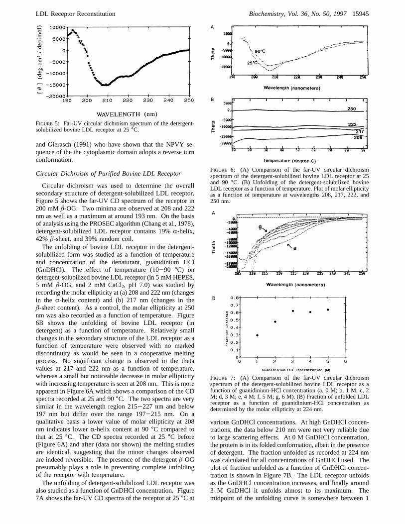
solubilized form was studied as a function of temperatureand concentration of the denaturant, guanidinium HCl(GnDHCl). The effect of temperature (10-90 °C) ondetergent-solubilized bovine LDL receptor (in 5 mM HEPES,5 mM â-OG, and 2 mM CaCl2, pH 7.0) was studied byrecording the molar ellipticity at (a) 208 and 222 nm (changesin the R-helix content) and (b) 217 nm (changes in theâ-sheet content). As a control, the molar ellipticity at 250nm was also recorded as a function of temperature. Figure6B shows the unfolding of bovine LDL receptor (indetergent) as a function of temperature. Relatively smallchanges in the secondary structure of the LDL receptor as afunction of temperature were observed with no markeddiscontinuity as would be seen in a cooperative meltingprocess. No significant change is observed in the thetavalues at 217 and 222 nm as a function of temperature,whereas a small but noticeable decrease in molar ellipticitywith increasing temperature is seen at 208 nm. This is moreapparent in Figure 6A which shows a comparison of the CDspectra recorded at 25 and 90°C. The two spectra are verysimilar in the wavelength region 215-227 nm and below197 nm but differ over the range 197-215 nm. On aqualitative basis a lower value of molar ellipticity at 208nm indicates lowerR-helix content at 90°C compared tothat at 25°C. The CD spectra recorded at 25°C before(Figure 6A) and after (data not shown) the melting studiesare identical, suggesting that the minor changes observedare indeed reversible. The presence of the detergentâ-OGpresumably plays a role in preventing complete unfoldingof the receptor with temperature.The unfolding of detergent-solubilized LDL receptor was
also studied as a function of GnDHCl concentration. Figure7A shows the far-UV CD spectra of the receptor at 25°C at
various GnDHCl concentrations. At high GnDHCl concen-trations, the data below 210 nm were not very reliable dueto large scattering effects. At 0 M GnDHCl concentration,the protein is in its folded conformation, albeit in the presenceof detergent. The fraction unfolded as recorded at 224 nmwas calculated for all concentrations of GnDHCl used. Theplot of fraction unfolded as a function of GnDHCl concen-tration is shown in Figure 7B. The LDL receptor unfoldsas the GnDHCl concentration increases, and finally around3 M GnDHCl it unfolds almost to its maximum. Themidpoint of the unfolding curve is somewhere between 1
FIGURE 5: Far-UV circular dichroism spectrum of the detergent-solubilized bovine LDL receptor at 25°C.
FIGURE 6: (A) Comparison of the far-UV circular dichroismspectrum of the detergent-solubilized bovine LDL receptor at 25and 90 °C. (B) Unfolding of the detergent-solubilized bovineLDL receptor as a function of temperature. Plot of molar ellipticityas a function of temperature at wavelengths 208, 217, 222, and250 nm.
FIGURE 7: (A) Comparison of the far-UV circular dichroismspectrum of the detergent-solubilized bovine LDL receptor as afunction of guanidinium-HCl concentration (a, 0 M; b, 1 M; c, 2M; d, 3 M; e, 4 M; f, 5 M; g, 6 M). (B) Fraction of unfolded LDLreceptor as a function of guanidinium-HCl concentration asdetermined by the molar ellipticity at 224 nm.
LDL Receptor Reconstitution Biochemistry, Vol. 36, No. 50, 199715945
and 2 M GnDHCl. Complete unfolding of the receptor doesnot occur even at 6 M GnDHCl.
Reconstitution of the LDL Receptor and CryoelectronMicroscopy
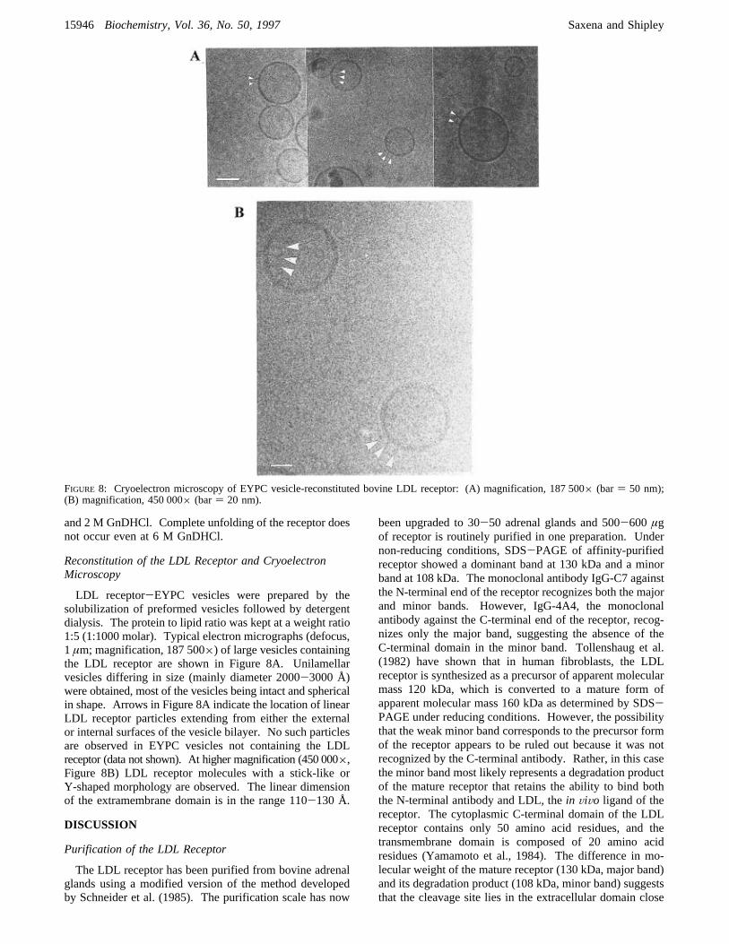
LDL receptor-EYPC vesicles were prepared by thesolubilization of preformed vesicles followed by detergentdialysis. The protein to lipid ratio was kept at a weight ratio1:5 (1:1000 molar). Typical electron micrographs (defocus,1 µm; magnification, 187 500×) of large vesicles containingthe LDL receptor are shown in Figure 8A. Unilamellarvesicles differing in size (mainly diameter 2000-3000 Å)were obtained, most of the vesicles being intact and sphericalin shape. Arrows in Figure 8A indicate the location of linearLDL receptor particles extending from either the externalor internal surfaces of the vesicle bilayer. No such particlesare observed in EYPC vesicles not containing the LDLreceptor (data not shown). At higher magnification (450 000×,Figure 8B) LDL receptor molecules with a stick-like orY-shaped morphology are observed. The linear dimensionof the extramembrane domain is in the range 110-130 Å.
DISCUSSION
Purification of the LDL Receptor
The LDL receptor has been purified from bovine adrenalglands using a modified version of the method developedby Schneider et al. (1985). The purification scale has now
been upgraded to 30-50 adrenal glands and 500-600 µgof receptor is routinely purified in one preparation. Undernon-reducing conditions, SDS-PAGE of affinity-purifiedreceptor showed a dominant band at 130 kDa and a minorband at 108 kDa. The monoclonal antibody IgG-C7 againstthe N-terminal end of the receptor recognizes both the majorand minor bands. However, IgG-4A4, the monoclonalantibody against the C-terminal end of the receptor, recog-nizes only the major band, suggesting the absence of theC-terminal domain in the minor band. Tollenshaug et al.(1982) have shown that in human fibroblasts, the LDLreceptor is synthesized as a precursor of apparent molecularmass 120 kDa, which is converted to a mature form ofapparent molecular mass 160 kDa as determined by SDS-PAGE under reducing conditions. However, the possibilitythat the weak minor band corresponds to the precursor formof the receptor appears to be ruled out because it was notrecognized by the C-terminal antibody. Rather, in this casethe minor band most likely represents a degradation productof the mature receptor that retains the ability to bind boththe N-terminal antibody and LDL, thein ViVo ligand of thereceptor. The cytoplasmic C-terminal domain of the LDLreceptor contains only 50 amino acid residues, and thetransmembrane domain is composed of 20 amino acidresidues (Yamamoto et al., 1984). The difference in mo-lecular weight of the mature receptor (130 kDa, major band)and its degradation product (108 kDa, minor band) suggeststhat the cleavage site lies in the extracellular domain close
FIGURE 8: Cryoelectron microscopy of EYPC vesicle-reconstituted bovine LDL receptor: (A) magnification, 187 500× (bar) 50 nm);(B) magnification, 450 000× (bar) 20 nm).
15946 Biochemistry, Vol. 36, No. 50, 1997 Saxena and Shipley

to the membrane surface. Thus, the 108 kDa band probablyrepresents LDL receptor lacking both the cytoplasmic andtransmembrane domains. As such, this shortened receptorlacking the hydrophobic transmembrane domain could beconsidered a useful candidate for crystallization studiesprovided it can be purified in sufficiently large amounts.
Secondary Structure
Lacking extensive information on the secondary andtertiary structures of membrane proteins in general andlipoprotein receptors in particular, we have used the predic-tive methods developed by Chou and Fasman (1974a,b) toderive secondary structure information for the LDL receptor.For this we used the amino acid sequence derived from thesequence of a full-length cloned 5.3 kb cDNA for the humanLDL receptor (Yamamoto et al., 1984). For the human LDLreceptor we obtain overall values of 23%R-helix, 25%â-sheet, and 39%â-turns. This distribution of secondarystructure is in reasonable agreement with that reportedpreviously by De Loof et al. (1986) using a similar algorithm(20%R-helix, 30%â-sheet, and 30%â-turns) and with theCD-derived distribution for the bovine receptor (19%R-helix,42%â-sheet, and 39% random coil).The amino acid sequence deduced from the cDNA
sequence suggests five domains in the 839 amino acidprotein. For the ligand-binding region (Domain I) the overallsecondary structure is predicted by the Chou-Fasmanalgorithm to be 15-20%R-helix andâ-sheet, together witha high level (approximately 50%) ofâ-turns (Figures 3Aand 4; Table 1). In good agreement with these predictions,recent NMR studies of the first and second cysteine-richrepeats indicate a high level ofâ-turn andâ-hairpin structuresparticularly toward the C-terminal end of these approximately40-residue sequences (Daly et al., 1995a,b). The EGF-precursor homology (Domain II) and O-linked sugar (Do-main III) regions show increases inR-helix and â-sheetcontent, with lower levels ofâ-turn. The cytoplasmicdomain (Domain V) is predicted to be rich inR-helix (44%)andâ-turn, with noâ-sheet being indicated.While the Chou-Fasman algorithm based on the crystal
structures of globular, nonmembrane proteins is clearlyappropriate for the extramembrane domains of the LDLreceptor (Domains I, II, III, and V), it is perhaps lessappropriate for the membrane-spanning Domain IV. Whilethe Chou-Fasman algorithm suggests a predominantlyâ-sheet structure for the short, 22-residue transmembranedomain (Domain IV), a high potential forR-helix structureis also predicted (see Figure 3). Because a single 22-residuetransmembraneâ-strand is energetically unfavorable, it islikely that the transmembrane region takes the form of anR-helix. A possible reason for predicting aâ-strand con-formation for this domain has been suggested by Jahnig(1989) indicating that the most hydrophobic residues, whichare associated withâ-strand conformations in water-solubleproteins, are also found inR-helix structures of membraneproteins [also, see Fariselli et al. (1993)].
Circular Dichroism
Far-UV CD spectroscopy has been used to study thefolding and unfolding of protein secondary structure eitherat equilibrium or kinetically. The thermal studies ofdetergent-solubilized LDL receptor by CD (Figure 6) show
that the receptor is resistant to change in overall secondarystructure with temperature. This could be partly due to adetergent effect whereby the secondary structure of thereceptor is stabilized by the detergent. In addition, theN-terminal domain (Domain 1) accounting for 35% of thereceptor is probably resistant to unfolding due to its stabiliza-tion by the multiple disulfide bonds present within each ofthe seven cysteine-rich repeats. The minor changes observedin secondary structure are reversible, demonstrating that thereceptor folds back into its original conformation whencooled back to 25°C. Interestingly, GnDHCl appears tohave a more pronounced effect than temperature on thesecondary structure of the receptor (see Figures 6 and 7).On the basis of data at 224 nm, unfolding of approximately70% of the LDL receptor occurs at 6 M GnDHCl with amidpoint of unfolding at approximately 1.5 M GnDHCl.
Reconstitution of the LDL Receptor and ElectronMicroscopy
Bovine LDL receptor has been successfully incorporatedinto phospholipid vesicles using two different methods: (i)by simple detergent dialysis (Schneider, 1983) and (ii) asdescribed here, by solubilization of preformed vesicles anddetergent dialysis. Multilamellar liposomes are very easyto prepare but they are not suitable for structural studies ofmembrane proteins. On the other hand, small unilamellarvesicles suffer from the disadvantage that the surface of thesevesicles has a high degree of curvature which affects manyof the physical properties of the phospholipid in the vesicles.Therefore, for most studies of reconstituted receptors, it isimportant that the membrane protein is incorporated intolarge unilamellar vesicles (diameter>1000 Å) which mostclosely resemble biological membranes. Large unilamellarvesicles (LUVs) have been prepared using a variety ofprocedures, e.g. detergent dialysis, solvent evaporation,extrusion, etc. Schneider (1983) showed that the bovine LDLreceptor could be reconstituted into EYPC bilayers using adetergent dialysis method. On the basis of gel filtrationexperiments, Schneider (1983) calculated the diameter of thereceptor-containing vesicles to be in the range 2000-2700Å. In the current study, the reconstituted EYPC vesiclescontaining the LDL receptor had diameters in the range 500-3000 Å as determined by electron microscopy, but with themajority in the range 2000-3000 Å. At a protein to lipidmolar ratio of 1:10 000, and considering the surface area ofphospholipid head group to be 70 Å2 and bilayer thickness40 Å, the expected number of receptor molecules per singlebilayer vesicle is 20-80. Previous studies (Saxena &Shipley, 1992; Saxena, 1994) using negative-stain electronmicroscopy showed that LDL receptor-containing vesiclesprepared by detergent dialysis do bind gold-labeled LDL;however, these studies showed that a high percentage ofvesicles prepared by simple detergent dialysis contained morethan one bilayer. A significant improvement in the produc-tion of unilamellar EYPC vesicles containing the LDLreceptor has been achieved by using preformed vesicles madeby extrusion, followed by the addition of detergent-solubi-lized receptor, and then dialysis to remove detergent. Thesereconstituted vesicles were almost exclusively unilamellar(diameter, 2000-3000 Å) and somewhat larger than the sizeof the original preformed vesicles. It is probable that at theconcentration of the detergent used, a complete disruptionof the bilayer of the vesicles occurs prior to reformation on
LDL Receptor Reconstitution Biochemistry, Vol. 36, No. 50, 199715947
removal of the detergent. As shown in Figure 8, cryoelectronmicroscopy shows linear projections from the bilayer surfaceand suggests an extended structure (length approximately 120Å) for the extracellular region (comprising Domains I, II,and III) of the LDL receptor.Electron microscopy has become a very powerful tech-
nique for structure determination of membrane proteinsreconstituted with lipids. Successful incorporation of theLDL receptor into large unilamellar vesicles has paved theway for detailed structural studies of the receptor and itsinteraction with its ligand, LDL. Our initial studies of thereconstituted vesicles using cryoelectron microscopy con-firmed the unilamellar nature of the reconstituted vesiclesand showed the receptor to be present in two orientations(i.e. on the outside and inside vesicle surfaces, see Figure8). Since the extracellular domain forms the major portion(92%) of the receptor, it is straightforward to distinguishbetween the two orientations. Labeling studies with mono-clonal antibodies directed against the N-terminus and theC-terminus of the receptor would clearly distinguish betweenthe two orientations. Future studies will focus on (i)incorporating more copies of the LDL receptor into eachvesicle, (ii) labeling of the reconstituted LDL receptor usingLDL, LDL receptor antibodies, or their gold-labeled coun-terparts, and (iii) attempts to produce 2D crystals of thereceptor. This approach should ultimately lead to a pictureof the LDL receptor in a membrane environment and alsoprovide a vehicle for studying structural aspects of theinteraction of the LDL receptor with its ligand LDL.
ACKNOWLEDGMENT
We thank Drs. Joseph L. Goldstein and Michael S. Brown(University of Texas Southwestern Medical Center, Dallas,TX) for providing us with the monoclonal antibody IgG-4A4. Also, we acknowledge helpful advice from Robert A.Reed on the purification of the LDL receptor and from Drs.Donna Cabral-Lilly and Mary T. Walsh on electron micros-copy and CD techniques, respectively. Technical assistancewas provided by D. Gantz, A. Tercyak, C. England, M.Gigliotti, and C. Curry.
REFERENCES
Bansal, A., & Gierasch, L. M. (1991)Cell 67, 1195-1201.Beisiegel, U., Schneider, W. J., Goldstein, J. L., Anderson, R. G.,& Brown, M. S. (1981)J. Biol. Chem. 256, 11923-11931.
Beisiegel, U., Schneider, W. J., Brown, M. S., & Goldstein, J. L.(1982)J. Biol. Chem. 257, 13150-13156.
Bieri, S., Djordjevic, J. T., Daly, N. L., Smith, R., & Kroon, P. A.(1995)Biochemistry 34, 13059-13065.
Brown, M. S., & Goldstein, J. L. (1986)Science 232, 34-47.Chang, C. T., Wu, C.-S. C., & Yang, J. T. (1978)AnalyticalBiochemistry 91, 13-31.
Chou, P. Y., & Fasman, G. D. (1974a)Biochemistry 13, 211-222.
Chou, P. Y., & Fasman, G. D. (1974b)Biochemistry 13, 222-245.
Cummings, R. D., Kornfeld, S., Schneider, W. J., Hobgood, K.K., Tolleshaug, H., Brown, M. S., & Goldstein, J. L. (1983)J.Biol. Chem. 258, 15261-15273.
Daly, N. L., Scanlon, M. J., Djordjevic, J. T., Kroon, P. A., & Smith,R. (1995a)Proc. Natl. Acad. Sci. U.S.A. 34, 14474-14481.
Daly, N. L., Djordjevic, J. T., Kroon, P. A., & Smith, R. (1995b)Biochemistry 34, 14474-14481.
De Loof, H., Rosseneu, M., Brasseur, R., & Ruysschaert, J. M.(1986)Proc. Natl. Acad. Sci. U.S.A. 83, 2295-2299.
Esser, V., Limbird, L. E., Brown, M. S., Goldstein, J. L., & Russell,D. W. (1988)J. Biol. Chem. 263, 13282-13290.
Fariselli, P., Compiani, M., & Casadio, R. (1993)Eur. Biophys. J.22, 41-51.
Goldstein, J. L., & Brown, M. S. (1982)Med. Clin. N. Am. 66,335-362.
Goldstein, J. L., & Brown, M. S. (1983) inThe Metabolic Basis ofInherited Disease(Stanbury, J. B., Wyngaarden, J. B., Freder-ickson, D. S., Goldstein, J. L., and Brown, M. S., Eds.) pp 672-712, McGraw-Hill, New York.
Goldstein, J. L., Brown, M. S., Anderson, R. G. W., Russell, D.W., & Schneider, W. J. (1985)Annu. ReV. Cell Biol. 1, 1-39.
Jahnig, F. (1989) inPrediction of Protein Structure and thePrinciples of Protein Conformation(Fasman, G. D., Ed.) pp707-717, Plenum Press, New York.
Laemmli, U. K. (1970)Nature (London) 227, 680-685.Lindgren, F. T., Jensen, L. C., & Hatch, F. T. (1972) inBloodLipids and Lipoproteins: Quantitation, Composition and Me-tabolism(Nelson, G. L., Ed.) Wiley Interscience, New York.
Madden, T. D., & Cullis, P. R. (1984)J. Biol. Chem. 259, 7655-7658.
Pittman, R. C., Carew, T. E., Attie, A. D., & Steinberg, D. (1982)J. Biol. Chem. 257, 7994-8000.
Saxena, K. (1994) Membrane Receptors: Low Density LipoproteinReceptor and Glycosphingolipids. Ph.D. Thesis, Boston Univer-sity, Boston, MA, 1994.
Saxena, K., & Shipley, G. G. (1992)Biophys. J. 61, A263.Schneider, W. J. (1983)J. Cell. Biochem. 23, 95-106.Schneider, W. J. (1989)Biochim. Biophys. Acta 988, 303-317.Schneider, W. J., Basu, S. K., McPhaul, M. J., Goldstein, J. L., &Brown, M. S. (1979)Proc. Natl. Acad. Sci. U.S.A. 76, 5577-5581.
Schneider, W. J., Goldstein, J. L., & Brown, M. S. (1980)J. Biol.Chem. 255, 11442-11447.
Schneider, W. J., Beisiegel, U., Goldstein, J. L., & Brown, M. S.(1982)J. Biol. Chem. 257, 2664-2673.
Schneider, W. J., Goldstein, J. L., & Brown, M. S. (1985)MethodsEnzymol. 109, 405-417.
Soutar, A. K. (1992)J. Intern. Med. 231, 633-641.Tolleschaug, H., Goldstein, J. L., Schneider, W. J., & Brown, M.S. (1982)Cell 30, 715-724.
Yamamoto, T., Davis, C. G., Brown, M. S., Schneider, W. J., Casey,M. L., Goldstein, J. L., & Russell, D. W. (1984)Cell 39,27-38.
Yokoyama, C., Wang, X., Briggs, M. R., Admon, A., Hua, X.,Goldstein, J. L., & Brown, M. S. (1993)Cell 75, 187-197.
BI971579P
15948 Biochemistry, Vol. 36, No. 50, 1997 Saxena and Shipley