Embed Size (px)
Citation preview
1
STRUCTURAL REARRANGEMENTS OF THE �10-23� DNAZYME TO β3
INTEGRIN SUBUNIT mRNA INDUCED BY CATIONS AND THEIR RELATIONS
TO THE CATALYTIC ACTIVITY
Marcin Cieslak1, Jacek Szymanski2, Ryszard Adamiak3, Czeslaw S. Cierniewski2,4
Center for Molecular and Macromolecular Research in Lodz1, Polish Academy of Sciences,
Department of Molecular and Medical Biophysics2, Medical University in Lodz, Institute of
Bioorganic Chemistry in Poznan3, Polish Academy of Sciences, Center for Medical Biology
and Microbiology in Lodz4, Polish Academy of Sciences, Poland
Correspondence to: Czeslaw S. Cierniewski, Ph.D.
Department of Molecular and Medical Biophysics, Medical University in Lodz, 92-215 Lodz,
6/8 Mazowiecka Street, Poland
Tel: (48-42) 6783393
Fax: (48-42) 6789433
E-mail: [email protected]
Running title: Structural transitions induced in the DNAzyme by Mg2+
Key words: DNAzyme, cations, structural transitions, binding affinity
Copyright 2003 by The American Society for Biochemistry and Molecular Biology, Inc.
JBC Papers in Press. Published on September 2, 2003 as Manuscript M300504200 by guest on February 28, 2019
http://ww
w.jbc.org/
Dow
nloaded from

2
The intracellular ability of the �10-23� DNAzyme to efficiently inhibit expression
of the targeted proteins has been evidenced by in vitro and in vivo studies. However,
standard conditions for kinetic measurements of the DNAzyme catalytic activity in vitro
include 25 mM Mg2+, a concentration that is very unlikely to be achieved intracellularly.
To study this discrepancy we analyzed the folding transitions of the �10-23� DNAzyme
induced by Mg2+. For this purpose spectroscopic analyses such as fluorescence
resonance energy transfer, fluorescence anisotropy, circular dichroism, and surface
plasmon resonance measurements were performed. The global geometry of the
DNAzyme in the absence of added Mg2+ seems to be essentially extended, it has no
catalytic activity and shows a very low binding affinity to its RNA substrate. The
folding of the DNAzyme induced by binding of Mg2+ may occur in several distinct stages.
The first stage, observed at 0.5 mM Mg2+, corresponds to the formation of a compact
structure with limited binding properties and without catalytic activity. Then, at 5 mM
Mg2+ flanking arms are projected at right position and angles to bind RNA. DNAzyme
in such a state shows substantial binding to its substrate and significant catalytic
activity. Finally, the transition occurring at 15 mM Mg2+ leads to the formation of the
catalytic domain and DNAzyme shows high binding affinity towards substrate and
efficient catalytic activity. Under conditions simulating intracellular conditions, the
DNAzyme was only partially folded, did not bind to its substrate and showed only
residual catalytic activity, suggesting that it may be inactive in the transfected cells and
behave like antisense oligodeoxynucleotide.
The typical DNAzyme, known as the "10�23" model, has tremendous potential in
gene suppression for both target validation and therapeutic applications (1). It is capable of
cleaving single-stranded RNA at specific sites under simulated physiological conditions, and
can be used to control even complex biological processes such as tumor angiogenesis. For
example, DNAzymes to β1 and β3 mRNA reduced expression of targeted integrin subunits in
endothelial cells and blocked proliferation, migration and network formation in a fibrin and
Matrigel matrix (2). In a cell culture system, a "10-23" deoxyribozyme designed against 12-
lipoxygenase mRNA specifically down regulated expression of this protein and its
metabolites, known to play a crucial role in tumor angiogenesis (3). Similarly, the DNAzyme
to VEGFR2 mRNA cleaved its substrate efficiently and inhibited the proliferation of
endothelial cells with a concomitant reduction of VEGFR2 mRNA, and blocked tumor growth
in vivo (4).
by guest on February 28, 2019http://w
ww
.jbc.org/D
ownloaded from

3
The origins of the DNAzyme catalytic activity are not yet fully understood, but the
observed rate enhancements probably are generated by a number of factors, including metal
ion and nucleobase catalysis and local stereochemical effects. The 10-23 DNAzyme has been
developed using an in vitro selection strategy on the basis of its ability to cleave RNA in the
presence of Mg2+ (1). It has a catalytic domain of 15 highly conserved deoxyribonucleotides
flanked by 2 substrate-recognition domains and can cleave effectively between any unpaired
purine and pyrimidine of mRNA transcripts. Like many other enzymes catalyzing
phosphoryl-transfer reactions, it is recognized as a metalloenzyme requiring divalent metal,
preferentially Mg2+ ions, for catalytic activity. Divalent cations play a crucial role in these
mechanisms as evidenced by a number of observations. For example, addition of La3+ to the
Mg2+-background reaction mixture inhibited the DNAzyme-catalyzed reactions, suggesting
the replacement of catalytically and/or structurally important Mg2+ by La3+ (5). The function
of divalent metal cations in DNAzyme activity is very complex and includes: (a) stabilization
of the transition state of reaction. The divalent metal cation dependence of the enzyme was
described as being the evidence supporting a chemical mechanism involving metal-assisted
deprotonation of the 2�-hydroxyl located adjacent to the cleavage site (6). (b) Neutralization
of negative charges of phosphate groups, thus facilitating DNA-RNA interactions. High
resolution X-ray crystal structures of Mg2+ and Ca2+ salts of the model B-DNA decamers
CCAACGTTGG and CCAGCGCTGG revealed sequence-specific binding of Mg2+ and Ca2+
to the major and minor grooves of DNA, as well as non-specific binding to backbone
phosphate oxygen atoms. This accounts for the neutralization of between 50 and 100% of the
negative charges of phosphate groups (7). (c) Some of these bound cations may also play a
purely structural role by inducing proper folding of the DNAzyme molecule, thus helping to
organize the enzyme into its active conformation. There are several reports showing that
Mg2+ helps to stabilize different types of double-stranded DNA structures (8, 9), and can
induce bending or enhance curvature in DNA (10). Furthermore, Mg2+ and other divalent
cations enhance end-to-end DNA interactions, particularly in the case of fragments with self-
complementary ends (11). These structural effects of cations may be even more profound in
a single-stranded and flexible DNAzyme molecule.
Mg2+-dependent cleavage has special relevance to biology because it is compatible
with intracellular conditions, raising the possibility that DNA enzymes might be made to
operate in vivo (12). However, at the present time it is hard to explain their intracellular
catalytic activity, keeping in mind the catalytic dependence upon high concentrations of Mg2+,
unlikely to be achieved in cytoplasm. To address this question, we attempted to correlate
by guest on February 28, 2019http://w
ww
.jbc.org/D
ownloaded from

4
changes in the catalytic activity and configuration of the DNAzyme induced by gradually
bound Mg2+. To characterize structural changes in the DNAzyme we performed FRET
analysis allowing us to monitor general folding of the molecule based on the measurements of
distances between fluorophores linked to 5� and 3� side bases, and SPR analysis of the
DNAzyme binding to its RNA substrate. Structural changes induced by cations in
DNAzymes were also monitored be circular dichroism and fluorescence anisotropy analysis.
EXPERIMENTAL PROCEDURES
Synthesis of DNAzyme to β3 mRNA. DNAzyme was chemically synthesized on a
solid support using an ABI-394 DNA Synthesizer as described before (13). This particular
DNA sequence (5�GAGTCCCATAg1g2c3t4a5g6c7t8a9c10a11a12c13g14a15AAGACTTGAG3�) was
used previously to analyze the enzymatic activity, specificity, exonuclease resistance, and
ability to inhibit expression of β3 integrins in endothelial cells (2). For BIAcore experiments,
the inactive DNAzyme, β3DE(15)In, with a single substitution (G6→A) in the reactive loop,
and antisense oligodeoxynucleotide β3(1245-1265) containing both flanking arms of the
β3DE (5�GAGTCCCATACAAGACTTGAG3�) were synthesized Two analogues of
β3DE(15)In and β3(1245-1265) were produced as well, containing the modified
oligonucleotides such as phosphorothioates or 2'-O-methyl-substituted residues introduced at
both 5' and 3' sides. Hence, S-β3DE(15)in and S-β3(1245-1265) have two phosphorothioate
substitutions while MeO-β3DE(15)in and MeO-β3(1245-1265) contain two 2'-O-methyl-
substituted residues at both their 5' and 3' sides, respectively. An additional mutated
DNAzyme [MeO-β3DE(11)] with a downsized catalytic loop of a deoxyribozyme from 15-mer
to 11-mer (5�GAGTCCCATAg1g2c3t4a9c10a11a12c13g14a15AAGACTTGAG3�) was synthesized
and used as a control. DNAzymes with the 11-mer catalytic loop were described to be Ca2+-
dependent deoxyribozymes and showed significantly reduced binding affinities and catalytic
activities in the presence of Mg2+ (14). All deoxyoligonucleotides were purified by HPLC
and ion exchange chromatography (to 98%), and their purity was checked by PAGE under
denaturing conditions.
The doubly-labeled β3 DNAzymes with the 15-mer and 11-mer catalytic loops were
synthesized by the solid state phosphoramidite approach on an ABI 392 synthesizer, starting
from Fluorescein CPG support (CPG, Inc). The 2�-O-Me RNA CE phosphoramidites (Glen
Research) were used for introduction of 2�-O-Me nucleotide units flanking the sequence from
by guest on February 28, 2019http://w
ww
.jbc.org/D
ownloaded from

5
the 3�- and 5�-ends. The synthesis was terminated with an addition of Rhodamine CE
phosphoramidite (CPG, Inc). Oligonucleotides were cleaved from the solid support and
deprotected by brief (4h at 55°C) treatment with 30% ammonia. Pure product was isolated by
preparative HPLC on a Hamilton PRP1 column. The peak fractions were evaporated to
dryness, redissolved in water and then ethanol precipitated.
The synthetic, biotinylated (at the 3�-end) 21-mer RNA (biotin-
CUCAGGGUAUGUUCUGAACUC) used in BIAcore experiments was purchased from
Bionovo (Poland). Its sequence corresponded to the 1245-1265 fragment of β3 mRNA.
After synthesis the product was purified by HPLC and its purity checked by PAGE.
Preparation of target RNA substrates and kinetic analysis. Aliquots of RNA
substrates (20 µl; 5 µM) dissolved in a T4 polynucleotide kinase buffer were mixed with [γ-32P]ATP (2 µl; 20 µCi) and T4 polynucleotide kinase (3 units). Reaction was carried out
for an hour at 37oC. All reported kinetic values were determined in multiple turnover
reactions. Vmax and KM values were determined from the y intercept and slope, respectively,
of the best-fit line to a Lineweaver-Burke plot of 1/V vs 1/[S]. Reactions (10 min., 37oC, total
volume 20 µl) were carried out in 50 mM Tris, pH 8.0 containing 15 mM MgCl2, 0.01% SDS,
DNAzyme (0.0125 µM) with the radiolabeled RNA substrate used in a wide range of
concentrations. The cleavage reaction was stopped by addition of 5 µl 0.5 M EDTA and the
products were separated by electrophoresis in 20% polyacrylamide gels under denaturing
conditions. Amounts of the product were evaluated by use of a PhosphorImager (Molecular
Dynamics).
Cell culture. Human umbilical vein endothelial cells (HUVEC) were isolated from freshly
collected umbilical cords by collagenase treatment (15, 16). Cells were grown in gelatin-
coated 75-cm2 tissue culture flasks and were maintained at confluence in RPMI 1640 medium
supplemented with streptomycin (100 µg/ml), penicillin (100 U/ml), fungizone (2.5 mg/ml),
heparin (90 µg/ml), L-glutamine (1 mM), sodium bicarbonate (2 mg/ml), 20 % fetal bovine
serum and epidermal growth factor (40 ng/ml) at 37oC in a humidified 10% CO2 atmosphere.
Primary cultures were harvested at confluence with trypsin/EDTA and transferred into
gelatin-coated dishes. For the experiments, confluent cultures were used at the second
passage.
For microscopic examination cells were plated at a density of 5x10⁴ cells/well on
Thermanox Cover slips in 8 well tissue culture chamber slides (NUNC) with detachable
by guest on February 28, 2019http://w
ww
.jbc.org/D
ownloaded from
6
chambered upper structures. Before performance of assays the serum-containing medium was
changed to a serum-free medium (Opti-MEM). The cultures were gently rinsed three times
with the medium and preincubated with fluorophore-labeled MeOβ3DE(15) (0.5 µM) for 6
hours in the presence of lipofectin (5 µg/ml). After that time the transfection mixture was
replaced by normal serum-containing medium and cells were grown for another 18 hours.
Attached treated intact cells were maintained in a CO2 incubator at 37oC. Two control assays
were carried out using either untreated cells or cells exposed to 0.25 µM fluorescein. After
incubation cells were washed three times with PBS, fixed with freshly prepared 3.5%
paraformaldehyde for 15 min at room temperature then washed three times with PBS and
mounted in 2.5% DABCO in glycerol and processed for microscopy.
Fluorescence spectroscopy. Fluorescence emission spectra were measured on a
Perkin-Elmer LS-50 spectrofluorometer, and spectra were corrected for lamp fluctuations and
instrumental variations. Polarization artifacts were avoided by setting excitation and emission
polarizers to magic angle conditions (54.74°). All the fluorescence measurements were
performed at the temperature of 23 ± 1°C. Emission spectra, excitation spectra and
luminescence intensity were recorded with 5 nm band passes for both the excitation and
emission monochromators. A cut-off filter in the emission beam was used to eliminate
second order wavelength interference. The excitation wavelengths used were 494 nm and 560
nm for fluorescein- and rhodamine-conjugated constructs, respectively. Emission spectra
were corrected for the blank contribution and for the instrument response, and normalized to
the DNA concentration in a quartz cell with a 1 cm path length. Excitation and emission
spectra were automatically corrected for lamp intensity variations. Buffers were degassed by
bubbling nitrogen to prevent quenching of fluorescence by dissolved oxygen. The
fluorescence emission signals were stable to photobleaching under the experimental
conditions of measurement. The apparent interchromophore separation, R, the distance
separating the energy donor and acceptor was calculated by the Forster equation: R = R0(1/E-
1)1/6, where E is the efficiency of energy transfer from donor to acceptor. E = 1-FDA/FD and
R0 is the distance for 50% transfer efficiency E. FDA and FD are the fluorescence intensities of
the donor in the presence and absence of the acceptor, respectively. FDA and FD were
measured at 520 nm, the emission maximum for fluorescein.
The fluorescence anisotropy of the fluorescein and rhodamine probes attached to the
DNAzyme (Fluo-MeO-β3DE(15)-Rhod, Fluo-MeO-β3DE(11)-Rhod) was monitored in a
by guest on February 28, 2019http://w
ww
.jbc.org/D
ownloaded from

7
Perkin-Elmer LS-50 spectrofluorometer equipped with an automatic anisotropy measuring
device. The anisotropy r is defined as:
r = (IVV -G x IVH) / (IVV + 2 x G x IVH)
where I is fluorescence intensity. The first and second indices refer to the orientation of
excitation and emission polarizers, respectively. G is the correction factor. The cell holder
was thermostated at 21°C.
Analysis of fluorescence data. Efficiencies of energy transfer were determined from
enhancement acceptor fluorescence (17, 18). The emission at a given wavelength (v1) of a
double-labeled sample excited primarily at the donor wavelength (v�) contains emission from
the donor, emission from directly excited acceptor and emission from acceptor excited by
energy transfer from the donor, i.e.:
F(v1,v�) = [S] . [εD (v�) . φD (v1) . d+ . {(1 � EFRET) . a+ + a−}
+ εA (v�) . φΑ (v1) . a+ + εD (v�) . φΑ (v1) . EFRET . d+ + a+} = FD (v1,v�) + FA (v1,v�)
where [S] is the concentration of DNAzyme, d+ and a+ are the molar fraction of DNAzyme
molecules labeled with donor and acceptor respectively, and a- is the molar fraction of
DNAzyme molecules unlabeled with acceptor. Superscripts D and A refer to donor and
acceptor, respectively. εD(v�) and εA(v�) are the molar absorption coefficients of donor and
acceptor, respectively and φD(v1) and φΑ(v1) are the fluorescent quantum yields of donor and
acceptor respectively. Thus the spectrum contains the components due to donor emission [FD
(v1,v�) i.e. the first term containing φD(v1)] and those due to acceptor emission [FA(v1,v�) i.e.,
the latter two terms containing φΑ(v1)]. The first stage of the analysis involves subtraction of
the spectrum of DNA labeled only with donor, leaving just the acceptor components, i.e. FA
(v1,v�). The pure acceptor spectrum thus derived is normalized to one from the same sample
excited at a wavelength (v��) at which only the acceptor is excited, with emission at v2. We
then obtain the acceptor ratio:
(ratio)A = FA(v1,v�)/ FA(v2,v��) =
{EFRET . d+ . [εD (v�)/ εA (v��) + εA (v�)/ εA (v��)]} . [φΑ (v1)/ φΑ (v2)]
EFRET is directly proportional to (ratio)A, and can be easily calculated since εD(v�)/εA(v��) and
εA(v�)/εA(v��) are measured from absorption spectra, and φΑ(v1)/φΑ(v2) is unity when v1 = v2.
Analysis of Circular Dichroism. The circular dichroism spectra of MeO-β3DE (1
µM), free or in the complex with the substrate (2 µM) was measured in a solution of 10 mM
by guest on February 28, 2019http://w
ww
.jbc.org/D
ownloaded from

8
Tris-HCl, pH 7.5, containing increasing concentrations of MgCl2 at 21°C. In control
experiments, 0.1 M and 1 M NaCl were used. Before measurement, the complex was
allowed to form by heating the solution at 95°C for 2.5 min followed by gradual cooling.
Measurements were made in a quartz cuvette (5 mm path length) with a CD
spectrophotometer (model Jobin Yvon) from 200 to 320 nm in triplicate. The spectra were
obtained by smoothing the averaged spectra with a calculator.
Surface Plasmon Resonance. The kinetic parameters (association and dissociation rate
constants, kon and koff, respectively) and the affinity constant (KD) between DNAzyme and the
mRNA substrate were measured by surface plasmon resonance using a BIAcoreX (Pharmacia
Biotech Inc.). Briefly, avidin was covalently attached to carboxymethyl dextran (CM5) chips
(BIAcore) previously activated with N-hydroxysuccinimide and N-ethyl-N'-
dimethylaminopropyl carbodiimide according to the manufacturer's instructions. Experiments
were performed at 37oC using 50 mM Tris, pH 8.0, containing divalent cations at the
indicated concentrations. 15 µl of 5�-end biotinylated RNA at 100 nM in 50 mM Tris, pH
8.0, was injected at the flow rate of 5 µl/min and consequently immobilized on the bound
avidin to give a response of about 500 RU. The levels of immobilized RNA were within the
low levels that have to be used to ensure that the observed binding rate will be limited by the
reaction kinetics rather than by the mass transport effects of the injected DNAzyme (19). In
typical experiments, DNAzyme was flowing in two channels of the sensor, the first one
contained the RNA substrate attached to avidin, and the second was without the RNA
substrate. The latter was used to correct SPR traces and remove the background binding
between DNAzyme and the immobilized avidin on the dextran. The concentration of the
injected DNAzyme was in the range of between 20 and 200 nM, and the flow rate was 5
µl/min. The amount of ligands bound to immobilized RNA substrate was monitored by
measuring the variation of the surface plasmon resonance angle as a function of time. Results
were expressed in resonance units (RU), an arbitrary unit specific for the BIAcore instrument.
In preliminary experiments, the data obtained for at least three different concentrations of
DNAzymes were fitted to several models and the best fits (Chi2 =1.4) were obtained assuming
an one-to-one interaction. Then, the association rate constant, kon, and dissociation rate
constant, koff, were determined separately from individual association and dissociation phases,
respectively. The overall affinity constant, KA, was derived from kon/koff . The sensor chip
was regenerated with three 10 µl pulses of 12.5% formamide.
by guest on February 28, 2019http://w
ww
.jbc.org/D
ownloaded from

9
RESULTS
Enzymatic characteristics of the DNAzyme to β3 integrin subunit. The �10-23�
DNAzyme used in these studies was designed to cleave β3 integrin subunit mRNA and
preliminary characterization was done in our recent work (2). Standard conditions for kinetic
measurements of DNAzyme�s catalytic activity in vitro include 25 mM MgCl2 (20). Under
such conditions the �10-23� DNAzyme shows optimal enzymatic activity while its mutant
MeO-β3DE(11) with the shortened catalytic loop is inactive (Figure 1A). In this experiment,
enzymatic reactions were performed in 50 mM Tris-HCl, pH 8.0, containing 0 to 25 mM
MgCl2, under multiple turnover conditions. The 32P-labelled mRNA substrate was mixed with
the DNAzyme (β3DE) in the molar ratio of 80:1, incubated at 37oC and aliquots were
withdrawn after 10 min. The cleavage reaction was stopped by addition of 5 µl 0.5 M EDTA
and products separated by electrophoresis in 20% polyacrylamide gels under denaturing
conditions. Amounts of the product were evaluated by use of a PhosphorImager. Figure 1B
shows a composition of cleavage mixtures obtained after 60 min incubation of 32P-labelled
mRNA substrates with 0.025 µM β3DE added at a molar ratio ranging from 5:1 to 80:1. Each
of the DNAzymes, unmodified (β3DE) and modified (MeO-β3DE), cleaved the substrate at
the predicted site. Interestingly, DNAzyme with the 2'-MeO residues showed a significantly
higher enzymatic activity than β3DE as evidenced by the catalytic efficiency kcat/KM of
3.62x10⁶ and 1.09x10⁶ M-1min-1, respectively.
Study of ion-induced folding of the DNAzyme by fluorescence resonance energy
transfer. To follow the folding transitions of the DNAzyme induced upon binding of Mg2+
ions, fluorescence resonance energy transfer (FRET) was utilized. For this purpose, β3DE
with the 15-mer and 11-mer catalytic loop were modified by attachment of the donor and
acceptor fluorophores, rhodamine and fluorescein, to the 5� and 3� ends of the flanking arms,
respectively. A series of experiments were next designed to analyze whether the fluorophores
attached to the terminal bases affect the ability of the oligodeoxynucleotide construct to
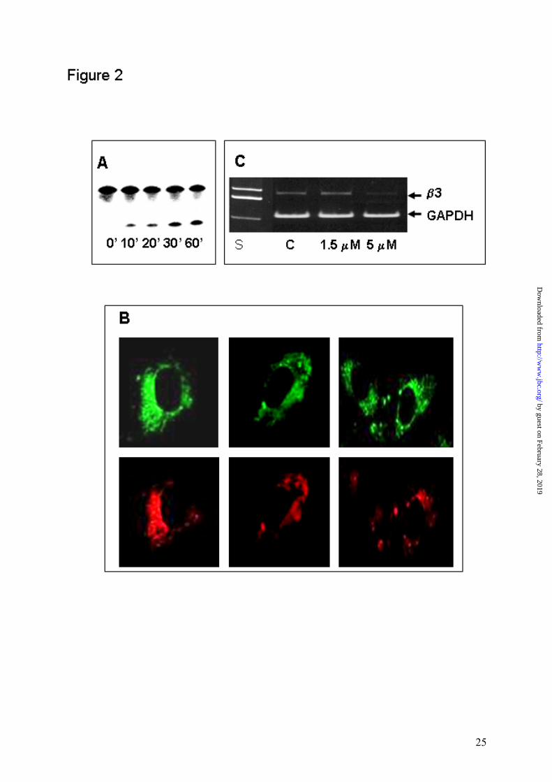
function as an active DNAzyme species. As is seen in Figure 2A, incubation of the Fluo-
MeO-β3DE(15)-Rhod with the 32P-labelled substrate in the presence of 25 mM Mg2+ under
multiple-turnover conditions at 37°C leads to cleavage at the correct site. When such a
construct was incubated with endothelial cells, it remained resistant to intracellular nucleases
and even after 24 hours was located exclusively within the cytoplasm, particularly in the
by guest on February 28, 2019http://w
ww
.jbc.org/D
ownloaded from

10
perinuclear organelles. The cellular uptake, intracellular distribution, and stability of the
Fluo-MeO-β3DE(15)-Rhod were the same as recently reported for another �10-23� DNAzyme
(21). Cellular transport of the Fluo-MeO-β3DE(15)-Rhod as a function of the external
oligonucleotide concentration was nonlinear, being more efficient at concentrations below 2
µM. The punctate fluorescence distribution observed even after 24 hours of exposure to the
DNAzyme seems to suggest that endosomal vesicles are the primary targets of the probes
under study (Figure 2B). Fluo-MeO-β3DE(15)-Rhod could be detected intracellularly, both
when emission of either fluorescein or rhodamine was measured and both fluorophores
showed full colocalization. Thus, the attached fluorophores did not influence the enzymatic
activity and biological properties of the DNAzyme, including the ability to interact with
cellular components responsible for its transport. Transfection of endothelial cells with the
DNAzyme (5 µM) efficiently reduces expression of β3 integrin subunit measured at the level
of β3 mRNA by RT PCR (Figure 2C) and at the cellular surface by flow cytometry (not
shown). Although MeO-β3DE(11)-Rhod had the same cellular distribution as MeO-β3DE(15)-
Rhod, it did not show any biological activity detectable at the level of β3 mRNA or β3
expression at the cell surface. These data provide the evidence that the �10-23� DNAzyme has
an intracellular catalytic or antisense activity, even at the much lower cation concentrations
than those normally used in in vitro analysis.
Both Fluo-MeO-β3DE(15)-Rhod and Fluo-MeO-β3DE(11)-Rhod were next used to
measure energy transfer resulting from a dipolar coupling between the transition moments of
the two fluorophores, fluorescein as the energy donor and rhodamine as the energy acceptor.
When the fluorescence spectrum of one fluorophore (the donor) overlaps with the excitation
spectrum of another fluorophore (the acceptor), the excitation of the donor induces
fluorescence of the acceptor, while its own fluorescence decreases. The extent of FRET is
extremely sensitive to the distance between the donor and the acceptor, being inversely
proportional to the sixth power of the distance. Attachment of rhodamine to Fluo-MeO-
β3DE(15) resulted in a decrease in the fluorescence emission at 520 nm characteristic for
fluorescein, and the fluorescence spectrum of the resulting construct, Fluo-MeO-β3DE(15)-
Rhod, had two peaks at 520 nm and 580 nm upon excitation at 494 nm (Figure 3). The
mutated β3DE, Fluo-MeO-β3DE(11)-Rhod, showed the same fluorescence properties. These
spectra clearly indicate that considerable energy from the excited fluorescein was transferred
to rhodamine, providing the evidence that both fluorophores are in close proximity. Cleavage
of the Fluo-MeO-β3DE(15)-Rhod with endonuclease from Serratia marcescens resulted in a
by guest on February 28, 2019http://w
ww
.jbc.org/D
ownloaded from

11
significant increase of the fluorescence intensity at 520 nm approaching the level of
fluorescein alone, indicating that both fluorophores are separated to the distance enabling the
energy transfer.
Variation in end-to-end distances during the ion-induced folding of the DNAzyme.
According to the model for the folding of the DNAzyme, the length of oligodeoxynucleotide
should shorten over the full range of Mg2+ concentration. Experimentally, we find that EFRET
increases rapidly upon addition of cations to Fluo-MeO-β3DE(15)-Rhod and reaches a plateau
value by 5 mM Mg2+. Assuming a Forster critical distance (R0) of 5.5 nm for donor-
fluoresceine (22), the distance between donor (fluorescein) and acceptor (rhodamine) in the
absence of Mg2+ was calculated to be 7.82 + 0.39 nm and was not significantly dependent
upon the DNAzyme concentration. The distance between two fluorophores shortens over this
range from 7.82 nm to 6.22 nm and further addition of Mg2+ ions essentially does not change
it (Figure 4A). At 25 mM Mg2+, the R value reaches 6.11 nm, and is almost identical to that
characteristic for the Fluo-MeO-β3DE-Rhod in the complex with its mRNA substrate.
Essentially the same changes in the interfluorophore distance were induced in the Fluo-MeO-
β3DE-Rhod upon binding of other divalent cations, such as Ca2+ and Mn2+ (Table I). Such
folding of the DNAzyme does not result simply from the neutralization of the polyanionic
nature of the oligodeoxynucleotide, since neither Na+ nor K+ added in place of Mg2+, even at
1M, showed any effect. In the case of DNAzyme with the 11-mer loop, Fluo-MeO-β3DE(11)-
Rhod, EFRET dramatically increased upon addition of 0.5 mM Mg2+ indicating that these
cations induce folding of the mutated deoxyribozyme via high affinity binding to sites(Figure
4B).
To evaluate overall changes induced in the DNAzyme by cations, the fluorescence
anisotropy of both fluorophores in Fluo-MeO-β3DE-Rhod was measured in the presence of
increasing concentrations of Mg2+, Ca2+, Li+, Na+ and K+. The fluorescence anisotropy r
reflects the local and global motions of the fluorophore, and is close to zero for a freely
rotating fluorophore. The theoretical upper limit of 0.4 corresponds to a totally non-rotating
fluorophore (23). The fluorescein- and rhodamine-labeled DNAzyme has a flexible single-
stranded molecule characterized by a low r value equal to 0.016 and 0.037, as measured for
fluorescein and rhodamine, respectively (Figure 5). In both cases, the fluorescence anisotropy
doubles when Mg2+ or Ca2+ concentration reached 5 mM or 1 mM, respectively, indicating the
increased condensation state of the molecule. However, there was no change in the
by guest on February 28, 2019http://w
ww
.jbc.org/D
ownloaded from
12
fluorescence anisotropy when monovalent cations were used even at much higher
concentrations (Figure 5). These results are fully consistent with the proposed mechanism
involving the divalent cation-induced folding of the DNAzyme and indicate that its molecule
becomes more compact upon Mg2+ binding.
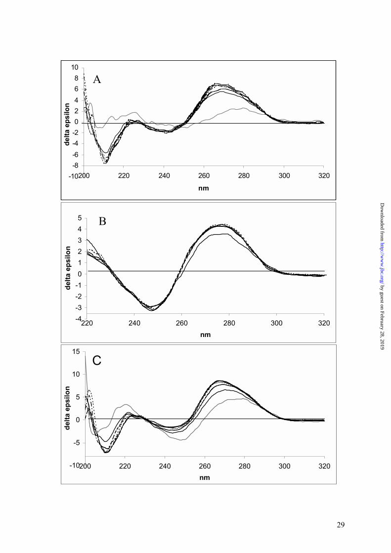
Complex formation between DNAzyme and its RNA substrate. The conformational
changes of the DNAzyme induced by Mg2+ were next analyzed by CD spectroscopy (Figure
6). CD spectra of an ion-free DNAzyme / RNA complex were reported earlier (24). We have
been interested in examining how far CD spectroscopy being sensitive to the structure helicity
and fold, could be applicable to probing binding of metal ions to single- and double stranded
species of interest. As the reference, CD spectra related to a regular complex formation
(Figure 6A), reflecting an antisense mechanism, were inspected first. CD spectrum of non-
enzymatic, single 21-mer DNA strand is irregular and of low magnitude. Addition of a
complementary RNA strand resulted in raising a regular positive Cotton effect at 269 nm, i.e.,
a region typical of the DNA/RNA hybrids (25). Influence of an increased Mg2+ concentration
on a double helical structure is not strong but clearly visible as indicated by increase of
amplitude of the Cotton effect. An initial addition of Mg2+ (0.5 mM) resulted in both a higher
Cotton effect than that produced by 1 mM Mg2+ and the formation of two characteristic,
discrete peaks (265 nm and 269 nm). At concentrations higher than 1 mM Mg2+, the height of
these hills is reversed and kept practically unchanged, even at higher Mg2+ concentrations of
up to 25 mM. The positive CD band of the Mg2+-free DNAzyme, although rather broad (276-
278 nm), is much more regular (Figure 6B) than that of the 21-mer oligodeoxynucleotide.
The spectrum also contains a negative effect at 249 nm and a weak, positive effect at 221 nm.
This indicates that some secondary structure exists for the 35-mer strand of MeO-β3DE, most
probably close to that predicted by the DNA folding algorithm (26). An addition of Mg2+ at
the initial concentration level (0.5 mM) led to a formation of a more regular and higher
amplitude positive band at 276 nm of a higher amplitude. Further Mg2+ additions (up to
25 mM) had no practical effect on the DNAzyme strand spectra. The binding of the Mg2+-
free MeO-β3DE strand to the target RNA leads to formation of a new type of spectrum with a
positive Cotton band at 270 nm of higher amplitude than that of the DNAzyme strand (Figure
6C). Both, the lack of the symmetry for this band and the appearance of the weaker negative
effect at 244 and positive at 223 nm are characteristic for the spectrum. Upon addition of an
initial amount of Mg2+ (0.5 mM), the amplitude of the positive Cotton effect rises
substantially and the peak is shifted down to 268 nm. No further increase of the Cotton effect
by guest on February 28, 2019http://w
ww
.jbc.org/D
ownloaded from

13
was observed above 1 mM Mg2+. The results presented above indicate that the global
geometry of the DNAzyme upon binding of Mg2+ adopts a compact structure projecting
flanking arms at right position and angles to bind substrate mRNA.
To further test this concept, the binding kinetics was directly measured by surface
plasmon resonance analysis in the presence of increasing concentrations of Mg2+. In these
experiments biotinylated RNA substrate was attached to the avidin coated sensor. To avoid
cleavage of the RNA substrate, the inactive DNAzyme containing a single nucleotide
substitution in the catalytic domain of β3DE was used. The antisense oligodeoxynucleotide
β3(1245-1265) consisting of both flanking arms (and thus antisense to β3 integrin subunit
mRNA) was tested as a control. To clarify the effect of cations on association and
dissociation processes between the DNAzyme and its RNA substrate, we have determined the
parameters of kon, koff, and KA in the binding reactions between RNA substrate and either
DNAzyme or the antisense oligodeoxynucleotide (Table II). The binding affinity of
β3DNAzyme to the RNA substrate was dependent upon the concentration of Mg2+. The
association constant determined in the presence of 15 mM Mg2+ was significantly higher
(p<0.001) than that observed at 5 mM of Mg2+. However, it was still much lower than the KA
describing the interaction of the antisense oligodeoxynucleotide with the same RNA
fragment. Interestingly, the binding affinity of the antisense oligodeoxynucleotide to the
RNA substrate did not depend upon cation concentration, and regardless of the Mg2+ presence
it was almost an order of magnitude higher than that of the DNAzyme.
DISCUSSION
The intracellular ability of various �10-23� DNAzymes to inhibit expression of the
targeted proteins was evidenced by several in vitro and in vivo studies indicating their
potential advantages as biocatalysts in oligonucleotide therapy (27). Despite tremendous
therapeutic potential, the ability of the DNAzyme to influence biological processes has not
been determined at the molecular level. Due to its high flexibility, the three dimensional
structure of the DNAzyme molecule is not yet known. Therefore, even a basic knowledge
about the mechanism by which Mg2+ and other divalent cations regulate enzymatic activity of
the 10-23 DNAzyme is at present speculative. The hypothetical mechanism for catalysis of
RNA cleavage by the DNAzyme is essentially based on assumptions that it behaves similarly
to the hammerhead ribozyme, which also is active in the presence of various divalent metal
by guest on February 28, 2019http://w
ww
.jbc.org/D
ownloaded from
14
cations (28). Despite different compositions, the �10-23� DNAzyme and the hammerhead
ribozyme show many common features, including the divalent cation dependence and kinetic
parameters of RNA cleavage. Exhaustive studies on chimeric DNAzymes and substrates
composed of DNA and RNA showed that both types of enzymes have a very similar catalytic
mechanism (5). The reactions have identical dependence on pH, both demonstrate an inverse
correlation between the pKa of metal hydrates and activity and solvent isotope effects and thio
effects on the reactions are identical (29). The crystal structure of several unmodified and
modified hammerhead RNA in the absence of divalent metal ions has been solved (28, 30-
32). Cation binding sites and the mechanism by which they control enzymatic activity have
been elucidated (30, 33). Five Mg2+ sites are seen in the crystal structure of the Mg2+-soaked
freeze-trapped conformational intermediate of the hammerhead ribozyme, which could be
divided into two groups based on their roles in catalytic activity. The first group consists of
Mg2+ sites that upon binding of cations induce folding of the enzyme into its active
conformation. The second group includes sites occupied by Mg2+ bound directly to the pro-R
oxygen at the cleavage site. The hammerhead ribozyme can cleave its own RNA and this
activity requires one or more catalytic divalent metal ions, one of which ionizes the 2�-
hydroxyl at the cleavage site. The newly generated nucleophile attacks the adjacent phosphate
by an in-line mechanism. The same metal ion, or perhaps another, stabilizes the
pentacoordinated phosphate transition state by binding directly to the pro-R phosphate
oxygen. The reaction generates 5�-hydroxyl and 2�,3�-cyclic phosphate termini at the cleavage
site (34).
The structural effect of Mg2+ is well established in the hammerhead ribozyme (35). In
the absence of divalent metal ions, the hammerhead structure is extended, with a disordered
core, but upon addition of metal ions folding occurs in two distinct steps. Both events are
well described by two-state transitions induced by the noncooperative binding of Mg2+. One
can assume that similarly to the hammerhead ribozyme, metal ions will induce the folding of
the DNAzyme molecule into the geometry required to facilitate the pathway into the transition
state, and will also bind at a specific location(s) where they can participate directly in the
chemistry of the cleavage reaction.
Data presented in this report show that binding of Mg2+ to the 10-23 DNAzyme
induces significant rearrangement of the catalytic loop, which leads to optimal folding of the
molecule. This folding may occur in several distinct stages. The first transition induced by
0.5 mM Mg2+ results in the formation of a compact structure of the DNAzyme. The
DNAzyme in such a state binds weakly to its RNA substrate and lacks catalytic activity. In
by guest on February 28, 2019http://w
ww
.jbc.org/D
ownloaded from

15
the next stage observed at concentrations up to 5 mM Mg2+, the flanking arms are projected
into the proper position to bind the RNA substrate. Under such conditions the DNAzyme
binds efficiently to the substrate and shows substantial catalytic activity. Further increase of
Mg2+ leads to the final transition involving formation of the completely organized catalytic
domain of the DNAzyme. Such a mechanism is supported by the following observations: (a)
EFRET of the Flu-MeO-β3DE(15)-Rhod rapidly increases in the range from 0 to 5 mM cations
and reaches a plateau value by 5 mM Mg2+, indicating at this concentration the shortest
distance between the energy donor and acceptor. In the absence of Mg2+, the DNAzyme is
inactive and its catalytic core essentially unfolded. With the addition of 5 mM Mg2+, the
orientation of the catalytic loop changes, and the distance between 5� and 3� ends almost
reaches the value characteristic for the DNAzyme in complex with its RNA substrate. Under
these conditions, the DNAzyme shows substantial enzymatic activity. A significant increase
in the catalytic activity of the DNAzyme is observed when the Mg2+ concentration is
increased from 5 to 15 mM suggesting that additional structural alterations within the catalytic
loop have occurred, even though there was no further change in EFRET. The hyperbolic
concentration dependence of the end-to-end distance with a midpoint of approximately 2 mM
Mg2+, significantly lower than that characteristic for the chemical cleavage step (Figure 2),
indicates that structural changes induced in the DNAzyme occur at much lower Mg2+
concentrations than those required for the catalytic properties. This conclusion is supported
by observation that the mutated inactive variant of the MeO-β3DE, with the shortened
catalytic loop, adopts the compact structure at a much lower Mg2+ concentration, indicating
that such a structural transition is not sufficient to gain catalytic activity. (b) The fluorescence
anisotropy of Flu-MeO-β3DE-Rhod doubles after exposure to the increasing concentrations
of Mg2+ or Ca2+ and reaches the maximum at their concentration of 1-5 mM, indicating the
increased condensation state of the molecule under these conditions. (c) Saturation effects of
Mg2+ concentrations detected by CD spectroscopy were produced in the range from 0.5 to 2.0
mM, i.e. somewhat lower than that described by other techniques. As expected, spectra of the
DNA / RNA hybrid (positive effect at 269 nm), used as a referenced structure close to a
regular A-type helix (36, 37), were much less sensitive to Mg2+ than those of the DNAzyme
strand and its complex with RNA. The Mg2+-free MeO-β3DE strand (effect at 276-278 nm)
undergoes a considerable change both upon Mg2+ binding (276 nm) and further structural
stabilization upon binding to the target RNA strand. It should be emphasized that these
conformational changes take place at a Mg2+ concentration as low as 0.5 mM. An overall
by guest on February 28, 2019http://w
ww
.jbc.org/D
ownloaded from
16
similarity of the DNAzyme / RNA complex spectra (Figure 6C) to that typical for A-type
RNA/DNA hybrids was observed (25). This also confirms an earlier observation based on
various Mg2+-free DNAzyme complexes, that the hybrid nature of the flanking arms strongly
influences their global geometry (24). (d) The final form adopted at 15 mM Mg2+, showing
high binding affinity towards the mRNA substrate, is in good agreement with the global form
of the structure observed under the same conditions for the hammerhead ribozyme (30, 38,
39). Interestingly, the binding affinity of the DNAzyme to RNA linearly increased in the
presence of divalent cations when they were used in the range from 0 to 15 mM, but it was
still much lower than that of the antisense oligodeoxynucleotide consisting of the DNAzyme
flanking arms. (e) Essentially the same effect on the folding, binding affinity to RNA
substrate and catalytic activity of the DNAzyme were found when other divalent cations such
as Ca2+ and Mn2+ were used in the same concentration range. Monovalent cations such as
Na+, K+, and Li+ added in place of Mg2+ even at much higher concentrations of up to 1M did
not show any effect.
Acknowledgements. This work was supported by projects Z-KBN 004/PO4/98 from the
Polish Committee for Scientific Research. The authors wish to thank Dr A. Okruszek for
synthesis of oligodeoxynucleotides and Dr M. Koziolkiewicz for stimulating discussions.
REFERENCES
1. Santoro, S.W., Joyce, G.F. (1997) Proc. Natl. Acad. Sci. USA 94, 4262-4266
2. Cieslak, M., Niewiarowska, J., Nawrot, M., Koziolkiewicz, M., Stec, W.J.,
Cierniewski, C.S. (2002) J. Biol. Chem. 277, 6779-87
3. Liu, C., Cheng, R., Sun, L.Q., Tien, P. (2001) Biochem. Biophys. Res. Commun.
284, 1077-1082
4. Zhang, L., Gasper, W.J., Stass, S.A., Ioffe, O.B., Davis, M.A., Mixson, A.J. (2002)
Cancer Res. 62, 5463-5469
5. He, Q.C., Zhou, J.M., Zhou, D.M., Nakamatsu, Y., Baba, T., Taira, K. (2002)
Biomacromolecules 3, 69-83
6. Santoro, S.W., Joyce, G.F. (1998) Biochemistry 37, 13330-13342
7. Chiu, T.K., Dickerson, R.E. (2000) J. Mol. Biol. 25, 915-45
8. Welche, J.B., Duckett, D.R., Lilley, D.M. (1993) Nucleic Acids Res. 21, 4548-
4555
by guest on February 28, 2019http://w
ww
.jbc.org/D
ownloaded from

17
9. Soyfer, V.N., Potaman, V.N.: Triple-helical nucleic acids. P. 360, Springer, New
York (1966)
10. Brukner, I., Susic, S., Dlakic, M., Savic, A., Pongor, S. (1994) J. Mol. Biol. 11, 26-
32
11. Dahlgreen, P.R., Lyubchenko, Y.L. (2002) Biochemistry 41, 11372-11378
12. Scott, W., Klug, A. (1996) Trends Biochem. Sci. 21, 220-224
13. Cierniewski, C.S., Babinska A., Swiatkowska M., Wilczynska, M., Okruszek, A.,
Stec, W. (1995) Eur. J. Biochem. 227, 494-499
14. Okumoto, Y., Sugimoto, N. (2000) J. Inorg. Chem. 82, 189-195)
15. Jaffe, E.A., Minich, R., Adelman, B., Becker, C., G., Nachman, R.L. (1976) J.
Exp. Med. 144, 209-221
16. Jaffe, E.A., Nachman, R.L., Becher, C.G., Minich, C.R., (1973) J. Clin. Invest. 52,
2745-2756
17. Clegg, R.M. (1992) Methods Enzymol. 211, 353-388
18. Clegg, R.M., Murchie, A.I.H., Zechel, A. and Lilley, D.M.J. (1993) Proc. Natl
Acad. Sci. USA 90, 2994-2998, (1993)
19. Bondeson, K., Frostell-Karlsson, A., Fagerstam, L., Magnusson, G. (1993) Anal.
Biochem. 214, 245-251
20. Kumar, P.K.R., Zhou, D.-M., Yoshinari, K. and Taira, K. (1996) In Eckstein, F.
and Lilley, D. M. J. (eds.), Catalytic RNA, Nucleic Acids and Molecular Biology,
volume 10, pp. 217-230, Springer-Verlag, Berlin, Germany
21. Dass, C.R., Saravolac, E.G., Li, Y., Sun, L.Q. (2002) Antisense Nucleic Acid Drug
Dev. 12, 289 �299
22. Zhou, D.-M., Usman, N., Wincott, F.E., Matulic-Adamic, J., Orita, M., Zhang, L.-
H., Komiyama, M., Kumar, P.K.R. and Taira, K. (1996) J. Am. Chem. Soc. 118,
5862-5866
23. Lakowicz, J.R. (1983) Principles of Fluorescence Spectroscopy, Plenum Press,
New York
24. Ota,N., Warashina,M., Hirano,K., Hatanaka, K., and Taira, K. (1998) Nucleic
Acids Res. 26, 3385-3391
25. Clark, C.L., Cecil, P.K., Singh, D., and Gray, D.M. (1997) Nucleic Acids Res. 25,
4098-4105
26. Zuker, M. (2003) Nucleic Acids Res. 31, 1-10
27. Khachigian, L.M. (2002) Curr. Opin. Mol. Ther. 4, 119-121
by guest on February 28, 2019http://w
ww
.jbc.org/D
ownloaded from

18
28. Dahm, S.C., Uhlenback, O.C. (1991) Biochemistry 30, 9464-9469
29. Ota, N., Warashina, M., Hirano, K., Hatanaka, K., Taira, K (1998) Nucleic Acids
Research 26, 3385-3391
30. Pley, H.W., Flaherty, K.M., McKay, D.B. (1994) Nature 372, 111-113
31. Scott, W.G., Finch, J.T. and Klug, A. (1995) Cell 81, 991-1002
32. Ruffner, D.E., Stormo, G.D., Uhlenbeck, O.C. (1990) Biochemistry 29, 10695-
10702
33. McKay, D.B. (1996) RNA 2, 395- 403
34. Scott, W.G., Murray, J.B., Arnold, J.R.P., Stoddard, B.L. and Klug, A. (1996)
Science 274, 2065-2069
35. Gurminder, S., Bassi., Alastair, I.H., Walter, F., Clegg, R.M., Lilley, M.J. (1997)
EMBO J. 16, 7481-7489
36. Moore, D.S. and Wagner, T.E. (1974) Biopolymers, 13, 977-986
37. Fairall, L., Martin, S. and Rhodes, D. (1989) EMBO J., 8, 1809-1817
38. Breaker, R.R., Joyce, G.F. (1995) Chem. Biol. 2, 655-660
39. Pley, H.W., Flaherty, K.M., McKay, D.B. (1994) Nature, 372, 68-74
by guest on February 28, 2019http://w
ww
.jbc.org/D
ownloaded from

19
Table I The estimated interfluorophore distance based on FRET analysis of the Fluo-MeO-β3DE-Rhod. The fluorophore-labeled MeO-β3DE (40 nM) was incubated with different divalent cations used in the concentration range from 0 to 25 mM and the interfluorophore distance R was evaluated based upon the Forster equation. Data represent a mean value of three separate determinations.
Interfluorophore distance [nm]
Concentration [mM] Mg2+ Ca2+ Mn2+
0 5 15 25
7.823 6.215 6.143 6.107
8.040 6.237 6.087 6.042
8.165 6.167 6.099 6.059
by guest on February 28, 2019http://w
ww
.jbc.org/D
ownloaded from

20
Table II.
Kinetic parameters for binding of β3DE or antisense oligodeoxynucleotide β3(1245-1265) to immobilized RNA substrate and their dependence upon Mg2+ concentration. The 3�-end biotinylated RNA substrate (GUUCCACUCGUUAUCUUC) was immobilized on a BIAcoreTM CM5 sensor chip coated with avidin (∆RU = 5000). The analyte was injected at a flow of 5 µl/min and measurements were done at 37oC using 50 mM Tris, pH 8.0, containing divalent cations at the indicated concentrations. MeO-β3DE used in these experiments was inactive due to a single base substitution (G6→A). The interaction of the corresponding oligodeoxynucleotide β3(1245-1265) containing both flanking arms of the β3DE with the same RNA substrate was analyzed under the same conditions. This oligodeoxynucleotide was modified as in MeO-β3DE. The kon and koff were determined from the association and dissociation phases, respectively, with four different concentrations of the DNAzyme and the oligodeoxynucleotide. Apparent KA corresponds to kon / koff ratio. Data are shown as a mean value of four separate analyses.
DNA Mg2+
[mM]Ca2+
[mM] kon
(1/Ms) koff
(1/s) KA
(1/M)
β3DE
5.0 15.0 25.0
5.0
15.0 25.0
(8.23+0.53)x104
(8.47+0.77)x104
(1.23+0.28)x105
1.20+0.11)x105 9.51+0.15)x104 8.55+0.79)x104
(1.98+0.94)x10-3 (2.99+1.05)x10-3 (2.84+1.53)x10-3
3.22+0.94) x10-3 3.09+0.86) x10-3 2.84+0.94) x10-3
(2.25+0.43)x107 (3.16+1.23)x107 (6.91+2.60)x107
2.74+0.84)x107 3.05+1.01)x107 5.71+2.71)x107
β3(1245-1265)
5.0 15.0 25.0
5.0 15.0 25.0
(3.44+1.04)x104
(6.04+2.10)x104
(8.49+2.15)x104
5.22+1.46)x104 7.84+1.48)x104 9.41+2.94)x104
(1.41+0.49) x10-4 (3.76+0.53) x10-4 (5.90+0.18) x10-4
8.40+1.88) x10-4 6.11+1.02) x10-4 7.21+1.50) x10-4
(1.64+0.29)x108 (1.78+0.88)x108 (1.46+0.40)x108
1.19+0.84)x108 1.37+0.52)x108 1.48+0.83)x108
Legends to Figures.
by guest on February 28, 2019http://w
ww
.jbc.org/D
ownloaded from

21
Figure 1. Cleavage of β3 integrin subunit mRNA substrate by DNAzymes
in vitro. Panel A shows the effect of Mg2+ concentration on enzymatic activity of
DNAzymes. MeO-β3DE(15) (•- •) and its mutant MeO-β3DE(11) (■-■) were incubated
with the RNA substrate (molar ratio 1:80) for 10 min in the presence of increasing
concentrations of Mg2+ ranging from 0 to 25 mM. The cleavage reaction was stopped
by addition of 0.5 M EDTA and the products were separated by electrophoresis in
20% polyacrylamide gels under denaturing conditions. Relative amounts of cleavage
products (%) are plotted versus Mg2+ concentration. Inserted is an autoradiogram of
the gel showing the cleavage product obtained at different Mg2+ concentrations (0 to
25 mM). Panel B shows catalytical activity of β3DE and MeO-β3DE. In these
experiments aliquots of the 32P-labelled mRNA substrate were incubated with
DNAzymes. β3DE and MeO-β3DE were used at a molar ratio (substrate to enzyme)
ranging from 5:1 to 80:1 for up to 60 min. at 37oC. The cleavage products obtained
after 10 min incubation of 32P-labelled mRNA substrate are shown alone (lane 1) or with
DNAzymes (β3DE, MeO-β3DE) mixed at the ratio 5:1 (lanes 2 and 7), 10:1 (lanes 3
and 8), 20:1 (lanes 4 and 9), 40:1 (lanes 5 and 10) and 80:1 (lanes 6 and 11).
DNAzymes were used at the concentration of 0.025 µM. Reactions were carried out
in 50 mM Tris, pH 8.0 containing 15 mM MgCl2, 0.01% SDS. Amounts of the product
were evaluated by a PhosphorImager (Molecular Dynamics) and used to calculate
kinetic parameters. They were determined in multiple turnover reactions and represent
a mean of three independent experiments.
Figure 2 Biological activity of Fluo-MeO-β3DE-Rhod. Panel A shows DNAzyme
activity of the fluorophore-labeled construct identical to that used for the FRET
analysis. The cleavage activity was examined after incubation of Fluo-MeO-β3DE-
Rhod with a 20-fold excess of 5�-32P-labelled RNA substrate in the presence of 15 mM
MgCl2 at 37°C. A sample was removed at different time points (lanes 1-5). Panel B
shows the fluorescence image of endothelial cells treated with Fluo-MeO-β3DE-Rhod.
Endothelial cells, exposed to 0.5 µM of the fluorophore-labeled construct for 24 hours
at 37oC and processed as described in Materials and Methods, were analyzed by
confocal fluorescence microscopy. The punctate fluorescence distribution within the
cytoplasm was detected by monitoring both fluorescein and rhodamine attached to
by guest on February 28, 2019http://w
ww
.jbc.org/D
ownloaded from

22
DNAzyme. Panel C shows the reduced expression of β3 mRNA in HUVECs treated
with MeO-β3DE when compared to unchanged expression in untreated cells. β3 mRNA
was evaluated by relative quantitative RT PCR using GAPDH mRNA as an intrinsic
control.
Figure 3. Fluorescence resonance energy transfer between fluorescein and rhodamine
attached to MeO-β3DE at 5� and 3� ends, respectively. Fluo-MeO-β3DE-
Rhod (40 nM) had two peaks at 520 nm and 580 nm upon excitation at 494 nm.
Inserted is an autoradiogram of a gel after electrophoresis of the Fluo-MeO-β3DE-
Rhod digested with endonuclease from Serratia marcescens. Degradation of the
fluorophore-labeled DNAzyme resulted in a significant increase in fluorescence
intensity at 520 nm and a disappearance of the 580 nm maximum.
Figure 4. FRET analysis for the MeO-β3DE as a function of Mg2+ concentration.
Panel A shows calculated interfluoropohore distances based on measured
efficiency of energy transfer presented as a function of MgCl2 concentration. The plot
shows the variation in FRET efficiency as a function of Mg2+ concentration up to
25 mM when Fluo-MeO-β3DE(15)-Rhod (•- •) or Fluo-MeO-β3DE(11)-Rhod (■-■) were
tested. In accordance with the model, the FRET efficiency is found to increase
(indicating a reducing end-to-end distance) over this complete range. Panel B shows
the scheme with the expected behavior of the free DNAzyme or complexed with its
RNA substrate in the presence of Mg2+. The distances R were calculated based on
FRET analysis.
Figure 5. Effect of Mg2+ on the fluorescence anisotropy of Fluo-MeO-β3DE-Rhod.
MgCl2 (●-●), CaCl2 (О-О), KCl (▲-▲), and NaCl (∆-∆) were added step-
wise to 0.5 µM Fluo-MeO-β3DE(15)-Rhod and incubated for 5 min before measuring
the fluorescence anisotropy. To monitor the fluorescence anisotropy of fluorescein
(Panel A) or rhodamine (Panel B), the samples were excited at 494 nm or 560 nm and
fluorescence emission was measured at 520 nm or 580 nm, respectively.
Figure 6. Effect of Mg2+ on CD spectra of the free MeO-β3DE and its complex with the
by guest on February 28, 2019http://w
ww
.jbc.org/D
ownloaded from

23
target RNA. CD spectra of the antisense oligodeoxynucleotide MeO-β3(1245-
1265)/ RNA complex (Panel A), the single stranded MeO-β3DE (Panel B) and the
MeO-β3DE / RNA complex (Panel C) were taken in the presence of Mg2+
concentrations ranging from 0 to 25 mM. However, since there was no change when
concentrations higher than 5 mM Mg2+ were used, those spectra were deleted for
clarity of the plot. Spectra of the single stranded antisense oligodeoxynucleotide
MeO-β3(1245-1265) and MeO-β3DE are shown as a grey line in Panels A and C,
respectively. Spectra were taken at the following Mg2+ concentrations: 0 mM ( ),
0.5 mM (-----), 1.0 mM (⋅⋅⋅⋅⋅⋅), 2.0 mM (-⋅-⋅-⋅-), 5.0 mM (-⋅⋅-⋅⋅-).
by guest on February 28, 2019http://w
ww
.jbc.org/D
ownloaded from
29
-10-8-6-4-20246810
200 220 240 260 280 300 320
nm
delta
eps
ilon
-4-3-2-1012345
220 240 260 280 300 320nm
delta
eps
ilon
-10
-5
0
5
10
15
200 220 240 260 280 300 320nm
delta
eps
ilon
C
A
B
by guest on February 28, 2019http://w
ww
.jbc.org/D
ownloaded from

Marcin Cieslak, Jacek Szymanski, Ryszard Adamiak and Czeslaw S. CierniewskimRNA induced by cations and their relations to the catalytic activity
Structural rearrangements of the "10-23" DNAzyme to beta 3 integrin subunit
published online September 2, 2003J. Biol. Chem.
10.1074/jbc.M300504200Access the most updated version of this article at doi:
Alerts:
When a correction for this article is posted•
When this article is cited•
to choose from all of JBC's e-mail alertsClick here
by guest on February 28, 2019http://w
ww
.jbc.org/D
ownloaded from

![[3,3]-Sigmatropic rearrangements - Massey Universitygjrowlan/stereo2/lecture11.pdf · 123.702 Organic Chemistry Claisen rearrangements • One of the most useful sigmatropic rearrangements](https://img.dokumen.tips/doc/110x75/5adcada77f8b9a213e8bd8b0/33-sigmatropic-rearrangements-massey-gjrowlanstereo2lecture11pdf123702.jpg)