Embed Size (px)
Citation preview
STRUCTURAL ELUCIDATION OF HUMAN OXIDATIVE METABOLITES OFMURAGLITAZAR: USE OF MICROBIAL BIOREACTORS IN THE BIOSYNTHESIS OF
METABOLITE STANDARDS
Donglu Zhang, Haiying Zhang, Nelly Aranibar, Ronald Hanson, Yande Huang, Peter T. Cheng,Shung Wu, Samuel Bonacorsi, Mingshe Zhu, Arun Swaminathan, and W. Griffith Humphreys
Pharmaceutical Candidate Optimization (D.Z., H.Z., N.A., M.Z., W.G.H.), Process Development (R.H.), Analytical A&D (Y.H.),Discovery Chemistry (P.T.C., S.B., S.W.), and Clinical Discovery (A.S.), Pharmaceutical Research Institute, Bristol-Myers
Squibb, Princeton, New Jersey
Received August 30, 2005; accepted November 4, 2005
ABSTRACT:
Muraglitazar (Pargluva), a dual �/� peroxisome proliferator-acti-vated receptor activator, is currently in clinical development fortreatment of type 2 diabetes. This study describes the structuralelucidation of the human oxidative metabolites of muraglitazarthrough the use of a combination of microbial bioreactors, NMRand accurate mass analyses, and organic synthesis. Plasma, urine,and feces were collected from six healthy subjects following oraladministration of 14C-labeled muraglitazar (10 mg, 100 �Ci) andpooled samples were analyzed. Approximately 96% of the recov-ered radioactive dose was found in the feces and 3.5% in the urine.The parent compound represented >85% of the radioactivity inplasma. The fecal radioactivity was distributed among 16 metab-olites (M1–M12, M14–M16, and M8a) and the parent drug, of whichhydroxylation and O-demethylation metabolites (M5, M10, M11,M14, and M15) represented the prominent human metabolites. Theurinary radioactivity was distributed into several peaks includingmuraglitazar glucuronide (M13) and the parent drug. Low concen-
trations of metabolites in human samples prevented direct identi-fication of metabolites beyond liquid chromatographic (LC)-massspectrometric analysis. Microbial strains Cunninghamella elegansand Saccharopolyspora hirsuta produced muraglitazar metabo-lites that had the same high performance liquid chromatographyretention times and the same tandem mass spectrometric (MS/MS)properties as the corresponding human metabolites. The microbialmetabolites M9, M10, M11, M14, M15, and M16 were isolated andanalyzed by NMR. Based on these LC-MS/MS and NMR analyses,and organic synthesis, the structures of 16 human oxidative me-tabolites were identified. The oxidative metabolism of muraglitazarwas characterized by hydroxylation, O-demethylation, oxazole-ring opening, and O-demethylation/hydroxylation, as well as O-dealkylation and carboxylic acid formation. This study demon-strated the utility of microbial bioreactors for the identification ofmetabolites.
Peroxisome proliferator-activated receptors (PPARs) are a set ofnuclear hormone receptors (comprising the �, �, and � subtypes). Thetwo most intensively investigated subtypes have been PPAR� (pri-marily expressed in the liver and demonstrated to play a critical rolein lipid metabolism) and PPAR� (primarily expressed in adiposetissue and implicated in insulin sensitization as well as glucose andfatty acid utilization). PPAR� is the target of the fibrate class ofhypolipidemic drugs such as fenofibrate (Balfour et al., 1990; Pack-ard, et al., 2002; Despres, 2001) and gemfibrozil (Spencer and Bar-radell, 1996), whereas PPAR� is the target of the thiazolidinedione(Mudaliar and Henry, 2001) class of antidiabetic drugs such as ros-iglitazone (Balfour and Plosker, 1999; Cheng-Lai and Levine, 2000;Goldstein, 2000) and pioglitazone (Gillies and Dunn, 2000). Mura-
glitazar (N-[(4-methoxyphenoxy)carbonyl]-N-[[4-[2-(5-methyl-2-phenyl-4-oxazolyl)ethoxy] phenyl]methyl]glycine, BMS-298585,Pargluva; Fig. 1), is an oxybenzylglycine analog (nonthiazolidinedi-one) dual �/� PPAR activator currently in clinical development forthe treatment of type 2 diabetes (Devasthale et al., 2005). This studydescribes the structural elucidation of oxidative metabolites of mura-glitazar in humans following oral administration of [14C]muraglitazarthrough the use of a combination of microbial bioreactors, NMR, andaccurate mass LC/MS.
The advantages of microbial bioreactors as a complementary invitro system for drug metabolism are the relatively low cost, mildconditions, ease of use, potential for efficient conversion with a highyield of metabolites, scale-up capability, and a potential to reduce theuse of animals. Microbial strains including the zygomycete fungusCunninghamella elegans and the actinomycete Saccharopolysporahirsuta have been used to study biotransformation and biosynthesis ofseveral drugs and have shown the ability to produce oxidative metab-
Article, publication date, and citation information can be found athttp://dmd.aspetjournals.org.
doi:10.1124/dmd.105.007153.
ABBREVIATIONS: PPAR, peroxisome proliferator-activated receptor; HPLC, high performance liquid chromatography; LC/MS, liquid chroma-tography/mass spectrometry; P450, cytochrome P450; TFA, trifluoroacetic acid; ESI(�), positive electrospray ionization; Q-TOF, quadrupole timeof flight; 1D, one-dimensional; 2-D, two-dimensional; TOCSY, total correlation spectroscopy; THF, tetrahydrofuran; EtOAc, ethyl acetate; TBS,Tris-buffered saline; MeOH, methanol; IBX, 1-hydroxy-1,2-benziodoxole-3(1H)-one-1-oxide; DMSO, dimethyl sulfoxide; ADME, absorption,distribution, metabolism, and excretion.
0090-9556/06/3402-267–280$20.00DRUG METABOLISM AND DISPOSITION Vol. 34, No. 2Copyright © 2006 by The American Society for Pharmacology and Experimental Therapeutics 7153/3079083DMD 34:267–280, 2006 Printed in U.S.A.
267
at ASPE
T Journals on June 23, 2017
dmd.aspetjournals.org
Dow
nloaded from

olite profiles similar to those found in mammalian species (Ikeda etal., 1985; Zhang et al., 1995, 1996a, 1997). Some of these microbialbiotransformation reactions are catalyzed by cytochrome P450 en-zymes (Zhang et al., 1995, 1996b; Yang et al., 1997). Therefore,microbial bioreactors should be useful for generation of metabolites insufficient quantity to aid in structural elucidation of human metabo-lites by spectroscopic and chromatographic methods.
Materials and Methods
Materials. [14C]Muraglitazar, with a radiospecific activity of 10 �Ci/mgand a radiochemical purity of 99.4%, was synthesized in multiple steps from[U-14C]phenol in a 20% overall yield at the Pharmaceutical Research Institute,Bristol-Myers Squibb. The structure of muraglitazar and the positions of 14Clabels are shown in Fig. 1. PEG-400 was purchased from Aldrich Chemical Co.(Milwaukee, WI). Trifluoroacetic acid (TFA) was purchased from EM Scien-tific (Gibbstown, NJ). Ecolite liquid scintillation cocktail was purchased fromPerkinElmer Life and Analytical Sciences (Boston, MA). Deuterated solventsD2O, CD3CN, and CDCl3 (D 99.8%) were obtained from Cambridge IsotopeLaboratories, Inc. (Andover, MA). All organic solvents and water were ofHPLC grade. Microbial strains were purchased from The American TypeCulture Collection (Manassas, VA).
Sample Collection and Preparation. The study in human subjects hadInstitutional Review Board approval and all subjects were required to giveinformed and written consent before participation in the study. Six healthyhuman subjects each received an oral dose of 10 mg (100 �Ci) of [14C]mu-raglitazar in PEG-400. Plasma (1, 4, 12, 24, and 48 h), urine (0–240 h), andfeces (0–240 h) were collected. Plasma was collected in EDTA and pooledacross the six subjects. The plasma (1 ml) was extracted with acetonitrile (3volumes) and centrifuged at 2000g for 10 min. The supernatant was saved andthe pellet was extracted twice with acetonitrile (3 ml). The radioactivityextraction efficiency was �95%. The combined supernatants were evaporatedto dryness by evaporation under a stream of nitrogen. The residue wasredissolved in 200 �l of acetonitrile/water (3:7) and centrifuged at 2000g for10 min before HPLC analysis. Urine and feces were collected for 0 to 240 hat 24-h intervals, and the volume of urine and the weight of feces weredetermined for mass balance. Fecal paste was prepared and pooled from allsamples collected up to 10 days across six subjects. Samples of pooled feces(1 g) were extracted by mixing with ethyl acetate (3 ml) and centrifuging at2000g for 10 min. The supernatant was saved and the pellet was extractedtwice with ethyl acetate (3 ml). The extraction efficiency of radioactivity was84.5%. The combined supernatants were evaporated to dryness by evaporationunder a stream of nitrogen. The residue was redissolved in 200 �l of aceto-nitrile/water (3:7) and centrifuged at 2000g for 10 min before HPLC analysis.Urine was concentrated directly under a stream of nitrogen. Plasma, urine, andfecal extracts were analyzed by HPLC separation, fraction collection, andradioactivity counting as well as by accurate mass LC/MS and LC-MS/MS.
Microbial Incubations and Isolation of Metabolites. The fungus Cun-ninghamella elegans (ATCC 20230) and the actinomycete Saccharopolysporahirsuta (ATCC 20501) were grown in 500-ml flasks at 28°C and 200 rpm ina rotary shaker on a medium consisting of 0.5% toasted NutriSoy (ADMHealth, Decatur, IL), 2% glucose, 0.5% yeast extract, 0.5% K2HPO4, 0.5%NaCl, adjusted to pH 7 with aqueous HCl (Rosazza and Smith,1979). Thebacterial strain was started from a 1-ml vial (stored in liquid nitrogen) in 100ml of medium grown for 3 days; then, 10 ml of the culture from this flask was
used to inoculate 100 ml of medium. The filamentous fungi were grown froma 1-ml spore suspension in 100 ml of medium. Muraglitazar (30 mg slurried in1 ml of methanol) was added to each flask after 24 h of growth of the secondstage bacterial culture or first stage fungal culture. The incubations werecontinued for 72 h, and then, the reactions were quenched with 100 ml ofacetonitrile. After storage for several hours at room temperature and 4 days at4°C, cells were removed by centrifugation at 3000g for 10 min. A 50-mlportion of each supernatant was extracted twice with ethyl acetate (50 ml foreach extraction). The combined ethyl acetate extracts were evaporated to neardryness under a stream of nitrogen. The residues were each reconstituted in 2ml of acetonitrile/water (3:7, v/v). The extracts were analyzed by accuratemass LC/MS and LC-MS/MS. For metabolite isolation, fractions were col-lected from five injections (100 �l) onto a 4.6 � 150 mm YMC HPLC column(YMC Co., Ltd., Kyoto, Japan). The microbial isolates were analyzed byLC/MS, LC-MS/MS, and NMR.
Radioactivity Detection. Radioactivity in the biological samples was de-termined by mixing an aliquot of the sample with 15 ml of Ecolite cocktail andcounting for 10 min using a Tri-Carb 2250 liquid scintillation analyzer(PerkinElmer Life and Analytical Sciences). The radioactivity in fecal homog-enates was determined by combusting the samples, followed by liquid scintil-lation counting. For HPLC profiling, the HPLC eluent was collected in96-deep-well Lumaplates and dried in a SpeedVac (Thermo Electron Corpo-ration, Waltham, MA). The plates were counted for 10 min per well using aTopCount scintillation analyzer (PerkinElmer Life and Analytical Sciences).
HPLC. HPLC was performed on a Shimadzu Class VP system equippedwith two pumps (model LC-10AT), an autoinjector (SIL 10AD), and a diodearray detector (SPD-M10A) (Shimadzu, Kyoto, Japan). YMC ODS AQ C-18columns (4.6 or 2.0 � 150 mm, 5 �) were used for all separations. HPLCeffluent fractions were collected into the 96-well plates at 0.26-min intervalsfor 70 min after sample injection with a Gilson model 202 fraction collector(Gilson Medical Electronics, Middleton, WI). The columns were eluted withsolvents A and B. Solvent A was water containing 0.06% TFA. Solvent B wasacetonitrile containing 0.06% TFA. The initial condition was 5% solvent B.The B composition was increased linearly to 25% (5 min), 40% (15 min), 53%(40 min), 60% (3 min), and 90% (2 min), and then was held at 90% for 7 min.The flow rates were 1 ml/min for the 4.6 � 150 mm column and 0.3 ml/minfor the 2.0 � 150 mm column. Metabolites in plasma, urine, and fecal extractswere monitored by radioactivity detection in collected fractions, and metabo-lites in microbial incubations were monitored by on-line UV detection at280 nm.
LC/MS and LC-MS/MS Analyses. A YMC ODS AQ C-18 column (2.0 �150 mm, 5 �) was used for LC/MS. A Thermo Electron LCQ deca XP massspectrometer with an ESI(�) source (Thermo Electron Corporation) was usedfor initial MS/MS analysis of fungal metabolites. The capillary temperatureused for analysis was 230°C. The nitrogen gas flow rate, spray current, andvoltages were adjusted to give maximum sensitivity for the parent compound.The collision energy was 20%. Accurate mass analysis was conducted on aMicromass Q-TOF Ultima mass spectrometer that was equipped with a Lock-Spray and an ESI(�) source (Waters, Milford, MA). LC-MS/MS analysis ofthe human and microbial metabolites was also performed with the Q-TOF witha collision energy of 15 to 25%. The HPLC eluent was directed to the massspectrometer. The desolvation temperature used for analysis was 300°C. Thenitrogen gas flow rate, and the spray and cone voltages were adjusted to givemaximum sensitivity for muraglitazar. The m/z 556.2771 of an infused 20ng/�l leucine enkephalin solution was used as lock mass. The Q-TOF wastuned to 18,000 resolution at half-peak height using an insulin tuning solution(at m/z 956.3), and was calibrated up to 1500 Da using a polyalanine calibra-tion solution. The experimentally obtained masses matched their respectivecalculated values with an error of less than 5 mDa (�10 ppm).
NMR. The synthetic materials and microbial isolates were analyzed on aJEOL ECL-500 MHz spectrometer (JEOL, Tokyo, Japan) or a Bruker Avance600 MHz system (Bruker, Newark, DE) equipped with a 5-mm Z-gradientprobe, or a 3-mm Nalorac probe. 1D 1H, 2D correlation spectroscopy, 2DTOCSY (total correlation spectroscopy), heteronuclear multiple quantum co-herence spectroscopy (one-bond carbon-proton correlation), heteronuclearmultiple-bond correlation spectroscopy (long-range carbon-proton correla-tion), and edited distortionless enhancement by polarization transfer experi-ments were performed. All chemical shifts are reported in ppm relative to
FIG. 1. Structure of muraglitazar with numbering system and major ESI(�)/MSfragmentation patterns.
268 ZHANG ET AL.
at ASPE
T Journals on June 23, 2017
dmd.aspetjournals.org
Dow
nloaded from

tetramethylsilane in CD3CN. For reference, a complete proton and carbon peakassignment of the NMR spectra was made on the parent drug.
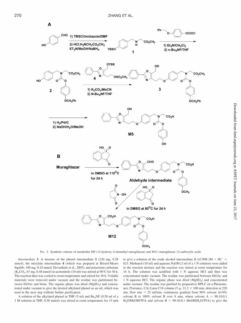
Synthesis of M5. The proposed structure for M5 based on mass spectralanalysis had a hydroxyl group on carbon-12 (Table 1). To confirm thisstructure, 12-hydroxy muraglitazar was synthesized in several steps (Fig. 2A),as described below.
Intermediate 1. A solution of 4-hydroxybenzaldehyde (1.0 g, 8.2 mmol),tetrabutylsilyl chloride (1.48 g, 9.8 mmol), and imidazole (660 mg, 9.8 mmol)in N,N-dimethylformamide (10 ml) was stirred at room temperature for 3 h.The reaction mixture was then partitioned between ethyl acetate (EtOAc) andbrine (saturated NaCl solution). The organic phase was washed with 10%aqueous lithium chloride, dried over anhydrous magnesium sulfate (MgSO4),and concentrated under vacuum to give the TBS ether, which was used in thenext step without further purification. A solution of the TBS ether, glycinemethyl ester hydrochloride (1.23 g, 9.8 mmol), and triethylamine (Et3N, 0.4ml, 9.8 mmol) in methanol (MeOH, 20 ml) was stirred at room temperature for18 h, and then sodium borohydride (NaBH4, 370 mg, 9.8 mmol) was cau-tiously added portion-wise. The reaction was stirred for 1 h at room temper-ature, followed by addition of saturated aqueous sodium bicarbonate(NaHCO3) and removal of volatiles under vacuum. The residue was partitioned
between EtOAc and saturated aqueous NaHCO3. The organic phase was dried(MgSO4) and concentrated under vacuum to give the amino ester intermediate1 (1.3 g, 51%) as an oil, LC/MS [M � H]� � 310. The oil was used in the nextreaction without further purification.
Intermediate 2. A solution of amino-ester intermediate 1 (500 mg, 1.6mmol), 4-benzyloxyphenyl chloroformate (510 mg, 1.9 mmol), and Et3N (0.27ml, 1.9 mmol) in methylene chloride (CH2Cl2, 10 ml) was stirred at roomtemperature for 2 h. The reaction mixture was then partitioned betweenCH2Cl2 and saturated aqueous NaHCO3. The organic phase was dried(MgSO4) and concentrated under vacuum to give the desired carbamate ester,LC/MS [M � H]� � 536, which was used in the next step without furtherpurification. To a solution of the TBS ether carbamate ester in tetrahydrofuran(THF, 10 ml) was added tetrabutylammonium fluoride (Bu4NF, 1.9 ml of a 1M solution in THF, 1.9 mmol) at room temperature. The reaction was stirredfor 1 h at room temperature and then was concentrated under vacuum. Theresidue was partitioned between EtOAc and aqueous 1 N hydrochloric acid(HCl). The organic phase was dried (MgSO4) and concentrated under vacuum.The residue was chromatographed on a silica gel column (stepwise gradientfrom 85:15 to 55:45 hexane/EtOAc) to give the phenol carbamate esterintermediate 2 (300 mg, 44%) as a clear oil, LC/MS [M � H]� � 422.
TABLE 1
Proposed structures of human oxidative muraglitazar metabolites
269HUMAN OXIDATIVE MURAGLITAZAR METABOLITES
at ASPE
T Journals on June 23, 2017
dmd.aspetjournals.org
Dow
nloaded from
Intermediate 3. A mixture of the phenol intermediate 2 (120 mg, 0.28mmol), the mesylate intermediate 4 (which was prepared at Bristol-MyersSquibb; 100 mg, 0.24 mmol; Devasthale et al., 2005), and potassium carbonate(K2CO3, 67 mg, 0.48 mmol) in acetonitrile (10 ml) was stirred at 90°C for 36 h.The reaction then was cooled to room temperature and stirred for 36 h. Volatilematerials were removed under vacuum and the residue was partitioned be-tween EtOAc and brine. The organic phase was dried (MgSO4) and concen-trated under vacuum to give the desired alkylated phenol as an oil, which wasused in the next step without further purification.
A solution of the alkylated phenol in THF (5 ml) and Bu4NF (0.50 ml of a1 M solution in THF, 0.50 mmol) was stirred at room temperature for 15 min
to give a solution of the crude alcohol intermediate 3, LC/MS [M � H]� �623. Methanol (10 ml) and aqueous NaOH (2 ml of a 1 N solution) were addedto the reaction mixture and the reaction was stirred at room temperature for16 h. The solution was acidified with 1 N aqueous HCl and then wasconcentrated under vacuum. The residue was partitioned between EtOAc and1 N aqueous HCl. The organic phase was dried (MgSO4) and concentratedunder vacuum. The residue was purified by preparative HPLC on a Phenome-nex (Torrance, CA) Luna C18 column (5 �, 21.2 � 100 mm; detection at 220nm; flow rate � 25 ml/min; continuous gradient from 90% solvent A/10%solvent B to 100% solvent B over 8 min, where solvent A � 90:10:0.1H2O/MeOH/TFA and solvent B � 90:10:0.1 MeOH/H2O/TFA) to give the
FIG. 2. Synthetic scheme of metabolite M5 (12-hydroxy O-demethyl muraglitazar) and M12 (muraglitazar 12-carboxylic acid).
270 ZHANG ET AL.
at ASPE
T Journals on June 23, 2017
dmd.aspetjournals.org
Dow
nloaded from

O-benzylphenyl carbamate acid 3 (69 mg, 40%) as a white solid. LC/MS[M � H]� � 609.
Metabolite M5. To a solution of the O-benzylphenyl carbamate acid 3 inMeOH (10 ml) was added 10% palladium on carbon catalyst (50 mg), and thereaction was stirred under an atmosphere of hydrogen at room temperature for30 min. After the catalyst was filtered off, the solution was concentrated undervacuum and the residue was purified as described for purification of theintermediate 3 to give metabolite M5 (25 mg; 43%) as a white powder. LC/MS[M � H]� � 519. 1H NMR (400 MHz, CD3OD): 7.91 (m, 2H), 7.40 (m, 3H),7.15 (m, 2H), 6.82 (m, 4H), 6.67 (m, 2H), 4.58 (s, 2H), 4.47 (2s, 2H), 4.18 (t,J � 6.5 Hz, 2H), 3.89 (2s, 2H), 2.98 (t, J � 6.5 Hz, 2H).
Synthesis of M12. The proposed structure for M12 based on mass spectralanalysis was oxidation of the methyl-12 to a carboxylic acid. To confirm thisstructure, muraglitazar 12-carboxylic acid was synthesized in two steps (Fig.2B), as described below.
Aldehyde Intermediate. To an 8-ml screw cap vial was added muraglitazar(52.5 mg, 0.10 mmol), 1-hydroxy-1,2-benziodoxole-3(1H)-one-1-oxide (IBX;281.0 mg, 1.0 mmol), dimethyl sulfoxide (DMSO; 4.0 ml), and a magnetic stirbar. The vial was capped and placed in an oil bath heated at 110°C for 24 h.The reaction mixture was diluted with 4.0 ml of acetonitrile and subjected topreparative HPLC isolation using a Phenomenex Luna C18 column, 21.2 �150 mm, 5 �, with solvent A (water with 0.05% trifluoroacetic acid) andsolvent B (acetonitrile with 0.05% TFA). The linear gradient used was 50% Bto 90% B in 10 min at a flow rate of 21.0 ml/min (monitored at 280 nm).Fractions containing the product were pooled, concentrated via rotary evapo-ration, and freeze-dried to afford an off-white powder (9.5 mg, 16% isolatedyield). 1H NMR (600 MHz, DMSO-d6) 9.9 (s, 1H), 8.08 (d, J � 7.6 Hz, 2H),7.63 (m, 1H), 7.58 (m, 2H), 7.26, 7.22 (d, J � 8.0 Hz, 2H, rotamers), 6.97 (m,2H), 6.90 (m, 4H), 4.53, 4.39 (s, 2H, rotamers), 4.33 (m, 2H), 3.84, 3.79 (s, 2H,rotamers), 3.72 (s, 3H), 3.38 (m, 2H); 13C NMR (100 MHz, DMSO-d6), 177.7,170.7, and 170.4 (rotamers), 163.0, 157.5, 156.5, 154.8, and 154.5 (rotamers),150.6, 145.7, 144.5, 132.4, 129.4 (4C), 129.0, 127.2 (2C), 125.4, 122.5, and122.4 (rotamers, 2C), 114.6 and 114.5 (rotamers, 2C), 114.2 (2C), 65.2, 55.4,50.8, and 50.7 (rotamers), 48.5 and 48.3 (rotamers), 26.3; high resolution MS[ESI(�)] calculated for C29H26N2O8Na ([M � Na]�): 553.1587; found:553.1575.
Metabolite M12. The reaction was carried out by mixing a sample of thealdehyde intermediate (9.0 mg) in 0.2 ml of DMSO and IBX (7.6 mg), andN-hydroxysuccinimide (9.8 mg). The reaction proceeded very slowly at am-bient temperature but achieved total conversion at 80°C for 24 h. The reactionmixture (about 0.14 ml) was diluted with acetonitrile (1.0 ml) and water (0.5ml). The resulting solution was used for product isolation via semipreparativeHPLC using a Phenomenex Luna C18 column, 10 � 150 mm, 5 �, withsolvent A (5% acetonitrile/95% water with 0.01 M NH4OAc) and solvent B(95% acetonitrile/5% water with 0.01 M NH4OAc). The linear gradient usedwas 10% B to 70% B in 20 min at a flow rate of 4.7 ml/min (monitored at 280nm). Fractions containing the product were pooled, concentrated via rotaryevaporation, and freeze-dried to afford a white powder (ca. 1 mg). 1H NMR(600 MHz, DMSO-d6) 7.96 (d, J � 6.6 Hz, 2H), 7.50 (m, 3H), 7.25 and 7.21(d, J � 8.2 Hz, 2H, rotamers), 6.89–7.00 (m 6H), 4.54 and 4.40 (s, 2H,rotamers), 4.26 (t, J � 7.3 Hz, 2H), 3.88 and 3.63 (s, 2H, rotamers), 3.72 (s,3H), 3.29 (t, J � 7.3 Hz, 2H); high resolution MS [ESI(�)] calculated forC29H27N2O9 ([M � H]�): 547.1717; found: 547.1727.
Results
Biotransformation Profiles in Human Plasma, Urine, and Fecesand in Microbial Incubations. After oral administration of [14C]mu-raglitazar (10 mg) to humans, urinary excretion was minimal (3.5% ofthe dose) and the majority of radioactivity was excreted in feces(approximately 96% of the recovered radioactive dose). The overallradioactivity recovery was low (64%) from this study, possibly be-cause of difficulties in accurately measuring the radioactivity in afecal paste preparation. An additional human ADME study showed�93% recovery of the radioactive dose in which feces was homoge-nized in a water and ethanol mixture. The fecal metabolite profile wassimilar in both studies (data not shown). The parent compound rep-
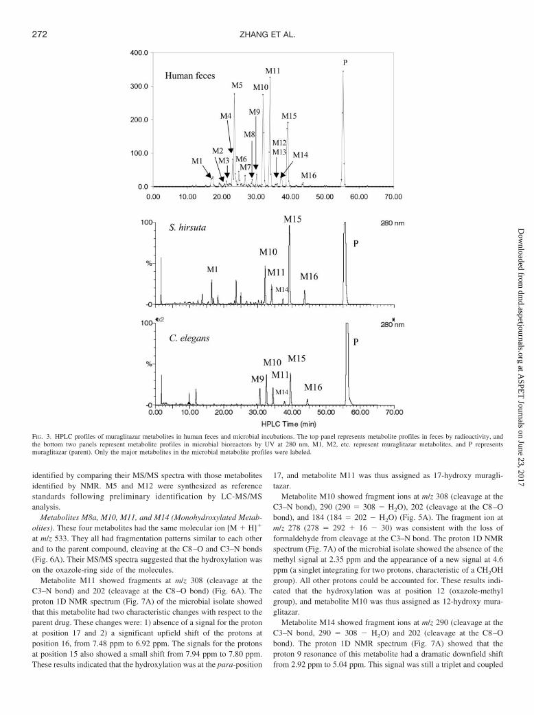
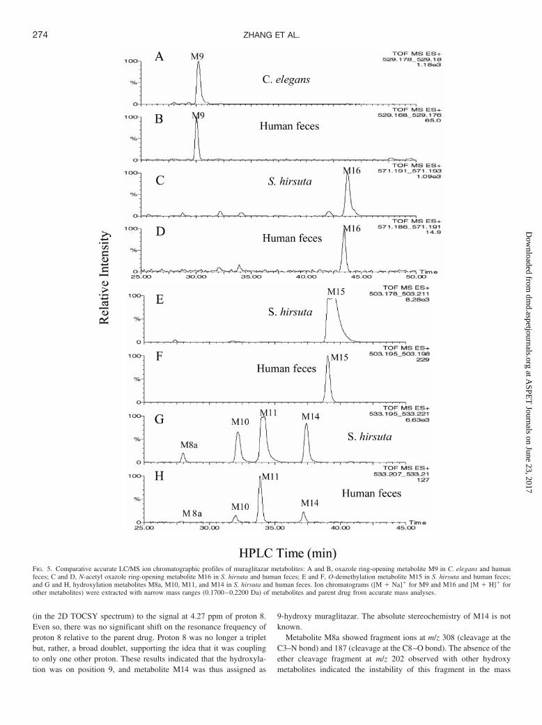
resents �90% of the radioactivity in human plasma at early timepoints and �85% at 48 h. No metabolites accounted for �5% of theradioactivity in plasma at any time point. Human metabolites identi-fied in plasma included M18, M5, M10, M11, M13, and M15. Figure3 shows metabolite profiles of human feces (by radioactivity) andmicrobial incubations (by UV at 280 nm). Four metabolite peaks (M5,M10, M11, and M15) and the parent compound (P) were the majorradioactive components in human feces and, together, accounted for�70% of the radioactivity in the fecal sample. Human urine containedseveral radioactive peaks including M13 and P, with M13 accountingfor approximately 1% of the dose. Figure 4 shows the selected ionchromatographic profiles of muraglitazar metabolites in human feces.There were four hydroxy metabolites (M8a, M10, M11, and M14),four hydroxy O-demethyl metabolites (M2, M5, M6, and M7), oneO-demethyl metabolite (M15), three dihydroxy metabolites (M3, M4,and M8), one dioxygenation with dehydrogenation metabolite (M12),and two oxazole ring-opening metabolites (M9 and M16). Becausehuman feces, as the major excretion route, contained low concentra-tions of metabolites of muraglitazar at a 10-mg dose, detailed struc-tural identification of fecal metabolites through isolation was notpractical. Microbial bioreactors were thus used to generate sufficientquantities for isolation and identification of the human metabolites.Microbes S. hirsuta and C. elegans produced high yields of metabo-lites which had the same retention times as human fecal metabolitesM9, M10, M11, M14, M15, and M16 (Fig. 3). In addition to thesemajor metabolites, the microbial strains also produced other oxidativemetabolites that had the same retention times and fragmentationpatterns as those found in human feces (partial data are shown inTable 2). Comparative HPLC/accurate MS chromatographic profileswere very similar between human feces and microbial incubations forselected muraglitazar metabolites as demonstrated in Fig. 5. In addi-tion, the mass spectral fragmentation patterns and molecular formulacalculated with a 5-ppm accuracy of these microbial metabolitesmatched the corresponding human metabolites (Table 2). Based onthese comparisons, the microbial metabolites were judged to be iden-tical to the human metabolites.
Identification of Metabolites. Typical fragmentation patterns ofmuraglitazar in a full-scan MS analysis showed cleavage at thebenzylic C–N bond adjacent to carbamate (C3–N) to give a fragmentat m/z 292 and cleavage at the ether bond (C8–O) to give a fragmentat m/z 186 (as shown in Fig. 1). Muraglitazar had a molecular ion [M� H]� at m/z 517 (Table 2). The key 1H NMR data (Table 3) formetabolite identification are the chemical shifts (ppm) at 2.35 for themethyl group at position 12, 2.92 for the methylene at position 9, 3.77for the methoxy group at position 23, and 7.47 or 7.48 for the aromaticprotons at positions 16 and 17.
Similar fragmentation patterns were observed with muraglitazarmetabolites M2 to M16. All monohydroxylation metabolites (M2,M5, M6, M7, M8a, M10, M11, and M14) showed a fragmentation ionat m/z 202 (186 � 16) or its dehydrated form at m/z 184 (202 � H2O),indicating that oxygen addition occurred on the oxazole-ring side ofthe molecules. Monohydroxylation metabolites, M2, M5, M7, M8a,M10, and M14 (but not M6 and M11, which were hydroxylated atposition 17), also showed a fragmentation ion at m/z 187.0756, whichhad a formula of C12H11O2, indicating that the nitrogen was lost in themass spectrometer after ether cleavage of the oxygenated molecules(187 � 186 � NH � oxygen). The mechanism for this gas phasereaction is not currently understood. NMR data of metabolites wereanalyzed by comparison to the spectra of the parent muraglitazar.Metabolites M9, M10, M11, M14, M15, and M16 were isolated fromincubations with C. elegans and S. hirsuta and identified by NMR andLC-MS/MS analyses. The structures of other human metabolites were
271HUMAN OXIDATIVE MURAGLITAZAR METABOLITES
at ASPE
T Journals on June 23, 2017
dmd.aspetjournals.org
Dow
nloaded from
identified by comparing their MS/MS spectra with those metabolitesidentified by NMR. M5 and M12 were synthesized as referencestandards following preliminary identification by LC-MS/MSanalysis.
Metabolites M8a, M10, M11, and M14 (Monohydroxylated Metab-olites). These four metabolites had the same molecular ion [M � H]�
at m/z 533. They all had fragmentation patterns similar to each otherand to the parent compound, cleaving at the C8–O and C3–N bonds(Fig. 6A). Their MS/MS spectra suggested that the hydroxylation wason the oxazole-ring side of the molecules.
Metabolite M11 showed fragments at m/z 308 (cleavage at theC3–N bond) and 202 (cleavage at the C8–O bond) (Fig. 6A). Theproton 1D NMR spectrum (Fig. 7A) of the microbial isolate showedthat this metabolite had two characteristic changes with respect to theparent drug. These changes were: 1) absence of a signal for the protonat position 17 and 2) a significant upfield shift of the protons atposition 16, from 7.48 ppm to 6.92 ppm. The signals for the protonsat position 15 also showed a small shift from 7.94 ppm to 7.80 ppm.These results indicated that the hydroxylation was at the para-position
17, and metabolite M11 was thus assigned as 17-hydroxy muragli-tazar.
Metabolite M10 showed fragment ions at m/z 308 (cleavage at theC3–N bond), 290 (290 � 308 � H2O), 202 (cleavage at the C8–Obond), and 184 (184 � 202 � H2O) (Fig. 5A). The fragment ion atm/z 278 (278 � 292 � 16 � 30) was consistent with the loss offormaldehyde from cleavage at the C3–N bond. The proton 1D NMRspectrum (Fig. 7A) of the microbial isolate showed the absence of themethyl signal at 2.35 ppm and the appearance of a new signal at 4.6ppm (a singlet integrating for two protons, characteristic of a CH2OHgroup). All other protons could be accounted for. These results indi-cated that the hydroxylation was at position 12 (oxazole-methylgroup), and metabolite M10 was thus assigned as 12-hydroxy mura-glitazar.
Metabolite M14 showed fragment ions at m/z 290 (cleavage at theC3–N bond, 290 � 308 � H2O) and 202 (cleavage at the C8–Obond). The proton 1D NMR spectrum (Fig. 7A) showed that theproton 9 resonance of this metabolite had a dramatic downfield shiftfrom 2.92 ppm to 5.04 ppm. This signal was still a triplet and coupled
FIG. 3. HPLC profiles of muraglitazar metabolites in human feces and microbial incubations. The top panel represents metabolite profiles in feces by radioactivity, andthe bottom two panels represent metabolite profiles in microbial bioreactors by UV at 280 nm. M1, M2, etc. represent muraglitazar metabolites, and P representsmuraglitazar (parent). Only the major metabolites in the microbial metabolite profiles were labeled.
272 ZHANG ET AL.
at ASPE
T Journals on June 23, 2017
dmd.aspetjournals.org
Dow
nloaded from

FIG. 4. Accurate LC/MS ion chromatographic profiles of [14C]muraglitazar metabolites in human feces oxazole ring-opening metabolite M9 (A); dioxygenation anddehydrogenation metabolite M12 (B); N-acetyl oxazole ring-opening metabolite M16 (C); the parent drug (D); dihydroxylation metabolites M3, M4, and M8 (E);O-demethylation metabolite M15 (F); hydroxylation and O-demethylation metabolites M2, M5, M6, and M7 (G); hydroxylation metabolites M8a, M10, M11, and M14(H); and radiochromatogram of human feces for comparison purpose (I). Ion chromatograms ([M � Na]� for M9 and M16 and [M � H]� for other metabolites) wereextracted with narrow mass ranges (0.1700–0.2200 Da) of metabolites and parent drug from accurate mass analyses. XIC, extracted ion chromatograms.
273HUMAN OXIDATIVE MURAGLITAZAR METABOLITES
at ASPE
T Journals on June 23, 2017
dmd.aspetjournals.org
Dow
nloaded from
(in the 2D TOCSY spectrum) to the signal at 4.27 ppm of proton 8.Even so, there was no significant shift on the resonance frequency ofproton 8 relative to the parent drug. Proton 8 was no longer a tripletbut, rather, a broad doublet, supporting the idea that it was couplingto only one other proton. These results indicated that the hydroxyla-tion was on position 9, and metabolite M14 was thus assigned as
9-hydroxy muraglitazar. The absolute stereochemistry of M14 is notknown.
Metabolite M8a showed fragment ions at m/z 308 (cleavage at theC3–N bond) and 187 (cleavage at the C8–O bond). The absence of theether cleavage fragment at m/z 202 observed with other hydroxymetabolites indicated the instability of this fragment in the mass
FIG. 5. Comparative accurate LC/MS ion chromatographic profiles of muraglitazar metabolites: A and B, oxazole ring-opening metabolite M9 in C. elegans and humanfeces; C and D, N-acetyl oxazole ring-opening metabolite M16 in S. hirsuta and human feces; E and F, O-demethylation metabolite M15 in S. hirsuta and human feces;and G and H, hydroxylation metabolites M8a, M10, M11, and M14 in S. hirsuta and human feces. Ion chromatograms ([M � Na]� for M9 and M16 and [M � H]� forother metabolites) were extracted with narrow mass ranges (0.1700–0.2200 Da) of metabolites and parent drug from accurate mass analyses.
274 ZHANG ET AL.
at ASPE
T Journals on June 23, 2017
dmd.aspetjournals.org
Dow
nloaded from
spectrometer. The result suggested that the hydroxylation was prob-ably at position 8 (methylene group). This hemiacetal structure wasexpected to have some degree of instability in aqueous environments,which was consistent with the low concentration of this metabolite inhuman feces and the subsequent formation of the O-dealkyl metabo-lite M1. Although the position for the hydroxyl group in M8a was notdetermined, available data supported a tentative assignment for M8aas 8-hydroxy muraglitazar.
Metabolite M15. M15 showed a molecular ion [M � H]� at m/z503 and major fragment ions at m/z 186 and 292 in the LC/MSanalysis, consistent with O-demethylation. 1H and 13C NMR analysesshowed that M15 lacked the OCH3 group (at 3.8 ppm for 1H in Fig.7B and 55 ppm for 13C) observed for the parent muraglitazar. Theonly change was the disappearance of the methoxy resonance ofposition 23. All other signals were in the same range as in the parent
molecule, including the ortho- and meta-positions in the correspond-ing aromatic ring (Table 3; Fig. 7B). This metabolite was assigned asO-demethyl muraglitazar.
Metabolites M9 and M16. These two metabolites had mass spectralcharacteristics distinct from those of muraglitazar and other metabo-lites. They showed a strong Na adduct of the molecular ions in LC/MSanalyses. Both of these metabolites did show typical fragmentationpatterns, cleaving at the C8–O and C3–N bonds.
Metabolite M9 had a molecular ion [M � H]� at m/z 507 (loss of10 Da from the parent compound) and fragment ions at m/z 282(cleavage at the C3–N bond, 282 � 292 � 10) and 176 (cleavage atthe C8–O bond, 176 � 186 � 10). Accurate mass analysis andformula calculation indicated that the 10-Da loss was due to elementalcomposition changes with an addition of oxygen and loss of ethylene(�O�C2H2). The UV spectral changes (from a strong absorptionpeak around 250–315 mm and a weak absorption peak around 210–240 mm to a strong absorption peak around 200–255 mm and a weakabsorption peak around 265–285 mm) suggested the removal of theextended aromatic system, consistent with an oxazole-ring opening.The proton 1D NMR spectrum (Fig. 7B) showed that the methylresonance at position 12 was lost and there was no new signal thatcould account for these protons, suggesting that this position was notsimply hydroxylated. These results indicated that the metabolite hadan oxazole ring-opening structure. Metabolite M9 was assigned as theoxazole ring-opening imide derivative of muraglitazar.
Metabolite M16 showed a molecular ion [M � H]� at m/z 549 (plus32 Da from the parent compound) and fragment ions at m/z 324(cleavage at the C3–N bond, 324 � 282 � 42, an acetyl group), 282(cleavage at the C3–N bond, 282 � 292 � 10), 218 (cleavage at theC8–O bond, 218 � 176 � 42, an acetyl group), and 176 (cleavage atthe C8–O bond, 176 � 186 � 10). This fragmentation patternsuggested that M16 was a derivative of M9 with the addition of anacetyl group. The proton 1D NMR spectrum showed a new signal inthe aliphatic region at 2.28 ppm that might be attributed to an acetylgroup (not shown). A theoretical prediction of the NMR spectrum ofthis proposed structure gave a value of 2.24 ppm for the acetyl group,in good agreement with the experimental value. These results sup-ported the idea that this metabolite was an N-acetyl derivative of M9,
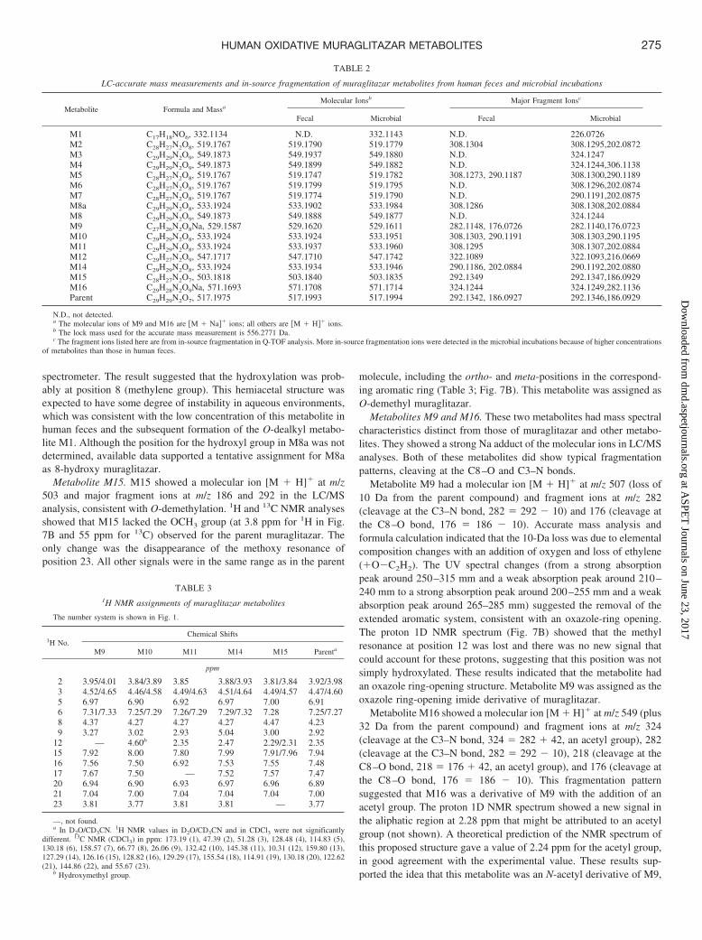
TABLE 2
LC-accurate mass measurements and in-source fragmentation of muraglitazar metabolites from human feces and microbial incubations
Metabolite Formula and MassaMolecular Ionsb Major Fragment Ionsc
Fecal Microbial Fecal Microbial
M1 C17H18NO6, 332.1134 N.D. 332.1143 N.D. 226.0726M2 C28H27N2O8, 519.1767 519.1790 519.1779 308.1304 308.1295,202.0872M3 C29H29N2O9, 549.1873 549.1937 549.1880 N.D. 324.1247M4 C29H29N2O9, 549.1873 549.1899 549.1882 N.D. 324.1244,306.1138M5 C28H27N2O8, 519.1767 519.1747 519.1782 308.1273, 290.1187 308.1300,290.1189M6 C28H27N2O8, 519.1767 519.1799 519.1795 N.D. 308.1296,202.0874M7 C28H27N2O8, 519.1767 519.1774 519.1790 N.D. 290.1191,202.0875M8a C29H29N2O8, 533.1924 533.1902 533.1984 308.1286 308.1308,202.0884M8 C29H29N2O9, 549.1873 549.1888 549.1877 N.D. 324.1244M9 C27H26N2O8Na, 529.1587 529.1620 529.1611 282.1148, 176.0726 282.1140,176.0723M10 C29H29N2O8, 533.1924 533.1924 533.1951 308.1303, 290.1191 308.1303,290.1195M11 C29H29N2O8, 533.1924 533.1937 533.1960 308.1295 308.1307,202.0884M12 C29H27N2O9, 547.1717 547.1710 547.1742 322.1089 322.1093,216.0669M14 C29H29N2O8, 533.1924 533.1934 533.1946 290.1186, 202.0884 290.1192,202.0880M15 C28H27N2O7, 503.1818 503.1840 503.1835 292.1349 292.1347,186.0929M16 C29H28N2O9Na, 571.1693 571.1708 571.1714 324.1244 324.1249,282.1136Parent C29H29N2O7, 517.1975 517.1993 517.1994 292.1342, 186.0927 292.1346,186.0929
N.D., not detected.a The molecular ions of M9 and M16 are �M � Na�� ions; all others are �M � H�� ions.b The lock mass used for the accurate mass measurement is 556.2771 Da.c The fragment ions listed here are from in-source fragmentation in Q-TOF analysis. More in-source fragmentation ions were detected in the microbial incubations because of higher concentrations
of metabolites than those in human feces.
TABLE 31H NMR assignments of muraglitazar metabolites
The number system is shown in Fig. 1.
1H No.Chemical Shifts
M9 M10 M11 M14 M15 Parenta
ppm
2 3.95/4.01 3.84/3.89 3.85 3.88/3.93 3.81/3.84 3.92/3.983 4.52/4.65 4.46/4.58 4.49/4.63 4.51/4.64 4.49/4.57 4.47/4.605 6.97 6.90 6.92 6.97 7.00 6.916 7.31/7.33 7.25/7.29 7.26/7.29 7.29/7.32 7.28 7.25/7.278 4.37 4.27 4.27 4.27 4.47 4.239 3.27 3.02 2.93 5.04 3.00 2.92
12 — 4.60b 2.35 2.47 2.29/2.31 2.3515 7.92 8.00 7.80 7.99 7.91/7.96 7.9416 7.56 7.50 6.92 7.53 7.55 7.4817 7.67 7.50 — 7.52 7.57 7.4720 6.94 6.90 6.93 6.97 6.96 6.8921 7.04 7.00 7.04 7.04 7.04 7.0023 3.81 3.77 3.81 3.81 — 3.77
—, not found.a In D2O/CD3CN. 1H NMR values in D2O/CD3CN and in CDCl3 were not significantly
different. 13C NMR (CDCl3) in ppm: 173.19 (1), 47.39 (2), 51.28 (3), 128.48 (4), 114.83 (5),130.18 (6), 158.57 (7), 66.77 (8), 26.06 (9), 132.42 (10), 145.38 (11), 10.31 (12), 159.80 (13),127.29 (14), 126.16 (15), 128.82 (16), 129.29 (17), 155.54 (18), 114.91 (19), 130.18 (20), 122.62(21), 144.86 (22), and 55.67 (23).
b Hydroxymethyl group.
275HUMAN OXIDATIVE MURAGLITAZAR METABOLITES
at ASPE
T Journals on June 23, 2017
dmd.aspetjournals.org
Dow
nloaded from

FIG. 6. Q-TOF MS/MS spectra of human fecal metabolites of muraglitazar: (A) hydroxylation metabolites M14, M11, M10, and M8a, (B) hydroxylation/ O-demethylationmetabolites M7, M6, M5, and M2, and (C) dihydroxylation metabolites M3, M4, and M8 and 12-carboxylic acid metabolite M12.
276 ZHANG ET AL.
at ASPE
T Journals on June 23, 2017
dmd.aspetjournals.org
Dow
nloaded from
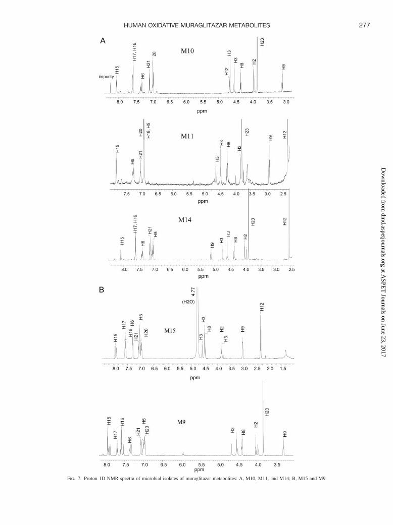
FIG. 7. Proton 1D NMR spectra of microbial isolates of muraglitazar metabolites: A, M10, M11, and M14; B, M15 and M9.
277HUMAN OXIDATIVE MURAGLITAZAR METABOLITES
at ASPE
T Journals on June 23, 2017
dmd.aspetjournals.org
Dow
nloaded from

and metabolite M16 was thus assigned as the oxazole ring-openingN-acetylimide derivative of muraglitazar.
Metabolites M2, M5, M6, and M7 (O-Demethylated and Hydroxy-lated Metabolites). These four metabolites had the same molecular ion[M � H]� at m/z 519 (519 � 517 � 16 � 14), consistent withhydroxylation and O-demethylation. They had typical fragmentationpatterns of cleavage at either the C8–O or the C3–N bond (Fig. 6B).Their MS/MS spectra also suggested that the hydroxylations were onthe oxazole-ring side of the molecules. M2, M5, M6, and M7 had thesame fragmentation patterns as monohydroxy metabolites M8a, M10,M11, and M14, respectively. Therefore, these compounds were as-signed as the O-demethylation metabolites corresponding to the hy-droxy metabolites M8a, M10, M11, and M14. M2 was tentativelyassigned as 8-hydroxy O-demethyl muraglitazar. Similar to the as-signment for M8a, available data supported the hydroxyl group at C8,although the final structure for M2 was not determined. M5 wasassigned as 12-hydroxy O-demethyl muraglitazar. M6 was assigned as17-hydroxy O-demethyl muraglitazar. M7 was assigned as 9-hydroxyO-demethyl muraglitazar.
The synthesis of the metabolite M5 is outlined in Fig. 2A. 4-Hy-droxybenzaldehyde was protected as the t-butyldimethylsilyl etherand then was subjected to reductive amination with glycine methylester hydrochloride to give the secondary amine 1. Acylation of amine1 with 4-benzyloxyphenyl chloroformate, followed by deprotection ofthe tert-butyldimethylsilyl ether, furnished the phenol carbamate ester2. Alkylation of phenol 2 with the hydroxylated phenyloxazole me-sylate 4, followed by deprotection of the TBS ether, provided thehydroxymethyl oxazole carbamate ester 3. Deprotection of 3 (hydrog-enolysis of the O-benzyl ether followed by ester hydrolysis) providedthe metabolite M5. Synthetic M5 had the same HPLC retention timeand MS/MS fragmentation patterns as the corresponding metabolitesfrom human feces (data not shown).
Metabolites M3, M4, M8, and M12. Metabolites M3, M4, and M8had the same molecular ion [M � H]� at m/z 549 (549 � 517 � 32),which was consistent with dihydroxylation. Metabolite M12 had amolecular ion [M � H]� at m/z 547 (549 � 517 � 32 � 2), whichwas consistent with dioxygenation and dehydrogenation. All of thesemetabolites showed a typical fragmentation pattern, with cleavage atthe C8–O and C3–N bonds (Table 2; Fig. 6C). The MS/MS spectrafor all the metabolites suggested that both hydroxylations were on theoxazole-ring side of the molecules.
Metabolite M4 showed fragment ions at m/z 531 (549 � H2O), 324(cleavage at the C3–N bond, 324 � 292 � 16 � 16), 306 (306 �324 � H2O), 218 (cleavage at the C8–O bond, 218 � 186 � 16 �16), and 200 (200 � 218 � H2O) (Fig. 6C). This fragmentationpattern was similar to that of the 12-monohydroxylation metaboliteM10 (Table 2; Fig. 6A). In addition, M4 had a characteristic fragmentat m/z 294, loss of formaldehyde (from cleavage at the C3–N bond,294 � 324 � CH2O); this fragmentation was also observed in the12-hydroxy metabolites M5 and M10 (at m/z 278). M4 also showed acharacteristic fragment at m/z 192.0687 (C10H10NO3), which could beformed from the cleavage between the C-9 and C-10 bond followedby a hydration. These results suggested dihydroxylation at the 9- and12-positions. Metabolite M4 was tentatively assigned as 9,12-dihydroxy muraglitazar.
Metabolite M8 showed fragment ions at m/z 324 (cleavage at theC3–N bond, 324 � 292 � 16 � 16) and 218 (cleavage at the C8–Obond, 218 � 186 � 16 � 16) (Fig. 6C). The fragmentation patternwas similar to that of 17-hydroxy muraglitazar except for the addi-tional 16-mass unit increase for each fragment. M8 was tentativelyassigned as 12,17-dihydroxy muraglitazar. Although another possibil-ity could be that the second hydroxylation occurs on the same phenyl
ring (16,17-dihydroxy muraglitazar), a trace level of a fragment at m/z306 (306 � 324 � H2O) in the MS/MS of M8 suggested that M8 wasmore likely to be 12,17-dihydroxy muraglitazar.
Metabolite M3 showed fragment ions at m/z 324 (cleavage at theC3–N bond, 324 � 292 � 16 � 16), 187 (cleavage at the C8–O bond,C12H11O2), and 192 (C10H10NO3) (Fig. 6C). Detection of the frag-ment ion at m/z 187 ruled out the possibility of 17-hydroxylation. Noother major fragment ions were observed with this metabolite. Theresult suggested dihydroxylation at positions 8 and 12. M3 wastentatively assigned as 8,12-dihydroxy muraglitazar.
Metabolite M12 showed fragments at m/z 322 (cleavage at theC3–N bond, 324 � 292 � 16 � 16 � 2) and 218 (cleavage at theC8–O bond, 218 � 186 � 16 � 16 � 2) (Fig. 6C). One structure thatwas consistent with these observations was muraglitazar 12-carboxylic acid. Other structures including 12-oxa-17-hydroxy mura-glitazar, 9-oxa-17-hydroxy muraglitazar, and 12-oxa-9-hydroxy mu-raglitazar could not be ruled out. To confirm the structure of M12,muraglitazar 12-carboxylic acid was synthesized with two oxidationsteps from muraglitazar. Literature procedures for the oxidation of amethyl group that is directly attached from an oxazole ring to analdehyde, and from an aldehyde to a carboxylic acid were used for thepreparation (Nicolaou et al., 2001; Mazitschek et al., 2002). SyntheticM12 had the same HPLC retention time and MS/MS fragmentationpatterns as the corresponding metabolites from human feces (data notshown).
Metabolites M13 and M18. A very small trace level of M13 wasdetected in human fecal extract by LC-MS/MS; however, this wasa relatively major metabolite in human urine. This metabolite hada molecular ion [M � H]� at m/z 693 and fragment ions at m/z 517,292, and 186 (the same fragment ions as muraglitazar), consistentwith an acyl glucuronide of the parent compound. M13 was ten-tatively assigned as an acyl glucuronide of muraglitazar. M18 wasa minor metabolite detected in plasma and urine. M18 had amolecular ion [M � H]� at m/z 709 and fragment ions of 533, 308,and 202, consistent with a glucuronide of hydroxy muraglitazar.M18 was assigned as the glucuronide of an isomer of hydroxymuraglitazar, although the position for the glucuronic acid was notdetermined.
Metabolite M1. This peak was a minor metabolite in human feces(Fig. 3). The radioactive peak had the same retention time as ametabolite in microbial incubations. Based on LC/MS and LC-MS/MS analyses, M1 was tentatively assigned as O-dealkyl muragli-tazar. The structures of muraglitazar metabolites are shown in Fig. 1.
Discussion
After oral administration of [14C]muraglitazar to healthy sub-jects, the parent drug was the major radioactive component in thecirculation. No single metabolite accounted for �5% of the totalplasma radioactivity. Muraglitazar acyl glucuronide, M13, ac-counted for approximately 1% of the dose in urine. Fecal excretionrepresented �96% of the recovered radioactivity. Major fecalmetabolites included M5, M10, M11, and M15. The parent drugaccounted for approximately 15% of the recovered radioactivedose in feces, whereas oxidative metabolites accounted for theremaining dose. Altogether, 16 oxidative and 2 glucuronide me-tabolites were identified from plasma, urine, and feces. Muragli-tazar and structurally characterized metabolites represented �95%of the radioactivity in excreta. The oxidative metabolites of[14C]muraglitazar in human feces were probably generated byintestinal and liver P450 enzymes followed by biliary excretion.An additional mechanism could include intestinal excretion ofmetabolites. It is unlikely that the fecal metabolites were generated
278 ZHANG ET AL.
at ASPE
T Journals on June 23, 2017
dmd.aspetjournals.org
Dow
nloaded from
by the enzymes in the intestinal microflora since these enzymes aregenerally reductive in nature. Consistent with these findings, in-cubation of [14C]muraglitazar in human liver microsomes in thepresence of NADPH generated M1, M9, M10, M11, M12, M14,M15, and M16 as prominent oxidative metabolites (data notshown). Although P450 enzymes are expected to catalyze forma-tion of the initial oxidation product at C-12 of muraglitazar (M10)and other oxidative metabolites, the additional oxidation steps ofthe hydroxyl group in M10 to a carboxylic acid group in M12 couldbe catalyzed by an alcohol dehydrogenase and an aldehyde oxi-dase.
During the development of new chemical entities, there are twogeneral questions regarding metabolite identification that need to beaddressed. The first concern is which metabolites (above what con-centration or what percentage of the dose) should be structurallycharacterized, and the second concerns the degree of characterizationthat is necessary for the metabolites. At the compound selection andoptimization stage, only major metabolites and potentially toxicand/or reactive metabolite(s) need to be identified to avoid com-pounds that may have unacceptably high clearance values or have thepotential to cause toxicity through reactive metabolite generation.However, radioactive materials are often not available at this time,and quantitation is often limited to estimation based on LC/UVdetection. Minor metabolites are often missed by UV detection at thisstage. Once in the development stage, ADME data in animals andhumans with radiolabeled materials will reveal the major circulatingmetabolite(s) and major clearance pathways. The ADME data willalso define relative concentrations of each metabolite in plasma, urine,bile, and feces. Since one of the main objectives for metabolismstudies is to support drug safety evaluation studies, the Metabolite InSafety Testing (MIST) committee suggested that any circulating me-tabolites and any significant metabolites in excreta of humans shouldbe characterized to understand the formation pathways of metabolites(Baillie et al., 2002). Identification of the major metabolites in humanexcreta aids not only in defining the major clearance pathways butalso in the design of drug-drug interaction studies. Major metabolitesin animals, which may not be as important as human metabolites, alsoshould be identified since they may explain the species-specific tox-icities observed in animals. Thus, there is a need to identify quanti-tatively and qualitatively important radioactive metabolites in studieswith radiolabeled materials.
The structural identification of metabolites can be carried out atmultiple levels. The goal of initial identification of metabolites isoften accomplished through LC/UV, LC/MS, and LC/MS/MS anal-yses to determine the biotransformation pathways involved in theclearance of the compound. Determination of the nature of themetabolites, such as oxygenation (hydroxylation or oxidation of aheteroatom), dioxygenation, dealkylation, and conjugation (glu-curonide, sulfate, glutathione, etc.), is often sufficient. The iden-tification of metabolites from biological matrices beyond thisinitial level is often challenging. Due to low concentrations andinterference from endogenous components, identification of me-tabolites with unusual structures presents additional challenges.Neutral loss and product ion scans have been used for real-time,data-dependent acquisition of full MS/MS data of expected metab-olites to improve the selectivity and sensitivity (Castro-Perez et al.,2002). We have used accurate mass spectrometry in this study forthe analysis of the metabolite profile of muraglitazar in humanfeces. The accurate mass measurements not only provided cleanerselective ion chromatograms (ion chromatograms extracted with amass accuracy to the second decimal place; Figs. 4 and 5) ofmetabolites in feces but also provided spectra for easy identifica-
tion of the molecular ions, which greatly enhanced the metaboliteidentification. Further refinement of this concept led to the devel-opment of a general mass defect filtering methodology (Zhang etal., 2003). Consequently, mass defect filtering can be used to aid inthe identification of molecular ions of metabolites with unusualstructures or present at low concentrations, as well as for metab-olites formed from expected biotransformations.
LC/MS and LC/MS/MS methods will often leave significant am-biguity in the exact structural identification of metabolites. The nextlevel of identification involves detailed structural elucidation of themetabolites by NMR and often requires quantities of materials notavailable from in vivo samples. In this study, we have demonstratedthe use of microbial bioreactors to produce large quantities of majormetabolites of muraglitazar for isolation and identification. Anothermajor human metabolite, M5, was not produced in a sufficient quan-tity from the microbial incubations for isolation; however, the hy-droxylation site in M5 was tentatively assigned from the isolation andpartial identification of the microbial metabolite, which directed or-ganic synthesis of the metabolite. The synthetic compound matchedthe human metabolite both by HPLC retention time and LC/MS andLC/MS/MS analyses.
In summary, the oxidative biotransformation pathways of muragli-tazar in humans include hydroxylation, O-demethylation, hydroxyla-tion/O-demethylation, oxazole-ring opening, O-dealkylation, and car-boxylic acid formation. A combination of microbial bioreactors, NMRand LC/accurate mass analyses, and organic synthesis helped tosuccessfully identify human fecal metabolites following oral admin-istration of [14C]muraglitazar. Microbial bioreactors proved to beextremely useful as a source for oxidative metabolites of muraglitazarand greatly aided in metabolite identification. This methodologyshould be broadly applicable to the determination of metabolite struc-tures for other new chemical entities.
Acknowledgments. We thank Dr. Jianmin Ren for review of themanuscript and Dr. Carl E. Cerniglia from National Center for Tox-icological Research for helpful discussions.
References
Baillie TA, Cayen MN, Fouda H, Gerson RJ, Green JD, Grossman SJ, Klunk LJ, LeBlanc B,Perkins DG, and Shipley LA (2002) Contemporary issues in toxicology. Drug metabolites insafety testing. Toxicol Appl Pharmacol 182:188–196.
Balfour JA, McTavish D, and Heel RC (1990) Fenofibrate. A review of its pharmacodynamic andpharmacokinetic properties and therapeutic use in dyslipidaemia. Drugs 40:260–290.
Balfour JA and Plosker GL (1999) Rosiglitazone. Drugs 57:921–930.Castro-Perez J, Hoyes J, Major H, and Preece S (2002) Advances in MS-based approaches for
drug and metabolism studies. Chromatographia 55:S-59.Cheng-Lai A and Levine A (2000) Rosiglitazone: an agent from the thiazolidinedione class for
the treatment of type 2 diabetes. Heart Dis 2:326–333.Despres JP (2001) Increasing high-density lipoprotein cholesterol: an update on fenofibrate. Am J
Cardiol 88(12A):30N–36N.Devasthale PV, Chen S, Jeon Y, Qu F, Shao C, Wang W, Zhang H, Cap M, Farrelly D, Golla
R, et al. (2005) Design and synthesis of N-[(4-methoxyphenoxy)carbonyl]-N-[[4-[2-(5-methyl-2-phenyl-4-oxazolyl)ethoxy] phenyl]methyl]glycine [Muraglitazar/BMS-298585], a novelperoxisome proliferator-activated receptor �/� dual agonist with efficacious glucose andlipid-lowering activities. J Med Chem 48:2248–2250.
Gillies PS and Dunn CJ (2000) Pioglitazone. Drugs 60:333–343 [discussion 344–345].Goldstein BJ (2000) Rosiglitazone. Int J Clin Pract 54:333–337.Ikeda Y, Kondo S, Kanai F, Sawa T, Hamada M, Takeuchi T, and Umezawa H (1985) A new
destomycin-family antibiotic produced by Saccharopolyspora hirsuta. J Antibiot (Tokyo)38:430–438.
Mazitschek R, Mulbaier M, and Giannis A (2002) IBX-mediated oxidation of primary alcoholsand aldehydes to form carboxylic acids. Angew Chem Int Ed Engl Suppl 41:4059–4061.
Mudaliar S and Henry RR (2001) New oral therapies for type 2 diabetes mellitus: the glitazonesor insulin sensitizers. Annu Rev Med 52:239–257.
Nicolaou KC, Barran PS, and Zhong YL (2001) Selective oxidation at carbon adjacent toaromatic systems with IBX. J Am Chem Soc 123:3183–3185.
Packard KA, Backes JM, Lenz TL, Wurdeman RL, Destache C, and Hilleman DE (2002)Comparison of gemfibrozil and fenofibrate in patients with dyslipidemic coronary heartdisease. Pharmacotherapy 22:1527–1532.
Rosazza JP and Smith RV (1979) Microbial models for drug metabolism. Adv Appl Microbiol25:169–203.
Spencer CM and Barradell LB (1996) Gemfibrozil. A reappraisal of its pharmacologicalproperties and place in the management of dyslipidaemia. Drugs 51:982–1018.
279HUMAN OXIDATIVE MURAGLITAZAR METABOLITES
at ASPE
T Journals on June 23, 2017
dmd.aspetjournals.org
Dow
nloaded from
Yang YF, Zhang DL, and Cerniglia CE (1997) Purification and characterization of a solublecytochrome P450 enzyme from yeast Trichosporon cutaneum. FEMS Microbiol Lett 154:347–353.
Zhang DL, Evans FE, Freeman JP, Duhart B, and Cerniglia CE (1995) Biotransformation of theamitriptyline by Cunninghamella elegans. Drug Metab Dispos 23:1417–1425.
Zhang DL, Evans FE, Freeman JP, Yang YF, Deck JD, and Cerniglia CE (1996a) Formation ofmammalian metabolites of cyclobenzaprine by the fungus, Cunninghamella elegans. Chem-Biol Interact 102:79–92.
Zhang DL, Hansen EB, Deck JD, Heinze TM, Luneau A, Korfmacher WA, and Cerniglia CE(1997) Fungal transformation of antihistamines: metabolism of cyproheptadine hydrochlorideby Cunninghamella elegans. Xenobiotica 27:301–315.
Zhang DL, Yang YF, Leakey J, and Cerniglia CE (1996b) Phase I and phase II enzyme produced
by Cunninghamella elegans for metabolism of xenobiotics. FEMS Microbiol Lett 138:221–226.
Zhang HY, Zhang DL, and Ray K (2003) A software filter to remove interference ions fromdrugs metabolites in accurate mass liquid chromatography/mass spectrometric analyses. JMass Spectrom 38:1110–1112.
Address correspondence to: Dr. Donglu Zhang, Pharmaceutical CandidateOptimization, Bristol-Myers Squibb, P.O. Box 4000, Princeton, NJ 08543. E-mail:[email protected]
280 ZHANG ET AL.
at ASPE
T Journals on June 23, 2017
dmd.aspetjournals.org
Dow
nloaded from