Embed Size (px)
Citation preview

Structural Changes in the Anuran MandibularArch during Metamorphosis, with referencetoRana Temporaria.1
By
H. K. Pusey, M.A.Assistant Lecturer, Department of Zoology, University College, London.
With Plates 33 to 45.
CONTENTS.
PAGE
1. F O R M E R W O R K A N D T H E P R O B L E M 4 8 0
2. A C K N O W L E D G E M E N T S . . . . . . . . 4 8 23 . N O M E N C L A T U R E A N D H O M O L O G I E S O F C E R T A I N C R A N I O - Q T T A D R A T E
A R T I C U L A T I O N S E M P L O Y E D B Y F O R M E R W O R K E R S . . . 4 8 3
4. M A T E R I A L , M E T H O D S , A N D F I G U R E S . . . . . 4 8 6
5. D E S C R I P T I O N O F S E V E N D E V E L O P M E N T A L S T A G E S — C A R T I L A G E
O N L Y 4 8 8
(a) S u m m a r y of a b o v e . . . . . . . 5176. D I S C U S S I O N . . . . . . . . . . 522
(a) T h e O t i c P r o c e s s e s a n d t h e M u s c u l a r P r o c e s s . . . 5 2 3(6) A s c e n d i n g P r o c e s s . . . . . . . 5 2 5(c) C o m m i s s u r a Q u a d r a t o - c r a n i a l i s A n t e r i o r . G a u p p ' s a n d
E d g e w o r t h ' s Theor i e s 5 2 6(d) Ligamentum Quadrato-ethmoidale and the Pterygoid
Process 527(e) Writer's view of the Nature of the Commissura . . 528(/) 'Pseudobasal Process' 529(g) Summary of the Homologies put forward in this Paper . 530
7. REDESCRIPTION OF THE Tir.MT>rm.Ar. REGION OF THE SKULLS OF . 531
(a) A s c a p h u s . . . . . . . . 532( 6 ) L i o p e l m a . . • • • • • • 535(c) A l y t e s 537
8. DISCUSSION . . . . . . . . . . 538(a) Probable course of Evolution from A s c a p h u s - t y p e to
Ranid-type . . . . . . . . 538(6) Nature of the Pseudobasal Process of A n u r a . . 539(c) A Theory of the Hyoid Suspension of the Quadrate . 542
9. THE PROBABLE EVOLUTION OF THE TADPOLE JAW APPARATUS . 5451 The substance of this paper was written in June 1936 and was success-
fully submitted as a thesis for the degree of Bachelor of Science in theUniversity of Oxford.
NO. 320 I i

480 H. K. PUSBYPAGE
1 0 . A N I N T E R P R E T A T I O N O F T H E M E A N I N G O F T H E J A W M E T A -
M O R P H O S I S . . . . . . • • • 5 4 61 1 . S U M M A R Y 5 4 7
1 2 . L I T E R A T U R E C I T E D . . . . . . . . 5 4 8
1 3 . A B B R E V I A T I O N S U S E D I N T H E P L A T E S . . . . . 5 5 0
1 4 . D E S C R I P T I O N O F P L A T E S . . . • • • . 5 5 1
1. FORMER WORK AND THE PROBLEM.
VERY many workers have undertaken investigations onvarious points of cranial morphology in the A n u r a. Of these,for the present purpose, Gaupp (1893 and 1906) is by far the mostoutstanding. Since Gaupp published his most excellent mono-graph in 1893, no other worker has investigated the problemsof jaw metamorphosis in such detail. Later work dealing withthe jaws has been related rather to muscles than to cartilages(Luther, 1914; Litzelmann, 1923). Passing reference has beenmade to jaw anatomy and metamorphosis by a very largenumber of other workers, but the whole matter is so complicatedby the relative movement of the parts, by the destruction ofpre-existing structures, and by the rapid growth and movementof new structures, that only a detailed stage-by-stage study iseffective in explaining them. The study of sections unaided byreconstructions is not an effective method of attack, and themanufacture of solid reconstructions must be hampered by thevery vague outlines shown by those cartilages which are under-going destruction. Consequently the description of the jawsduring the metamorphosis has not been advanced in any detailedway since 1893.
No attempt will be made to sum up or to assess the valueof work done prior to 1893; this labour has been fully and satis-factorily undertaken by Gaupp. Eeference to more recent workwill be made as required in the discussions at the end.
Gaupp's paper (1893) was very detailed, and I have foundhis observation of fact to have been extremely accurate asregards the jaws; I have detected no major errors, and have•found very little to correct even in the details. His accountis least full at the time of the fusion of the quadrate to theneurocranium by the otic and pseudobasal processes; I havetried to make good this deficiency.
SKULL AND ARCHES IN ANUEA 481
The description of jaw metamorphosis given here is takenpurely from my own investigations and is related particularlyto the reconstructions shown in the figures. The work wasundertaken independently of the accounts given by Gaupp andwas intended to serve as a check on them. The two accountsare so similar that it would be wearisome to make continualacknowledgement of Gaupp's findings; only occasionally willsuch reference be made. My own account is somewhat morecomplete in certain details of the destruction and buckling ofthe quadrate, and of its re-attachment in the temporal region.But though Gaupp's account is very excellent and can easilybe understood by a reader who is familiar with the details ofthe jaw metamorphosis and who has sections and reconstruc-tions available for study, it becomes somewhat involved forone not so equipped. The chief criticism to be brought againsthis paper is the scanty use he makes of figures. Such figures ashe gives are frequently oblique views of somewhat isolated partsof the skull or jaws, and are therefore not easily comparableone with another. It is to be hoped that these defects are absentfrom the present series of figures.
But though Gaupp's description of the facts is so thoroughand so reliable, his interpretation of those facts has now becomesomewhat out of date, and even at the time of publishing wasunable to account for the homology of certain structures. Morerecent work has made a reinterpretation imperative. A schemeof homologies has been devised in this paper, which takesaccount of all the structures concerned and relates them tostructures found in other frogs and in the Urodeles. The schememust, however, remain in part hypothetical until further workhas been undertaken, particularly on the development of theprimitive frog A s c a p h u s . It may be pointed out at oncethat the view here adopted is at variance in certain points fromthe newly developed theory of the hyostylic attachment of theAnuran jaws (Kruijtzer, 1931), and in these points containsnew—though as yet unproved—suggestions.
When Gaupp published his paper he was still unable to finda homologue in the Urode l a for his Anuran 'commissuraquadrato-cranialis anterior'; in addition, he was faced with the

482 H. K. PUSEY
problem of the possession by the frogs of separate larval andadult otic processes; whilst he disposed of the tadpole' muscularprocess' by saying that it was a larval specialization unrepre-sented in the U r o d e l a . More recently his interpretation ofthe 'basal articulation' has been called in question by de Beer(1926). Very recently, too, the primitive frogs A s c a p h u sand L i o p e l m a have been described by de Villiers, and byWagner; the descriptions given raise new problems. Whilstfinally the views supported by Kruijtzer call for further in-vestigation in the light of this newest work.
The main problems to be answered then seem to be:1. What is the homology of the 'commissura quadrato-
cranialis anterior' ?2. What light can be thrown on the possession by the frogs
during development of two otic processes and a muscularprocess ?
8. What is the true nature of the 'pseudobasal' articulation?4. What was the structure of the temporal region of the
skull of the ancestral Anuran?In addition, the present paper sets out to give a more detailed
account, with figures, of the jaw metamorphosis. An attemptis also made to see how far the evolution of the larval jawarrangement can be explained from comparative anatomy, andto give an interpretation of the metamorphic changes.
2. ACKNOWLEDGEMENTS.
The work was begun in the Department of Zoology andComparative Anatomy, Oxford, and was completed in theDepartment of Zoology, University College, London.
I wish to record my sincere thanks to Professor E. S. Good-rich, F.E.S., of Oxford, and to Professor D. M. S. Watson,F.E.S., of London, for their continual encouragement and adviceand for very kindly reading the manuscript.
I would particularly thank Dr. G. R. de Beer for suggestingthe problem, for supplying part of the material ready fixed, forhis helpful criticism and advice, and for drawing my attentionto certain relevant papers; Miss de Vos, of Stellenbosch, forsending me two drawings from sections of A s c a p h u s and for

SKULL AND AECHBS IN ANURA 488
checking certain facts and corrections on material in the Uni-versity Department of Zoology; and Miss Joyce Townend foradvice on the preparation of the figures.
3. THE NOMENCLATUEB AND HOMOLOGIES OF CERTAIN CRANIO-
QUADRATE ARTICULATIONS EMPLOYED BY FORMER WORKERS.
The interpretation of the attachments of the Anuran quadrateto the skull has long been obscure; the obscurity was at firstdue as much to confusion in the use of nomenclature as toinaccuracy in observation. An attempt will, therefore, be madeto follow through the names used by earlier workers and torelate them to the names at present in use; in this way it ishoped that a new value may be added to this earlier work.
In his famous Croonian lecture in 1858 Huxley introducedthe word ' suspensorium' as the name for a c o m m o n attach-ment of both the mandibular and the hyoid arches to the skull.In the frog-tadpole he then stated that the tadpole otic processwas the 'whole suspensorium, and not merely the quadratumof the branchiate vertebrate'. He thus introduced the erroneousidea of a hyoid element in this structure in the tadpole. Henoted correctly the ascending process and the commissuraquadrato-cranialis anterior but gave them no definite names.
Parker, in 1871, followed Huxley's false lead and in additionmade some original wrong observations. He believed that theupper end of the hyoid arch was fused on to the upper end of themandibular arch (tadpole quadrate cartilage) behind it. This,in fact, is quite untrue. It led him to call the tadpole otic processthe ' supra-hyomandibular'. He noted (1) the knob on thequadrate behind the ceratohyal; (2) apparently in later stages,the debris of the folded quadrate bar; and (3) in later stagesagain, the pseudobasal process. In their respective stages henamed a l l t h e s e s t r u c t u r e s 'infra-hyomandibular'. Thelower end of his supposedly fused ' hyomandibular' he called' symplectic bar ' ; this was the ' posterior spur of the quadrate'of this paper.
He had also seen the ascending process which he rightlybelieved to be of mandibular origin; this he variously namedthe 'metapterygoid connective or root', or 'mandibular root'.

484 H. K. PUSEY
In 1874 Huxley examined M e n o b r a n c h u s ( = N e c -t u r u s), and compared it with the frog. He noted the ascendingprocess, and gave its true relations to the branches of the Vthnerve; he called the basal process of the quadrate the ' pedicleof the suspensorium', and he noted the otic process and describedthe relations of the branches of the VHth nerve. But in hiscomparison of this U r o d e l e with the frog he made a numberof mistakes. In the tadpole he described the ascending processand homologized it with the 'pedicle' of M e n o b r a n c h u s ,thus making it a basal process, which is wrong; he even statedthat its relations to the branches of the Vth nerve were the sameas in the U r o d e l e !
In the metamorphosis of the frog he believed that his ' pedicleof the suspensorium' (correctly, the ascending process) wascarried outwards to become the inner attachment of the adult(the pseudobasal process). This is not the case, as the ascendingprocess is wholly destroyed; but in this way, by attributing toit an origin from the ascending process, he made the pseudo-basal process of the frog also the homologue of the basal processof the U r o d e l e , an error which persisted far into the presentcentury, with support from Gaupp (1893 and 1906). Hedescribed the frog's otic process. He was left to find in the froga homologue of the U r o d e l e ascending process, and in theU r o d e l e a homologue of the tadpole 'orbitar process'(muscular process). He was unable to meet this demand inview of his previous statements.
In the same year (1874) Huxley published his researches onthe columella auris. The findings of this investigation allowedhim to correct Parker's error of 1871 which had sprung fromhis own error expressed in his Croonian lecture. He now showedthat the mandibular arch (quadrate cartilage) of the tadpolecontained no fused hyoid element. In consequence he was ableto rename Parker's ' supra-hyomandibular' as an ' otic process',and show it to be homologous with that of the U r o d e 1 e s.
In 1876, in his second paper on the skull in the frogs, Parkeradmitted Huxley's correction, and changed his' supra-hyomandi-bular ' to ' otic process', but introduced a new error by sayingthat it was separated off to give the 'annulus tympanicus';
SKULL AND ABCHES IN ANUEA 485
(in 1871 he believed that it gave the columella cartilages of theadult).
He believed that the ascending process—now his 'pedicle',following Huxley's error—remained intact into the adult inthe toad (Bufo vu lga r i s ) , whilst in other frogs its outerend only remained, and developed a 'condyle of the pedicle'(the pseudobasal process) to abut against the auditory capsule;he thus assumed that the pseudobasal process was of quadrateorigin. Parker was therefore nearer to the truth than Huxley,in that he admitted the (partial) destruction of the pedicle(ascending process) and looked on the 'condyle of the pedicle'(the pseudobasal process) as a n e w g r o w t h ; but he was wrongin believing it to be an outgrowth of the ascending process or ofthe quadrate at all. Parker states these views more shortly inhis book on the Skull written jointly with Bettany, 1877.
Huxley (1876) in a paper on C e r a t o d u s confirmed theuse of the name ' pedicle' for the basal process of the quadrate.In the same year Parker, in his paper on U r o d e l e s I, madeclear his views on the homologies of the quadrate attachmentsin U r o d e l e s and frogs. In the U r o d e l e he recognized anotic, and an ascending process and also a pedicle (true basalprocess). But he went on to say that this 'pedicle' is thehomologue of the frog's pedicle which by description is thefrog's ascending process. And he was then compelled to saythat the frog had no ascending process. This is exactly Huxley'sview in 1874.
Stohr in 1879 gave a correct description of the otic, ascendingand basal processes in the U r o d e l a and, in 1881, was the firstworker to realize that the 'pedicle' of the Anuran (i.e. itsascending process) was the homologue of the ascending processof the U r o d e l a .
These continual errors in homology, coupled with a somewhatloose use of terms, and very real errors in the interpretation ofthe facts of development, greatly detract from the clarity andusefulness of the earlier work.
Gaupp in 1893 made a very exhaustive investigation of thefrog's chondrocranium and jaws; he reviewed work done upto that date, and corrected the errors of fact. But his system
486 H. K. PUSEY
of homologies for the cranio-quadrate attachments seems to thepresent writer not to be entirely satisfactory. However, hedescribed a tadpole and an adult-otic process, the ascendingprocess, and the' basal process', in addition to other attachmentswith which we are not immediately concerned. He believed thatthe basal process was the homologue of the structure of similarname in the Urodeles—this may possibly still prove to betrue, though it is not the view put forward in this paper. Buthis implied suggestion that the attachment of this process tothe auditory capsule is homologous with that of the U r o d e 1 eis certainly wrong. However, his view has been accepted sinceby Edgeworth (1925) and other workers (de Villiers and hispupils), at least to the extent that they use his nomenclaturefor naming these structures.
In 192G (pp. 810 and 886), however, de Beer pointed out thatthe articulation of this ' basal process' with the auditory capsulewas not similar to that of U r o d e 1 e s; he explained the differ-ences in the relations of the branches of the Vllth nerve in thetwo groups and he used the word 'pseudobasal' to describethis articulation. Nevertheless, he followed Gaupp in attributinga quadrate origin to the ' basal' process and retained the name'basal' to describe it. Goodrich (1909, fig. 59 B) had alreadyfigured this articulation and showed the correct relations of theVllth nerve to it, but he made no comment in the text.
In this paper an attempt will be made to show that this'basal process' is not a process of the quadrate at all, but israther to be considered either as the isolated end of the (cranial)'basitrabecular process', or as a forward outgrowth of the(cranial) 'post-palatine commissure'. Other recent workers(Kruijtzer, 1931) have put a different interpretation on thefacts and would return in effect to the standpoint of Parker(1871) with his 'supra- and infra-hyomandibular'. The longstory of the attempts to explain this process is therefore probablynot yet at an end.
4. MATERIAL, METHODS, AND FIGURES.
The tadpoles used were taken during three seasons as spawnor newly hatched larvae from water near Oxford. They were
SKULL AND ARCHES IN ANURA 487
reared in tanks in the laboratory and were fed on a liberal mixeddiet of animal and vegetable food and on D r o s o p h i l a aftermetamorphosis. Samples from the stocks were fixed daily;some in 4 per cent, formol, some in corrosive formol, and somein Bouin's fixative; those fixed in corrosive formol were sub-sequently treated with iodine solution and acid alcohol. Inaddition, young frog stages were captured in the wild aftermetamorphosis and were fixed similarly.
The material was studied by three methods:1. In serial sections cut at 15 or 20 p. both transversely and
sagittally for each stage. Borax carmine was usually used asa bulk stain (though sometimes omitted); and this was followedby picro-nigrosin.
2. Dissections of whole chondrocrania were made by vanWijhe's method, using victoria blue (van Wijhe, 1902 and 1922).
3. Alizarin transparencies were made by methods describedby Gray (1929), for the study of bone development.
Victoria blue preparations proved useful in showing thegeneral changes in the jaws as metamorphosis proceeds, andthey were used as a check on the proportions of the recon-structions made from sections. However, the dye failed tostain either young or degenerate cartilage, so that the resultingpicture obtained from the preparations was necessarily incom-plete, and to this extent proved most misleading in the inter-pretation of sections. Sometimes the dye failed to stain anycartilage at all (compare van Seters, 1922).
To overcome this difficulty two dimensional reconstructionswere made of seven stages. For each stage a series of pictureswas drawn with the aid of a microprojector at a magnificationof 33 x linear, from sagittal sections. Direct composite tracingsfrom these pictures gave accurate' contour-line reconstructions'.Such formed the basis of figs. 1 to 12, and 20, 23, and 24.Using the same series of pictures it was possible, by a pro-jection method, to produce the dorsal and anterior views shownin figs. 13 to 19, and 21, 22, 25, and 31. Figs. 26 to 30 weremade by the contour-line method from the published drawingsof de Villiers, Wagner, and van Seters. The writer takes fullresponsibility for any errors which may have crept into these

488 H. K. PUSEY
last five pictures on account of the small number of originaldrawings which could be combined to produce them. Miss deVos, of Stellenbosch, very kindly sent me two camera lucidadrawings from the series of A s c a p h u s sections worked on byde Villiers, which represent sections in advance of those figuredin his paper. Drawings from both sources were combined togive figs. 26 and 27.
The descriptions which follow are based on the study of somefifty animals prepared by the various methods given above.In addition, use was made of sections of E a n a , and of waxplate reconstructions made by the late Dr. J. W. Jenkinson,which are now in the collections of the Department of Zoologyand Comparative Anatomy, Oxford.
The writer hopes to publish the details of his methods ofreconstruction as a separate paper.
5. THE DESCRIPTION OF SEVEN DEVELOPMENTAL
STAGES—CABTILAGE ONLY.
S t a g e I (fig. 1, PL 33; fig. 8, PI. 36; fig. 17, PI. 39).
E x t e r n a l a p p e a r a n c e : Length of body 13 mm.; lengthof tail 23 mm.; hind legs less than 1 cm.; front legs hidden;horny teeth present on jaws and 'lips'.
The quadrate cartilage (q.) of the tadpole skull is a long fiatstrap of cartilage which lies laterally to the cranial floor; itreaches from the anterior third of the auditory capsule almostto the front of the snout and slopes downward as it passesforward, so that its anterior end is below the level of the cranialfloor (fig. 1, PI. 33; fig. 8, PL 36). It is also inclined at an angle sothat its upper surface faces obliquely inward toward the middleline. Behind, it is anchored to the chondrocranium by two attach-ments, the otic process and the ascending process. The oticprocess (fig. 1, PL 33, pot, and fig. 17, PL 39, pot.) is a small rodof cartilage which projects forwards, outwards, and downwardsfrom the wall of the lateral semicircular canal (Ic.) of the audi-tory capsule. It is completely fused at its two ends, above, tothe auditory capsule, and below, to the outer edge of the
SKULL AND ARCHES IN ANURA 489
quadrate cartilage (q.), and is thickest at its hinder end. Op-posite the otic process the quadrate cartilage is continuedinwards on its inner, hinder corner, as the ascending process(pa.) which is a rod of circular section sloping gently upward,as it passes to the wall of the neurocranium. It is completelyfused with the trabecular region of the skull immediately behindand a little below the foramen for the oculomotor nerve (focn.).Its root thus lies across the front of the vertically elongatedforamen pro-oticum (fp.) in such a way that nerves V2 and Vs
pass out of the foramen above and laterally to it, while Vx andVII palatine pass forward, below, and medially to it; VIIhyomandibular passes out laterally behind and below it. Theouter end of the process and the back of the quadrate bar lieacross the front face of the auditory capsule, but there is noconnexion between these structures until the otic process isreached at the side.
The quadrate cartilage bows out laterally as it passes forwardfrom the auditory capsule (fig. 17, PI. 39) and then swings backagain towards the middle line to the articular region (paq.).Here its inner border is suspended to the neurocranium by the'commissura quadrato-cranialis anterior' (Gaupp's nomen-clature) (fig. 8, PL 36, cqa., and fig. 17, PL 39, ego.). This isa flattened band of cartilage which sweeps steeply upward andinward to fuse completely with the neurocranial wall along itsventro-lateral edge, immediately behind the root of the trabe-cular horn (ct.). At this stage of development, as at earlierstages, the commissura passes backwards as it passes inwards(fig. 8, PL 36); this backward slope is more marked in youngeranimals. The base of attachment of the commissura to thetrabecula is very broad, so that its hind border slowly curvesbackward into the subocular shelf (fig. 8, PL 36, sst,, and fig. 17,PL 39, sst.). This shelf projects outwards and downwards fromthe side of the trabecula; in front it is sharp-edged laterally,but it becomes blunter on passing back and it finally disappearsin the region below the optic foramen (fig. 8, PL 36, fon.);however, even after this point, the trabecula continues toproject beyond the plane of the side wall of the cranium aboveit, as a flat-faced ledge which is continued backward and is

490 H. K. PUSBY
finally obliterated, as such, by the fusion of the ascendingprocess with it.
In the angle formed by the commissura and the subocularledge behind it, we find a flattened triangular cartilage pro-jecting backward into the subocular space; Gaupp has calledthis the 'processus pseudo-pterygoideus' (fig. 8, PL 36, ppq.,and fig. 17, PL 89, ppq.). It is very variable in its degree ofdevelopment in different animals of an otherwise similar stageand seems sometimes to be absent altogether.
The ' processus quadrato-ethmoidalis' (of Gaupp) lies oppositethe processus pseudo pterygoideus but at a lower level and onthe front edge of the commissura (fig. 1, PL 33, fig. 8, PL 36,and fig. 17, PL S9,pqe.). It projects forwards, upwards, andinwards, and is continued by means of the' ligamentum quadrato-ethmoidale' (of Gaupp) (fig. 8, PL 36, Iq.) to the side of thetrabecular horn; the ligament embraces the tip of the processand is continued backward along the inner edge of the quadratebar down to the pars articularis (paq.) as a fibrous band oftissue (the future pterygoid bone). Together the process and theligament make a lateral and anterior border to the inner nostrilwhich opens from the nasal sac into the mouth cavity; thetrabecular horn and the commissura make the inner and hinderborder of the nostril. Below the processus quadrato-ethmoidalis,the front edge of the commissura passes downward to theinner angle of the pars articularis of the quadrate (fig. 17, PL89, paq.).
The inner comer of the pars articularis is the lowest pointof the quadrate cartilage. From it the front border of thequadrate slopes upwards somewhat steeply, and at the sametime passes forwards (fig. 8, PL 36, and fig. 17, PL 39) to its mostanterior position; then, from here, again passes backward.Only the inner side of the spear-head so formed is the surfaceof articulation for the lower jaw. Prom the tip of the speara ligament runs forwards, upwards, and inwards to the tip ofthe trabecula horn (fig. 8, PL 36, tql.); it has not been describedbefore and it may be called the ' trabecular-quadrate ligament'.
Exactly opposite the commissura the outer edge of thequadrate cartilage is carried up into a high triangular ' processus
SKULL AND ARCHES IN ANUEA 491
muscularis' (Gaupp) (fig. 1, PI. 33, pmq., and fig. 17, PI. 39, pmq.)It is a thin flange of cartilage with slightly thickened, out-turned edges; it leans inwards and somewhat forwards (fig. 17,PI. 39) and is concave outwards. Its upper end is supportedto the commissura by loose fibrous strands. These and themuscular process complete the roof of a tunnel whose floor isthe quadrate cartilage, and whose inner and outer walls arethe commissura and muscular process respectively. Throughthis tunnel the mandibular muscles, nerves, and blood-vesselspass to the jaw region in front.
Finally, the quadrate cartilage is notched below the hinderpart of the muscular process to give a surface for the articulationof the ceratohyal (ch.) of the branchial apparatus (fig. 1, PL 33,we). In the region of this notch the quadrate is narrowest fromside to side and is thin in section, whilst just in front it isthickened (fig. 8, PL 36); these are points of importance in thesubsequent buckling of the quadrate bar, for this notch suppliesa point of weakness.
There are, then, three c a r t i l a g e attachments of thequadrate to the skull in the tadpole, (1) the commissura infront, (2) the ascending process medially behind, and (3) the(tadpole) otic process laterally behind. In addition it is heldby two ligaments to the trabecular horn, (1) the ligamentumquadrato-ethmoidale, and (2) the trabecular-quadrate ligament.
In the tadpole the whole of the quadrate bar lies behind thepars articularis and the lower jaw; consequently it does notsupply the cartilage skeleton of the upper jaw. This deficiencyis made good by the specialization of the trabecular horn andits apparent derivative, the supra-rostral cartilage (fig. 1,PL 33, els., and fig. 17, PL 39, els.) (= cartilago labialis superior,of Gaupp). The trabecular horn (fig. 1, PI. 33, ct., fig. 8, PL 36,fig. 17, PL 39, ct.) is a long flat strap of cartilage diverging gentlyfrom its fellow and projecting forward from the cranial floor.It expands slightly as it passes forward, and its outer end iscurved downward and is squarely truncated. The straight outerends of the two horns supply surfaces for the articulation of theupper edge of the supra-rostral cartilage. This latter cartilage(els.) is crescentic in form and deep dorso-ventrally; there is

492 H. K. PUSEY
a V-shaped notch in its upper border medially (fig. 17, PI. 39),while at the sides the horns of the crescent are obliquely cutaway from below and behind (fig. 1, PI. 38). It is supplied bya mandibular muscle, and it is opened by means of ligamentsfrom the lower jaw; so that it is movable within limits on theends of the trabecular horns, and acts as the functional upperjaw. Its lower edge, particularly on its inner, posterior side, isshod by a single crescentic horny tooth blade, formed by thecornification of the overlying epidermal tissue.
The lower jaw is made up of two elements on each side;these will here be called the anterior and posterior jaw cartilages(cartilago labialis inferior and cartilago Meckelii of Gaupp). Thefour elements comprising the entire mandible are set t r a n s -v e r s e l y across the snout in the form of a depressed / x - / \ ,the anterior cartilages lying in front of the posterior ones. Theanterior cartilages (fig. I, PI. 33, ajc, and fig. 17, PI. 39, ajc.) arerectangular blocks, somewhat curved, with their convexitydirected forward; they abut against one another medially withsquare-cut inner ends (fig. 17, PI. 39) and are held together bya copula—a ball of tissue whose histological character is thatof young cartilage; fibrous strands are also present. At theouter ends the ventral posterior corners are recurved and fitinto sockets on the anterior faces of the posterior jaw cartilages.These posterior elements (fig. 1, PI. 33, pjc, and fig. 17, PL 39,•pjc.) are of more complicated form. They are concave forward(fig. 17, PI. 39) and have a slight sigmoid curvature when seenfrom in front. The front face of the inner end is cupped andreceives the posterior corner of the anterior cartilage, whilethe lateral end is hooked. The hook fits over the pars articularisso that the outer end lies below it, while the remainder of thecartilage passes upwards and inwards, across the articulation;more medially the cartilage rises upward somewhat morerapidly to complete the S bend begun at its outer end (fig. 1,PI. 33). The posterior cartilage is supplied with mandibularand hyoid muscles which shut and open the mouth, and, inaddition, it is connected to the anterior cartilage, and to thesupra-rostral, by ligaments. A single U-shaped horny toothblade is bound on to the front faces of the anterior jaw cartilages
SKULL AND ARCHES IN ANUEA 493
by tough connective tissue, so that these three structures moveas a unit on the posterior cartilages behind; and these latter,in turn, move on the quadrates.
At this stage the cartilages of the nasal capsule have begunto be laid down. The hind wall of the capsule is formed by acurved band of cartilage which stands up vertically from theinner end of the commissura along its front border (fig. 8,PI. 36, ppn.). Behind, it abuts against the side wall of theneurocranium and fuses with it above and below, enclosingthus an orbitonasal foramen (forn.), through which nerve Y1
enters the nasal capsule over the root of the commissura.At its upper end the band of cartilage is turned forward to makethe roof (fig. 8, PI. 36, tn.) of the nasal capsule and is continuousmedially with cartilage (fig. 17, PI. 39, tn.) which itself is con-tinuous with the front, vertical wall of the neurocranium. Atthe side the roofing cartilage curves downward to form thelateral wall of the capsule behind. From the lateral wall,immediately above the front edge of the commissura, and restingon it, the rudiment of the 'proeessus antorbitalis' (fig. 8, PL 36,fan., and fig. 17, PI. 39, fan.) is seen to project straight forward.Its outer end, as also the free edges of the nasal roof, are, asyet, only formed in dense mesenchyme or in very youngcartilage, but they have been drawn with firm outlines in thefigures for the sake of clarity.
Farther forward in the nasal roof independent chondrificationhas begun (fig. 17, PI. 39, tn.). From it the 'cartilago obliqua'(co.) projects backward and downward; the 'cartilago alaris'(cal.) is present and in front of it lies the ' cartilago praenasalissuperior' (cps.); both these last in mesenchyme only. Also inmesenchyme, and lying on the upper surface of the trabecularhorn, is the rudiment of the 'cartilago praenasalis inferior'(cpi.), while behind it the side wall and the lateral part of thefloor of the nasal capsule are already present as young cartilage(pn.). A considerable nasal septum (sn.) is also developed inthe middle line between the trabecular horns. This stage isonly a little earlier than that figured obliquely from in front byGaupp (1893, fig. 22).
A description of the neurocranium itself may be obtained

494 H. K. PUSEY
from Gaupp's accounts (1893 and 1906); but certain featuresclosely affected by the changes in the jaws may be noted here.The optic foramen (fig. 1, PI. 38, fon.), and the oculomotorforamen (focn.), lie immediately above the projecting ledge of thetrabecula; this separates them from the two palatine foraminabelow for the carotid artery and its palatine branch—the'foramen caroticum primarium' behind (fcp.), and the 'foramencranio-palatinum' in front (Jc). The four foramina mark theupper and lower limits of the original trabecula, and theirchanges in size indicate the degree of destruction of this barwhich takes place in later stages. As yet no destruction hastaken place and the arrangement is typical of the tadpole skull.
Skul l s E a r l i e r t h a n S tage 1.
Gaupp has figured and described such earlier skulls (his stage 2and figs. 12 and 13). The skull figured as stage 1 in this paperhas advanced towards the adult condition in that the rudimentsof the antorbital process, and of the nasal cartilages, havedeveloped. Earlier skulls differ in the absence of these cartilages.In a typical fully formed tadpole skull nerve Vx passes overthe root of the commissura, close against the side wall of theskull. Below it the anterior border of the commissura is thick-ened but as yet no antorbital process extends forwards from it,nor has the back wall of the nasal capsule grown up laterallyto nerve Vx or arched up over it to fuse above and below it withthe cranial wall. Vx is, therefore, not enclosed in an orbito-nasalforamen.
Apart from these differences the figures and description ofstage 1 may be taken as typical of the tadpole skull.
S tage 2 (fig.2,PL33;fig.9,PI.37;fig. 18,PI.40;fig.20,PI.41).
E x t e r n a l a p p e a r a n c e : length of body, 11-7 mm.; length oftail, 20-5 mm.; hind legs more than 1 cm.; arms enclosedbut bulging to the sides; body triangular; tail web narrowing;no horny jaw- or ' lip '-teeth present.
This stage shows a number of advances on stage 1. The lowerjaw is set less transversely across the snout and this is particularlytrue of the posterior jaw cartilage (fig. 2, PI. 33, pjc, and fig. 18,

SKULL AND ARCHES IN ANURA 495
PL 40, jy'c.); the anterior jaw cartilage (ajc.) is not curved backin the middle line. The posterior cartilage is undergoing arotation on its long axis, so that its former dorsal surface isnow facing d o r s o - l a t e r a l l y and its articular notch is facingbackward and u p w a r d rather than backward and down-w a r d . (These are significant differences in spite of the factthat animal 1 died with its mouth shut and animal 2 with itsmouth open). Both jaw cartilages have been remodelled by aprocess of cartilage resorption; the matrix has been removedin places leaving only a rather indeterminate shell of peri-chondrium bounding now empty spaces. Only definitive carti-lage is shown in fig. 2, PL 33, but in fig. 18, PL 40, the formeroutlines of the pre-existing cartilage have been put in in dottedlines to assist description. A small area of cartilage matrixhas been removed from the anterior lower face of the posteriorjaw cartilage in front of the articular region, whilst a muchgreater erosion has taken place along its postero-ventral border;the external dotted line (fig. 18, PL 40) shows the posterior formerlimit, and the internal (anterior) dotted line the anterior limitof the matrix so removed. In the anterior jaw cartilage at leasta third of its former volume of matrix has been removed alongits antero-ventral face, whilst some erosion has also taken placeposteriorly toward the middle line. The firm lines (fig. 18,PL 40) show the limits of the uneroded, definitive jaw cartilages.
The horny tooth blades have been lost from both jaws andthe teeth have also been shed from the 'lips', which have becomemuch reduced.
The three cartilage processes attaching the quadrate cartilageto the neurocranium have all undergone changes—eithersoftening and distortion, or actual cartilage erosion. Thecartilage along the posterior margin of the commissura has beenremoved, leaving only a ragged casing of perichondrium (dottedlines, fig. 9, PL 37, and fig. 18, PL 40); this erosion has completelydestroyed the processus pseudopterygoideus. In addition, thedestruction has extended to the subocular shelf along the sideof the trabecula; it has taken place from the commissura to theascending process. In front, the medial part of the trabeeularemains as the ventro-lateral wall of the skull, but it shows a
NO. 320 K k

496 H. K. PUSBY
raw outer edge from which the shelf has been eroded. Fartherback, however, in the region of the optic and oculomotorforamina, the destruction has been more extensive, the wholethickness of the trabecula being involved. Below the opticforamen (fig. 2, PL 33, fon.) only the perichondrial walls of thetrabecula remain, except for a ventral cartilage bridge justdorsal to the foramen cranio-palatinum (fc). With the exception,therefore, of the presence of the perichondrium, the area of theoptic foramen has been greatly increased ventrally at theexpense of the trabecula. The presence of debris in this regionis shown in fig. 2, PI. 33, by a dotted margin to the formeroptic foramen; the foramen cranio-palatinum still retainsuneroded margins.
The destruction has gone farther between the oculomotorforamen (focn.) and the foramen caroticum primarium (fcp.).The two foramina are now virtually joined into one, except fora bridge of perichondrium. Between the now enlarged opticand oculomotor foramina there still remains a narrow pillar ofuneroded cartilage representing about half the original thicknessof the trabecula. The destruction of the subocular shelf, thetrabecula, the commissura, and the ascending process, and thesoftening of the top of the muscular process and of the tip ofthe tadpole otic process, all take place simultaneously. Thechanges are thus probably due to the same causes, which maywell be some hormone release.
Now that the posterior angle between the commissura andthe trabecula has been softened and freed of the obstructionoffered by the subocular shelf and the processus pseudo-pterygoideus, the whole commissura has rotated backwardabout its attachment to the brain case, so that it slopes b a c k -w a r d s rather than forwards in passing out from the skull(compare fig. 8, PL 36, and fig. 9, PI. 37). In addition, alongits anterior edge, low down near the pars articularis, there hasbeen some further slight erosion leading to a narrowing of widthof the effective articulating surface of the quadrate (dottedline, fig. 9, PL 37).
The ascending process (pa.) has undergone destruction with-out any change in its position. The matrix of the middle of its

SKULL AND AECHBS IN ANURA 497
length has been removed and there remains only the vaguesttrace of perichondrium joining its two ends. This residue ofperichondrium is not shown in fig. 2, PI. 33, and fig. 18, PI. 40,which show only the stump projecting from the front borderof the pro-otic foramen and the outer end projecting in fromthe quadrate cartilage (fig. 2, PL 33, pa.; fig. 9, PL 37, pa.;and fig. 18, PL 40, pa.).
The otic process (fig. 2, PL 3S,pot., and fig. 18, PL 40,pot.) hasbeen softened, if one may judge by its buckled condition, andits subsequent partial destruction in stage 3. Its base, con-necting it with the auditory capsule, has been extended, andthe edge of the capsule itself has been widened laterallyforming the beginning of a 'crista parotica' (fig. 9, PL 37, cp~).At its outer end the process has been forced backward and isnow bent on itself (fig. 2, PL 33). Histologically its cells showno degeneration, and there is no loss of matrix.
The rotation about the top of the commissura has allowed thewhole quadrate bar to move backward; it has apparently metwith resistance from the otic process. As shown above, the oticprocess itself has been bent, but the outer edge of the quadratecartilage has also been thrown into undulating folds (fig. 9,PL 37, and fig. 18, PL 40). There has been a resorption of matrixfrom the cartilage along its extreme lateral border in front ofthe otic process (dotted line, fig. 18, PL 40) but the perichondriumremains intact. In spite of the forces which must be actingin this process of buckling, the hind border of the quadrate—and its ascending process—have not moved back to any extent,and the lateral head vein and the hyomandibular branch ofnerve VII he uncrushed between them and the front face of theauditory capsule below the otic process.
In addition to moving backward the quadrate bar has becomeless horizontal and more vertical in position, thus opening upthe angle which the whole bar makes with the cranial floor(compare fig. 8, PL 36, and fig. 9, PL 37). There is already a veryslight tendency for the 'pars metapterygoidea' of the quadratebar, in front of the notch (nc.) for the ceratohyal, to set morevertically than the remainder of the bar; this tendency isincreased in subsequent stages.
498 H. K. PUSEY
The pars artieularis (paq.) and the processus quadrato-ethmoidalis (pqe.) are, in this stage, still attached to thetrabecular horn (fig. 2, PL 33, ct.) by the two ligaments shownin stage 1 (fig. 8, PL 36). The movement of the quadratebackward is therefore presumably responsible for the down-ward and backward bending of the trabecular horn (comparefig. 1, PL 33, and fig. 2, PL 33). The horn has undergone muchdegeneration particularly at its outer end where both itslateral and its median corners are reduced almost to shells ofperichondrium; the former is much ' telescoped' on the moresolid cartilage behind, but this telescoping is not shown in thefigures.
Farther back the trabecular horn is being functionally re-placed as the floor of the nasal capsule laterally by the growthof young cartilage on its upper face; whilst anteriorly itsfunction as the skeleton of the snout is being taken over bythe inferior pre-nasal cartilage (cpi.) which has increased greatlyin size from the previous stage. Toward the middle line, however,the posterior part of the horn is still the floor of the nasalcapsule; its inner edge has become fused with a lateral out-growth from the base of the nasal septum (sn.)
The roof of the nasal capsule has become complete by thefusion of the various roof elements with one another and withthe top of the nasal septum. These changes, coupled with thedownward deflection of the trabecular horn and of the nasalcapsule above it, have greatly altered the appearance of thedorsal view (fig. 18, PL 40) from that of stage 1 (fig. 17, PL 39).The other complicated nasal structures will not be dealt withhere, but attention must be directed to the antorbital process(pan.). It has developed considerably from the previous stageso that now its outer end has become a flattened plate bifurcatedlaterally. The forwardly directed ' processus maxillaris anterior'(fig. 2, PL 33, pma., and fig. 9, PL 37, pma.) is the first to bedeveloped and is already present, in miniature, as the tip of theprocess in stage 1 (fig. 8, PL 36). The 'processus maxillarisposterior' pmp.), on the other hand, is still incompletelychondrified. Its inner end curves backwards and inwards, andis laid against the tip of the processus quadrato-ethmoidalis
SKULL AND AECHES IN ANURA 499
(pqe.) of the quadrate, on its outer side. The inner end of theprocess is still formed in mesenchyme only, whilst more later-ally the tissue becomes denser and passes into the youngcartilage which comprises the whole outer end of the antorbitalprocess.
The base of the antorbital process—the 'pars plana' (ppn.)rests on the front border of the commissura from which it canbe distinguished histologically by the difference in the amountof matrix in its cartilage. The rotation of the commissuramentioned above leads necessarily to the rotation of the ant-orbital process above it. Consequently the axis of this process,which in stage 1 pointed forward, now points forward, down-ward, and outward.
Like the trabecular horn, the supra-rostral cartilage hasundergone very considerable reduction. Except for its lateralwings the whole of its cartilage is showing a reduction of itsmatrix, and the narrow ventral bridge in the middle line hasalready broken through (fig. 18, PL 40, ds.), thus dividing itinto lateral halves. Emargination has taken place at its upperand lower edges so that its dorso-ventral depth has been greatlyreduced (compare figs. 1 and 3, PL 33). Like the lower jaw,it has lost the horny tooth blade which underlies it in thetypical tadpole.
Many of the changes so far mentioned are related to thebackward movement of the quadrate, but the changes in themuscular process cannot be attributed to this cause. The upperedge of the muscular process (fig. 2, PL 33, pmq.) has becomesoftened and some matrix absorption has taken place. Theorbito hyoideus muscle takes its origin all round the edge ofthe process and is inserted on the ceratohyal (fig. 2, PL 33, dh.)below. The pull of this muscle is apparently responsible forthe downfolding of the upper edge of the process which hasbecome turned over, outwards, and downwards (see fig. 2,PL 33, and fig. 18, PL 40), thus reducing the height of the processand allowing the attachment of the muscle to pass more ventrally.[This procedure is not always followed; in some animals theupper edge of the process folds down on itself like the foldsof a closed fan, and is not turned outwards. However, in all
500 H. K. PUSBY
cases the height is reduced, the peripheral cartilage being firstsoftened and then destroyed.]
For clarity the muscular process is drawn with firm lines infig. 2, PL 33, and fig. 18, PL 40.
There remains for description one other structure related tothe jaws which makes its appearance at this stage for the firsttime. This is the rudiment of the 'processus basalis (quadrati!)'of Gaupp (fig. 20, PL 41). To avoid the implications of this nameit will be called the 'pseudobasal process' throughout thispaper, and its probable homology will be suggested in the dis-cussion which follows. [The name 'pseudobasal articulation'was first used by de Beer, 1926, p. 336, for the articulation ofthis process with the auditory capsule in the adult frog. Follow-ing Gaupp and other workers, de Beer assumed the process tobe of quadrate origin—a view with which the present authordisagrees—but his nomenclature may usefully be retained inthis connexion.]
The subocular palatal vacuity of the tadpole skull is flooredby a sheet of fibrous connective tissue which is slung from thebounding structures of the vacuity, i.e. from the inner and outeredges of the quadrate bar laterally, the hind edge of the com-missura and the processus pseudo-pterygoideus in front, thesubocular trabecular shelf medially, the lower edge of the pro-otic foramen, and the face of the auditory capsule behind. Onthe auditory capsule the sheet is attached somewhat ventrallyto the ascending process and, laterally, swings upwards towardthe front edge of the fenestra ovalis. It is in this latter regionthat pseudobasal process begins to condense (fig. 20, PL 41). Instage 1 the subocular sheet is already somewhat more fibrousin this region, while in stage 2 there has been a very considerableincrease in the number of cells here, so that there is a patchof denser mesenchyme, with fairly sharp limits, lying in frontof the ventro-lateral, anterior face of the auditory capsule. Atits postero-lateral corner the cells of this mesenchyme con-densation appear to have migrated out from the front face ofthe capsule whose cartilage in this region is histologicallyyoung, for there is no perichondrium over it and the scatter ofnuclei is continuous from the capsule to the pseudobasal process.

SKULL AND ARCHES IN ANUEA 501
More medially, however, the perichondrium of the capsule isintact, so that the pseudobasal process, in this region, isprobably a condensation in s i t u . In front the process passesinto the subocular sheet and medially into the long fibrousstrands which attach the fibres of the pterygoid muscle to thefront, lower face of the auditory capsule. The limits of theprocess are sharp enough to allow of its reconstruction, andfig. 20, PL 41, shows it as a conical structure with a concaveupper surface which underlies the back of the quadrate bar.The perichondrium of the quadrate in this region is intact andthere is no i n d i c a t i o n w h a t s o e v e r that the pseudobasalprocess owes its origin in any way to the quadrate, as Gauppbelieved. The broad base of the cone is applied to the auditorycapsule, the more intimate connexion being shown laterally andbehind. The hyomandibular branch of nerve VII and the lateralhead vein run out laterally along its upper surface close in frontof the auditory capsule; VII palatine leaves the pro-oticforamen medially (morphologically anteriorly) to it, whilst thecarotid artery passes from its lateral position behind, to itsmedial position in front, below the process.
S t a g e 3 (fig. 3, PL 34; fig. 10, PL 37; and fig. 19, PL 41).
E x t e r n a l a p p e a r a n c e : length of body, 9 mm.; length oftail, 18 mm.; hind legs large and flexed at the knee; armsfree; fin web of tail very narrow; corners of mouth almostback to the eye.
[This animal has an unusually small skull, and is also peculiarin having no trace of the cartilage roof of the neurocraniumother than the teetum synoticum.]
The changes mentioned in the previous stage have beencontinued and accentuated in this stage.
The two elements of the lower jaw are coming more into linewith one another (fig. 3, PL 34), and they are more closelypressed together; they are beginning to fuse at the joint, thoughthe division is still clearly marked externally, and internally isshown by the enclosure of perichondrium. There has been afurther rotation of the posterior jaw cartilage on its long axis

502 H. K. PUSEY
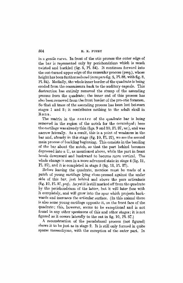
so that now the articular notch (fig. 19, PI. 41, an.) faces directlyupward and the anterior spur, which in stage 2 o v e r h u n gthe anterior element, now lies along its o u t e r side and em-braces it (fig. 8, PL 34). What is now the most anterior upperpoint of the posterior cartilage is being eroded away (comparefig. 18, PL 40, and fig. 19, PL 41). The anterior jaw element hasbecome hooked backwards at its outer end, whilst its inner endhas been trimmed by erosion into a small rounded rod with aclubbed inner end which is somewhat upturned in the middleline. From the histology of the cartilage, as well as by a com-parison of fig. 18, PL 40, and fig. 19, PL 41, it may be seen thatboth jaw elements are growing in length; this is particularlytrue of the posterior element, when it is remembered that theanimal of stage 8 is so much smaller than that of stage 2. Theresult of the straightening and growth of the jaw is shown inthe more posterior position now taken up by its articular region(compare fig. 2, PL 33, with fig. 8, PL 34).
In this stage much of the commissura has been entirelydestroyed; the destruction has taken place in the angle betweenit and the skull wall, and has progressed from behind forwards.The antorbital process in consequence is now becoming free ofthe commissura whose cartilage only remains as a ragged lowerposterior edge to it (dotted line, fig. 19, PL 41); more laterally,however, the connexion still remains intact, though the matrixof the commissura is even here much eroded. Laterally to thisagain, the cartilage of the former commissura, far from beingeroded, is showing an increase in the number of its nuclei—anindication of active life. This active cartilage (fig. 3, PL 34, ptc.;fig. 10, PL 37, ptc.; and fig. 19, PL 41, ptc.) is that part of thecommissural band from which the adult pterygoid process willbe carved out, by the erosion of the neighbouring cartilage fromits surface, both above and below. In front, this pterygoidbar (ptc.) is continued into the projecting processus quadrato-ethmoidalis (pqe.) and, with it, is destined to give the wholeadult pterygoid process. [The two names of the parts of whatbecomes a single adult structure are retained for convenienceof description.] What remains then of the commissura at thisstage is the adult pterygoid process and a band of cartilage
SKULL AND ARCHES IN ANUEA 503
debris on its upper and inner side still connecting it to the ant-orbital process. Cartilage is also being destroyed on the underside of the future pterygoid process, between it and the parsartieularis of the quadrate (dotted line, fig. 10, PL 37). This isallowing the process to emerge as a round rod projecting fromthe inner edge of the quadrate cartilage. At the same time itis narrowing the width of the quadrate bar; a similar erosion ofcartilage below the muscular process, laterally, is assisting in.this narrowing process (dotted line, fig. 19, PL 41).
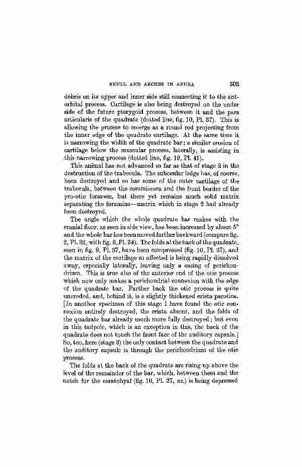
This animal has not advanced so far as that of stage 2 in thedestruction of the trabecula. The subocular ledge has, of course,been destroyed and so has some of the outer cartilage of thetrabecula, between the commissura and the front border of thepro-otic foramen, but there yet remains much solid matrixseparating the foramina—matrix which in stage 2 had alreadybeen destroyed.
The angle which the whole quadrate bar makes with thecranial floor, as seen in side view, has been increased by about 5°and the whole bar has been moved farther backward (compare fig.2, PL 33, with fig. 3,P1.34). The folds at the back of the quadrate,seen in fig. 9, PL 37, have been compressed (fig. 10, PL 37), andthe matrix of the cartilage so affected is being rapidly dissolvedaway, especially laterally, leaving only a casing of perichon-drium. This is true also of the anterior end of the otic processwhich now only makes a perichondrial connexion with the edgeof the quadrate bar. Farther back the otic process is quiteuneroded, and, behind it, is a slightly thickened crista parotica.[In another specimen of this stage I have found the otic con-nexion entirely destroyed, the crista absent, and the folds ofthe quadrate bar already much more fully destroyed; but evenin this tadpole, which is an exception in this, the back of thequadrate does not touch the front face of the auditory capsule.]So, too, here (stage 3) the only contact between the quadrate andthe auditory capsule is through the perichondrium of the oticprocess.
The folds at the back of the quadrate are rising up above thelevel of the remainder of the bar, which, between them and thenotch for the ceratohyal (fig. 10, PL 37, nc.) is being depressed
504 H. K. PUSEY
in a gentle curve. In front of the otic process the outer edge ofthe bar is represented only by perichondrium which is muchtwisted and buckled (fig. 3, PL 34). It continues forward intothe out-turned upper edge of the muscular process (pmq.), whoseheight has been further reduced (compare fig. 2, PL 33, with fig. 3,PL 34). Medially, the whole inner border of the quadrate is beingeroded from the eommissura back to the auditory capsule. Thisdestruction has entirely removed the stump of the ascendingprocess from the quadrate; the inner end of this process hasalso been removed from the front border of the pro-otic foramen.So that all trace of the ascending process has been lost betweenstages 1 and 3; it contributes nothing to the adult skull inE a n a .
The matrix in the c e n t r e of the quadrate bar is beingremoved in the region of the notch for the ceratohyal; herethe cartilage was already thin (figs. 9 and 10, PL 37, we.), and wasnarrow laterally. As a result, this is a point of weakness in thebar and, already in this stage (fig. 10, PL 37), we see the secondmain process of buckling beginning. This consists in the bendingof the bar about the notch, so that the part behind becomesdepressed into a U, as mentioned above, while the part in frontbends downward and backward to become more vertical. Thewhole change is seen in a more advanced state in stage 4 (fig. 11,PL 37), and it is completed in stage 5 (fig. 12, PL 37).
Before leaving the quadrate, mention must be made of apatch of young cartilage lying close-pressed against the underside of this bar, just behind and above the pars articularis(fig. 10, PL 37,psq). As yet it is still marked off from the quadrateby the perichondrium of the latter, but it will later fuse withit completely, and will grow into the spur which projects back-wards and increases the articular surface. (In this animal thereis also some young cartilage opposite it, on the front face of thequadrate; this, however, seems to be exceptional and is notfound in any other specimens of this and other stages; it is notfigured as it occurs laterally to the cut in fig. 10, PL 37.)
A reconstruction of the pseudobasal process (not figured)shows it to be just as in stage 2. It is still only formed in quitesparse mesenchyme, with the exception of the outer part. In
SKULL AND ARCHES IN ANUEA 505
this animal this lateral part happens to be already chondrifiedas a cap of cartilage on the face of the auditory capsule (fig. 3,PL 34). No perichondrium intervenes between them laterally,whilst, in front, its cells pass insensibly into the mesenchymeof the remainder of the process. The early chondrification inthis region is probably exceptional.
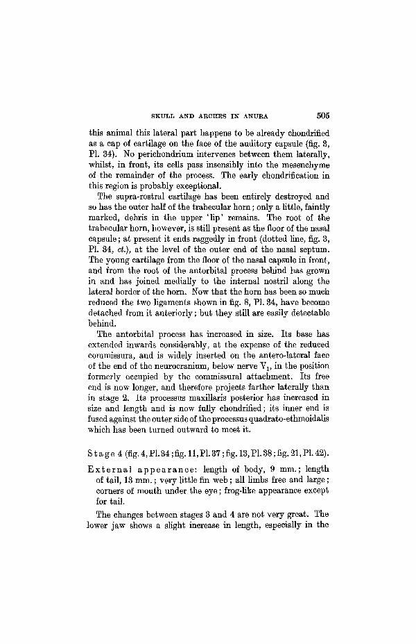
The supra-rostral cartilage has been entirely destroyed andso has the outer half of the trabecular horn; only a little, faintlymarked, debris in the upper ' l ip' remains. The root of thetrabecular horn, however, is still present as the floor of the nasalcapsule; at present it ends raggedly in front (dotted line, fig. 3,PI. 34, ct.), at the level of the outer end of the nasal septum.The young cartilage from the floor of the nasal capsule in front,and from the root of the antorbital process behind has grownin and has joined medially to the internal nostril along thelateral border of the horn. Now that the horn has been so muchreduced the two ligaments shown in fig. 8, PL 34, have becomedetached from it anteriorly; but they still are easily detectablebehind.
The antorbital process has increased in size. Its base hasextended inwards considerably, at the expense of the reducedcommissura, and is widely inserted on the antero-lateral faceof the end of the neurocranium, below nerve Yv in the positionformerly occupied by the commissural attachment. Its freeend is now longer, and therefore projects farther laterally thanin stage 2. Its processus maxillaris posterior has increased insize and length and is now fully chondrified; its inner end isfused against the outer side of the processus quadrato-ethmoidaliswhich has been turned outward to meet it.
Stage4(fig.4,PL34;fig.ll,P1.37;fig.l3,P1.38;fig.21,P1.42).
E x t e r n a l a p p e a r a n c e : length of body, 9 mm.; lengthof tail, 13 mm.; very little fin web; all limbs free and large;corners of mouth under the eye; frog-like appearance exceptfor tail.The changes between stages 3 and 4 are not very great. The
lower jaw shows a slight increase in length, especially in the

506 H. K. PUSEY
anterior jaw cartilage; but much of the difference in size be-tween the jaws of fig. 19, PI. 41, and fig. 21, PL 42, can beaccounted for by the different absolute sizes of the skulls ofthe animals concerned. The jaw as a who le has moved backso that its anterior end protrudes less far forward into the snout,while its articular region is now coming under the optic foramen.The two elements are far more closely fused and very littletrace of the double origin of the jaw is visible externally; eveninternally the enclosed perichondrium is beginning to disappear.Throughout the length of the jaw the cartilage shows an in-creased number of nuclei and gives a histological appearanceof active growth. All erosion of cartilage has now ceased.
What remains of the commissura has been detached from thebase of the antorbital process and lies as an eroded plate over-lying the pterygoid process, along its upper and inner side (fig.21, PL 42, cqa.; fig. 4, PL 34, cqa.; fig. 11, PL 37, cqa.; fig. 13, PL 38,cqa.). The quadrate bar has moved farther back and its forwardextremity is becoming more erect (fig. 11, PL 37). As a resultof this rotation the pterygoid process (ptc.) is coming to liemore horizontally. It should be noted also that, whereas instage 1 the quadrate bar was so inclined that it faced inwards,now, in stage 4, it faces almost directly forward, without theinward tilt. This tendency, begun in stage 3, is possibly dueto the backward pressure exerted by the jaw on the outer partof the pars articularis at a time when the inner border is stillanchored by the commissura and the processus maxillarisposterior. In any case it results in the anterior end of thepterygoid process passing more and more laterally away fromthe skull wall. In fig. 21, PL 42, it is seen now to point inwardsonly to a slight extent compared with fig. 19, PL 41; the break-down of the commissural attachment to the antorbital processmust assist this movement. The change of tilt of the quadrateis obscured by the apparent replacement, in the figures, of thelowest corner of the quadrate (fig. 1, PL 33 and fig. 8, PL 36) by theposterior spur (fig. 4, PL 34, and fig. 11, PL 37), but it is notice-able in that the muscular process is caused to stand up morevertically and to lean less toward the middle line (comparefig. 17, PL 39, and fig. 19, PL 41).

SKULL AND ABCHES IN ANUEA 507
The outer edge of the quadrate bar is more buckled and isformed only of perichondrium. Behind it the root of the oticprocess is thickened into a strong knob; the outer end of theotic process is bent back and has been carried underneath thisknob. A cloud of mesenchyme surrounds these structureslaterally, and stretches forward toward the muscular process,lying along the outer side of the quadrate debris; it will subse-quently chondrify as the adult crista parotica.
The small folds of cartilage at the posterior end of thequadrate (fig. 11, PL 37) are less eroded in this animal thanin stage 3 ; they are still parted by a space from the face ofthe auditory capsule. Between them and the notch (nc.) forthe ceratohyal the main quadrate bar is folding into a U(fig. 11, PL 37). The inner under side of this U rests onthe concave upper face of the pseudobasal process (fig. 13,PL 38, psp.) and does much to mould it. The process, inits turn, probably prevents the quadrate from collapsing backagainst the auditory capsule and it also forces the whole barupward against the under side of the otic process. The pseudo-basal process is now made up of dense mesenchyme, but nochondrification has yet begun, even laterally. The lateral headvein has been displaced to a position largely above the foldedquadrate whilst nerve VII hyomandibular passes through thenarrow space just above the pseudobasal process behind thequadrate.
The antorbital process shows some increase in size and alsoa change of position. It now stands out more laterally from theskull (compare fig. 19, PL 41, and fig. 21, PL 42). The change isdue to an actual backward rotation, which is made possible bythe softening of the cartilage between its root and the skullwall; this cartilage is the last remnant of the commissural rootand is now being removed and replaced by the younger cartilageof the antorbital process, which is usurping its base of attach-ment. The rotation has allowed the whole palato-pterygoid barto move back without any marked increase in its length; it hasalso increased the distance between the front border of theantorbital process and the nasal cartilages (compare figs. 3and 4, PL 34). The under edge of the process still shows a
508 H. K. PUSEY
ragged scar where the commissura has been detached (fig. 21,PL 42, cqa., and dotted line).
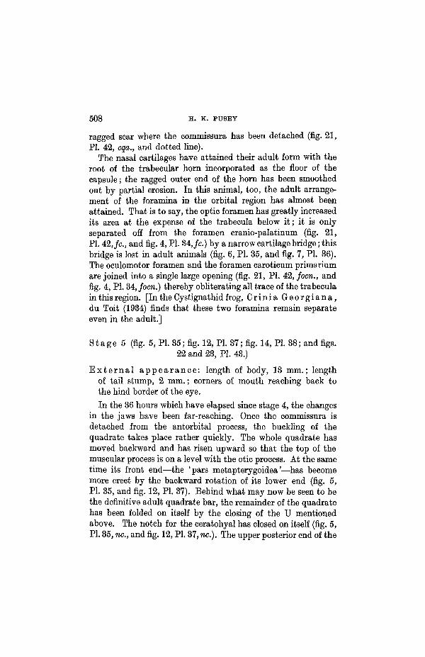
The nasal cartilages have attained their adult form with theroot of the trabecular horn incorporated as the floor of thecapsule; the ragged outer end of the horn has been smoothedout by partial erosion. In this animal, too, the adult arrange-ment of the foramina in the orbital region has almost beenattained. That is to say, the optic foramen has greatly increasedits area at the expense of the trabecula below it; it is onlyseparated off from the foramen cranio-palatinum (fig. 21,PL 42,/c, and fig. 4, PL 34,/c.) by a narrow cartilage bridge; thisbridge is lost in adult animals (fig. 6, PL 35, and fig. 7, PL 36).The oculomotor foramen and the foramen caroticum primariumare joined into a single large opening (fig. 21, PL 42, focn., andfig. 4, PL 34, focn.) thereby obliterating all trace of the trabeculain this region. [In the Cystignathid frog, C r in i a G e o r g i a n a ,du Toit (1934) finds that these two foramina remain separateeven in the adult.]
S t a g e 5 (fig. 5, PL 35; fig. 12, PL 37; fig. 14, PL 38; and figs.22 and 23, PL 43.)
E x t e r n a l a p p e a r a n c e : length of body, 13 mm.; lengthof tail stump, 2 mm.; corners of mouth reaching back tothe hind border of the eye.
In the 36 hours which have elapsed since stage 4, the changesin the jaws have been far-reaching. Once the commissura isdetached from the antorbital process, the buckling of thequadrate takes place rather quickly. The whole quadrate hasmoved backward and has risen upward so that the top of themuscular process is on a level with the otic process. At the sametime its front end—the 'pars metapterygoidea'—has becomemore erect by the backward rotation of its lower end (fig. 5,PL 35, and fig. 12, PL 37). Behind what may now be seen to bethe definitive adult quadrate bar, the remainder of the quadratehas been folded on itself by the closing of the U mentionedabove. The notch for the ceratohyal has closed on itself (fig. 5,PL 35, nc, and fig. 12, PL 37, no.). The upper posterior end of the
SKULL AND ARCHES IN ANURA 509
original quadrate bar stands up above the definitive quadrate,between it and the auditory capsule (fig. 14, PL 38, qd.); it isnow little more than a hollow shell of perichondrium, whosefolding still shows the arrangement of small undulations ofearlier stages (fig. 12, PI. 37, qd.); it is beginning to fall forwardand cover the upper end of the adult quadrate bar.
The lateral edge of the auditory capsule is now somewhatexpanded, by the growth of i t s own cartilage cells, into aprojecting ledge—the crista parotica (fig. 5, PI. 35, cp.). Theperichondrial covering of this ledge is lost and its cells arecontinuous with those of a surrounding mass of dense mesen-chyme, the inner part of which is already chondrified as veryyoung cartilage continuous with the older, histologically dis-tinguishable, cartilage of the auditory capsule. This mesen-chyme passes forwards half the way along the upper edge of themuscular process and, medially, has grown into the spaces inthe debris of the degenerate quadrate bar (the full complicationof these debris is not shown in fig. 5, PL 35). The mesenchymelies laterally to the muscular process, but median to the top ofthe squamosal which is now growing upward to cover this region.
For the first time in this series of stages the debris of thequadrate have collapsed backward so that they are in contactwith the front face of the auditory capsule (fig. 12, PL 37).As a consequence, the lumen of the lateral head vein has beenalmost closed over part of its length and nerve VII hyomandi-bular is squashed between the two structures. What remainsof the tip of the (tadpole) otic process has been twisted inwardsbelow the crista against whose front face it is impacted alongwith the debris of the quadrate. There is, in consequence, abuckled mass of degenerate cartilage and perichondrium com-pressed between the muscular process in front and the cristaand the auditory capsule behind. Intermingled with it are cellswhich have grown inwards from the lateral band of mesenchymementioned above.
The collapse of the quadrate has had its effect also on thepseudobasal process below and behind it (fig. 14, PL 38, psp.,and fig. 23, PL 43, psp.). Where the pressure was greatest thesoft tissue of this process has been forced to the two sides with

510 H. K. PUSEY
the result that its upper border has been notched (dotted line,fig. 14, PL 38, and fig. 28, PL 43). Toward the middle line theinner upper edge of the definitive quadrate bar (just above theroot of the pterygoid process) has also cut into its front edge,again notching it (fig. 23, PL 43, nq.). Here the quadrate andthe pseudobasal process come into very intimate contact, forthe quadrate in this region is already devoid of perichondrium(fig. 12, PL 42, dotted line). More medially still, the twostructures are parted by the inclusion of a shelf of the developingpterygoid bone in the joint. As yet the pseudobasal process isunchondrified.
The shaping of the quadrate bar and the reduction of itswidth above the pars articularis is still going on; this process wasmentioned on p. 508 in stage 8. Laterally, a curved notch isbeing removed, by matrix absorption, between the pars arti-cularis and the muscular process and similar erosion is takingplace below the pterygoid process on its inner side (comparewidths, figs. 14 and 13, PL 38).
All trace of the commissura has been removed from the uppersurface of the pterygoid bar, which has now been shaped intoa round rod (fig. 14, PL 38, ptc). Its anterior end—the processusquadrato ethmoidalis—is still inclined at a slight angle to thatpart of the bar which was cut out of the commissura (fig. 12,PL 37, pqe. and ptc), but this angle is already much smaller thanin stage 4 (compare figs. 12 and 11, PL 37). The whole bar lieshorizontally (fig. 5, PL 35), but still slopes slightly inwards atits anterior end (fig. 22, PL 43); it makes an angle with thequadrate bar which is still less than a right angle below (fig.5, PL 35).
The antorbital process has again rotated farther backward(compare fig. 21, PL 42, and fig. 22, PL 43). The movement,coupled with a slight increase in the length of its processusmaxillaris posterior, has allowed the quadrate to take up itsmore posterior position. The whole palato-pterygoid bar, formedas it is of the processus maxillaris posterior in front, the pro-cessus quadrato ethmoidalis medially, and the (commissural)pterygoid process behind, has become much straightened andnow lies approximately parallel to the cranial floor. The last

SKULL AND ARCHES IN ANURA 511
debris of the commissura have been removed from the back ofthe antorbital process.
There is a considerable difference in the absolute sizes of theanimals of stages 4 and 5; when this has been taken into accountit will be seen that there has been an increase in the length ofthe lower jaw; this applies to both jaw cartilages though moreparticularly to the posterior element. As a result the articularregion has passed behind the level of the optic foramen, and theanterior end has again been slightly withdrawn from the snout.This latter change, however, may rather be due to the down-ward and backward flexure of the jaw in the symphysial region.The apparent distortion in this region has been noted also invictoria blue preparations of similar stages of development.The general set of the jaw has altered; it now lies parallel tothe cranial floor and to the upper jaw, instead of sloping upwardin front. Though invisible now from the outside, the point ofjunction of the jaw elements may still be found in sections;it has been put in in dotted lines in fig. 5, PL 35, and fig. 22,PL 43, for convenience of comparison with the other figures.
S t a g e 6 (fig. 6, PL 35; fig. 15, PL 38; fig. 24, PL 43).
E x t e r n a l a p p e a r a n c e : length of body, 11-8 mm.; minutetail stump; corner of mouth behind the eye.
The lower jaw now shows a great increase in length and atthe same time a decrease in area of transverse section. As therehas been no active process of erosion taking place, it is to beassumed that the growth in length may be due rather to astretching or redistribution of material than to an addition ofnew material. This elongation—whatever its method—is re-stricted entirely to the posterior jaw cartilage. The downwardand backward flexure of the anterior end of the jaw has beenreduced to more normal proportions.
The antorbital process has rotated a little further, particularlyat its outer end, and the upper jaw cartilages have becomesomewhat elongated and are much straightened. The increasein length seems to have taken place equally in the processusmaxillaris posterior and in the pterygoid process; this is the
NO. 320 L 1

512 H. K. PUSBY
first marked growth of this latter process, in the series heredescribed. In dorsal view (not figured) the pterygoid process isseen to curve outwards at its anterior end on passing forwardfrom the quadrate; this is characteristic of the adult skull(fig. 25, PL 44) and had not previously been attained in theseries.
The quadrate bar has rotated backward at its lower end sothat the angle which it makes with the pterygoid process abovehas become obtuse; in stage 5 it was still acute. As a result ofthe rotation the muscular process slopes downwards and for-wards from the crista parotica with which it is now in carti-lagenous continuity. The width of the quadrate bar appears tohave been further reduced, but all erosion has now ceased(compare figs. 14 and 15, PI. 88). Behind the pars articularisthe posterior spur of the quadrate has increased in size (fig. 6,PI. 85, jjsg.).
Special attention must now be given to the manner in whichthe adult quadrate bar is attached to the neurocranium. It isa difficult problem and its elucidation from the study of sectionsis rendered difficult by the large amount of degenerate cartilageand debris in this region, as well as by the dense mesenchymelaterally. However, the present method of reconstruction hasbeen very helpful, and the pictures so obtained give a clearanswer to the problem. A close comparison of figs. 13, 14, 15,and 16, PL 38, makes the position clear.
The otic process of the adult, so far as this is supplied by thequadrate, is formed of what remains of the base of the muscularprocess of the tadpole; that is, its anterior basal part which liesin front of the notch for the ceratohyal. This is clearly seen inthe series of lateral views of the skull. The remainder of theadult quadrate bar—its 'pars metapterygoidea'—is made upof that part of the tadpole quadrate which lies in front of thisnotch, i.e. in front of a line from the middle of the muscularprocess, on its inner side, to the back of the pterygoid process.This line is seen as the sloping upper end of the quadrate infig. 14, PL 88, as a darkly shaded line in fig. 13, PL 88, and as adot and dashlineinfig. 15, PL 38 (- .- .- .- .) . The folded remainderof the quadrate bar lies behind the pars metapterygoidea in
SKULL AND ARCHES IN ANURA 513
fig. 14, PL 38, and rises up above it. Laterally lie the debrisof the back of the muscular process, of the buckled lateral edgeof the quadrate, and of the tip of the tadpole otic process (fig. 5,PI. 35). The soft, partly chondrified pseudobasal process lieson the inner side of the adult bar (fig. 14, PI. 38, psp.).
In the time which elapses between stages 5 and 6, most of thematrix has been removed from the buckled posterior part ofthe quadrate (fig. 12, PI. 39, qd.), thus reducing its bulk andleaving chiefly the more resistant perichondrium. The shell soformed then falls, or is forced, forward and folds itself over theupper edge of the definitive quadrate bar like a ridge-tile(compare figs. 14 and 15, PL 38, qd.). At the same timethe fold becomes fused on its inner side to the pseudobasalprocess. The quadrate cartilage has already cut into thesoft tissue of this pseudobasal process, making a groove (fig. 23,PL 43, nq.) whose upper part also lies over the top of thequadrate bar as a ridge-tile. Though in very intimate con-tact with this overlying fold of dual origin the quadrate barcan still be clearly distinguished from it histologically; in fig. 24,PL 43, therefore, the quadrate bar has been removed and thepseudobasal process, psp., is shown with the quadrate element,qd., joined to its antero-lateral face; behind this quadratetissue is the groove, nq., in which lies the definitive quadratebar. In fig. 15, PL 38, the posterior limit of the pseudobasal
process is shown by a dotted line ( ) and the lateral limitof the auditory capsule (with the root of the tadpole otic process)by a broken line ( ).
The attachment of the muscular process (= adult otic processof the quadrate) to the auditory capsule is also of dual origin.(1) The debris (of the muscular process, of the lateral edge ofthe quadrate, and of the tip of the tadpole otic process), witha probable infilling of mesenchyme cells from the lateral source,are by some means converted into a mass of cartilage; it liesbetween the auditory capsule with its tadpole otic processbehind, and the back of the muscular process (adult otic process)in front. Its appearance in sections is histologically a littledifferent from cartilage formed directly from mesenchyme, andfurthermore it contains liberal streaks of perichondrium within

514 H. K. PUSBY
its substance, streaks which still show the original folding of thequadrate in this region. (2) On passing laterally this peculiarcartilage shades off imperceptibly into more typical youngcartilage formed by direct condensation from the lateral bandof mesenchyme mentioned above. This young cartilage liesalong the outer upper edge of the muscular process (adult oticprocess), almost to its anterior border; it is now covered bythe squamosal which has grown up laterally to it. Behind, itforms a lateral casing round the tadpole otic process and theoriginal tadpole crista parotica of the auditory capsule, andextends to the side and curves downward as an overhangingroof to the region of the middle ear. Together all these structuresform the adult crista parotica (fig. 6, PL 35, cp.) and the oticattachment (poa.).
The pseudobasal process is chondrified, for the first time inthe series, as young cartilage. Between its base and the auditorycapsule there, now lies a thin sheet of dense fibrous tissue makinga joint between them. Similarly, the root of the pterygoidprocess is separated from the pseudobasal process by fibroustissue which is becoming calcified as part of the pterygoid bone;dorso-laterally, however, the two structures are fused together.
Too much attention cannot be given to these quadrateattachments in view of the interpretation put on them by recentworkers (Kruijtzer, 1931). It may be pointed out, then, thatthe definitive quadrate bar may be separated from the neuro-cranium, and from its temporal attachments, in virtue of thedifferent histological character of its older cartilage. When thishas been done, a bridge of cartilage is left which arches over thecranio-quadrate passage for nerve VII hyomandibular and thelateral head vein. This bridge is made up of three main elements;(1) the pseudobasal process medially; (2a) the middle part ofthe back of the folded quadrate bar of the tadpole, i.e. the'ridge-tile' mentioned above: (26) the lateral debris of thequadrate bar, its muscular and (tadpole) otic processes, all ofwhich are reconverted into true cartilage; and (3) the band oflateral mesenchyme cartilage fused on laterally. It cannot betoo fully stressed that the middle of this bridge, in E a n a, iscomposed of c o n v e r t e d q u a d r a t e t i s s u e .

SKULL AND AECHBS IN ANUKA 515
S t a g e 7 (fig. 7, PL 36; fig. 16, PL 38; fig. 25, PL 44,and fig. 31, PL 45).
E x t e r n a l a p p e a r a n c e : typical young frog some weeksafter leaving the water; it had fed on land; no tail; bodylength, 14-3 mm.
The animal had begun to grow again after the metamorphosis.Its skull is the first in the series to show a general increase insize in a l l its structures. (During metamorphosis there seemsto be a tendency for the skull to d e c r e a s e slightly in size.)
The lower jaw again shows a considerable increase in lengthand, as in the previous stage, this growth is restricted to theposterior jaw cartilage almost entirely (compare fig. 6, PL 35, andfig. 7, PL 36). The diameter of its cross-section is slightlyreduced, suggesting a farther stretching of its material. As a resultof the growth the articular region has passed yet farther back.
To accommodate the now elongated jaw the quadrate hasrotated backward about its upper end, thereby changing the slopeboth of the muscular process and of the crista parotica behind(compare fig. 6, PL 35, and fig. 7, PL 36). As a result, too, thepterygoid process has undergone a slight downward rotation,though to a smaller extent than the quadrate bar, so that the anglebetween them has again become more obtuse. The pterygoidprocess has also rotated outward at its anterior end (fig. 16, PL38, pic, and fig. 25, PL 44, ptc.).
The various elements of the otic and pseudobasal processeshave become more fully fused and are therefore the less easilydistinguishable. The amount of cartilage in this region has beenreduced, especially laterally, where the otic process and theupper end of the quadrate bar are of more delicate build. Theotic attachment, however, still shows flecks of perichondriumin its substance; whilst the cap of cartilage may still be tracedrunning down from it, along the top of the quadrate bar to thepseudobasal process. But these distinctions are now purelyhistological and are no longer structural. It is to be noticedthat it is only the outer, upper side of the pseudobasal processwhich is fused with the quadrate (fig. 16, PL 38); medially thepseudobasal process (marked with a dotted line ( ) is separated

516 H. K. PUSBY
by a cleft (which contains part of the pterygoid bone) from theroot of the pterygoid process. At the back of the pars arti-cularis the spur (fig. 7, PI. 36, psq.) has become much enlargedand now takes part in the actual articulating surface.
For the rest, this stage differs little from the previous stageor from the typical adult frog's chondrocranium. The ant-orbital process slopes backward to a more marked degree andits outer end has taken up a more ventral position, therebycarrying the processus maxillaris posterior downward with it.In dorsal view the whole upper jaw is seen to bow out to theside, whilst its two elements have now come fully into line withone another (compare fig. 22, PL 43, and fig. 25, PL 44).
THE SKULL OF FROGS OLDER THAN STAGE 7.
The changes noted in older skulls are continuations of thechanges begun in earlier stages.
The quadrate bar rotates yet farther backward. The middleof its length is firmly fused to the pseudobasal process and itsupper end to the crista parotica; consequently it is only itslower end which is free to move to any great extent. Its back-ward movement, therefore, bends the bar on the pseudobasalprocess as a fulcrum. The lower end of the bar lies close underthe auditory capsule and is only parted from it by the widthof the hyoid cornu which intervenes between them. The appear-ance of increased backward rotation is enhanced by the increasein the size of the posterior spur, which has grown upward andbackward from the now almost horizontal pars articularis. Thehorizontal position of this region, coupled with the increase ofthe spur, supplies the quadrate with a much greater articulatingsurface for the lower jaw. This is met by an increase in the sizeof the articular region of the jaw, which has become extendedinto a clubbed end with a notch in the upper surface to fit onthe quadrate; in front of the articular region the transversesection of the jaw is relatively smaller than in earlier stages.
But beside rotating backward the quadrate bar has under-gone another change of position. In stage 7 its flat anterior facewas directed f o r w a r d and downwards, but in later stages itcomes to face o u t w a r d as well as forward and downward.
SKULL AND ARCHES IN ANURA 517
This applies chiefly to the region lateral to the pterygoid process,where the outer edge has been twisted backward, while themedian edge has been held in its former position by the buttress-ing action of the pseudobasal process behind it. More ventrallythe bar again becomes partially untwisted so that it once morefaces forward in the articular region. This lateral twisting is butthe completion of a process already beginning in stage 2; it isresponsible, in part, for the outward rotation of the pterygoidprocess.
The otic attachment of the quadrate does not seem to havebeen entirely rigid in stage 7, since the postero-lateral corner ofthe crista parotica has come to be twisted upward above theneighbouring wall of the auditory capsule. This is probably dueto the rotation of the quadrate and the consequent change inslope of the muscular process and therefore of the crista behindit. Further, the crista shows a change, in that part of its lateralborder is now continuous with part of the upper border of theannulus tympanicus; while on its lower surface there is a fusionwith the extra-stapedial of the middle ear apparatus; this lasthad already taken place in stage 7.
This description is taken from my own reconstructions of thetemporal region of young adult frogs—both in anterior andlateral views. It was supplemented by a study of a solid recon-struction of this region made by Dr. J. W. Jenkinson and nowin the University Museum, Oxford.
(a) Shor t Summary of t he M e t a m o r p h o s i s .
The lower jaw cartilages increase in length and decrease inthickness; part of the decrease is due to erosion and part,possibly, to a stretching process. The anterior cartilage elon-gates slightly at first, but very little later; it becomes morehorizontal and its outer posterior end hooks backwards andbegins to fuse with the posterior cartilage. This latter cartilagerotates on its long axis so that its former upper surface now lieslaterally and its upstanding innermost peak lies laterally besidethe anterior cartilage, with which it fuses. Its S shape isstraightened out largely by erosion and it then increases inlength and comes to lie with its long axis more nearly parallel

518 H. K. PUSBY
to that of the body, instead of transversely to it. The twocartilages come more and more into a horizontal line whenviewed laterally. The articular notch on the jaw formerlyfaced backwards and downwards and now faces upwards; itpasses back by the stretching (?) of the jaw until it lies farback under the auditory capsule. At the same time the frontend of the jaw is withdrawn more under the nasal capsule.
Before the metamorphosis of the quadrate begins the ant-orbital process is formed from the anterior edge of the com-missura close against the side wall of the skull. Its anterior andupper borders are continuous with the nasal roof and side wall.Its main base of attachment to the root of the commissura liesventral to nerve Yv Its anterior end develops a forwardlydirected processus maxillaris anterior and its posterior outercomer develops a backwardly, and inwardly, directed processusmaxillaris posterior.
Meanwhile the cartilage of the following structures has beensoftened: the supra-rostral cartilage, the outer ends of thetrabecular horns, the posterior part of the quadrate cartilagewith the ascending process on its inner, and the tip of the tadpoleotic process on its outer, side; and the upper edge of themuscular process. In addition, the subocular shelf is beingremoved from the trabecular and, with it, the processus pseudo-pterygoideus is being destroyed and the back of the upper partof the commissura softened. These all appear to be simultaneousprocesses (they may prove to be due to some hormone release).
By some agency unknown the whole quadrate bar is nowmoved backward, rotating on the commissura, which is thereforeitself rotated backward. The quadrate is still attached to thetrabecular horn by the ligamentum quadrato-ethmoidale andby the 'trabecular quadrate ligament'. The trabecular horn,and the nasal capsule above it, are therefore rotated downwardand backward; the outer half of this horn, and the supra-rostralcartilage in front of it, are entirely destroyed and leave no tracein the adult. The cartilages of the nasal capsule are at this timerapidly developing, fusing with one another and becomingchondrified.
The middle section of the ascending process is entirely

SKULL AND ARCHES IN ANURA 519
destroyed, leaving temporary stumps attached to the cranialwall medially and to the quadrate laterally; even these arevery soon entirely destroyed, so that the ascending processforms no structure in the adult skull.
The backward movement of the softened quadrate baragainst the rigid auditory capsule behind, causes the bar tofold on itself in gentle undulations; the outer tip of the tadpoleotic process is bent backward, but offers sufficient resistanceto prevent the collapse of the quadrate back on to the face ofthe auditory capsule. Appearances in sections lead one tosuppose that the mandibular muscles may play a part in thisinitial buckling.
Meanwhile the pseudobasal process is condensing as mesen-chyme in front of the antero-lateral corner of the auditorycapsule. Unlike Gaupp, I can find no indication that it owesits origin in any way to the quadrate; it is rather related to theauditory capsule, especially laterally, where no perichondriumintervenes between the two structures.
These various changes continue rapidly. The upper part ofthe commissura is destroyed from behind forward and theresidue is rotated backward. Its base of attachment to the sidewall of the skull and the root of the trabecular horn is usurpedby the inward and backward growth of the root of the ant-orbital process. From the lower outer part of the commissurathe pterygoid process of the adult now emerges. The residueof the commissura is eroded from its upper, inner, surface and,below, the angle which it makes with the quadrate bar is openedby further cartilage erosion. This destruction, coupled with asimilar destruction laterally, below the muscular process,narrows the transverse width of the quadrate bar in the regionabove the pars articularis.
In front, the antorbital process has increased in size and hasbeen rotated backward; it also stands out more laterally. Itsprocessus maxillaris posterior has increased in length, and isnow fused at its inner end with the tip of the processus quadrato-ethmoidalis of the pterygoid process. This element of the ant-orbital process grows greatly in length, becomes straightened,and so forms the anterior half of the adult cartilage upper jaw.
520 H. K. PUSEY
The processus quadrato-ethmoidalis and the pterygoid processbehind it show very little growth in length.
The upper edge of the muscular process is turned outwardand downward and is finally destroyed. The height of the pro-cess is so reduced to a third; in the subsequent buckling theposterior half of the process is much deformed and is practicallydestroyed. Only the lower anterior part of the tadpole muscularprocess, therefore, remains to the adult stage: this part becomesthe adult otic process of the quadrate.
The backward movement of the quadrate bar causes theundulations of cartilage behind to collapse on one another;they are piled up with the more posterior folds dorsally. Allbut the perichondrium of these folds is soon destroyed, a state-ment which applies also to the front part of the tadpole oticprocess, which is now in contact with the quadrate only throughperichondrium. The basal part of the process, however, becomesextended backward to join with a (tadpole) crista paroticawhich is formed by an expansion of the auditory capsule. Thesurface of this crista tends to lose its perichondrium and itscells become more or less continuous with a cloud of mesenchymecells which surround it on all sides and stretch forward laterallyto the muscular process in front.
Meanwhile, in front of the notch for the articulation of theceratohyal, the quadrate bar is turning downward and backwardso that it sets more vertically. This is the 'pars metaptery-goidea' of Parker and Gaupp and is all of the quadrate of thetadpole which is retained into the adult stage (with the exceptionof certain quadrate debris to be described). As a consequence ofthis rotation the notch for the hyoid is beginning to close onitself. Behind the notch the remaining unbuckled part of thequadrate is beginning to fold into a single large U which sooncollapses on itself and comes to lie behind, and somewhat above,the pars metapterygoidea. This folded part of the quadrate isthen much eroded and its perichondrium falls forward andembraces the inwardly sloping upper edge of the pars meta-pterygoidea between the muscular process laterally and thepseudobasal process medially. With the collapse of this U themuscular process has passed back, and has been raised upwards
SKULL AND ARCHES IN ANURA 521
and is now forced backward under the overhanging crista ofthe auditory capsule, whose anterior outer edge is carried inwardin a fold below itself. The notch for the hyoid has now fullycollapsed.
Meanwhile the pseudobasal process has become more denseand has been moulded by the pressure of the quadrate upon it.For a time it seems to prevent the total occlusion of the eranio-qnadrate passage above it, but later the debris of the quadrateare forced back into the passage against the face of the auditorycapsule. When it takes up this most posterior position thecurve of the quadrate—where the base of the pterygoid processSAvings laterally into the pars metapterygoidea—cuts into thesoft tissue of the pseudobasal process and begins to fuse withits lateral edge; more medially, the two structures are partedby the inclusion, in the joint, of the pterygoid bone which isdeveloping on the side of the pterygoid process and the inneredge of the quadrate.' The pseudobasal process also fuses withthe debris of the quadrate bar which lie along the top edge of thepars metapterygoidea; it gives the appearance, therefore, ofhaving grown up laterally to pass into the tissue of the oticprocess, but this is not the case.
The otic process, so far as the quadrate is concerned, is madeout of the undestroyed cartilage of the tadpole muscular pro-cess. It is fused to the crista parotica, (a) by the conversion ofthe q u a d r a t e debris behind it (= lateral folded part of thequadrate bar, posterior part of muscular process, and anteriorperichondrium of the tadpole otic process) into cartilage; and(b) by the fusion along its outer upper edge of a rod of cartilagewhich has formed from the cloud of mesenchyme previouslymentioned. This rod is indistinguishably fused with the medial(debris) cartilage, but stands out clearly (in transverse sections)where it lies beside the upper edge of the muscular process moreanteriorly. Posteriorly it has fused on all sides with the tadpolecrista parotica and now extends this structure outward anddownward. All these elements of the otic articulation laterbecome fully fused up.
The remaining quadrate bar continues to rotate backwards,a process which at first alters the inclination of the crista and
522 II. K. PUSEY
in later stages causes an actual bending of the bar itself acrossthe rigid fulcrum of the now fully chondrified pseudobasalprocess. The back of the pars articularis has meanwhile ob-tained a large posterior spur which is formed from an independentcondensation of cartilage cells which formed behind it and soonfused with it.
During the backward movement of the quadrate bar theanterior face of the pars metapterygoidea has changed itsinclination. In early stages it sloped in such a way that it facedforward and in Avar d towards the side wall of the skull. Inlater stages it comes to face directly forwards and in the adultit faces outwards and forwards. This movement assisted theroot of the pterygoid process to emerge from the quadrato barand enhances what is, in fact, probably only an appearance ofgrowth in length in this structure. 'I'his twisting of the bargreatly alters the appearances seen in transverse sections, onthe one hand of recently metamorphosed frogs and on the otherof adult animals: it is also responsible for the outward rotationof tin* tip of the pterygoid process into its adult (lateral) position.
(i. DISCUSSION.
An attempt will here be made to analyse the facts of jawmetamorphosis to see what light can be thrown on the peculiari-ties of structure and attachment of the jaws in the tadpole andthe adult frog and to relate these peculiarities to the more normalstructures of other groups. It may be mentioned here that thereis a great constancy of structure among most of the A n u r ain the relations of the jaws to the skull, and in allied structures.We are largely indebted for our knowledge of the skull in thegroup as a whole to Parker's third paper on the B a t r a c h i a(1881). But it may well be that more extensive work will showthat this constancy is not so great as now appears. Alreadythe work of de Villiers, Wagner, and van Seters has shown thatA s c a p h u s and L i o p e l m a are very far from the commonAnuran type in certain points of jaw suspension, while A1 y t e s,B o m b i n a t o r (Luther, 1924, fig. 20), and D i s c o g l o s s u s(Pusey) also retain certain characters which must be lookedupon as primitive from this point of view. Nevertheless, on
SKULL AND ARCHES IN ANUEA 523
our present knowledge, we may assume that the descriptionof jaw metamorphosis here given for Eana temporar iaapplies, at least in broad outline, to most other Anurans. Thereis, however, but little comparative evidence of this metamor-phosis from other species, and the similarity has rather to beinferred from a few scattered observations, and from the factthat tadpole skulls of essentially similar construction give similaradult skulls. Our evidence on this point comprises the workof Parker on several species, Litzelmann on Bombinator ,Kotthaus on Xenopus, and Kruijtzer on Megalophrysand Alytes .
(«) Perhaps the most obvious fact to be noticed in the studyof the above reconstructions is that the anterior lower thirdof the tadpole muscular process becomes the otic process ofthe adult quadrate. Though both Parker and Gaupp noticedthat the adult otic process carried traces of the residue of themuscular process, neither fully realized that these processes areone and the same structure. Litzelmann's figures show the samefor Bombinator . In the tadpole skull , then, wemay say tha t the lower front border of the mus-cular process is the same s t ruc ture as the adultotic process. When this is established it can then be saidthat the upper part of the tadpole muscular process is a specializa-tion of the upper part of the adult otic process—a specializationto give increased muscular attachment, and one which isdestroyed in the adult. Such a specialization is peculiar onlyto the Anura. Gaupp considered the whole muscularprocess to be such a special larval feature, but we may now saythat it is only the upper part of the process which is an exag-gerated adult otic process. The hinder border of the muscularprocess in the tadpole slopes downwards and backwards andpasses insensibly into the outer edge of the quadrate bar. Inthe auditory region this outer edge is continued backward tofuse with the auditory capsule as the 'tadpole otic process'. Itwould seem reasonable then to believe that in the tadpole theanterior edge of the muscular process is the front border of anotic process which, behind, reaches right back along the borderof the quadrate bar to its attachment with the auditory capsule

524 H. K. PUSEY
in the tadpole otic process; that is to say, that a shorter oticprocess in the ancestor has, in phylogeny, been stretched forwardto the front of the head, whilst its anterior upper border hasbeen extended upward to give the muscular process.
At metamorphosis the top of the muscular process is destroyedand the outer edge of the quadrate bar becomes buckled andfolded on itself and is then impacted between what remainsof the muscular process and what remains of the tadpole oticprocess. The debris so formed—themselves of otic-processorigin—are then reconverted into cartilage to assist in thefusion of the two ends of a previous single structure. Thelateral band of binding cartilage which is fused on to this jointlaterally seems to owe its origin rather to the lateral wall of theauditory capsule, and since it gives rise to the crista parotieaof the adult and was already in continuity with the tadpolerepresentative of that structure in its origin, it would seem tobe fair to call it the crista parotiea, or a specialization of itformed probably to assist in the rapid attachment of the adultotic process. Another view has been put forward by Kruijtzerfor an apparently similar structure in M e g a l o p h r y s and inA1 y t e s; he believes it to be a ' laterohyal' of hyoid origin; thisview will be discussed later. Van Kampen has found the samestructure in P i p a.
In the view put forward in this paper, then, the whole outerborder of the quadrate bar, from the edge of the auditorycapsule to the anterior edge of the muscular process, is to belooked upon as a single very much stretched otic process; thetadpole otic process would then be the original connexion ofthis process with the auditory capsule.1 At metamorphosis theeffect of the stretching during phylogeny is reversed by a pro-cess of telescoping in ontogeny. The middle of the process isnecessarily weakened and partially destroyed and the debrisare rechondrified to join up the anterior and posterior limitsinto a shorter more typical process again fused to the auditorycapsule through what remains of the base of the tadpole oticattachment. The joint is then made stronger by a specialization
1 However, Alytes (Kruijtzer) and Discoglossus (Pusey), bothprimitive frogs, are without 'tadpole otic processes'.

SKULL AND ARCHES IN ANUBA 525
and forward growth and fusion of the crista parotica laterallyto the muscular process.
If this interpretation is correct the question does not ariseas to which of two otic processes is the homologue of the singleotic process of other animals. For the attachment to the skull isalways through the base of the tadpole otic process. The fusionof the adult otic process with it is but the completion of thereduction in length of a single process which was very muchelongated in the larva. Gaupp was of the opinion that thesecond fusion of the quadrate with the auditory capsule wasthe homologue of the otic process of the U r o d e 1 e s. He con-sidered the tadpole otic process to be a larval specialization con-fined only to the A n u r a.
In support of this view of the phylogenetic stretching of asingle otic process, the following facts may be brought forward:(1) The rudiment of the annulus tympanicus is found in stage 2at the base of the muscular process; it passes back with thisprocess during metamorphosis to the auditory region. (2) Theanterior (morphologically outer) end of the Eustachian tubereaches to the same point and also moves back at metamor-phosis. (3) The same is true of the rudiment of the columellaauris. (4) The jaw muscles inserted on the muscular process,especially on its inner surface, pass back to the auditory regionwithout change of attachment. (5) The squamosal rudimentforms on the base of the muscular process in stage 2 and growsupward to about a third of its height; that is to the top of thatpart which becomes the adult process, and remains so placedinto the adult. (6) Finally the forward position of the parsarticularis and of the lower jaw, and of the ceratohyal whichlies immediately below—morphologically just posterior to themuscular process—all point to the fact that the anterior edgeof a single otic process has been carried forward and has takenwith it all those structures normally associated with it.
(6) The homology of the ascending process of the tadpolewith that of the U r o d e l e s and other forms, has not been indoubt since its true nature was established by Stohr in 1881.Its relations to the branches of the Vth and Vllth nerves andto neighbouring blood-vessels are correct for such a process.
526 H. K. PUSBY
It is only surprising that a structure so common in skulls ofother forms should be entirely absent in that of the a d u l t frog.It may, however, persist in part in the adult skull of A s c a p h u s(see the subsequent discussion, pp. 534 and 535).
(c) The homology of the commissura quadrato-cranialisanterior is by no means so clear. In 1893 Gaupp was led tothe view that it represented the connexion found in the salmonbetween the palatopterygoid bar of the quadrate and the ant-orbital process of the nasal capsule. In 1906 he was in greaterdoubt aboitt this structure and stated that it had no ' analogue'in the U rode la . In 1925 Edgeworth made a survey of theoccurrence of an attachment of the tip of the pterygoid processof U r o d e l e s to the trabecular wall of the cranium belowthe later-developed antorbital process. As a result of hisresearches he said:
'These phenomena indicate an ancestral Urodelan conditionin which the pterygoid process was continuous anteriorly withthe trabecula, i.e. one in which there was a pterygoquadrate.(laupp held that the commissura quadrato-cranialis anteriorof the larval stage of E a n a has no 'analogue' in the Uro -d e 1 a, but the new information above mentioned shows that itis homologous with the pterygoid process of U r o d e l a andshould be similarly named. The later developed pterygoidprocess of Gaupp might have a new name.'
A number of criticisms may be raised against this view.Firstly, Edgeworth is forced by it to believe that the frog'spterygoid process, which itself is formed from the distal partof the commissura, is not homologous with that of U r o d e 1 e s.Apart from his hypothesis there seems to be no other obstaclein the way of this latter homology; he therefore solves onedifficulty only to supply a new one. His view is that the tipof the pterygoid process in both young U r o d e 1 e s and in thetadpole is fused to the trabecula below the antorbital process;in the U r o d e l e s by tissue which is subsequently destroyed,and in the tadpole by the commissura which is also destroyed.In some adult U r o d e l e s there is a cartilaginous connexionof the tip of the pterygoid process with the antorbital processas in the adult frog. From his figures and descriptions I am left
SKULL AND ARCHES IN ANURA 527
in doubt as to how far these two connexions in the U r o d e l e sare to be considered as separate.
But his view seems to be untenable since the processusquadrato-ethmoidalis—which on non-Bdgeworth homologiesbecomes the t i p of the pterygoid process—projects forwardfrom the f ron t face of the commissura. So that here wehave part of the pterygoid process beyond the fusion of thecommissura with the skull. However, Edgeworth disposes ofthis difficulty by disowning the subsequently developed ptery-goid process of the frog and saying that a new name should befound for it; he offers no homology for it.
(d) If there are two possible connexions of the pterygoid pro-cess to the skull in U r o d e l e s as Edgeworth suggests, (1) to theroot of the antorbital process, and (2) to its processus maxillarisposterior, only the latter seems to be represented, in the frog,in the adult. The processus quadrato-ethmoidalis is admittedlycontinued forward by a ligament—ligamentum quadrato-ethmoidale—to the side of the trabecula, but this cannot repre-sent connexion 1 above, of the U r o d e 1 e, as both the processand the ligament lie laterally to the inner nostril and the liga-ment joins the trabecula in front of it. In the U r o d e l e s thisconnexion must lie behind and therefore medially to the nostril.So that even if we do not accept Edgeworth's homology, if weaccept his facts, we are still left with a connexion in the Uro -deles unparalleled in the frogs. And the ligament of the frogsis not found in the U r o d e l e s . This ligament may probablybe explained as a specialization of the rudiment of the pterygoidbone. On becoming detached from the trabecula, its hinderpart ossifies as that part of the pterygoid which lies mediallyto, and overlaps, the maxilla. However, it seems to me thatthis difficulty is not greater than that left unfaced by Edge-worth, namely of finding a homologue in the U r o d e 1 e (or inany other animal) for the cartilages which give the apparentpterygoid process of the frog.
Gaupp's view also raises difficulties. He believed that thecommissura of the frog was the whole palatopterygoid barshortened up and joined to the cranial wall by the fusion of theethmopalatine-articulation seen in the salmon (de Beer, 1927).
NO. 320 M m

528 H. K. PUSEY
The processus quadrato-ethmoidalis then represented that partof the palatine bar in front of this hinder articulation. The greatgrowth of the processus maxillaris posterior of the antorbitalprocess was then to be considered as a special anuran modifica-tion. However, the whole pterygoid bar up to the antorbitalprocess seems to belong to the quadrate in those U r o d e l e swhich retain it (i.e. in E a n o d o n and, perhaps, H y n o b i u sand M e n o p o m a ; Edgeworth, 1925). In the salmon we findthe anterior end of the palato-pterygoid bar chondrifyingseparately from the part behind (de Beer, 1927). The factsthen from the U r o d e l e s and the salmon suggest that theprocessus maxillaris posterior (which, in the frog, belongs to theantorbital process) is really a detached front end of the palato-pterygoid bar secondarily attached to and developing from thefintorbital process. Much a view cannot bo made to agree withthat of Gaupp.
(<•) An alternative interpretation will now be put forward,which is not concerned with these difficulties.
The descriptions given of these parts in the tadpole show thatthe adult frog pterygoid process emerges from the distal partof the commissura where the latter passes into the quadratebar. To this pterygoid process is added the small forwardlyprojecting processus quadrato-ethmoidalis which projects for-ward from the middle of the front face of the commissura;together these two cartilages make up the adult pterygoid bar.On the medial and upper side of this bar, then, there lies theremainder of the commissura—that part which will subse-quently be detached from the skull wall and destroyed. Whenso detached—had it not been so fully eroded and bent so that itpoints forward—it would lie as an inwardly directed processof the quadrate on the inner side of the base of the pterygoidbar close to the point where this swings laterally into the bodyof the adult quadrate bar. It would, in fact, have all the rela-tions to the quadrate and the pterygoid process of a basalprocess. The v iew here p u t fo rward t h e n is t h a tthe comrn i s su ra is t he b a s a l p r o c e s s of t he q u a d -r a t e which has passed e x c e p t i o n a l l y far f o rwardto fuse w i t h t h e t r a b e c u l a j u s t b e h i n d t h e n a s a l

SKULL AND AECHES IN ANUEA 529
s a c . Compare the conditions described in S c y l l i u m (deBeer, 1931). It may be assumed that in phylogeny it ceasedto abut against a basitrabecular process from the cranial floorand passed progressively forward, probably in subsequentontogenies, fusing with the edge of the trabecula. In the tadpoleit has taken up the most anterior position possible—as here itis stopped by the nasal sac and its inner nostril to which thecommissura makes a posterior border.
The relations of this attachment to surrounding structuresmust now be examined. It should lie dorsally to nerve VIIpalatine and ventrally to nerve Vj; it does so. Other structuressuch as the lateral head vein and the carotid artery do notreach far enough forward to assist us. But it may here benoted that there seem to be no obstacles in the way of such aforward movement. In addition, as shown above, the anteriorborder of the otic process (tadpole muscular process) lies oppositeit on the outer side of the quadrate bar. It also bears the correctrelations to the pars articularis of the quadrate and the rudimentof the pterygoid bone. Much of this evidence is of a negativenature, but later it will be shown that the conditions foundin A s c a p h u s and L i o p e l m a probably may support thisview.
(/) It will be assumed then that the commissura is the basalprocess of the quadrate. In that case the ' pseudobasal process'cannot also be the basal process. Gaupp believed that it was,and his view has been accepted by most other workers up tothe present. What then is the pseudo-basal process ? From thestudy of its origin Gaupp found it hard to believe that it be-longed to the quadrate; however, in a 37 mm. embryo he saidthat he found a connexion. I cannot confirm this point. Bather,as Gaupp had to admit, it seems to belong to the lateral anteriorface of the auditory capsule in front of the foramen ovale.Kruijtzer has described the independent origin of this struc-ture in M e g a l o p h r y s and A l y t e s , and its s e c o n d a r yfusion to the quadrate. In this region its cells are continuouswith those of the capsule, from which, laterally, it is notseparated by any perichondrium during development. Later,however, it becomes separated from the capsule by a fibrous
580 II. K. I'USEY
joint and it becomes fused to the quadrate—though at a pointfar from the region of the quadrate from which Gaupp sug-gested that it might be developed. Gaupp was impressed bythe similarity of the adult process to the basal process of theUrode l e quadrate, and almost against his own evidence heAvas forced to advocate this homology. Since then, however, thenature of the articulation of this process with the capsule hasbeen culled in question by de Beer (1920).
In this paper it is assumed that this 'psoudobasal process'of the adult frog is tho outer end of the basitrabecular processfrom the basal plnte •which has been separated off from thecranial Hour by the destruction of its root. It is suggestedthat it, has become fused secondarily to tho quadrate followingthe complete destruction of tho true basal process (commissura)and IIIHO secondarily has obtained a jointed articulation withthe auditory capsule, thus restoring a more Urodele-likoarrangement in this region. The reasons for this belief will bediscussed when the skulls of A s e a p h u s , L i o p o l m a , andAlyteH have been described (see p. 5!!9). It should here benoted that, had tho commissura not been distorted and destroyedduring metamorphosis in ii an a, but had merely been detachedut its upper end from the skull, it would have abutted againstthe pseudobasal process, making thus a joint between a truequadrate basal process and the end of a cranial basitrabecularprocess (albeit detached).1
Little mention need be made of the other processes of thejaw apparatus. The antorbital process is undoubtedly thehomologue of the structure found in other animals. Only itsprocessus maxillaris posterior seems to be unusually welldeveloped as a unique elongated rod making up about half thecartilage upper jaw in the adult. The pterygoid process hasbeen already dealt with.
{<!) A summary may here be given of the homologies putforward in this paper:
' Since writing thin account I. have found a small basal process in tad-poles ill' 1) i H « o H 1 o H H II 8 before metamorphosis; it lies below tho ascendingprocess in tho typical vertebrate position; its presence probably does notinvalidate the prenoiit, argument (l.'iisey).
SKULL AND ARCHES IN ANURA 531
1. The commissura quadrato-cranialis anterior is the basalprocess of the quadrate.
2. The whole outer border of the quadrate bar from theanterior edge of the muscular process to the tadpole otic processis the single much elongated otic process of other forms. Themiddle of its length only is destroyed and so it collapses onitself to give a shortened adult otic process.
3. The lower anterior part of the muscular process gives thecartilage which forms the adult otic process. The remainder isa unique specialization found only in the A n u r a.
4. The 'pseudobasal process' ( = basal process of Gaupp) isthe isolated outer end of the basitrabecular process of the skullplus post-palatine commissure behind (see pp. 529 and 539).
5. The processus quadrato-ethmoidalis and the pterygoidprocess (from the commissura) are together equal to theU r o dele pterygoid process.
(>. The ascending process and the antorbital process arehomologous to the similar structures in other forms.
7. The excessive length of the processus maxillaris posterioris either a unique specialization of the A n u r a , or the detachedfront end of the palatopterygoid bar.
8. The ligamentum quadrato-ethmoidale is an early specializa-tion of the rudiment of the pterygoid bone.
9. The 'trabecular-quadrate ligament' is a special Anuranfeature; its hinder part ossifies as the quadrato jugal.
Apart, then, from a possible additional attachment in theU r o d e l e s between the tip of the pterygoid process and thetrabecula, below the antorbital process, we find a close agree-ment between the parts found in the U r o d e l e s and theA n u r a . The differences in the relations of the Vth and Vllthnerves in the two groups will be discussed later in this paper.
7. A EEDBSCRIPTION OF THE TEMPORAL REGIONS OF THE
SKULLS OF ASCAPHUS, LIOPELMA, AND ALYTES.
By far the most important work on the A n u r a in respectof the jaw anatomy and the temporal region has been thedescriptions recently given of the adult structure of singlespecimens of A s c a p h u s and L i o p e l m a . These two species
532 H. K. PUSBY
are placed in a separate family—the Liopelmidae—by Noble,who appears to consider them to be the most primitive livingAnurans; other workers have placed them with the Discoglos-sidae. So far as the temporal region of their skulls is concerned,it seems very reasonable to agree with Noble's views.
(a) A s c a p h u s t r u e i .
The account here given of the temporal anatomy of A s c a -p h u s is taken from the work of de Villiers (1934). His publishedpictures (fig. 7 A, B, C, and D), together with two drawings takenmore anteriorly and most kindly supplied to me by Miss de Vosof Stellenbosch, have been reconstructed by the method pre-viously described. These reconstructions (figs. 26 and 27,PI. 45) are seen from in front. In fig. 27, PI. 45, much of thequadrate has been removed and in both figures the part abovethe pars articularis had been drawn in freehand (hence in dottedlines) by analogy with E a n a and L i o p e 1 m a; however, theposition of the lower jaw is known to be correct from fig. 7 D.In fig. 27, PL 45, the nerves are shown. The present author issolely responsible for any differences of description or nomencla-ture here introduced and for any errors brought in the recon-structions.
De Villiers draws attention to the Urodele-like characterof the nerve foramina and arrangements generally of thetemporal region. The facial nerve is separated from the tri-geminal nerve; some part of the separating cartilage mustrepresent a prefacial commissure—a structure entirely absentin R a n a and the bulk of frogs. In addition, the foramen forthe trigeminal nerve is divided into an upper and a lowerdivision by a bony bridge (tb.) in front. From the upperopening nerves V2 and V3 pass out, and from the lower openingV1( a nerve connecting V to VII palatine, and the lateral headvein; these latter two structures are not shown in fig. 27, PI. 45,but the main branches of nerve V and the divided trigeminalforamen are clearly shown.
The Vllth nerve ganglion (fig. 27, PI. 45, VII g) lies in aforamen ventral and lateral to the trigeminal foramina; fromit the palatine branch of VII passes forwards (VII p.). VII
SKULL AND ARCHES IN ANUEA 533
hyomandibular is separated from VII palatine by a skeletalbridge behind which it runs out laterally through its ownforamen, which is situated between the auditory capsule aboveand 'a ventral cartilage ledge of the auditory capsule' below.This ledge is carried out laterally to the auditory capsule and,behind, is developed into a hook which articulates with thehyoid cornu; VII hyomandibular runs out over this ledge, givesoff two branches to the IXth nerve, and then runs down behindand alongside the hyoid cornu.
The root of the Vth nerve runs in the cranial cavity mediallyto the functional inner wall of the auditory capsule; but theroot of the Vllth nerve runs at first with the Vlllth and thenpasses through the foramen acusticum anterius and forwardin t h e c a v i t y of a u d i t o r y c a p s u l e to its ganglion inthe palatine foramen.
The pterygo-quadrate complex is peculiar in that it carriesa median process (fig. 26, PI. 45, bpr. ?) on the inner side of theroot of the pterygoid bar (ptc.) where the latter passes into thebody of the quadrate., This process is a t h i n plate which abutsagainst the antero-lateral floor of the auditory capsule andunderlies the front part of the 'ventral cartilage ledge' men-tioned above. It lies in front of the palatine branch of the VII.The outer upper part of the quadrate carries an upstandingprocess (fig. 26, PL 45, pmq. ?).
In the absence of any embryological evidence we can onlyinterpret these structures by an analogy (if not a homology)with the U r o d e l e condition (see Goodrich, 1930, pp. 259and 278). Firstly, since the root of the Vllth nerve runs int h e c a v i t y of t h e a u d i t o r y c a p s u l e , we may assumethat the true floor of the capsule is missing.1 That being so, theacting floor to the capsule must be a lateral outgrowth fromthe basal plate of the skull floor, with which the capsule hasbecome secondarily fused. Clearly part of this lateral out-growth lies in front of the palatine branch of VII (de Villiers,1934; fig. 7 c and D) ; it is therefore to be considered as a true
1 This is supported by my own unpublished observations on Disco-glossus; here the ventral capsular floor above the Vllth nerve develops,but only very late.

534 H. K. PUSBY
basitrabecular process (bt. ?); part lies behind this nerve andis a lateral commissure. The capsule has presumably growndown laterally as well as medially on to this plate as in U r o -de l e s , thus separating nerve VII palatine from VII hyomandi-bular. The acting median wall of the capsule—since it separatesthe root of the Vth nerve from the root of the Vllth nerve—isa pre-facial commissure; no such structure has previously beenso named in the A n u r a , though van Seters has described asimilar state of affairs in A l y t e s (van Seters, 1922). A pre-facial commissure also occurs in B o m b i n a t o r (Luther, 1924,fig. 20) and in D i s c o g l o s s u s (Pusey); de Beer (1926) remarksthat it is absent in the A n u r a. The conditions in this region,then, are almost identical with those of U r o d e l e s .
In discussing the trigeminal bridge (tb.), and the separationof the trigeminal and facial foramina, de Villiers says: 'One isinclined to consider this condition primitive, but it is equallypossible that it may be a case of partial neoteny, as a separationof the facial from the trigeminal foramen is recapitulated inAnuran ontogeny.' As it stands, this statement appears to meto be untrue; though he does not say so, he is presumablyreferring to the separation of nerves V2 and V3 above, fromnerves Vx and VII palatine and hyomandibular below, by theascending process in the tadpole. This separation is not com-plete, in that the outer part of the ascending process is notfused to the front face of the auditory capsule. Were this thecase, however, and should this bridge remain intact into theadult, we should have a condition as shown by the trigeminalbridge in A s c a p h u s. Only embryological evidence can showwhether this is the true nature of this bridge, as may well bethe case. [From the reconstructions it may be seen that theattachment of the ascending process would then have been madeto the pila pro-otica well above the level of the trabecula itself;such a high-level attachment occurs in A l y t e s (van Seters)1
and in Uro deles . ] But the ascending process can neverseparate off al l the branches of the Vth nerve from the branchesof Vllth as de Villiers seems to suggest. Only a prefacial com-missure is able to separate the roo t of V from the r o o t of
1 Also in Discoglossus (Pusey).

SKULL AND ARCHES IN ANUEA 535
VII. We have in Ascaphus not one bridge but two inthis region—the upper, possibly the inner end of the ascendingprocess secondarily fused laterally to the front face of theauditory capsule; the lower, a prefacial commissure. It seemsprobable that this arrangement is primitive; but de Villiers'sapparent contention that the retention of an ascending processinto the adult is neotenic might be correct did it occur inE a n a and the modern type Anuran, but this retention musthave been primitive in the early Anuran ancestor (and possiblyin Ascaphus too) where the presence of the ascending processin the adult would have been the normal state of affairs—asin the Urode les to-day.
In the absence of embryological evidence we may assumethat Ascaphus has a basitrabecular process. Against itabuts a median process from the quadrate—giving thereforean articulation typical of Urode les and other forms. Thisquadrate process can only be a true 'basal process' therefore;de Villiers has marked it so in his figures; but in his otherworks he also marks the pseudobasal process as a basal process.The discussion of this point will be deferred. It is to be noticedtoo that the quadrate carries an upstanding process; this maywell be the muscular process of the larva and has been markedpmq.? in fig. 26, PI. 45. The actual otic attachment is then madeout of a more median part of the quadrate bar than is the casein E a n a .
(b) Liopelnia h o c h s t e t t e r i .
This description is taken from the two papers of Wagner,1934, and from a study of the reconstructions here made fromher published pictures. (These pictures were found to containcertain errors. In her second paper the order of the figuredsections should be A, C, B, and not A, B, C, in fig. 8; and thestructure marked aso. should have been marked prbs. in fig. 8A ;these corrections were very kindly confirmed for me on theoriginal sections by Miss de Vos of Stellenbosch.)
The root of the Vth nerve is parted from that of the Vllthby a cartilage bridge (figs. 28 and 29, PI. 45, pfc). (In herfigures Wagner does not show nerve Y1 as her sections do not

536 H. K. PUSBY
go far enough forward; in fig. 29, PL 45, it has been put in byanalogy with other frogs; it is marked V^). The trigeminalforamen is undivided. The root of nerve VII does not run inthe cavity of the auditory capsule in passing to its geniculateganglion. This ganglion lies in a groove in the front face of theauditory capsule (fig. 28, PI. 45, ff.). The groove is roofed bythe overhanging auditory capsule (ac.) laterally, and by whatmust be a prefacial commissure medially (fig. 28, PL 45, pfc).Its floor is made up of a ledge which runs out from the floorof the skull and passes laterally to project beyond the auditorycapsule at the side (lac.); the capsule is fused downward on toit laterally to the facial ganglion. Nerve VII hyomandibularruns backward and laterally from the ganglion, behind part ofthis fused bridge, as in A s c a p h us ; it passes out of its ownforamen between the ledge below and the auditory capsuleabove, and runs back clown the hind face of the hyoid cornu,after giving off a branch to nerve IX (see fig. 28, PL 45, VII km.,IX re). The hyoid cornu is fused with the hinder lateral marginof the ledge (fig. 29, PL 45, clt,).
The palatal branch of nerve VII passes forward from theganglion. I t is only s e p a r a t e d from the b r a n c h e sof ne rve V by the p re fac ia l commissu re (pfc);c o n s e q u e n t l y t h e r e is no b a s i t r a b e c u l a r p rocessin front of i t . In addition, the facial ganglion lies moremedially than in A s c a p h u s . Wagner shows a considerablenotch behind VII palatine in her fig. 8 B—a notch which appearsto be bridged across by dense mesenchyme.
If we use the method of interpretation by analogy as forAscaph us , we note the presence of a prefacial commissure,the absence of a basitrabecular process, but the presence of alarge post-palatine commissure (the ledge, lac., behind andbelow the facial ganglion) which is continued far forward,laterally to the ganglion and the palatine nerve. The auditorycapsule has its own floor above the commissure.
The pterygo-quadrate is just as in A s c a p h u s. It has a largeflat basal process (which here rests on the lateral ledge, lac,and not unde r it as in Ascaphus) which arises from theinner side of the base of the pterygoid process. The ? muscular
SKULL AND ARCHES IN ANURA 537
process (pmq. ?) is not carried up into such a high ridge as inA s c a p h u s .
(c) Aly tes o b s t e t r i c a n s .This description is taken from van Seters's account of the
larval skull and from the reconstruction here made from hisfigures (a-h of his fig. 3). This reconstruction (fig. 30, PI. 45)has been reversed for comparison with Ascaphus andL i o p e l m a .
The roots of the Vth and Vllth nerves are separated by aplate (fig. 30, PI. 45, pfc.) which van Seters calls the ' cloisonmediale de la capsule auditive'; this can only be a prefacialcommissure and therefore part of the side wall of the skull andnot part of the auditory capsule. The root of nerve VII passesthrough the auditory capsule for a very short distance beforepassing under the true ventral floor of the capsule (= 'unepetite cloison oblique', van Seters) into a tunnel whose floormust represent a post-palatine commissure (= 'une partiehorizontale, provenant de la paroi ventrale de la capsuleauditive et du plan basal'). It then passes out of the mouth ofthis tunnel below the trigeminal foramen which lies above it.Apparently the trigeminal and facial ganglia are partly fusedinto a ganglion pro-oticum in front of these foramina. TheVllth hyomandibular nerve passes to the side after the rootof VII has left its tunnel; though it is not shown in the figures,VII palatine must also pass ventrally after VII has left itstunnel. There can therefore be no basitrabecular process infront of the palatine. How far laterally the post-palatine com-missure may stretch is not described.1
In the adult A l y t e s , Kruijtzer finds the pseudobasalprocess (his pharyngohyal) as an independent knob of cartilagejust in front of the foramen ovale, as in E a n a . The Vllthnerve takes the typical anuran course in relation to it.
Fig. 31, PI. 45, shows an anterior view of the temporal regionof a young adult E a n a with the main branches of nerves V,VII (and some of IX) put in, for comparison with the frogs
1 The nature of these structures is fully confirmed by my work onDiscoglossus (Pusey).
538 II. K. PUSEY
described above. In E a n a , of course, there is no prefacialcommissure, so that the branches of V and VII are not parted,and all emerge from the single ganglion pro-oticum (gp.) in theforamen pro-oticum (fp.).
8. DISCUSSION.
It seems impossible to doubt that the temporal arrangementof the skull of A s c a p h u s is primitive and shows the originalmorphology of this part of the skull in the ancestral frogs. Itis a striking fact that not only does this frog contain a fullcomplement of those structures usually found in this region inother groups—i.e. basitrabecular process, post-palatine com-missure, prefacial commissure, and possibly part of the ascend-ing process—but has them specialized in just the peculiarmanner found in the U r o d e l e skull, i.e. with the auditorycapsule carried far forward over the Vllth nerve and fused withthe prefacial commissure and basitrabecular process below it,so that VII palatine is parted from VII hyomandibular. Witha knowledge, therefore, of the anatomy of A s c a p h u s itbecomes far more easy to trace the morphology, on the one handof the U r o d e l e s , and on the other of the more specializedfrogs, back to a common ancestral type. This is of importancein that it must necessarily reduce one of the arguments of thosewho would derive the frogs and the U r o d e 1 e s independentlyfrom the fish (Holmgren, 1934, and Save-So" debergh, 1934 and1935). The evidence seems rather to strengthen what may becalled the classical view of the common origin of these twogroups within the Amphibia.
(a) In the absence of adequate palaeontological evidence onthe problem of the origin of the A n u r a it will be interestingfrom a study of comparative anatomy to see how the Eanidtype of temporal region must have been derived from such acondition as that shown in A s c a p h u s. A comparison offigs. 27 and 31, PI. 45, will show what changes are necessary.The changes consist in the collecting together of all the branchesboth of nerves V and VII into a single pro-otic foramen. Thiscan only be done, in effect, first by pulling VII palatine throughthe basitrabecular process in front of it. I t is to be n o t i c e d
SKULL AND ARCHES IN ANURA 539
tha t this is the stage of development shown byLiopelma [if, in addition, the trigeminal bridge of A s c a p h us(ascending process?) is also destroyed].
This change can be made in two ways: (1) by destroyinga 11 of the basitrabecular portion of the ' lateral cartilage ledge'from the basal plate and leaving only the post-palatine portionbehind it; or (2) by destroying only the root of the basi-trabecular process and leaving its lateral part fused to the post-palatine commissure behind, and its front part projectingforward alongside, and laterally to, the facial ganglion. Froma mere anatomical study it is suggested here that Liopelmashows condition number 2. However, whatever its geneticorigin, such a block of cartilage lying behind the Vllth palatinenerve becomes, by definition, post-palatine commissure.
(b) The next step towards the Eanid condition will beachieved if nerve VII hyomandibular is pulled through thelateral junction of the auditory capsule with the underlying'lateral cartilage ledge' in a Liopelmid stage (see fig. 29, PL 45).This will bring both VII palatine and hyomandibular into acommon facial foramen separated from the trigeminal foramenonly by the prefacial commissure. It will also leave the outerend of the 'lateral cartilage ledge' projecting forward freelyfrom the antero-lateral face of the auditory capsule just infront of the foramen ovale; behind, the process will still be fusedto the auditory capsule by the post-palatine commissure and itsouter posterior corner will still give an attachment to the hyoidcornu. This is the condition found in Alytes andwe may now suggest that the forwardly project ingprocess of the la te ra l car t i lage ledge is repre-sented in Alytes and all other Frogs by whathas, in this paper , been called the pseudobasalprocess. (Gaupp's basal process.) The pseudobasal processwould thus be the isolated outer end of the basitrabecularprocess plus the post-palatine commissure behind it. Thissuggestion is in agreement with its development in Kan a,where the hinder part develops in continuity with the auditorycapsule (= post-palatine commissure, therefore) and the frontpart is a condensation in situ of mesenchyme (= outer end of

540 H. K. PUSBY
the basitrabecular process). The attachment of the hyoid cornuis also identical and so are the relations of the branches ofnerve VII.
Finally, to bring the A l y t e s condition to that of E a n a ,it is necessary to destroy the prefacial commissure entirely, andso allow the facial and the trigeminal foramina to run into one.[A prefacial commissure occurs also in B o m b i n a t o r (VanSeters, 1922; Stadmuller. 1931; and Luther, 1924, fig. 20).1
Parker's figures (1881) seem to show a divided pro-otic foramenin his H y l a and H y l a p l e s i a . ]
It should be noted that van Seters sums up the evidencerelating to the segmentation of the otic region of the skullsof A n u r a and U r o d e l e s . He comes to the conclusion thatthe A n u r a have two metotic somites while the U r o d e l e shave three. It will be interesting to await investigations on thispoint in A s c a p h u s and L i o p e l m a ; it may well prove thatthese two frogs add to their other Urodele-like charactersthe possession of t h r e e metotic segments in the skull; for,like the U r o d e l e s , their auditory capsules undoubtedly reachmorphologically farther forward over the facial nerve and inaddition they possess three foramina for the entrance of theVHIth nerve into the capsule—another U r o d e 1 e feature—asagainst two in most other frogs. A s c a p h u s and L i o p e l m acontain three auditory foramina (de Villiers and Wagner);Kruijtzer has found three in M e g a l o p h r y s , and Miyiwiki(1927) has described the same for a number of oriental frogs.Other known Anurans contain only two. It may well be thatthe loss of part of the basitrabecular process, &c, may be relatedto the suppression of the one out of three metotic segments.
So important are A s c a p h u s and L i o p e l m a for theinterpretation of the frog's skull that it is very necessary thattheir development should be worked out in detail. Detailedwork should also be undertaken in the temporal anatomy bothof other primitive frogs and also of U r o d e l e s . It may wellprove that the presence or absence of such structures as thebasitrabecular process, the bridge between it and the auditorycapsule above, the prefacial commissure, and the foramen
1 And in Discoglossus (Pusey).

SKULL AND ARCHES IN ANURA 541
acusticum medium may be of use in supplying independentevidence of the evolution of the families of frogs within theclass A n u r a .
It is suggested here then that the pseudobasal process of moretypical Anurans is of basitrabecular and post-palatine originand belongs to the skull and not to the quadrate. Followingthe steps of Eanid development it is suggested that while itdevelops from the base of the auditory capsule it is onlys e c o n d a r i l y fused on to the quadrate and only secon-d a r i l y jointed on to the capsule. Since, then, the pseudobasalprocess of typical frogs is probably represented by the basi-trabecular process of A s c a p h u s (i.e. its lateral cartilage ledgeof the auditory capsule), the process in A s c a p h u s andL i o p e l m a from the root of the pterygoid process of the quad-rate cannot be the pseudobasal process of such frogs. It mustthen be some other structure altogether. In the absence of anyembryological evidence the suggestion is here made that thisquadrate process is the commissura of the tadpole which hasbeen detached from the skull without destruction in theseLiopelmid frogs, and has been carried back to make a typicalbasal-basitrabecular articulation behind. It is, in fact, atsuccessive stages of development first the commissura andsecondly the basal process.1 Such a backward movement ofa pre-existing larval structure would account for the fact thatin A s c a p h u s it rests below the 'lateral cartilage ledge'and in L i o p e l m a a b o v e it, the position it takes up beingrather a matter of chance from the morphological standpoint.
This discussion has contained very much unsupported theory,but it is clear that a study of the development of A s c a p h u s ,which has a free-swimming larva, can settle certain points oneway or the other. [L iope lma hatches from the egg as ajumping frog (Archey, 1922) and therefore its development maybe expected to be less enlightening]. It can settle whether orno the commissura quadrato-cranialis anterior becomes thebasal process of A s c a p h u s and L i o p e l m a . If this is
1 Discoglossus gives no support to this view. Here the commissura istotally destroyed at metamorphosis, and a pseudobasal process is formedin the usual way (Pusey).
542 H. K. PUSEY
proved to be the case it then proves that the pseudobasal processof other frogs canno t be the basal process of the quadrateas most workers have believed up till now. So, for this reasonalso, it is imperative that the development and metamorphosisof Ascaphus should be worked out. The confirmation of thewhole argument here set out awaits this evidence.
(c) There remains for discussion another theory of the natureof the pseudobasal process, a theory which is at variance bothwith the older view of Gaupp and with that put forward here;it is rather a return to Parker's and Huxley's idea of the hyoidsuspension of the jaws.
Kruijtzer, 1931, examined the development of the chondro-crania of Megalophrys m on t an a, and of A l y t e s . Heobserved in Mega lophrys an independent cartilage nodulebetween the crista parotica and the (adult) otic process of thepalatoquadrate; he called it the 'laterohyal'. In addition, henoted the pseudobasal process as a chondrification independentof the quadrate and connected to the lower edge of the fenestraovalis; he called this process the 'pharyngohyal'. In Alyteshe also found the pseudobasal process as an independentcartilage connected by a ligament to the plectrum of the middleear apparatus. He also noted a 'laterohyal' as in Megalo-phrys and found it to be c o n t i n u o u s wi th thep h a r y n g o h y a l . His idea is that these joined processes arethe ventral and dorsal attachments of the hyoid arch to theskull and that the quadrate bar is fused to them in front. VanKampen in 1926 found an independent 'laterohyal' in Pip a.
We may take it, from their figures and descriptions, thatthe 'laterohyal' seen by these workers is the same structurewhich I have described in Ban a—namely, the 'cloud ofmesenchyme' from the tadpole crista parotica which chondrifiesto extend this shelf laterally and which is fused farther forwardalong the upper outer edge of the muscular process (adult oticprocess of the quadrate). The pharyngohyal is equally certainlythe pseudobasal process of l i a n a . Is the connexion whichKruijtzer has seen between them the band of quadrate cartilageformed from the debris of the buckled quadrate bar behind?for, as I have been at pains to explain, this is of q u a d r a t e
SKULL AND ARCHES IN ANURA 543
origin in E a n a and makes a continuous bridge from thelatero-dorsal side of the pseudobasal process along the top ofthe quadrate bar to the mass of otic cartilage laterally—a massmade up in part of reconstituted q u a d r a t e debris and in partof the 'mesenchyme cloud', when this has chondrified. If thisis so, as seems probable, then the theory is already weakenedin that the middle of the bridge, at least, may be of quadrateorigin in these frogs. It may also be pointed out that in A1 y t e sthe tadpole otic process is absent (van Seters) and only one caseof its occurrence was noted by Kruijtzer in M e g a l o p h r y s . 1
If then the 'laterohyal' described in these forms is n o t theequivalent of the mesenchyme cloud in E a n a , there remainsthe possibility that it is the otic process of the tadpole delayedin appearance; in such a case it would be of quadrate or atleast of cranial origin. (It may be noted here that Litzelmann'sfigures of B o m b i n a t o r show a continuous cartilage bridgefrom the pseudobasal process to the crista parotica.)
Kruijtzer's theory of the jaw attachment has been built upby the application of the views of Schmahlhausen (1923), vanKampen (1926), and Stadmiiller (1931) on the hyoid arch, tothe particular problems of the A n u r a . The chief criticismwhich can now be brought against it is that it takes no accountof the structures found in A s c a p h u s and L i o p e l m a (theanatomy of these frogs had not been described in 1931 whenKruijtzer formulated his scheme). It appears to break downwhen applied to them, if the present interpretation of theirstructure proves to be correct. In addition, the theory seems tointroduce additional difficulties into the already difficult problemof finding homologies for the parts of the Anuran auditorycartilages. Kruijtzer believes that the columella of the frog(i.e. the plectrum) is also part of the hyoid arch in addition tohis pharygo- and latero-hyals. He is faced, then, with the pro-blem of t w o dorsal attachments of the hyoid arch to the cristaparotica. If he assumes the insertion of the base of the plectruminto the front of the fenestra ovalis to be the ventral attachmentof the hyoid arch, he is also faced with a further duplication,in that his pharyngohyal already fulfils this function. The
1 Discoglossus has no tadpole otic process (Pusey).
NO. 320 N n
544 H. K. PUSEY
settlement of the claims of his view and of the view put forwardin this paper must await further work, particularly on thedevelopment of A s c a p hu s. Both cannot be correct. Violette(1930), in a short note, has supported the view that the Anuranplectrum belongs, not to the hyoid arch, but to the first branchialarch behind it. He has worked on E a n a p a l u s t r i s and is ledto this theory by a consideration of the relations of the plectrumand columella to the IXth nerve and its ganglion. On the otherhand, Salvadori (1928) has found a connexion both in Bufoand in E a n a between the outer end of the columella and theceratohyal. Both these workers cannot be correct, though,should Violette prove his point, the application of his findingsto Kruijtzer's theory would ease the difficulty of findinghomologies. However, his theory seems to be most improbable.It cuts across all previous work and his own published evidenceis negligible.
My own investigations on the development of columella inE a n a lead me to the conclusion that it is undoubtedly a hyoidstructure. In such a stage as 3 of my series I find the rudimentof the columella stretching from the fenestra ovalis almost tothe front edge of the muscular process. It consists of the verytenuous but quite distinct mesenchyme band—the ' suspensoriocolumellar ligament' of other workers—with two condensationson it, the extra stapedial in front and the columella behind.Eunning parallel with it, and close on its inner, morphologicallyanterior side lies the Eustachian tube which also stretchesforward toward the muscular process. Somewhat below it andlateral to it—on its morphologically posterior side—runs themain branch of nerve VII. It is hard to believe that thesestructures do not represent a spiracular pouch with the hyoidskeleton and nerve behind it.
On the whole, then, it seems that neither Kruijtzer's norViolette's theory is tenable. However, more work must be doneon the temporal region of the skulls of both primitive frogs andU r o d e 1 e s before the very old and long-disputed question ofthe nature of the Anuran pseudobasal process can finally besettled.
SKULL AND ARCHES IN ANUEA 545
9. THE PROBABLE EVOLUTION OF THE TADPOLE JAW
APPARATUS.
When once a scheme of homologies has been built up so that alltadpole and frog structures can be homologized with structuresin the U r o d e l e s , it is possible to visualize the processes whichmust have gone to the establishment of the extraordinarilyspecialized larval arrangement of the jaws in the tadpole.
Clearly the otic process has been very greatly elongated togive the whole lateral border of the quadrate bar from theauditory capsule to the front border of the muscular process;this latter process has, then, been developed as an exaggerationof the otic process to give increased muscular attachment inconnexion with a specialized jaw and branchial apparatus.Medially, the basal process has slipped forward and passedalong the trabecula to its very advanced position as the tadpolecommissura; the movement of these two processes forward hasstretched the body of the quadrate into a long flat strap—thequadrate bar of the tadpole; the whole of this must representthe small thick rod found far back in U r o d e l e s . The sub-ocular space between the skull and the quadrate may well havebeen much smaller in ancestral larval frogs; all that is absolutelynecessary is a small foramen for Y1 behind; in D a c t y l e t h r a(= Xenopus) and in P i pa (Parker, 'Batrachia II') the spaceis very small. It may be that the subocular shelf on the trabeculaand the processus pseudopterygoideus are relics of the cartilagewhich may have filled this space; apart from this suggestionno other homology can be found for this last peculiar processwhich is of short duration in the larval life and seems to have noobvious function; in many other frogs it is a much largerstructure than in E a n a . Gaupp could find no homologue forit among the structures of other groups. The basal processwould seem to have increased its base of attachment to thepterygoid process so that this latter becomes masked in theouter part of the commissura above and below by the extensionof the pars articularis of the quadrate. As a result, the pterygoidbar must have become useless as an upper jaw cartilage. Thetrabecular cornu and its probable derivative, the supra-rostral

546 H. K. PUSEY
cartilage, then became the skeleton of the tadpole upper jaw.With the forward movement of the quadrate the lower jawbecame progressively shortened and therefore would havebecome transverse; it became divided into two segments.Probably the ligaments from the quadrate to the trabecularhorn were evolved in response to the mechanical strains setup by the very vigorous rasping method of feeding; the liga-mentum quadrato-ethmoidale would seem to be a specializationof the pterygoid bone rudiment, while the ' trabecular-quadrateligament' seems to be associated with the quadrato-jugal.
Meanwhile the changes in the temporal region would havegone on independently of this stretching and, as has beensuggested, they may have been due to the suppression of ametotic segment from the skull.
10. AN INTEBPEETATION OP THE MEANING OF THE
JAW METAMORPHOSIS.
The metamorphic changes in the jaws of the tadpole seemto be a reversal of those phylogenetic changes which had broughtabout the specialized larval organization. That is to say, thosestructures which were stretched during phylogeny are nowhastily reduced again to more typical adult proportions (e.g.the otic process, the whole quadrate bar, and the muscularprocess). Those which had been shortened or obscured arelengthened or carved out again (the lower jaw and the pterygoidbar). Those structures which were carried into new positionsreturn again (commissura ?; annulus tympanicus, columellaapparatus, Eustachian tube, and ceratohyal; jaw muscles—both origins and insertions; nasal cartilages, pterygoid, andsquamosal), whilst those whose development had been sup-pressed in the tadpole organization are now rapidly reformed(nasal cartilages, pseudobasal process, crista parotica, andseveral bones). Finally, those structures which were developedpurely as larval specializations are destroyed without leavinga trace (outer ends of the trabecular cornua, the supra-rostralcartilage, many of the contours of the lower jaw, and themuscular process, in part). It is not easy to explain the totaldestruction of the ascending process. Also the changes in the
SKULL AND ARCHES IN ANURA 547
temporal regions seem to have gone on independently of thestretching of the quadrate; as suggested, they may be due toa loss of a metotic segment from the skull and the consequentshortening of the auditory capsule. The late development ofthe basitrabecular process (= pseudobasal process) may haveassisted the change in relations of the nerves and their foramina.
11. SUMMARY.
1. The chondrocranium and the mandibular arch have beenstudied in seven stages. A summary of the changes in themandibular arch during metamorphosis is given on p. 517 et seq.
2. These changes are discussed on p. 522 et seq.3. A summary of the homologies which can be established
or suggested by a study of the stages is given on p. 531. Thereare four new suggestions.
4. The temporal region of the skulls of A s c a p h u s , Lio-pe lma , and Aly t e s are redescribed on p. 531, and arediscussed on p. 538.
5. Ascaphus is shown to possess (in the adult) separatetrigeminal and facial foramina divided by a prefacial com-missure; a post-palatine commissure; with less certainty, acomplete basitrabecular process articulating with a true basalprocess; and a trigeminal foramen divided possibly by the rootof the ascending process.
6. L iope lma is shown to possess the above structuresexcept for the divided trigeminal foramen, with the differencethat the root of the basitrabecular process has been lost.
7. A l y t e s , B o m b i n a t o r , and Discoglossus eachpossess a prefacial commissure. In them, and in all the otherA n u r a, the isolated outer end of the basitrabecular processis probably represented as the pseudobasal process (Gaupp'sbasal process).
The structures described in sections 5, 6, and 7 of this sum-mary have not previously been given these homologies.
8. The change from the Ascaphus to the Ranid type oftemporal arrangement is shown to be due to a collecting togetherof the branches of nerves V and VII into a single pro-oticforamen.

548 H. K. PUSBY
9. Kruijtzer's theory of the hyoid suspension of the mandi-bular arch is criticized.
10. An interpretation is given of the probable evolution ofthe tadpole mandibular apparatus, and of the metamorphicchanges required to produce the adult skull.
12. LITERATURE CITED.
1. Archey, G., 1922—"Habitat and life history of Liopelma hoohstetteri",'Bee. Canterbury (N.Z.) Mus.', 2.
2. de Beer, G. R., 1926.—"Studies on Vertebrate head, I I" , 'Quart.Journ. Micr. Sci.', 70.
3. 1927.—"Early development of Chondrocranium of Salmo fario",ibid., 71.
4. 1931.—"Development of skull of Scyllium canicula", ibid., 74.5. Edgeworth, F. H., 1923.—"Quadrate in Cryptobranchus, Menopoma,
and Hynobius", 'Journ. Anat., Lond.', 57.6. 1925.—"Autostylism of Dipnoi and Amphibia", ibid., 59.7. Gaupp, E., 1893.—" Primordialcranium u. Kierferbogen v. Rana",
'Morph. Arb.', 2.8. 1906.—"Entwicklung des Kopfskelettes", in Hertwig's 'Hand-
buch der vergl. und exper. Entwick. der Wirb.' Jena.9. Goodrich, E. S., 1909.—"Vertebrata Craniata. Cyclostomes and
Fishes" in 'A Treatise on Zoology', ed. Lankester. London.10. 1930.—'Studies on the Structure and Development of Verte-
brates.' London.11. Gray, P., 1929.—"Preparation of Alizarin transparencies',' 'Mus. J.,
Lond.', 28.12. Holmgren, N., 1934.—"Origin of the tetrapod limb", 'Acta Zool.,
Stockh.', 14.13. Huxley, T. H., 1858.—"Theory of the Vertebrate skull", 'Proc. Roy.
Soc.', 9.14. Huxley, T. H., 1874.—"Skull and heart of Menobranchus", 'Proc.
Zool. Soc, Lond.'15. 1875.—"Development of Columella auris in Amphibia",
'Nature', Lond., 11.16. 1876.—"On Ceratodus Foateri", 'Proc. Zool. Soc. Lond.'17. van Kampen, P. N., 1926.—"Mededeeling over het laterohyale",
'Tijdschr. ned. dierk. Ver.', 20.18. Kotthaus, A., 1933.—"Die Entwicklung des Primordialcraniums von
Xenopus loevis", 'Z. Wiss. Zool.', 144.19. Kruijtzer, E. M., 1931.—"De Ontwikkeling van het Chondrocranium
en enkele Kopzennuwen van Megalophrys montana", ' Proefsehrift,Leiden.'

SKULL AND ARCHES IN ANUKA 549
20. Litzelmann, E., 1923.—"Visceralapparat der Amphibien", 'Zeitsch.Anat. EntwGesch.', 67.
21. Luther, A., 1914.—"t)ber die vom N. trigeminus versorgte Musku-latur der Amphibien", 'Acta Soe. Sci. fenn.', 44.
22. 1924.—" Entwicklungsmechaniaehe Untersuchungen am Laby-rinth einiger Anuren", 'Comment, biol., Helsingf.', 2.
23. Miyiwiki, S., 1927.—"Das Vorhandensein des Foramen aeustioummedium an der Ohrkapsel von Anuren", 'Folia anat. japon.', 5.
24. Noble, G. K., 1924.—"A new spadefoot toad from the Oligoeene ofMongolia, with a summary of the evolution of the Pelobatidae",'Amer. Mus. Novit.', 132.
25. 1931.—'The biology of the Amphibia.' McGraw-Hill, New York.26. Parker, W. K., 1871.—"Development of skull of common frog",
'Philos. Trans.', 161.27. 1876.—"Structure and development of skull in Batrachia,
Part II", ibid., 166.28. 1877.—"Structure and development of skull in Urodelous
Amphibia", ibid., 167.29. 1881.—"Structure and development of skull in Batrachia,
Part III", ibid., 172.30. Parker, W. K., and Bettany, G. T., 1877.—'Morphology of the Skull.'
London.31. Pusey, H. K.—'Methods of reconstruction from serial sections.' To
be published.32. ' Chondrocranium of Discoglossus pictus.' To be published.33. Salvadori, G., 1928.—"Origine e sviluppo della columella auris negli
Anfibi Anuri", 'Ric. Morf. Biol. anim.', 1.34. Save-Soderbergh, G., 1934.—" Evolution of the Vertebrates, and
Classification of this Group", 'Ark. Zool.', 26, A, no. 17.35. 1935.—"Dermal bones of the head of Labyrinthodont Stego-
cephalians and primitive Eeptilia", 'Medd. Gronland', 98, no. 3.36. van Seters, W. H., 1922.—"Developpement du Chondrocrane d'Alytes
obstetricans avant la metamorphose", 'Arch. Biol., Paris.'37. Stadmiiller, F., 1931.—"Cartilago para-articularis v. Bombinator, u.
d. Schmalhausensche Theorie d. Gehorknochelehen", 'ZeitschriftAnat. EntwGesch.', 94.
38. 1931.—"VariantenimMittelohrgebiet bei Bombinator." 'Morph.Jahrb.', 66.
39. Stohr, P., 1879.—"Z. Entwicklungsg. d. Urodelen-Schadels", 'Z. Wiss.Zool.', 33.
40. 1881.—"Z. Entwicklungsg. d. Anuren-Schadels", ibid., 36.41. du Toit, C. A., 1934.—-"On the Cystignathid, Crinia georgiana",
'Proc. Zool. Soc. Lond.'42. de Villiers, C. G. S., 1934.—"Cranial anatomy of Ascaphus truei, the
American 'Liopelmid'", 'Bull. Mus. comp. Zool. Harv.', 77.
550 H. K. PUSEY
43. Violette, H. N., 1930.—'Anat. Rec.', 45.44. Wagner, D. S., 1934.—(i) "Structure of inner ear in Liopelmidae
(Noble)", 'Anat. Anz.', 79.45. 1934.—(ii) 'Cranial characters of Liopelma hochstetteri.' ibid., 79.46. van Wijhe, 1902.—"A new method for demonstrating Cartilagenous
microskeletons", 'Verb. Akad. Wet. Amst.', 5.47. 1922.—"Prime Entwicklungsstadien des Kopf- und Rumpf-
Skoletts von Acantliias vulgaris", 'Uijdr. Dicrk.', 22.
18. ABBREVIATIONS USED IN THE PLATES.
HC, auditory capsule; ajc, anterior jaw cartilage (cartilago labialisinferior, Oaupp); an., articular notch for lower jaw; tr.sc., anterior semi-circular canal; bpr., banal process (of quadrate); bt,, basitrabeeular process ;en., columella auris (= plectrum); ail,, cartilago alaris (nasi); <:/'., cranialfloor; ch., eeratohyal (cornu hyale); ci., erista intermedia (nasi); eh.,ttupra-rostral cartilago (cartilago labialis superior, Oaupp); ('«.. cartilagoobliqua (nnttl); <•/>., crista parotiea; epi., cartilago pmmasalis inferior;t'pH., eartilago prumasalis wuporioi'; or/., cranio-quadrate passage; cqa.,commissure quadrato-eranialis anterior; c,t., cornu trabeculae;/«., foramenaj)icale, Vl; j'uu,, foramen acusticum anterius; fc, foramen cranio pala-tiiiuni; fcp., foramen eurotieum primurium; fen., forainen endolympliati-cum ;,//., facial foramen;/;'., foramen jugulare;/«;«., forameTi oculomotorii;/<)(., foramen oliiictoriimi; fun., foramen opticiim; font., foramen orbito-niiHalis, V,; fp., foraiiKMi ])ro-oticum; fpi,, forainen pcrilymphatieuminferius; //., foramen trigemini; fid., foramen trigemini (dorsal division,V2 and Va);ftrn., foramen for IV ;flv., foramen trigemini (ventral division,Vi); tip; ganglion pro-oticum; ha., attachment for hyoid cornu; lac,'cartilage ledge of auditory capsule'; /<;., lateral semicircular canal; (/.,lower jaw; /</., ligamentum quadrato-cthnioidale; lie, lateral cranial wall;n., notocliord; tic, notch for articulation of ceratohyal; tiq., notch for(juadrate; op., opereuliini; p<i., processiis aseendens (quadrati); pan.,processus antorbitalis; paq., pars articularis (quadrati); pf., foramen forVII palatine; pfc, prefacial commissure; pft., posterior fontanelle; pjc,posterior jaw cartilage (eartilago Meckelii, Oaupp); pma., processus maxil-laris anterior; pmp., processus maxillaris posterior; pmq., processusmuscularis (quadrati); pn., paries nasi; poa., processus oticus (adult;;pot., processus otieus (tadpole); ppn., pars plana (nasi); ppq., processuspseudopterygoideus (quadrati?); pqe., processus quadrato-ethmoidalis(quadrati); psc, posterior semicircular canal; psp., 'pseudobasal process';psq., posterior spur of the quadrate; pic, processus pterygoideus (quadrati);q., quadrate; qd., quadrate debris; mi., septum nasi; sow., soliiin nasi;asl., subocular shelf of trabecula; /., trabecula cranii; tb., bridge dividingtrigominal foramen; /•;»., taenia tcoti marginalis; tme., taenia tecti medialis;In., tectum noHi; tql., 'trabocular-quadrate ligament'; is., tectum synoti-cum; It., taenia tecti transversalis; Vd., 'dorsal nerve' of V (Wagnor);
SKULL AND ARCHES IN ANURA 551
Vg., Trigeminal ganglion; Vr., root of V; Vv., 'ventral root'of V(Wagner),V2 and V3?; Vv profundus nerve; F2, maxillary branch of V; Vz, mandi-bular branch of V; VIlet., mandibularis internus of VII (= chordatympani); Vllg., facial ganglion; Vllhm., hyomandibular branch of VII;VII])., palatine branch of VII; IXg., IX (glossopharangeal) ganglion;IXd., dorsal branch of IX; IXrc, ramus communicans IX to VII.
14. DESCKIPTION OF PLATES 33-45.Figs. 1-25, and fig. 31, of Rana t e m p o r a r i a .
PLATE 33.
Fig. 1.—Stage 1. Lateral view.Fig. 2.—Stage 2. Lateral view.
PLATE 34.
Fig. 3.—Stage 3. Lateral view.Fig. 4.—Stage 4. Lateral view.
PLATE 35.
Fig. 5.—Stage 5. Lateral view.Fig. 0.—Stage 6. Lateral view.
PLATE 36.
Fig. 7.—Stage 7. Lateral view.Fig. 8.—Stage 1. Aa fig. 1, but with part of quadrate bar and auditory
capsule cut away; the jaw cartilages are not figured.
PLATE 37.
Fig. 9.—Stage 2. As fig. 2, but with part of quadrate bar, auditory andnasal capsules and lower jaw removed.
Fig. 10.—Stage 3. As fig. 3, but with part of quadrate bar, auditory andnasal capsules and lower jaw removed.
Fig. 11.—Stage 4. As fig. 4, but with part of quadrate bar and part ofauditory and nasal capsules and the lower jaw removed.
Fig. 12.—Stage 5. As part of fig. 5; lateral part of quadrate and auditorycapsule removed.
PLATE 38.
Fig. 13.—Stage 4. Anterior view of posterior § of the skull and jaws ofthe right side; the out passes vertically through fig. 4 just in front of thetip of the commissura quadrato-cranialis anterior.
Fig. 14.—Stage 5. Anterior view of right side of the skull and jaws.The cut passes vertically through fig. 5, in the region of the optic foramen.
Fig. 15.—Stage C. Anterior view of right half of skull and jaws. The cutpasses vertically through fig. 6, between the optic and oculomotor foramina.
Fig. 16.—Stage 7. Anterior view of right half of skull and jaws. The cutpasses vertically through fig. 7, between the optic and oculomotor foramina.