Embed Size (px)
Citation preview

REVIEW
Structural biology of supramolecularassemblies by magic-angle spinningNMR spectroscopy
Caitlin M. Quinn1,2 and Tatyana Polenova1,2*
1Department of Chemistry and Biochemistry, University of Delaware, Newark, DE 19716, USA2Pittsburgh Center for HIV Protein Interactions, University of Pittsburgh School of Medicine, Pittsburgh, PA 15306, USA
Quarterly Reviews of Biophysics (2017), 50, e1, page 1 of 44 doi:10.1017/S0033583516000159
Abstract. In recent years, exciting developments in instrument technology and experimental methodology have advanced the field of magic-angle spinning (MAS) nuclear magnetic resonance (NMR) to new heights. Contemporary MAS NMR yields atomic-level insights into struc-ture and dynamics of an astounding range of biological systems, many of which cannot be studied by other methods. With the advent of fastMAS, proton detection, and novel pulse sequences, large supramolecular assemblies, such as cytoskeletal proteins and intact viruses, are nowaccessible for detailed analysis. In this review, we will discuss the current MAS NMR methodologies that enable characterization of complexbiomolecular systems and will present examples of applications to several classes of assemblies comprising bacterial and mammalian cyto-skeleton as well as human immunodeficiency virus 1 and bacteriophage viruses. The body of work reviewed herein is representative ofthe recent advancements in the field, with respect to the complexity of the systems studied, the quality of the data, and the significance tothe biology.
1. Introduction 2
2. Current methodology for structural and dynamics analysis of biological assemblies by MAS NMR 22.1. Isotopic labeling 42.2. Resonance assignments and structure determination 4
2.2.1. Through-space multidimensional correlation spectroscopy 52.2.2. Through-bond multidimensional correlation spectroscopy 62.2.3. Proton detection and fast MAS 62.2.4. Protein structure determination by MAS NMR 7
2.3. MAS NMR for the study of protein dynamics 92.3.1. Microsecond to nanosecond timescale dynamics 102.3.2. Millisecond to microsecond timescale dynamics 12
2.4. Intermolecular interactions 132.4.1. Chemical shift perturbations 132.4.2. Dipolar-edited correlation spectroscopy 14
3. MAS NMR of cytoskeleton-associated proteins 153.1. MTs and MT-associated proteins (MAPs) 16
3.1.1. Structure of CAP-Gly domain of dynactin 163.1.2. Interface of CAP-Gly with MTs 17
* Author for correspondence: T. Polenova, Department of Chemistry and Biochemistry, University of Delaware, 036 Brown Labs, Newark, DE 19716, USA.
Email: [email protected]; Tel.: 302-831-1968
© Cambridge University Press 2017.
https://www.cambridge.org/core/terms. https://doi.org/10.1017/S0033583516000159Downloaded from https://www.cambridge.org/core. IP address: 54.39.106.173, on 27 May 2020 at 18:51:06, subject to the Cambridge Core terms of use, available at

3.1.3. Dynamics of CAP-Gly 173.2. Bactofilins 18
4. MAS NMR of viral assemblies and intact viral particles 194.1. HIV-1 capsid and maturation intermediates 20
4.1.1. Structural characterization of HIV-1 capsid assemblies 214.1.2. Conformational dynamics of HIV-1 capsid assemblies by MAS NMR 234.1.3. MAS NMR of HIV-1 maturation intermediates 26
4.2. Bacteriophages 284.2.1. Structural characterization of bacteriophage capsid proteins 284.2.2. Characterization of bacteriophage capsid dynamics 294.2.3. Characterization of protein–nucleic acid interactions in bacteriophages 30
4.3. Other viral assemblies 31
5. Conclusions and future perspectives 32
Acknowledgements 32
References 32
1. IntroductionIn the past decade, the field of magic-angle spinning (MAS) nuclear magnetic resonance (NMR) has made significant strides.This technique has advanced to the level where we can now determine structures and characterize dynamics of complex sys-tems, including large protein assemblies, at atomic resolution. A decade ago, this effort was in its infancy with the demon-stration of the proof of principle that structures of small proteins can be solved de novo. Now we are tackling a wide range ofbiologically pressing problems, where traditional techniques yield only limited insights or are powerless. Recent instrumenttechnology and methodological advancements have been conducive to the study of increasingly complex biological systems.Such advancements include the development of fast MAS capabilities (up to ∼110 kHz at present) and very high magneticfields (up to 1 GHz at present with 1.2 GHz magnets currently in production) that yield unprecedented gains in sensitivityand resolution and enable proton detection (Holland et al. 2010; Lewandowski et al. 2011a; Zhou et al. 2007b).
As a biophysical method, MAS NMR offers many advantages over other techniques. There are no theoretical size limitations(though challenges with respect to sensitivity and resolution arise with increasing molecular weight), no solubility limitations,and no requirements for well-formed crystals or long-range order. MAS NMR can achieve atomic-level resolution and alsotackle very large systems such as whole cells and intact viral particles. MAS NMR can probe both structure and dynamics at orclose to physiologically relevant experimental conditions including temperature and pH. These advantages allow for the char-acterization of highly complex biological systems to address compelling questions in biology. MAS NMR can provide uniqueinsights into an astounding range of biological systems, including proteins embedded in native membrane environments(Brown & Ladizhansky, 2015; Naito et al. 2015), aggregates of misfolded or disordered proteins (Comellas & Rienstra,2013; Tycko, 2011), biomaterials (Goobes, 2014), and metalloproteins (Jaroniec, 2012; Knight et al. 2012, 2013). MASNMR is also well suited for the study of biological assemblies comprised of multiple components or multiple copies of thesame molecule, including entire viruses and cells (Goldbourt, 2013; Loquet et al. 2013b; Weingarth & Baldus, 2013; Yanet al. 2013b). In this review, we will discuss the current MAS NMR methodology for structural and dynamics studies of bio-logical systems with specific focus on applications to supramolecular assemblies represented by proteins associated with thecytoskeleton and viral assemblies.
2. Current methodology for structural and dynamics analysis of biologicalassemblies by MAS NMRThe general work flow for MAS NMR studies of biological systems (Fig. 1) first entails preparation of samples isotopicallyenriched with NMR active nuclei (namely, 13C and 15N). Proteins are subsequently prepared for MAS NMR studies by crys-tallization (Martin & Zilm, 2003), assembly, sedimentation (Bertini et al. 2011b, 2013), or similar approaches, and packed intorotors. Optimization is key as sample conditions can have a significant impact on spectral quality. Before advanced structuralor dynamics studies can be executed, site-specific resonance assignments must be obtained. This is accomplished by acquiringa suite of multidimensional spectra (typically two-dimensional (2D) and three-dimensional (3D)), and establishing through-
2
https://www.cambridge.org/core/terms. https://doi.org/10.1017/S0033583516000159Downloaded from https://www.cambridge.org/core. IP address: 54.39.106.173, on 27 May 2020 at 18:51:06, subject to the Cambridge Core terms of use, available at

space (dipolar) and/or through-bond (scalar) intra- and inter-residue homonuclear correlations and heteronuclear correla-tions (HETCOR).
MAS NMR can access information of great interest in biology, including protein structure and dynamics, as well as protein–protein and protein–ligand interactions. Protein structure determination by MAS NMR requires the quantification of struc-tural restraints, such as short- and long-range (separated by more than four residues) internuclear distances, as well as back-bone torsion angles. Structural restraints are integrated into simulated annealing protocols for structure calculation. MASNMR has been increasingly combined with other biophysical methods such as cryo-electron microscopy (cryo-EM) for
Fig. 1. Workflow for studies of biological supramolecular assemblies by MAS NMR. Preparation of homogeneous, isotopically labeledsamples and resonance assignments are the first steps of any structural biology study by MAS NMR. Resonance assignments and otherexperiments exploit two types of inter-nuclear correlations: through-space (dipolar-based), which selects for rigid residues, and through-bond (scalar or J coupling based), which selects for dynamic residues. Biological questions that can be addressed by MAS NMR includestructure determination, protein dynamics, and intermolecular interactions. Protein structure determination generally entails first obtain-ing long-range, inter-nuclear distance correlations, often combined with other structural restraints, and subsequently input into simulatedannealing protocols for structure calculation. Two approaches commonly used for the determination of site-specific millisecond to nano-second protein dynamics are relaxation dispersion and measurement of reduced anisotropic interactions (e.g., chemical shift anisotropy ordipolar interactions). Finally, MAS NMR can characterize protein–protein and protein–ligand intermolecular interactions. Methods forobserving these intermolecular interfaces include chemical shift perturbations, dipolar filtered experiments such as dREDOR, and quanti-tative distance measurements with REDOR/TEDOR-based experiments. Isotopic labeling schematic reprinted with permission fromHigman et al. (2009). Copyright 2009 Springer. Sedimented solute NMR (SedNMR) figure adapted with permission from Bertini et al.(2013). Copyright 2013 American Chemical Society. CA-SP1 A92E TEM image and through-space and through-bond correlation experi-ments reprinted with permission from Han et al. (2013). Copyright 2013 American Chemical Society. Structure determination and chemi-cal shift perturbation figures adapted with permission Yan et al. (2013a). Copyright 2013 Elsevier. Anisotropic spin interactions andprotein dynamics/structure figures adapted with permission from Lu et al. (2015a). Copyright 2015 National Academy of Sciences.dREDOR figure and CAP-Gly/MT complex TEM adapted with permission from Yan et al. (2015a). Copyright 2015 National Academy ofSciences. TEDOR/REDOR distances figure reprinted with permission from Nieuwkoop & Rienstra (2010). Copyright 2010 AmericanChemical Society. Relaxation dispersion figure reprinted with permission from Lewandowski et al. (2011b). Copyright 2011 AmericanChemical Society.
3
https://www.cambridge.org/core/terms. https://doi.org/10.1017/S0033583516000159Downloaded from https://www.cambridge.org/core. IP address: 54.39.106.173, on 27 May 2020 at 18:51:06, subject to the Cambridge Core terms of use, available at

structure determination of supramolecular assemblies. Anisotropic interactions, such as magnitudes and orientations of dipo-lar and chemical shift tensors are highly sensitive to both structure and dynamics. A wide range of methods exists to studyprotein dynamics with MAS NMR over timescales from picoseconds to seconds. Chemical shift perturbations and dipolar-edited correlation methods can be used to characterize protein–protein and protein–ligand interactions.
Two essential considerations for successful MAS NMR experiments are sensitivity and resolution, which can be affected bynumerous factors such as protein size and dynamics, sample homogeneity, and nuclear spin interactions including dipolarand J (scalar) couplings. Challenges related to resolution and sensitivity can be alleviated or overcome using advanced hard-ware (i.e., faster spinning probes and higher magnetic fields) and appropriate choice of isotopic labeling schemes.Spectroscopic methods employed are a fundamental factor to maximize sensitivity and resolution, including choice of mag-netization transfer method (i.e., through-space versus through-bond) and detection method (1H versus heteronuclear detec-tion). In the following sections, we provide an overview of methods commonly employed for the study of biological systemsincluding supramolecular assemblies by MAS NMR.
2.1 Isotopic labeling
Isotopic enrichment with magnetically active 13C and 15N is essential for the study of proteins by NMR. Beyond uniformisotopic labeling with 13C-glucose and 15NHCl4, there are many alternative labeling schemes for selective incorporation ofisotopes into desired sites. Spectral crowding is a substantial challenge in MAS NMR, and beyond 3D- and 4D spectra, highermagnetic fields, and fast MAS, isotope editing is used to alleviate the congestion. Sparse as well as selective isotopic labelingmethods are often employed for the determination of long-range 13C–13C distance restraints and torsion angles. The commonprotocols include preparation of recombinant proteins from minimal media containing [2-13C]glycerol, [1,3-13C]glycerol,[1,6-13C]glucose, and [2-13C]glucose as the sole carbon source (Higman et al. 2009; Hong, 1999; LeMaster & Kushlan,1996). Selective labeling with [2-13C]glycerol and [1,3-13C]glycerol was essential to the first protein structure determinationby MAS NMR (Castellani et al. 2002). These labeling schemes exploit bacterial metabolic pathways to achieve known patternsof amino acid labeling (Goldbourt et al. 2007a). These selective labeling schemes also serve to reduce line broadening byreducing strong 13C–13C dipolar couplings and J-couplings. For further spectral simplification, amino acid specific labelscan be incorporated (Mcintosh & Dahlquist, 1990), which also allow for the study of critical protein properties, such asamino acid protonation state, as well as the determination of select distance restraints with fewer ambiguities. WithHis-to-Gln mutations and selective labeling of His37 of M2(21-97), Hong and co-workers showed that the protonationstate of this transmembrane domain residue is perturbed by the presence of the cytoplasmic domain, suggesting a mechanismof 1H conduction (Liao et al. 2015). Perdeuteration with back exchange of amide protons enables the acquisition of high-resolution proton-detected spectra and the determination of 1H–1H distance restraints by reducing the very strong 1H–1Hdipolar couplings (Chevelkov et al. 2006; Reif et al. 2003; Zhou et al. 2007b) and is further discussed below. Additional selec-tive 13C and 2H labeling schemes for aliphatic groups of Ala, Val, Leu, and Ile, developed by Kay and co-workers (Rosen et al.1996) and first applied in the solid state by Reif and co-workers (Agarwal et al. 2006), can also be used for structural restraintsas demonstrated for structure determination of ubiquitin (Agarwal et al. 2014). To characterize intermolecular interfaces anddistances, differential labeling schemes have been developed. In this family of labeling schemes one region of the protein,monomer in an assembly, or binding partner contains one set of labels (e.g., 13C or 13C,15N), while its interaction partnerhas different labeling (e.g, 15N). In these differentially labeled samples, intermolecular interactions are then measured byexperiments where magnetization is selectively transferred across the intermolecular interface, demonstrated for distancedetermination of select 13C–15N spin pairs in gramicidin A in early work (Fu et al. 2000), and later applied to protein studiesby Baldus (Etzkorn et al. 2004) and Polenova (Marulanda et al. 2004; Yang et al. 2008). Generally, there are many isotopiclabeling approaches available to an experimentalist, and an appropriate combination of isotopic labeling schemes is selected toaddress specific questions.
2.2 Resonance assignments and structure determination
Performing resonance assignments entails obtaining homo- and heteronuclear intra-residue and sequential inter-residue cor-relations and is the necessary first step to any study of protein structure or dynamics by MAS NMR. Early work of noteincludes complete or near complete resonance assignments of BPTI (58 residues, (McDermott et al. 2000)), SH3 (62 residues,(Pauli et al. 2001)), ubiquitin (76 residues, (Igumenova et al. 2004a, b), and Crh (85 residues, (Bockmann et al. 2003)).Isotropic chemical shifts yield information on secondary structure, protonation states, and dynamics (Williamson, 1990;Wishart & Sykes, 1994). For structure determination, long-range distance restraints must also be obtained. Determining res-onance assignments and distance restraints requires collecting a suite of multidimensional spectra using dipolar and/or scalarbased correlations. From homonuclear correlations and HETCOR experiments, the spin system belonging to a given amino
4
https://www.cambridge.org/core/terms. https://doi.org/10.1017/S0033583516000159Downloaded from https://www.cambridge.org/core. IP address: 54.39.106.173, on 27 May 2020 at 18:51:06, subject to the Cambridge Core terms of use, available at

acid is first identified from experiments including 2D 13C–13C experiments and 2D/3D 15N–13C NCACX experiments.Inter-residue correlation experiments such as 2D/3D NCOCX are then used to establish sequential, residue specific assign-ments. These experiments are further detailed in Section 2.2.1. In large systems, assignments could be challenging due to spec-tral congestion and typically require a large number of experiments in conjunction with sparse isotopic labeling discussedabove, as demonstrated for assignment of the 189 residue protein DsbA by Rienstra and co-workers (Sperling et al. 2010).Modern technological advancements including fast MAS (frequencies of 40–110 kHz), which provides both sensitivity andresolution enhancement (Barbet-Massin et al. 2014b; Bertini et al. 2010; Laage et al. 2009; Parthasarathy et al. 2013;Samoson et al. 2005), and proton detection (Chevelkov et al. 2006; Paulson et al. 2003; Reif & Griffin, 2003; Zhou et al.2007a) enabled the development of new experiments for time-efficient resonance assignments and recording distancerestraints. Fast MAS and proton detection are further discussed in Section 2.2.3.
2.2.1 Through-space multidimensional correlation spectroscopy
Through-space correlation experiments rely on distance-dependent internuclear dipolar couplings (DIS ∝ γIγS/r3). Observed
correlations can be short or long range, depending on the chosen pulse sequence and experimental parameters (e.g., mixingtime). Early through-space correlation experiments were optimized for MAS frequencies of 10–30 kHz. With advances inprobe technology and faster spinning speeds, methods have been developed to achieve efficient polarization transfer at higherMAS rates. Common through-space homonuclear correlation experiments optimized for the slower spinning regime (10–30kHz) include dipolar-assisted rotational resonance (DARR) (Takegoshi et al. 2001), RF-assisted diffusion (RAD) (Morcombeet al. 2004), proton-driven spin diffusion (PDSD) (Szeverenyi et al. 1982), and dipolar recoupling enhanced by amplitudemodulation (DREAM) (Verel et al. 2001) to obtain 13C–13C correlations. Some applications of note on biological assembliesinclude PDSD for resonance assignments and structure determination of the type III secretion system (T3SS) needle (Demerset al. 2014; Loquet et al. 2011), BacA filament (Shi et al. 2015; Vasa et al. 2015), HET-s amyloid (Wasmer et al. 2008), andDARR for detection of the Pf1 bacteriophage DNA signals (Sergeyev et al. 2011) and characterization of the human immu-nodeficiency virus 1 (HIV-1) capsid and CA-SP1 maturation intermediate (Han et al. 2010, 2013). For determination of het-eronuclear NCA, NCO, NCACX, and NCOCX correlations, 15N–13C double cross-polarization (DCP), first presented bySchaefer et al. (1979), is commonly employed. Baldus et al. developed frequency-selective DCP (known as SPECIFIC-CP)(Baldus et al. 1998) for selective NCA or NCO excitation, as demonstrated for resonance assignments of SH3 (Pauli et al.2001). SPECIFIC-CP has been shown to be broadly applicable (Luca et al. 2003). Other recoupling sequences such as dipolarinsensitive nuclei enhanced by polarization transfer (INEPT) for selective C–H excitation at both moderate (De Vita &Frydman, 2001; Wickramasinghe et al. 2008) and fast (Holland et al. 2010) spinning speeds have been also reported.
At MAS frequencies faster than 30 kHz, the conventional spin diffusion-based experiments for recording homonuclear cor-relations are no longer efficient. Under these conditions, DREAM and fpRFDR (finite pulse rf driven recoupling (Ishii, 2001))are efficient for recording one- and two-bond correlations. Another family of experiments that is particularly useful forrecording long-range 13C–13C distance restraints is COmbined R2n
ν-Driven (CORD) dipolar recoupling sequences, wherethe magnetization transfer is driven by rotor-synchronized R2n
ν symmetry-based recoupling (Hou et al. 2011a, 2013a; Luet al. 2015b). The RNn
ν and CNnν symmetry recoupling schemes were originally presented by Levitt and co-workers
(Carravetta et al. 2000). CORD utilizes a super-cycled R2nν recoupling to achieve broadband homonuclear correlations
with high polarization transfer efficiency at both moderate and fast MAS rates while not suffering from dipolar truncationeffects. Proton-assisted recoupling (PAR) is another method that performs well at fast MAS to obtain long distance13C–13C (De Paepe et al. 2008; Lewandowski et al. 2009b) or 15N–15N (Lewandowski et al. 2009a) correlations. PAR isbased on third spin assisted recoupling (TSAR), in which two spins are connected via dipolar couplings with a third spinleading to zero quantum (ZQ) polarization transfer. Distances of ∼6–7 Å can be observed with PAR and CORD.
Beyond DCP, several methods have been developed for the acquisition of long range 15N–13C correlations. PAIN-CP(proton-assisted insensitive nuclei cross-polarization) is a third-spin-assisted heteronuclear polarization transfer (Agarwalet al. 2013; De Paepe et al. 2011; Lewandowski et al. 2007) first presented by Griffin and co-workers, which like its homo-nuclear counterpart discussed above, utilizes neighboring proton spins to enhance magnetization transfer efficiency withappropriate choice of 13C, 15N, and 1H rf fields. Transferred echo double resonance (TEDOR) (Hing et al. 1992) is aREDOR (rotational echo double resonance (Gullion & Schaefer, 1989)) derived scheme that can also be used to detect15N–13C distances up to ∼8 Å. In REDOR-based pulse sequences, the dipolar coupling between two spins is reintroducedby a train of rotor-synchronized 180° pulses (Gullion & Schaefer, 1989). The resulting dephasing of magnetization is propor-tional to the magnitude of the dipolar coupling (and hence distance between the two spins). A variation of the TEDOR pulsesequence developed by Jaroniec et al. (2002), z-filtered TEDOR, is shown in Fig. 2e. The inclusion of a z-filter is needed toeliminate artifacts due to 13C–13C J couplings in uniformly labeled systems. TEDOR-derived distance restraints have been
5
https://www.cambridge.org/core/terms. https://doi.org/10.1017/S0033583516000159Downloaded from https://www.cambridge.org/core. IP address: 54.39.106.173, on 27 May 2020 at 18:51:06, subject to the Cambridge Core terms of use, available at

applied to a range of systems including structure determination of microcrystalline GB1 by Rienstra and co-workers(Nieuwkoop et al. 2009) and L7Ae-bound Box C/D RNA by Carlomagno and co-workers (Marchanka et al. 2015). Pulsesequences, schematics, and model compound spectra for several through-space correlation methods are presented in Fig. 2.
2.2.2 Through-bond multidimensional correlation spectroscopy
Complementary to through-space, dipolar-based correlation experiments, scalar-based through-bond transfer mechanismscan be exploited to obtain inter-nuclear correlations. Through-bond experiments utilize the electron-mediated J couplingbetween neighboring atoms. This transfer mechanism can be especially valuable in cases of dynamics (Heise et al. 2005)and at fast MAS frequencies, situations where dipolar couplings are partially or fully averaged. J-based experiments arealso ideal at faster spinning frequencies due to the lower required decoupling power, and can allow for the necessary longercoherence evolution times (Bertini et al. 2011a). Experiments such as heteronuclear (Elena et al. 2005) or homonuclear(Linser et al. 2008) INEPT (Morris & Freeman, 1979), homonuclear total through-bond correlation spectroscopy(TOBSY) (Hardy et al. 2001), homonuclear constant-time uniform sign cross-peak COSY (CTUC-COSY), (Chen et al.2006), as well as solid-state INADEQUATE (Lesage et al. 1997) and refocused INADEQUATE (Lesage et al. 1999) havebeen used to complement dipolar-based correlation spectroscopy in the study of protein assemblies (Fig. 3). With these meth-ods, sufficient sensitivity is attained despite the relatively small size of the J-couplings (e.g. 50 Hz 13C–13C J coupling versus 2kHz dipolar coupling). TOBSY experiments utilize the POST-C7 symmetry sequence (Hohwy et al. 1998) to achieve efficient,scalar-based polarization transfer. CTUC-COSY offers excellent sensitivity by converting both zero-quantum and double-quantum magnetization, and has been applied to detect dynamic regions of the Y145Stop human prion protein (Helmuset al. 2010) and α-Synuclein fibrils (Comellas et al. 2011), as well as to obtain pure one-bond correlations in 13C–13C spectraof CAP-Gly (Sun et al. 2009). INADEQUATE experiments in the solid state use double-quantum coherence transfer identicalto solution NMR. More recent modifications of solid-state INADEQUATE have included the addition of a z-filter (Cadarset al. 2007) and FSLG (frequency-switched Lee–Goldberg) homonuclear 1H–1H decoupling (Baltisberger et al. 2011) toreduce artifacts, and development of band-selective INADEQUATE using the spin state selective excitation (S3E) scheme,which has been demonstrated at 60 kHz MAS (Bertini et al. 2011a). The use of scalar transfers in proton-detected experimentsat fast MAS has recently been demonstrated in the solid state as well including 13C–13C INEPT transfer for resonance assign-ments of superoxide dismutase (SOD) (Knight et al. 2011). Pintacuda and co-workers reported the application of‘out-and-back’ 13C–13C scalar-based transfer for resonance assignments with fast MAS (frequencies of 60 kHz and higher(Barbet-Massin et al. 2013)). These proton-detected 3D experiments can be applied to both fully protonated samples aswell as perdeuterated samples with 100% HN back exchange, and were demonstrated on AP205 bacteriophage as well asnumerous other diverse classes of proteins (Barbet-Massin et al. 2014b).
2.2.3 Proton detection and fast MAS
In contrast to solution NMR where dipolar couplings are averaged out by molecular tumbling, the strong 1H–1H dipolar cou-plings present in solid-state NMR (SSNMR) samples lead to very broad 1H lines. As a consequence, MAS NMR experimentshave customarily been acquired with direct detection of low γ nuclei such as 13C and 15N, which greatly limits sensitivity.Proton detection takes advantage of the high gyromagnetic ratio of protons for increased sensitivity and with advances inhardware is increasingly applied in SSNMR. Early work by Reif, Griffin, and Zilm demonstrated that with perdeuterationto reduce 1H–1H dipolar couplings and 100% amide 1H-back exchange, proton-detected HETCOR experiments could beapplied in the solid state and that the anticipated sensitivity gains are realized, while dipolar truncation is avoided(Paulson et al. 2003; Reif & Griffin, 2003; Reif et al. 2001). Subsequent work demonstrated that increased 1H resolutioncan be achieved with higher levels of deuteration (i.e., only 10–40% 1H back exchange) (Akbey et al. 2010) and/or fasterMAS frequencies (Chevelkov et al. 2006; Samoson et al. 2001). Linser et al. first demonstrated the application of 1H detectionto amyloids and membrane proteins (Linser et al. 2011b). With the advent of fast MAS (⩾40 kHz), proton-detection even onfully protonated proteins, first demonstrated by Rienstra and co-workers (Zhou et al. 2007a), has become feasible withimprovements in resolution and sensitivity scaling with the MAS rate (Agarwal et al. 2014; Lewandowski et al. 2011a;Marchetti et al. 2012). Further, the sensitivity gains of 1H detection enable the use of very small sample amounts(Agarwal et al. 2014; Dannatt et al. 2015). Recent works of note in the application of proton detection include studies ofRNA–protein interfaces by Asami et al. (2013), structure determination of SOD by Knight et al. (2012), and measurementsof heteronuclear dipolar couplings in Pf1 bacteriophage by Opella and co-workers (Park et al. 2013). Additional capabilities of1H detection include obtaining direct information on hydrogen-bond length from 1H chemical shifts (Zhou & Rienstra, 2008).Pintacuda and co-workers have recently demonstrated a suite of 3D proton-detected experiments to enable rapid data acqui-sition and assignment, with data sets of sufficient quality for the automated assignment routines to be applicable
6
https://www.cambridge.org/core/terms. https://doi.org/10.1017/S0033583516000159Downloaded from https://www.cambridge.org/core. IP address: 54.39.106.173, on 27 May 2020 at 18:51:06, subject to the Cambridge Core terms of use, available at

(Barbet-Massin et al. 2014b). They demonstrated the use of these sequences on several challenging systems, including assem-blies of AP205 bacteriophage and Measles virus (MeV) nucleocapsid (Barbet-Massin et al. 2014a, b).
2.2.4 Protein structure determination by MAS NMR
Structure determination by SSNMR was first demonstrated on small peptides oriented in lipid bilayers with static methods byCross and co-workers (Ketchem et al. 1996; Wang et al. 2001) and Opella and co-workers (Opella et al. 1999) MAS NMRstructure determination of a protein was first reported by Oschkinat and co-workers for the α-spectrin SH3 domain(Castellani et al. 2002). Technical and methodological advances have enabled the application of MAS NMR to structure deter-mination of increasingly complex systems (Fig. 4a). MAS NMR is particularly valuable for the high-resolution structure deter-mination of supramolecular assemblies, which are often insoluble or non-crystalline. Protein structure determination by MASNMR requires determination of a sufficient number of quantitative or semi-quantitative structural restraints including dis-tance restraints obtained from homonuclear correlation and HETCOR spectra, using long-range magnetization transfer tech-niques described above, which can probe interatomic distances of up to ∼7 Å (Fig. 4b). 13C–13C distances are the mostfrequently utilized and often make use of selectively labeled samples for spectral simplification and semi-quantitative cross-peak intensity analysis. Additional distance restraints that have been utilized include 15N–13C (Nieuwkoop et al. 2009),15N–15N (Hu et al. 2012; Lewandowski et al. 2009a), and increasingly 1H–1H (Andreas et al. 2016; Linser et al. 2011a;Zhou et al. 2007b) distances. Very recently, Pintacuda and co-workers presented the first protein structures determined onfully protonated samples with 1H detection (Andreas et al. 2016). They acquired RFDR-based 1H–1H distance restraintswith ⩾100 kHz MAS to determine the structures of two proteins: GB1 and the AP205 nucleocapsid assembly. Less than0·5 mg of U–15N, 13C protein and 2 weeks of experiment time and ‘unsupervised’ structure determination were sufficientto derive the protein structures.
Fig. 2. (a) Schematic representation for homonuclear and heteronuclear third spin assisted recoupling, a second-order mechanism,which uses the dipolar couplings with a third spin to achieve magnetization transfer (De Paepe et al. 2011). (b) Pulse sequence for 2D15N–13C PAIN–CP heteronuclear correlation experiment (De Paepe et al. 2011). (c) 2D homonuclear PAR pulse sequence (De Paepeet al. 2008). (d) 15N–13C correlation spectra of MLF: (top) DCP, (bottom) PAIN–CP, demonstrating the more efficient magnetizationtransfer of PAIN-CP (Lewandowski et al. 2007). (e) Pulse sequence for 15N–13C heteronuclear z-filtered TEDOR correlations (Jaroniecet al. 2002). Shaded portions indicate z-filters incorporated to eliminate artifacts arising 13C–13C J couplings in uniformly labeled samples.(a, b) Reprinted with permission from De Paepe et al. (2011). Copyright 2011 AIP Publishing. (c) Reprinted with permission from DePaepe et al. (2008). Copyright 2008 AIP Publishing. (d) Reprinted with permission from Lewandowski et al. (2007). Copyright 2007American Chemical Society. (e) Reprinted with permission from Jaroniec et al. (2002). Copyright 2002 American Chemical Society.
7
https://www.cambridge.org/core/terms. https://doi.org/10.1017/S0033583516000159Downloaded from https://www.cambridge.org/core. IP address: 54.39.106.173, on 27 May 2020 at 18:51:06, subject to the Cambridge Core terms of use, available at

In addition to inter-atomic distance restraints, anisotropic spin interactions including dipolar and chemical shift tensor mag-nitudes and orientations are a powerful tool for protein structure determination. These interactions exhibit secondary struc-ture, orientation, and amino acid type dependence that can be exploited in structure determination, demonstrated extensivelyby Rienstra and co-workers on GB1 (Franks et al. 2008; Wylie et al. 2009, 2011). In structure calculations, dipolar couplingsand CSA tensors can constrain backbone torsion angles (Ladizhansky et al. 2003; Rienstra et al. 2002) as demonstrated onAβ11–25 with recoupling of chemical shift anisotropy (ROCSA) measurements (Chan & Tycko, 2003). Recent applicationsinclude the use of 1H–15N and 1H–13Cα dipolar couplings determined from separated local field (SLF) measurements(Das et al. 2013) as orientation restraints (with dihedral angles derived from isotropic chemical shifts) in determining thestructure of CXCR1, a chemokine receptor involved in inflammatory response ((Park et al. 2012), Fig. 5). Anisotropicspin interactions can also provide valuable dynamics information, as further discussed in Section 2.3. Additional structuralrestraints that may be incorporated into a structure calculation include predicted torsion angles based on backbone chemicalshifts from TALOS (Shen & Bax, 2015; Shen et al. 2009), hydrogen bonding, and paramagnetic relaxation enhancements(PREs, (Nadaud et al. 2007)). PREs utilize the enhanced R1 relaxation of residues in close proximity to a paramagnetic centeras a structural restraint and were applied to structure determination of SOD (Knight et al. 2012) and the membrane proteinAnabaena sensory rhodopsin (ASR) (Wang et al. 2013). Beyond intra-subunit restraints, long-range distances, such as thoseacquired in zf-TEDOR experiments, can contribute inter-subunit restraints for structure determination of supramolecularassemblies (Nieuwkoop & Rienstra, 2010).
Structural restraints are incorporated in simulated annealing calculations in a program such as Xplor-NIH (Schwieters et al.2003, 2006) or CYANA (Guntert, 2004), with optimization protocols, which include molecular dynamics (MD) and Monte
Fig. 3. Scalar-based correlation experiments frequently used in the solid state. (a) Heteronuclear 1H−13C INEPT pulse sequence (Elenaet al. 2005), (b) homonuclear 13C–13C TOBSY pulse sequence (Hardy et al. 2001), (c) homonuclear 13C–13C INADEQUATE pulsesequences, (top) solid-state INADEQUATE, (bottom) refocused INADEQUATE (Lesage et al. 1999). (d) 1H–13C INEPT (black) and13C–13C INEPT-TOBSY spectra (green) of HET-s amyloids (Wasmer et al. 2009). (e) Direct (black) and CP (orange) INADEQUATEspectra of CA-SP1 tubular assemblies (Han et al. 2013). (a) Adapted with permission from Elena et al. (2005). Copyright 2005 AmericanChemical Society. (b) Adapted with permission from Hardy et al. (2001). Copyright 2001 Elsevier. (c) Reprinted with permission fromLesage et al. (1999). Copyright 1999 American Chemical Society. (d) Reprinted with permission from Wasmer et al. (2009). Copyright2009 Elsevier. (e) Reprinted with permission from Han et al. (2013). Copyright 2013 American Chemical Society.
8
https://www.cambridge.org/core/terms. https://doi.org/10.1017/S0033583516000159Downloaded from https://www.cambridge.org/core. IP address: 54.39.106.173, on 27 May 2020 at 18:51:06, subject to the Cambridge Core terms of use, available at

Carlo simulations. Recently, multiple laboratories have demonstrated the use of de novo structure prediction based on isotro-pic chemical shifts and amino acid sequence with CS-ROSETTA (Das et al. 2009; Shen et al. 2008), without requiring distancerestraints. CS-ROSETTA has been incorporated into structure determination of the biological assemblies T3SS (Demers et al.2014; Loquet et al. 2012) and the M13 bacteriophage (Morag et al. 2015). (Whether this approach is generally applicable to awide range of systems remains to be investigated.) Rosetta enables the modeling of symmetric macromolecular assemblies andhas been a key development for the atomic-resolution structure determination of these large and complex systems (DiMaioet al. 2011).
The capabilities of MAS NMR for structure determination have been further expanded in recent years by the application ofintegrated structure determination, wherein MAS NMR restraints are combined with other biophysical methods, such as EM,solution NMR, and MD simulations. While exact approaches may differ, in general, the secondary structure as determinedfrom secondary chemical shifts or high-resolution monomeric structure is mapped into the lower resolution electron densitymap (typically by rigid body modeling), and this structure is further refined with simulated annealing, using structurerestraints such as cryo-EM structure factors and NMR distance restraints. Fig. 6e illustrates the iterative protocol.Lower-resolution microscopy can provide information on the symmetry and macromolecular organization, while MASNMR data provide atomic-level structural details including inter-subunit contacts. This approach is proving to be particularlyauspicious for the study of macromolecular assemblies including structure determination of αB-crystallin (with small-angleX-ray scattering (SAXS), MD (Jehle et al. 2010)), T3SS (with cryo-EM (Demers et al. 2014; Loquet et al. 2012)), DsbB (withX-ray crystallography, MD (Tang et al. 2013)), FimA (with solution NMR, STEM (scanning transmission electron micros-copy) (Habenstein et al. 2015)), and the mouse ASC inflammasome (with cryo-EM, Fig. 6 (Sborgi et al. 2015)).
2.3 MAS NMR for the study of protein dynamics
Protein dynamics are an essential attribute of biological function including intra-cellular transport (Desai & Mitchison, 1997)and inter-cellular signaling (Alenghat & Golan, 2013), as well as detrimental pathologies, such as in the case of amyloids(Chiti & Dobson, 2006). Relevant motions include both faster, small-amplitude motions such as backbone fluctuationsand larger amplitude motions such as whole domain reorganization (Tzeng & Kalodimos, 2012). In contrast to other tech-niques that are used for characterization of biomolecular dynamics, such as SAXS, FRET (fluorescence resonance energytransfer), and AFM (atomic force microscopy), NMR (both solution and MAS) yields information on multiple sites withina protein simultaneously. Furthermore, nuclear spin interactions, including the chemical shift, dipolar, and quadrupolar ten-sors, are sensitive probes of dynamics over many decades of motional timescales, from picoseconds to seconds, making NMRa unique technique for probing motions over the entire range of functionally relevant timescales, often in a single sample asdemonstrated for GB1 (Lewandowski et al. 2015) and thioredoxin (Yang et al. 2009). MAS NMR is particularly well suited forprobing protein dynamics in large biological assemblies and has shed light on a number of intriguing biological questions,such as gating of membrane proteins (Hu et al. 2010; Wang & Ladizhansky, 2014; Weingarth et al. 2014; Wylie et al.2014), mechanisms of enzyme catalysis (Caulkins et al. 2015; Rozovsky & McDermott, 2001; Schanda et al. 2014; Ullrich
Fig. 4. (a) PDB structures determined by solid-state NMR each year. Blue indicates structures determined by MAS NMR alone whileorange indicates structures determined with an integrated approach, including methods such as electron microscopy or solution NMR inaddition to SSNMR data. Year 2015 includes structures deposited through February 2016. (b) Contact map of MT-associated CAP-Glyillustrating all intra- and inter-residue correlations observed from MAS NMR distance restraints used in the structure calculation (Yanet al. 2015a). (b) Adapted with permission from Yan et al. (2015a). Copyright 2015 National Academy of Sciences.
9
https://www.cambridge.org/core/terms. https://doi.org/10.1017/S0033583516000159Downloaded from https://www.cambridge.org/core. IP address: 54.39.106.173, on 27 May 2020 at 18:51:06, subject to the Cambridge Core terms of use, available at

& Glaubitz, 2013), and the regulation of protein–protein interactions in supramolecular assemblies (Hoop et al. 2014; Opellaet al. 2008; Yan et al. 2015b). Unlike in solution NMR, the anisotropic tensorial spin interactions are recorded in MAS NMRrather than the motionally averaged residual interactions. Dipolar, CSA, and quadrupolar tensors contain orientational infor-mation and thus bear a wealth of information on the motional symmetry and amplitudes, which can be inferred only indi-rectly from the isotropic chemical shifts or residual dipolar interactions. Nuclear spin relaxation is also extensively used as aprobe of dynamics over a wide range of conditions, and yields unprecedented insights into hierarchical protein dynamics, aswas recently demonstrated (Lewandowski et al. 2015). For more extensive review of MAS NMR for the study of proteindynamics see the following review articles (Krushelnitsky et al. 2013; Watt & Rienstra, 2014).
Recently, several groups have presented comprehensive studies of protein dynamics for systems of interest using a combina-tion of the methods described below to gain insight into protein dynamics across multiple timescales. These works includedipolar order parameter (DOP) and 15N R1ρ studies of ASR (Good et al. 2014), R1 and R1ρ studies of GB1 (Lewandowski et al.2010, 2011b, 2015), SH3 (Zinkevich et al. 2013) and SOD (Knight et al. 2012), 1H–15N DOP, 15N R1, and
15N CSA measure-ments of thioredoxin (Yang et al. 2009), DOP, 15N CSA, and peak intensity experiments on HIV-1 capsid (Byeon et al. 2012;Lu et al. 2015a), DOP and peak intensity measurements of CAP-Gly (Yan et al. 2015b), and DOP, R1, R1ρ, and R2 studies ofubiquitin (Haller & Schanda, 2013; Schanda et al. 2010).
2.3.1 Microsecond to nanosecond timescale dynamics
Dynamic processes on the microsecond-to-nanosecond timescale include backbone fluctuations and rotation of side chainmethyl groups. These motions can be observed with 1H–15N and 1H–13C dipolar as well as 13C, 15N, and 1H chemicalshift anisotropy (CSA) tensor measurements, and R1 relaxation experiments. Dynamic processes on this timescale result innarrowing of the tensors below their rigid-limit value (Torchia & Szabo, 1985). The rigid limit 1H–15NH and 1H–13Cα dipolarcoupling constants are 11.34 kHz (Yao et al. 2008) and 22.7 kHz (Alkaraghouli & Koetzle, 1975), respectively. Reduced1H–13C and 1H–15N dipolar coupling strengths due to dynamics are also reflected in peak intensities in 1H–13C or1H–15N cross polarization experiments as demonstrated by the differential 1H–15N CP buildup for the soluble and transmem-brane domains of the N-terminal FMN-binding domain of NADPH-cytochrome P450 oxidoreductase, a redox partner ofcytochrome P450 (Huang et al. 2014). For further characterization of microsecond-to-nanosecond dynamics, several methodsthat rely on quantitative measurement of T1 relaxation, dipolar couplings, and CSA tensors have been developed. Reducinginterference from strong 1H–1H couplings and spin diffusion have been important components in the development of meth-ods to study microsecond to nanosecond dynamics.
Longitudinal spin-lattice relaxation (R1) is used to probe protein backbone mobility on the nanosecond timescale.Pseudo-quantitative R1 measurements were first conducted on Crh by Emsley and co-workers (Giraud et al. 2004).Quantitative R1 measurements have subsequently been applied to several microcrystalline systems including GB1(Lewandowski et al. 2010), SH3 (Zinkevich et al. 2013), and ubiquitin (Haller & Schanda, 2013; Schanda et al. 2010), as
Fig. 5. Structure determination of CXCR1 with dipolar couplings as a structural restraint (Park et al. 2012). (a) CO–Cα correlationsfrom NCACX 3D. (b) Strip plots from SLF measurements, indicating the 1H–15N dipolar coupling strength at a given 13Cα chemicalshift, corresponding to the residues indicated. (c) 1H–15N dipolar coupling versus residue number. The ‘wave’ pattern (cyan) is a featureof the transmembrane helices. (d) 10 lowest energy structures of CXCR1. Adapted with permission from Park et al. (2012). Copyright2012 Nature Publishing Group.
10
https://www.cambridge.org/core/terms. https://doi.org/10.1017/S0033583516000159Downloaded from https://www.cambridge.org/core. IP address: 54.39.106.173, on 27 May 2020 at 18:51:06, subject to the Cambridge Core terms of use, available at

well as the transmembrane protein ASR (Good et al. 2014), the metalloprotein SOD (Knight et al. 2012), and an amyloid-likefragment of the yeast prion protein Sup35p (Lewandowski et al. 2011c). While 15N R1 measurements are relatively straight-forward, 13C R1 measurements require fast MAS (>60 kHz, (Lewandowski et al. 2010)) or selective labeling (Asami et al.2015), in order to reduce 13C–13C proton driven spin diffusion.
Dipolar chemical shift correlation (DIPSHIFT), first presented by Griffin and co-workers (Munowitz et al. 1981, 1982) andextended to slower dynamics by DeAzevedo et al. (2008) is a common technique for the measurement of 1H–15N and 1H–13Cdipolar couplings and characterization of microsecond-to-nanosecond timescale dynamics. In traditional DIPSHIFT experi-ments, the magnetization evolves under the influence of the heteronuclear dipolar coupling, while 1H–1H couplings are sup-pressed with phase-modulated Lee–Goldburg decoupling (PMLG) (Vinogradov et al. 1999). Alternatively, DISPSHIFT-basedRN-symmetry recoupling experiments can be used for the measurement of 1H–15N and 1H–13C dipolar couplings (Fig. 7,(Hou et al. 2011b)). These sequences selectively reintroduce heteronuclear dipolar couplings while reducing interferencefrom homonuclear dipolar couplings (Carravetta et al. 2000). In addition, these pulse sequences are suitable for fast MASfrequencies and can be used in fully protonated systems (Hou et al. 2011b). In these R symmetry sequences, the heteronucleardipolar coupling is reintroduced with a rotor-synchronized RNn
ν radio frequency pulse train applied on the proton channel.An NCA, NCO, or 13C–13C correlation dimension is incorporated for site-specific determination of dynamics. Recently amodification of the RN-DIPSHIFT experiment, phase-alternating R-symmetry (PARS), was developed (Hou et al. 2014).This sequence incorporates a phase-shifted RN symmetry block applied on 1H, with π pulses applied on the X channeland efficiently suppresses effects from the 1H CSA. Further, the series of X channel pulses refocuses the chemical shift, elim-inating the need for a Hahn echo and giving the experiment inherently higher sensitivity than RN-DIPSHIFT. Under fastMAS conditions (⩾60 kHz), cross-polarization with variable contact time (CPVC) is another promising approach for char-acterization of motions on these timescales (Paluch et al. 2013, 2015b; Zhang et al. 2015). This approach has been recentlydemonstrated for recording motions in aromatic groups in GB1 and dynein light-chain LC8 proteins (Paluch et al. 2015a).
CN (Chan & Tycko, 2003) and RN (Hou et al. 2010, 2012, 2013b) symmetry sequences for measurement of chemical shifttensors have also been established (dubbed ROCSA and RNCSA, respectively). Like their dipolar counterparts, theseexperiments can be used under fast MAS and in fully protonated, uniformly 13C enriched systems, with effective suppressionof homonuclear dipolar couplings. Several variations of these RN symmetry pulse sequences for the study of
Fig. 6. Combined use of MAS NMR and cryo-EM to determine the structure of the mouse ASC inflammasome (ASC-PYD) (Sborgiet al. 2015). (a) Electron density map determined by cryo-EM. (b) Strips from 13C–13C–13C 3D. (c) Strips from 13C–13C 2D (top) andCHHC 2D (bottom). (d) Secondary chemical shift plot, indicating the predominantly α-helical content of the protein. (e) Flow chart illus-trating the protocol for structure refinement. MAS NMR data contributions are shaded yellow and cryo-EM data are shaded green.(f) Cryo-EM density reconstruction superimposed with a monomer of ASC-PYD. (g, h) Superposition of the 20 lowest energy structuresof the filament and monomer. Positions of 10 arbitrary residues as determined by structure refinement are shown in orange.(i) Inter-residue interactions in a monomer of ASC-PYD. Orange lines indicate ambiguous distance restraints between Tyr 60, Leu 68(orange) and neighboring residues (gray). Reprinted with permission from Sborgi et al. (2015). Copyright 2015 National Academy ofSciences.
11
https://www.cambridge.org/core/terms. https://doi.org/10.1017/S0033583516000159Downloaded from https://www.cambridge.org/core. IP address: 54.39.106.173, on 27 May 2020 at 18:51:06, subject to the Cambridge Core terms of use, available at

microsecond-to-nanosecond dynamics are presented in Fig. 7. RN-DIPSHIFT, PARS, and RNCSA experiments have beensuccessfully applied to a range of supramolecular assemblies, with several studies highlighted below.
2.3.2 Millisecond to microsecond timescale dynamics
Biological processes on the millisecond-to-microsecond timescale include domain motions and enzyme catalysis. Rotatingframe (R1ρ) and transverse (R2*) spin-lattice relaxation and 15N–13C dipolar couplings are sensitive to dynamics on this time-scale. Quantitative spin-lattice relaxation methods can measure exchange rates, population distributions, and chemical shiftdifferences among exchanging sites. An important consideration for relaxation-based dynamics studies is interference fromrelaxation mechanisms unrelated to dynamics. These issues can be overcome by the use of deuteration and/or fast MAS(Lewandowski et al. 2011b; Quinn & McDermott, 2012; Tollinger et al. 2012).
Rotating frame-based experiments measure relaxation resulting from spatial reorientation of a CSA or dipolar tensor and aresensitive to microsecond dynamics. Lewandowski et al. quantified site-specific backbone dynamics of GB1 with 15N and 13CR1ρ relaxation measurements (Fig. 8a,b, (Lamley et al. 2015; Lewandowski et al. 2011b)). The method has been recentlyapplied to the membrane protein ASR by Ladizhansky and co-workers (Good et al. 2014). Krushelnitsky and co-workers pre-sented a suite of relaxation studies on SH3 over a range of timescales that included R1ρ relaxation, as well as
1H–15N dipolarcouplings and R1 relaxation (Zinkevich et al. 2013). As technology and methodology in the field of MAS NMR continue toadvance, these experiments are being applied to increasingly complex systems.
Carr–Purcell–Meiboom–Gill (CPMG) measurements, sensitive to sub-millisecond motions, have been well established for thestudy of protein dynamics in solution state NMR (Epstein et al. 1995; Farrow et al. 1994; Lorieau et al. 2010; Mandel et al.1995; Meiboom & Gill, 1958; Volkman et al. 2001; Zhang et al. 2006). These experiments have recently been extended toapplications in the solid state, as demonstrated by Schanda and co-workers on microcrystalline ubiquitin (Tollinger et al.2012). In this study, differential line broadening of zero- and double-quantum coherences as an initial indicator of dynamicswas also utilized (Dittmer & Bodenhausen, 2004). Dipolar evolution time from REDOR dephasing curves can also serve as ameasure of millisecond timescale dynamics. In this case, reduced effective 15N–13C dipolar couplings at dynamic sites result inthe absence of REDOR dephasing, as observed for the important linker residue Tyr145 in HIV-1 capsid assemblies (Byeonet al. 2012). Reduced 15N–13C dipolar couplings and hence the presence of millisecond timescale dynamics can also bereflected in reduced N–C cross-peak intensities (Helmus et al. 2008).
Fig. 7. RN-symmetry based sequences for the measurement of dipolar and chemical shift anisotropy lineshapes. (Hou et al. 2014)(a) conventional RN-based DIPSHIFT, (b) 1H CSA recoupling with or without heteronuclear decoupling, (c) PARS, (d) constant timePARS, (e) 3D PARS for dipolar lineshapes measurements. Reproduced with permission from Hou et al. (2014). Copyright 2014 AIPPublishing.
12
https://www.cambridge.org/core/terms. https://doi.org/10.1017/S0033583516000159Downloaded from https://www.cambridge.org/core. IP address: 54.39.106.173, on 27 May 2020 at 18:51:06, subject to the Cambridge Core terms of use, available at
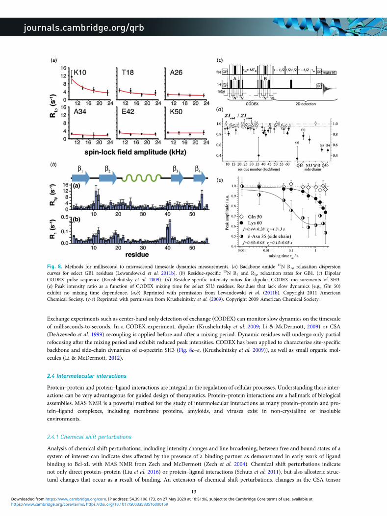
Exchange experiments such as center-band only detection of exchange (CODEX) can monitor slow dynamics on the timescaleof milliseconds-to-seconds. In a CODEX experiment, dipolar (Krushelnitsky et al. 2009; Li & McDermott, 2009) or CSA(DeAzevedo et al. 1999) recoupling is applied before and after a mixing period. Dynamic residues will undergo only partialrefocusing after the mixing period and exhibit reduced peak intensities. CODEX has been applied to characterize site-specificbackbone and side-chain dynamics of α-spectrin SH3 (Fig. 8c–e, (Krushelnitsky et al. 2009)), as well as small organic mol-ecules (Li & McDermott, 2012).
2.4 Intermolecular interactions
Protein–protein and protein–ligand interactions are integral in the regulation of cellular processes. Understanding these inter-actions can be very advantageous for guided design of therapeutics. Protein–protein interactions are a hallmark of biologicalassemblies. MAS NMR is a powerful method for the study of intermolecular interactions as many protein–protein and pro-tein–ligand complexes, including membrane proteins, amyloids, and viruses exist in non-crystalline or insolubleenvironments.
2.4.1 Chemical shift perturbations
Analysis of chemical shift perturbations, including intensity changes and line broadening, between free and bound states of asystem of interest can indicate sites affected by the presence of a binding partner as demonstrated in early work of ligandbinding to Bcl-xL with MAS NMR from Zech and McDermott (Zech et al. 2004). Chemical shift perturbations indicatenot only direct protein–protein (Liu et al. 2016) or protein–ligand interactions (Schutz et al. 2011), but also allosteric struc-tural changes that occur as a result of binding. An extension of chemical shift perturbations, changes in the CSA tensor
Fig. 8. Methods for millisecond to microsecond timescale dynamics measurements. (a) Backbone amide 15N R1ρ relaxation dispersioncurves for select GB1 residues (Lewandowski et al. 2011b). (b) Residue-specific 15N R1 and R1ρ relaxation rates for GB1. (c) DipolarCODEX pulse sequence (Krushelnitsky et al. 2009). (d) Residue-specific intensity ratios for dipolar CODEX measurements of SH3.(e) Peak intensity ratio as a function of CODEX mixing time for select SH3 residues. Residues that lack slow dynamics (e.g., Gln 50)exhibit no mixing time dependence. (a,b) Reprinted with permission from Lewandowski et al. (2011b). Copyright 2011 AmericanChemical Society. (c-e) Reprinted with permission from Krushelnitsky et al. (2009). Copyright 2009 American Chemical Society.
13
https://www.cambridge.org/core/terms. https://doi.org/10.1017/S0033583516000159Downloaded from https://www.cambridge.org/core. IP address: 54.39.106.173, on 27 May 2020 at 18:51:06, subject to the Cambridge Core terms of use, available at
magnitude can also serve as a probe of intermolecular interactions, such as applied to studies of cytb(5) in complex withcytP4502B4 (Pandey et al. 2013). Further, MAS NMR methods exist to characterize the bona fide intermolecular bindinginterface. For example, PRE in sites distal to the spin label can also be used as an indicator of intermolecular interactions(Wang et al. 2012). Dipolar edited correlation techniques have been particularly productive for the study of direct intermo-lecular interactions in biological macromolecular assemblies.
2.4.2 Dipolar-edited correlation spectroscopy
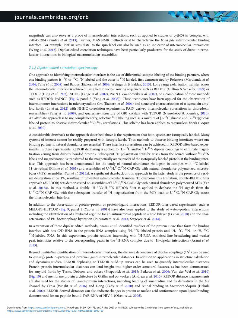
One approach to identifying intermolecular interfaces is the use of differential isotopic labeling of the binding partners, whereone binding partner is 13C or 13C,15N labeled and the other is 15N labeled, first demonstrated by Polenova (Marulanda et al.2004; Yang et al. 2008) and Baldus (Etzkorn et al. 2004; Weingarth & Baldus, 2013). Long range polarization transfer acrossthe intermolecular interface is achieved using heteronuclear mixing sequences such as REDOR (Gullion & Schaefer, 1989) orTEDOR (Hing et al. 1992), NHHC (Lange et al. 2002), PAIN (Lewandowski et al. 2007), or a combination of these methodssuch as REDOR–PAINCP (Fig. 9, panel 2 (Yang et al. 2008)). These techniques have been applied for the observation ofintermonomer interactions in microcrystalline Crh (Etzkorn et al. 2004) and structural characterization of α-synuclein amy-loid fibrils (Lv et al. 2012) with NHHC correlation experiments, PAIN-derived intermolecular correlations in thioredoxinreassemblies (Yang et al. 2008), and quaternary structure of GB1 crystals with TEDOR (Nieuwkoop & Rienstra, 2010).An alternate approach is to use complementary, selective 13C labeling such as a mixture of [1-13C]glucose and [2-13C]glucoselabeled protein to observe intermolecular 13C–13C correlations. This scheme has been applied to α-synuclein fibrils (Loquetet al. 2010).
A considerable drawback to the approach described above is the requirement that both species are isotopically labeled. Manysystems of interest cannot be readily prepared with isotopic labels. Thus methods to observe binding interfaces where onebinding partner is natural abundance are essential. These interface correlations can be achieved in REDOR-filter-based exper-iments. In these experiments, REDOR dephasing is applied to 1H–13C and/or 1H–15N dipolar couplings to eliminate magne-tization arising from directly bonded protons. Subsequent 1H polarization transfer arises from the source without 13C/15Nlabels and magnetization is transferred to the magnetically active nuclei of the isotopically labeled protein at the binding inter-face. This approach has been demonstrated for the study of natural abundance rhodopsin in complex with 13C-labeled11-cis-retinal (Kiihne et al. 2005) and assemblies of U-2H,13C,15N-CAP-Gly with natural abundance polymerized microtu-bules (MTs) assemblies (Yan et al. 2015a). A significant drawback of this approach in the latter study is the presence of resid-ual deuteration at ca. 1%, resulting in unwanted intramolecular transfers. To overcome this limitation, double-REDOR filterapproach (dREDOR) was demonstrated on assemblies of U-13C,15N-CAP-Gly with natural abundance polymerized MTs (Yanet al. 2015a). In this method, a double 1H–13C/1H–15N REDOR filter is applied to dephase the 1H signals from theU-13C,15N-CAP-Gly, with the subsequent transfer of 1H magnetization from the MTs back to U-13C,15N-CAP-Gly acrossthe intermolecular interface.
In addition to the observation of protein–protein or protein–ligand interactions, REDOR-filter-based experiments, such asMELODI-HETCOR (Fig. 9, panel 1 (Yao et al. 2001)) have also been applied to the study of water–protein interactions,including the identification of a hydrated arginine for an antimicrobial peptide in a lipid bilayer (Li et al. 2010) and the char-acterization of Pf1 bacteriophage hydration (Purusottam et al. 2013; Sergeyev et al. 2014).
In a variation of these dipolar edited methods, Asami et al. identified residues of the protein L7Ae that form the bindinginterface with box C/D RNA in the protein-RNA complex using 2H, 15N-labeled protein and 1H, 13C, 15N- or 2H, 13C,15N-labeled RNA. In this experiment, protein residues interacting with 1H-RNA exhibited line broadening and weakerpeak intensities relative to the corresponding peaks in the 2H-RNA complex due to 1H–dipolar interactions (Asami et al.2013).
Beyond qualitative identification of intermolecular interfaces, the distance dependence of dipolar couplings (1/r3) can be usedto quantify protein–protein and protein–ligand intermolecular distances. In addition to applications in structure calculationand dynamics studies, REDOR dephasing or TEDOR build-up curves can be used to quantify intermolecular distances.Protein–protein intermolecular distances can lend insight into higher-order structural features, as has been demonstratedfor amyloid fibrils by Tycko, Dobson, and others (Fitzpatrick et al. 2013; Petkova et al. 2006; Van der Wel et al. 2010)(Fig. 10) and membrane protein architecture by Griffin and co-workers (Andreas et al. 2015). REDOR distance measurementsare also used for the studies of ligand–protein interactions, including binding of amantadine and its derivatives in the M2channel by Cross (Wright et al. 2016) and Hong (Cady et al. 2010) and retinal binding in bacteriorhodopsin (Helmleet al. 2000). REDOR-derived distances can also indicate changes in protein or nucleic acid conformation upon ligand binding,demonstrated for tat peptide-bound TAR RNA of HIV-1 (Olsen et al. 2005).
14
https://www.cambridge.org/core/terms. https://doi.org/10.1017/S0033583516000159Downloaded from https://www.cambridge.org/core. IP address: 54.39.106.173, on 27 May 2020 at 18:51:06, subject to the Cambridge Core terms of use, available at
Various classes of supramolecular assemblies studied by MAS NMR include amyloid systems (reviewed in (Comellas &Rienstra, 2013; Tycko, 2011)), the Shigella type-III secretion system (TSS3) (Demers et al. 2014; Loquet et al. 2012,2013a), the Escherichia coli pilus protein FimA (Habenstein et al. 2015), and the MAVS (mitochondrial antiviral signaling)protein (He et al. 2015, 2016). This review focuses on two particular classes of supramolecular assemblies: cytoskeletal pro-teins and viruses.
3. MAS NMR of cytoskeleton-associated proteinsThe cytoskeleton is an essential cellular component in all domains of life. Functions of the cytoskeleton include maintenanceof cell shape, motility, intracellular transport, endocytosis, and cell signaling (Fischer & Fowler, 2015). In eukaryotes, the cel-lular cytoskeleton is composed of three main filament types: microfilaments (actin filaments), MTs (Nogales, 2000), and inter-mediate filaments. While most filaments of the prokaryotic cytoskeleton have eukaryotic analogues, there are filament typesthat are unique to prokaryotes (Lowe et al. 2004). Function of the cytoskeleton is crucially dependent on interactions with
Fig. 9. Two methods for the study of intermolecular interactions in protein assemblies. (Panel 1) MELODI–HETCOR (a) MELODI–HETCOR pulse sequence. (b–d) LG-HETCOR 1H–15N spectra of an Arg-rich membrane-embedded peptide (b) no REDOR dephasing,(c) only 1H–13C REDOR dephasing, (d) both 1H–13C and 1H–15N REDOR dephasing. (Li et al. 2010) (Panel 2) REDOR-PAINCP(e) pulse sequence for REDOR–PAINCP experiment. (f) 2D 15N–13C REDOR–PAINCP spectra of thioredoxin. (g) Observed intermolecu-lar correlations plotted onto the structure of thioredoxin. (Yang et al. 2008) (a–d) Adapted with permission fron Li et al. (2010).Copyright 2010 American Chemical Society. (e–g) Adapted with permission from Yang et al. (2008). Copyright 2008 American ChemicalSociety.
15
https://www.cambridge.org/core/terms. https://doi.org/10.1017/S0033583516000159Downloaded from https://www.cambridge.org/core. IP address: 54.39.106.173, on 27 May 2020 at 18:51:06, subject to the Cambridge Core terms of use, available at

binding partners, including the motor proteins dynein, kinesin, and myosin (Vale, 2003). The disruption of these interactionsby small molecules is a key mechanism in therapeutics for the treatment of cancers (Wood & Bergnes, 2004) and neurode-generative diseases (Gunawardena, 2013).
3.1 MTs and MT-associated proteins (MAPs)
MTs andMAPs perform several vital physiological functions in the cell includingmitosis and transport of signalingmolecules (Vale,2003). MTs are an important target of chemotheraputics due to their essential roles in cell division. MTs are highly dynamic andcontinually polymerizing and de-polymerizing in the cellular matrix (Howard & Hyman, 2003). Despite extensive structural andbiochemical characterization,manyopen questions remainwith respect to the function ofMTs and their associated proteins, includ-ing the atomic-level understanding of protein–protein interactions, of the role of protein dynamics in different conformational states,and of how protein–protein interactions and dynamics come together to orchestrate cellular processes. MAS NMR can lend insightinto the structure and dynamics of MT–MAP complexes at atomic resolution. To date, in-depth MAS NMR studies have been per-formed on only a handful of systems, including dynactin’s CAP-Gly domain assembled with MTs (Fig. 11a,b), bactofilin (Fig. 11c),and MTs interacting with small molecules, such as paclitaxel (Taxol) (Li et al. 2000; Paik et al. 2007), epothilone B (Kumar et al.2010), and their derivatives. In the following, we review the work on the first two cytoskeletal assemblies.
3.1.1 Structure of CAP-Gly domain of dynactin
Dynactin, an activator of the dynein motor assembly, is a protein complex involved in intracellular transport (Caviston &Holzbaur, 2006). Dynactin regulates dynein transport along MTs, and mutations within its p150Glued subunit lead to neuro-degenerative disorders, such as Huntington’s disease, Charcot–Marie Tooth disease, amyotropic lateral sclerosis, distal spinalbulbar muscular atrophy, and Perry syndrome (Chen et al. 2014). Within the p150Glued subunit of dynactin, CAP-Gly(cytoskeleton-associated protein glycine-rich) is an 89 residue MT-binding domain (Vaughan et al. 2002;Waterman-Storer et al. 1995). Dynactin CAP-Gly is the first MAP assembled with MTs, whose structure and dynamicshave been investigated in depth by MAS NMR, yielding atomic-resolution insights unavailable from other techniques andestablishing a proof of principle for investigations of other cytoskeleton-associated assemblies (Ahmed et al. 2010; Sunet al. 2009; Yan et al. 2013a, 2015a, b). Recently, the atomic-level resolution structure of CAP-Gly bound to polymericMTs was reported (Yan et al. 2015a) (PDB ID code 2MPX), the first structure of any cytoskeleton-associated protein assem-bled with cytoskeleton.
To determine the structure of CAP-Gly in complex with MTs, three different isotopic labeling schemes were used: U-15N,13C;U-15N, [2-13C]glucose; and U-15N, [1,6-13C]glucose. The structure was determined using hundreds of medium- and long-range
Fig. 10. Application of REDOR distance measurements to a selectively labeled amyloid protofilament revealed anti-parallel stacking of theβ-sheets. (a) 2D 15N–13C ZF-TEDOR spectrum, (b) 2D 13C–13C PDSD spectrum, (c) cross-section of anti-parallel β-sheets, red and blue linesindicate intermolecular distances measured, (d) REDOR dephasing curve of residues Y105 and S115, indicating head-to-tail arrangement of theprotofilament. Reproduced with permission from Fitzpatrick et al. (2013). Copyright 2013 National Academy of Sciences.
16
https://www.cambridge.org/core/terms. https://doi.org/10.1017/S0033583516000159Downloaded from https://www.cambridge.org/core. IP address: 54.39.106.173, on 27 May 2020 at 18:51:06, subject to the Cambridge Core terms of use, available at

distance restraints collected from 13C–13C CORD and 15N–13C PAIN-CP experiments, as well as hydrogen-bonding restraintsand torsion angles from TALOS+. The equivalent resolution in the structure was 1.9–2.5 Å with a tightly constrained ensembleof lowest-energy conformers. A very similar approach was previously used to determine the structure of free CAP-Gly (PDB IDcode 2M02) (Yan et al. 2013a). The structure of CAP-Gly assembled on MTs indicates that, while the overall secondary structureis retained, the flexible loops of CAP-Gly have remarkably different conformations when associated with MTs (Fig. 12). Loop β3/β4 adopts a more open conformation in the free state of CAP-Gly, and rearranges to a more closed conformation when bound toMTs. The different sidechain orientations of residues in this loop may play a role in CAP-Gly’s structural plasticity and ability tointeract with different binding partners.
EB1 is another MAP that, like dynactin, localizes at the plus end of the growing MT. EB1 is thought to have a role in MTdynamics; specifically it may promote MT elongation (Rogers et al. 2002). EB1 interacts with the p150Glued subunit of dynac-tin and it is hypothesized that the two proteins form a plus end complex to regulate MT dynamics (Ligon et al. 2003). Thep150Glued subunit may play a role in recruiting EB1 to the MTs. Residues of CAP-Gly that are perturbed by binding of EB1were identified by chemical shift changes. Chemical shift perturbations indicate that free CAP-Gly exists in multiple conform-ers, but is conformationally homogeneous when bound to MTs (Yan et al. 2013a).
3.1.2 Interface of CAP-Gly with MTs
The main challenge for NMR characterization of MTs and their assemblies with associated proteins is that to date there havebeen no efficient isotopic labeling protocols established for tubulin. This precludes in-depth structural characterization of MTsand limits the approaches for determination of intermolecular interfaces formed by MTs and their binding partners. To over-come this challenge, the application of dREDOR filters was explored to characterize the intermolecular interfaces that dynac-tin’s CAP-Gly forms with MTs and EB1 (Yan et al. 2015a). In these experiments, 1H–13C and 1H–15N REDOR filters weresimultaneously applied to dephase all 1H magnetization arising from U-13C,15N CAP-Gly. Subsequently, polarization wastransferred from 1H of natural abundance MTs or EB1 to their binding interface on the surface of CAP-Gly. A 13C-13CCORD dimension was included to enable site-specific assignment of the binding interface. Figure 13 shows dREDOR–CORD and dREDOR–HETCOR spectra of CAP-Gly in complex with MTs (b), EB1 (c), and the intermolecular interfacesas determined by dREDOR (a left, green) and chemical shift perturbations (a right, orange/purple). The results confirmedthat loop β3/β4 including the GKNDG motif comprises the primary binding interface with MTs. CAP-Gly interacts withits binding partners on the flat side of the protein, where most of the surface-exposed hydrophobic residues are located.dREDOR experiments of the CAP-Gly/EB1 complex were consistent with the known binding interface for this complex,which has been determined previously (Hayashi et al. 2005; Honnappa et al. 2006; Yan et al. 2013a), validating the approachfor characterizing the CAP-Gly/MT interface.
3.1.3 Dynamics of CAP-Gly
MT-associated motors and their activators possess conformational plasticity, which is essential for their ability to bind to andslide along the MTs (Howard, 2001; Vale, 2003). Conformational plasticity is directly related to internal flexibility, and
Fig. 11. Transmission electron microscopy of cytoskeleton-associated proteins for MAS NMR experiments. (a, b) 2H,13C,15N CAP-Gly/MT complex before and after MAS. Adapted with permission from Yan et al. (2015a). Copyright 2015 National Academy of Sciences. (c)13C, 15N BacA. Filament bundles are indicated by arrows, sheets are indicated by asterisks, and single filaments are indicated by arrow-heads. Adapted with permission from Vasa et al. (2015). Copyright 2015 National Academy of Sciences.
17
https://www.cambridge.org/core/terms. https://doi.org/10.1017/S0033583516000159Downloaded from https://www.cambridge.org/core. IP address: 54.39.106.173, on 27 May 2020 at 18:51:06, subject to the Cambridge Core terms of use, available at

therefore, knowledge of dynamics is essential for understanding the biological function of MAP assemblies. The dynamics ofCAP-Gly free, in complex with EB1, and bound to polymeric MTs have been characterized using MAS NMR, over a range offunctionally relevant timescales from nanoseconds to milliseconds (Yan et al. 2015b). Global dynamics were probed through atemperature series of 1D- and 2D 13C spectra (Fig. 14a-c). Site-specific dynamics were characterized using 1H–15N and1H–13C DOPs (Fig. 14d) (Hou et al. 2011b). As indicated by the temperature series in Fig. 14a–c, free and MT-boundCAP-Gly are dynamic across the entire range of timescales under investigation, and the motions are strongly temperaturedependent. In contrast, the CAP-Gly in complex with EB1 is largely rigid on these timescales and its spectra are not temper-ature dependent. From the measurement of DOPs, it was found that the loops of CAP-Gly are mobile in the free protein aswell as in complex with MTs. However, consistent with 1D and 2D spectra, the dynamics of CAP-Gly are notably attenuatedwhen in complex with EB1. Remarkably, the loops of CAP-Gly show an increase in fast timescale backbone fluctuations(nanosecond-to-microsecond), but a decrease in slower dynamics (microsecond-to-millisecond) upon binding to MTs(Fig. 14d). It was proposed that these observed changes in dynamics have a critical function in CAP-Gly/MT interactions.The combined structural and dynamics studies of CAP-Gly highlight the structural plasticity of this protein and the essentialrole this flexibility plays in CAP-Gly’s ability to adopt different conformations and interact with different binding partners.
3.2 Bactofilins
Bactofilins are a recently discovered class of bacterial cytoskeletal proteins. Similar to eukaryotic cystoskeletal proteins, theseproteins have diverse functions, such as cellular mobility, cell shape, and attachment. Bactofilins assemble rapidly and spon-taneously, making them not amenable for characterization by many biophysical techniques (Kuhn et al. 2010). Bactofilinscontain a conserved central rigid DUF583 domain, and terminal regions that are more dynamic. Lange and co-workershave recently reported resonance assignments and structure for BacA from Caulobacter crescentus (Shi et al. 2015; Vasaet al. 2015). Only the core DUF583 domain (residues 39–137) was observed in dipolar based 13C–13C and 15N–13C spectra,supporting the hypothesis that while the core is rigid, the termini are dynamic and believed to have a role in protein–protein
Fig. 12. (a) Structure of CAP-Gly bound to polymerized MTs (purple, 2MPX) and free CAP-Gly (orange, 2M02), both determined withMAS NMR, and CAP-Gly bound to EB1 (green, 2HKQ). (b) Expansion of loop regions of CAP-Gly in the three systems, indicating thedifferences in loop position and side-chain orientation for CAP-Gly in its three different states (Yan et al. 2015a). (c) Chemical shift per-turbations for several residues in CAP-Gly indicating multiple conformers of free CAP-Gly (black) that collapse to a single conformer incomplex with EB1 (Yan et al. 2013a), (a) and (b) Adapted with permission from Yan et al. (2015a). Copyright 2015 National Academyof Sciences. (c) Adapted with permission from Yan et al. (2013a). Copyright 2013 Elsevier.
18
https://www.cambridge.org/core/terms. https://doi.org/10.1017/S0033583516000159Downloaded from https://www.cambridge.org/core. IP address: 54.39.106.173, on 27 May 2020 at 18:51:06, subject to the Cambridge Core terms of use, available at

or protein–membrane interactions. Secondary structure analysis revealed at least ten distinct β-sheet segments (Fig. 15). Toobserve dynamic residues, through-bond INEPT (Morris & Freeman, 1979) 13C–13C correlation spectra were acquired.Chemical shifts for residues in the INEPT spectra indicated random coil secondary structure. Interestingly, fewer resonanceswere observed in the INEPT spectrum than would be expected to arise from the N- and C-termini, indicating that not allresidues in these regions are dynamic. While the secondary structure and dynamic behavior of BacA have similar featuresto amyloids (Daebel et al. 2012; Heise et al. 2005; Helmus et al. 2010), BacA has a distinct β-helical tertiary structure, as indi-cated by mass-per-length measurements by scanning transmission EM.
The atomic resolution structure determined by Shi et al. revealed that BacA is a right-handed β-helix with a triangular hydro-phobic core and six windings (Shi et al. 2015). With sparsely labeled BacA samples (1,3-13C and 2-13C glycerol), medium- andlong-range distance restraints were obtained, as well as torsion angle restraints from TALOS+ and β-sheet hydrogen bondrestraints. Additional 1H–1H distance restraints from a 4D HN(H)(H)NH spectrum (acquired with sine weightedPoisson-gap non-uniform sampling (Hyberts et al. 2010, 2012)) and a 2D NHHC spectrum were essential for determinationof the handedness of the β-helical structure. The presence of a right-handed β-helix had not been previously reported for anycytoskeletal protein. The hydrophobic core is triangular with highly conserved glycines at many of the corners and three par-allel β-sheets per winding (Fig. 16). It is believed that hydrophobic interactions mediate polymerization/folding of bactofilins(Kuhn et al. 2010). Hydrogen bonds between adjacent β-strands help to stabilize the overall structure. Windings 1 and 6 werenot as well restrained due to a lack of intermolecular distance restraints, which is attributed to increased dynamics in theseregions of the protein. Mutations of hydrophobic residues in winding 6 affect BacA assembly in vivo (Kuhn et al. 2010). It islikely that dynamics in this region of the protein has a role in BacA assembly.
4. MAS NMR of viral assemblies and intact viral particlesViruses are small pathogens that can impact all forms of life from bacteria, to plants and animals (Brussaard et al. 2004;Nelson & Citovsky, 2005; Pearson et al. 2009; Prangishvili, 2013; Smith & Helenius, 2004). The general virion structure isa single- or double-stranded DNA or RNA encapsulated by a protein coat or capsid. Some viruses include a lipid envelopesurrounding the capsid as well. Viruses invade target cells and seize the host cell’s machinery to reproduce, while evadingcellular defense mechanisms (Kaminskyy & Zhivotovsky, 2010). These key aspects of viral replication are of great interestas targets for the treatment of viral infections, but can also be exploited for nanotechnology and drug development. MASNMR is an excellent tool to probe structure and dynamics of viral protein assemblies, and particularly promising is an inte-grated approach, where this technique is combined with other experimental (e.g., cryo-EM, X-ray crystallography, and sol-ution NMR) and/or computational (e.g., MD simulations) methods to yield atomic-level understanding of structure and
Fig. 13. (a) Intermolecular interfaces of CAP-Gly with MT and EB1 determined with dREDOR (left, green), and observed chemical shiftperturbations (right, purple/orange). For chemical shift perturbations, purple residues indicate large shifts >1 ppm, orange indicates shiftsbetween 0.5 and 1 ppm. (b) dREDOR–HETCOR and dREDOR–CORD spectra of U-13C,15N CAP-Gly bound to MT and (c) in complexwith EB1. (Yan et al. 2015a) Reproduced with permission from Yan et al. (2015a). Copyright 2015 National Academy of Sciences.
19
https://www.cambridge.org/core/terms. https://doi.org/10.1017/S0033583516000159Downloaded from https://www.cambridge.org/core. IP address: 54.39.106.173, on 27 May 2020 at 18:51:06, subject to the Cambridge Core terms of use, available at

dynamics of viral assemblies. Below we discuss two important classes of viruses, HIV-1 and bacteriophages, which to datehave been the most thoroughly characterized by MAS NMR (Fig. 17).
4.1 HIV-1 capsid and maturation intermediates
Acquired immunodeficiency syndrome (AIDS), caused by HIV, is a global pandemic and affects approximately 37 millionpeople globally (World Health Organization, 2015). A key step in the HIV lifecycle is maturation, where an immatureviral particle is converted into a mature, infectious virion through a cascade of cleavage steps of the Gag polyprotein(Fig. 18a, Fig. 22, panel 1). The final step of maturation is the cleavage of the SP1 tail from CA (capsid protein) and the reor-ganization of CA into the mature conical capsid, which encapsulates the retroviral RNA (Briggs & Krausslich, 2011; Engelman& Cherepanov, 2012). Viral maturation is of poignant interest as the target of a novel class of therapeutics termed maturationinhibitors (Adamson et al. 2009; Salzwedel et al. 2007). Maturation inhibitors such as Bevirimat (BVM) inhibit HIV-1 mat-uration by binding to the CA-SP1 cleavage site and preventing cleavage. Tubular assemblies of CA mimic the native capsidlattice (Byeon et al. 2009; Zhao et al. 2013). Obtaining stable, morphologically homogeneous samples for MAS NMR typicallyrequires high ionic strength (∼1–2 M NaCl). With the use of low-E and Efree probes designed to minimize heating due to
Fig. 14. 15N–13C SPECIFIC CP NCA 1D spectra indicating temperature dependence of global conformational dynamics of (a) freeCAP-Gly, (b) CAP-Gly/EB1 complex, (c) CAP-Gly bound to MTs. (d) DOPs of free CAP-Gly at −2 °C (purple), and MT-boundCAP-Gly at −2 °C (green) and −19 °C (black). The micro-to-nanosecond timescale dynamics of MT-bound CAP-Gly are enhanced at−2° in comparison to the free protein. Adapted with permission from Yan et al. (2015b). Copyright 2015 The American Society forBiochemistry and Molecular Biology.
Fig. 15. (a) 13C–13C PDSD correlation spectrum of U–13C, 15N BacA. (b) Secondary structure of the core domain DUF583 of BacAdetermined from secondary chemical shift analysis. Adapted with permission from Vasa et al. (2015). Copyright 2015 National Academyof Sciences.
20
https://www.cambridge.org/core/terms. https://doi.org/10.1017/S0033583516000159Downloaded from https://www.cambridge.org/core. IP address: 54.39.106.173, on 27 May 2020 at 18:51:06, subject to the Cambridge Core terms of use, available at

radiofrequency irradiation, outstanding quality MAS NMR spectra can be obtained (Byeon et al. 2012; Bayro et al. 2014; Chen& Tycko, 2010; Han et al. 2010, 2013; Lu et al. 2015a).
4.1.1 Structural characterization of HIV-1 capsid assemblies
CA is a 231 residue, predominantly α-helical protein with independently folding N-terminal (NTD, residues 1–143) andC-terminal (CTD, residues 148–231) domains, connected by an inter-domain linker (Deshmukh et al. 2013; Gres et al.2015). Early studies (Chen & Tycko, 2010; Han et al. 2010) revealed that most residues in both NTD and CTD are relativelyrigid, and CA assemblies retain the secondary and tertiary structure determined from solution NMR and x-ray crystallographystudies. Upon maturation, the viral capsid morphology changes from spherical to conical. Mature conical HIV-1 capsids arebuilt from hexameric and pentameric CA assemblies, whose stoichiometry in the final capsid is variable, and so is the capsid’scone shape. Four kinds of intermolecular contacts are critical for capsid morphology and stability both in vivo and in vitro:intra-hexameric NTD–NTD and NTD–CTD, and inter-hexameric CTD–CTD interfaces around pseudo twofold and pseudothreefold axes (Fig. 18c–e) (Byeon et al. 2009; Pornillos et al. 2009). The capsid’s structural polymorphism is also observed invitro, where the morphology can be controlled by assembly conditions. CA can assemble into cones, tubes, and spheres(Barklis et al. 1998; Ehrlich et al. 2001; Ganser-Pornillos et al. 2007; Gross et al. 1997; Han et al. 2010, 2013). MAS NMRstudies of spheres, cones, and tubes indicated no major differences in secondary or tertiary structure among the three mor-phologies (Bayro et al. 2014; Chen & Tycko, 2010; Han et al. 2010, 2013), but no 3D structure is available so atomic-leveldetails of structural organization of these in vitro assemblies remain poorly understood.
HIV-1 exhibits significant sequence variability and sequence-dependent viral infectivity (Price et al. 2012). To date, twoHIV-1 strains have been studied by MAS NMR: HXB2 and NL4-3 (Byeon et al. 2012; Bayro et al. 2014; Chen & Tycko,2010; Han et al. 2010, 2013; Lu et al. 2015a). The wild-type sequences of these proteins differ by only four amino acids,but this variation causes considerable conformational changes across the CA sequence (Han et al. 2013). These structuralperturbations may play a role in the differing viral infectivity of the two strains. Viral infectivity is also regulated throughthe interactions with host factors. Cyclophilin A (CypA) is one such host factor protein that modulates HIV-1 infectivityand uncoating through direct interactions with the HIV-1 capsid’s CypA-binding loop located in the NTD. The mechanismof CypA is complex and poorly understood (Colgan et al. 1996; Luban et al. 1993). A recent study yielded the structure ofCypA in complex with the assembled HIV-1 capsid at 8 Å resolution (Liu et al. 2016). It was discovered, usingcryo-EM-guided all-atom MD simulations and MAS NMR spectroscopy that CypA simultaneously interacts with two CAsubunits in different hexamers. This binding mechanism established through the integrated cryo-EM/MAS NMR/MD
Fig. 16. Top view (a) and side view (b) of BacA structure determined by MAS NMR. (c) Schematic representation of the 6 windings.Colors are as follows: white – hydrophobic residues, red – acidic residues, blue – basic residues, and green – other residues. Mutations ofasterisked residues in winding 6 affect in vivo assembly. Reprinted with permission from Shi et al. (2015). Copyright 2015 AmericanAssociation for the Advancement of Science.
21
https://www.cambridge.org/core/terms. https://doi.org/10.1017/S0033583516000159Downloaded from https://www.cambridge.org/core. IP address: 54.39.106.173, on 27 May 2020 at 18:51:06, subject to the Cambridge Core terms of use, available at

approach provided insights into the mechanism of HIV-1 capsid stabilization by and recruitment of CypA to promote theviral infectivity.
Additional mutations in the various regions of CA sequence have been shown to affect the assembly morphology and effi-ciency, capsid stability, and interactions with host factors (Bocanegra et al. 2012; Forshey et al. 2002; Gres et al. 2015;Jiang et al. 2011; Manocheewa et al. 2013; McCarthy et al. 2013; Qi et al. 2008; Yang & Aiken, 2007). For example, mutationsin the linker region of CA render the capsid assembly inefficient or abolish it completely (Jiang et al. 2011). On the contrary,the E45A mutation makes a ‘hyperstable’ capsid, which does not disassemble, thus dramatically reducing the viral infectivity(Forshey et al. 2002; Hulme et al. 2015; Yang et al. 2012). Mutations in the CypA-binding loop, such as A92E and G94Dexhibit drastically reduced viral infectivity in the presence of the normally required CypA. Full infectivity can be restoredby inhibition of CypA by cyclosporin. MAS NMR analysis of these mutants revealed that the mutations induce conforma-tional changes, as well as changes in the dynamics, vide infra (Lu et al. 2015a).
To get insights into the structural changes occurring upon formation of the hexagonal CA lattice (predominant symmetry inthe cones), Bayro et al. used MAS NMR-derived structural restraints in tubular CA assemblies for refinement against solutionNMR and x-ray crystallography structures of monomeric CA (Bayro et al. 2014). 15N–15N backbone distances, determined by15N–15N dipolar recoupling (15N-BARE), have been used by Tycko and co-workers to derive backbone torsion angles andsecondary structure (Hu et al. 2012). In α-helices and tight turns, 15N–15N distances are short, leading to rapid signaldecay, while the decay is slower in loop regions (Fig. 19a). Deviations of experimental 15N-BARE curves of CA tubular assem-blies from simulated curves derived from crystal or solution NMR structures indicated differences in secondary structure.Using the 15N–15N distances and TALOS torsion angle predictions as structural restraints, structure refinement was carriedout against CA coordinates derived from solution NMR (monomer, 2LF4 (Shin et al. 2011)) and crystal structures (hexamer,3MGE (Pornillos et al. 2010)). Figure 19b–d shows regions of the CA structure that adopt a more extended conformation intubular assemblies than in their respective initial structures. Among these regions that undergo changes are the 310 helix nearthe start of the CTD, as well as the loops between helices 3 and 4, and helices 10 and 11. These perturbations were attributedto conformational changes arising from higher-order oligomerization and/or lattice curvature.
Very recently Bayro and Tycko characterized the structure of tubular assemblies of the capsid pseudo-twofold inter-hexamericinterface (Bayro & Tycko, 2016) using mixed and selective labeling schemes and dipolar-based distance measurements, withadditional restraints derived from the cryo-EM structure determined by Zhang and co-workers (Zhao et al. 2013). In thisstudy, quantitative W184 to M185 intermolecular distances across the dimerization interface were obtained with 15N–13CREDOR dephasing experiments. A series of experiments were performed to characterize intra-residue distances of W184and M185 in order to constrain the side-chain conformations, including 15N–13C REDOR distances, 13C–13C BroBaRR(BroadBand Rotational Resonance) distances for M185 (Chan & Tycko, 2004), and tensor correlation experiments(Dabbagh et al. 1994) of W184 to determine the angle between the backbone Cα–N bond vector and sidechain Cδ1–Nε1bond vector. The distances and angles determined differed somewhat from prior solution NMR (Byeon et al. 2009) andX-ray crystallography studies (Gres et al. 2015; Worthylake et al. 1999), suggesting the structure of the dimerization interfacein tubular assemblies differs from that in crystals or CA in solution. Further intra-CA monomer distance constraints wereobtained from 13C–13C correlations with long mixing (700 ms). With an integrated structure calculation approach describedabove, they were able to determine the structure of the inter-hexameric dimer interface in CA tubular assemblies, and furtherdemonstrated that this interface is well ordered in this morphology.
Fig. 17. Transmission electron microscopy images of viral assemblies and viruses for MAS NMR studies: (a) tubular assemblies ofHIV-1 capsid (CA), HXB2 strain, (b) CA in complex with CypA (Lu et al. 2015a), (c) tubular assemblies of CA-SP1 maturation interme-diate, NL4–3 strain, A92E mutant (Han et al. 2013). (d) T7 bacteriophage (Abramov & Goldbourt, 2014). (a, b) Adapted with permissionfrom Lu et al. (2015a). Copyright 2015 National Academy of Sciences. (c) Reprinted with permission from Han et al. (2013). Copyright2013 American Chemical Society. (d) Adapted with permission from Abramov & Goldbourt (2014). Copyright 2014 Springer.
22
https://www.cambridge.org/core/terms. https://doi.org/10.1017/S0033583516000159Downloaded from https://www.cambridge.org/core. IP address: 54.39.106.173, on 27 May 2020 at 18:51:06, subject to the Cambridge Core terms of use, available at

4.1.2 Conformational dynamics of HIV-1 capsid assemblies by MAS NMR
Protein dynamics have indispensible functions in many stages of the HIV-1 lifecycle, including assembly, release, and mat-uration (Freed, 2015). While CA is relatively rigid in the individual α-helices, residues in loops and the flexible linker, whichare key functional regions of the protein, are dynamic, giving rise to conformational plasticity, which is directly connected tocapsid morphology, interactions with host factors, and viral infectivity.
Lu et al. have characterized the site-specific dynamics of several capsid constructs, including the HXB2 and NL4-3 strainsdiscussed above, as well as CypA-bound CA and the A92E and G94D ‘escape mutants’ (Lu et al. 2015a). These mutantshave approximately 10% the infectivity of the wild-type virus; however, infectivity can be restored upon CypA inhibition(Ylinen et al. 2009). The CypA loop of CA (residues 84–100) undergoes conformational changes upon binding of CypAas well as in the A92E and G94D ‘escape’ mutants. 1H–15N and 1H–13C dipolar tensors and resonance intensities reported
Fig. 18. (a) Late stages of HIV-1 viral life cycle from assembly at the host cell membrane to budding and virion maturation. (Freed,2015) (b) All-atom MD derived model of the HIV-1 capsid based on cryo-ET and solution NMR, with a hexamer of hexamers subunitshown in the expansion (Lu et al. 2015a). (c) Hexmer of hexamers of HIV-1 capsid assembly. The interhexameric trimer interface is cir-cled. (d, e) Helix 10 trimer interface and helix 9 dimer interface respectively. Blue: hexamer of hexamers, orange: pentamer of hexamers(Zhao et al. 2013). (f) CA monomer. Residues for which chemical shift perturbations are observed upon binding of CypA are highlightedorange. (g) 15N–13C and 13C–13C correlation spectra of free CA tubular assemblies (black) and CA tubular assemblies in complex withCypA (Lu et al. 2015a) (a) Adapted with permission from Freed (2015). Copyright 2015 Macmillan Publishers. (b, f, g) Adapted with per-mission from Lu et al. (2015a). Copyright 2015 National Academy of Sciences. (c–e) Adapted with permission from Zhao et al. (2013).Copyright 2013 Nature Publishing Group.
23
https://www.cambridge.org/core/terms. https://doi.org/10.1017/S0033583516000159Downloaded from https://www.cambridge.org/core. IP address: 54.39.106.173, on 27 May 2020 at 18:51:06, subject to the Cambridge Core terms of use, available at

Fig. 19. (a) 15N–15N BARE curves for selected CA residues. Circles indicate experimental curves for CA tubular assemblies. Simulated curves correspond to the following structures: (- -) 2LF4, (−)3MGE, (−) 2KOD. (b) Initial (red) and final (blue) structure refinement against 2LF4, indicating the change in the conformation of the 310 helix. (c, d) Initial (orange) and final (blue) structure refine-ment against 3MGE, indicating the change in conformation of loop 3/4 and loop 10/11, respectively. Adapted with permission from Bayro et al. (2014). Copyright 2014 Elsevier.
24
https://ww
w.cam
bridge.org/core/terms. https://doi.org/10.1017/S0033583516000159
Dow
nloaded from https://w
ww
.cambridge.org/core. IP address: 54.39.106.173, on 27 M
ay 2020 at 18:51:06, subject to the Cambridge Core term
s of use, available at
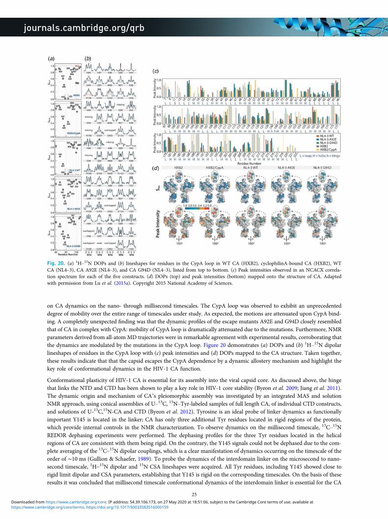
on CA dynamics on the nano- through millisecond timescales. The CypA loop was observed to exhibit an unprecedenteddegree of mobility over the entire range of timescales under study. As expected, the motions are attenuated upon CypA bind-ing. A completely unexpected finding was that the dynamic profiles of the escape mutants A92E and G94D closely resembledthat of CA in complex with CypA: mobility of CypA loop is dramatically attenuated due to the mutations. Furthermore, NMRparameters derived from all-atom MD trajectories were in remarkable agreement with experimental results, corroborating thatthe dynamics are modulated by the mutations in the CypA loop. Figure 20 demonstrates (a) DOPs and (b) 1H–15N dipolarlineshapes of residues in the CypA loop with (c) peak intensities and (d) DOPs mapped to the CA structure. Taken together,these results indicate that that the capsid escapes the CypA dependence by a dynamic allostery mechanism and highlight thekey role of conformational dynamics in the HIV-1 CA function.
Conformational plasticity of HIV-1 CA is essential for its assembly into the viral capsid core. As discussed above, the hingethat links the NTD and CTD has been shown to play a key role in HIV-1 core stability (Byeon et al. 2009; Jiang et al. 2011).The dynamic origin and mechanism of CA’s pleiomorphic assembly was investigated by an integrated MAS and solutionNMR approach, using conical assemblies of U–13C, 15N–Tyr-labeled samples of full length CA, of individual CTD constructs,and solutions of U-13C,15N-CA and CTD (Byeon et al. 2012). Tyrosine is an ideal probe of linker dynamics as functionallyimportant Y145 is located in the linker; CA has only three additional Tyr residues located in rigid regions of the protein,which provide internal controls in the NMR characterization. To observe dynamics on the millisecond timescale, 13C–15NREDOR dephasing experiments were performed. The dephasing profiles for the three Tyr residues located in the helicalregions of CA are consistent with them being rigid. On the contrary, the Y145 signals could not be dephased due to the com-plete averaging of the 13C–15N dipolar couplings, which is a clear manifestation of dynamics occurring on the timescale of theorder of ∼10 ms (Gullion & Schaefer, 1989). To probe the dynamics of the interdomain linker on the microsecond to nano-second timescale, 1H–15N dipolar and 15N CSA lineshapes were acquired. All Tyr residues, including Y145 showed close torigid limit dipolar and CSA parameters, establishing that Y145 is rigid on the corresponding timescales. On the basis of theseresults it was concluded that millisecond timescale conformational dynamics of the interdomain linker is essential for the CA
Fig. 20. (a) 1H–15N DOPs and (b) lineshapes for residues in the CypA loop in WT CA (HXB2), cyclophilinA-bound CA (HXB2), WTCA (NL4–3), CA A92E (NL4–3), and CA G94D (NL4–3), listed from top to bottom. (c) Peak intensities observed in an NCACX correla-tion spectrum for each of the five constructs. (d) DOPs (top) and peak intensities (bottom) mapped onto the structure of CA. Adaptedwith permission from Lu et al. (2015a). Copyright 2015 National Academy of Sciences.
25
https://www.cambridge.org/core/terms. https://doi.org/10.1017/S0033583516000159Downloaded from https://www.cambridge.org/core. IP address: 54.39.106.173, on 27 May 2020 at 18:51:06, subject to the Cambridge Core terms of use, available at

assembly. Solution NMR experiments revealed that, while these millisecond timescale motions of the linker are essential foropening up the conformational space, the number of accessible conformers is finite, and their populations are controlledthrough electrostatic intermolecular interactions between side chains of W184 and E175. This integrated approach establisheda molecular switch mechanism by which dynamics and electrostatic interactions work together to permit the formation ofvaried capsid morphologies in the mature HIV (Byeon et al. 2012).
In another study, Tycko and co-workers observed that while CA is generally rigid, there are regions of crucial biological func-tion that exhibit static or dynamic disorder (Bayro et al. 2014). Using scalar-based 13C–13C TOBSY (Hardy et al. 2001) exper-iments, dynamically disordered regions of the protein were identified. Residues attributed to the interdomain linker, loop 8/9,and the C-terminal tails were observed in this data set, indicating that these residues are dynamically disordered. To distin-guish residues that are mobile on the sub-millisecond timescale, 1H T2 filtered NCA data sets were acquired. In this exper-iment, a 1H T2 filter is incorporated and signals decay according to the local 1H–1H dipolar coupling strength. Thus, peakintensities of rigid residues are weakened to 20–30% of their peak intensity, while dynamic residues retain more than 40%of their peak intensity. 1H T2 filtered experiments indicated that many dynamic residues are in loops and/or solvent exposed,with the most mobile residues being in the CypA loop (Fig. 21), in agreement with the DOP measurements by Lu et al.(2015a). The N-terminal segment was also observed to be mobile.
4.1.3 MAS NMR of HIV-1 maturation intermediates
Understanding the viral maturation process has been of interest both from the fundamental science standpoint and for thedevelopment of small molecule maturation inhibitors as a venue for HIV-1 treatment. Despite intense research into the mat-uration mechanism, many key questions remain open, including how the conical CA capsids form from the immature Gaglattice. Two hypotheses have emerged to explain how capsid reorganization takes place upon maturation: (i) during
Fig. 21. (a) NCA spectrum of CA tubular assemblies, with sufficiently resolved peaks labeled. (b) 1H T2 filtered NCA spectrum with168 µs spin echo. Label colors correspond to peak intensity from 1H T2 filtered NCA experiment: 15–30%, dark blue; 31–40%, light blue;41–55%, green; 56–85%, magneta. Reprinted with permission from Bayro et al. (2014). Copyright 2014 Elsevier.
26
https://www.cambridge.org/core/terms. https://doi.org/10.1017/S0033583516000159Downloaded from https://www.cambridge.org/core. IP address: 54.39.106.173, on 27 May 2020 at 18:51:06, subject to the Cambridge Core terms of use, available at

Fig. 22. (Panel 1) Sequence of Gag polyprotein cleavage during maturation. (Panel 2) (a–d) Direct-DARR, (e–j) INADEQUATE, and (k)CP-DARR spectra of CA (orange) and CA-SP1 (black) NL4–3 strain assembled into tubes. Selected regions show the presence of SP1peaks not observed in the CA spectra. Reprinted with permission from Han et al. (2013). Copyright 2013 American Chemical Society.
27
https://www.cambridge.org/core/terms. https://doi.org/10.1017/S0033583516000159Downloaded from https://www.cambridge.org/core. IP address: 54.39.106.173, on 27 May 2020 at 18:51:06, subject to the Cambridge Core terms of use, available at
maturation, the capsid gradually changes shape from spherical to conical, and (ii) cleavage of SP1 triggers disassembly of theimmature lattice and subsequent de novo reassembly of the mature capsid core. Recently, a small molecule BVM was discov-ered, which was shown to inhibit viral maturation by abolishing the final step, the cleavage of the SP1 peptide from theCA-SP1 maturation intermediate (Fig. 22, panel 1) (Adamson et al. 2010; de Marco et al. 2010; Fontana et al. 2016;Nguyen et al. 2011). Studies on the BVM interaction with the Gag and maturation intermediates by EM and biochemicalmethods suggest that the second hypothesis is correct, i.e. capsid formation takes place through de novo assembly ratherthan by the gradual lattice remodeling (Keller et al. 2013). These EM studies are limited by resolution, and to gain atomic-levelinsights on the final step of HIV-1 maturation, MAS NMR spectroscopy was pursued. In a recent investigation, the SP1 pep-tide was characterized in tubular assemblies of two strains of the CA-SP1 maturation intermediate: wild type HXB2 andNL4-3 A92E mutant (Han et al. 2013). Dipolar and scalar-based correlation experiments (Fig. 22, panel 2) revealed thatthe SP1 peptide in the assembled state is dynamically disordered and does not adopt a stable helical structure proposedon the basis of early solution NMR experiments on isolated SP1 peptide in organic solvents (Datta et al. 2011). The presenceof the SP1 peptide affects the conformation and dynamics of CA protein: residues 226–231 of the CTD directly preceding theSP1 peptide, and which are mobile on microsecond timescale in CA assemblies, become more rigid, with the motions occur-ring on millisecond timescale in the CA-SP1 tubes. Most surprisingly, the dynamics of the CypA loop is modulated by thepresence of the SP1 peptide: the loop becomes more flexible. Taken together, the results of this investigation support the denovo reassembly hypothesis (Han et al. 2013; Keller et al. 2013; Lu et al. 2015a).
To summarize, MAS NMR is a powerful emerging method for atomic-level characterization of HIV-1 viral assemblies. Recentwork has demonstrated the significance of sequence-dependent conformational changes and dynamics of the HIV-1 capsidassemblies, as well as brought to light the importance of dynamic allostery in HIV maturation, infectivity, and interactionswith host factor proteins.
4.2 Bacteriophages
Bacteriophages are a diverse class of viruses that target and infect bacteria. Bacteriophages have applications in many areas,including molecular biology (Bax & Grishaev, 2005; Messing, 2001; Smith, 1985), nanotechnology (Huang et al. 2005; Namet al. 2006), and drug development (Clark & March, 2006; Omidfar & Daneshpour, 2015). For review see (Henry &Debarbieux, 2012; Salmond & Fineran, 2015). Two classes of bacteriophages have been studied by MAS NMR: filamentousand icosahedral bacteriophages. Filamentous phages, which include Pf1, fd, and M13 are long rod-like structures with asingle-stranded DNA (ssDNA) encapsulated in a capsid, composed of symmetric repeats of several thousand copies of anα-helical coat protein (Marvin, 1998). A second class is represented by tailed icosahedral viruses, such as T4 and T7,which are composed of nucleic acid encased in an icosahedral capsid, with a tail to attach to the target host cell(Ackermann, 1999). In addition to structural studies of bacteriophage capsids, protein–nucleic acid interactions in bacterio-phages have been addressed by MAS NMR.
4.2.1 Structural characterization of bacteriophage capsid proteins
McDermott and co-workers first characterized the intact Pf1 bacteriophage by MAS NMR, where resonance assignments andsecondary structure determination were performed and the feasibility of this approach for atomic-level analysis was estab-lished (Goldbourt et al. 2007b). Pf1 undergoes a cooperative phase transition at 10 °C (Thiriot et al. 2005), with the high-and low-temperature forms having slightly different helical symmetries. Goldbourt et al. mapped chemical shift perturbationsobserved in 13C–13C correlation spectra to the structure of Pf1 (Goldbourt et al. 2010). Most chemical shift perturbationsappeared in the side chains of residues in the hydrophobic region of the protein, at the interface between subunits. They pos-tulated that adjustments in these hydrophobic regions enabled the temperature transition of what is an overall rigid structure.
Hydration waters play critical roles in biological function, including maintenance of structure and facilitating protein dynam-ics. McDermott and co-workers (Sergeyev et al. 2014), and Sinha and co-workers (Purusottam et al. 2013) used 1H–15N and1H–13C HETCOR spectroscopy, including MEdium-to-LOng Distance (MELODI) HETCOR (Yao & Hong, 2001) to probethe hydration state of Pf1. REDOR dephasing pulses (Gullion & Schaefer, 1989) were applied during HETCOR experimentsto dephase magnetization arising from directly bonded protons, such that observed magnetization is known to come fromdirect contact with water. McDermott and co-workers also utilized water-selective T2′-filtering for spectral simplificationin some HETCOR experiments and a 3D MELODI HETCOR experiment which incorporates a train of 13C and 15NREDOR dephasing pulses on both 13C and 15N with a DARR 13C–13C mixing component which allowed for the assignmentof numerous protein residues in contact with water, as shown in Fig. 23. SD-HETCOR experiments (HETCOR with 1H spindiffusion (Kumashiro et al. 1998)) were also included to allow for the observation of hydration waters on the encapsulatedDNA. Hydration sites include surface exposed residues at the N-terminus, as well as C-terminal residues, which contact
28
https://www.cambridge.org/core/terms. https://doi.org/10.1017/S0033583516000159Downloaded from https://www.cambridge.org/core. IP address: 54.39.106.173, on 27 May 2020 at 18:51:06, subject to the Cambridge Core terms of use, available at

the interior cavity of the phage, including Arg 44 and Lys 45, which participate in protein–DNA interactions, as discussedbelow. These results indicate that hydration waters are essential in the protein–DNA interactions, which stabilize the capsidstructure. The data also revealed hydrophilic groves in the coat protein, which allow for water exchange between the interior ofthe phage and the exterior surface.
Goldbourt and co-workers reported the structure of the capsid protein from intact M13 bacteriophage (Morag et al. 2015). Thisis the first structure of an intact filamentous virus capsid solved by MAS NMR. The intra- and inter-subunit distance restraintswere established using selectively labeled samples (Fig. 24a) (Morag et al. 2011). The ‘fold-and-dock’ protocol of CS-ROSETTA(Das et al. 2009; Shen et al. 2008) containing 35 M13 subunits to capture all unique interactions was used (DiMaio et al. 2011).This protocol is designed to determine optimum structures of higher-order oligomers. M13 and other Ff class bacteriophageshave fivefold subunit symmetry around the virion axis with largely α-helical secondary structure. Many inter-subunit interac-tions were observed in hydrophobic regions, highlighting the importance of the hydrophobic pockets for subunit packing.Each monomer was found to participate in four hydrophobic pockets, with participating residues distributed across the mono-mer (Fig. 24c). Many of these key aromatic/hydrophobic residues are conserved among filamentous bacteriophages. The struc-ture also reveals hydrogen bonding and electrostatic interactions that further contribute to capsid stability.
4.2.2 Characterization of bacteriophage capsid dynamics
Lorieau et al. probed backbone dynamics in Pf1 by Lee–Goldburg cross-polarization experiments (Lorieau et al. 2008).1H–13C DOPs were measured for several Cα, Cβ, Cγ, and Cδ sites. The backbone of Pf1 was found to be remarkably rigid.
Fig. 23. (a) 1H-13C MELODI-HETCOR of Pf1. (b) 13C–13C slice of a 1H–13C–13C 3D spectrum at the water frequency. (c) Pf1 subunit;residues interacting with water are shaded purple. Hydrated residues are concentrated at the N- and C-termini. Reprinted with permissionfrom Sergeyev et al. (2014). Copyright 2014 AIP Publishing.
29
https://www.cambridge.org/core/terms. https://doi.org/10.1017/S0033583516000159Downloaded from https://www.cambridge.org/core. IP address: 54.39.106.173, on 27 May 2020 at 18:51:06, subject to the Cambridge Core terms of use, available at

As would be expected, many solvent-exposed side chains exhibited reduced order parameters. Significantly, Arg 44 and Lys 45side chains, which are directed into the phage cavity, were also observed to be highly mobile. It was postulated that thedynamics of these two residues is essential for mediating DNA–protein interactions. Opella and co-workers also measured1H–15N dipolar lineshapes of this system with 1H-detected experiments at 50 kHz MAS (Park et al. 2013).
4.2.3 Characterization of protein–nucleic acid interactions in bacteriophages
Nucleic acids are an integral component of viruses (Speir & Johnson, 2012). Recently several labs have demonstrated thatMAS NMR can characterize nucleic acids (Cherepanov et al. 2010; Huang et al. 2012) and protein–nucleic acid interactions(Asami et al. 2013; Asami & Reif, 2013; Marchanka et al. 2015, 2013) including studies of protein–DNA interactions in intactbacteriophages (Morag et al. 2014; Sergeyev et al. 2011; Yu & Schaefer, 2008).
In contrast to other bacteriophages, Pf1 has an unusually high nucleotide-to-capsid subunit ratio of 1:1 (Day et al. 1988) and ahighly extended conformation. (Other inoviruses have ratios of between 2–2.5 and 1.) Using dynamic nuclear polarization(DNP, (Ni et al. 2013)) for sensitivity enhancement, Sergeyev et al. were able to detect nucleotide resonances in the intactPf1 bacteriophage (Sergeyev et al. 2011). The reported chemical shifts for the deoxyribose moieties capture the unusualDNA conformation of Pf1. As compared with average B-form DNA chemical shifts, C2′, C3′, C4′, and C5′ shifts in Pf1appeared downfield, indicating a C2′-endo/gauche conformation with an anti glycosidic bond orientation. Nucleotide basechemical shifts, such as those of TC7 and CC5 fall outside the averages reported in the BMRB (Ulrich et al. 2008) indicatingthat Pf1 DNA does not form typical base-pairing interactions.
Goldbourt, Morag, and Abramov used 13C–13C and 1H-mediated 31P–13C correlation experiments to characterize capsid–ssDNA interactions in the fd bacteriophage (Abramov et al. 2011; Morag et al. 2014). Initial studies demonstrated that well-resolved spectra with narrow peaks could be attained for the wild-type coat protein, enabling resonance assignments andfurther structural characterization (Abramov et al. 2011). Prior structural studies with static SSNMR, cryo-EM, and fiber dif-fraction relied on the rigid Y21M mutant (Zeri et al. 2003), as the wild-type protein yielded poor data quality (i.e., broad lines)in SSNMR (Tan et al. 1999) or lack of structural convergence in the case of cryo-EM (Wang et al. 2006). For the study of DNAwithin fd, the choice of labeling scheme was key (Morag et al. 2014). By including unlabeled aromatic amino acids in theexpression media, the nucleotide resonances were not obscured by protein peaks. Broadband and efficient CORD 13C–13Ccorrelation experiments (Hou et al. 2011a, 2013a), described above in Section 2.2.1, were found to be ideal for the observationof DNA resonances, particularly for quaternary carbons with no directly attached protons. Nucleotide-specific assignmentswere achieved using a nucleotide walk, as shown in Fig. 25. In the CORD spectra acquired with long mixing times,sugar-to-capsid and base-to-capsid DNA–protein interactions were also observed. PHHC proton-mediated 31P–13C correla-tion spectra detected capsid-to-phosphate backbone interactions. Altogether, 56 capsid–ssDNA interactions could be
Fig. 24. (a) 13C–13C CORD spectrum of [1,3–13C]glycerol, U-15N M13 bacteriophage with 500 ms mixing time. Intra-residue contactsare labeled in black. Inter-residue contacts within the same subunit are labeled in green. Inter-residue contacts between residues in differ-ent subunits are labeled in blue. Select inter-subunit correlations are shown on the structure. (b) Sideview of the NMR-ROSETTA modelof M13 containing 35 subunits, the minimum number of subunits required to contain all unique interactions. (c) Hydrophobic pocketsformed by several subunits. Adapted with permission from Morag et al. (2015). Copyright 2015 National Academy of Sciences.
30
https://www.cambridge.org/core/terms. https://doi.org/10.1017/S0033583516000159Downloaded from https://www.cambridge.org/core. IP address: 54.39.106.173, on 27 May 2020 at 18:51:06, subject to the Cambridge Core terms of use, available at

assigned. These protein–DNA interactions occur primarily between positively charged Lys side chains near the C-terminusand the DNA phosphate backbone. Fd was found to have a similar sugar conformation to Pf1 (Sergeyev et al. 2011) butgreater tendency for base stacking and a different protein–DNA interface.
Due to challenges with respect to spectral overlap and sample preparation, MAS NMR of nucleic acids has been much morelimited than protein NMR. Abramov and Goldbourt were able to characterize the 40 kbp double-stranded DNA packagedwithin bacteriophage T7, a 50 MDa icosahedral phage (Abramov & Goldbourt, 2014). Using 13C–13C DARR and 15N–13CTEDOR correlations, all carbons and most nitrogens of the nucleic acid could be detected. The DNA packaged inside thephage was determined to be B-form with expected Watson–Crick base pairing. This study, along with the studies of nucleicacids in fd and Pf1, adds to the growing database of nucleic acid chemical shifts (particularly for large, native systems) and therelationship of chemical shifts to structure.
Yu and Schaefer used 15N–31P REDOR experiments to characterize DNA packaging in bacteriophage T4 with U-15N and[ε-15N]Lys-labeled T4 (Yu & Schaefer, 2008). The dependence of REDOR dephasing on the 15N–31P distance enabled thedetermination that the DNA packaged inside T4 is B-form by determining the distance between the phosphate backboneand nitrogen atoms of the nucleic acid bases. T4 is packed into the phage capsid with over 1000 molecules of three lysine-richproteins (Karam, 1994). The very close phosphate–lysine distances (as close as 3·5 Å) indicate that side-chain amine groupshave an important role in charge balance in the phage. Further charge balance of DNA inside the phage comes from poly-amines and ammonium cations.
Bacteriophages including Pf1, fd, and M13 have also been studied in membrane-associated states, as well as by static SSNMRtechniques by Opella and others. These topics are beyond the scope of the current review. We direct readers to the followingreviews and key publications: (Bechinger, 1997; Cross et al. 1983; Glaubitz et al. 2000; Marassi & Opella, 2003; Opella et al.2008; Park et al. 2010; Shon et al. 1991; Tan et al. 1999; Thiriot et al. 2004, 2005; Zeri et al. 2003).
4.3 Other viral assemblies
Pintacuda and co-workers reported a study of the MeV nucleocapsid (Barbet-Massin et al. 2014a). Prior to this work, noatomic-level information was available for assembled nucleocapsid. The MeV nucleocapsid is comprised a relatively rigidNCORE domain and a more dynamic NTAIL domain. Proton detection was applied to both intact and cleaved assemblies
Fig. 25. Spin system assignments of DNA in fd bacteriophage. Nucleotide walks are shown for (a) dG – blue and dC – red, and (b) dA– green, and dT – pink. (c) Assignment grid indicating all observed DNA correlations. (d) Expansion of 13C–13C CORD spectrum.Capsid-to-sugar correlations are labeled green, while intra-nucleotide resonances are labeled black. (e) Model of protein–DNA interactionsin fd. Adapted with permission from Morag et al. (2014). Copyright 2014 American Chemical Society.
31
https://www.cambridge.org/core/terms. https://doi.org/10.1017/S0033583516000159Downloaded from https://www.cambridge.org/core. IP address: 54.39.106.173, on 27 May 2020 at 18:51:06, subject to the Cambridge Core terms of use, available at

with MAS at 60 kHz. Dipolar 15N–1H correlations revealed rigid residues of NCORE, while scalar-based correlations detectedmobile residues of NTAIL. Capsid hydration levels were also evaluated by observing T1ρ relaxation of 15N–1H peak intensities.The reduced intensities of the cleaved nucleocapsid relative to intact capsid indicate that the presence of the NTAIL domainleads to a less ordered and hence more hydrated conformation.
5. Conclusions and future perspectivesMAS NMR has developed into a compelling technique for the characterization of biological assemblies at the atomic level.Integrated approaches to protein structure determination utilizing MAS NMR structural restraints in conjunction with infor-mation from other methods, such as cryo-EM and MD, are very promising for structural and dynamics analysis of highmolecular weight assemblies. Continued advancements in sensitivity, resolution, and methodology will enable the detailedcharacterization of increasingly complex systems, such as whole cells, where substantial progress has already been made(Curtis-Fisk et al. 2008; Janssen et al. 2010; Pius et al. 2012; Reichhardt & Cegelski, 2014).
AcknowledgementsThis work is supported by the National Institutes of Health NIGMS grants P50GM082251 and F32GM113452 and is a con-tribution from the Pittsburgh Center for HIV Protein Interactions. C.M.Q. acknowledges the support of the NationalInstitutes of Health Postdoctoral Fellowship grant F32GM113452.
ReferencesABRAMOV, G. & GOLDBOURT, A. (2014). Nucleotide-type chemical shift assignment of the encapsulated 40 kbp dsDNA in intact bacteriophageT7 by MAS solid-state NMR. Journal of Biomolecular NMR 59, 219–230.
ABRAMOV, G., MORAG, O. & GOLDBOURT, A. (2011). Magic-angle spinning NMR of a class I filamentous bacteriophage virus. Journal of PhysicalChemistry B 115, 9671–9680.
ACKERMANN, H.W. (1999). Tailed bacteriophages: the order Caudovirales. Advances in Virus Research 51, 135–201.ADAMSON, C. S., SAKALIAN, M., SALZWEDEL, K. & FREED, E. O. (2010). Polymorphisms in Gag spacer peptide 1 confer varying levels of resistanceto the HIV-1 maturation inhibitor bevirimat. Retrovirology 7, 1–8.
ADAMSON, C. S., SALZWEDEL, K. & FREED, E. O. (2009). Virus maturation as a new HIV-1 therapeutic target. Expert Opinion on TherapeuticTargets 13, 895–908.
AGARWAL, V., DIEHL, A., SKRYNNIKOV, N. & REIF, B. (2006). High resolution 1H detected 1H,13C correlation spectra in MAS solid-state NMR usingdeuterated proteins with selective 1H, 2H, isotopic labeling of methyl groups. Journal of the American Chemical Society 128, 12620–12621.
AGARWAL, V., PENZEL, S., SZEKELY, K., CADALBERT, R., TESTORI, E., OSS, A., PAST, J., SAMOSON, A., ERNST, M., BOCKMANN, A. & MEIER, B. H. (2014).De novo 3D structure determination from sub-milligram protein samples by solid-state 100 kHz MAS NMR spectroscopy. AngewandteChemie – International Edition 53, 12253–12256.
AGARWAL, V., SARDO, M., SCHOLZ, I., BOCKMANN, A., ERNST, M. & MEIER, B. H. (2013). PAIN with and without PAR: variants for third-spinassisted heteronuclear polarization transfer. Journal of Biomolecular NMR 56, 365–377.
AHMED, S., SUN, S., SIGLIN, A. E., POLENOVA, T. & WILLIAMS, J. C. (2010). Disease-associated mutations in the p150Glued subunit destabilize theCAP-Gly domain. Biochemistry 49, 5083–5085.
AKBEY, U., LANGE, S., FRANKS, W. T., LINSER, R., REHBEIN, K., DIEHL, A., VAN ROSSUM, B. J., REIF, B. & OSCHKINAT, H. (2010). Optimum levels ofexchangeable protons in perdeuterated proteins for proton detection in MAS solid-state NMR spectroscopy. Journal of Biomolecular NMR46, 67–73.
ALENGHAT, F. J. & GOLAN, D. E. (2013). Membrane protein dynamics and functional implications in mammalian cells. Functional Organizationof Vertebrate Plasma Membrane 72, 89–120.
ALKARAGHOULI, A. R. & KOETZLE, T. F. (1975). Neutron-diffraction study of L-phenylalanine hydrochloride. Acta Crystallographica Section B:Structural Science 31, 2461–2465.
ANDREAS, L. B., JAUDZEMS, K., STANEK, J., LALLI, D., BERTARELLO, A., LE MARCHAND, T., CALA-DE PAEPE, D., KOTELOVICA, S., AKOPJANA, I., KNOTT, B.,WEGNER, S., ENGELKE, F., LESAGE, A., EMSLEY, L., TARS, K., HERRMANN, T. & PINTACUDA, G. (2016). Structure of fully protonated proteins byproton-detected magic-angle spinning NMR. Proceedings of the National Academy of Sciences of the United States of America 113, 9187–9192.
ANDREAS, L. B., REESE, M., EDDY, M. T., GELEV, V., NI, Q. Z., MILLER, E. A., EMSLEY, L., PINTACUDA, G., CHOU, J. J. & GRIFFIN, R. G. (2015). Structureand mechanism of the influenza A M2(18–60) dimer of dimers. Journal of the American Chemical Society 137, 14877–14886.
ASAMI, S., PORTER, J. R., LANGE, O. F. & REIF, B. (2015). Access to Cα backbone dynamics of biological solids by 13C T1 relaxation and moleculardynamics simulation. Journal of the American Chemical Society 137, 1094–1100.
32
https://www.cambridge.org/core/terms. https://doi.org/10.1017/S0033583516000159Downloaded from https://www.cambridge.org/core. IP address: 54.39.106.173, on 27 May 2020 at 18:51:06, subject to the Cambridge Core terms of use, available at

ASAMI, S., RAKWALSKA-BANGE, M., CARLOMAGNO, T. & REIF, B. (2013). Protein-RNA interfaces probed by 1H-detected MAS solid-state NMRspectroscopy. Angewandte Chemie – International Edition 52, 2345–2349.
ASAMI, S. & REIF, B. (2013). Proton-detected solid-state NMR spectroscopy at aliphatic sites: application to crystalline systems. Accounts ofChemical Research 46, 2089–2097.
BALDUS, M., PETKOVA, A. T., HERZFELD, J. & GRIFFIN, R. G. (1998). Cross polarization in the tilted frame: assignment and spectral simplificationin heteronuclear spin systems. Molecular Physics 95, 1197–1207.
BALTISBERGER, J. H., MUSAPELO, T., SUTTON, B., REYNOLDS, A. & GURUNG, L. (2011). Reduction of spin diffusion artifacts from 2Dzfr-INADEQUATE MAS NMR spectra. Journal of Magnetic Resonance 208, 70–75.
BARBET-MASSIN, E., FELLETTI, M., SCHNEIDER, R., JEHLE, S., COMMUNIE, G., MARTINEZ, N., JENSEN, M. R., RUIGROK, R.W. H., EMSLEY, L., LESAGE, A.,BLACKLEDGE, M. & PINTACUDA, G. (2014a). Insights into the structure and dynamics of measles virus nucleocapsids by 1H-detected solid-stateNMR. Biophysical Journal 107, 941–946.
BARBET-MASSIN, E., PELL, A. J., JAUDZEMS, K., FRANKS, W. T., RETEL, J. S., KOTELOVICA, S., AKOPJANA, I., TARS, K., EMSLEY, L., OSCHKINAT, H., LESAGE,A. & PINTACUDA, G. (2013). Out-and-back 13C–13C scalar transfers in protein resonance assignment by proton-detected solid-state NMRunder ultra-fast MAS. Journal of Biomolecular NMR 56, 379–386.
BARBET-MASSIN, E., PELL, A. J., RETEL, J. S., ANDREAS, L. B., JAUDZEMS, K., FRANKS, W. T., NIEUWKOOP, A. J., HILLER, M., HIGMAN, V., GUERRY, P.,BERTARELLO, A., KNIGHT, M. J., FELLETTI, M., LE MARCHAND, T., KOTELOVICA, S., AKOPJANA, I., TARS, K., STOPPINI, M., BELLOTTI, V., BOLOGNESI,M., RICAGNO, S., CHOU, J. J., GRIFFIN, R. G., OSCHKINAT, H., LESAGE, A., EMSLEY, L., HERRMANN, T. & PINTACUDA, G. (2014b). Rapidproton-detected NMR assignment for proteins with fast magic angle spinning. Journal of the American Chemical Society 136, 12489–12497.
BARKLIS, E., MCDERMOTT, J., WILKENS, S., FULLER, S. & THOMPSON, D. (1998). Organization of HIV-1 capsid proteins on a lipid monolayer.Journal of Biological Chemistry 273, 7177–7180.
BAX, A. & GRISHAEV, A. (2005). Weak alignment NMR: a hawk-eyed view of biomolecular structure. Current Opinion in Structural Biology 15,563–570.
BAYRO, M. J., CHEN, B., YAU, W. M. & TYCKO, R. (2014). Site-specific structural variations accompanying tubular assembly of the HIV-1 capsidprotein. Journal of Molecular Biology 426, 1109–1127.
BAYRO, M. J. & TYCKO, R. (2016). Structure of the dimerization interface in the mature HIV-1 capsid protein lattice from solid state NMR oftubular assemblies. Journal of the American Chemical Society 138, 8538–8546.
BECHINGER, B. (1997). Structure and dynamics of the M13 coat signal sequence in membranes by multidimensional high-resolution and solid-state NMR spectroscopy. Proteins – Structure Function and Genetics 27, 481–492.
BERTINI, I., EMSLEY, L., FELLI, I. C., LAAGE, S., LESAGE, A., LEWANDOWSKI, J. R., MARCHETTI, A., PIERATTELLI, R. & PINTACUDA, G. (2011a).High-resolution and sensitivity through-bond correlations in ultra-fast magic angle spinning (MAS) solid-state NMR. Chemical Science2, 345–348.
BERTINI, I., EMSLEY, L., LELLI, M., LUCHINAT, C., MAO, J. & PINTACUDA, G. (2010). Ultrafast MAS solid-state NMR permits extensive 13C and 1Hdetection in paramagnetic metalloproteins. Journal of the American Chemical Society 132, 5558–5559.
BERTINI, I., LUCHINAT, C., PARIGI, G. & RAVERA, E. (2013). SedNMR: on the edge between solution and solid-state NMR. Accounts of ChemicalResearch 46, 2059–2069.
BERTINI, I., LUCHINAT, C., PARIGI, G., RAVERA, E., REIF, B. & TURANO, P. (2011b). Solid-state NMR of proteins sedimented by ultracentrifugation.Proceedings of the National Academy of Sciences of the United States of America 108, 10396–10399.
BOCANEGRA, R., RODRIGUEZ-HUETE, A., FUERTES, M. A., DEL ALAMO, M. & MATEU, M. G. (2012). Molecular recognition in the human immuno-deficiency virus capsid and antiviral design. Virus Research 169, 388–410.
BOCKMANN, A., LANGE, A., GALINIER, A., LUCA, S., GIRAUD, N., JUY, M., HEISE, H., MONTSERRET, R., PENIN, F. & BALDUS, M. (2003). Solid state NMRsequential resonance assignments and conformational analysis of the 2×10·4 kDa dimeric form of the Bacillus subtilis protein Crh. Journalof Biomolecular NMR 27, 323–339.
BRIGGS, J. A. & KRAUSSLICH, H. G. (2011). The molecular architecture of HIV. Journal of Molecular Biology 410, 491–500.BROWN, L. S. & LADIZHANSKY, V. (2015). Membrane proteins in their native habitat as seen by solid-state NMR spectroscopy. Protein Science 24,1333–1346.
BRUSSAARD, C. P. D., NOORDELOOS, A. A. M., SANDAA, R. A., HELDAL, M. & BRATBAK, G. (2004). Discovery of a dsRNA virus infecting the marinephotosynthetic protist Micromonas pusilla. Virology 319, 280–291.
BYEON, I. J., MENG, X., JUNG, J., ZHAO, G., YANG, R., AHN, J., SHI, J., CONCEL, J., AIKEN, C., ZHANG, P. & GRONENBORN, A. M. (2009). Structuralconvergence between Cryo-EM and NMR reveals intersubunit interactions critical for HIV-1 capsid function. Cell 139, 780–790.
BYEON, I. J. L., HOU, G. J., HAN, Y., SUITER, C. L., AHN, J., JUNG, J., BYEON, C. H., GRONENBORN, A. M. & POLENOVA, T. (2012). Motions on themillisecond time scale and multiple conformations of HIV-1 capsid protein: implications for structural polymorphism of CA assemblies.Journal of the American Chemical Society 134, 6455–6466.
CADARS, S., SEIN, J., DUMA, L., LESAGE, A., PHAM, T. N., BALTISBERGER, J. H., BROWN, S. P. & EMSLEY, L. (2007). The refocused INADEQUATE MASNMR experiment in multiple spin-systems: interpreting observed correlation peaks and optimising lineshapes. Journal of MagneticResonance 188, 24–34.
CADY, S. D., SCHMIDT-ROHR, K., WANG, J., SOTO, C. S., DEGRADO, W. F. & HONG, M. (2010). Structure of the amantadine binding site of influenzaM2 proton channels in lipid bilayers. Nature 463, 689–693.
CARRAVETTA, M., EDEN, M., ZHAO, X., BRINKMANN, A. & LEVITT, M. H. (2000). Symmetry principles for the design of radiofrequency pulsesequences in the nuclear magnetic resonance of rotating solids. Chemical Physics Letters 321, 205–215.
33
https://www.cambridge.org/core/terms. https://doi.org/10.1017/S0033583516000159Downloaded from https://www.cambridge.org/core. IP address: 54.39.106.173, on 27 May 2020 at 18:51:06, subject to the Cambridge Core terms of use, available at

CASTELLANI, F., VAN ROSSUM, B., DIEHL, A., SCHUBERT, M., REHBEIN, K. & OSCHKINAT, H. (2002). Structure of a protein determined by solid-statemagic-angle-spinning NMR spectroscopy. Nature 420, 98–102.
CAULKINS, B. G., YANG, C., HILARIO, E., FAN, L., DUNN, M. F. & MUELLER, L. J. (2015). Catalytic roles of beta Lys87 in tryptophan synthase: 15Nsolid state NMR studies. Biochimica et Biophysica Acta – Proteins and Proteomics 1854, 1194–1199.
CAVISTON, J. P. & HOLZBAUR, E. L. (2006). Microtubule motors at the intersection of trafficking and transport. Trends in Cell Biology 16, 530–537.CHAN, J. C. C. & TYCKO, R. (2003). Recoupling of chemical shift anisotropies in solid-state NMR under high-speed magic-angle spinning andin uniformly 13C-labeled systems. Journal of Chemical Physics 118, 8378–8389.
CHAN, J. C. C. & TYCKO, R. (2004). Broadband rotational resonance in solid state NMR spectroscopy. Journal of Chemical Physics 120, 8349–8352.CHEN, B. & TYCKO, R. (2010). Structural and dynamical characterization of tubular HIV-1 capsid protein assemblies by solid state nuclearmagnetic resonance and electron microscopy. Protein Science 19, 716–730.
CHEN, L. L., OLSEN, R. A., ELLIOTT, D.W., BOETTCHER, J. M., ZHOU, D. H. H., RIENSTRA, C. M. & MUELLER, L. J. (2006). Constant-time through-bond 13C correlation spectroscopy for assigning protein resonances with solid-state NMR spectroscopy. Journal of the AmericanChemical Society 128, 9992–9993.
CHEN, X. J., XU, H., COOPER, H. M. & LIU, Y. B. (2014). Cytoplasmic dynein: a key player in neurodegenerative and neurodevelopmental dis-eases. Science China – Life Sciences 57, 372–377.
CHEREPANOV, A. V., GLAUBITZ, C. & SCHWALBE, H. (2010). High-resolution studies of uniformly 13C,15N-labeled RNA by solid-state NMR spec-troscopy. Angewandte Chemie – International Edition 49, 4747–4750.
CHEVELKOV, V., REHBEIN, K., DIEHL, A. & REIF, B. (2006). Ultrahigh resolution in proton solid-state NMR spectroscopy at high levels of deut-eration. Angewandte Chemie – International Edition 45, 3878–3881.
CHITI, F. & DOBSON, C. M. (2006). Protein misfolding, functional amyloid, and human disease. Annual Review of Biochemistry 75, 333–366.CLARK, J. R. & MARCH, J. B. (2006). Bacteriophages and biotechnology: vaccines, gene therapy and antibacterials. Trends in Biotechnology 24,212–218.
COLGAN, J., YUAN, H. E. H., FRANKE, E. K. & LUBAN, J. (1996). Binding of the human immunodeficiency virus type 1 Gag polyprotein to cyclo-philin A is mediated by the central region of capsid and requires Gag dimerization. Journal of Virology 70, 4299–4310.
COMELLAS, G., LEMKAU, L. R., NIEUWKOOP, A. J., KLOEPPER, K. D., LADROR, D. T., EBISU, R., WOODS, W. S., LIPTON, A. S., GEORGE, J. M. & RIENSTRA,C. M. (2011). Structured regions of α-synuclein fibrils include the early-onset Parkinson’s disease mutation sites. Journal of MolecularBiology 411, 881–895.
COMELLAS, G. & RIENSTRA, C. M. (2013). Protein structure determination by magic-angle spinning solid-state NMR, and insights into the for-mation, structure, and stability of amyloid fibrils. Annual Review of Biophysics 42, 515–536.
CROSS, T. A., TSANG, P. & OPELLA, S. J. (1983). Comparison of protein and deoxyribonucleic acid backbone structures in fd and Pf1 bacteri-ophages. Biochemistry 22, 721–726.
CURTIS-FISK, J., SPENCER, R. M. & WELIKY, D. P. (2008). Native conformation at specific residues in recombinant inclusion body protein in wholecells determined with solid-state NMR spectroscopy. Journal of the American Chemical Society 130, 12568–12569.
DABBAGH, G., WELIKY, D. P. & TYCKO, R. (1994). Determination of monomer conformations in noncrystalline solid polymers by 2-dimensionalNMR exchange spectroscopy. Macromolecules 27, 6183–6191.
DAEBEL, V., CHINNATHAMBI, S., BIERNAT, J., SCHWALBE, M., HABENSTEIN, B., LOQUET, A., AKOURY, E., TEPPER, K., MULLER, H., BALDUS, M., GRIESINGER,C., ZWECKSTETTER, M., MANDELKOW, E., VIJAYAN, V. & LANGE, A. (2012). β-Sheet core of tau paired helical filaments revealed by solid-stateNMR. Journal of the American Chemical Society 134, 13982–13989.
DANNATT, H. R.W., TAYLOR, G. F., VARGA, K., HIGMAN, V. A., PFEIL, M. P., ASILMOVSKA, L., JUDGE, P. J. & WATTS, A. (2015). 13C and 1H-detectionunder fast MAS for the study of poorly available proteins: application to sub-milligram quantities of a 7 trans-membrane protein. Journal ofBiomolecular NMR 62, 17–23.
DAS, B. B., LIN, E. C. & OPELLA, S. J. (2013). Experiments optimized for magic angle spinning and oriented sample solid-state NMR of proteins.Journal of Physical Chemistry B 117, 12422–12431.
DAS, R., ANDRE, I., SHEN, Y., WU, Y., LEMAK, A., BANSAL, S., ARROWSMITH, C. H., SZYPERSKI, T. & BAKER, D. (2009). Simultaneous prediction of proteinfolding and docking at high resolution. Proceedings of the National Academy of Sciences of the United States of America 106, 18978–18983.
DATTA, S. A. K., TEMESELEW, L. G., CRIST, R. M., SOHEILIAN, F., KAMATA, A., MIRRO, J., HARVIN, D., NAGASHIMA, K., CACHAU, R. E. & REIN, A. (2011).On the role of the SP1 domain in HIV-1 particle assembly: a molecular switch? Journal of Virology 85, 4111–4121.
DAY, L. A., MARZEC, C. J., REISBERG, S. A. & CASADEVALL, A. (1988). DNA packing in filamentous bacteriophages. Annual Review of Biophysicsand Biophysical Chemistry 17, 509–539.
DE MARCO, A., MULLER, B., GLASS, B., RICHES, J. D., KRAUSSLICH, H. G. & BRIGGS, J. A. G. (2010). Structural analysis of HIV-1 maturation usingcryo-electron tomography. PLoS Pathogens 6, 1–9.
DE PAEPE, G., LEWANDOWSKI, J. R., LOQUET, A., BOCKMANN, A. & GRIFFIN, R. G. (2008). Proton assisted recoupling and protein structure deter-mination. Journal of Chemical Physics 129, 1–21.
DE PAEPE, G., LEWANDOWSKI, J. R., LOQUET, A., EDDY, M., MEGY, S., BOCKMANN, A. & GRIFFIN, R. G. (2011). Heteronuclear proton assisted recou-pling. Journal of Chemical Physics 134, 1–18.
DE VITA, E. & FRYDMAN, L. (2001). Spectral editing in 13C MAS NMR under moderately fast spinning conditions. Journal of MagneticResonance 148, 327–337.
DEAZEVEDO, E. R., HU, W. G., BONAGAMBA, T. J. & SCHMIDT-ROHR, K. (1999). Centerband-only detection of exchange: efficient analysis ofdynamics in solids by NMR. Journal of the American Chemical Society 121, 8411–8412.
34
https://www.cambridge.org/core/terms. https://doi.org/10.1017/S0033583516000159Downloaded from https://www.cambridge.org/core. IP address: 54.39.106.173, on 27 May 2020 at 18:51:06, subject to the Cambridge Core terms of use, available at
DEAZEVEDO, E. R., SAALWACHTER, K., PASCUI, O., DE SOUZA, A. A., BONAGAMBA, T. J. & REICHERT, D. (2008). Intermediate motions as studied bysolid-state separated local field NMR experiments. Journal of Chemical Physics 128, 1–12.
DEMERS, J. P., HABENSTEIN, B., LOQUET, A., VASA, S. K., GILLER, K., BECKER, S., BAKER, D., LANGE, A. & SGOURAKIS, N. G. (2014). High-resolutionstructure of the Shigella type-III secretion needle by solid-state NMR and cryo-electron microscopy. Nature Communications 5, 1–12.
DESAI, A. & MITCHISON, T. J. (1997). Microtubule polymerization dynamics. Annual Review of Cell and Developmental Biology 13, 83–117.DESHMUKH, L., SCHWIETERS, C. D., GRISHAEV, A., GHIRLANDO, R., BABER, J. L. & CLORE, G. M. (2013). Structure and dynamics of full-length HIV-1capsid protein in solution. Journal of the American Chemical Society 135, 16133–16147.
DIMAIO, F., LEAVER-FAY, A., BRADLEY, P., BAKER, D. & ANDRE, I. (2011). Modeling symmetric macromolecular structures in Rosetta3. PLoS ONE6, e20450.
DITTMER, J. & BODENHAUSEN, G. (2004). Evidence for slow motion in proteins by multiple refocusing of heteronuclear nitrogen/proton multiplequantum coherences in NMR. Journal of the American Chemical Society 126, 1314–1315.
EHRLICH, L. S., LIU, T. B., SCARLATA, S., CHU, B. & CARTER, C. A. (2001). HIV-1 capsid protein forms spherical (immature-like) and tubular(mature-like) particles in vitro: structures switching by pH-induced conformational changes. Biophysical Journal 81, 2992–2992.
ELENA, B., LESAGE, A., STEUERNAGEL, S., BOCKMANN, A. & EMSLEY, L. (2005). Proton to carbon-13 INEPT in solid-state NMR spectroscopy.Journal of the American Chemical Society 127, 17296–17302.
ENGELMAN, A. & CHEREPANOV, P. (2012). The structural biology of HIV-1: mechanistic and therapeutic insights. Nature Reviews Microbiology10, 279–290.
EPSTEIN, D. M., BENKOVIC, S. J. & WRIGHT, P. E. (1995). Dynamics of the dihydrofolate-reductase folate complex - catalytic sites and regionsknown to undergo conformational change exhibit diverse dynamical features. Biochemistry 34, 11037–11048.
ETZKORN, M., BOCKMANN, A., LANGE, A. & BALDUS, M. (2004). Probing molecular interfaces using 2D magic-angle-spinning NMR on proteinmixtures with different uniform labeling. Journal of the American Chemical Society 126, 14746–14751.
FARROW, N. A., MUHANDIRAM, R., SINGER, A. U., PASCAL, S. M., KAY, C. M., GISH, G., SHOELSON, S. E., PAWSON, T., FORMANKAY, J. D. & KAY, L. E.(1994). Backbone dynamics of a free and a phosphopeptide-complexed Src homology-2 domain studied by 15N NMR relaxation.Biochemistry 33, 5984–6003.
FISCHER, R. S. & FOWLER, V. M. (2015). Thematic minireview series: the state of the cytoskeleton in 2015. Journal of Biological Chemistry 290,17133–17136.
FITZPATRICK, A.W. P., DEBELOUCHINA, G. T., BAYRO, M. J., CLARE, D. K., CAPORINI, M. A., BAJAJ, V. S., JARONIEC, C. P., WANG, L. C., LADIZHANSKY, V.,MULLER, S. A., MACPHEE, C. E., WAUDBY, C. A., MOTT, H. R., DE SIMONE, A., KNOWLES, T. P. J., SAIBIL, H. R., VENDRUSCOLO, M., ORLOVA, E. V.,GRIFFIN, R. G. & DOBSON, C. M. (2013). Atomic structure and hierarchical assembly of a cross-β amyloid fibril. Proceedings of the NationalAcademy of Sciences of the United States of America 110, 5468–5473.
FONTANA, J., KELLER, P. W., URANO, E., ABLAN, S. D., STEVEN, A. C. & FREED, E. O. (2016). Identification of an HIV-1 mutation in spacer peptide 1that stabilizes the immature CA-SP1 lattice. Journal of Virology 90, 972–978.
FORSHEY, B. M., VON SCHWEDLER, U., SUNDQUIST, W. I. & AIKEN, C. (2002). Formation of a human immunodeficiency virus type 1 core of optimalstability is crucial for viral replication. Journal of Virology 76, 5667–5677.
FRANKS, W. T., WYLIE, B. J., SCHMIDT, H. L., NIEUWKOOP, A. J., MAYRHOFER, R. M., SHAH, G. J., GRAESSER, D. T. & RIENSTRA, C. M. (2008). Dipoletensor-based atomic-resolution structure determination of a nanocrystalline protein by solid-state NMR. Proceedings of the NationalAcademy of Sciences of the United States of America 105, 4621–4626.
FREED, E. O. (2015). HIV-1 assembly, release and maturation. Nature Reviews Microbiology 13, 484–496.FU, R. Q., COTTEN, M. & CROSS, T. A. (2000). Inter- and intramolecular distance measurements by solid-state MAS NMR: determination ofgramicidin A channel dimer structure in hydrated phospholipid bilayers. Journal of Biomolecular NMR 16, 261–268.
GANSER-PORNILLOS, B. K., CHENG, A. & YEAGER, M. (2007). Structure of full-length HIV-1 CA: a model for the mature capsid lattice. Cell 131,70–79.
GIRAUD, N., BOCKMANN, A., LESAGE, A., PENIN, F., BLACKLEDGE, M. & EMSLEY, L. (2004). Site-specific backbone dynamics from a crystalline pro-tein by solid-state NMR spectroscopy. Journal of the American Chemical Society 126, 11422–11423.
GLAUBITZ, C., GROBNER, G. & WATTS, A. (2000). Structural and orientational information of the membrane embedded M13 coat protein by13C-MAS NMR spectroscopy. Biochimica et Biophysica Acta 1463, 151–161.
GOLDBOURT, A. (2013). Biomolecular magic-angle spinning solid-state NMR: recent methods and applications. Current Opinion inBiotechnology 24, 705–715.
GOLDBOURT, A., DAY, L. A. & MCDERMOTT, A. E. (2007a). Assignment of congested NMR spectra: carbonyl backbone enrichment via theEntner–Doudoroff pathway. Journal of Magnetic Resonance 189, 157–165.
GOLDBOURT, A., DAY, L. A. & MCDERMOTT, A. E. (2010). Intersubunit hydrophobic interactions in Pf1 filamentous phage. Journal of BiologicalChemistry 285, 37051–37059.
GOLDBOURT, A., GROSS, B. J., DAY, L. A. & MCDERMOTT, A. E. (2007b). Filamentous phage studied by magic-angle spinning NMR: resonanceassignment and secondary structure of the coat protein in Pf1. Journal of the American Chemical Society 129, 2338–2344.
GOOBES, G. (2014). Past and future solid-state NMR spectroscopic studies at the convergence point between biology and materials research.Israel Journal of Chemistry 54, 113–124.
GOOD, D. B., WANG, S., WARD, M. E., STRUPPE, J., BROWN, L. S., LEWANDOWSKI, J. R. & LADIZHANSKY, V. (2014). Conformational dynamics of aseven transmembrane helical protein Anabaena sensory rhodopsin probed by solid-state NMR. Journal of the American ChemicalSociety 136, 2833–2842.
35
https://www.cambridge.org/core/terms. https://doi.org/10.1017/S0033583516000159Downloaded from https://www.cambridge.org/core. IP address: 54.39.106.173, on 27 May 2020 at 18:51:06, subject to the Cambridge Core terms of use, available at
GRES, A. T., KIRBY, K. A., KEWALRAMANI, V. N., TANNER, J. J., PORNILLOS, O. & SARAFIANOS, S. G. (2015). X-ray crystal structures of native HIV-1capsid protein reveal conformational variability. Science 349, 99–103.
GROSS, I., HOHENBERG, H. & KRAUSSLICH, H. G. (1997). In vitro assembly properties of purified bacterially expressed capsid proteins of humanimmunodeficiency virus. European Journal of Biochemistry 249, 592–600.
GULLION, T. & SCHAEFER, J. (1989). Rotational-echo double-resonance NMR. Journal of Magnetic Resonance 81, 196–200.GUNAWARDENA, S. (2013). Nanoparticles in the brain: a potential therapeutic system targeted to an early defect observed in many neurodegen-erative diseases. Pharmaceutical Research 30, 2459–2474.
GUNTERT, P. (2004). Automated NMR structure calculation with CYANA. Protein NMR Techniques 278, 353–378.HABENSTEIN, B., LOQUET, A., HWANG, S., GILLER, K., VASA, S., BECKER, S., HABECK, M. & LANGE, A. (2015). Hybrid structure of the type 1 pilus ofuropathogenic Escherichia coli. Angewandte Chemie – International Edition 54, 11691–11695.
HALLER, J. D. & SCHANDA, P. (2013). Amplitudes and time scales of picosecond-to-microsecond motion in proteins studied by solid-state NMR:a critical evaluation of experimental approaches and application to crystalline ubiquitin. Journal of Biomolecular NMR 57, 263–280.
HAN, Y., AHN, J., CONCEL, J., BYEON, I. J. L., GRONENBORN, A. M., YANG, J. & POLENOVA, T. (2010). Solid-state NMR studies of HIV-1 capsid pro-tein assemblies. Journal of the American Chemical Society 132, 1976–1987.
HAN, Y., HOU, G. J., SUITER, C. L., AHN, J., BYEON, I. J. L., LIPTON, A. S., BURTON, S., HUNG, I., GOR’KOV, P. L., GAN, Z. H., BREY, W., RICE, D.,GRONENBORN, A. M. & POLENOVA, T. (2013). Magic angle spinning NMR reveals sequence-dependent structural plasticity, dynamics, and thespacer peptide 1 conformation in HIV-1 capsid protein assemblies. Journal of the American Chemical Society 135, 17793–17803.
HARDY, E. H., VEREL, R. & MEIER, B. H. (2001). Fast MAS total through-bond correlation spectroscopy. Journal of Magnetic Resonance 148,459–464.
HAYASHI, I., WILDE, A., MAL, T. K. & IKURA, M. (2005). Structural basis for the activation of microtubule assembly by the EB1 and p150Glued
complex. Molecular Cell 19, 449–460.HE, L. C., BARDIAUX, B., AHMED, M., SPEHR, J., KONIG, R., LUNSDORF, H., RAND, U., LUHRS, T. & RITTER, C. (2016). Structure determination of helicalfilaments by solid-state NMR spectroscopy. Proceedings of the National Academy of Sciences of the United States of America 113, E272–E281.
HE, L. C., LUHRS, T. & RITTER, C. (2015). Solid-state NMR resonance assignments of the filament-forming CARD domain of the innate immu-nity signaling protein MAVS. Biomolecular NMR Assignments 9, 223–227.
HEISE, H., HOYER, W., BECKER, S., ANDRONESI, O. C., RIEDEL, D. & BALDUS, M. (2005). Molecular-level secondary structure, polymorphism, anddynamics of full-length alpha-synuclein fibrils studied by solid-state NMR. Proceedings of the National Academy of Sciences of the UnitedStates of America 102, 15871–15876.
HELMLE, M., PATZELT, H., OCKENFELS, A., GARTNER, W., OESTERHELT, D. & BECHINGER, B. (2000). Refinement of the geometry of the retinal bindingpocket in dark-adapted bacteriorhodopsin by heteronuclear solid-state NMR distance measurements. Biochemistry 39, 10066–10071.
HELMUS, J. J., SUREWICZ, K., NADAUD, P. S., SUREWICZ, W. K. & JARONIEC, C. P. (2008). Molecular conformation and dynamics of the Y145Stopvariant of human prion protein. Proceedings of the National Academy of Sciences of the United States of America 105, 6284–6289.
HELMUS, J. J., SUREWICZ, K., SUREWICZ, W. K. & JARONIEC, C. P. (2010). Conformational flexibility of Y145Stop human prion protein amyloidfibrils probed by solid-state nuclear magnetic resonance spectroscopy. Journal of the American Chemical Society 132, 2393–2403.
HENRY, M. & DEBARBIEUX, L. (2012). Tools from viruses: bacteriophage successes and beyond. Virology 434, 151–161.HIGMAN, V. A., FLINDERS, J., HILLER, M., JEHLE, S., MARKOVIC, S., FIEDLER, S., VAN ROSSUM, B. J. & OSCHKINAT, H. (2009). Assigning large proteins inthe solid state: a MAS NMR resonance assignment strategy using selectively and extensively 13C-labelled proteins. Journal of BiomolecularNMR 44, 245–260.
HING, A.W., VEGA, S. & SCHAEFER, J. (1992). Transferred-echo double-resonance NMR. Journal of Magnetic Resonance 96, 205–209.HOHWY, M., JAKOBSEN, H. J., EDEN, M., LEVITT, M. H. & NIELSEN, N. C. (1998). Broadband dipolar recoupling in the nuclear magnetic resonanceof rotating solids: a compensated C7 pulse sequence. Journal of Chemical Physics 108, 2686–2694.
HOLLAND, G. P., CHERRY, B. R., JENKINS, J. E. & YARGER, J. L. (2010). Proton-detected heteronuclear single quantum correlation NMR spectro-scopy in rigid solids with ultra-fast MAS. Journal of Magnetic Resonance 202, 64–71.
HONG, M. (1999). Determination of multiple ϕ-torsion angles in proteins by selective and extensive 13C labeling and two-dimensional solid-state NMR. Journal of Magnetic Resonance 139, 389–401.
HONNAPPA, S., OKHRIMENKO, O., JAUSSI, R., JAWHARI, H., JELESAROV, I., WINKLER, F. K. & STEINMETZ, M. O. (2006). Key interaction modes ofdynamic +TIP networks. Molecular Cell 23, 663–671.
HOOP, C. L., LIN, H. K., KAR, K., HOU, Z. P., POIRIER, M. A., WETZEL, R. & VAN DER WEL, P. C. A. (2014). Polyglutamine amyloid core boundariesand flanking domain dynamics in Huntingtin fragment fibrils determined by solid-state nuclear magnetic resonance. Biochemistry 53,6653–6666.
HOU, G., BYEON, I. J., AHN, J., GRONENBORN, A. M. & POLENOVA, T. (2012). Recoupling of chemical shift anisotropy by R-symmetry sequences inmagic angle spinning NMR spectroscopy. Journal of Chemical Physics 137, 134201.
HOU, G., PARAMASIVAM, S., BYEON, I. J., GRONENBORN, A. M. & POLENOVA, T. (2010). Determination of relative tensor orientations by γ-encodedchemical shift anisotropy/heteronuclear dipolar coupling 3D NMR spectroscopy in biological solids. Physical Chemistry Chemical Physics12, 14873–14883.
HOU, G., YAN, S., SUN, S., HAN, Y., BYEON, I. J., AHN, J., CONCEL, J., SAMOSON, A., GRONENBORN, A. M. & POLENOVA, T. (2011a). Spin diffusiondriven by R-symmetry sequences: applications to homonuclear correlation spectroscopy in MAS NMR of biological and organic solids.Journal of the American Chemical Society 133, 3943–3953.
36
https://www.cambridge.org/core/terms. https://doi.org/10.1017/S0033583516000159Downloaded from https://www.cambridge.org/core. IP address: 54.39.106.173, on 27 May 2020 at 18:51:06, subject to the Cambridge Core terms of use, available at
HOU, G., YAN, S., TREBOSC, J., AMOUREUX, J. P. & POLENOVA, T. (2013a). Broadband homonuclear correlation spectroscopy driven by combinedR2n
v sequences under fast magic angle spinning for NMR structural analysis of organic and biological solids. Journal of Magnetic Resonance232, 18–30.
HOU, G. J., BYEON, I. J. L., AHN, J., GRONENBORN, A. M. & POLENOVA, T. (2011b). 1H-13C/1H-15N heteronuclear dipolar recoupling byR-symmetry sequences under fast magic angle spinning for dynamics analysis of biological and organic solids. Journal of the AmericanChemical Society 133, 18646–18655.
HOU, G. J., LU, X. Y., VEGA, A. J. & POLENOVA, T. (2014). Accurate measurement of heteronuclear dipolar couplings by phase-alternatingR-symmetry (PARS) sequences in magic angle spinning NMR spectroscopy. Journal of Chemical Physics 141, 1–11.
HOU, G. J., PARAMASIVAM, S., YAN, S., POLENOVA, T. & VEGA, A. J. (2013b). Multidimensional magic angle spinning NMR spectroscopy forsite-resolved measurement of proton chemical shift anisotropy in biological solids. Journal of the American Chemical Society 135,1358–1368.
HOWARD, J. (2001). Mechanics of Motor Proteins and the Cytoskeleton. Sunderland, MA: Sinauer Associates.HOWARD, J. & HYMAN, A. A. (2003). Dynamics and mechanics of the microtubule plus end. Nature 422, 753–758.HU, F. H., LUO, W. B. & HONG, M. (2010). Mechanisms of proton conduction and gating in influenza M2 proton channels from solid-stateNMR. Science 330, 505–508.
HU, K. N., QIANG, W., BERMEJO, G. A., SCHWIETERS, C. D. & TYCKO, R. (2012). Restraints on backbone conformations in solid state NMR studiesof uniformly labeled proteins from quantitative amide 15N-15N and carbonyl 13C-13C dipolar recoupling data. Journal of MagneticResonance 218, 115–127.
HUANG, R., YAMAMOTO, K., ZHANG, M., POPOVYCH, N., HUNG, I., IM, S. C., GAN, Z., WASKELL, L. & RAMAMOORTHY, A. (2014). Probing the trans-membrane structure and dynamics of microsomal NADPH-cytochrome P450 oxidoreductase by solid-state NMR. Biophysical Journal 106,2126–2133.
HUANG, W., BARDARO, M. F., VARANI, G. & DROBNY, G. P. (2012). Preparation of RNA samples with narrow line widths for solid state NMRinvestigations. Journal of Magnetic Resonance 223, 51–54.
HUANG, Y., CHIANG, C. Y., LEE, S. K., GAO, Y., HU, E. L., DE YOREO, J. & BELCHER, A. M. (2005). Programmable assembly of nanoarchitecturesusing genetically engineered viruses. Nano Letters 5, 1429–1434.
HULME, A. E., KELLEY, Z., OKOCHA, E. A. & HOPE, T. J. (2015). Identification of capsid mutations that alter the rate of HIV-1 uncoating ininfected cells. Journal of Virology 89, 643–651.
HYBERTS, S. G., MILBRADT, A. G., WAGNER, A. B., ARTHANARI, H. & WAGNER, G. (2012). Application of iterative soft thresholding for fast recon-struction of NMR data non-uniformly sampled with multidimensional Poisson Gap scheduling. Journal of Biomolecular NMR 52, 315–327.
HYBERTS, S. G., TAKEUCHI, K. & WAGNER, G. (2010). Poisson-Gap sampling and forward maximum entropy reconstruction for enhancing theresolution and sensitivity of protein NMR data. Journal of the American Chemical Society 132, 2145–2147.
IGUMENOVA, T. I., MCDERMOTT, A. E., ZILM, K.W., MARTIN, R.W., PAULSON, E. K. & WAND, A. J. (2004a). Assignments of carbon NMR reso-nances for microcrystalline ubiquitin. Journal of the American Chemical Society 126, 6720–6727.
IGUMENOVA, T. I., WAND, A. J. & MCDERMOTT, A. E. (2004b). Assignment of the backbone resonances for microcrystalline ubiquitin. Journal ofthe American Chemical Society 126, 5323–5331.
ISHII, Y. (2001). 13C-13C dipolar recoupling under very fast magic angle spinning in solid-state nuclear magnetic resonance: applications todistance measurements, spectral assignments, and high-throughput secondary-structure determination. Journal of Chemical Physics 114,8473–8483.
JANSSEN, G. J., DAVISO, E., VAN SON, M., DE GROOT, H. J. M., ALIA, A. & MATYSIK, J. (2010). Observation of the solid-state photo-CIDNP effect inentire cells of cyanobacteria Synechocystis. Photosynthesis Research 104, 275–282.
JARONIEC, C. P. (2012). Solid-state nuclear magnetic resonance structural studies of proteins using paramagnetic probes. Solid State NuclearMagnetic Resonance 43–44, 1–13.
JARONIEC, C. P., FILIP, C. & GRIFFIN, R. G. (2002). 3D TEDOR NMR experiments for the simultaneous measurement of multiple carbon-nitrogen distances in uniformly 13C, 15N-labeled solids. Journal of the American Chemical Society 124, 10728–10742.
JEHLE, S., RAJAGOPAL, P., BARDIAUX, B., MARKOVIC, S., KUHNE, R., STOUT, J. R., HIGMAN, V. A., KLEVIT, R. E., VAN ROSSUM, B. J. & OSCHKINAT, H.(2010). Solid-state NMR and SAXS studies provide a structural basis for the activation of αB-crystallin oligomers. Nature Structural &Molecular Biology 17, 1037–U1031.
JIANG, J. Y., ABLAN, S. D., DEREBAIL, S., HERCIK, K., SOHEILIAN, F., THOMAS, J. A., TANG, S. X., HEWLETT, I., NAGASHIMA, K., GORELICK, R. J., FREED, E.O. & LEVIN, J. G. (2011). The interdomain linker region of HIV-1 capsid protein is a critical determinant of proper core assembly and stabil-ity. Virology 421, 253–265.
KAMINSKYY, V. & ZHIVOTOVSKY, B. (2010). To kill or be killed: how viruses interact with the cell death machinery. Journal of Internal Medicine267, 473–482.
KARAM, J. D. (1994). Molecular Biology of Bacteriophage T4. Washington, DC: American Society for Microbiology Press.KELLER, P.W., HUANG, R. K., ENGLAND, M. R., WAKI, K., CHENG, N. Q., HEYMANN, J. B., CRAVEN, R. C., FREED, E. O. & STEVEN, A. C. (2013). Atwo-pronged structural analysis of retroviral maturation indicates that core formation proceeds by a disassembly-reassembly pathway ratherthan a displacive transition. Journal of Virology 87, 13655–13664.
KETCHEM, R. R., LEE, K. C., HUO, S. & CROSS, T. A. (1996). Macromolecular structural elucidation with solid-state NMR-derived orientationalconstraints. Journal of Biomolecular NMR 8, 1–14.
37
https://www.cambridge.org/core/terms. https://doi.org/10.1017/S0033583516000159Downloaded from https://www.cambridge.org/core. IP address: 54.39.106.173, on 27 May 2020 at 18:51:06, subject to the Cambridge Core terms of use, available at

KIIHNE, S. R., CREEMERS, A. F. L., DE GRIP, W. J., BOVEE-GEURTS, P. H.M., LUGTENBURG, J. & DE GROOT, H. J. M. (2005). Selective interface detection:mapping binding site contacts in membrane proteins by NMR spectroscopy. Journal of the American Chemical Society 127, 5734–5735.
KNIGHT, M. J., FELLI, I. C., PIERATTELLI, R., EMSLEY, L. & PINTACUDA, G. (2013). Magic angle spinning NMR of paramagnetic proteins. Accounts ofChemical Research 46, 2108–2116.
KNIGHT, M. J., PELL, A. J., BERTINI, I., FELLI, I. C., GONNELLI, L., PIERATTELLI, R., HERRMANN, T., EMSLEY, L. & PINTACUDA, G. (2012). Structure andbackbone dynamics of a microcrystalline metalloprotein by solid-state NMR. Proceedings of the National Academy of Sciences of the UnitedStates of America 109, 11095–11100.
KNIGHT, M. J., WEBBER, A. L., PELL, A. J., GUERRY, P., BARBET-MASSIN, E., BERTINI, I., FELLI, I. C., GONNELLI, L., PIERATTELLI, R., EMSLEY, L., LESAGE, A.,HERRMANN, T. & PINTACUDA, G. (2011). Fast resonance assignment and fold determination of human superoxide dismutase by high-resolution proton-detected solid-state MAS NMR spectroscopy. Angewandte Chemie – International Edition 50, 11697–11701.
KRUSHELNITSKY, A., DEAZEVEDO, E., LINSER, R., REIF, B., SAALWACHTER, K. & REICHERT, D. (2009). Direct observation of millisecond to secondmotions in proteins by dipolar CODEX NMR spectroscopy. Journal of the American Chemical Society 131, 12097–12099.
KRUSHELNITSKY, A., REICHERT, D. & SAALWACHTER, K. (2013). Solid-state NMR approaches to internal dynamics of proteins: from picoseconds tomicroseconds and seconds. Accounts of Chemical Research 46, 2028–2036.
KUHN, J., BRIEGEL, A., MORSCHEL, E., KAHNT, J., LESER, K., WICK, S., JENSEN, G. J. & THANBICHLER, M. (2010). Bactofilins, a ubiquitous class ofcytoskeletal proteins mediating polar localization of a cell wall synthase in Caulobacter crescentus. EMBO Journal 29, 327–339.
KUMAR, A., HEISE, H., BLOMMERS, M. J. J., KRASTEL, P., SCHMITT, E., PETERSEN, F., JEGANATHAN, S., MANDELKOW, E. M., CARLOMAGNO, T., GRIESINGER,C. & BALDUS, M. (2010). Interaction of epothilone B (patupilone) with microtubules as detected by two-dimensional solid-state NMR spec-troscopy. Angewandte Chemie – International Edition 49, 7504–7507.
KUMASHIRO, K. K., SCHMIDT-ROHR, K., MURPHY, O. J., OUELLETTE, K. L., CRAMER, W. A. & THOMPSON, L. K. (1998). A novel tool for probing mem-brane protein structure: solid-state NMR with proton spin diffusion and X-nucleus detection. Journal of the American Chemical Society 120,5043–5051.
LAAGE, S., SACHLEBEN, J. R., STEUERNAGEL, S., PIERATTELLI, R., PINTACUDA, G. & EMSLEY, L. (2009). Fast acquisition of multi-dimensional spectra insolid-state NMR enabled by ultra-fast MAS. Journal of Magnetic Resonance 196, 133–141.
LADIZHANSKY, V., JARONIEC, C. P., DIEHL, A., OSCHKINAT, H. & GRIFFIN, R. G. (2003). Measurement of multiple psi torsion angles in uniformly13C,15N-labeled alpha-spectrin SH3 domain using 3D 15N–13C–13C–15N MAS dipolar-chemical shift correlation spectroscopy. Journal ofthe American Chemical Society 125, 6827–6833.
LAMLEY, J. M., LOUGHER, M. J., SASS, H. J., ROGOWSKI, M., GRZESIEK, S. & LEWANDOWSKI, J. R. (2015). Unraveling the complexity of protein back-bone dynamics with combined 13C and 15N solid-state NMR relaxation measurements. Physical Chemistry Chemical Physics 17, 21997–22008.
LANGE, A., LUCA, S. & BALDUS, M. (2002). Structural constraints from proton-mediated rare-spin correlation spectroscopy in rotating solids.Journal of the American Chemical Society 124, 9704–9705.
LEMASTER, D. M. & KUSHLAN, D. M. (1996). Dynamical mapping of E. coli thioredoxin via 13C NMR relaxation analysis. Journal of theAmerican Chemical Society 118, 9255–9264.
LESAGE, A., AUGER, C., CALDARELLI, S. & EMSLEY, L. (1997). Determination of through-bond carbon-carbon connectivities in solid-state NMRusing the INADEQUATE experiment. Journal of the American Chemical Society 119, 7867–7868.
LESAGE, A., BARDET, M. & EMSLEY, L. (1999). Through-bond carbon–carbon connectivities in disordered solids by NMR. Journal of theAmerican Chemical Society 121, 10987–10993.
LEWANDOWSKI, J. R., DE PAEPE, G., EDDY, M. T. & GRIFFIN, R. G. (2009a). 15N–15N proton assisted recoupling in magic angle spinning NMR.Journal of the American Chemical Society 131, 5769–5776.
LEWANDOWSKI, J. R., DE PAEPE, G., EDDY, M. T., STRUPPE, J., MAAS, W. & GRIFFIN, R. G. (2009b). Proton assisted recoupling at high spinningfrequencies. Journal of Physical Chemistry B 113, 9062–9069.
LEWANDOWSKI, J. R., DE PAEPE, G. & GRIFFIN, R. G. (2007). Proton assisted insensitive nuclei cross polarization. Journal of the AmericanChemical Society 129, 728–729.
LEWANDOWSKI, J. R., DUMEZ, J. N., AKBEY, U., LANGE, S., EMSLEY, L. & OSCHKINAT, H. (2011a). Enhanced resolution and coherence lifetimes in thesolid-state NMR spectroscopy of perdeuterated proteins under ultrafast magic-angle spinning. Journal of Physical Chemistry Letters 2,2205–2211.
LEWANDOWSKI, J. R., HALSE, M. E., BLACKLEDGE, M. & EMSLEY, L. (2015). Direct observation of hierarchical protein dynamics. Science 348, 578–581.
LEWANDOWSKI, J. R., SASS, H. J., GRZESIEK, S., BLACKLEDGE, M. & EMSLEY, L. (2011b). Site-specific measurement of slow motions in proteins.Journal of the American Chemical Society 133, 16762–16765.
LEWANDOWSKI, J. R., SEIN, J., SASS, H. J., GRZESIEK, S., BLACKLEDGE, M. & EMSLEY, L. (2010). Measurement of site-specific 13C spin-lattice relaxationin a crystalline protein. Journal of the American Chemical Society 132, 8252–8254.
LEWANDOWSKI, J. R., VAN DER WEL, P. C. A., RIGNEY, M., GRIGORIEFF, N. & GRIFFIN, R. G. (2011c). Structural complexity of a composite amyloidfibril. Journal of the American Chemical Society 133, 14686–14698.
LI, S. H., SU, Y. C., LUO, W. B. & HONG, M. (2010). Water-protein interactions of an arginine-rich membrane peptide in lipid bilayers inves-tigated by solid-state nuclear magnetic resonance spectroscopy. Journal of Physical Chemistry B 114, 4063–4069.
LI, W. B. & MCDERMOTT, A. (2012). Investigation of slow molecular dynamics using R-CODEX. Journal of Magnetic Resonance 222, 74–80.
38
https://www.cambridge.org/core/terms. https://doi.org/10.1017/S0033583516000159Downloaded from https://www.cambridge.org/core. IP address: 54.39.106.173, on 27 May 2020 at 18:51:06, subject to the Cambridge Core terms of use, available at
LI, W. B. & MCDERMOTT, A. E. (2009). Characterization of slow conformational dynamics in solids: dipolar CODEX. Journal of BiomolecularNMR 45, 227–232.
LI, Y. K., POLIKS, B., CEGELSKI, L., POLIKS, M., GRYCZYNSKI, Z., PISZCZEK, G., JAGTAP, P. G., STUDELSKA, D. R., KINGSTON, D. G. I., SCHAEFER, J. & BANE,S. (2000). Conformation of microtubule-bound paclitaxel determined by fluorescence spectroscopy and REDOR NMR. Biochemistry 39,281–291.
LIAO, S. Y., YANG, Y., TIETZE, D. & HONG, M. (2015). The influenza M2 cytoplasmic tail changes the proton-exchange equilibria and the back-bone conformation of the transmembrane histidine residue to facilitate proton conduction. Journal of the American Chemical Society 137,6067–6077.
LIGON, L. A., SHELLY, S. S., TOKITO, M. & HOLZBAUR, E. L. F. (2003). The microtubule plus-end proteins EB1 and dynactin have differential effectson microtubule polymerization. Molecular Biology of the Cell 14, 1405–1417.
LINSER, R., BARDIAUX, B., HIGMAN, V., FINK, U. & REIF, B. (2011a). Structure calculation from unambiguous long-range amide and methyl 1H–1Hdistance restraints for a microcrystalline protein with MAS solid-state NMR spectroscopy. Journal of the American Chemical Society 133,5905–5912.
LINSER, R., DASARI, M., HILLER, M., HIGMAN, V., FINK, U., DEL AMO, J. M. L., MARKOVIC, S., HANDEL, L., KESSLER, B., SCHMIEDER, P., OESTERHELT, D.,OSCHKINAT, H. & REIF, B. (2011b). Proton-detected solid-state NMR spectroscopy of fibrillar and membrane proteins. Angewandte Chemie –International Edition 50, 4508–4512.
LINSER, R., FINK, U. & REIF, B. (2008). Proton-detected scalar coupling based assignment strategies in MAS solid-state NMR spectroscopyapplied to perdeuterated proteins. Journal of Magnetic Resonance 193, 89–93.
LIU, C., PERILLA, J. R., NING, J., LU, M., HOU, G., RAMALHO, R., HIMES, B. A., ZHAO, G., BEDWELL, G., BYEON, I. J., AHN, J., GRONENBORN, A. M.,PREVELIGE, P. E., ROUSSO, I., AIKEN, C., POLENOVA, T., SCHULTEN, K. & ZHANG, P. (2016). Cyclophilin A stabilizes the HIV-1 capsid througha novel non-canonical binding site. Nature Communications 7.
LOQUET, A., GILLER, K., BECKER, S. & LANGE, A. (2010). Supramolecular interactions probed by 13C–13C solid-state NMR spectroscopy. Journalof the American Chemical Society 132, 15164–15166.
LOQUET, A., HABENSTEIN, B., CHEVELKOV, V., VASA, S. K., GILLER, K., BECKER, S. & LANGE, A. (2013a). Atomic structure and handedness of thebuilding block of a biological assembly. Journal of the American Chemical Society 135, 19135–19138.
LOQUET, A., HABENSTEIN, B. & LANGE, A. (2013b). Structural investigations of molecular machines by solid-state NMR. Accounts of ChemicalResearch 46, 2070–2079.
LOQUET, A., LV, G., GILLER, K., BECKER, S. & LANGE, A. (2011). 13C spin dilution for simplified and complete solid-state NMR resonance assign-ment of insoluble biological assemblies. Journal of the American Chemical Society 133, 4722–4725.
LOQUET, A., SGOURAKIS, N. G., GUPTA, R., GILLER, K., RIEDEL, D., GOOSMANN, C., GRIESINGER, C., KOLBE, M., BAKER, D., BECKER, S. & LANGE, A.(2012). Atomic model of the type III secretion system needle. Nature 486, 276–281.
LORIEAU, J. L., DAY, L. A. & MCDERMOTT, A. E. (2008). Conformational dynamics of an intact virus: order parameters for the coat protein of Pf1bacteriophage. Proceedings of the National Academy of Sciences of the United States of America 105, 10366–10371.
LORIEAU, J. L., LOUIS, J. M. & BAX, A. (2010). The complete influenza hemagglutinin fusion domain adopts a tight helical hairpin arrangementat the lipid: water interface. Proceedings of the National Academy of Sciences of the United States of America 107, 11341–11346.
LOWE, J., VAN DEN ENT, F. & AMOS, L. A. (2004). Molecules of the bacterial cytoskeleton. Annual Review of Biophysics and BiomolecularStructure 33, 177–198.
LU, M., HOU, G., ZHANG, H., SUITER, C. L., AHN, J., BYEON, I. J., PERILLA, J. R., LANGMEAD, C. J., HUNG, I., GOR’KOV, P. L., GAN, Z., BREY, W.,AIKEN, C., ZHANG, P., SCHULTEN, K., GRONENBORN, A. M. & POLENOVA, T. (2015a). Dynamic allostery governs cyclophilin A-HIV capsid inter-play. Proceedings of the National Academy of Sciences of the United States of America 112, 14617–14622.
LU, X., GUO, C., HOU, G. & POLENOVA, T. (2015b). Combined zero-quantum and spin-diffusion mixing for efficient homonuclear correlationspectroscopy under fast MAS: broadband recoupling and detection of long-range correlations. Journal of Biomolecular NMR 61, 7–20.
LUBAN, J., BOSSOLT, K. L., FRANKE, E. K., KALPANA, G. V. & GOFF, S. P. (1993). Human-immunodeficiency-virus type-1 Gag protein binds tocyclophilin-A and cyclophilin-B. Cell 73, 1067–1078.
LUCA, S., HEISE, H. & BALDUS, M. (2003). High-resolution solid-state NMR applied to polypeptides and membrane proteins. Accounts ofChemical Research 36, 858–865.
LV, G. H., KUMAR, A., GILLER, K., ORCELLET, M. L., RIEDEL, D., FERNANDEZ, C. O., BECKER, S. & LANGE, A. (2012). Structural comparison of couseand human α-synuclein amyloid fibrils by solid-state NMR. Journal of Molecular Biology 420, 99–111.
MANDEL, A. M., AKKE, M. & PALMER, A. G. (1995). Backbone dynamics of Escherichia coli ribonuclease H – correlations with structure andfunction in an active enzyme. Journal of Molecular Biology 246, 144–163.
MANOCHEEWA, S., SWAIN, J. V., LANXON-COOKSON, E., ROLLAND, M. & MULLINS, J. I. (2013). Fitness costs of mutations at the HIV-1 capsid hex-amerization interface. PLoS ONE 8, 1–10.
MARASSI, F. M. & OPELLA, S. J. (2003). Simultaneous assignment and structure determination of a membrane protein from NMR orientationalrestraints. Protein Science 12, 403–411.
MARCHANKA, A., SIMON, B., ALTHOFF-OSPELT, G. & CARLOMAGNO, T. (2015). RNA structure determination by solid-state NMR spectroscopy.Nature Communications 6, 1–7.
MARCHANKA, A., SIMON, B. & CARLOMAGNO, T. (2013). A suite of solid-state NMR experiments for RNA intranucleotide resonance assignmentin a 21 kDa protein–RNA complex. Angewandte Chemie – International Edition 52, 9996–10001.
39
https://www.cambridge.org/core/terms. https://doi.org/10.1017/S0033583516000159Downloaded from https://www.cambridge.org/core. IP address: 54.39.106.173, on 27 May 2020 at 18:51:06, subject to the Cambridge Core terms of use, available at

MARCHETTI, A., JEHLE, S., FELLETTI, M., KNIGHT, M. J., WANG, Y., XU, Z. Q., PARK, A. Y., OTTING, G., LESAGE, A., EMSLEY, L., DIXON, N. E. &PINTACUDA, G. (2012). Backbone assignment of fully protonated solid proteins by 1H detection and ultrafast magic-angle-spinning NMRspectroscopy. Angewandte Chemie – International Edition 51, 10756–10759.
MARTIN, R.W. & ZILM, K. W. (2003). Preparation of protein nanocrystals and their characterization by solid state NMR. Journal of MagneticResonance 165, 162–174.
MARULANDA, D., TASAYCO, M. L., MCDERMOTT, A., CATALDI, M., ARRIARAN, V. & POLENOVA, T. (2004). Magic angle spinning solid-state NMRspectroscopy for structural studies of protein interfaces. Resonance assignments of differentially enriched Escherichia coli thioredoxin reas-sembled by fragment complementation. Journal of the American Chemical Society 126, 16608–16620.
MARVIN, D. A. (1998). Filamentous phage structure, infection and assembly. Current Opinion in Structural Biology 8, 150–158.MCCARTHY, K. R., SCHMIDT, A. G., KIRMAIER, A., WYAND, A. L., NEWMAN, R. M. & JOHNSON, W. E. (2013). Gain-of-sensitivity mutations in aTrim5-resistant primary isolate of pathogenic SIV identify two independent conserved determinants of Trim5α specificity. PLoSPathogens 9, 1–15.
MCDERMOTT, A., POLENOVA, T., BOCKMANN, A., ZILM, K. W., PAULSON, E. K., MARTIN, R.W. & MONTELIONE, G. T. (2000). Partial NMR assignmentsfor uniformly (13C, 15N)-enriched BPTI in the solid state. Journal of Biomolecular NMR 16, 209–219.
MCINTOSH, L. P. & DAHLQUIST, F. W. (1990). Biosynthetic incorporation of 15N and 13C for assignment and interpretation ofnuclear-magnetic-resonance spectra of proteins. Quarterly Reviews of Biophysics 23, 1–38.
MEIBOOM, S. & GILL, D. (1958). Modified spin-echo method for measuring nuclear relaxation times. Review of Scientific Instruments 29, 688–691.MESSING, J. (2001). The universal primers and the shotgun DNA sequencing method. Protein NMR Techniques 167, 13–31.MORAG, O., ABRAMOV, G. & GOLDBOURT, A. (2011). Similarities and differences within members of the Ff family of filamentous bacteriophageviruses. Journal of Physical Chemistry B 115, 15370–15379.
MORAG, O., ABRAMOV, G. & GOLDBOURT, A. (2014). Complete chemical shift assignment of the ssDNA in the filamentous bacteriophage fdreports on its conformation and on its interface with the capsid shell. Journal of the American Chemical Society 136, 2292–2301.
MORAG, O., SGOURAKIS, N. G., BAKER, D. & GOLDBOURT, A. (2015). The NMR-Rosetta capsid model of M13 bacteriophage reveals a quadrupledhydrophobic packing epitope. Proceedings of the National Academy of Sciences of the United States of America 112, 971–976.
MORCOMBE, C. R., GAPONENKO, V., BYRD, R. A. & ZILM, K.W. (2004). Diluting abundant spins by isotope edited radio frequency field assisteddiffusion. Journal of the American Chemical Society 126, 7196–7197.
MORRIS, G. A. & FREEMAN, R. (1979). Enhancement of nuclear magnetic-resonance signals by polarization transfer. Journal of the AmericanChemical Society 101, 760–762.
MUNOWITZ, M., AUE, W. P. & GRIFFIN, R. G. (1982). Two-dimensional separation of dipolar and scaled isotropic chemical-shift interactions inmagic angle NMR spectra. Journal of Chemical Physics 77, 1686–1689.
MUNOWITZ, M. G., GRIFFIN, R. G., BODENHAUSEN, G. & HUANG, T. H. (1981). Two-dimensional rotational spin-echo nuclear magnetic-resonancein solids – correlation of chemical-shift and dipolar interactions. Journal of the American Chemical Society 103, 2529–2533.
NADAUD, P. S., HELMUS, J. J., HOFER, N. & JARONIEC, C. P. (2007). Long-range structural restraints in spin-labeled proteins probed by solid-statenuclear magnetic resonance spectroscopy. Journal of the American Chemical Society 129, 7502–7503.
NAITO, A., KAWAMURA, I. & JAVKHLANTUGS, N. (2015). Recent solid-state NMR studies of membrane-bound peptides and proteins. AnnualReports on NMR Spectroscopy 86, 333–411.
NAM, K. T., KIM, D.W., YOO, P. J., CHIANG, C. Y., MEETHONG, N., HAMMOND, P. T., CHIANG, Y. M. & BELCHER, A. M. (2006). Virus-enabled syn-thesis and assembly of nanowires for lithium ion battery electrodes. Science 312, 885–888.
NELSON, R. S. & CITOVSKY, V. (2005). Plant viruses. Invaders of cells and pirates of cellular pathways. Plant Physiology 138, 1809–1814.NGUYEN, A. T., FEASLEY, C. L., JACKSON, K. W., NITZ, T. J., SALZWEDEL, K., AIR, G. M. & SAKALIAN, M. (2011). The prototype HIV-1 maturationinhibitor, bevirimat, binds to the CA-SP1 cleavage site in immature Gag particles. Retrovirology 8, 1–13.
NI, Q. Z., DAVISO, E., CAN, T. V., MARKHASIN, E., JAWLA, S. K., SWAGER, T. M., TEMKIN, R. J., HERZFELD, J. & GRIFFIN, R. G. (2013). High frequencydynamic nuclear polarization. Accounts of Chemical Research 46, 1933–1941.
NIEUWKOOP, A. J. & RIENSTRA, C. M. (2010). Supramolecular protein structure determination by site-specific long-range intermolecular solidstate NMR spectroscopy. Journal of the American Chemical Society 132, 7570–7571.
NIEUWKOOP, A. J., WYLIE, B. J., FRANKS, W. T., SHAH, G. J. & RIENSTRA, C. M. (2009). Atomic resolution protein structure determination by three-dimensional transferred echo double resonance solid-state nuclear magnetic resonance spectroscopy. Journal of Chemical Physics 131, 1–8.
NOGALES, E. (2000). Structural insights into microtubule function. Annual Review of Biochemistry 69, 277–302.OLSEN, G. L., EDWARDS, T. E., DEKA, P., VARANI, G., SIGURDSSON, S. T. & DROBNY, G. P. (2005). Monitoring tat peptide binding to TAR RNA bysolid-state 31P-19F REDOR NMR. Nucleic Acids Research 33, 3447–3454.
OMIDFAR, K. & DANESHPOUR, M. (2015). Advances in phage display technology for drug discovery. Expert Opinion on Drug Discovery 10, 651–669.OPELLA, S. J., MARASSI, F. M., GESELL, J. J., VALENTE, A. P., KIM, Y., OBLATT-MONTAL, M. & MONTAL, M. (1999). Structures of the M2 channel-lining segments from nicotinic acetylcholine and NMDA receptors by NMR spectroscopy. Nature Structural Biology 6, 374–379.
OPELLA, S. J., ZERI, A. C. & PARK, S. H. (2008). Structure, dynamics, and assembly of filamentous bacteriophages by nuclear magnetic resonancespectroscopy. Annual Review of Physical Chemistry 59, 635–657.
PAIK, Y., YANG, C., METAFERIA, B., TANG, S. B., BANE, S., RAVINDRA, R., SHANKER, N., ALCARAZ, A. A., JOHNSON, S. A., SCHAEFER, J., O’CONNOR, R.D., CEGELSKI, L., SNYDER, J. P. & KINGSTON, D. G. I. (2007). Rotational-echo double-resonance NMR distance measurements for the tubulin-bound paclitaxel conformation. Journal of the American Chemical Society 129, 361–370.
40
https://www.cambridge.org/core/terms. https://doi.org/10.1017/S0033583516000159Downloaded from https://www.cambridge.org/core. IP address: 54.39.106.173, on 27 May 2020 at 18:51:06, subject to the Cambridge Core terms of use, available at
PALUCH, P., PAWLAK, T., AMOUREUX, J. P. & POTRZEBOWSKI, M. J. (2013). Simple and accurate determination of X-H distances under ultra-fastMAS NMR. Journal of Magnetic Resonance 233, 56–63.
PALUCH, P., PAWLAK, T., JEZIORNA, A., TREBOSC, J., HOU, G., VEGA, A. J., AMOUREUX, J. P., DRACINSKY, M., POLENOVA, T. & POTRZEBOWSKI, M. J.(2015a). Analysis of local molecular motions of aromatic sidechains in proteins by 2D and 3D fast MAS NMR spectroscopy and quantummechanical calculations. Physical Chemistry Chemical Physics 17, 28789–28801.
PALUCH, P., TREBOSC, J., NISHIYAMA, Y., POTRZEBOWSKI, M. J., MALON, M. & AMOUREUX, J. P. (2015b). Theoretical study of CP-VC: a simple, robustand accurate MAS NMR method for analysis of dipolar C–H interactions under rotation speeds faster than ca. 60 kHz. Journal of MagneticResonance 252, 67–77.
PANDEY, M. K., VIVEKANANDAN, S., AHUJA, S., HUANG, R., IM, S. C., WASKELL, L. & RAMAMOORTHY, A. (2013). Cytochrome-P450-cytochrome-b(5)interaction in a membrane environment changes 15N chemical shift anisotropy tensors. Journal of Physical Chemistry B 117, 13851–13860.
PARK, S. H., DAS, B. B., CASAGRANDE, F., TIAN, Y., NOTHNAGEL, H. J., CHU, M., KIEFER, H., MAIER, K., DE ANGELIS, A. A., MARASSI, F. M. & OPELLA,S. J. (2012). Structure of the chemokine receptor CXCR1 in phospholipid bilayers. Nature 491, 779–783.
PARK, S. H., MARASSI, F. M., BLACK, D. & OPELLA, S. J. (2010). Structure and dynamics of the membrane-bound form of Pf1 coat protein: impli-cations of structural rearrangement for virus assembly. Biophysical Journal 99, 1465–1474.
PARK, S. H., YANG, C., OPELLA, S. J. & MUELLER, L. J. (2013). Resolution and measurement of heteronuclear dipolar couplings of a noncrystallineprotein immobilized in a biological supramolecular assembly by proton-detected MAS solid-state NMR spectroscopy. Journal of MagneticResonance 237, 164–168.
PARTHASARATHY, S., NISHIYAMA, Y. & ISHII, Y. (2013). Sensitivity and resolution enhanced solid-state NMR for paramagnetic systems and bio-molecules under very fast magic angle spinning. Accounts of Chemical Research 46, 2127–2135.
PAULI, J., BALDUS, M., VAN ROSSUM, B., DE GROOT, H. & OSCHKINAT, H. (2001). Backbone and side-chain 13C and 15N signal assignments of thealpha-spectrin SH3 domain by magic angle spinning solid-state NMR at 17.6 tesla. Chembiochem 2, 272–281.
PAULSON, E. K., MORCOMBE, C. R., GAPONENKO, V., DANCHECK, B., BYRD, R. A. & ZILM, K. W. (2003). Sensitive high resolution inverse detectionNMR spectroscopy of proteins in the solid state. Journal of the American Chemical Society 125, 15831–15836.
PEARSON, M. N., BEEVER, R. E., BOINE, B. & ARTHUR, K. (2009). Mycoviruses of filamentous fungi and their relevance to plant pathology.Molecular Plant Pathology 10, 115–128.
PETKOVA, A. T., YAU, W. M. & TYCKO, R. (2006). Experimental constraints on quaternary structure in Alzheimer’s beta-amyloid fibrils.Biochemistry 45, 498–512.
PIUS, J., MORROW, M. R. & BOOTH, V. (2012). 2H solid-state nuclear magnetic resonance investigation of whole Escherichia coli interacting withantimicrobial peptide MSI-78. Biochemistry 51, 118–125.
PORNILLOS, O., GANSER-PORNILLOS, B. K., BANUMATHI, S., HUA, Y. Z. & YEAGER, M. (2010). Disulfide bond stabilization of the hexameric capsomerof human immunodeficiency virus. Journal of Molecular Biology 401, 985–995.
PORNILLOS, O., GANSER-PORNILLOS, B. K., KELLY, B. N., HUA, Y. Z., WHITBY, F. G., STOUT, C. D., SUNDQUIST, W. I., HILL, C. P. & YEAGER, M. (2009).X-ray structures of the hexameric building block of the HIV capsid. Cell 137, 1282–1292.
PRANGISHVILI, D. (2013). The wonderful world of archaeal viruses. Annual Review of Microbiology 67, 565–585.PRICE, A. J., FLETCHER, A. J., SCHALLER, T., ELLIOTT, T., LEE, K., KEWALRAMANI, V. N., CHIN, J. W., TOWERS, G. J. & JAMES, L. C. (2012). CPSF6 definesa conserved capsid interface that modulates HIV-1 replication. PLoS Pathogens 8, 1–14.
PURUSOTTAM, R. N., RAI, R. K. & SINHA, N. (2013). Mechanistic insights into water-protein interactions of filamentous bacteriophage. Journal ofPhysical Chemistry B 117, 2837–2840.
QI, M. L., YANG, R. F. & AIKEN, C. (2008). Cyclophilin A-dependent restriction of human immunodeficiency virus type 1 capsid mutants forinfection of nondividing cells. Journal of Virology 82, 12001–12008.
QUINN, C. M. & MCDERMOTT, A. E. (2012). Quantifying conformational dynamics using solid-state R1ρ experiments. Journal of MagneticResonance 222, 1–7.
REICHHARDT, C. & CEGELSKI, L. (2014). Solid-state NMR for bacterial biofilms. Molecular Physics 112, 887–894.REIF, B. & GRIFFIN, R. G. (2003). 1H detected 1H, 15N correlation spectroscopy in rotating solids. Journal of Magnetic Resonance 160, 78–83.REIF, B., JARONIEC, C. P., RIENSTRA, C. M., HOHWY, M. & GRIFFIN, R. G. (2001). 1H-1H MAS correlation spectroscopy and distance measurementsin a deuterated peptide. Journal of Magnetic Resonance 151, 320–327.
REIF, B., VAN ROSSUM, B. J., CASTELLANI, F., REHBEIN, K., DIEHL, A. & OSCHKINAT, H. (2003). Characterization of 1H-1H distances in a uniformly2H,15N-labeled SH3 domain by MAS solid-state NMR spectroscopy. Journal of the American Chemical Society 125, 1488–1489.
RIENSTRA, C. M., TUCKER-KELLOGG, L., JARONIEC, C. P., HOHWY, M., REIF, B., MCMAHON, M. T., TIDOR, B., LOZANO-PEREZ, T. & GRIFFIN, R. G. (2002).De novo determination of peptide structure with solid-state magic-angle spinning NMR spectroscopy. Proceedings of the National Academyof Sciences of the United States of America 99, 10260–10265.
ROGERS, S. L., ROGERS, G. C., SHARP, D. J. & VALE, R. D. (2002). Drosophila EB1 is important for proper assembly, dynamics, and positioning ofthe mitotic spindle. Journal of Cell Biology 158, 873–884.
ROSEN, M. K., GARDNER, K. H., WILLIS, R. C., PARRIS, W. E., PAWSON, T. & KAY, L. E. (1996). Selective methyl group protonation of perdeuteratedproteins. Journal of Molecular Biology 263, 627–636.
ROZOVSKY, S. & MCDERMOTT, A. E. (2001). The time scale of the catalytic loop motion in triosephosphate isomerase. Journal of MolecularBiology 310, 259–270.
SALMOND, G. P. & FINERAN, P. C. (2015). A century of the phage: past, present and future. Nature Reviews Microbiology 13, 777–786.
41
https://www.cambridge.org/core/terms. https://doi.org/10.1017/S0033583516000159Downloaded from https://www.cambridge.org/core. IP address: 54.39.106.173, on 27 May 2020 at 18:51:06, subject to the Cambridge Core terms of use, available at

SALZWEDEL, K., MARTIN, D. E. & SAKALIAN, M. (2007). Maturation inhibitors: a new therapeutic class targets the virus structure. Aids Reviews 9,162–172.
SAMOSON, A., TUHERM, T. & GAN, Z. (2001). High-field high-speed MAS resolution enhancement in 1H NMR spectroscopy of solids. Solid StateNuclear Magnetic Resonance 20, 130–136.
SAMOSON, A., TUHERM, T., PAST, J., REINHOLD, A., ANUPOLD, T. & HEINMAA, I. (2005). New horizons for magic-angle spinning NMR. NewTechniques in Solid-State NMR 246, 15–31.
SBORGI, L., RAVOTTI, F., DANDEY, V. P., DICK, M. S., MAZUR, A., RECKEL, S., CHAMI, M., SCHERER, S., HUBER, M., BOCKMANN, A., EGELMAN, E. H.,STAHLBERG, H., BROZ, P., MEIER, B. H. & HILLER, S. (2015). Structure and assembly of the mouse ASC inflammasome by combined NMRspectroscopy and cryo-electron microscopy. Proceedings of the National Academy of Sciences of the United States of America 112,13237–13242.
SCHAEFER, J., MCKAY, R. A. & STEJSKAL, E. O. (1979). Double-cross-polarization NMR of solids. Journal of Magnetic Resonance 34, 443–447.SCHANDA, P., MEIER, B. H. & ERNST, M. (2010). Quantitative analysis of protein backbone dynamics in microcrystalline ubiquitin by solid-stateNMR spectroscopy. Journal of the American Chemical Society 132, 15957–15967.
SCHANDA, P., TRIBOULET, S., LAGURI, C., BOUGAULT, C. M., AYALA, I., CALLON, M., ARTHUR, M. & SIMORRE, J. P. (2014). Atomic model of a cell-wallcross-linking enzyme in complex with an intact bacterial peptidoglycan. Journal of the American Chemical Society 136, 17852–17860.
SCHUTZ, A. K., SORAGNI, A., HORNEMANN, S., AGUZZI, A., ERNST, M., BOCKMANN, A. & MEIER, B. H. (2011). The amyloid-Congo red interface atatomic resolution. Angewandte Chemie – International Edition 50, 5956–5960.
SCHWIETERS, C. D., KUSZEWSKI, J. J. & CLORE, G. M. (2006). Using Xplor-NIH for NMR molecular structure determination. Progress in NuclearMagnetic Resonance Spectroscopy 48, 47–62.
SCHWIETERS, C. D., KUSZEWSKI, J. J., TJANDRA, N. & CLORE, G. M. (2003). The Xplor-NIH NMR molecular structure determination package.Journal of Magnetic Resonance 160, 65–73.
SERGEYEV, I. V., BAHRI, S., DAY, L. A. & MCDERMOTT, A. E. (2014). Pf1 bacteriophage hydration by magic angle spinning solid-state NMR.Journal of Chemical Physics 141, 1–13.
SERGEYEV, I. V., DAY, L. A., GOLDBOURT, A. & MCDERMOTT, A. E. (2011). Chemical shifts for the unusual DNA structure in Pf1 bacteriophagefrom dynamic-nuclear-polarization-enhanced solid-state NMR spectroscopy. Journal of the American Chemical Society 133, 20208–20217.
SHEN, Y. & BAX, A. (2015). Protein structural information derived from NMR chemical shift with the neural network program TALOS-N.Protein NMR Techniques 1260, 17–32.
SHEN, Y., DELAGLIO, F., CORNILESCU, G. & BAX, A. (2009). TALOS+: a hybrid method for predicting protein backbone torsion angles from NMRchemical shifts. Journal of Biomolecular NMR 44, 213–223.
SHEN, Y., LANGE, O., DELAGLIO, F., ROSSI, P., ARAMINI, J. M., LIU, G., ELETSKY, A., WU, Y., SINGARAPU, K. K., LEMAK, A., IGNATCHENKO, A.,ARROWSMITH, C. H., SZYPERSKI, T., MONTELIONE, G. T., BAKER, D. & BAX, A. (2008). Consistent blind protein structure generation fromNMR chemical shift data. Proceedings of the National Academy of Sciences of the United States of America 105, 4685–4690.
SHI, C., FRICKE, P., LIN, L., CHEVELKOV, V., WEGSTROTH, M., GILLER, K., BECKER, S., THANBICHLER, M. & LANGE, A. (2015). Atomic-resolution struc-ture of cytoskeletal bactofilin by solid-state NMR. Science Advances 1, 1–5.
SHIN, R., TZOU, Y. M. & KRISHNA, N. R. (2011). Structure of a monomeric mutant of the HIV-1 capsid protein. Biochemistry 50, 9457–9467.SHON, K. J., KIM, Y. G., COLNAGO, L. A. & OPELLA, S. J. (1991). NMR studies of the structure and dynamics of membrane-bound bacteriophagePf1 coat protein. Science 252, 1303–1304.
SMITH, A. E. & HELENIUS, A. (2004). How viruses enter animal cells. Science 304, 237–242.SMITH, G. P. (1985). Filamentous fusion phage: novel expression vectors that display cloned antigens on the virion surface. Science 228, 1315–1317.SPEIR, J. A. & JOHNSON, J. E. (2012). Nucleic acid packaging in viruses. Current Opinion in Structural Biology 22, 65–71.SPERLING, L. J., BERTHOLD, D. A., SASSER, T. L., JEISY-SCOTT, V. & RIENSTRA, C. M. (2010). Assignment strategies for large proteins by magic-anglespinning NMR: The 21-kDa disulfide-bond-forming enzyme DsbA. Journal of Molecular Biology 399, 268–282.
SUN, S., SIGLIN, A., WILLIAMS, J. C. & POLENOVA, T. (2009). Solid-state and solution NMR studies of the CAP-Gly domain of mammalian dynac-tin and its interaction with microtubules. Journal of the American Chemical Society 131, 10113–10126.
SZEVERENYI, N. M., SULLIVAN, M. J. & MACIEL, G. E. (1982). Observation of spin exchange by two-dimensional fourier-transform 13C crosspolarization-magic-angle spinning. Journal of Magnetic Resonance 47, 462–475.
TAKEGOSHI, K., NAKAMURA, S. & TERAO, T. (2001). 13C–1H dipolar-assisted rotational resonance in magic-angle spinning NMR. ChemicalPhysics Letters 344, 631–637.
TAN, W. M., JELINEK, R., OPELLA, S. J., MALIK, P., TERRY, T. D. & PERHAM, R. N. (1999). Effects of temperature and Y21M mutation on confor-mational heterogeneity of the major coat protein (pVIII) of filamentous bacteriophage fd. Journal of Molecular Biology 286, 787–796.
TANG, M., NESBITT, A. E., SPERLING, L. J., BERTHOLD, D. A., SCHWIETERS, C. D., GENNIS, R. B. & RIENSTRA, C. M. (2013). Structure of the disulfidebond generating membrane protein DsbB in the lipid bilayer. Journal of Molecular Biology 425, 1670–1682.
THIRIOT, D. S., NEVZOROV, A. A. & OPELLA, S. J. (2005). Structural basis of the temperature transition of Pf1 bacteriophage. Protein Science 14,1064–1070.
THIRIOT, D. S., NEVZOROV, A. A., ZAGYANSKIY, L., WU, C. H. & OPELLA, S. J. (2004). Structure of the coat protein in Pf1 bacteriophage determinedby solid-state NMR spectroscopy. Journal of Molecular Biology 341, 869–879.
TOLLINGER, M., SIVERTSEN, A. C., MEIER, B. H., ERNST, M. & SCHANDA, P. (2012). Site-resolved measurement of microsecond-to-millisecondconformational-exchange processes in proteins by solid-state NMR spectroscopy. Journal of the American Chemical Society 134,14800–14807.
42
https://www.cambridge.org/core/terms. https://doi.org/10.1017/S0033583516000159Downloaded from https://www.cambridge.org/core. IP address: 54.39.106.173, on 27 May 2020 at 18:51:06, subject to the Cambridge Core terms of use, available at

TORCHIA, D. A. & SZABO, A. (1985). Information content of powder lineshapes in the fast motion limit. Journal of Magnetic Resonance 64, 135–141.TYCKO, R. (2011). Solid-state NMR studies of amyloid fibril structure. Annual Review of Physical Chemistry 62, 279–299.TZENG, S. R. & KALODIMOS, C. G. (2012). Protein activity regulation by conformational entropy. Nature 488, 236–240.ULLRICH, S. J. & GLAUBITZ, C. (2013). Perspectives in enzymology of membrane proteins by solid-state NMR. Accounts of Chemical Research 46,2164–2171.
ULRICH, E. L., AKUTSU, H., DORELEIJERS, J. F., HARANO, Y., IOANNIDIS, Y. E., LIN, J., LIVNY, M., MADING, S., MAZIUK, D., MILLER, Z., NAKATANI, E.,SCHULTE, C. F., TOLMIE, D. E., KENT WENGER, R., YAO, H. & MARKLEY, J. L. (2008). BioMagResBank. Nucleic Acids Research 36(Database issue),D402–408.
VALE, R. D. (2003). The molecular motor toolbox for intracellular transport. Cell 112, 467–480.VAN DER WEL, P. C. A., LEWANDOWSKI, J. R. & GRIFFIN, R. G. (2010). Structural characterization of GNNQQNY amyloid fibrils by magic anglespinning NMR. Biochemistry 49, 9457–9469.
VASA, S., LIN, L., SHI, C., HABENSTEIN, B., RIEDEL, D., KUHN, J., THANBICHLER, M. & LANGE, A. (2015). β-Helical architecture of cytoskeletal bactofilinfilaments revealed by solid-state NMR. Proceedings of the National Academy of Sciences of the United States of America 112, E127–E136.
VAUGHAN, P. S., MIURA, P., HENDERSON, M., BYRNE, B. & VAUGHAN, K. T. (2002). A role for regulated binding of p150Glued to microtubule plusends in organelle transport. Journal of Cell Biology 158, 305–319.
VEREL, R., ERNST, M. & MEIER, B. H. (2001). Adiabatic dipolar recoupling in solid-state NMR: the DREAM scheme. Journal of MagneticResonance 150, 81–99.
VINOGRADOV, E., MADHU, P. K. & VEGA, S. (1999). High-resolution proton solid-state NMR spectroscopy by phase-modulated Lee–Goldburgexperiment. Chemical Physics Letters 314, 443–450.
VOLKMAN, B. F., LIPSON, D., WEMMER, D. E. & KERN, D. (2001). Two-state allosteric behavior in a single-domain signaling protein. Science 291,2429–2433.
WANG, J. F., KIM, S., KOVACS, F. & CROSS, T. A. (2001). Structure of the transmembrane region of the M2 protein H+ channel. Protein Science10, 2241–2250.
WANG, S., MUNRO, R. A., KIM, S. Y., JUNG, K. H., BROWN, L. S. & LADIZHANSKY, V. (2012). Paramagnetic relaxation enhancement reveals oligo-merization interface of a membrane protein. Journal of the American Chemical Society 134, 16995–16998.
WANG, S., MUNRO, R. A., SHI, L., KAWAMURA, I., OKITSU, T., WADA, A., KIM, S. Y., JUNG, K. H., BROWN, L. S. & LADIZHANSKY, V. (2013). Solid-stateNMR spectroscopy structure determination of a lipid-embedded heptahelical membrane protein. Nature Methods 10, 1007–1012.
WANG, S. L. & LADIZHANSKY, V. (2014). Recent advances in magic angle spinning solid state NMR of membrane proteins. Progress in NuclearMagnetic Resonance Spectroscopy 82, 1–26.
WANG, Y. A., YU, X., OVERMAN, S., TSUBOI, M., THOMAS, Jr., G. J. & EGELMAN, E. H. (2006). The structure of a filamentous bacteriophage. Journalof Molecular Biology 361, 209–215.
WASMER, C., LANGE, A., VAN MELCKEBEKE, H., SIEMER, A. B., RIEK, R. & MEIER, B. H. (2008). Amyloid fibrils of the HET-s(218–289) prion form aβ-solenoid with a triangular hydrophobic core. Science 319, 1523–1526.
WASMER, C., SCHUTZ, A., LOQUET, A., BUHTZ, C., GREENWALD, J., RIEK, R., BOCKMANN, A. & MEIER, B. H. (2009). The molecular organization of thefungal prion HET-s in its amyloid form. Journal of Molecular Biology 394, 119–127.
WATERMAN-STORER, C.M., KARKI, S. & HOLZBAUR, E. L. (1995). The p150Glued component of the dynactin complex binds to both microtubules andthe actin-related protein centractin (Arp-1). Proceedings of the National Academy of Sciences of the United States of America 92, 1634–1638.
WATT, E. D. & RIENSTRA, C. M. (2014). Recent advances in solid-state nuclear magnetic resonance techniques to quantify biomolecular dynam-ics. Analytical Chemistry 86, 58–64.
WEINGARTH, M. & BALDUS, M. (2013). Solid-state NMR-based approaches for supramolecular structure elucidation. Accounts of ChemicalResearch 46, 2037–2046.
WEINGARTH, M., VAN DER CRUIJSEN, E. A.W., OSTMEYER, J., LIEVESTRO, S., ROUX, B. & BALDUS, M. (2014). Quantitative analysis of the water occu-pancy around the selectivity filter of a K+ channel in different gating modes. Journal of the American Chemical Society 136, 2000–2007.
WICKRAMASINGHE, N. P., SHAIBAT, M. A., JONES, C. R., CASABIANCA, L. B., DE DIOS, A. C., HARWOOD, J. S. & ISHII, Y. (2008). Progress in 13C and 1Hsolid-state nuclear magnetic resonance for paramagnetic systems under very fast magic angle spinning. Journal of Chemical Physics 128, 1–15.
WILLIAMSON, M. P. (1990). Secondary-structure dependent chemical-shifts in proteins. Biopolymers 29, 1428–1431.WISHART, D. S. & SYKES, B. D. (1994). The 13C chemical-shift index - a simple method for the identification of protein secondary structureusing 13C chemical-shift data. Journal of Biomolecular NMR 4, 171–180.
WOOD, K.W. & BERGNES, G. (2004). Mitotic kinesin inhibitors as novel anti-cancer agents. Annual Reports in Medicinal Chemistry 39, 173–183.World Health Organization (2015). HIV/AIDS (Fact sheet No 360).WORTHYLAKE, D. K., WANG, H., YOO, S., SUNDQUIST, W. I. & HILL, C. P. (1999). Structures of the HIV-1 capsid protein dimerization domain at2·6 Å resolution. Acta Crystallographica Section D, Biological Crystallography 55(Pt 1), 85–92.
WRIGHT, A. K., BATSOMBOON, P., DAI, J., HUNG, I., ZHOU, H. X., DUDLEY, G. B. & CROSS, T. A. (2016). Differential binding of rimantadine enan-tiomers to influenza A M2 proton channel. Journal of the American Chemical Society 138, 1506–1509.
WYLIE, B. J., BHATE, M. P. & MCDERMOTT, A. E. (2014). Transmembrane allosteric coupling of the gates in a potassium channel. Proceedings ofthe National Academy of Sciences of the United States of America 111, 185–190.
WYLIE, B. J., SCHWIETERS, C. D., OLDFIELD, E. & RIENSTRA, C. M. (2009). Protein structure refinement using 13Cα chemical shift tensors. Journalof the American Chemical Society 131, 985–992.
43
https://www.cambridge.org/core/terms. https://doi.org/10.1017/S0033583516000159Downloaded from https://www.cambridge.org/core. IP address: 54.39.106.173, on 27 May 2020 at 18:51:06, subject to the Cambridge Core terms of use, available at

WYLIE, B. J., SPERLING, L. J., NIEUWKOOP, A. J., FRANKS, W. T., OLDFIELD, E. & RIENSTRA, C. M. (2011). Ultrahigh resolution protein structures usingNMR chemical shift tensors. Proceedings of the National Academy of Sciences of the United States of America 108, 16974–16979.
YAN, S., GUO, C., HOU, G., ZHANG, H., LU, X., WILLIAMS, J. C. & POLENOVA, T. (2015a). Atomic-resolution structure of the CAP-Gly domain ofdynactin on polymeric microtubules determined by magic angle spinning NMR spectroscopy. Proceedings of the National Academy ofSciences of the United States of America 112, 14611–14616.
YAN, S., HOU, G. J., SEHWIETERS, C. D., AHMED, S., WILLIAMS, J. C. & POLENOVA, T. (2013a). Three-dimensional structure of CAP-Gly domain ofmammalian dynactin determined by magic angle spinning NMR spectroscopy: conformational plasticity and interactions with end-bindingprotein EB1. Journal of Molecular Biology 425, 4249–4266.
YAN, S., SUITER, C. L., HOU, G. J., ZHANG, H. L. & POLENOVA, T. (2013b). Probing structure and dynamics of protein assemblies by magic anglespinning NMR spectroscopy. Accounts of Chemical Research 46, 2047–2058.
YAN, S., ZHANG, H. L., HOU, G. J., AHMED, S., WILLIAMS, J. C. & POLENOVA, T. (2015b). Internal dynamics of dynactin CAP-Gly is regulated bymicrotubules and plus end tracking protein EB1. Journal of Biological Chemistry 290, 1607–1622.
YANG, J., TASAYCO, M. L. & POLENOVA, T. (2008). Magic angle spinning NMR experiments for structural studies of differentially enriched pro-tein interfaces and protein assemblies. Journal of the American Chemical Society 130, 5798–5807.
YANG, J., TASAYCO, M. L. & POLENOVA, T. (2009). Dynamics of reassembled thioredoxin studied by magic angle spinning NMR: snapshots fromdifferent time scales. Journal of the American Chemical Society 131, 13690–13702.
YANG, R. F. & AIKEN, C. (2007). A mutation in α-helix 3 of CA renders human immunodeficiency virus type 1 cyclosporin A resistant anddependent: rescue by a second-site substitution in a distal region of CA. Journal of Virology 81, 3749–3756.
YANG, R. F., SHI, J., BYEON, I. J. L., AHN, J., SHEEHAN, J. H., MEILER, J., GRONENBORN, A. M. & AIKEN, C. (2012). Second-site suppressors of HIV-1capsid mutations: restoration of intracellular activities without correction of intrinsic capsid stability defects. Retrovirology 9, 1–14.
YAO, L. S., VOGELI, B., YING, J. F. & BAX, A. (2008). NMR determination of amide N–H equilibrium bond length from concerted dipolar cou-pling measurements. Journal of the American Chemical Society 130, 16518–16520.
YAO, X. L. & HONG, M. (2001). Dipolar filtered 1H–13C heteronuclear correlation spectroscopy for resonance assignment of proteins. Journal ofBiomolecular NMR 20, 263–274.
YAO, X. L., SCHMIDT-ROHR, K. & HONG, M. (2001). Medium- and long-distance 1H–13C heteronuclear correlation NMR in solids. Journal ofMagnetic Resonance 149, 139–143.
YLINEN, L. M., SCHALLER, T., PRICE, A., FLETCHER, A. J., NOURSADEGHI, M., JAMES, L. C. & TOWERS, G. J. (2009). Cyclophilin A levels dictate infec-tion efficiency of human immunodeficiency virus type 1 capsid escape mutants A92E and G94D. Journal of Virology 83, 2044–2047.
YU, T. Y. & SCHAEFER, J. (2008). REDOR NMR characterization of DNA packaging in bacteriophage T4. Journal of Molecular Biology 382,1031–1042.
ZECH, S. G., OLEJNICZAK, E., HAJDUK, P., MACK, J. & MCDERMOTT, A. E. (2004). Characterization of protein-ligand interactions by high-resolutionsolid-state NMR spectroscopy. Journal of the American Chemical Society 126, 13948–13953.
ZERI, A. C., MESLEH, M. F., NEVZOROV, A. A. & OPELLA, S. J. (2003). Structure of the coat protein in fd filamentous bacteriophage particles deter-mined by solid-state NMR spectroscopy. Proceedings of the National Academy of Sciences of the United States of America 100, 6458–6463.
ZHANG, Q., SUN, X. Y., WATT, E. D. & AL-HASHIMI, H. M. (2006). Resolving the motional modes that code for RNA adaptation. Science 311,653–656.
ZHANG, R., DAMRON, J., VOSEGAARD, T. & RAMAMOORTHY, A. (2015). A cross-polarization based rotating-frame separated-local-field NMR exper-iment under ultrafast MAS conditions. Journal of Magnetic Resonance 250, 37–44.
ZHAO, G., PERILLA, J. R., YUFENYUY, E. L., MENG, X., CHEN, B., NING, J., AHN, J., GRONENBORN, A. M., SCHULTEN, K., AIKEN, C. & ZHANG, P. (2013).Mature HIV-1 capsid structure by cryo-electron microscopy and all-atom molecular dynamics. Nature 497, 643–646.
ZHOU, D. H. & RIENSTRA, C. M. (2008). Rapid analysis of organic compounds by proton-detected heteronuclear correlation NMR spectroscopywith 40 kHz magic-angle spinning. Angewandte Chemie – International Edition 47, 7328–7331.
ZHOU, D. H., SHAH, G., CORMOS, M., MULLEN, C., SANDOZ, D. & RIENSTRA, C. M. (2007a). Proton-detected solid-state NMR spectroscopy of fullyprotonated proteins at 40 kHz magic-angle spinning. Journal of the American Chemical Society 129, 11791–11801.
ZHOU, D. H., SHEA, J. J., NIEUWKOOP, A. J., FRANKS, W. T., WYLIE, B. J., MULLEN, C., SANDOZ, D. & RIENSTRA, C. M. (2007b). Solid-state protein-structure determination with proton-detected triple-resonance 3D magic-angle-spinning NMR spectroscopy. Angewandte Chemie –
International Edition 46, 8380–8383.ZINKEVICH, T., CHEVELKOV, V., REIF, B., SAALWACHTER, K. & KRUSHELNITSKY, A. (2013). Internal protein dynamics on ps to μs timescales as studiedby multi-frequency 15N solid-state NMR relaxation. Journal of Biomolecular NMR 57, 219–235.
44
https://www.cambridge.org/core/terms. https://doi.org/10.1017/S0033583516000159Downloaded from https://www.cambridge.org/core. IP address: 54.39.106.173, on 27 May 2020 at 18:51:06, subject to the Cambridge Core terms of use, available at


![Supporting Information · Supporting Information Decamethylcucurbit[5]uril based Supramolecular Assemblies as Efficient Electrocatalysts for Oxygen Reduction Reaction Ruru Chen,ab](https://img.dokumen.tips/doc/110x75/5eaec9fbe603423ba5066208/supporting-supporting-information-decamethylcucurbit5uril-based-supramolecular.jpg)