Embed Size (px)
Citation preview

Structural and Morphological properties of Lithium
Doped Copper Oxide Nanoparticles
S.Janet Priscilla1,2
, R.Daniel1,2
, S.Gayathri2, John.D.Rodney
2, Merin Thomas
2,
Caroline Ponraj3, K.Sivaji
1*
1 Department of Nuclear Physics, University of Madras, Chennai-600025,
Tamil Nadu, India, 2Department of Physics, Madras Christian College, Chennai-600059,
Tamil Nadu, India, 3Department of Physics,
VIT, Chennai, Tamil Nadu, India.
*E-mail: [email protected]
Abstract — The developments of nano size metal oxide particles are concerted
because of their notable advancements and its idiosyncratic nature. Metal oxide
nanoparticles have proven heed due to their unique physical and chemical properties
differing from bulk. Among all the metal oxides, copper oxide nanomaterials have
attracted more attention due to its distinctive properties in the field of catalysis,
optoelectronics, sensing and solar cells. Monoclinic copper oxide nanoparticles have
enthralled due to its wide range of applications in multifarious fields. Pure copper
oxide and lithium doped copper oxide nanoparticles were synthesized using energy
efficient and rapid solution combustion technique were glycine is employed as a fuel.
Samples are characterized by X-ray diffraction (XRD) and FTIR. The surface
morphology of these nanoparticles was carried out using high resolution scanning
electron microscopy (HR-SEM). Nanoparticles have many active sites as compared to
the bulk, because of its large surface to volume ratio. The advantages of producing
nanoparticles by this method are swift, versatile and cost effective.
Keywords—copper oxide;solution combustion synthesis; optoelectronic devices.
INTRODUCTION
The exotic properties of nanoscale metal oxide particles have encouraged wide
ranging research activity on their heterogeneous application [1]. In the field of green
chemistry, nano metal oxide catalyzed reactions has avowed as captivating and
environmentally enticing methods of organic synthesis. Among the known varieties of
metal oxides, copper oxide nanoparticles (NPs) possess remarkable physiochemical
properties such as small particle size, large surface area, significant reactive
morphology and surface active sites. CuO nanoparticles are of great interest due to its
potential applications in a wide variety of areas including electronic and
optoelectronic devices, such as photo conducting and photo thermal [2],
electrochemical cells [3], gas sensors [4], and solar cells [5]. The monoclinic copper
International Journal of Materials Science ISSN 0973-4589 Volume 12, Number 2 (2017) © Research India Publications http://www.ripublication.com
345

oxide is known for its size-dependent chemical properties and due to their interactive
properties like their large surface to volume ratio. In the present study lithium has
been incorporated with CuO and its effect on structural and morphological features
have been studied.
EXPERIMENTAL PROCEDURE
Pure CuO nanoparticles were synthesized by solution combustion method [6]
with analytical grade chemicals, copper nitrate Cu(NO3)2.3H2O was used as an
oxidizer and glycine as a fuel and their required amounts were dissolved in double
distilled water. The Lithium dopant was added in 1% in nitrate form with the solution.
Stoichiometric composition of the redox mixture was calculated based on the
principles of propellant chemistry, keeping the O/F ratio to unity. Aqueous solution is
prepared in flat bottom flask and stirred constantly for 45 minutes. The mixture
was then kept on the mantle to evaporate excess water and to auto-ignite with the
rapid evolution of large volume of gases to produce fine powder. As-prepared powder
was heat treated at 400°C for 2 h to remove the nitrate impurity. Finally, the
precursors were annealed at 800°C for 1 h. After this duration, the annealing was put
off and cooled to room temperature. Hence pure and well crystalline powder was
obtained. The obtained material was grounded and was characterized for the structural
and morphological properties.
Structural studies of these samples were carried out by XRD powder X-ray
diffractometer in the 2θ range using Cu Kα1 radiation (1.54056Å). The XRD patterns
were compared with standard ICDD files of CuO (89-5895) The Morphology of the
powder was studied using scanning electron microscopy (SEM).FTIR absorption
behavior of the synthesized samples was obtained using Perkin-Elmer Spectrum
FTIR spectrometer with scanning range 4000–400 cm-1
.
RESULTS AND DISCUSSIONS
Structural analysis: X-Ray diffraction
The distinctive XRD pattern of the pure CuO and 1% Lithium doped CuO
nano particles annealed at 800°C is shown in fig.1a,b. The peak positions of the
sample exhibited the monoclinic structure of CuO which was confirmed from the
ICDD card No (89-5895). Further, no other impurity peaks was observed in the XRD
pattern.
International Journal of Materials Science ISSN 0973-4589 Volume 12, Number 2 (2017) © Research India Publications http://www.ripublication.com
346
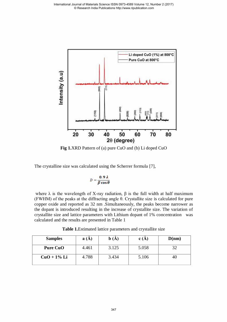
Fig 1.XRD Pattern of (a) pure CuO and (b) Li doped CuO
The crystalline size was calculated using the Scherrer formula [7],
where λ is the wavelength of X-ray radiation, β is the full width at half maximum
(FWHM) of the peaks at the diffracting angle θ. Crystallite size is calculated for pure
copper oxide and reported as 32 nm .Simultaneously, the peaks become narrower as
the dopant is introduced resulting in the increase of crystallite size. The variation of
crystallite size and lattice parameters with Lithium dopant of 1% concentration was
calculated and the results are presented in Table 1
Table 1.Estimated lattice parameters and crystallite size
Samples a (Ǻ) b (Ǻ) c (Ǻ) D(nm)
Pure CuO 4.461 3.125 5.058 32
CuO + 1% Li 4.788 3.434 5.106 40
International Journal of Materials Science ISSN 0973-4589 Volume 12, Number 2 (2017) © Research India Publications http://www.ripublication.com
347
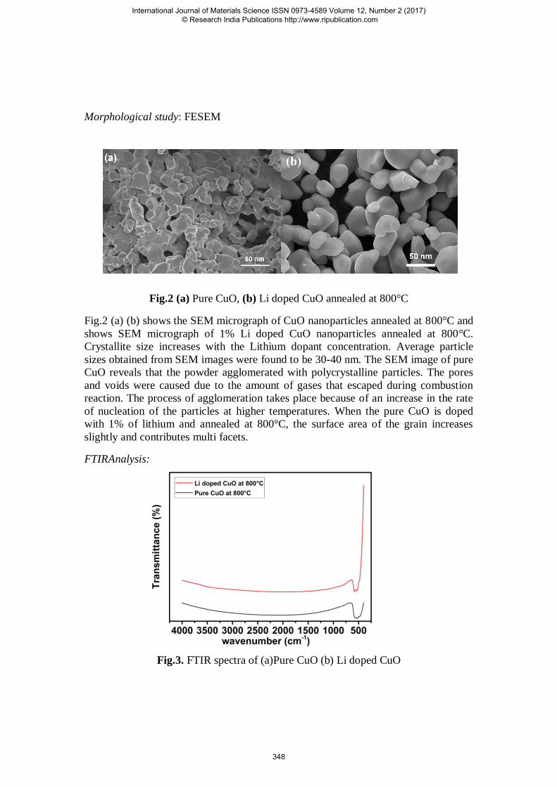
Morphological study: FESEM
Fig.2 (a) Pure CuO, (b) Li doped CuO annealed at 800°C
Fig.2 (a) (b) shows the SEM micrograph of CuO nanoparticles annealed at 800°C and
shows SEM micrograph of 1% Li doped CuO nanoparticles annealed at 800°C.
Crystallite size increases with the Lithium dopant concentration. Average particle
sizes obtained from SEM images were found to be 30-40 nm. The SEM image of pure
CuO reveals that the powder agglomerated with polycrystalline particles. The pores
and voids were caused due to the amount of gases that escaped during combustion
reaction. The process of agglomeration takes place because of an increase in the rate
of nucleation of the particles at higher temperatures. When the pure CuO is doped
with 1% of lithium and annealed at 800°C, the surface area of the grain increases
slightly and contributes multi facets.
FTIRAnalysis:
Fig.3. FTIR spectra of (a)Pure CuO (b) Li doped CuO
International Journal of Materials Science ISSN 0973-4589 Volume 12, Number 2 (2017) © Research India Publications http://www.ripublication.com
348

The FTIR spectra of pure and alkali metal doped CuO nanocrystallites
recorded at room temperature in the range 400-4500 cm-1
. Fig.3 shows an obvious
transmittance band around 580 cm-1
and 535 cm-1
which were assigned to vibration of
Cu(II)-O bands. The broad absorption band at 1712 cm-1
is due to the vibration mode
of absorbed water molecules.The shift in transmittance band from 580 to 572 cm-1
and
535 to 521 cm-1
indicates that there is a complexation of CuO and lithium.
CONCLUSION
A simple and swift combustion synthesis method to prepare high-quality
lithium doped copper oxide nanoparticles are adopted, in which an aqueous solution
of the metal nitrates and glycine is heated to the point of auto ignition. The resultant
ash was found to consist of finely divided particles of Lithium doped copper oxide
depending on the fuel/oxidant ratio in the precursor. This mixture is readily converted
to the desired phase by annealing at 800oC. XRD patterns showed that the synthesized
structures have good crystallinity with monoclinic crystal structure. Using of glycine
as fuel in preparation of copper oxide with lithium as dopant affects the structural and
morphological properties of these materials. The crystallite size varies in the range of
32–40 nm. FTIR spectra also validate the purity of CuO nanoparticles. SEM analysis
shows that the morphology and specific surface area of the samples are influenced by
the calcinations temperature and the dopant concentration.
ACKNOWLEDGEMENT
The authors thank DST-FIST for providing research facilities in Madras Christian
College, Chennai. The authors also thank, Dr. Selvakumar Sellaiyan, Division of
Applied science, University of Tsukuba, Japan for extending his support.
REFERENCES
[1] Djerad S, Geiger B, Schott F J P, Kureti S. Catal Commun, 2009, 10: 1103.
[2] Chiang CY, Aroh K, Ehrman SH (2012) Copper Oxide nanoparticle made by
flame spray pyrolysis for photochemical water splitting-part I. CuO nanoparticle
preparation. Int J Hydro ener 37:4871-4879.
[3] J. Morales, L. Sanchez, F.Martin, J.R. Ramos-Barrrado, M. Sanchez, Thin Solid
Flims 474 (2005) 133.
[4] A. Cruccolini, R.Palombari, Sens. Actuators B 98 (2004) 227.
[5] P.O. Larsson, A. Andersson, R.L. Wallengerg, B. Svensson, J. catal. 163 (1996)
279.
[6] Chandrappa G T, Ghosh S, Patil K C. J Matter Synth Processing, 1999, 7:273.
[7] Chihiro M, Nagamoto H, Tekemura I, Kitano K, Komatsu H, Sekiguchi K, Tabusa
F, Mori T, Tominaga M, Yabuuchi Y. J Med Chem, 1995, 38:353.
International Journal of Materials Science ISSN 0973-4589 Volume 12, Number 2 (2017) © Research India Publications http://www.ripublication.com
349

![Morphological and Optical Properties of SnO2 Doped ZnO ... · The structural, optical, and electronic properties are determined by the particle size [3,4]. SnO 2 and ZnO are belonging](https://img.dokumen.tips/doc/110x75/5f81daa8eb6da10c0c76a647/morphological-and-optical-properties-of-sno2-doped-zno-the-structural-optical.jpg)