Embed Size (px)
Citation preview

Structural and Functional Characterization of MonomericEphrinA1 Binding Site to EphA2 Receptor*□S
Received for publication, October 10, 2011, and in revised form, February 14, 2012 Published, JBC Papers in Press, February 23, 2012, DOI 10.1074/jbc.M111.311670
Carla M. Lema Tomé‡1, Enzo Palma‡1,2, Sara Ferluga‡, W. Todd Lowther§, Roy Hantgan¶, Jill Wykosky‡3,and Waldemar Debinski‡4
From the ‡Brain Tumor Center of Excellence, Department of Neurosurgery, the §Department of Biochemistry, and the ¶Departmentof Biochemistry and Molecular Medicine, Wake Forest School of Medicine, Winston-Salem, North Carolina 27157
Background:Monomeric ephrinA1 exhibits anti-tumor activity and possesses a class-characteristic G-H loop for receptorbinding.Results:Alanine-scanning mutagenesis of the G-H loop revealed amino acids with specific contributions to the function of theligand.Conclusion:Our results document a critical importance of the G-H loop for ephrinA1 tumor-suppressing activity.Significance:More potent anti-tumor isoforms of ephrinA1 can be generated.
The EphA2 receptor is overexpressed in glioblastoma multi-forme and has been to shown to contribute to cell transforma-tion, tumor initiation, progression, andmaintenance. EphrinA1(eA1) is a preferred ligand for the receptor. Treatment withmonomeric eA1, the formof eA1 found in the extracellular envi-ronment, causes receptor phosphorylation, internalization, anddown-regulation with subsequent anti-tumor effects. Here, weinvestigated the structure-function relationship of amonomericeA1 focusing on its G-H loop (108FQRFTPFTLGKEFKE123G), ahighly conserved region among eAs that mediates binding totheir receptors. Alanine substitution mutants of the G-H loopamino acids were transfected into U-251MG glioblastomamul-tiforme cells, and functional activity of each mutant in condi-tionedmedia was assessed by EphA2 down-regulation, ERK andAKT activation and cellular response assays. Alanine substitu-tions at positions Pro-113 Thr-115, Gly-117, Glu-122, and alsoGln-109 enhanced the EphA2 receptor down-regulation anddecreased p-ERK and p-AKT. Substitution mutants of eA1 atpositions Phe-108, Arg-110, Phe-111, Thr-112, Phe-114, Leu-116, Lys-118, Glu-119, and Phe-120 had a deleterious effect onEphA2 down-regulation when compared with eA1-WT.Mutants at positions Phe-108, Lys-18, Lys-121, Gly-123retained similar properties to eA1-WT. Recombinant eA1-R110A, -T115A, -G117A, and -F120A have been found toexhibit the same characteristics as the ligands contained in theconditioned media mainly due to the differences in their bind-ing to the receptor. Thus, we have identified variants of eA1 that
possess either superagonistic or antagonistic properties. Thesenew findings will be important in the understanding of thereceptor/ligand interactions and in further design of anti-can-cer therapies targeting the eA/EphA system.
The Eph receptors constitute the largest classified family ofvertebrate receptor-tyrosine kinases. The interaction betweenthe Eph receptors and their ligands, the ephrins, regulates cel-lular repulsion and adhesion, an effect that plays a majorinstructive role in tissue patterning, neuronal targeting, andvascular and neuronal development during embryogenesis(1–3) aswell as neuronal plasticity and regeneration in the adultcentral nervous system (4–7). Similar to other receptor-tyro-sine kinases, Ephs and ephrins have also been implicated incarcinogenesis, assuming critical roles in oncogenic transfor-mation, metastasis, and angiogenesis (8–10). Accordingly,Ephs and ephrins have been shown to be overexpressed in avariety of solid tumors (11–13), including breast (14, 15), pan-creas (16), gastric (17), colorectal (18), prostate (19), and brain(20, 21).Eph receptors and ephrin ligands are classified into A and B
classes according to their sequence homology. There are nineEphA receptors encoded in the human genome that bind to fiveglycosylphosphatidylinositol-anchored class A ephrins (eAs)5and five EphB receptors that bind to three transmembrane-tethered ephrinB ligands (2, 3). In general, binding within eachclass is prominent; however, interclass interactions such as thebinding of eA5 to EphB2 or ephrinB2 to EphA4 (22, 23) havebeen documented. Eph receptors and ephrin ligands are uniquein that their binding results in bidirectional signaling thataffects the Eph-expressing cell (forward signaling) and the eph-rin-expressing cell (reverse signaling) and often involves signal-ing pathways with opposite effects, e.g. cellular adhesion versusrepulsion (3, 24).Our laboratory has documented that the EphA2 receptor,
but not its preferred ligand eA1, is prominently overexpressed
* This work was supported, in whole or in part, by National Institutes of HealthGrant CA 74145 (NCI; to W. D.). This work was supported by the BrainTumor Center of Excellence (to W. D.) and Institutional Development Grant2006-IDG-1004 from the North Carolina Biotechnology Center (to R. H.).
□S This article contains supplemental Table S1 and Figs. S1–S4.1 Both authors contributed equally to this work.2 A National Institutes of Health T32 CA113267 TRADONC fellow. Present
address: Genentech, Inc., 1 DNA Way, South San Francisco, CA, 94080.3 Present address: Ludwig Institute for Cancer Research, University of Califor-
nia San Diego, 9500 Gilman Dr. La Jolla, CA 92093-0660.4 To whom correspondence should be addressed: Depts. of Neurosurgery,
Radiation Oncology, and Cancer Biology, Wake Forest School of Medicine,Medical Center Blvd., Winston-Salem, NC 27157. Tel.: 336-716-9712; Fax:336-713-7639; E-mail: [email protected].
5 The abbreviations used are: eA1, ephrinA1; GBM, glioblastoma multiforme;CM, conditioned media; RU, response units.
THE JOURNAL OF BIOLOGICAL CHEMISTRY VOL. 287, NO. 17, pp. 14012–14022, April 20, 2012© 2012 by The American Society for Biochemistry and Molecular Biology, Inc. Published in the U.S.A.
14012 JOURNAL OF BIOLOGICAL CHEMISTRY VOLUME 287 • NUMBER 17 • APRIL 20, 2012
by guest on April 12, 2018
http://ww
w.jbc.org/
Dow
nloaded from
in the highly aggressive and lethal primary brain tumor glio-blastoma multiforme (GBM). Moreover, EphA2 overexpres-sion in GBM has been significantly linked to malignancy grade(20, 21), malignant progression (25), and patient survival (26,27). Thus, the EphA2 receptor represents an attractive molec-ular target for therapeutic and diagnostic/imaging applicationsfor GBM.We have also shown that eA1-Fc, a recombinant sol-uble homodimer of eA1, induces EphA2 phosphorylation,internalization, and subsequent down-regulation thatmediatesthe reversal of several of the malignant properties of GBMcells, i.e. invasion, migration, and anchorage-independentgrowth (20).This effect was also observed when eA1 was ectopically
expressed in GBM cells; however, in this case eA1 was beingreleased into the extracellular environment as a soluble, mono-meric protein capable of mediating its effects in a paracrinemanner (28). Importantly, this finding supports the develop-ment of a soluble, monomeric eA1-based ligand that may beused as a vector for targeted delivery of therapeutic or imagingagents to GBM tumors. With that in mind, we have recentlyconjugated a bacterial toxin to eA1-Fc and demonstratedremarkable EphA2-specificity and cytotoxic potency of thisconjugate on GBM cells (29). Taken together, these observa-tions prompted us to study the structure-function relationshipof wild type (WT) eA1 by generating structural mutants bysite-directed mutagenesis and analyzing their effect on EphA2down-regulation as a readily measured functional end point oftheir activity.In general, the structure of eA ligands is a variation of the
Greek key �-barrel-fold with parallel and anti-parallel�-strands that are connected by several highly flexible loops
(30–32). Of particular importance for receptor binding is theconserved G-H loop (Fig. 1A). This loop for eA1 packs within asurface pocket of the N-terminal ligand binding domain ofEphA2 (Fig. 1B). In this work the G-H loop of eA1 (residues108–123) was subjected to alanine-scanningmutagenesis in aneffort to identify variants that alter cellular effects. Most vari-ants had either no or deleterious consequences on EphA2 bind-ing. At least three variants within the turn of the loop (P113A,T115A, and G117A) had increased activity when comparedwith eA1-WT. These improved variants should help in thedesign of more potent anti-tumor agents based on eA1.
EXPERIMENTAL PROCEDURES
Cell Culture—GBMcell linesU-251MG,U-373, and SNB-19were obtained from the American Type Culture Collection(Manassas, VA). U-251MG cells were grown in DMEM � glu-tamine, 10% (v/v) FBS, 0.1mmol/liter nonessential amino acids,and 1% (v/v) penicillin/streptomycin. eA1-mutant cell lines,which are derived from the U-251 MG parental cell line, weregrown in the above medium supplemented with 200 �g/mlGeneticin. U-373 cells were grown inMEM, 10% (v/v) FBS, and1% (v/v) penicillin/streptomycin, and SNB-19 cells were grownin RPMI, 10% (v/v) FBS, 0.1 mmol/liter nonessential aminoacids, sodium pyruvate, and 1% (v/v) penicillin/streptomycin.All cultures were maintained at 37 °C and 5% CO2.Alanine-scanning Mutagenesis—eA1 mutants were pre-
pared using a transformer site-directed mutagenesis kit fromClontech (Mountain View, CA) and an eA1-pcDNA3.1� plas-mid generated from full-length human ephrin mRNA obtainedfrom human umbilical vein endothelial cells using specificprimers for the eA1 gene. Mutagenesis and selection primers
FIGURE 1. The eA1 G-H loop and its interaction with EphA2. A, sequence alignment of the G-H loop of human eA ligands. The residues indicated in red areconserved. Blue shading indicates the most common residue or residues with comparable biophysical properties. The G and H �-strands are indicated by greenarrows. B, structure model of the eA1-EphA2 complex. The G-H loop of eA1 (green) docks within a surface cavity of the ligand binding domain of the EphA2receptor (white). The side chains of the G-H loop are highlighted in light blue (PDB 3CZU (31)). The figure was produced using PyMOL (The PyMOL MolecularGraphics System, Version 1.3, Schrödinger, LLC.).
Structure-Function Analysis of Monomeric EphrinA1
APRIL 20, 2012 • VOLUME 287 • NUMBER 17 JOURNAL OF BIOLOGICAL CHEMISTRY 14013
by guest on April 12, 2018
http://ww
w.jbc.org/
Dow
nloaded from

used are listed in supplemental Table S1. BMH71-18 mutSchemically competent Escherichia coli cells were then trans-formedwithmutant ephrinA1 plasmids and cultured overnightat 37 °C with shaking at 280 rpm; plasmid DNA was subse-quently isolated with aQiaprepminiprep kit (Qiagen, Valencia,CA). Sequence-verified eA1-mutant plasmids (Wake ForestUniversity DNASequencing Laboratory) were then transfectedinto U-251 MG cells as described below.Transfection Experiments—Mutant plasmids were trans-
fected into U-251 MG glioma cells in Opti-MEM (Invitrogen)using Lipofectamine 2000 (Invitrogen) as per the manufactur-er’s instructions. After 24 h, Opti-MEM media was replacedwith growth medium containing 20% FBS and then again, 24 hlater, with 10% FBS growth medium. Forty-eight hours latercells were split into 100-mm2 dishes and selected with growthmedium containing 800 �g/ml Geneticin. Individual cloneswere isolated with cloning rings, transferred into a 24-wellplate, andmaintained in growthmediumwith 200�g/mlGene-ticin. Mutant eA1 expression in the cell media of clones wasverified by eA1 immunoblotting, and high-expressing cloneswere further cultured in growthmediumwithout Geneticin forat least 48 h to obtain conditioned media for EphA2 down-regulation assays.EphA2Down-regulation Assay—U-251MGcells were grown
to 60% confluence in 60-mm dishes at 37 °C, 5% CO2, and sub-sequently dosed with conditioned media containing equivalentamounts of wild type eA1 or mutant eA1 as evaluated by eA1Western blotting and densitometry analysis. For determinationof the effect of all the mutants together on EphA2 down-regu-lation and activation of AKT and ERK as well as in the migra-tion/cell morphology assays, eA1 was measured by ELISA (seebelow). Negative controls for these experiments included freshmedia and conditioned media from vector-transfected U-251MGcells, whereas positive controlswere recombinant homodi-meric eA1-Fc at 1�g/ml (R&DSystems,Minneapolis,MN) andwild type eA1-conditioned media. U-251 MG cells were incu-bated for 24 h at 37 °C, 5% CO2 with the corresponding condi-tioned media or controls before obtaining cell lysates.Sandwich ELISAAnalysis of eA1 in ConditionedMedia (CM)—
eA1 inCMwas determined using a sandwich ELISA assay. CMswere collected from subconfluent monolayers of U-251 MGparental and vector-, WT-, and mutant eA1-transfected cells.Media were centrifuged for 5 min �1000 rpm to pellet anyinsoluble debris, and supernatant was collected. A 96-well platewas coated with 5 �g/ml polyclonal goat anti-mouse humaneA1 (R&D Systems), blocked in 5% milk, and incubated withserial concentrations of eA1-Fc (R&D Systems) or CM. Detec-tion was achieved using a primary antibody against human eA1(clone V18, Santa Cruz Biotechnology, Santa Cruz, CA) fol-lowed by HRP-conjugated anti-rabbit secondary antibodyand 2,2�-azino-bis(3-ethylbenzothiazoline-6-sulfonic acid)(ABTS). Detectionwas performed at 405 nm. Both primary andsecondary antibodies were raised against epitopes outside ofthe eA1 GH-loop and thus enable detection of eA1 GH-loopmutants. A standard curve was constructed using the signalscorresponding to the eA1-Fc serial dilutions, and mutant eA1content in CM was calculated.
ConditionedMedia Treatment of U-251MGCells—Subcon-fluent cultures of U-251 MG cells were grown in 60-mm2
dishes. Cells were treated with conditioned media dilutionsbased on the results from the sandwich ELISA assay, recombi-nant mouse eA1/Fc chimera (R&D Systems), or conditionedgrowth media control for the indicated times. Cells were pho-tographed by phase contrast microscopy with a 20� objectivelens, and images were processed using Photoshop (Adobe Sys-tems). Cell lysates were collected and used for Westernblotting.Western Blotting—Cell lysates were prepared by washing
treated U-251 MG cells with PBS and lysed in radioimmuno-precipitation assay buffer (0.5% (w/v) sodium deoxycholate,0.1% (w/v) sodium dodecyl sulfate, 0.5% (w/v) Igepal in 10 mM
phosphate-buffered saline) containing mammalian proteaseinhibitor mixture and 0.5% v/v sodium vanadate (Sigma). Celllysates were separated by SDS-PAGE in 10% polyacrylamidegels (EphA2/actin immunoblotting) or 12% polyacrylamidegels (ephrinA1-immunoblotting for conditioned media) andthen transferred to a polyvinylidene difluoride membrane(Pierce).Membraneswere subsequently blocked (5% (w/v)milkin PBS containing 0.05% (v/v) Tween 20 (Sigma)) for at least 1 hat room temperature and then incubated overnight at 4 h withshaking with the appropriate primary antibodies includinganti-EphA2 clone D7 at 1:1000 (Santa Cruz Biotechnology),anti-�-actin diluted at 1:50,000 (Sigma), anti-ephrinA1 cloneV18 at 1:300 (Santa Cruz Biotechnology), anti-phospho-p44/42MAPK (ERK1/2) (Thr-202/Tyr-204) XPTM, anti-p44/42MAPK (Erk1/2), anti-phospho-AKT (Se-r473) XPTM, anti-Akt(Cell Signaling Technology, Danvers, MA), and anti-� tubulin(Thermo Scientific, Fremont, CA). After three 5-min washes in0.05% (v/v) Tween 20 in PBS, membranes were incubated withsecondary antibody conjugated to horseradish peroxidase (goatanti-mouse IgG or goat anti-rabbit IgG) at a dilution of 1:5000in blotto for 1 h at room temperature with shaking.Membraneswere then again washed three times with 0.05% (v/v) Tween 20in PBS, and proteins were detected using the Enhanced Chemi-luminescence Plus Western blotting detection system (Amer-sham Biosciences). Membranes were subsequently exposed toBioMax XAR autoradiographic film (Eastman Kodak Co.) for10–20 s. Films were scanned at a resolution of 600 dpi using aHP ScanJet3979 and Adobe Photoshop 5.0 Software. Densi-tometry analysis was conducted using Image J Software(National Institutes of Health).Migration Assay—Wounds were made in a confluent mono-
layer of U-251 MG cells with a sterile 200-�l tip, and growthmedia containingWTandmutant eA1was added. Conditionedgrowth medium from untransfected cells was used as the neg-ative control, whereas eA1-Fc (1�g/ml) was added as a positivecontrol. Phase contrast microscopy pictures were taken of thesame field at 0, 8, 16, and 24 h. Distance of thewound in�mwasmeasured in five places for each of three wounds for each treat-ment or cell type at each time point using ImagePro Plus soft-ware, and the percent wound closure over 24 h was calculatedfor graphical representation.Production of Recombinant Variants of eA1—Recombinant
eA1-WT and its variants R110A, T115A, G117A, and F120Awere produced using the baculovirus expression vector system
Structure-Function Analysis of Monomeric EphrinA1
14014 JOURNAL OF BIOLOGICAL CHEMISTRY VOLUME 287 • NUMBER 17 • APRIL 20, 2012
by guest on April 12, 2018
http://ww
w.jbc.org/
Dow
nloaded from

from BD Biosciences. eA1 (19–175) WT, one of the forms ofeA1 that we identified in the media of cancer cells (unpub-lished) and the four eA1 mutants were amplified by PCR usingthe forward primer 5�-TATAGGATCCCATCACCATCAC-CATCACGATCGCCACACCGTC-3� (BamHI restriction siteand the coding sequence for the N terminus Histidine tag), andthe reverse primer 5�-CACGAATTCCTATTATTAAAC-CCGCACCTCTGGGTCATC-3� (EcoRI restriction site andstop codons). The amplified fragments were cloned intoBamHI-EcoRI sites in the Baculovirus transfer vectorpAcGP67-B (BD Biosciences) and sequenced. This vector car-ries the gp67 Baculovirus-encoded secretion signal sequenceupstream the MCS forcing the secretion of the recombinantproteins. The generated recombinant bacmids pBeA1, pBeA1-110, pBeA1-115, pBeA1-117, and pBEe1-120, respectively,were amplified in E. coli DH5� cells.
Sf9 insect cells were co-transfected with pBEA1, pBeA1-110,pBEA1-115, pBEA1-117, and pBEA1-120 recombinant bac-mids and the linearized BaculogoldTM Baculovirus DNA (BDBiosciences) using the BaculogoldTM Transfection kit (BD Bio-sciences) following the instructions of the supplier. Infectiousrecombinant baculoviruses were amplified two times in BDBaculogoldTM MaX-XP Serum-free Insect Cell Medium (BDBiosciences) in Sf9 serum-free media-adapted cells at 27 °C for5 days to obtain high titer virus stocks. Sf9 cells, serum-freemedia-adapted, were infected with high titer virus stocks andgrown at 27 °C for 5 days to produce the recombinant proteins.Sf9-baculovirus infected media containing the recombinant
proteinswere collected, and floating cells were removed by cen-trifugation (at 3000 � g for 10 min). Supernatant was filteredthrough a 0.22-�mpore filter, 0.1 M ureawas added, and the pHwas adjusted to 7.4. The His6-tagged recombinant proteinswere purified by affinity chromatography with HisTrap HPaffinity column (GE Healthcare). The column was equilibratedin bufferA (50mMNaH2PO4�H2O, 150mMNaCl, pH7.4). Afterloading, the column was washed with 10 column volumes ofbuffer A, and the recombinant proteins were eluted with a stepgradient with buffer B (50 mM NaH2PO4�H2O, 300 mM NaCl,250mM imidazole, pH 7.4). Recombinant proteins were filtered(0.22 �m) and stored at �20 °C.Surface Plasmon Resonance Assay—Binding interactions
between eA1 (wild type and mutants) and EphA2 were mea-sured by surface plasmon resonance in a BIAcore T100 instru-ment (33, 34). Recombinant human EphA2 receptor (R&D Sys-tems) was immobilized on the dextran matrix of a CM5biosensor chip using amine coupling chemistry (29�g/ml at pH4) to achieve a sparse monolayer in the sample chamber(5991 � 139 response units (RU), n � 3) and a blank immobi-lization in the reference chamber. Unreacted sites in bothchambers were blocked with ethanolamine. All surface plas-mon resonance procedures were carried out at 25.0 °C.Binding curves were obtained with eA1 monomer (0–3000
nM) and dimer (0–2400 nM) by delivering aliquots of each pro-tein in HEPES-buffered saline, pH 7.4, to both sample and ref-erence chambers at 30 �l/min. RU versus time profiles weremonitored for 700 s during the binding step; dissociation wasthen monitored for 1500 s as buffer was delivered. Boundligands were removed by regeneration with 1 M NaCl followed
by 0.1% SDS. Time-dependent changes obtained with bufferdelivery were subtracted from each protein binding profile(sample � reference RU signals) to obtain double-correctedkinetic traces.Anchorage-independent Growth—U-251 MG cells (2 � 103)
were plated in 6-well plates in growth medium plus 0.35% agaron a base layer of BactoTMAgar, BD Biosciences, growthmedium plus 0.5% agar. Cells were supplemented with eA1-WT, eA1-R110A, eA1-T115A, eA1-G117A, and eA1-F120Aat 1.0 and 0.1 �g/ml or vehicle alone. Fresh media-eA1 wasadded to the cells 3 days and 1 week after plating. Colonieswere counted after 14 days. Clusters of colonies greater than50 cells were counted in 10 random fields at low power. Eachexperimental condition was done in triplicate for everyassay.Statistical Analysis—Probability (p) values were calculated
using the analysis of variance one-way test using MS Excel; pvalues �0.05 were considered to be statistically significant.
RESULTS
EphA2 Is Differentially Down-regulated by G-H Loop eA1Alanine Mutants—We demonstrated that EphA2 is differen-tially down-regulated in U-251 MG cells treated with anequimolar amount of CM obtained from U-251[eA1](�) andvariousU-251[G-H loopmutant eA1s](�) cells. First, we deter-mined that there are three principal groups of alanine-mutatedeA1 variants. In the first group a diminished EphA2 down-reg-ulation was observed among CM from R110A, F111A, T112A,L116A, E119A, F120A, and G123A eA1 mutants when com-pared with eA1-WT CM (Fig. 2A). In the second group threesubstitution mutations, namely F108A, K118A, and K121Ayielded activity similar to those obtained with U-251[eA1](�)CM (supplemental Fig. S1). Interestingly, the third group ofvariants (Pro-113, Thr-115, Gly-117, and Glu-122) exhibited a39, 67, 39, and 57% (Fig. 2B) enhanced down-regulation ofEphA2, respectively. Q109A demonstrated a similar effect (notshown). It is noteworthy that T115A and E122A are two of fewother residues that are unique to eA1 G-H loop. Further exper-iments revealed that non-conservative substitutions at position115, such as T115R, T115D, and T115G, not only abolished theenhancing effect on EphA2 down-regulation that had beenobtained with T115A CM, but they made the variants inactive(supplemental Fig. S2). One substitution, F114A, was the onlymutant of eA1 found in small amounts in the cell lysate and notreleased into the media and, thus, of unknown activity (notshown).G-H LoopMutant CMs Promote Cell Rounding and Decrease
Migration of GBM Cells—Next, U-251 MG parental and G48aGBM cells were treated with equal concentrations of the differ-entmutant eA1 CMs asmeasured directly by ELISA.We inves-tigated an ability of the mutants to elicit a characteristic mor-phological response to eA1 in a form of cell rounding (20, 35).Cells treated with the alanine mutant CMs became roundedwithin 20 min of treatment as did cells exposed toU-251[eA1](�) CM or homodimeric eA1-Fc (not shown; moredetailed analysis of cell rounding was performed with recombi-nant forms of eA1 as shown in Fig. 5E). Cell rounding wasaccompanied by EphA2 receptor down-regulation (Fig. 2C).
Structure-Function Analysis of Monomeric EphrinA1
APRIL 20, 2012 • VOLUME 287 • NUMBER 17 JOURNAL OF BIOLOGICAL CHEMISTRY 14015
by guest on April 12, 2018
http://ww
w.jbc.org/
Dow
nloaded from

FIGURE 2. EphA2 down-regulation ability of alanine point-mutants of human ephrinA1. A, eA1 G-H loop mutants with diminished ability to down-regulateEphA2. Shown are Western blots of EphA2 immunoreactivity in U-251 MG cells treated with CM from eA1-R110A, eA1-F111A, eA1-T112A, eA1-L116A, eA1-E119A, eA1-F120Am and eA1-G123A for 24 h. Equivalent dosing (1.0� to 0.1�) was verified by Western blotting; eA1-Fc (1 mg/ml) and eA1-WT CM were usedas positive controls. B, eA1 G-H loop mutants with enhanced effect on EphA2 receptor down-regulating ability. Shown is a Western blot of EphA2 immunore-activity in U-251 MG cells treated with CM from eA1-P113A, eA1-T115A, eA1-G117A, and eA1-E122A for 24 h. C, temporal analysis of EphA2 down-regulationin response to the treatment with eA1-Fc (1 �g/ml), U-251 vector, eA1-WT, or eA1-mutant CM. Western blot of EphA2 immunoreactivity in U-251 MG cellstreated for 1 h and for 8 h are shown. ELISA was used to confirm equivalent dosing of eA1 mutants.
Structure-Function Analysis of Monomeric EphrinA1
14016 JOURNAL OF BIOLOGICAL CHEMISTRY VOLUME 287 • NUMBER 17 • APRIL 20, 2012
by guest on April 12, 2018
http://ww
w.jbc.org/
Dow
nloaded from
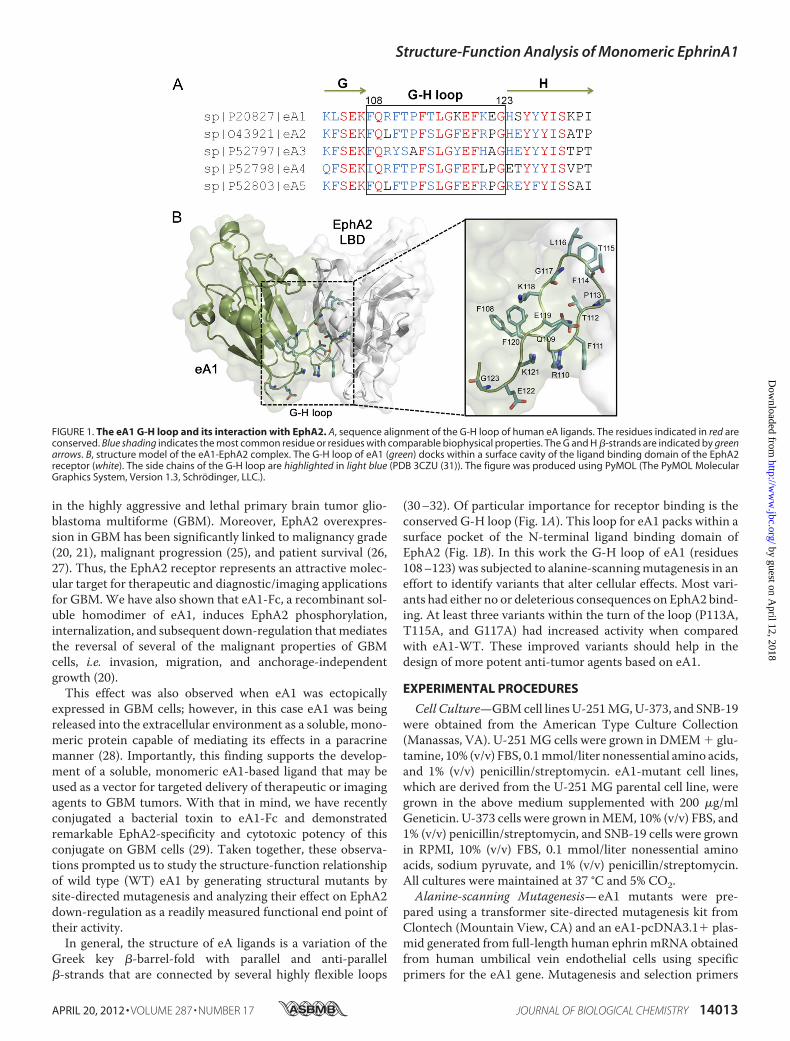
Interestingly, P113A, T115A, and G117A CM induced rapid (1h) EphA2 receptor down-regulation (Fig. 2C). This prominenteffect of the threemutants was sustained for 8 h post-treatmentwhen also the Q109A mutant showed an increased activity.Furthermore, the treatment of U-251 MG cells with similarconcentrations of CM from P113A, T115A, G117A, K121A,and E122Amutants also decreased the migration of U-251MGcells (Fig. 3).Ras-MAPK and PI3K-AKT Pathways Are Differentially
Affected by eA1 G-H Loop Mutants—We next investigated theeffect of WT and mutant eA1s on intracellular signaling, spe-cifically on classical oncogenic pathways such as Ras-MAPKand the phosphoinositide 3-kinase/AKT pathways known to beresponsive to eA1 stimulation. Dimeric eA1-Fc suppresses sig-naling through theRas-MAPKpathway (36–38) as does solublemonomeric eA1 (28); hence, we examined whether or not analtered suppression of this pathway would occur after treat-ment with themutant eA1CM. Indeed, treatment withQ109A,P113A, T115A, G117A, and E122A CM led to a decrease inphosphorylation of ERK at 30 min (Fig. 4A). Surprisingly, thiseffect was sustained for 8 h and less so at 24 h post-treatment(Fig. 4A). It appears that eA1-F120A actually activated p-ERK.
DecreasedphosphorylationofAKTwasshownto followEphA2receptor activation by eA1-Fc (39). Therefore, we investigated theeffect ofmutantmonomeric eA1son thePI3K/AKTpathway.Thetreatment of U-251 MG cells with P113A, T115A, G117A, andE122ACM led to a decrease in p-AKT at Ser-473 30min after thetreatment (Fig. 4B). This effect was at least partially sustained byP113A, T115A, and G117Amutants for 8 h (Fig. 4B).eA1 and Its Variants Bind to Immobilized EphA2 with Differ-
entAffinities—Purified recombinantmonomericeA1-WTand itsfour variants (Fig. 5A), chosen on the basis of varying activitiesidentified in the experimentswithCM,were further characterizedfor their binding abilities and biological properties. In the bindingassays using BIAcore system, plotting the maximum signalchanges versus eA1 concentration yieldedhyperbolic bindingpro-files (supplemental Fig. S3). Analysis by nonlinear regression (Sig-maPlot 11, SYSTAT Software, Santa Fe, CA) with a single site-saturablemodel yieldedBmax � 229� 7RU andKd � 89� 21 nMfor eA1-Fc (dimer); eA1 monomer yielded Bmax � 163 � 26 RUandKd � 580 � 240 nM.
EphA2 binding experiments with wild type eA1 andmutantscarried out at 1 �M yielded rapid signal changes during thebinding step that approached a plateau by 30–60 s; dissociation
FIGURE 3. Effect of eA1-WT and eA1-mutant CMs on the migration of GBM cells. A, phase contrast micrographs of a wound on confluent U-251 MG cells(20� magnification) treated with control, eA1-Fc (1 �g/ml), eA1-WT, and mutant eA1 CM over a period of 24 h. B, shown is quantification of migration as thepercentage of wound closure at 24 h of U251-MG cells treated with control, eA1-Fc (1 mg/ml), eA1-WT, and mutant eA1 CM. ELISA was used to confirmequivalent dosing of eA1 mutants.
Structure-Function Analysis of Monomeric EphrinA1
APRIL 20, 2012 • VOLUME 287 • NUMBER 17 JOURNAL OF BIOLOGICAL CHEMISTRY 14017
by guest on April 12, 2018
http://ww
w.jbc.org/
Dow
nloaded from

rates exhibited half-times �50 s (Fig. 5B). Plotting the maxi-mum binding signals observed in replicate experiments witheA1-WT (n� 11) and themutants T115A (n� 4), F120A ( n�4), R110A (n � 3) and G117A (n � 3) yielded a consistentpattern of tighter binding by eA1-T115 and eA1-G117A vari-antswhen comparedwith eA1-WT, and a partial (eA1-R110) orcomplete loss (eA1-F120) of the binding by the others (Fig. 5C).Recombinant Variants of eA1 Display Enhanced Biological
Properties Relative to Wild Type eA1—The recombinant vari-ants of eA1 were then tested for their ability to down-regulatethe EphA2 receptor, to induce cell rounding, and to affectanchorage-independent growth ofGBMcells as an in vitro anti-tumor activity measure. The most active variants in all theseassays were eA1-T115A and eA1-G117A, whereas eA1-R110Aand eA1-F120A were either significantly less active or lost theactivity altogetherwhen comparedwith eA1-WT. For example,eA1-T115A and -G117A down-regulated the EphA2 receptorat concentrations as low as 0.05�g/mlwhereas eA1-WT lackedactivity at 0.1 �g/ml (Fig. 5D). Furthermore, the same wasobserved in a cell-rounding assay when these two super agonis-tic variants of eA1 were still able to change the GBM cells mor-phology at the lowest concentrations used (Fig. 5E). In the
anchorage-independent growth assay, eA1-T115A and eA1-G117A showed significant inhibition of this growth compara-ble with eA1-WT at 1.0 �g/ml, but they demonstrated morepronounced inhibition than eA1-WT at the lower 0.1 �g/mlconcentration (Fig. 5F and supplemental Fig. S4, A and 4B).
DISCUSSION
In thisworkwehave directly documented a critical role of theeA1 G-H loop in mediating functional soluble monomeric eA1activity. Importantly, the P113A,T115A,G117A, E122A, and tosome extent Q109A mutants of the eA1 ligand exhibited anenhanced down-regulation of the EphA2 receptor, the typicalmorphological changes of cancer cell-rounding, and altered cellmigration, all at significantly lower concentrations (Table 1). Inaddition, the treatment with mutant eA1 ligands had profoundand sustained effects on classical oncogenic pathways in whichthe eA1/EphA2 system is ultimately involved. These resultswere recapitulated with the use of recombinant purified vari-ants of eA1. Thus, eA1-T115A and eA1-G117A bind tighter tothe immobilized EphA2 receptor than eA1-WT whereasA1-R110A and eA1-F120A bind less tightly or lose the bindingcompletely. The two superagonistic variants of eA1 have alsosignificantly more pronounced biological activities in down-regulation of EphA2, cell rounding, and anchorage-indepen-dent growth assays when compared with eA1-WT or eA1-R110A or eA1-F120A (Fig. 5).The G-H loop of eA1 packs into a surface pocket of the
EphA2 receptor (Fig. 1). Several of the variants lost part or all ofthe ability to interact with the EphA2 (R110A, F111A, T112A,L116A, E119A, and G123A). The most likely scenarios for theobserved loss in activity are that the structure of loop may havebeen distorted or altered in its conformation dynamics. Forexample, L116 fills completely a pocket primary generated bythe side chains ofMet-59 and Val-61 (Fig. 6). Amutation to Alawould result in a loss of this hydrophobic interaction.Mutationof eA1 at position 119 was detrimental in terms of EphA2receptor down-regulation, a result that is also in line with itsrole as the “latch” that strengthens the eA1/EphA2 interaction(31). The F108A, K118A, and K121A variants exhibited WTlevels of activity.In contrast, the P113A, T115A, G117A, E122A, and Q109A
mutants had improved interactionwith EphA2, as supported bytheir ability to bind at significantly lower concentrations (Fig.5C). A closer inspection at the details of the interactions ofP113A, T115A, and G117A with EphA2 (Fig. 6) provides somerationale for the consequences of themutations. Pro-113 packsagainst the disulfide bond between residues Cys-70 and Cys-188 and the backbone atoms of residues Val-89 and Ala-190 ofEphA2. Perhaps the removal of the conformational rigidity ofPro-113 enabled the loop to have more facile binding. Thr-115fills a pocket lined by several residues including Thr-101, Thr-151, Phe-156, Asp-155, and Val-61. It is unclear why an Alasubstitutionwould bemore favorable, but the requirement for asmall, uncharged amino acid at this position is supported by thecomplete loss of EphA2 binding by the T115D and T115R vari-ants (supplemental Fig. S2). It is also unclear why the mutationof Gly-117, Glu-122, and Gln-109 to Ala would be favorable.Clearly more work will be required to ascertain the molecular
FIGURE 4. Effect of eA1 G-H loop point mutants on oncogenic signaling. A,Western blots of p-ERK and p42/p44 MAPK immunoreactivity in U-251 MGcells treated with control, eA1-Fc (1 �g/ml), eA1-WT, and eA1-mutant CMs for30 min, 8 h, and 24 h. B, Western blots of p-AKT and total AKT immunoreac-tivity in U-251 MG cells treated with control CM, eA1-Fc (1 �g/ml), eA1-WT,and eA1-mutant CMs for 30 min and 8 h. ELISA was used to confirm equivalentdosing of eA1 mutants in both assays.
Structure-Function Analysis of Monomeric EphrinA1
14018 JOURNAL OF BIOLOGICAL CHEMISTRY VOLUME 287 • NUMBER 17 • APRIL 20, 2012
by guest on April 12, 2018
http://ww
w.jbc.org/
Dow
nloaded from

FIGURE 5. Binding and biological activity of recombinant eA1 G-H loop mutants. A, Western blot of recombinant forms of eA1 mutants. B, surface plasmonresonance analysis of the binding of eA1-WT and recombinant eA1 mutants to immobilized EphA2. Each protein was delivered at 1 �M for 700 s followed bya 1500-s dissociation step. Solid line, eA1-WT; long dash, eA1-T115A; dash-dot, eA1-G117A; short dash, eA1-R110A; dotted line, eA1-F120A. C, quantification ofbinding of recombinant eA1 mutants as assayed by surface plasmon resonance. Significant differences (p � 0.05) versus eA1-WT are indicated by asterisks. D,down-regulation of the EphA2 receptor in GBM cells by recombinant eA1 mutants as shown by EphA2 western blotting. E, cell rounding of GBM cells inresponse to various concentrations of recombinant eA1 mutants. F, quantification of the effect of recombinant eA1 mutants on anchorage-independentgrowth of GBM cells. Significant differences (p � 0.05) versus untreated group are indicated by asterisk.
Structure-Function Analysis of Monomeric EphrinA1
APRIL 20, 2012 • VOLUME 287 • NUMBER 17 JOURNAL OF BIOLOGICAL CHEMISTRY 14019
by guest on April 12, 2018
http://ww
w.jbc.org/
Dow
nloaded from

basis for the observed improvements. Nonetheless, it is encour-aging to find that single mutations can lead to significantimprovements in the eA1-EphA2 interaction. Further optimi-zation of theG-H loop andperhaps othersmayultimately proveto yield eA1 variants that will be useful for cancer therapy ordiagnosis.Mutation of ephrinA1 at the five critical residues led to sus-
tained EphA2 receptor down-regulation in GBM cells. It hadbeen previously documented that upon exposure to dimericeA1-Fc, the EphA2 receptor present on tumor cells undergoestyrosine phosphorylation and is down-regulated (40–42). Theeffects of binding of eA1 to EphA2 have been hypothesized toconstitute a dual process comprised of direct signaling via
ligand-receptor interaction and the effects resulting from thedown-regulation of the receptor. The five mutants showed anenhanced ability to down-regulate the receptor when com-pared with eA1-WT treatment.Binding of eA1 to EphA2 leads to SHP-2 recruitment and
subsequent FAK dephosphorylation that suppresses integrinfunction and diminishes cell adhesion to the extracellularmatrix (35). The effects of this signaling cascade are visible in ashort period of time after eA1 administration, when tumor cellrounding ensues most likely from the formation of the kinase-active Src-FAK complex, which leads to contraction of thecytoskeleton via myosin II and RhoA activity (43). Mutant eA1CMs corresponding to the four critical residues cause a rapidand profound cell rounding (Fig. 5E) (28). In addition, admin-istration of eA1-Fc, eA1-WT, or the four mutants CM led todecreased migration of GBM cells, in line with the resultobtained in breast and prostate cancer cells (37, 44) and in con-trast with data obtained in osteosarcoma (45).Previous reports indicated that eA1-Fc suppressed signaling
through theRas-MAPKpathway (28, 36–38). In contrast, otherreports indicate that ERK1/2 activity is actually increased aftertreatment with eA1-Fc (44, 45). The treatment of U-251 MGcells with eA1-P113A, -T115A, -G117A, and -E122A CMdecreased phosphorylation of extracellular signal-regulatedkinase beyond to what was seen with eA1-WT CM. Of partic-ular interest is the fact that this effect was sustained for anextended period of time, as decreased p-ERK was observed at8 h of post-treatment in cells treated with these four mutants.AKT activation in the majority of GBM has been reported to
result fromPTEN inactivation, receptor-tyrosine kinase activa-tion, orAKT amplification (46, 47). Recent reports also indicatethe existence of a negative regulatory loop between ligand-ac-tivated EphA2 and AKT (39, 48). In accordance with thesereports, treatment of U-251 MG cells with eA1-WT andmutant eA1 CM corresponding to the critical residues in theG-H loop led to a significant reduction in AKT phosphoryla-tion. The effect of the four eA1mutant CMon the p-AKT levels
FIGURE 5—continued
FIGURE 6. Molecular interactions of the G-H loop near eA1 residues 113–117. The coloring scheme is the same as Fig. 1 (PDB 3CZU (31)).
TABLE 1Activity of eA1 G-H loop mutants in GBM cells� to ���, arbitrary degree of potentiation of eA1 and its substitution mutantsactivity. - to—, arbitrary degree of alteration of eA1mutants activity. NA, not done.
MutantEphA2
down-regulationp-ERK
down-regulationp-Akt
down-regulation
WT �� �� �F108A �� - �Q109A ��� ��� ��R110A – – -F111A – NA NAT112A – NA NAP113A ��� ��� ���F114A NA NA NAT115A ��� ��� ���L116A – NA NAG117A ��� ��� ���K118A �� NA NAE119A – NA NAF120A – — –K121A �� - –E122A ��� ��� ���G123A – – �
Structure-Function Analysis of Monomeric EphrinA1
14020 JOURNAL OF BIOLOGICAL CHEMISTRY VOLUME 287 • NUMBER 17 • APRIL 20, 2012
by guest on April 12, 2018
http://ww
w.jbc.org/
Dow
nloaded from

was greater than that of the eA1-WT. The results further sup-port the importance of eA1 signaling axis and its involvement inGBM progression/maintenance.Our previous work had determined that EphA2 represents a
promising target for new therapeutics to combat GBM (21).The construction of a targeted cytotoxin wherein eA1-Fc wasconjugated to the truncated form of Pseudomonas exotoxin Ayielded a highly potent and specific agent that kills GBM,breast, andprostate cancer cells at very low concentrations (29).The results of the work presented here should prove helpful infurther rational design of these and similar cytotoxins. Beingthat eA1 is a tumor-suppressing factor inGBMby itself, findingan optimal variant(s) will have an important impact on thedesign and production of novel anti-tumor agents.
Acknowledgment—We thank Dr. Young A. Choi for help with exper-iments involving recombinant eA1s.
REFERENCES1. Merlos-Suárez, A., and Batlle, E. (2008) Eph-ephrin signaling in adult tis-
sues and cancer. Curr Opin Cell Biol. 20, 194–2002. Pasquale, E. B. (2005) Eph receptor signaling casts a wide net on cell
behavior. Nat. Rev. Mol. Cell Biol. 6, 462–4753. Pasquale, E. B. (2008) Eph-ephrin bidirectional signaling in physiology and
disease. Cell 133, 38–524. Drescher, U., Kremoser, C., Handwerker, C., Löschinger, J., Noda,M., and
Bonhoeffer, F. (1995) In vitro guidance of retinal ganglion cell axons byRAGS, a 25-kDa tectal protein related to ligands for Eph receptor-tyrosinekinases. Cell 82, 359–370
5. Knöll, B., and Drescher, U. (2002) Ephrin-As as receptors in topographicprojections. Trends Neurosci 25, 145–149
6. Lai, K. O., and Ip, N. Y. (2009) Synapse development and plasticity. Rolesof ephrin/Eph receptor signaling. Curr. Opin. Neurobiol. 19, 275–283
7. Nakamoto, M., Cheng, H. J., Friedman, G. C., McLaughlin, T., Hansen,M. J., Yoon, C. H., O’Leary, D. D., and Flanagan, J. G. (1996) Topograph-ically specific effects of ELF-1 on retinal axon guidance in vitro and retinalaxon mapping in vivo. Cell 86, 755–766
8. Surawska, H., Ma, P. C., and Salgia, R. (2004) The role of ephrins and Ephreceptors in cancer. Cytokine Growth Factor Rev. 15, 419–433
9. Wimmer-Kleikamp, S. H., and Lackmann, M. (2005) Eph-modulated cellmorphology, adhesion, and motility in carcinogenesis. IUBMB Life 57,421–431
10. Beauchamp, A., and Debinski, W. (2012) Ephs and ephrins in cancer.Ephrin-A1 signaling. Semin. Cell Dev. Biol. 23, 109–115
11. Ireton, R. C., and Chen, J. (2005) EphA2 receptor-tyrosine kinase as apromising target for cancer therapeutics. Curr. Cancer Drug Targets 5,149–157
12. Kinch, M. S., and Carles-Kinch, K. (2003) Overexpression and functionalalterations of the EphA2-tyrosine kinase in cancer. Clin. Exp. Metastasis20, 59–68
13. Walker-Daniels, J., Hess, A. R., Hendrix, M. J., and Kinch, M. S. (2003)Differential regulation of EphA2 in normal and malignant cells. Am. J.Pathol. 162, 1037–1042
14. Wu, Q., Suo, Z., Risberg, B., Karlsson, M. G., Villman, K., and Nesland,J. M. (2004) Expression of Ephb2 and Ephb4 in breast carcinoma. Pathol.Oncol. Res. 10, 26–33
15. Zelinski, D. P., Zantek, N. D., Stewart, J. C., Irizarry, A. R., and Kinch,M. S.(2001) EphA2 overexpression causes tumorigenesis of mammary epithe-lial cells. Cancer Res. 61, 2301–2306
16. Mudali, S. V., Fu, B., Lakkur, S. S., Luo, M., Embuscado, E. E., and Iacobu-zio-Donahue, C. A. (2006) Patterns of EphA2 protein expression in pri-mary and metastatic pancreatic carcinoma and correlation with geneticstatus. Clin. Exp. Metastasis 23, 357–365
17. Nakamura, R., Kataoka, H., Sato, N., Kanamori, M., Ihara,M., Igarashi, H.,
Ravshanov, S.,Wang, Y. J., Li, Z. Y., Shimamura, T., Kobayashi, T., Konno,H., Shinmura, K., Tanaka, M., and Sugimura, H. (2005) EPHA2/EFNA1expression in human gastric cancer. Cancer Sci. 96, 42–47
18. Kataoka, H., Igarashi, H., Kanamori,M., Ihara,M.,Wang, J. D.,Wang, Y. J.,Li, Z. Y., Shimamura, T., Kobayashi, T., Maruyama, K., Nakamura, T.,Arai, H., Kajimura, M., Hanai, H., Tanaka, M., and Sugimura, H. (2004)Correlation of EPHA2 overexpression with high microvessel count in hu-man primary colorectal cancer. Cancer Sci. 95, 136–141
19. Zeng, G., Hu, Z., Kinch, M. S., Pan, C. X., Flockhart, D. A., Kao, C., Gard-ner, T. A., Zhang, S., Li, L., Baldridge, L. A., Koch, M. O., Ulbright, T. M.,Eble, J. N., and Cheng, L. (2003) High level expression of EphA2 receptor-tyrosine kinase in prostatic intraepithelial neoplasia. Am. J. Pathol. 163,2271–2276
20. Wykosky, J., and Debinski, W. (2008) The EphA2 receptor and ephrinA1ligand in solid tumors. Function and therapeutic targeting. Mol. CancerRes. 6, 1795–1806
21. Wykosky, J., Gibo, D.M., Stanton, C., and Debinski,W. (2005) EphA2 as anovel molecularmarker and target in glioblastomamultiforme.Mol. Can-cer Res. 3, 541–551
22. Qin, H., Noberini, R., Huan, X., Shi, J., Pasquale, E. B., and Song, J. (2010)Structural characterization of the EphA4-Ephrin-B2 complex reveals newfeatures enabling Eph-ephrin binding promiscuity. J. Biol. Chem. 285,644–654
23. Himanen, J. P., Chumley, M. J., Lackmann, M., Li, C., Barton, W. A.,Jeffrey, P. D., Vearing, C., Geleick, D., Feldheim, D. A., Boyd, A. W., Hen-kemeyer, M., and Nikolov, D. B. (2004) Repelling class discrimination.Ephrin-A5 binds to and activates EphB2 receptor signaling.Nat. Neurosci.7, 501–509
24. Pasquale, E. B. (2010) Eph receptors and ephrins in cancer. Bidirectionalsignaling and beyond. Nat. Rev. Cancer 10, 165–180
25. Li, X., Wang, Y., Wang, Y., Zhen, H., Yang, H., Fei, Z., Zhang, J., Liu, W.,Wang, Y., and Zhang, X. (2007) Expression of EphA2 in human astrocytictumors. Correlation with pathologic grade, proliferation, and apoptosis.Tumour Biol. 28, 165–172
26. Liu, F., Park, P. J., Lai, W., Maher, E., Chakravarti, A., Durso, L., Jiang, X.,Yu, Y., Brosius, A., Thomas, M., Chin, L., Brennan, C., DePinho, R. A.,Kohane, I., Carroll, R. S., Black, P. M., and Johnson, M. D. (2006) A ge-nome-wide screen reveals functional gene clusters in the cancer genomeand identifies EphA2 as a mitogen in glioblastoma. Cancer Res. 66,10815–10823
27. Wang, L. F., Fokas, E., Bieker,M., Rose, F., Rexin, P., Zhu, Y., Pagenstecher,A., Engenhart-Cabillic, R., and An, H. X. (2008) Increased expression ofEphA2 correlates with adverse outcome in primary and recurrent glio-blastoma multiforme patients. Oncol Rep 19, 151–156
28. Wykosky, J., Palma, E., Gibo,D.M., Ringler, S., Turner, C. P., andDebinski,W. (2008) Soluble monomeric EphrinA1 is released from tumor cells andis a functional ligand for the EphA2 receptor. Oncogene 27, 7260–7273
29. Wykosky, J., Gibo, D. M., and Debinski, W. (2007) A novel, potent, andspecific ephrinA1-based cytotoxin against EphA2 receptor-expressing tu-mor cells.Mol. Cancer Ther. 6, 3208–3218
30. Qin, H., Lim, L., and Song, J. (2012) Protein dynamics at Eph receptor-ligand interfaces as revealed by crystallography, NMR and MD simula-tions. BMC Biophys. 5, 2
31. Himanen, J. P., Goldgur, Y., Miao, H., Myshkin, E., Guo, H., Buck, M.,Nguyen, M., Rajashankar, K. R., Wang, B., and Nikolov, D. B. (2009) Li-gand recognition by A-class Eph receptors. Crystal structures of theEphA2 ligand binding domain and the EphA2/ephrin-A1 complex.EMBORep. 10, 722–728
32. Himanen, J. P., Yermekbayeva, L., Janes, P. W., Walker, J. R., Xu, K., Ata-pattu, L., Rajashankar, K. R., Mensinga, A., Lackmann, M., Nikolov, D. B.,and Dhe-Paganon, S. (2010) Architecture of Eph receptor clusters. Proc.Natl. Acad. Sci. U.S.A. 107, 10860–10865
33. Hantgan, R. R., Stahle,M. C., and Lord, S. T. (2010) Dynamic regulation offibrinogen. Integrin �IIb�3 binding. Biochemistry 49, 9217–9225
34. Hantgan, R. R., and Stahle, M. C. (2009) Integrin priming dynamics.Mechanisms of integrin antagonist-promoted �IIb�3:PAC-1 molecularrecognition. Biochemistry 48, 8355–8365
35. Miao,H., Burnett, E., Kinch,M., Simon, E., andWang, B. (2000)Activation
Structure-Function Analysis of Monomeric EphrinA1
APRIL 20, 2012 • VOLUME 287 • NUMBER 17 JOURNAL OF BIOLOGICAL CHEMISTRY 14021
by guest on April 12, 2018
http://ww
w.jbc.org/
Dow
nloaded from

of EphA2 kinase suppresses integrin function and causes focal-adhesion-kinase dephosphorylation. Nat. Cell Biol. 2, 62–69
36. Guo, H., Miao, H., Gerber, L., Singh, J., Denning, M. F., Gilliam, A. C., andWang, B. (2006) Disruption of EphA2 receptor-tyrosine kinase leads toincreased susceptibility to carcinogenesis in mouse skin. Cancer Res. 66,7050–7058
37. Macrae,M.,Neve, R.M., Rodriguez-Viciana, P., Haqq,C., Yeh, J., Chen,C.,Gray, J. W., and McCormick, F. (2005) A conditional feedback loop regu-lates Ras activity through EphA2. Cancer Cell 8, 111–118
38. Miao, H., Wei, B. R., Peehl, D. M., Li, Q., Alexandrou, T., Schelling, J. R.,Rhim, J. S., Sedor, J. R., Burnett, E., and Wang, B. (2001) Activation ofEphA receptor-tyrosine kinase inhibits the Ras/MAPK pathway.Nat. CellBiol. 3, 527–530
39. Miao, H., Li, D. Q., Mukherjee, A., Guo, H., Petty, A., Cutter, J., Basilion,J. P., Sedor, J.,Wu, J., Danielpour, D., Sloan, A. E., Cohen,M. L., andWang,B. (2009) EphA2 mediates ligand-dependent inhibition and ligand-inde-pendent promotion of cell migration and invasion via a reciprocal regula-tory loop with Akt. Cancer Cell 16, 9–20
40. Duxbury, M. S., Ito, H., Zinner, M. J., Ashley, S. W., and Whang, E. E.(2004) Ligation of EphA2 by Ephrin A1-Fc inhibits pancreatic adenocar-cinoma cellular invasiveness. Biochem. Biophys. Res. Commun. 320,1096–1102
41. Shao, H., Pandey, A., O’Shea, K. S., Seldin, M., and Dixit, V. M. (1995)Characterization of B61, the ligand for the Eck receptor protein-tyrosinekinase. J. Biol. Chem. 270, 5636–5641
42. Walker-Daniels, J., Riese, D. J., 2nd, and Kinch, M. S. (2002) c-Cbl-depen-dent EphA2protein degradation is induced by ligand binding.Mol. Cancer
Res. 1, 79–8743. Parri, M., Taddei, M. L., Bianchini, F., Calorini, L., and Chiarugi, P. (2009)
EphA2 reexpression prompts invasion of melanoma cells shifting frommesenchymal to amoeboid-like motility style. Cancer Res. 69, 2072–2081
44. Pratt, R. L., and Kinch, M. S. (2002) Activation of the EphA2-tyrosinekinase stimulates the MAP/ERK kinase signaling cascade. Oncogene 21,7690–7699
45. Fritsche-Guenther, R., Noske, A., Ungethüm, U., Kuban, R. J., Schlag,P. M., Tunn, P. U., Karle, J., Krenn, V., Dietel, M., and Sers, C. (2010) Denovo expression of EphA2 in osteosarcoma modulates activation of themitogenic signaling pathway. Histopathology 57, 836–850
46. CancerGenomeAtlas ResearchNetwork (2008) Comprehensive genomiccharacterization defines human glioblastoma genes and core pathways.Nature 455, 1061–1068
47. Parsons, D. W., Jones, S., Zhang, X., Lin, J. C., Leary, R. J., Angenendt, P.,Mankoo, P., Carter, H., Siu, I. M., Gallia, G. L., Olivi, A., McLendon, R.,Rasheed, B. A., Keir, S., Nikolskaya, T., Nikolsky, Y., Busam,D.A., Tekleab,H., Diaz, L. A., Jr., Hartigan, J., Smith, D. R., Strausberg, R. L., Marie, S. K.,Shinjo, S. M., Yan, H., Riggins, G. J., Bigner, D. D., Karchin, R., Papado-poulos, N., Parmigiani, G., Vogelstein, B., Velculescu, V. E., and Kinzler,K.W. (2008) An integrated genomic analysis of human glioblastomamul-tiforme. Science 321, 1807–1812
48. Yang, N. Y., Fernandez, C., Richter, M., Xiao, Z., Valencia, F., Tice, D. A.,and Pasquale, E. B. (2011) Cross-talk of the EphA2 receptor with a serine/threonine phosphatase suppresses the Akt-mTORC1 pathway in cancercells. Cell. Signal. 23, 201–212
Structure-Function Analysis of Monomeric EphrinA1
14022 JOURNAL OF BIOLOGICAL CHEMISTRY VOLUME 287 • NUMBER 17 • APRIL 20, 2012
by guest on April 12, 2018
http://ww
w.jbc.org/
Dow
nloaded from

Wykosky and Waldemar DebinskiCarla M. Lema Tomé, Enzo Palma, Sara Ferluga, W. Todd Lowther, Roy Hantgan, Jill
to EphA2 ReceptorStructural and Functional Characterization of Monomeric EphrinA1 Binding Site
doi: 10.1074/jbc.M111.311670 originally published online February 23, 20122012, 287:14012-14022.J. Biol. Chem.
10.1074/jbc.M111.311670Access the most updated version of this article at doi:
Alerts:
When a correction for this article is posted•
When this article is cited•
to choose from all of JBC's e-mail alertsClick here
Supplemental material:
http://www.jbc.org/content/suppl/2012/02/23/M111.311670.DC1
http://www.jbc.org/content/287/17/14012.full.html#ref-list-1
This article cites 48 references, 12 of which can be accessed free at
by guest on April 12, 2018
http://ww
w.jbc.org/
Dow
nloaded from